Note: Descriptions are shown in the official language in which they were submitted.
CA 02703790 2010-04-26
WO 2009/073160 PCT/US2008/013248
ANTI-VEGF ANTIBODIES
FIELD OF THE INVENTION
[0001] This invention relates generally to anti-VEGF selected polypeptide
sequences and antibodies with beneficial properties for research, therapeutic
and
diagnostic purposes.
BACKGROUND OF THE INVENTION
[0002] Development of a vascular system is a fundamental requirement for
many physiological and pathological processes. Actively growing tissues such
as
embryos and tumors require adequate blood supply. They satisfy this need by
producing pro-angiogenic factors, which promote new blood vessel formation via
a
process called angiogenesis. Vascular tube formation is a complex but orderly
biological event involving all or many of the following steps: a) Endothelial
cells
(ECs) proliferate from existing ECs or differentiate from progenitor cells; b)
ECs
migrate and coalesce to form cord-like structures; c) vascular cords then
undergo
tubulogenesis to form vessels with a central lumen; d) existing cords or
vessels send
out sprouts to form secondary vessels; e) primitive vascular plexus undergo
further
remodeling and reshaping; and f) peri-endothelial cells are recruited to
encase the
endothelial tubes, providing maintenance and modulatory functions to the
vessels;
such cells including pericytes for small capillaries, smooth muscle cells for
larger
vessels, and myocardial cells in the heart. Hanahan, D. Science 277:48-50
(1997);
Hogan, B. L. & Kolodziej, P. A. Nature Reviews Genetics. 3:513-23 (2002);
Lubarsky, B. & Krasnow, M. A. Cell. 112:19-28 (2003).
[0003] It is well established that angiogenesis is implicated in the
pathogenesis of a variety of disorders. These include solid tumors and
metastasis,
atherosclerosis, retrolental fibroplasia, hemangiomas, chronic inflammation,
intraocular neovascular diseases such as proliferative retinopathies, e.g.,
diabetic
retinopathy, age-related macular degeneration (AMD), neovascular glaucoma,
immune rejection of transplanted corneal tissue and other tissues, rheumatoid
arthritis,
and psoriasis. Folkman et al., J. Biol. Chem., 267:10931-10934 (1992);
Klagsbrun et
t
CA 02703790 2010-04-26
WO 2009/073160 PCT/US2008/013248
al., Annu. Rev. Physiol. 53:217-239 (1991); and Garner A., "Vascular
diseases," In:
Pathobiology of Ocular Disease. A Dynamic Approach, Garner A., Klintworth GK,
eds., 2nd Edition (Marcel Dekker, NY, 1994), pp 1625-1710.
[0004] In the case of tumor growth, angiogenesis appears to be crucial for the
transition from hyperplasia to neoplasia, and for providing nourishment for
the growth
and metastasis of the tumor. Folkman et al., Nature 339:58 (1989). The
neovascularization allows the tumor cells to acquire a growth advantage and
proliferative autonomy compared to the normal cells. A tumor usually begins as
a
single aberrant cell, which can proliferate only to a size of a few cubic
millimeters due
to the distance from available capillary beds, and it can stay 'dormant'
without further
growth and dissemination for a long period of time. Some tumor cells then
switch to
the angiogenic phenotype to activate endothelial cells, which proliferate and
mature
into new capillary blood vessels. These newly formed blood vessels not only
allow
for continued growth of the primary tumor, but also for the dissemination and
recolonization of metastatic tumor cells. Accordingly, a correlation has been
observed between density of microvessels in tumor sections and patient
survival in
breast cancer as well as in several other tumors. Weidner et al., N. Engl. J.
Med
324:1-6 (1991); Horak et al., Lancet 340:1120-1124 (1992); Macchiarini et al.,
Lancet 340:145-146 (1992). The precise mechanisms that control the angiogenic
switch is not well understood, but it is believed that neovascularization of
tumor mass
results from the net balance of a multitude of angiogenesis stimulators and
inhibitors
(Folkman, Nat Med 1(1):27-31 (1995)).
[0005] The process of vascular development is tightly regulated. To date, a
significant number of molecules, mostly secreted factors produced by
surrounding
cells, have been shown to regulate EC differentiation, proliferation,
migration and
coalescence into cord-like structures. For example, vascular endothelial
growth factor
(VEGF) has been identified as the key factor involved in stimulating
angiogenesis and
in inducing vascular permeability. Ferrara et al., Endocr. Rev. 18:4-25
(1997). The
finding that the loss of even a single VEGF allele results in embryonic
lethality points
to an irreplaceable role played by this factor in the development and
differentiation of
the vascular system. Furthermore, VEGF has been shown to be a key mediator of
neovascularization associated with tumors and intraocular disorders. Ferrara
et al.,
Endocr. Rev. supra. The VEGF mRNA is overexpressed by the majority of human
2
CA 02703790 2010-04-26
WO 2009/073160 PCT/US2008/013248
tumors examined. Berkman et al., J. Clin. Invest. 91:153-159 (1993); Brown et
al.,
Human Pathol. 26:86-91 (1995); Brown et al., Cancer Res. 53:4727-4735 (1993);
Mattern et al., Brit. J. Cancer 73:931-934 (1996); Dvorak et al., Am. J.
Pathol.
146:1029-1039 (1995).
[0006] Also, the concentration levels of VEGF in eye fluids are highly
correlated to the presence of active proliferation of blood vessels in
patients with
diabetic and other ischemia-related retinopathies. Aiello et al., N. Engl. J.
Med.
331:1480-1487 (1994). Furthermore, studies have demonstrated the localization
of
VEGF in choroidal neovascular membranes in patients affected by AMD. Lopez et
al., Invest. Ophthalmol. Vis. Sci. 37:855-868 (1996).
[0007] Anti-VEGF neutralizing antibodies suppress the growth of a variety of
human tumor cell lines in nude mice (Kim et al., Nature 362:841-844 (1993);
Warren
et al., J. Clin. Invest. 95:1789-1797 (1995); Borgstrom et al., Cancer Res.
56:4032-
4039 (1996); Melnyk et al., Cancer Res. 56:921-924 (1996)) and also inhibit
intraocular angiogenesis in models of ischemic retinal disorders. Adamis et
al., Arch.
Ophthalmol. 114:66-71 (1996). Therefore, anti-VEGF monoclonal antibodies or
other inhibitors of VEGF action are promising candidates for the treatment of
tumors
and various intraocular neovascular disorders. Such antibodies are described,
for
example, in EP 817,648 published January 14, 1998; and in W098/45331 and
W098/45332, both published October 15, 1998. One of the anti-VEGF antibodies,
bevacizumab, has been approved by the FDA for use in combination with a
chemotherapy regimen to treat metastatic colorectal cancer (CRC) and non-small
cell
lung cancer (NSCLC). And bevacizumab is being investigated in many ongoing
clinical trials for treating various cancer indications.
[0008] Other anti-VEGF antibodies, anti-Nrpl antibodies and anti-Nrp2
antibodies are also known, and described, for example, in Liang et al., JMo1
Biol 366,
815-829 (2007) and Liang et al., JBiol Chem 281, 951-961 (2006), PCT
publication
number W02007/056470 and PCT Application No. PCT/US2007/069179, the content
of these patent applications are expressly incorporated herein by reference.
SUMMARY OF THE INVENTION
[0009] The invention provides novel anti-VEGF antibodies and uses thereof.
3
CA 02703790 2010-04-26
WO 2009/073160 PCT/US2008/013248
[0010] A number of anti-VEGF antibodies are provided in the invention. For
example, an antibody that binds to VEGF or a fragment thereof is provided,
wherein
the antibody comprises six HVRs selected from:
(i) an HVR-Ll comprising the amino acid sequence of X1X2R X3SL
wherein the HVR-L1 comprises 1, 2 or 3 substitutions in any
combination of the following positions: X, is G or A; X2 is V or I;
and/or X3 is T or R;
(ii) an HVR-L2 comprising the amino acid sequence of DASSLA (SEQ ID
NO:6);
(iii) an HVR-L3 comprising the amino acid sequence of SYKSPL (SEQ ID
NO:7);
(iv) an HVR-H1 comprising the amino acid sequence of SISGSWIF (SEQ
ID NO:1);
(v) an HVR-H2 comprising the amino acid sequence of GAIWPFGGYTH
(SEQ ID NO:2).; and
(vi) an HVR-H3 comprising the amino acid sequence of
RWGHSTSPWAMDY (SEQ ID NO:3).
[0011] In another embodiment, an antibody that binds to VEGF or a fragment
thereof is provided, wherein the antibody comprises:
(1) an HVR-H 1 comprising the amino acid sequence of SEQ ID NO:1;
(2) an HVR-H2 comprising the amino acid sequence of SEQ ID NO:2;
(3) an HVR-H3 comprising the amino acid sequence of SEQ ID NO:3
(4) an HVR-L1 comprising the amino acid sequence of SEQ ID NO:4;
(5) an HVR-L2 comprising the amino acid sequence of SEQ ID NO:6; and
(6) an HVR-L3 comprising the amino acid sequence of SEQ ID NO:7.
[0012] In another embodiment, an antibody that binds to VEGF or a fragment
thereof is provided, wherein the antibody comprises:
(1) an HVR-H1 comprising the amino acid sequence of SEQ ID NO:1;
(2) an HVR-H2 comprising the amino acid sequence of SEQ ID NO:2;
(3) an HVR-H3 comprising the amino acid sequence of SEQ ID NO:3
(4) an HVR-L1 comprising the amino acid sequence of SEQ ID NO:5;
(5) an HVR-L2 comprising the amino acid sequence of SEQ ID NO:6; and
4
CA 02703790 2010-04-26
WO 2009/073160 PCT/US2008/013248
(6) an HVR-L3 comprising the amino acid sequence of SEQ ID NO:7.
[00131 In another embodiment, an antibody that binds to VEGF or a fragment
thereof is provided, wherein the light chain variable domain comprises the
amino acid
sequence of SEQ ID NO:44 or SEQ ID NO:45.
[00141 In another embodiment, an antibody that binds to VEGF or a fragment
thereof is provided, wherein the anti-VEGF antibody comprises the heavy chain
variable domain comprising the amino acid sequence of SEQ ID NO:43, and the
light
chain variable domain comprises the amino acid sequence of SEQ ID NO:44 or 45.
In yet another embodiment, the anti-VEGF antibody comprises the heavy chain
variable domain comprising the amino acid sequence of SEQ ID NO:43, and the
light
chain variable domain comprises the amino acid sequence of SEQ ID NO:44. In
yet
another embodiment, the anti-VEGF antibody comprises the heavy chain variable
domain comprising the amino acid sequence of SEQ ID NO:43, and the light chain
variable domain comprises the amino acid sequence of SEQ ID NO:45.
[00151 In certain embodiments, any of the above antibodies is a monoclonal
antibody. In one embodiment, the antibody is an antibody fragment selected
from a
Fab, Fab'-SH, Fv, scFv, or (Fab')2 fragment. In one embodiment, the antibody
is
humanized. In one embodiment, the antibody is human. In yet another
embodiment,
at least a portion of the framework sequence is a human consensus framework
sequence.
[00161 Polynucleotides encoding any of the above antibodies are provided, as
well as vectors comprising the polynucleotides and host cells comprising the
vectors
of the invention. In one embodiment, the host cell is eukaryotic. In another
embodiment, the host cell is a CHO cell. A method of making an anti-VEGF
antibody is also provided. For example, a method comprises culturing the host
cell
under conditions suitable for expression of the polynucleotide encoding the
antibody,
and isolating the antibody.
[00171 In one aspect, a method of detecting the presence of VEGF in a
biological sample is provided, the method comprising contacting the biological
sample with an antibody of the invention under conditions permissive for
binding of
the antibody to VEGF, and detecting whether a complex is formed between the
antibody and VEGF. In one embodiment, the method comprises detecting VEGF-
anti-VEGF antibody complex in a biological sample wherein the amino acid
sequence
5
CA 02703790 2010-04-26
WO 2009/073160 PCT/US2008/013248
of the anti-VEGF antibody comprises a heavy chain variable domain comprising
the
amino acid sequence of SEQ ID NO:43, and a light chain variable domain
comprising
the amino acid sequence of SEQ ID NO:44 or 45. In yet another embodiment, the
method comprises detecting VEGF-anti-VEGF antibody complex in a biological
sample wherein the amino acid sequence of the anti-VEGF antibody comprises a
heavy chain variable domain comprising the amino acid sequence of SEQ ID
NO:43,
and a light chain variable domain comprising the amino acid sequence of SEQ ID
NO:44. In yet another embodiment, the method comprises detecting VEGF-anti-
VEGF antibody complex in a biological sample wherein the amino acid sequence
of
1o the anti-VEGF antibody comprises a heavy chain variable domain comprising
the
amino acid sequence of SEQ ID NO:43, and a light chain variable domain
comprising
the amino acid sequence of SEQ ID NO:45. In yet another embodiment, the anti-
VEGF antibody is detectably labeled.
[00181 Pharmaceutical compositions as well as methods of treatment are also
provided. In one aspect, a pharmaceutical composition is provided comprising
an
antibody of the invention and a pharmaceutically acceptable carrier. In
another
aspect, a method of treating cancer is provided, e.g., the method comprises
administering to an individual the pharmaceutical composition comprising any
of the
above antibodies. Cancers treated by the methods of the invention include, but
are not
limited to, squamous cell cancer, small-cell lung cancer, non-small cell lung
cancer,
adenocarcinoma of the lung, squamous carcinoma of the lung, cancer of the
peritoneum, hepatocellular cancer, gastric cancer, gastrointestinal cancer,
gastrointestinal stromal cancer, pancreatic cancer, glioblastoma, cervical
cancer,
ovarian cancer, liver cancer, bladder cancer, hepatoma, breast cancer, colon
cancer,
colorectal cancer, endometrial or uterine carcinoma, salivary gland carcinoma,
kidney
or renal cancer, liver cancer, prostate cancer, vulval cancer, thyroid cancer,
hepatic
carcinoma and various types of head and neck cancer, melanoma, superficial
spreading melanoma, lentigo maligna melanoma, acral lentiginous melanomas,
nodular melanomas, B-cell lymphoma, chronic lymphocytic leukemia (CLL); acute
lymphoblastic leukemia (ALL); hairy cell leukemia; chronic myeloblastic
leukemia;
post-transplant lymphoproliferative disorder (PTLD), abnormal vascular
proliferation
associated with phakomatoses, edema associated with brain tumors, or Meigs'
syndrome. In certain embodiments, the tumor, cancer or cell proliferative
disorder
6
CA 02703790 2010-04-26
WO 2009/073160 PCT/US2008/013248
being treated colon cancer, lung cancer, breast cancer or glioblastoma. In yet
another
embodment, the subject being treated is human.
[0019] The invention further provides immunoconjugates comprising an
antibody conjugated to an agent, such as a drug or cytotoxic agent.
BRIEF DESCRIPTION OF THE DRAWINGS
[0020] Figure 1: Heavy chain and light chain HVR loop sequences of anti-
VEGF antibodies. The figures show the heavy chain HVR sequences, H1, H2, and
H3, and light chain HVR sequences, L1, L2, and L3. Sequence numbering is as
follows: clone B20-4.1.1 (HVR-H 1 is SEQ ID NO:1; HVR-H2 is SEQ ID NO:2;
HVR-H3 is SEQ ID NO:3; HVR-L1 is SEQ ID NO:4; HVR-L2 is SEQ ID NO:6;
HVR-L3 is SEQ ID NO:7); and clone B20-4.1.1RR (HVR-H1 is SEQ ID NO:1;
HVR-H2 is SEQ ID NO:2; HVR-H3 is SEQ ID NO:3; HVR-L1 is SEQ ID NO:5;
HVR-L2 is SEQ ID NO:6; HVR-L3 is SEQ ID NO:7).
[0021] Amino acid positions are numbered according to the Kabat numbering
system as described below.
[0022] Figures 2a and 2b: depict exemplary acceptor human consensus
framework sequences for use in practicing the instant invention with sequence
identifiers as follows:
Variable heavy (VH) consensus frameworks
human VH subgroup I consensus framework minus Kabat CDRs (SEQ ID
NO:8)
human VH subgroup I consensus framework minus extended hypervariable
regions (SEQ ID NOs:9-1 1)
human VH subgroup II consensus framework minus Kabat CDRs (SEQ ID
NO:12)
human VH subgroup II consensus framework minus extended hypervariable
regions (SEQ ID NOs:13-15)
human VH subgroup 11 consensus framework minus extended
human VH subgroup III consensus framework minus Kabat CDRs (SEQ ID
NO: 16)
human VH subgroup III consensus framework minus extended hypervariable
regions (SEQ ID NOs: 17-19)
7
CA 02703790 2010-04-26
WO 2009/073160 PCT/US2008/013248
human VH acceptor framework minus Kabat CDRs (SEQ ID NO:20)
human VH acceptor framework minus extended hypervariable regions (SEQ
ID NOs:21-22)
human VH acceptor 2 framework minus Kabat CDRs (SEQ ID NO:23)
human VH acceptor 2 framework minus extended hypervariable regions (SEQ
ID NOs:24-26)
[0023] Amino acid positions are numbered according to the Kabat numbering
system as described below.
[0024] Figure 3: depicts exemplary acceptor human consensus framework
sequences for use in practicing the instant invention with sequence
identifiers as
follows:
Variable light (VL) consensus frameworks
human VL kappa subgroup I consensus framework (SEQ ID NO:27)
human VL kappa subgroup II consensus framework (SEQ ID NO:28)
human VL kappa subgroup III consensus framework (SEQ ID NO:29)
human VL kappa subgroup IV consensus framework (SEQ ID NO:30)
[0025] Figure 4: depicts framework region sequences of huMAb4D5-8 heavy
and light chains. Numbers in superscript/bold indicate amino acid positions
according
to Kabat.
[0026] Figure 5: depicts modified/variant framework region sequences of
huMAb4D5-8 heavy and light chains. Numbers in superscript/bold indicate amino
acid positions according to Kabat.
[0027] Figure 6: The amino acid sequences of the light chain HVR
sequences L 1, L2 and L3 for anti-VEGF antibodies B20-4. 1.1 and B20-4.1.1 RR.
[0028] Figure 7: The amino acid sequences of the heavy chain HVR
sequences H1, H2, and H3 for anti-VEGF antibodies B20-4.1.1 and B20-4.1.1RR.
[0029] Figure 8: depicts the light chain variable regions of antibody clones
B20-4.1.1 (SEQ ID NO:44) and B20-4.1.1 RR (SEQ ID NO:45).
[0030] Figure 9: depicts the heavy chain variable regions of antibody clones
B20-4.1.1 and B20-4.1.1 RR (SEQ ID NO:43).
[0031] Figure 10: Table summarizing the kinetic binding affinity
measurement of B20 variants IgGs to human VEGF and murine VEGF. Human or
murine VEGF was immobilized to achieve approximately 60 response units.
8
CA 02703790 2010-04-26
WO 2009/073160 PCT/US2008/013248
[00321 Figure 11: Table summarizing the kinetic binding affinity
measurement of B20 variants IgGs to human VEGF and murine VEGF. Human or
murine VEGF was immobilized to achieve approximately 1000 response units.
[0033] Figure 12: HUVEC thymidine incorporation assay, which shows that
the B20 variants can effectively inhibit the HUVEC cell proliferation.
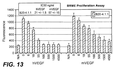
[0034] Figure 13: depicts the effect of B20-4. 1.1 on VEGF-induced BRME
proliferation.
[0035] Figure 14: depicts the effect of B20-4. 1.1 on tumor growth in nude
mice with xenografted human tumor cells (A549 cells), as measured by tumor
volumes over the number of treatment days.
[0036] Figure 15: depicts the effect of B20-4. 1.1 on tumor growth in nude
mice with xenografted human tumor cells (MDA-MB231 cells), as measured by
tumor volumes over the number of treatment days.
[0037] Figure 16: depicts the effect of the avastin antibody on VEGF-induced
BRME proliferation. Inhibition of mVEGF was not observed at a concentration of
up
to 1500 nM of the avastin antibody.
DETAILED DESCRIPTION OF THE INVENTION
[0038] The invention herein provides isolated antibodies that bind to VEGF
and uses thereof. Pharmaceutical compositions as well as methods of treatment
are
also provided.
[0039] The invention further provides methods of making anti-VEGF
antibodies, and polynucleotides encoding anti-VEGF antibodies.
General techniques
[0040] The techniques and procedures described or referenced herein are
generally well understood and commonly employed using conventional methodology
by those skilled in the art, such as, for example, the widely utilized
methodologies
described in Sambrook et al., Molecular Cloning: A Laboratory Manual 3rd.
edition
(2001) Cold Spring Harbor Laboratory Press, Cold Spring Harbor, N.Y. CURRENT
PROTOCOLS IN MOLECULAR BIOLOGY (F. M. Ausubel, et al. eds., (2003)); the
series METHODS IN ENZYMOLOGY (Academic Press, Inc.): PCR 2: A
PRACTICAL APPROACH (M. J. MacPherson, B. D. Hames and G. R. Taylor eds.
9
CA 02703790 2010-04-26
WO 2009/073160 PCT/US2008/013248
(1995)), Harlow and Lane, eds. (1988) ANTIBODIES, A LABORATORY
MANUAL, and ANIMAL CELL CULTURE (R. I. Freshney, ed. (1987));
Oligonucleotide Synthesis (M. J. Gait, ed., 1984); Methods in Molecular
Biology,
Humana Press; Cell Biology: A Laboratory Notebook (J. E. Cellis, ed., 1998)
Academic Press; Animal Cell Culture (R. I. Freshney), ed., 1987); Introduction
to
Cell and Tissue Culture (J. P. Mather and P. E. Roberts, 1998) Plenum Press;
Cell and
Tissue Culture: Laboratory Procedures (A. Doyle, J. B. Griffiths, and D. G.
Newell,
eds., 1993-8) J. Wiley and Sons; Handbook of Experimental Immunology (D. M.
Weir
and C. C. Blackwell, eds.); Gene Transfer Vectors for Mammalian Cells (J. M.
Miller
and M. P. Calos, eds., 1987); PCR: The Polymerase Chain Reaction, (Mullis et
al.,
eds., 1994); Current Protocols in Immunology (J. E. Coligan et al., eds.,
1991); Short
Protocols in Molecular Biology (Wiley and Sons, 1999); Immunobiology (C. A.
Janeway and P. Travers, 1997); Antibodies (P. Finch, 1997); Antibodies: A
Practical
Approach (D. Catty., ed., IRL Press, 1988-1989); Monoclonal Antibodies: A
Practical
Approach (P. Shepherd and C. Dean, eds., Oxford University Press, 2000); Using
Antibodies: A Laboratory Manual (E. Harlow and D. Lane (Cold Spring Harbor
Laboratory Press, 1999); The Antibodies (M. Zanetti and J. D. Capra, eds.,
Harwood
Academic Publishers, 1995); and Cancer: Principles and Practice of Oncology
(V. T.
DeVita et al., eds., J.B. Lippincott Company, 1993).
Definitions
[0041] For purposes of interpreting this specification, the following
definitions will apply and, whenever appropriate, terms used in the singular
will also
include the plural and vice versa. In the event that any definition set forth
below
conflicts with any document incorporated herein by reference, the definition
set forth
below shall control.
[0042] The term "antibody" is used in the broadest sense and specifically
covers monoclonal antibodies (including full length monoclonal antibodies),
polyclonal antibodies, multispecific antibodies (e.g., bispecific antibodies),
and
antibody fragments so long as they exhibit the desired biological activity.
[0043] The term "monoclonal antibody" as used herein refers to an antibody
obtained from a population of substantially homogeneous antibodies, i.e., the
individual antibodies comprising the population are identical except for
possible
CA 02703790 2010-04-26
WO 2009/073160 PCT/US2008/013248
mutations, e.g., naturally occurring mutations, that may be present in minor
amounts.
Thus, the modifier "monoclonal" indicates the character of the antibody as not
being a
mixture of discrete antibodies. In certain embodiments, such a monoclonal
antibody
typically includes an antibody comprising a polypeptide sequence that binds a
target,
wherein the target-binding polypeptide sequence was obtained by a process that
includes the selection of a single target binding polypeptide sequence from a
plurality
of polypeptide sequences. For example, the selection process can be the
selection of a
unique clone from a plurality of clones, such as a pool of hybridoma clones,
phage
clones, or recombinant DNA clones. It should be understood that a selected
target
binding sequence can be further altered, for example, to improve affinity for
the
target, to humanize the target binding sequence, to improve its production in
cell
culture, to reduce its immunogenicity in vivo, to create a multispecific
antibody, etc.,
and that an antibody comprising the altered target binding sequence is also a
monoclonal antibody of this invention. In contrast to polyclonal antibody
preparations, which typically include different antibodies directed against
different
determinants (epitopes), each monoclonal antibody of a monoclonal antibody
preparation is directed against a single determinant on an antigen. In
addition to their
specificity, monoclonal antibody preparations are advantageous in that they
are
typically uncontaminated by other immunoglobulins.
[00441 The modifier "monoclonal" indicates the character of the antibody as
being obtained from a substantially homogeneous population of antibodies, and
is not
to be construed as requiring production of the antibody by any particular
method. For
example, the monoclonal antibodies to be used in accordance with the present
invention may be made by a variety of techniques, including, for example, the
hybridoma method (e.g., Kohler and Milstein, Nature, 256:495-97 (1975); Hongo
et
al., Hybridoma, 14 (3): 253-260 (1995), Harlow et al., Antibodies: A
Laboratory
Manual, (Cold Spring Harbor Laboratory Press, 2nd ed. 1988); Hammerling et
al., in:
Monoclonal Antibodies and T-Cell Hybridomas 563-681 (Elsevier, N.Y., 1981)),
recombinant DNA methods (see, e.g., U.S. Patent No. 4,816,567), phage-display
technologies (see, e.g., Clackson et al., Nature, 352: 624-628 (1991); Marks
et al., J.
Mol. Biol. 222: 581-597 (1992); Sidhu et al., J. Mol. Biol. 338(2): 299-310
(2004);
Lee et al., J. Mol. Biol. 340(5): 1073-1093 (2004); Fellouse, Proc. Natl.
Acad. Sci.
USA 101(34): 12467-12472 (2004); and Lee et al., J. Immunol. Methods 284(1-2):
11
CA 02703790 2010-04-26
WO 2009/073160 PCT/US2008/013248
119-132(2004), and technologies for producing human or human-like antibodies
in
animals that have parts or all of the human immunoglobulin loci or genes
encoding
human immunoglobulin sequences (see, e.g., WO 1998/24893; WO 1996/34096; WO
1996/33735; WO 1991/10741; Jakobovits et al., Proc. Natl. Acad. Sci. USA 90:
2551
(1993); Jakobovits et al., Nature 362: 255-258 (1993); Bruggemann et al., Year
in
Immunol. 7:33 (1993); U.S. Patent Nos. 5,545,807; 5,545,806; 5,569,825;
5,625,126;
5,633,425; and 5,661,016; Marks et al., Bio/Technology 10: 779-783 (1992);
Lonberg
et al., Nature 368: 856-859 (1994); Morrison, Nature 368: 812-813 (1994);
Fishwild
et al., Nature Biotechnol. 14: 845-851 (1996); Neuberger, Nature Biotechnol.
14: 826
(1996); and Lonberg and Huszar, Intern. Rev. Immunol. 13: 65-93 (1995).
[0045] The monoclonal antibodies herein specifically include "chimeric"
antibodies in which a portion of the heavy and/or light chain is identical
with or
homologous to corresponding sequences in antibodies derived from a particular
species or belonging to a particular antibody class or subclass, while the
remainder of
the chain(s) is identical with or homologous to corresponding sequences in
antibodies
derived from another species or belonging to another antibody class or
subclass, as
well as fragments of such antibodies, so long as they exhibit the desired
biological
activity (see, e.g.,U.S. Patent No. 4,816,567; and Morrison et al., Proc.
Natl. Acad.
Sci. USA 81:6851-6855 (1984)). Chimeric antibodies include PRIMATIZED
antibodies wherein the antigen-binding region of the antibody is derived from
an
antibody produced by, e.g., immunizing macaque monkeys with the antigen of
interest.
[0046] "Humanized" forms of non-human (e.g., murine) antibodies are
chimeric antibodies that contain minimal sequence derived from non-human
immunoglobulin. In one embodiment, a humanized antibody is a human
immunoglobulin (recipient antibody) in which residues from a HVR of the
recipient
are replaced by residues from a HVR of a non-human species (donor antibody)
such
as mouse, rat, rabbit, or nonhuman primate having the desired specificity,
affinity,
and/or capacity. In some instances, FR residues of the human immunoglobulin
are
replaced by corresponding non-human residues. Furthermore, humanized
antibodies
may comprise residues that are not found in the recipient antibody or in the
donor
antibody. These modifications may be made to further refine antibody
performance.
In general, a humanized antibody will comprise substantially all of at least
one, and
12
CA 02703790 2010-04-26
WO 2009/073160 PCT/US2008/013248
typically two, variable domains, in which all or substantially all of the
hypervariable
loops correspond to those of a non-human immunoglobulin, and all or
substantially all
of the FRs are those of a human immunoglobulin sequence. The humanized
antibody
optionally will also comprise at least a portion of an immunoglobulin constant
region
(Fe), typically that of a human immunoglobulin. For further details, see,
e.g., Jones et
al., Nature 321:522-525 (1986); Riechmann et al., Nature 332:323-329 (1988);
and
Presta, Curr. Op. Struct. Biol. 2:593-596 (1992). See also, e.g., Vaswani and
Hamilton, Ann. Allergy, Asthma & Immunol. 1:105-115 (1998); Harris, Biochem.
Soc.
Transactions 23:1035-1038 (1995); Hurle and Gross, Curr. Op. Biotech. 5:428-
433
(1994); and U.S. Pat. Nos. 6,982,321 and 7,087,409.
[0047] A "human antibody" is one which possesses an amino acid sequence
which corresponds to that of an antibody produced by a human and/or has been
made
using any of the techniques for making human antibodies as disclosed herein.
This
definition of a human antibody specifically excludes a humanized antibody
comprising non-human antigen-binding residues. Human antibodies can be
produced
using various techniques known in the art, including phage-display libraries.
Hoogenboom and Winter, J Mol. Biol., 227:381 (1991); Marks et al., J. Mol.
Biol.,
222:581 (1991). Also available for the preparation of human monoclonal
antibodies
are methods described in Cole et al., Monoclonal Antibodies and Cancer
Therapy,
Alan R. Liss, p. 77 (1985); Boerner et al., J. Immunol., 147(1):86-95 (1991).
See also
van Dijk and van de Winkel, Curr. Opin. Pharmacol., 5: 368-74 (2001). Human
antibodies can be prepared by administering the antigen to a transgenic animal
that
has been modified to produce such antibodies in response to antigenic
challenge, but
whose endogenous loci have been disabled, e.g., immunized xenomice (see, e.g.,
U.S.
Pat. Nos. 6,075,181 and 6,150,584 regarding XENOMOUSETM technology). See
also, for example, Li et al., Proc. Natl. Acad. Sci. USA, 103:3557-3562 (2006)
regarding human antibodies generated via a human B-cell hybridoma technology.
[0048] A "species-dependent antibody" is one which has a stronger binding
affinity for an antigen from a first mammalian species than it has for a
homologue of
that antigen from a second mammalian species. Normally, the species-dependent
antibody "binds specifically" to a human antigen (i.e., has a binding affinity
(Kd)
value of no more than about I x 10-7 M, preferably no more than about I x 10"8
M and
most preferably no more than about I x 10-9 M), but has a binding affinity for
a
13
CA 02703790 2010-04-26
WO 2009/073160 PCT/US2008/013248
homologue of the antigen from a second nonhuman mammalian species which is at
least about 50 fold, or at least about 500 fold, or at least about 1000 fold,
weaker than
its binding affinity for the human antigen. The species-dependent antibody can
be
any of the various types of antibodies as defined above, but preferably is a
humanized
or human antibody.
[0049] As used herein, "antibody mutant" or "antibody variant" refers to an
amino acid sequence variant of the species-dependent antibody wherein one or
more
of the amino acid residues of the species-dependent antibody have been
modified.
Such mutants necessarily have less than 100% sequence identity or similarity
with the
species-dependent antibody. In one embodiment, the antibody mutant will have
an
amino acid sequence having at least 75% amino acid sequence identity or
similarity
with the amino acid sequence of either the heavy or light chain variable
domain of the
species-dependent antibody, in another embodiment at least 80%, in another
embodiment at least 85%, in another embodiment at least 90%, and yet in
another
embodiment at least 95%. Identity or similarity with respect to this sequence
is
defined herein as the percentage of amino acid residues in the candidate
sequence that
are identical (i.e, same residue) or similar (i.e., amino acid residue from
the same
group based on common side-chain properties, see below) with the species-
dependent
antibody residues, after aligning the sequences and introducing gaps, if
necessary, to
achieve the maximum percent sequence identity. None of N-terminal, C-terminal,
or
internal extensions, deletions, or insertions into the antibody sequence
outside of the
variable domain shall be construed as affecting sequence identity or
similarity.
[0050] An "isolated" antibody is one which has been identified and separated
and/or recovered from a component of its natural environment. Contaminant
components of its natural environment are materials which would interfere with
research, diagnostic or therapeutic uses for the antibody, and may include
enzymes,
hormones, and other proteinaceous or nonproteinaceous solutes. In some
embodiments, an antibody is purified (1) to greater than 95% by weight of
antibody as
determined by, for example, the Lowry method, and in some embodiments, to
greater
than 99% by weight; (2) to a degree sufficient to obtain at least 15 residues
of N-
terminal or internal amino acid sequence by use of, for example, a spinning
cup
sequenator, or (3) to homogeneity by SDS-PAGE under reducing or nonreducing
conditions using, for example, Coomassie blue or silver stain. Isolated
antibodies
14
CA 02703790 2010-04-26
WO 2009/073160 PCT/US2008/013248
includes the antibody in situ within recombinant cells since at least one
component of
the antibody's natural environment will not be present. Ordinarily, however,
isolated
antibody will be prepared by at least one purification step.
[0051] "Native antibodies" are usually heterotetrameric glycoproteins of about
150,000 daltons, composed of two identical light (L) chains and two identical
heavy
(H) chains. Each light chain is linked to a heavy chain by one covalent
disulfide
bond, while the number of disulfide linkages varies among the heavy chains of
different immunoglobulin isotypes. Each heavy and light chain also has
regularly
spaced intrachain disulfide bridges. Each heavy chain has at one end a
variable
domain (VH) followed by a number of constant domains. Each light chain has a
variable domain at one end (VL) and a constant domain at its other end; the
constant
domain of the light chain is aligned with the first constant domain of the
heavy chain,
and the light chain variable domain is aligned with the variable domain of the
heavy
chain. Particular amino acid residues are believed to form an interface
between the
light chain and heavy chain variable domains.
[0052] The "variable region" or "variable domain" of an antibody refers to the
amino-terminal domains of the heavy or light chain of the antibody. The
variable
domain of the heavy chain may be referred to as "VH." The variable domain of
the
light chain may be referred to as "VL." These domains are generally the most
variable parts of an antibody and contain the antigen-binding sites.
[0053] The term "variable" refers to the fact that certain portions of the
variable domains differ extensively in sequence among antibodies and are used
in the
binding and specificity of each particular antibody for its particular
antigen.
However, the variability is not evenly distributed throughout the variable
domains of
antibodies. It is concentrated in three segments called hypervariable regions
(HVR5)
both in the light-chain and the heavy-chain variable domains. The more highly
conserved portions of variable domains are called the framework regions (FR).
The
variable domains of native heavy and light chains each comprise four FR
regions,
largely adopting a beta-sheet configuration, connected by three HVRs, which
form
loops connecting, and in some cases forming part of, the beta-sheet structure.
The
HVRs in each chain are held together in close proximity by the FR regions and,
with
the HVRs from the other chain, contribute to the formation of the antigen-
binding site
of antibodies (see Kabat et al., Sequences of Proteins of Immunological
Interest, Fifth
CA 02703790 2010-04-26
WO 2009/073160 PCT/US2008/013248
Edition, National Institute of Health, Bethesda, MD (1991)). The constant
domains
are not involved directly in the binding of an antibody to an antigen, but
exhibit
various effector functions, such as participation of the antibody in antibody-
dependent
cellular toxicity.
[0054] The "light chains" of antibodies (immunoglobulins) from any
vertebrate species can be assigned to one of two clearly distinct types,
called kappa
(K) and lambda (X), based on the amino acid sequences of their constant
domains.
[0055] Depending on the amino acid sequences of the constant domains of
their heavy chains, antibodies (immunoglobulins) can be assigned to different
classes.
There are five major classes of immunoglobulins: IgA, IgD, IgE, IgG, and IgM,
and
several of these may be further divided into subclasses (isotypes), e.g.,
IgGi, IgG2,
IgG3, IgG4, IgA1, and IgA2. The heavy chain constant domains that correspond
to the
different classes of immunoglobulins are called a, 8, E, y, and l t,
respectively. The
subunit structures and three-dimensional configurations of different classes
of
immunoglobulins are well known and described generally in, for example, Abbas
et
al., Cellular and Mol. Immunology, 4th ed. (W.B. Saunders, Co., 2000). An
antibody
may be part of a larger fusion molecule, formed by covalent or non-covalent
association of the antibody with one or more other proteins or peptides.
[0056] The terms "full length antibody," "intact antibody" and "whole
antibody" are used herein interchangeably to refer to an antibody in its
substantially
intact form, not antibody fragments as defined below. The terms particularly
refer to
an antibody with heavy chains that contain an Fc region.
[0057] A "naked antibody" for the purposes herein is an antibody that is not
conjugated to a cytotoxic moiety or radiolabel.
[0058] "Antibody fragments" comprise a portion of an intact antibody,
preferably comprising the antigen binding region thereof. Examples of antibody
fragments include Fab, Fab', F(ab')2, and Fv fragments; diabodies; linear
antibodies;
single-chain antibody molecules; and multispecific antibodies formed from
antibody
fragments.
[0059] Papain digestion of antibodies produces two identical antigen-binding
fragments, called "Fab" fragments, each with a single antigen-binding site,
and a
residual "Fc" fragment, whose name reflects its ability to crystallize
readily. Pepsin
16
CA 02703790 2010-04-26
WO 2009/073160 PCT/US2008/013248
treatment yields an F(ab')2 fragment that has two antigen-combining sites and
is still
capable of cross-linking antigen.
[0060] "Fv" is the minimum antibody fragment which contains a complete
antigen-binding site. In one embodiment, a two-chain Fv species consists of a
dimer
of one heavy- and one light-chain variable domain in tight, non-covalent
association.
In a single-chain Fv (scFv) species, one heavy- and one light-chain variable
domain
can be covalently linked by a flexible peptide linker such that the light and
heavy
chains can associate in a "dimeric" structure analogous to that in a two-chain
Fv
species. It is in this configuration that the three HVRs of each variable
domain
interact to define an antigen-binding site on the surface of the VH-VL dimer.
Collectively, the six HVRs confer antigen-binding specificity to the antibody.
However, even a single variable domain (or half of an Fv comprising only three
HVRs specific for an antigen) has the ability to recognize and bind antigen,
although
at a lower affinity than the entire binding site.
[0061] The Fab fragment contains the heavy- and light-chain variable domains
and also contains the constant domain of the light chain and the first
constant domain
(CH1) of the heavy chain. Fab' fragments differ from Fab fragments by the
addition
of a few residues at the carboxy terminus of the heavy chain CH 1 domain
including
one or more cysteines from the antibody hinge region. Fab'-SH is the
designation
herein for Fab' in which the cysteine residue(s) of the constant domains bear
a free
thiol group. F(ab')2 antibody fragments originally were produced as pairs of
Fab'
fragments which have hinge cysteines between them. Other chemical couplings of
antibody fragments are also known.
[0062] "Single-chain Fv" or "scFv" antibody fragments comprise the VH and
VL domains of antibody, wherein these domains are present in a single
polypeptide
chain. Generally, the scFv polypeptide further comprises a polypeptide linker
between the VH and VL domains which enables the scFv to form the desired
structure
for antigen binding. For a review of scFv, see, e.g., Pluckthiin, in The
Pharmacology
of Monoclonal Antibodies, vol. 113, Rosenburg and Moore eds., (Springer-
Verlag,
New York, 1994), pp. 269-315.
[0063] The term "diabodies" refers to antibody fragments with two antigen-
binding sites, which fragments comprise a heavy-chain variable domain (VH)
connected to a light-chain variable domain (VL) in the same polypeptide chain
(VH-
17
CA 02703790 2010-04-26
WO 2009/073160 PCT/US2008/013248
VL). By using a linker that is too short to allow pairing between the two
domains on
the same chain, the domains are forced to pair with the complementary domains
of
another chain and create two antigen-binding sites. Diabodies may be bivalent
or
bispecific. Diabodies are described more fully in, for example, EP 404,097; WO
1993/01161; Hudson et al., Nat. Med. 9:129-134 (2003); and Hollinger et al.,
Proc.
Natl. Acad. Sci. USA 90: 6444-6448 (1993). Triabodies and tetrabodies are also
described in Hudson et al., Nat. Med. 9:129-134 (2003).
[0064] The term "hypervariable region," "HVR," or "HV," when used herein
refers to the regions of an antibody variable domain which are hypervariable
in
sequence and/or form structurally defined loops. Generally, antibodies
comprise six
HVRs; three in the VH (H1, H2, H3) and three in the VL (L1, L2, L3). In native
antibodies, H3 and L3 display the most diversity of the six HVRs, and H3 in
particular is believed to play a unique role in conferring fine specificity to
antibodies.
See, e.g., Xu et al., Immunity 13:37-45 (2000); Johnson and Wu, in Methods in
Molecular Biology 248:1-25 (Lo, ed., Human Press, Totowa, NJ, 2003). Indeed,
naturally occurring camelid antibodies consisting of a heavy chain only are
functional
and stable in the absence of light chain. See, e.g., Hamers-Casterman et al.,
Nature
363:446-448 (1993); Sheriff et al., Nature Struct. Biol. 3:733-736 (1996).
[0065] A number of HVR delineations are in use and are encompassed herein.
The Kabat Complementarity Determining Regions (CDRs) are based on sequence
variability and are the most commonly used (Kabat et al., Sequences of
Proteins of
Immunological Interest, 5th Ed. Public Health Service, National Institutes of
Health,
Bethesda, MD. (1991)). Chothia refers instead to the location of the
structural loops
(Chothia and Lesk, J. Mol. Biol. 196:901-917 (1987)). The AbM HVRs represent a
compromise between the Kabat HVRs and Chothia structural loops, and are used
by
Oxford Molecular's AbM antibody modeling software. The "contact" HVRs are
based on an analysis of the available complex crystal structures. The residues
from
each of these HVRs are noted below.
18
CA 02703790 2010-04-26
WO 2009/073160 PCT/US2008/013248
Loop Kabat AbM Chothia Contact
---- ----- --- ------- -------
L1 L24-L34 L24-L34 L26-L32 L30-L36
L2 L50-L56 L50-L56 L50-L52 L46-L55
L3 L89-L97 L89-L97 L91-L96 L89-L96
H1 H31-H35B H26-H35B H26-H32 H30-H35B
(Kabat Numbering)
Hl H31-H35 H26-H35 H26-H32 H30-H35
(Chothia Numbering)
H2 H50-H65 H50-H58 H53-H55 H47-H58
H3 H95-H102 H95-H102 H96-HlOl H93-HlOl
[00661 HVRs may comprise "extended HVRs" as follows: 24-36 or 24-34
(L1), 46-56 or 50-56 (L2) and 89-97 or 89-96 (L3) in the VL and 26-35 (HI), 50-
65
or 49-65 (H2) and 93-102, 94-102, or 95-102 (H3) in the VH. The variable
domain
residues are numbered according to Kabat et al., supra, for each of these
definitions.
[00671 "Framework" or "FR" residues are those variable domain residues
other than the HVR residues as herein defined.
[00681 The term "variable domain residue numbering as in Kabat" or "amino
acid position numbering as in Kabat," and variations thereof, refers to the
numbering
system used for heavy chain variable domains or light chain variable domains
of the
compilation of antibodies in Kabat et al., supra. Using this numbering system,
the
actual linear amino acid sequence may contain fewer or additional amino acids
corresponding to a shortening of, or insertion into, a FR or HVR of the
variable
domain. For example, a heavy chain variable domain may include a single amino
acid insert (residue 52a according to Kabat) after residue 52 of H2 and
inserted
residues (e.g., residues 82a, 82b, and 82c, etc. according to Kabat) after
heavy chain
FR residue 82. The Kabat numbering of residues may be determined for a given
antibody by alignment at regions of homology of the sequence of the antibody
with a
"standard" Kabat numbered sequence.
[00691 The Kabat numbering system is generally used when referring to a
residue in the variable domain (approximately residues 1-107 of the light
chain and
residues 1-113 of the heavy chain) (e.g, Kabat et al., Sequences of
Immunological
Interest. 5th Ed. Public Health Service, National Institutes of Health,
Bethesda, Md.
19
CA 02703790 2010-04-26
WO 2009/073160 PCT/US2008/013248
(1991)). The "EU numbering system" or "EU index" is generally used when
referring
to a residue in an immunoglobulin heavy chain constant region (e.g., the EU
index
reported in Kabat et al., supra). The "EU index as in Kabat" refers to the
residue
numbering of the human IgG 1 EU antibody. Unless stated otherwise herein,
references to residue numbers in the variable domain of antibodies means
residue
numbering by the Kabat numbering system. Unless stated otherwise herein,
references to residue numbers in the constant domain of antibodies means
residue
numbering by the EU numbering system (e.g., see United States Provisional
Application No. 60/640,323, Figures for EU numbering).
[0070] An "affinity matured" antibody is one with one or more alterations in
one or more HVRs thereof which result in an improvement in the affinity of the
antibody for antigen, compared to a parent antibody which does not possess
those
alteration(s). In one embodiment, an affinity matured antibody has nanomolar
or even
picomolar affinities for the target antigen. Affinity matured antibodies may
be
produced using certain procedures known in the art. For example, Marks et al.,
Bio/Technology 10:779-783 (1992) describes affinity maturation by VH and VL
domain shuffling. Random mutagenesis of HVR and/or framework residues is
described by, for example, Barbas et al., Proc Nat. Acad. Sci. USA 91:3809-
3813
(1994); Schier et al., Gene 169:147-155 (1995); Yelton et al., J. Immunol.
155:1994-
2004 (1995); Jackson et al., J. Immunol. 154(7):3310-9 (1995); and Hawkins et
al., J.
Mol. Biol. 226:889-896 (1992).
[0071] A "blocking" antibody or an "antagonist" antibody is one which
inhibits or reduces biological activity of the antigen it binds. Certain
blocking
antibodies or antagonist antibodies substantially or completely inhibit the
biological
activity of the antigen.
[0072] An "agonist antibody," as used herein, is an antibody which partially
or
fully mimics at least one of the functional activities of a polypeptide of
interest.
[0073] "Growth inhibitory" antibodies are those that prevent or reduce
proliferation of a cell expressing an antigen to which the antibody binds.
[0074] Antibody "effector functions" refer to those biological activities
attributable to the Fc region (a native sequence Fc region or amino acid
sequence
variant Fc region) of an antibody, and vary with the antibody isotype.
Examples of
antibody effector functions include: Cl q binding and complement dependent
CA 02703790 2010-04-26
WO 2009/073160 PCT/US2008/013248
cytotoxicity (CDC); Fc receptor binding; antibody-dependent cell-mediated
cytotoxicity (ADCC); phagocytosis; down regulation of cell surface receptors
(e.g., B
cell receptor); and B cell activation.
[0075] The term "Fc region" herein is used to define a C-terminal region of an
immunoglobulin heavy chain, including native sequence Fc regions and variant
Fc
regions. Although the boundaries of the Fc region of an immunoglobulin heavy
chain
might vary, the human IgG heavy chain Fc region is usually defined to stretch
from an
amino acid residue at position Cys226, or from Pro230, to the carboxyl-
terminus
thereof. The C-terminal lysine (residue 447 according to the EU numbering
system)
of the Fc region may be removed, for example, during production or
purification of
the antibody, or by recombinantly engineering the nucleic acid encoding a
heavy
chain of the antibody. Accordingly, a composition of intact antibodies may
comprise
antibody populations with all K447 residues removed, antibody populations with
no
K447 residues removed, and antibody populations having a mixture of antibodies
with
and without the K447 residue.
[0076] A "functional Fc region" possesses an "effector function" of a native
sequence Fc region. Exemplary "effector functions" include C I q binding; CDC;
Fc
receptor binding; ADCC; phagocytosis; down regulation of cell surface
receptors
(e.g., B cell receptor; BCR), etc. Such effector functions generally require
the Fc
region to be combined with a binding domain (e.g., an antibody variable
domain) and
can be assessed using various assays as disclosed, for example, in definitions
herein.
[0077] A "native sequence Fc region" comprises an amino acid sequence
identical to the amino acid sequence of an Fc region found in nature. Native
sequence
human Fc regions include a native sequence human IgG 1 Fc region (non-A and A
allotypes); native sequence human IgG2 Fc region; native sequence human IgG3
Fc
region; and native sequence human IgG4 Fc region, as well as naturally
occurring
variants thereof.
[0078] A "variant Fc region" comprises an amino acid sequence which differs
from that of a native sequence Fc region by virtue of at least one amino acid
modification, preferably one or more amino acid substitution(s). Preferably,
the
variant Fc region has at least one amino acid substitution compared to a
native
sequence Fc region or to the Fc region of a parent polypeptide, e.g., from
about one to
about ten amino acid substitutions, and preferably from about one to about
five amino
21
CA 02703790 2010-04-26
WO 2009/073160 PCT/US2008/013248
acid substitutions in a native sequence Fc region or in the Fc region of the
parent
polypeptide. The variant Fc region herein will preferably possess at least
about 80%
homology with a native sequence Fc region and/or with an Fc region of a parent
polypeptide, and most preferably at least about 90% homology therewith, more
preferably at least about 95% homology therewith.
[00791 "Fc receptor" or "FcR" describes a receptor that binds to the Fc region
of an antibody. In some embodiments, an FcR is a native human FcR. In some
embodiments, an FcR is one which binds an IgG antibody (a gamma receptor) and
includes receptors of the FcyRI, FcyRII, and FcyRIII subclasses, including
allelic
variants and alternatively spliced forms of those receptors. FcyRII receptors
include
FcyRIIA (an "activating receptor") and FcyRIIB (an "inhibiting receptor"),
which
have similar amino acid sequences that differ primarily in the cytoplasmic
domains
thereof. Activating receptor FcyRIIA contains an immunoreceptor tyrosine-based
activation motif (ITAM) in its cytoplasmic domain. Inhibiting receptor FcyRIIB
contains an immunoreceptor tyrosine-based inhibition motif (ITIM) in its
cytoplasmic
domain. (See, e.g., Daeron, Annu. Rev. Immunol. 15:203-234 (1997)). FcRs are
reviewed, for example, in Ravetch and Kinet, Annu. Rev. Immunol 9:457-92
(1991);
Capel et al., Immunomethods 4:25-34 (1994); and de Haas et al., J. Lab. Clin.
Med.
126:330-41 (1995)). Other FcRs, including those to be identified in the
future, are
encompassed by the term "FcR" herein.
[00801 The term "Fc receptor" or "FcR" also includes the neonatal receptor,
FcRn, which is responsible for the transfer of maternal IgGs to the fetus
(Guyer et al.,
J. Immunol. 117:587(1976) and Kim et al., J. Immunol. 24:249 (1994)) and
regulation of homeostasis of immunoglobulins. Methods of measuring binding to
FcRri are known (see, e.g., Ghetie and Ward., Immunol. Today 18(12):592-598
(1997); Ghetie et al., Nature Biotechnology, 15(7):637-640 (1997); Hinton et
al., J.
Biol. Chem. 279(8):6213-6216 (2004); WO 2004/92219 (Hinton et al.).
[00811 Binding to human FcRn in vivo and serum half life of human FcRn
high affinity binding polypeptides can be assayed, e.g., in transgenic mice or
transfected human cell lines expressing human FcRn, or in primates to which
the
polypeptides with a variant Fc region are administered. WO 2000/42072 (Presta)
describes antibody variants with improved or diminished binding to FcRs. See
also,
e.g., Shields et al., J. Biol. Chem. 9(2):6591-6604 (2001).
22
CA 02703790 2010-04-26
WO 2009/073160 PCT/US2008/013248
[0082] "Human effector cells" are leukocytes which express one or more FcRs
and perform effector functions. In certain embodiments, the cells express at
least
FcyRIII and perform ADCC effector function(s). Examples of human leukocytes
which mediate ADCC include peripheral blood mononuclear cells (PBMC), natural
killer (NK) cells, monocytes, cytotoxic T cells, and neutrophils. The effector
cells
may be isolated from a native source, e.g., from blood.
[0083] "Antibody-dependent cell-mediated cytotoxicity" or "ADCC" refers to
a form of cytotoxicity in which secreted Ig bound onto Fc receptors (FcRs)
present on
certain cytotoxic cells (e.g., NK cells, neutrophils, and macrophages) enable
these
cytotoxic effector cells to bind specifically to an antigen-bearing target
cell and
subsequently kill the target cell with cytotoxins. The primary cells for
mediating
ADCC, NK cells, express FcyRIII only, whereas monocytes express FcyRI, FcyRII,
and FcyRIII. FcR expression on hematopoietic cells is summarized in Table 3 on
page 464 of Ravetch and Kinet, Annu. Rev. Immunol 9:457-92 (1991). To assess
ADCC activity of a molecule of interest, an in vitro ADCC assay, such as that
described in U.S. Patent No. 5,500,362 or 5,821,337 or U.S. Patent No.
6,737,056
(Presta), may be performed. Useful effector cells for such assays include PBMC
and
NK cells. Alternatively, or additionally, ADCC activity of the molecule of
interest
may be assessed in vivo, e.g., in an animal model such as that disclosed in
Clynes et
al., PNAS (USA) 95:652-656 (1998).
[0084] "Complement dependent cytotoxicity" or "CDC" refers to the lysis of a
target cell in the presence of complement. Activation of the classical
complement
pathway is initiated by the binding of the first component of the complement
system
(Cl q) to antibodies (of the appropriate subclass), which are bound to their
cognate
antigen. To assess complement activation, a CDC assay, e.g., as described in
Gazzano-Santoro et al., J. Immunol. Methods 202:163 (1996), may be performed.
Polypeptide variants with altered Fc region amino acid sequences (polypeptides
with
a variant Fc region) and increased or decreased Cl q binding capability are
described,
e.g., in U.S. Patent No. 6,194,551 B1 and WO 1999/51642. See also, e.g.,
Idusogie et
al., J. Immunol. 164: 4178-4184 (2000).
[0085] The term "Fc region-comprising antibody" refers to an antibody that
comprises an Fc region. The C-terminal lysine (residue 447 according to the EU
numbering system) of the Fc region may be removed, for example, during
purification
23
CA 02703790 2010-04-26
WO 2009/073160 PCT/US2008/013248
of the antibody or by recombinant engineering of the nucleic acid encoding the
antibody. Accordingly, a composition comprising an antibody having an Fc
region
according to this invention can comprise an antibody with K447, with all K447
removed, or a mixture of antibodies with and without the K447 residue.
[0086] "Binding affinity" generally refers to the strength of the sum total of
noncovalent interactions between a single binding site of a molecule (e.g., an
antibody) and its binding partner (e.g., an antigen). Unless indicated
otherwise, as
used herein, "binding affinity" refers to intrinsic binding affinity which
reflects a 1:1
interaction between members of a binding pair (e.g., antibody and antigen).
The
affinity of a molecule X for its partner Y can generally be represented by the
dissociation constant (Kd). Affinity can be measured by common methods known
in
the art, including those described herein. Low-affinity antibodies generally
bind
antigen slowly and tend to dissociate readily, whereas high-affinity
antibodies
generally bind antigen faster and tend to remain bound longer. A variety of
methods
of measuring binding affinity are known in the art, any of which can be used
for
purposes of the present invention. Specific illustrative and exemplary
embodiments
for measuring binding affinity are described in the following.
(0087] In one embodiment, the "Kd" or "Kd value" according to this invention
is measured by a radiolabeled antigen binding assay (RIA) performed with the
Fab
version of an antibody of interest and its antigen as described by the
following assay.
Solution binding affinity of Fabs for antigen is measured by equilibrating Fab
with a
minimal concentration of (125I)-labeled antigen in the presence of a titration
series of
unlabeled antigen, then capturing bound antigen with an anti-Fab antibody-
coated
plate (see, e.g., Chen et al., J. Mol. Biol. 293:865-881(1999)). To establish
conditions
for the assay, MICROTITER multi-well plates (Thermo Scientific) are coated
overnight with 5 .tg/ml of a capturing anti-Fab antibody (Cappel Labs) in 50
mM
sodium carbonate (pH 9.6), and subsequently blocked with 2% (w/v) bovine serum
albumin in PBS for two to five hours at room temperature (approximately 23 C).
In a
non-adsorbent plate (Nunc #269620), 100 pM or 26 pM [125I]-antigen are mixed
with
serial dilutions of a Fab of interest (e.g., consistent with assessment of the
anti-VEGF
antibody, Fab-12, in Presta et al., Cancer Res. 57:4593-4599 (1997)). The Fab
of
interest is then incubated overnight; however, the incubation may continue for
a
longer period (e.g., about 65 hours) to ensure that equilibrium is reached.
Thereafter,
24
CA 02703790 2010-04-26
WO 2009/073160 PCT/US2008/013248
the mixtures are transferred to the capture plate for incubation at room
temperature
(e.g., for one hour). The solution is then removed and the plate washed eight
times
with 0.1 % TWEEN-20TM in PBS. When the plates have dried, 150 p1/well of
scintillant (MICROSCINT-20 TM; Packard) is added, and the plates are counted
on a
TOPCOUNT TM gamma counter (Packard) for ten minutes. Concentrations of each
Fab that give less than or equal to 20% of maximal binding are chosen for use
in
competitive binding assays.
[00881 According to another embodiment, the Kd or Kd value is measured by
using surface plasmon resonance assays using a BIACORE -2000 or a BIACORE -
3000 (BlAcore, Inc., Piscataway, NJ) at 25 C with immobilized antigen CM5
chips at
-'10 response units (RU). Briefly, carboxymethylated dextran biosensor chips
(CM5,
BIACORE, Inc.) are activated with N-ethyl-N'- (3-dimethylaminopropyl)-
carbodiimide hydrochloride (EDC) and N-hydroxysuccinimide (NHS) according to
the supplier's instructions. Antigen is diluted with 10 mM sodium acetate, pH
4.8, to
5 g/ml (-0.2 M) before injection at a flow rate of 5 l/minute to achieve
approximately 10 response units (RU) of coupled protein. Following the
injection of
antigen, 1 M ethanolamine is injected to block unreacted groups. For kinetics
measurements, two-fold serial dilutions of Fab (0.78 nM to 500 nM) are
injected in
PBS with 0.05% TWEEN-20TM surfactant (PBST) at 25 C at a flow rate of
approximately 25 l/min. Association rates (k n) and dissociation rates (k ff)
are
calculated using a simple one-to-one Langmuir binding model (BIACORE
Evaluation Software version 3.2) by simultaneously fitting the association and
dissociation sensorgrams. The equilibrium dissociation constant (Kd) is
calculated as
the ratio k ff/k ,,. See, e.g., Chen et al., J. Mol. Biol. 293:865-881 (1999).
If the on-
rate exceeds 106 M-1 s-1 by the surface plasmon resonance assay above, then
the on-
rate can be determined by using a fluorescent quenching technique that
measures the
increase or decrease in fluorescence emission intensity (excitation = 295 nm;
emission
= 340 rim, 16 nm band-pass) at 25 C of a 20 nM anti-antigen antibody (Fab
form) in
PBS, pH 7.2, in the presence of increasing concentrations of antigen as
measured in a
spectrometer, such as a stop-flow equipped spectrophometer (Aviv Instruments)
or a
8000-series SLM-AMINCOTM spectrophotometer (ThermoSpectronic) with a stirred
cuvette.
CA 02703790 2010-04-26
WO 2009/073160 PCT/US2008/013248
[0089] An "on-rate," "rate of association," "association rate," or "koõ"
according to this invention can also be determined as described above using a
BIACORE -2000 or a BIACORE -3000 system (BlAcore, Inc., Piscataway, NJ).
[0090] The term "substantially similar" or "substantially the same," as used
herein, denotes a sufficiently high degree of similarity between two numeric
values
(for example, one associated with an antibody of the invention and the other
associated with a reference/comparator antibody), such that one of skill in
the art
would consider the difference between the two values to be of little or no
biological
and/or statistical significance within the context of the biological
characteristic
measured by said values (e.g., Kd values). The difference between said two
values is,
for example, less than about 50%, less than about 40%, less than about 30%,
less than
about 20%, and/or less than about 10% as a function of the
reference/comparator
value.
[0091] The phrase "substantially reduced," or "substantially different," as
used
herein, denotes a sufficiently high degree of difference between two numeric
values
(generally, one associated with a molecule and the other associated with a
reference/comparator molecule) such that one of skill in the art would
consider the
difference between the two values to be of statistical significance within the
context
of the biological characteristic measured by said values (e.g., Kd values).
The
difference between said two values is, for example, greater than about 10%,
greater
than about 20%, greater than about 30%, greater than about 40%, and/or greater
than
about 50% as a function of the value for the reference/comparator molecule.
[0092] An "acceptor human framework" for the purposes herein is a
framework comprising the amino acid sequence of a VL or VH framework derived
from a human immunoglobulin framework or a human consensus framework. An
acceptor human framework "derived from" a human immunoglobulin framework or a
human consensus framework may comprise the same amino acid sequence thereof,
or
it may contain pre-existing amino acid sequence changes. In some embodiments,
the
number of pre-existing amino acid changes are 10 or less, 9 or less, 8 or
less, 7 or less,
6 or less, 5 or less, 4 or less, 3 or less, or 2 or less. Where pre-existing
amino acid
changes are present in a VH, preferably those changes occur at only three,
two, or one
of positions 71H, 73H and 78H; for instance, the amino acid residues at those
positions may be 71A, 73T and/or 78A. In one embodiment, the VL acceptor human
26
CA 02703790 2010-04-26
WO 2009/073160 PCT/US2008/013248
framework is identical in sequence to the VL human immunoglobulin framework
sequence or human consensus framework sequence.
[00931 A "human consensus framework" is a framework which represents the
most commonly occurring amino acid residues in a selection of human
immunoglobulin VL or VH framework sequences. Generally, the selection of human
immunoglobulin VL or VH sequences is from a subgroup of variable domain
sequences. Generally, the subgroup of sequences is a subgroup as in Kabat et
al.,
supra. In one embodiment, for the VL, the subgroup is subgroup kappa I as in
Kabat
et al., supra. In one embodiment, for the VH, the subgroup is subgroup III as
in
Kabat et al., supra.
[00941 A "VH subgroup III consensus framework" comprises the consensus
sequence obtained from the amino acid sequences in variable heavy subgroup III
of
Kabat et al. In one embodiment, the VH subgroup III consensus framework amino
acid sequence comprises at least a portion or all of each of the following
sequences:
EVQLVESGGGLVQPGGSLRLSCAAS (SEQ ID NO:31)-Hl-
WVRQAPGKGLEWV (SEQ ID NO:32)-H2-
RFTISADTSKNTLYLQMNSLRAEDTAVYYC (SEQ ID NO:33)-H3-
WGQGTLVTVSS (SEQ ID NO:34). See Figure 4.
[00951 A "VL subgroup I consensus framework" comprises the consensus
sequence obtained from the amino acid sequences in variable light kappa
subgroup I
of Kabat et al. In one embodiment, the VH subgroup I consensus framework amino
acid sequence comprises at least a portion or all of each of the following
sequences:
DIQMTQSPSSLSASVGDRVTITC (SEQ ID NO:35)-L1-WYQQKPGKAPKLLIY
(SEQ ID NO:36)-L2-GVPSRFSGSGSGTDFTLTISSLQPEDFATYYC (SEQ ID
NO:37)-L3-FGQGTKVEIK (SEQ ID NO:38). See Figure 5.
[00961 As used herein, "codon set" refers to a set of different nucleotide
triplet
sequences used to encode desired variant amino acids. A set of
oligonucleotides can
be synthesized, for example, by solid phase synthesis, including sequences
that
represent all possible combinations of nucleotide triplets provided by the
codon set
and that will encode the desired group of amino acids. A standard form of
codon
designation is that of the IUB code, which is known in the art and described
herein. A
codon set typically is represented by 3 capital letters in italics, e.g., NNK,
NNS, XYZ,
DVK and the like. A "non-random codon set," as used herein, thus refers to a
codon
27
CA 02703790 2010-04-26
WO 2009/073160 PCT/US2008/013248
set that encodes select amino acids that fulfill partially, preferably
completely, the
criteria for amino acid selection as described herein. Synthesis of
oligonucleotides
with selected nucleotide "degeneracy" at certain positions is well known in
that art,
for example, the TRIM approach (Knappek et al. (1999) J. Mol. Biol. 296:57-
86);
Garrard & Henner (1993) Gene 128:103). Such sets of oligonucleotides having
certain codon sets can be synthesized using commercial nucleic acid
synthesizers
(available from, for example, Applied Biosystems, Foster City, CA), or can be
obtained commercially (for example, from Life Technologies, Rockville, MD).
Therefore, a set of oligonucleotides synthesized having a particular codon set
will
typically include a plurality of oligonucleotides with different sequences,
the
differences established by the codon set within the overall sequence.
Oligonucleotides, as used according to the invention, have sequences that
allow for
hybridization to a variable domain nucleic acid template and also can, but
does not
necessarily, include restriction enzyme sites useful for, for example, cloning
purposes.
[00971 The expression "linear antibodies" refers to the antibodies described
in
Zapata et al. (1995 Protein Eng, 8(10):1057-1062). Briefly, these antibodies
comprise a pair of tandem Fd segments (VH-CH 1-VH-CH 1) which, together with
complementary light chain polypeptides, form a pair of antigen binding
regions.
Linear antibodies can be bispecific or monospecific.
[00981 As used herein, "library" refers to a plurality of antibody or antibody
fragment sequences (for example, polypeptides of the invention), or the
nucleic acids
that encode these sequences, the sequences being different in the combination
of
variant amino acids that are introduced into these sequences according to the
methods
of the invention.
[00991 "Phage display" is a technique by which variant polypeptides are
displayed as fusion proteins to at least a portion of coat protein on the
surface of
phage, e.g., filamentous phage, particles. A utility of phage display lies in
the fact
that large libraries of randomized protein variants can be rapidly and
efficiently sorted
for those sequences that bind to a target antigen with high affinity. Display
of peptide
and protein libraries on phage has been used for screening millions of
polypeptides
for ones with specific binding properties. Polyvalent phage display methods
have
been used for displaying small random peptides and small proteins through
fusions to
either gene III or gene VIII of filamentous phage. Wells and Lowman (1992)
Curr.
28
CA 02703790 2010-04-26
WO 2009/073160 PCT/US2008/013248
Opin. Struct. Biol. 3:355-362, and references cited therein. In a monovalent
phage
display, a protein or peptide library is fused to a gene III or a portion
thereof, and
expressed at low levels in the presence of wild type gene III protein so that
phage
particles display one copy or none of the fusion proteins. Avidity effects are
reduced
relative to polyvalent phage so that sorting is on the basis of intrinsic
ligand affinity,
and phagemid vectors are used, which simplify DNA manipulations. Lowman and
Wells (1991) Methods: A companion to Methods in Enzymology 3:205-0216.
[00100] A "phagemid" is a plasmid vector having a bacterial origin of
replication, e.g., Co1El, and a copy of an intergenic region of a
bacteriophage. The
phagemid may be used on any known bacteriophage, including filamentous
bacteriophage and lambdoid bacteriophage. The plasmid will also generally
contain a
selectable marker for antibiotic resistance. Segments of DNA cloned into these
vectors can be propagated as plasmids. When cells harboring these vectors are
provided with all genes necessary for the production of phage particles, the
mode of
replication of the plasmid changes to rolling circle replication to generate
copies of
one strand of the plasmid DNA and package phage particles. The phagemid may
form infectious or non-infectious phage particles. This term includes
phagemids,
which contain a phage coat protein gene or fragment thereof linked to a
heterologous
polypeptide gene as a gene fusion such that the heterologous polypeptide is
displayed
on the surface of the phage particle.
[00101] The term "phage vector" means a double stranded replicative form
of a bacteriophage containing a heterologous gene and capable of replication.
The
phage vector has a phage origin of replication allowing phage replication and
phage
particle formation. The phage is preferably a filamentous bacteriophage, such
as an
M13, fl, fd, Pf3 phage or a derivative thereof, or a lambdoid phage, such as
lambda,
21, phi80, phi8l, 82, 424, 434, etc., or a derivative thereof
[00102] As used herein, "solvent accessible position" refers to a position of
an amino acid residue in the variable regions of the heavy and light chains of
a source
antibody or antigen binding fragment that is determined, based on structure,
ensemble
of structures and/or modeled structure of the antibody or antigen binding
fragment, as
potentially available for solvent access and/or contact with a molecule, such
as an
antibody-specific antigen. These positions are typically found in the CDRs and
on the
exterior of the protein. The solvent accessible positions of an antibody or
antigen
29
CA 02703790 2010-04-26
WO 2009/073160 PCT/US2008/013248
binding fragment, as defined herein, can be determined using any of a number
of
algorithms known in the art. In one embodiment, solvent accessible positions
are
determined using coordinates from a 3-dimensional model of an antibody,
preferably
using a computer program such as the InsightlI program (Accelrys, San Diego,
CA).
Solvent accessible positions can also be determined using algorithms known in
the art
(e.g., Lee and Richards (1971) J. Mol. Biol. 55, 379 and Connolly (1983) J.
App!.
Cryst. 16, 548). Determination of solvent accessible positions can be
performed using
software suitable for protein modeling and 3-dimensional structural
information
obtained from an antibody. Software that can be utilized for these purposes
includes
SYBYL Biopolymer Module software (Tripos Associates). Generally, where an
algorithm (program) requires a user input size parameter, the "size" of a
probe which
is used in the calculation is set at about 1.4 Angstrom or smaller in radius.
In
addition, determination of solvent accessible regions and area methods using
software
for personal computers has been described by Pacios (1994) Comput. Chem.
18(4):
377-386.
[001031 An "angiogenic factor or agent" is a growth factor which stimulates
the development of blood vessels, e.g., promote angiogenesis, endothelial cell
growth,
stabiliy of blood vessels, and/or vasculogenesis, etc. For example, angiogenic
factors,
include, but are not limited to, e.g., VEGF and members of the VEGF family,
P1GF,
PDGF family, fibroblast growth factor family (FGFs), TIE ligands
(Angiopoietins),
ephrins, Delta-like ligand 4 (DLL4), Del-1, fibroblast growth factors: acidic
(aFGF)
and basic (bFGF), Follistatin, Granulocyte colony-stimulating factor (G-CSF),
Hepatocyte growth factor (HGF) /scatter factor (SF), Interleukin-8 (IL-8),
Leptin,
Midkine, neuropilins, Placental growth factor, Platelet-derived endothelial
cell growth
factor (PD-ECGF), Platelet-derived growth factor, especially PDGF-BB or PDGFR-
beta, Pleiotrophin (PTN), Progranulin, Proliferin, Transforming growth factor-
alpha
(TGF-alpha), Transforming growth factor-beta (TGF-beta), Tumor necrosis factor-
alpha (TNF-alpha), etc. It would also include factors that accelerate wound
healing,
such as growth hormone, insulin-like growth factor-I (IGF-I), VIGF, epidermal
growth factor (EGF), CTGF and members of its family, and TGF-alpha and TGF-
beta. See, e.g., Klagsbrun and D'Amore (1991) Annu. Rev. Physiol. 53:217-39;
Streit
and Detmar (2003) Oncogene 22:3172-3179; Ferrara & Alitalo (1999) Nature
CA 02703790 2010-04-26
WO 2009/073160 PCT/US2008/013248
Medicine 5(12):1359-1364; Tonini et al. (2003) Oncogene 22:6549-6556 (e.g.,
Table
1 listing known angiogenic factors); and, Sato (2003) Int. J. Clin. Oncol.
8:200-206.
[00104] An "anti-angiogenesis agent" or "angiogenesis inhibitor" refers to a
small molecular weight substance, a polynucleotide (including, e.g., an
inhibitory
RNA (RNAi or siRNA)), a polypeptide, an isolated protein, a recombinant
protein, an
antibody, or conjugates or fusion proteins thereof, that inhibits
angiogenesis,
vasculogenesis, or undesirable vascular permeability, either directly or
indirectly. It
should be understood that the anti-angiogenesis agent includes those agents
that bind
and block the angiogenic activity of the angiogenic factor or its receptor.
For
example, an anti-angiogenesis agent is an antibody or other antagonist to an
angiogenic agent as defined above, e.g., antibodies to VEGF-A or to the VEGF-A
receptor (e.g., KDR receptor or Flt-I receptor), anti-PDGFR inhibitors such as
GleevecTM (Imatinib Mesylate), small molecules that block VEGF receptor
signaling
(e.g., PTK787/ZK2284, SU6668, SUTENT /SU11248 (sunitinib malate), AMG706,
or those described in, e.g., international patent application WO 2004/113304).
Anti-
angiogensis agents also include native angiogenesis inhibitors, e.g.,
angiostatin,
endostatin, etc. See, e.g., Klagsbrun and D'Amore (1991) Annu. Rev. Physiol.
53:217-39; Streit and Detmar (2003) Oncogene 22:3172-3179 (e.g., Table 3
listing
anti-angiogenic therapy in malignant melanoma); Ferrara & Alitalo (1999)
Nature
Medicine 5(12):1359-1364; Tonini et al. (2003) Oncogene 22:6549-6556 (e.g.,
Table
2 listing known antiangiogenic factors); and, Sato (2003) Int. J. Clin. Oncol.
8:200-
206 (e.g., Table 1 listing anti-angiogenic agents used in clinical trials).
[00105] The term "VEGF" or "VEGF-A" as used herein refers to the 165-
amino acid human vascular endothelial cell growth factor and related 121-, 189-
, and
206- amino acid human vascular endothelial cell growth factors, as described
by
Leung et al. (1989) Science 246:1306, and Houck et al. (1991) Mol. Endocrin,
5:1806,
together with the naturally occurring allelic and processed forms thereof. The
term
"VEGF" also refers to VEGFs from non-human species such as mouse, rat, or
primate. Sometimes the VEGF from a specific species are indicated by terms
such as
hVEGF for human VEGF, mVEGF for murine VEGF, etc. The term "VEGF" is also
used to refer to truncated forms of the polypeptide comprising amino acids 8
to 109 or
1 to 109 of the 165-amino acid human vascular endothelial cell growth factor.
Reference to any such forms of VEGF may be identified in the present
application,
31
CA 02703790 2010-04-26
WO 2009/073160 PCT/US2008/013248
e.g., by "VEGF (8-109)," "VEGF (1-109)" or "VEGF165." The amino acid positions
for a "truncated" native VEGF are numbered as indicated in the native VEGF
sequence. For example, amino acid position 17 (methionine) in truncated native
VEGF is also position 17 (methionine) in native VEGF. The truncated native
VEGF
has binding affinity for the KDR and Flt-1 receptors comparable to native
VEGF.
[00106] An "anti-VEGF antibody" is an antibody that binds to VEGF with
sufficient affinity and specificity. In one embodiment, the anti-VEGF antibody
of the
invention is antibody B20-4.1.1. In yet another embodiment, the the anti-VEGF
antibody of the invention is antibody B20-4.1.1RR. In yet another embodiment,
the
anti-VEGF antibody of the invention can be used as a therapeutic agent in
targeting
and interfering with diseases or conditions wherein the VEGF activity is
involved.
An anti-VEGF antibody will usually not bind to other VEGF homologues such as
VEGF-B or VEGF-C, nor other growth factors such as PIGF, PDGF or bFGF.
[00107] The anti-VEGF antibody "Bevacizumab (BV)," also known as
"rhuMAb VEGF" or "AVASTIN ," is a recombinant humanized anti-VEGF
monoclonal antibody generated according to Presta et al. (1997) Cancer Res.
57:4593-4599. It comprises mutated human IgG 1 framework regions and antigen-
binding complementarity-determining regions from the murine anti-hVEGF
monoclonal antibody A.4.6.1 that blocks binding of human VEGF to its
receptors.
Approximately 93% of the amino acid sequence of Bevacizumab, including most of
the framework regions, is derived from human IgGI, and about 7% of the
sequence
is derived from the murine antibody A4.6. 1. Bevacizumab has a molecular mass
of
about 149,000 daltons and is glycosylated.
[00108] The term "B20 series polypeptide" as used herein refers to a
polypeptide, including an antibody that binds to VEGF. B20 series polypeptides
include, but are not limited to, antibodies derived from a sequence of the B20
antibody or a B20-derived antibody described in U.S. Publication No.
20060280747,
U.S. Publication No. 20070141065 and/or U.S. Publication No. 20070020267, the
content of these patent applications are expressly incorporated herein by
reference. In
one embodiment, B20 series polypeptide is B20-4.1 as described in U.S.
Publication
No. 20060280747, U.S. Publication No. 20070141065 and/or U.S. Publication No.
20070020267.
32
CA 02703790 2010-04-26
WO 2009/073160 PCT/US2008/013248
[00109] The term "B20-4.1.1 " as used herein refers to that an antibody that
binds to VEGF and comprieses an antibody wherein HVR-HI is SEQ ID NO:1;
HVR-H2 is SEQ ID NO:2; HVR-H3 is SEQ ID NO:3; HVR-Ll is SEQ ID NO:4;
HVR-L2 is SEQ ID NO:6; HVR-L3 is SEQ ID NO:7.
[00110] The term "B20-4.1.1 RR" as used herein refers to that an antibody
that binds to VEGF and comprises an antibody wherein HVR-H1 is SEQ ID NO:1;
HVR-H2 is SEQ ID NO:2; HVR-H3 is SEQ ID NO:3; HVR-L1 is SEQ ID NO:5;
HVR-L2 is SEQ ID NO:6; HVR-L3 is SEQ ID NO:7.
[00111] The term"G6-31" as used herein is one of the G-6 series
polypeptides as described in U.S. Publication No. 20060280747, U.S.
Publication No.
20070141065 and/or U.S. Publication No. 20070020267. Other G-6 series
polypeptides, as described in U.S. Publication No. 20060280747, U.S.
Publication
No. 20070141065 and/or U.S. Publication No. 20070020267 include, but not
limited
to, G6-8 and G6-23.
[00112] The term "biological activity" and "biologically active," with
regard to a VEGF polypeptide, refer to physical/chemical properties and
biological
functions associated with VEGF.
[00113] A "VEGF antagonist" refers to a molecule capable of neutralizing,
blocking, inhibiting, abrogating, reducing or interfering with VEGF activities
including, but not limited to, its binding to one or more VEGF receptors. VEGF
antagonists include, without limitation, anti-VEGF antibodies and antigen-
binding
fragments thereof, receptor molecules and derivatives which bind specifically
to
VEGF thereby sequestering its binding to one or more receptors, anti-VEGF
receptor
antibodies and VEGF receptor antagonists such as small molecule inhibitors of
the
VEGFR tyrosine kinases. The term "VEGF antagonist," as used herein,
specifically
includes molecules, including antibodies, antibody fragments, other binding
polypeptides, peptides, and non-peptide small molecules, that bind to VEGF and
are
capable of neutralizing, blocking, inhibiting, abrogating, reducing or
interfering with
VEGF activities. Thus, the term "VEGF activities" specifically includes VEGF
mediated biological activities (as hereinabove defined) of VEGF. In one
embodiment, the VEGF-antagonist is anti-VEGF antibody B20-4.1.1. In yet
another
embodiment, the VEGF-antagonist is anti-VEGF antibody B20-4.1.1 RR.
33
CA 02703790 2010-04-26
WO 2009/073160 PCT/US2008/013248
[001141 As used herein, "treatment" (and variations such as "treat" or
"treating") refers to clinical intervention in an attempt to alter the natural
course of the
individual or cell being treated, and can be performed either for prophylaxis
or during
the course of clinical pathology. Desirable effects of treatment include
preventing
occurrence or recurrence of disease, alleviation of symptoms, diminishment of
any
direct or indirect pathological consequences of the disease, preventing
metastasis,
decreasing the rate of disease progression, amelioration or palliation of the
disease
state, and remission or improved prognosis. In some embodiments, antibodies of
the
invention are used to delay development of a disease or disorder or to slow
the
progression of a disease or disorder.
[001151 "Chronic" administration refers to administration of the agent(s) in
a continuous mode as opposed to an acute mode, so as to maintain the initial
therapeutic effect (activity) for an extended period of time. "Intermittent"
administration is treatment that is not consecutively done without
interruption, but
rather is cyclic in nature.
[001161 A "disorder" is any condition that would benefit from treatment
(for example, mammals who suffer from or need prophylaxis against abnormal
angiogenesis (excessive, inappropriate or uncontrolled angiogenesis) or
vascular
permeability). This includes chronic and acute disorders or diseases including
those
pathological conditions, which predispose the mammal to the disorder in
question.
Non-limiting examples of disorders to be treated herein include malignant and
benign
tumors; non-leukemias and lymphoid malignancies; and, in particular, tumor
(e.g.,
cancer) metastasis.
[001171 The terms "cell proliferative disorder" and "proliferative disorder"
refer to disorders that are associated with some degree of abnormal cell
proliferation.
In one embodiment, the cell proliferative disorder is cancer.
[001181 "Tumor," as used herein, refers to all neoplastic cell growth and
proliferation, whether malignant or benign, and all pre-cancerous and
cancerous cells
and tissues. The terms "cancer," "cancerous," "cell proliferative disorder,"
"proliferative disorder" and ""tumor" are not mutually exclusive as referred
to herein.
[001191 The terms "cancer" and "cancerous" refer to or describe the
physiological condition in mammals that is typically characterized by
unregulated cell
growth. Examples of cancer include, but are not limited to, carcinoma,
lymphoma,
34
CA 02703790 2010-04-26
WO 2009/073160 PCT/US2008/013248
blastoma, sarcoma, and leukemia. More particular examples of such cancers
include,
but are not limited to, squamous cell cancer, lung cancer (including small-
cell lung
cancer, non-small cell lung cancer, adenocarcinoma of the lung, and squamous
carcinoma of the lung), cancer of the peritoneum, hepatocellular cancer,
gastric or
stomach cancer (including gastrointestinal cancer and gastrointestinal stromal
cancer),
pancreatic cancer, glioblastoma, cervical cancer, ovarian cancer, liver
cancer, bladder
cancer, hepatoma, breast cancer, colon cancer, colorectal cancer, endometrial
or
uterine carcinoma, salivary gland carcinoma, kidney or renal cancer, liver
cancer,
prostate cancer, vulval cancer, thyroid cancer, hepatic carcinoma and various
types of
head and neck cancer, melanoma, superficial spreading melanoma, lentigo
maligna
melanoma, acral lentiginous melanomas, nodular melanomas, as well as B-cell
lymphoma (including low grade/follicular non-Hodgkin's lymphoma (NHL); small
lymphocytic (SL) NHL; intermediate grade/follicular NHL; intermediate grade
diffuse NHL; high grade immunoblastic NHL; high grade lymphoblastic NHL; high
grade small non-cleaved cell NHL; bulky disease NHL; mantle cell lymphoma;
AIDS-related lymphoma; and Waldenstrom's Macroglobulinemia); chronic
lymphocytic leukemia (CLL); acute lymphoblastic leukemia (ALL); Hairy cell
leukemia; chronic myeloblastic leukemia; and post-transplant
lymphoproliferative
disorder (PTLD), as well as abnormal vascular proliferation associated with
phakomatoses, edema (such as that associated with brain tumors), and Meigs'
syndrome.
[00120] An "individual," "subject," or "patient" is a vertebrate. In certain
embodiments, the vertebrate is a mammal. Mammals include, but are not limited
to,
farm animals (such as cows), sport animals, pets (such as cats, dogs, and
horses),
primates, mice and rats. In certain embodiments, a mammal is a human.
[00121] The term "pharmaceutical formulation" refers to a preparation
which is in such form as to permit the biological activity of the active
ingredient to be
effective, and which contains no additional components which are unacceptably
toxic
to a subject to which the formulation would be administered. Such formulations
may
be sterile.
[00122] A "sterile" formulation is aseptic or free from all living
microorganisms and their spores.
CA 02703790 2010-04-26
WO 2009/073160 PCT/US2008/013248
[00123] An "effective amount" refers to an amount effective, at dosages and
for periods of time necessary, to achieve the desired therapeutic or
prophylactic result.
[00124] A "therapeutically effective amount" of a substance/molecule of
the invention may vary according to factors such as the disease state, age,
sex, and
weight of the individual, and the ability of the substance/molecule, to elicit
a desired
response in the individual. A therapeutically effective amount encompasses an
amount in which any toxic or detrimental effects of the substance/molecule are
outweighed by the therapeutically beneficial effects. A "prophylactically
effective
amount" refers to an amount effective, at dosages and for periods of time
necessary, to
achieve the desired prophylactic result. Typically, but not necessarily, since
a
prophylactic dose is used in subjects prior to or at an earlier stage of
disease, the
prophylactically effective amount would be less than the therapeutically
effective
amount.
[00125] Administration "in combination with" one or more further
therapeutic agents includes simultaneous (concurrent) and consecutive
administration
in any order.
[00126] "Carriers" as used herein include pharmaceutically acceptable
carriers, excipients, or stabilizers which are nontoxic to the cell or mammal
being
exposed thereto at the dosages and concentrations employed. Often the
physiologically acceptable carrier is an aqueous pH buffered solution.
Examples of
physiologically acceptable carriers include buffers such as phosphate,
citrate, and
other organic acids; antioxidants including ascorbic acid; low molecular
weight (less
than about 10 residues) polypeptide; proteins, such as serum albumin, gelatin,
or
immunoglobulins; hydrophilic polymers such as polyvinylpyrrolidone; amino
acids
such as glycine, glutamine, asparagine, arginine or lysine; monosaccharides,
disaccharides, and other carbohydrates including glucose, mannose, or
dextrins;
chelating agents such as EDTA; sugar alcohols such as mannitol or sorbitol;
salt-
forming counterions such as sodium; and/or nonionic surfactants such as
TWEENTM,
polyethylene glycol (PEG), and PLURONICSTM.
[00127] A "liposome" is a small vesicle composed of various types of
lipids, phospholipids and/or surfactant which is useful for delivery of a drug
(such as
a VEGF polypeptide or antibody thereto) to a mammal. The components of the
36'
CA 02703790 2010-04-26
WO 2009/073160 PCT/US2008/013248
liposome are commonly arranged in a bilayer formation, similar to the lipid
arrangement of biological membranes.
[00128] The term "anti-neoplastic composition" refers to a composition
useful in treating cancer comprising at least one active therapeutic agent,
e.g., "anti-
cancer agent." Examples of therapeutic agents (anti-cancer agents) include,
but are
limited to, e.g., chemotherapeutic agents, growth inhibitory agents, cytotoxic
agents,
agents used in radiation therapy, anti-angiogenesis agents, apoptotic agents,
anti-
tubulin agents, and other-agents to treat cancer, such as anti-HER-2
antibodies, anti-
CD20 antibodies, an epidermal growth factor receptor (EGFR) antagonist (e.g.,
a
tyrosine kinase inhibitor), HER1/EGFR inhibitor (e.g., erlotinib (TarcevaTM),
platelet
derived growth factor inhibitors (e.g., GleevecTM (Imatinib Mesylate)), a COX-
2
inhibitor (e.g., celecoxib), interferons, cytokines, antagonists (e.g.,
neutralizing
antibodies) that bind to one or more of the following targets ErbB2, ErbB3,
ErbB4,
PDGFR-beta, B1yS, APRIL, BCMA or VEGF receptor(s), TRAIL/Apo2, and other
bioactive and organic chemical agents, etc. Combinations thereof are also
included in
the invention.
[00129] The term "cytotoxic agent" as used herein refers to a substance that
inhibits or prevents a cellular function and/or causes cell death or
destruction. The
term is intended to include radioactive isotopes e. At211 1131 1125, Y90,
Re186 Re188
Sm153 Bi212, p32 Pb212 and radioactive isotopes of Lu), chemotherapeutic
agents (e.g.,
methotrexate, adriamicin, vinca alkaloids (vincristine, vinblastine,
etoposide),
doxorubicin, melphalan, mitomycin C, chlorambucil, daunorubicin or other
intercalating agents, enzymes and fragments thereof such as nucleolytic
enzymes,
antibiotics, and toxins such as small molecule toxins or enzymatically active
toxins of
bacterial, fungal, plant or animal origin, including fragments and/or variants
thereof,
and the various antitumor or anticancer agents disclosed below. Other
cytotoxic
agents are described below. A tumoricidal agent causes destruction of tumor
cells.
[00130] A "toxin" is any substance capable of having a detrimental effect
on the growth or proliferation of a cell.
[00131] A "chemotherapeutic agent" is a chemical compound useful in the
treatment of cancer. Examples of chemotherapeutic agents include alkylating
agents
such as thiotepa and cyclosphosphamide (CYTOXAN ); alkyl sulfonates such as
busulfan, improsulfan and piposulfan; aziridines such as benzodopa,
carboquone,
37
CA 02703790 2010-04-26
WO 2009/073160 PCT/US2008/013248
meturedopa, and uredopa; ethylenimines and methylamelamines including
altretamine, triethylenemelamine, triethylenephosphoramide,
triethylenethiophosphoramide and trimethylomelamine; acetogenins (especially
bullatacin and bullatacinone); delta-9-tetrahydrocannabinol (dronabinol,
MARINOL ); beta-lapachone; lapachol; colchicines; betulinic acid; a
camptothecin
(including the synthetic analogue topotecan (HYCAMTIN ), CPT-11 (irinotecan,
CAMPTOSAR ), acetylcamptothecin, scopolectin, and 9-aminocamptothecin);
bryostatin; callystatin; CC-1065 (including its adozelesin, carzelesin and
bizelesin
synthetic analogues); podophyllotoxin; podophyllinic acid; teniposide;
cryptophycins
(particularly cryptophycin 1 and cryptophycin 8); dolastatin; duocarmycin
(including
the synthetic analogues, KW-2189 and CB 1-TM 1); eleutherobin; pancratistatin;
a
sarcodictyin; spongistatin; nitrogen mustards such as chlorambucil,
chlornaphazine,
chlorophosphamide, estramustine, ifosfamide, mechlorethamine, mechlorethamine
oxide hydrochloride, melphalan, novembichin, phenesterine, prednimustine,
trofosfamide, uracil mustard; nitrosoureas such as carmustine, chlorozotocin,
fotemustine, lomustine, nimustine, and ranimnustine; antibiotics such as the
enediyne
antibiotics (e.g., calicheamicin, especially calicheamicin gammall and
calicheamicin
omegaI1 (see, e.g., Nicolaou et al., Angew. Chem Intl. Ed. Engl., 33: 183-186
(1994));
CDP323, an oral alpha-4 integrin inhibitor; dynemicin, including dynemicin A;
an
esperamicin; as well as neocarzinostatin chromophore and related chromoprotein
enediyne antibiotic chromophores), aclacinomysins, actinomycin, authramycin,
azaserine, bleomycins, cactinomycin, carabicin, cacminomycin, carzinophilin,
chromomycins, dactinomycin, daunorubicin, detorubicin, 6-diazo-5-oxo-L-
norleucine, doxorubicin (including ADRIAMYCIN , morpholino-doxorubicin,
cyanomorpholino-doxorubicin, 2-pyrrolino-doxorubicin, doxorubicin HC1 liposome
injection (DOXIL(k), liposomal doxorubicin TLC D-99 (MYOCET(R), peglylated
liposomal doxorubicin (CAELYX ), and deoxydoxorubicin), epirubicin,
esorubicin,
idarubicin, marcellomycin, mitomycins such as mitomycin C, mycophenolic acid,
nogalamycin, olivomycins, peplomycin, porfiromycin, puromycin, quelamycin,
rodorubicin, streptonigrin, streptozocin, tubercidin, ubenimex, zinostatin,
zorubicin;
anti-metabolites such as methotrexate, gemcitabine (GEMZAR ), tegafur
(UFTORAL ), capecitabine (XELODA ), an epothilone, and 5-fluorouracil (5-FU);
combretastatin; folic acid analogues such as denopterin, methotrexate,
pteropterin,
38
CA 02703790 2010-04-26
WO 2009/073160 PCT/US2008/013248
trimetrexate; purine analogs such as fludarabine, 6-mercaptopurine,
thiamiprine,
thioguanine; pyrimidine analogs such as ancitabine, azacitidine, 6-azauridine,
carmofur, cytarabine, dideoxyuridine, doxifluridine, enocitabine, floxuridine;
androgens such as calusterone, dromostanolone propionate, epitiostanol,
mepitiostane,
testolactone; anti-adrenals such as aminoglutethimide, mitotane, trilostane;
folic acid
replenisher such as frolinic acid; aceglatone; aldophosphamide glycoside;
aminolevulinic acid; eniluracil; amsacrine; bestrabucil; bisantrene;
edatraxate;
defofamine; demecolcine; diaziquone; elfornithine; elliptinium acetate; an
epothilone;
etoglucid; gallium nitrate; hydroxyurea; lentinan; lonidainine; maytansinoids
such as
maytansine and ansamitocins; mitoguazone; mitoxantrone; mopidanmol;
nitraerine;
pentostatin; phenamet; pirarubicin; losoxantrone; 2-ethylhydrazide;
procarbazine;
PSK polysaccharide complex (JHS Natural Products, Eugene, OR); razoxane;
rhizoxin; sizofiran; spirogermanium; tenuazonic acid; triaziquone; 2,2',2'-
trichlorotriethylamine; trichothecenes (especially T-2 toxin, verracurin A,
roridin A
and anguidine); urethan; vindesine (ELDISINE , FILDESIN ); dacarbazine;
mannomustine; mitobronitol; mitolactol; pipobroman; gacytosine; arabinoside
("Ara-
C"); thiotepa; taxoid, e.g., paclitaxel (TAXOL ), albumin-engineered
nanoparticle
formulation of paclitaxel (ABRAXANETM), and docetaxel (TAXOTERE );
chloranbucil; 6-thioguanine; mercaptopurine; methotrexate; platinum agents
such as
cisplatin, oxaliplatin (e.g., ELOXATIN ), and carboplatin; vincas, which
prevent
tubulin polymerization from forming microtubules, including vinblastine
(VELBAN ), vincristine (ONCOVIN ), vindesine (ELDISINE , FILDESIN ),
and vinorelbine (NAVELBINE ); etoposide (VP-16); ifosfamide; mitoxantrone;
leucovorin; novantrone; edatrexate; daunomycin; aminopterin; ibandronate;
topoisomerase inhibitor RFS 2000; difluoromethylornithine (DMFO); retinoids
such
as retinoic acid, including bexarotene (TARGRETIN ); bisphosphonates such as
clodronate (for example, BONEFOS or OSTAC(P), etidronate (DIDROCAL ),
NE-58095, zoledronic acid/zoledronate (ZOMETA ), alendronate (FOSAMAX ),
pamidronate (AREDIA ), tiludronate (SKELID ), or risedronate (ACTONEL );
troxacitabine (a 1,3-dioxolane nucleoside cytosine analog); antisense
oligonucleotides, particularly those that inhibit expression of genes in
signaling
pathways implicated in aberrant cell proliferation, such as, for example, PKC-
alpha,
Raf, H-Ras, and epidermal growth factor receptor (EGF-R) (e.g., erlotinib
39
CA 02703790 2010-04-26
WO 2009/073160 PCT/US2008/013248
(TarcevaTM)); and VEGF-A that reduce cell proliferation; vaccines such as
THERATOPE vaccine and gene therapy vaccines, for example, ALLOVECTIN
vaccine, LEUVECTIN vaccine, and VAXID vaccine; topoisomerase 1 inhibitor
(e.g., LURTOTECAN ); rmRH (e.g., ABARELIX ); BAY439006 (sorafenib;
Bayer); SU-11248 (sunitinib, SUTENT , Pfizer); perifosine, COX-2 inhibitor
(e.g.,
celecoxib or etoricoxib), proteosome inhibitor (e.g., PS341); bortezomib
(VELCADE ); CCI-779; tipifarnib (R11577); orafenib, ABT510; Bcl-2 inhibitor
such as oblimersen sodium (GENASENSE ); pixantrone; EGFR inhibitors; tyrosine
kinase inhibitors; serine-threonine kinase inhibitors such as rapamycin
(sirolimus,
RAPAMUNE ); farnesyltransferase inhibitors such as lonafarnib (SCH 6636,
SARASARTM); and pharmaceutically acceptable salts, acids or derivatives of any
of
the above; as well as combinations of two or more of the above such as CHOP,
an
abbreviation for a combined therapy of cyclophosphamide, doxorubicin,
vincristine,
and prednisolone; and FOLFOX, an abbreviation for a treatment regimen with
oxaliplatin (ELOXATINTM) combined with 5-FU and leucovorin, and
pharmaceutically acceptable salts, acids or derivatives of any of the above;
as well as
combinations of two or more of the above.
[001321 Chemotherapeutic agents as defined herein include "anti-hormonal
agents" or "endocrine therapeutics" which act to regulate, reduce, block, or
inhibit the
effects of hormones that can promote the growth of cancer. They may be
hormones
themselves, including, but not limited to: anti-estrogens and selective
estrogen
receptor modulators (SERMs), including, for example, tamoxifen (including
NOLVADEX tamoxifen), raloxifene, droloxifene, 4-hydroxytamoxifen, trioxifene,
keoxifene, LY117018, onapristone, and FARESTON= toremifene; aromatase
inhibitors that inhibit the enzyme aromatase, which regulates estrogen
production in
the adrenal glands, such as, for example, 4(5)-imidazoles, aminoglutethimide,
MEGASE megestrol acetate, AROMASIN exemestane, formestanie, fadrozole,
RIVISOR vorozole, FEMARA letrozole, and ARIMIDEX anastrozole; and anti-
androgens such as flutamide, nilutamide, bicalutamide, leuprolide, and
goserelin; as
well as troxacitabine (a 1,3-dioxolane nucleoside cytosine analog); antisense
oligonucleotides, particularly those which inhibit expression of genes in
signaling
pathways implicated in abherant cell proliferation, such as, for example, PKC-
alpha,
Raf and H-Ras; ribozymes such as a VEGF expression inhibitor (e.g.,
CA 02703790 2010-04-26
WO 2009/073160 PCT/US2008/013248
ANGIOZYME ribozyme) and a HER2 expression inhibitor; vaccines such as gene
therapy vaccines, for example, ALLOVECTIN vaccine, LEUVECTIN vaccine,
and VAXID vaccine; PROLEUKIN rIL-2; LURTOTECAN topoisomerase 1
inhibitor; ABARELIX rmRH; Vinorelbine and Esperamicins (see U.S. Patent No.
4,675,187), and pharmaceutically acceptable salts, acids or derivatives of any
of the
above; as well as combinations of two or more of the above.
1001331 A "growth inhibitory agent" when used herein refers to a
compound or composition which inhibits growth of a cell (such as a cell
expressing
VEGF) either in vitro or in vivo. Thus, the growth inhibitory agent may be one
which
significantly reduces the percentage of cells (such as a cell expressing VEGF)
in S
phase. Examples of growth inhibitory agents include agents that block cell
cycle
progression (at a place other than S phase), such as agents that induce G1
arrest and
M-phase arrest. Classical M-phase blockers include the vincas (vincristine and
vinblastine), taxanes, and topoisomerase II inhibitors such as doxorubicin,
epirubicin,
daunorubicin, etoposide, and bleomycin. Those agents that arrest G1 also spill
over
into S-phase arrest, for example, DNA alkylating agents such as tamoxifen,
prednisone, dacarbazine, mechlorethamine, cisplatin, methotrexate, 5-
fluorouracil,
and ara-C. Further information can be found in Mendelsohn and Israel, eds.,
The
Molecular Basis of Cancer, Chapter 1, entitled "Cell cycle regulation,
oncogenes, and
antineoplastic drugs" by Murakami et al. (W.B. Saunders, Philadelphia, 1995),
e.g., p.
13. The taxanes (paclitaxel and docetaxel) are anticancer drugs both derived
from the
yew tree. Docetaxel (TAXOTERE , Rhone-Poulenc Rorer), derived from the
European yew, is a semisynthetic analogue of paclitaxel (TAXOL , Bristol-Myers
Squibb). Paclitaxel and docetaxel promote the assembly of microtubules from
tubulin
dimers and stabilize microtubules by preventing depolymerization, which
results in
the inhibition of mitosis in cells.
1001341 The "pathology" of a disease includes all phenomena that
compromise the well-being of the patient. For cancer, this includes, without
limitation, abnormal or uncontrollable cell growth, metastasis, interference
with the
normal functioning of neighboring cells, release of cytokines or other
secretory
products at abnormal levels, suppression or aggravation of inflammatory or
immunological response, etc.
41
CA 02703790 2010-04-26
WO 2009/073160 PCT/US2008/013248
[00135] The term "prodrug" as used in this application refers to a precursor
or derivative form of a pharmaceutically active substance that is less
cytotoxic to
tumor cells compared to the parent drug and is capable of being enzymatically
activated or converted into the more active parent form. See, e.g., Wilman
(1986)
"Prodrugs in Cancer Chemotherapy" Biochemical Society Transactions, 14, pp.
375-
382, 615th Meeting Belfast and Stella et al. (1985). "Prodrugs: A Chemical
Approach
to Targeted Drug Delivery," Directed Drug Delivery, Borchardt et al., (ed.),
pp. 247-
267, Humana Press. The prodrugs of this invention include, but are not limited
to,
phosphate-containing prodrugs, thiophosphate-containing prodrugs, sulfate-
containing
prodrugs, peptide-containing prodrugs, D-amino acid-modified prodrugs,
glycosylated prodrugs, (3-lactam-containing prodrugs, optionally substituted
phenoxyacetamide-containing prodrugs or optionally substituted phenylacetamide-
containing prodrugs, 5-fluorocytosine and other 5-fluorouridine prodrugs which
can
be converted into the more active cytotoxic free drug. Examples of cytotoxic
drugs
that can be derivatized into a prodrug form for use in this invention include,
but are
not limited to, those chemotherapeutic agents described above.
[00136] A "small molecule" is defined herein to have a molecular weight
below about 500 Daltons.
[00137] "Purified" means that a molecule is present in a sample at a
concentration of at least 95% by weight, or at least 98% by weight of the
sample in
which it is contained.
[00138] An "isolated" nucleic acid molecule is a nucleic acid molecule that
is separated from at least one other nucleic acid molecule with which it is
ordinarily
associated, for example, in its natural environment. An isolated nucleic acid
molecule
further includes a nucleic acid molecule contained in cells that ordinarily
express the
nucleic acid molecule, but the nucleic acid molecule is present
extrachromosomally or
at a chromosomal location that is different from its natural chromosomal
location.
[00139] The term "vector," as used herein, is intended to refer to a nucleic
acid molecule capable of transporting another nucleic acid to which it has
been linked.
One type of vector is a "plasmid," which refers to a circular double stranded
DNA
into which additional DNA segments may be ligated. Another type of vector is a
phage vector. Another type of vector is a viral vector, wherein additional DNA
segments may be ligated into the viral genome. Certain vectors are capable of
42
CA 02703790 2010-04-26
WO 2009/073160 PCT/US2008/013248
autonomous replication in a host cell into which they are introduced (e.g.,
bacterial
vectors having a bacterial origin of replication and episomal mammalian
vectors).
Other vectors (e.g., non-episomal mammalian vectors) can be integrated into
the
genome of a host cell upon introduction into the host cell, and thereby are
replicated
along with the host genome. Moreover, certain vectors are capable of directing
the
expression of genes to which they are operatively linked. Such vectors are
referred to
herein as "recombinant expression vectors," or simply, "expression vectors."
In
general, expression vectors of utility in recombinant DNA techniques are often
in the
form of plasmids. In the present specification, "plasmid" and "vector" may be
used
interchangeably as the plasmid is the most commonly used form of vector.
[001401 "Polynucleotide," or "nucleic acid," as used interchangeably herein,
refer to polymers of nucleotides of any length, and include DNA and RNA. The
nucleotides can be deoxyribonucleotides, ribonucleotides, modified nucleotides
or
bases, and/or their analogs, or any substrate that can be incorporated into a
polymer
15. by DNA or RNA polymerase or by a synthetic reaction. A polynucleotide may
comprise modified nucleotides, such as methylated nucleotides and their
analogs. If
present, modification to the nucleotide structure may be imparted before or
after
assembly of the polymer. The sequence of nucleotides may be interrupted by non-
nucleotide components. A polynucleotide may comprise modification(s) made
after
synthesis, such as conjugation to a label. Other types of modifications
include, for
example, "caps," substitution of one or more of the naturally occurring
nucleotides
with an analog, internucleotide modifications such as, for example, those with
uncharged linkages (e.g., methyl phosphonates, phosphotriesters,
phosphoamidates,
carbamates, etc.) and with charged linkages (e.g., phosphorothioates,
phosphorodithioates, etc.), those containing pendant moieties, such as, for
example,
proteins (e.g., nucleases, toxins, antibodies, signal peptides, ply-L-lysine,
etc.), those
with intercalators (e.g., acridine, psoralen, etc.), those containing
chelators (e.g.,
metals, radioactive metals, boron, oxidative metals, etc.), those containing
alkylators,
those with modified linkages (e.g., alpha anomeric nucleic acids, etc.), as
well as
unmodified forms of the polynucleotides(s). Further, any of the hydroxyl
groups
ordinarily present in the sugars may be replaced, for example, by phosphonate
groups,
phosphate groups, protected by standard protecting groups, or activated to
prepare
additional linkages to additional nucleotides, or may be conjugated to solid
or semi-
43
CA 02703790 2010-04-26
WO 2009/073160 PCT/US2008/013248
solid supports. The 5' and 3' terminal OH can be phosphorylated or substituted
with
amines or organic capping group moieties of from 1 to 20 carbon atoms. Other
hydroxyls may also be derivatized to standard protecting groups.
Polynucleotides can
also contain analogous forms of ribose or deoxyribose sugars that are
generally
known in the art, including, for example, 2'-O-methyl-, 2'-O-allyl-, 2'-fluoro-
or 2'-
azido-ribose, carbocyclic sugar analogs, a-anomeric sugars, epimeric sugars
such as
arabinose, xyloses or lyxoses, pyranose sugars, furanose sugars,
sedoheptuloses,
acyclic analogs, and basic nucleoside analogs such as methyl riboside. One or
more
phosphodiester linkages may be replaced by alternative linking groups. These
alternative linking groups include, but are not limited to, embodiments
wherein
phosphate is replaced by P(O)S ("thioate"), P(S)S ("dithioate"), (O)NR2
("amidate"),
P(O)R, P(O)OR', CO, or CH2 ("formacetal"), in which each R or R' is
independently
H or substituted or unsubstituted alkyl (1-20 C) optionally containing an
ether (-0-)
linkage, aryl, alkenyl, cycloalkyl, cycloalkenyl or araldyl. Not all linkages
in a
polynucleotide need be identical. The preceding description applies to all
polynucleotides referred to herein, including RNA and DNA.
[00141] "Oligonucleotide," as used herein, generally refers to short,
generally single-stranded, generally synthetic polynucleotides that are
generally, but
not necessarily, less than about 200 nucleotides in length. The terms
"oligonucleotide" and "polynucleotide" are not mutually exclusive. The
description
above for polynucleotides is equally and fully applicable to oligonucleotides.
[00142] "Percent (%) amino acid sequence identity" with respect to a
reference polypeptide sequence is defined as the percentage of amino acid
residues in
a candidate sequence that are identical with the amino acid residues in the
reference
polypeptide sequence, after aligning the sequences and introducing gaps, if
necessary,
to achieve the maximum percent sequence identity, and not considering any
conservative substitutions as part of the sequence identity. Alignment for
purposes of
determining percent amino acid sequence identity can be achieved in various
ways
that are within the skill in the art, for instance, using publicly available
computer
software such as BLAST, BLAST-2, ALIGN or Megalign (DNASTAR) software.
Those skilled in the art can determine appropriate parameters for aligning
sequences,
including any algorithms needed to achieve maximal alignment over the full
length of
the sequences being compared. For purposes herein, however, % amino acid
44
CA 02703790 2010-04-26
WO 2009/073160 PCT/US2008/013248
sequence identity values are generated using the sequence comparison computer
program ALIGN-2. The ALIGN-2 sequence comparison computer program was
authored by Genentech, Inc., and the source code has been filed with user
documentation in the U.S. Copyright Office, Washington D.C., 20559, where it
is
registered under U.S. Copyright Registration No. TXU510087. The ALIGN-2
program is publicly available from Genentech, Inc., South San Francisco,
California,
or may be compiled from the source code. The ALIGN-2 program should be
compiled for use on a UNIX operating system, preferably digital UNIX V4.OD.
All
sequence comparison parameters are set by the ALIGN-2 program and do not vary.
[00143] In situations where ALIGN-2 is employed for amino acid sequence
comparisons, the % amino acid sequence identity of a given amino acid sequence
A
to, with, or against a given amino acid sequence B (which can alternatively be
phrased
as a given amino acid sequence A that has or comprises a certain % amino acid
sequence identity to, with, or against a given amino acid sequence B) is
calculated as
follows:
100 times the fraction X/Y,
where X is the number of amino acid residues scored as identical matches by
the
sequence alignment program ALIGN-2 in that program's alignment of A and B, and
where Y is the total number of amino acid residues in B. It will be
appreciated that
where the length of amino acid sequence A is not equal to the length of amino
acid
sequence B, the % amino acid sequence identity of A to B will not equal the %
amino
acid sequence identity of B to A. Unless specifically stated otherwise, all %
amino
acid sequence identity values used herein are obtained as described in the
immediately
preceding paragraph using the ALIGN-2 computer program.
[001441 The expression "control sequences" refers to DNA sequences
necessary for the expression of an operably linked coding sequence in a
particular
host organism. The control sequences that are suitable for prokaryotes, for
example,
include a promoter, optionally an operator sequence, and a ribosome binding
site.
Eukaryotic cells are known to utilize promoters, polyadenylation signals, and
enhancers.
[001451 Nucleic acid is "operably linked" when it is placed into a functional
relationship with another nucleic acid sequence. For example, DNA for a
presequence or secretory leader is operably linked to DNA for a polypeptide if
it is
CA 02703790 2010-04-26
WO 2009/073160 PCT/US2008/013248
expressed as a preprotein that participates in the secretion of the
polypeptide; a
promoter or enhancer is operably linked to a coding sequence if it affects the
transcription of the sequence; or a ribosome binding site is operably linked
to a coding
sequence if it is positioned so as to facilitate translation. Generally,
"operably linked"
means that the DNA sequences being linked are contiguous, and, in the case of
a
secretory leader, contiguous and in reading phase. However, enhancers do not
have to
be contiguous. Linking is accomplished by ligation at convenient restriction
sites. If
such sites do not exist, the synthetic oligonucleotide adaptors or linkers are
used in
accordance with conventional practice.
[00146] As used herein, the expressions "cell," "cell line," and "cell
culture"
are used interchangeably and all such designations include progeny. Thus, the
words
"transformants" and "transformed cells" include the primary subject cell and
cultures
derived therefrom without regard for the number of transfers. It is also
understood
that all progeny may not be precisely identical in DNA content, due to
deliberate or
inadvertent mutations. Mutant progeny that have the same function or
biological
activity as screened for in the originally transformed cell are included.
Where distinct
designations are intended, it will be clear from the context.
[00147] The term "concurrently" is used herein to refer to administration of
two or more therapeutic agents, where at least part of the administration
overlaps in
time. Accordingly, concurrent administration includes a dosing regimen when
the
administration of one or more agent(s) continues after discontinuing the
administration of one or more other agent(s).
[00148] "Cancer recurrence" herein refers to a return of cancer following
treatment. In one embodiment, cancer recurrence includes return of cancer in
the
breast, as well as distant recurrence, where the cancer returns outside of the
breast.
Compositions of the invention
[00149] The invention encompasses isolated antibody and polynucleotide
embodiments. In one embodiment, an anti-VEGF antibody is purified.
[00150] This invention also encompasses compositions, including
pharmaceutical compositions, comprising an anti-VEGF antibody; and
polynucleotides comprising sequences encoding an anti-VEGF antibody. As used
herein, compositions comprise one or more antibodies that bind to VEGF, and/or
one
46
CA 02703790 2010-04-26
WO 2009/073160 PCT/US2008/013248
or more polynucleotides comprising sequences encoding one or more antibodies
that
bind to VEGF. These compositions may further comprise suitable carriers, such
as
pharmaceutically acceptable excipients including buffers, which are well known
in
the art.
[001511 In one embodiment, the anti-VEGF antibodies of the invention are
monoclonal. In yet another embodiment, the anti-VEGF antibodies are
polyclonal.
Also encompassed within the scope of the invention are Fab, Fab', Fab'-SH and
F(ab')2 fragments of the anti-VEGF antibodies provided herein. These antibody
fragments can be created by traditional means, such as enzymatic digestion, or
may be
generated by recombinant techniques. Such antibody fragments may be chimeric
or
humanized. These fragments are useful for the diagnostic and purposes set
forth
below. In one embodiment, an anti-VEGF antibody is a chimeric, humanized, or
human antibody.
[001521 Monoclonal antibodies are obtained from a population of
substantially homogeneous antibodies, i.e., the individual antibodies
comprising the
population are identical except for possible naturally occurring mutations
that may be
present in minor amounts. Thus, the modifier "monoclonal" indicates the
character of
the antibody as not being a mixture of discrete antibodies.
1001531 Exemplary monoclonal antibodies derived from a phage library are
provided herein and described in Example 1. Those antibodies are designated
B20-
4.1.1 and B20-4.1.1 RR. The sequences of the heavy and light chain variable
domains
of B20-4.1.1 and B20-4.1.1 RR are shown in Figures 1 and 6-9.
[001541 To screen for antibodies which bind to a particular epitope on the
antigen of interest, a routine cross-blocking assay such as that described in
Antibodies,
A Laboratory Manual, Cold Spring Harbor Laboratory, Ed Harlow and David Lane
(1988), can be performed. Alternatively, epitope mapping, e.g., as described
in
Champe et al. (1995) J. Biol. Chem. 270:1388-1394, can be performed to
determine
whether the antibody binds an epitope of interest. Further exemplary
embodiments of
anti-VEGF antibodies are provided below.
47
CA 02703790 2010-04-26
WO 2009/073160 PCT/US2008/013248
Specific embodiments of anti- VEGF antibodies
[00155] In one embodiment, the invention provides an antibody
comprising:
(i) a HVR-H1 sequence comprising the sequence of SEQ ID NO: 1;
(ii) a HVR-H2 sequence comprising the sequence of SEQ ID NO:2;
(iii) a HVR-H3 sequence comprising the sequence of SEQ ID NO:3.
(iv) a HVR-L1 sequence comprising the sequence of SEQ ID NO:4.
(v) a HVR-L2 sequence comprising the sequence of SEQ ID NO:6.
(vi) a HVR-L3 sequence comprising the sequence of SEQ ID NO:7.
[00156] In another embodiment, the invention provides an antibody
comprising:
(i) a HVR-Hl sequence comprising the sequence of SEQ ID NO: 1;
(ii) a HVR-H2 sequence comprising the sequence of SEQ ID NO:2;
(iii) a HVR-H3 sequence comprising the sequence of SEQ ID NO:3.
(iv) a HVR-L1 sequence comprising the sequence of SEQ ID NO:5.
(v) a HVR-L2 sequence comprising the sequence of SEQ ID NO:6.
(vi) a HVR-L3 sequence comprising the sequence of SEQ ID NO:7.
[00157] The amino acid sequences of SEQ ID NOs:1 to 7 are numbered
with respect to individual HVR (i.e., H1, H2 or H3) as indicated in Figure 1,
the
numbering being consistent with the Kabat numbering system as described
herein.
[00158] In one aspect, the invention provides antibodies comprising heavy
chain HVR sequences as depicted in Figure 1.
[00159] Some embodiments of antibodies of the invention comprise a light
chain variable domain of humanized 4D5 antibody (huMAb4D5-8) (HERCEPTIN ,
Genentech, Inc., South San Francisco, CA, USA) (also referred to in U.S.
Patent No.
6,407,213 and Lee et al., J. Mol. Biol. (2004), 340(5):1073-93) as depicted in
SEQ ID
NO:39 below.
[00160] 'Asp Ile Gln Met Thr Gln Ser Pro Ser Ser Leu Ser Ala Ser Val Gly
Asp Arg Val Thr Ile Thr Cys Arg Ala Ser Gln Asp Val Asn Thr Ala Val Ala Trp
Tyr
Gln Gln Lys Pro Gly Lys Ala Pro Lys Leu Leu Ile Tyr Ser Ala Ser Phe Leu Tyr
Ser
Gly Val Pro Ser Arg Phe Ser Gly Ser Arg Ser Gly Thr Asp Phe Thr Leu Thr Ile
Ser
Ser Leu Gln Pro Glu Asp Phe Ala Thr Tyr Tyr Cys Gln Gln His Tyr Thr Thr Pro
Pro
48
CA 02703790 2010-04-26
WO 2009/073160 PCT/US2008/013248
Thr Phe Gly Gin Gly Thr Lys Val Glu Ile Lys107 (SEQ ID NO:39) (HVR residues
are underlined).
[00161] In one embodiment, the huMAb4D5-8 light chain variable domain
sequence is modified at one or more of positions 30, 66 and 91 (Asn, Arg and
His as
indicated in bold/italics above, respectively). In one embodiment, the
modified
huMAb4D5-8 sequence comprises Ser in position 30, Gly in position 66 and/or
Ser in
position 91. Accordingly, in one embodiment, an antibody of the invention
comprises
a light chain variable domain comprising the sequence depicted in SEQ ID NO:40
below:
[00162] 'Asp Ile Gln Met Thr Gln Ser Pro Ser Ser Leu Ser Ala Ser Val Gly
Asp Arg Val Thr Ile Thr Cys Arg Ala Ser Gin Asp Val Ser Thr Ala Val Ala Trp
Tyr
Gln Gln Lys Pro Gly Lys Ala Pro Lys Leu Leu Ile Tyr Ser Ala Ser Phe Leu Tyr
Gly Val Pro Ser Arg Phe Ser Gly Ser Gly Ser Gly Thr Asp Phe Thr Leu Thr Ile
Ser
Ser Leu Gln Pro Glu Asp Phe Ala Thr Tyr Tyr Cys Gln Gin Ser Tyr Thr Thr Pro
Pro
Thr Phe Gly Gin Gly Thr Lys Val Glu Ile Lys107 (SEQ ID NO:40) (HVR residues
are
underlined)
[00163] Substituted residues with respect to huMAb4D5-8 are indicated in
bold/italics above.
[00164] Antibodies of the invention can comprise any suitable framework
variable domain sequence, provided binding activity to VEGF is substantially
retained. For example, in some embodiments, antibodies of the invention
comprise a
human subgroup III heavy chain framework consensus sequence. In one embodiment
of these antibodies, the framework consensus sequence comprises substitution
at
position 71, 73 and/or 78. In some embodiments of these antibodies, position
71 is A,
73 is T and/or 78 is A. In one embodiment, these antibodies comprise heavy
chain
variable domain framework sequences of huMAb4D5-8 (HERCEPTIN , Genentech,
Inc., South San Francisco, CA, USA) (also referred to in U.S. Patent Nos.
6,407,213
& 5,821,337, and Lee et al., J. Mol. Biol. (2004), 340(5):1073-93). In one
embodiment, these antibodies further comprise a human xI light chain framework
consensus sequence. In one embodiment, these antibodies comprise light chain
HVR
sequences of huMAb4D5-8 as described in U.S. Pat. Nos. 6,407,213 & 5,821,337).
In
one embodiment, these antibodies comprise light chain variable domain
sequences of
huMAb4D5-8 (HERCEPTIN , Genentech, Inc., South San Francisco, CA, USA)
49
CA 02703790 2010-04-26
WO 2009/073160 PCT/US2008/013248
(also referred to in U.S. Patent Nos. 6,407,213 & 5,821,337, and Lee et al.,
J. Mol.
Biol. (2004), 340(5):1073-93).
[00165] In one embodiment, an antibody of the invention comprises a
heavy chain variable domain, wherein the framework sequence comprises the
sequences of SEQ ID NOS:8, 9, 10, 11, 12, 13, 14, 15, 16, 17, 18, 19, 20, 21,
22, 23,
24, 25, and/or 26 (Figures 2a and 2b), and HVR H1, H2 and H3 sequences are SEQ
ID NOS:1, 2 and/or 3, respectively. In one embodiment, an antibody of the
invention
comprises a light chain variable domain, wherein the framework sequence
comprises
the sequences of SEQ ID NOS:27, 28, 29 and/or 30 (Figure 3), HVR L1 sequence
is
SEQ ID NOS:4 or 5, HVR L2 sequence is SEQ ID NO:6, and HVR L3 sequence is
SEQ ID NO:7.
[00166] In one embodiment, an antibody of the invention comprises a
heavy chain variable domain comprising the sequence of SEQ ID NO:43. In one
embodiment, an antibody of the invention comprises a light chain variable
domain
comprising the sequence of SEQ ID NO:44 or 45. In one embodiment, an antibody
of
the invention comprises a heavy chain variable domain comprising the sequence
of
SEQ ID NO:43 and a light chain variable domain comprising the sequence of SEQ
ID
NO:45. In yet another embodiment, an antibody of the invention comprises a
heavy
chain variable domain comprising the sequence of SEQ ID NO:44 and a light
chain
variable domain comprising the sequence of SEQ ID NO:45.
[00167] In one aspect, the invention provides an antibody that competes
with any of the above-mentioned antibodies for binding to VEGF. In one aspect,
the
invention provides an antibody that binds to the same epitope on VEGF as any
of the
above-mentioned antibodies.
Antibody Fragments
[00168] The present invention encompasses antibody fragments. Antibody
fragments may be generated by traditional means, such as enzymatic digestion,
or by
recombinant techniques. In certain circumstances there are advantages of using
antibody fragments, rather than whole antibodies. The smaller size of the
fragments
allows for rapid clearance, and may lead to improved access to solid tumors.
For a
review of certain antibody fragments, see Hudson et al. (2003) Nat. Med. 9:129-
134.
CA 02703790 2010-04-26
WO 2009/073160 PCT/US2008/013248
[00169] Various techniques have been developed for the production of
antibody fragments. Traditionally, these fragments were derived via
proteolytic
digestion of intact antibodies (see, e.g., Morimoto et al., Journal of
Biochemical and
Biophysical Methods 24:107-117 (1992); and Brennan et al., Science, 229:81
(1985)).
However, these fragments can now be produced directly by recombinant host
cells.
Fab, Fv and ScFv antibody fragments can all be expressed in and secreted from
E.
coli, thus allowing the facile production of large amounts of these fragments.
Antibody fragments can be isolated from the antibody phage libraries discussed
above. Alternatively, Fab'-SH fragments can be directly recovered from E. coli
and
chemically coupled to form F(ab')2 fragments (Carter et al., Bio/Technology
10:163-
167 (1992)). According to another approach, F(ab')2 fragments can be isolated
directly from recombinant host cell culture. Fab and F(ab')2 fragment with
increased
in vivo half-life comprising salvage receptor binding epitope residues are
described in
U.S. Patent No. 5,869,046. Other techniques for the production of antibody
fragments
will be apparent to the skilled practitioner. In certain embodiments, an
antibody is a
single chain Fv fragment (scFv). See WO 93/16185; U.S. Patent Nos. 5,571,894;
and
5,587,458. Fv and scFv are the only species with intact combining sites that
are
devoid of constant regions; thus, they may be suitable for reduced nonspecific
binding
during in vivo use. scFv fusion proteins may be constructed to yield fusion of
an
effector protein at either the amino or the carboxy terminus of an scFv. See
Antibody
Engineering, ed. Borrebaeck, supra. The antibody fragment may also be a
"linear
antibody", e.g., as described in U.S. Patent No. 5,641,870, for example. Such
linear
antibodies may be monospecific or bispecific.
Humanized Antibodies
[00170] The invention encompasses humanized antibodies. Various
methods for humanizing non-human antibodies are known in the art. For example,
a
humanized antibody can have one or more amino acid residues introduced into it
from
a source which is non-human. These non-human amino acid residues are often
referred to as "import" residues, which are typically taken from an "import"
variable
domain. Humanization can be essentially performed following the method of
Winter
and co-workers (Jones et al. (1986) Nature 321:522-525; Riechmann et al.
(1988)
Nature 332:323-327; Verhoeyen et al. (1988) Science 239:1534-1536), by
51
CA 02703790 2010-04-26
WO 2009/073160 PCT/US2008/013248
substituting hypervariable region sequences for the corresponding sequences of
a
human antibody. Accordingly, such "humanized" antibodies are chimeric
antibodies
(U.S. Patent No. 4,816,567) wherein substantially less than an intact human
variable
domain has been substituted by the corresponding sequence from a non-human
species. In practice, humanized antibodies are typically human antibodies in
which
some hypervariable region residues and possibly some FR residues are
substituted by
residues from analogous sites in rodent antibodies.
[00171] The choice of human variable domains, both light and heavy, to be
used in making the humanized antibodies can be important to reduce
antigenicity.
According to the so-called "best-fit" method, the sequence of the variable
domain of a
rodent antibody is screened against the entire library of known human variable-
domain sequences. The human sequence which is closest to that of the rodent is
then
accepted as the human framework for the humanized antibody. See, e.g., Sims et
al.
(1993) J. Immunol. 151:2296; Chothia et al. (1987) J. Mol. Biol. 196:901.
Another
method uses a particular framework derived from the consensus sequence of all
human antibodies of a particular subgroup of light or heavy chains. The same
framework may be used for several different humanized antibodies. See, e.g.,
Carter
et al. (1992) Proc. Natl. Acad. Sci. USA, 89:4285; Presta et al. (1993) J.
Immunol.,
151:2623.
[00172] It is further generally desirable that antibodies be humanized with
retention of high affinity for the antigen and other favorable biological
properties. To
achieve this goal, according to one method, humanized antibodies are prepared
by a
process of analysis of the parental sequences and various conceptual humanized
products using three-dimensional models of the parental and humanized
sequences.
Three-dimensional immunoglobulin models are commonly available and are
familiar
to those skilled in the art. Computer programs are available which illustrate
and
display probable three-dimensional conformational structures of selected
candidate
immunoglobulin sequences. Inspection of these displays permits analysis of the
likely
role of the residues in the functioning of the candidate immunoglobulin
sequence, i.e.,
the analysis of residues that influence the ability of the candidate
immunoglobulin to
bind its antigen. In this way, FR residues can be selected and combined from
the
recipient and import sequences so that the desired antibody characteristic,
such as
increased affinity for the target antigen(s), is achieved. In general, the
hypervariable
52
CA 02703790 2010-04-26
WO 2009/073160 PCT/US2008/013248
region residues are directly and most substantially involved in influencing
antigen
binding.
Human Antibodies
[00173] Human antibodies of the invention can be constructed by
combining Fv clone variable domain sequence(s) selected from human-derived
phage
display libraries with known human constant domain sequences(s) as described
above. Alternatively, human monoclonal antibodies of the invention can be made
by
the hybridoma method. Human myeloma and mouse-human heteromyeloma cell
lines for the production of human monoclonal antibodies have been described,
for
example, by Kozbor, J. Immunol., 133: 3001 (1984); Brodeur et al., Monoclonal
Antibody Production Techniques and Applications, pp. 51-63 (Marcel Dekker,
Inc.,
New York, 1987); and Boerner et al., J. Immunol., 147: 86 (1991).
[00174] It is now possible to produce transgenic animals (e.g., mice) that
are capable, upon immunization, of producing a full repertoire of human
antibodies in
the absence of endogenous immunoglobulin production. For example, it has been
described that the homozygous deletion of the antibody heavy-chain joining
region
(JH) gene in chimeric and germ-line mutant mice results in complete inhibition
of
endogenous antibody production. Transfer of the human germ-line immunoglobulin
gene array in such germ-line mutant mice will result in the production of
human
antibodies upon antigen challenge. See, e.g., Jakobovits et al., Proc. Natl.
Acad. Sci
US.A, 90: 2551 (1993); Jakobovits et al., Nature, 362: 255 (1993); Bruggermann
et
al., Year in Immunol., 7: 33 (1993).
[00175] Gene shuffling can also be used to derive human antibodies from
non-human, e.g. rodent, antibodies, where the human antibody has similar
affinities
and specificities to the starting non-human antibody. According to this
method,
which is also called "epitope imprinting," either the heavy or light chain
variable
region of a non-human antibody fragment obtained by phage display techniques
as
described herein is replaced with a repertoire of human V domain genes,
creating a
population of non-human chain/human chain scFv or Fab chimeras. Selection with
antigen results in isolation of a non-human chain/human chain chimeric scFv or
Fab
wherein the human chain restores the antigen binding site destroyed upon
removal of
the corresponding non-human chain in the primary phage display clone, i.e. the
53
CA 02703790 2010-04-26
WO 2009/073160 PCT/US2008/013248
epitope governs (imprints) the choice of the human chain partner. When the
process
is repeated in order to replace the remaining non-human chain, a human
antibody is
obtained (see PCT WO 93/06213 published April 1, 1993). Unlike traditional
humanization of non-human antibodies by CDR grafting, this technique provides
completely human antibodies, which have no FR or CDR residues of non-human
origin.
Bispecific Antibodies
[00176] Bispecific antibodies are monoclonal antibodies that have binding
specificities for at least two different antigens. In certain embodiments,
bispecific
antibodies are human or humanized antibodies. In certain embodiments, one of
the
binding specificities is for VEGF and the other is for any other antigen. In
certain
embodiments, bispecific antibodies may bind to two different epitopes of VEGF.
Bispecific antibodies may also be used to localize cytotoxic agents to cells
which
express VEGF. These antibodies possess a VEGF-binding arm and an arm which
binds a cytotoxic agent, such as, e.g., saporin, anti-interferon-a, vinca
alkaloid, ricin
A chain, methotrexate or radioactive isotope hapten. Bispecific antibodies can
be
prepared as full length antibodies or antibody fragments (e.g., F(ab')2
bispecific
antibodies).
[00177] Methods for making bispecific antibodies are known in the art.
Traditionally, the recombinant production of bispecific antibodies is based on
the co-
expression of two immunoglobulin heavy chain-light chain pairs, where the two
heavy chains have different specificities (Milstein and Cuello, Nature, 305:
537
(1983)). Because of the random assortment of immunoglobulin heavy and light
chains, these hybridomas (quadromas) produce a potential mixture of 10
different
antibody molecules, of which only one has the correct bispecific structure.
The
purification of the correct molecule, which is usually done by affinity
chromatography
steps, is rather cumbersome, and the product yields are low. Similar
procedures are
disclosed in WO 93/08829 published May 13, 1993, and in Traunecker et al.,
EMBO
J., 10: 3655 (1991).
[00178] According to a different approach, antibody variable domains with
the desired binding specificities (antibody-antigen combining sites) are fused
to
immunoglobulin constant domain sequences. The fusion, for example, is with an
54
CA 02703790 2010-04-26
WO 2009/073160 PCT/US2008/013248
immunoglobulin heavy chain constant domain, comprising at least part of the
hinge,
CH2, and CH3 regions. In certain embodiments, the first heavy-chain constant
region
(CH 1), containing the site necessary for light chain binding, is present in
at least one
of the fusions. DNAs encoding the immunoglobulin heavy chain fusions and, if
desired, the immunoglobulin light chain, are inserted into separate expression
vectors,
and are co-transfected into a suitable host organism. This provides for great
flexibility in adjusting the mutual proportions of the three polypeptide
fragments in
embodiments when unequal ratios of the three polypeptide chains used in the
construction provide the optimum yields. It is, however, possible to insert
the coding
sequences for two or all three polypeptide chains in one expression vector
when the
expression of at least two polypeptide chains in equal ratios results in high
yields or
when the ratios are of no particular significance.
[001791 In one embodiment of this approach, the bispecific antibodies are
composed of a hybrid immunoglobulin heavy chain with a first binding
specificity in
one arm, and a hybrid immunoglobulin heavy chain-light chain pair (providing a
second binding specificity) in the other arm. It was found that this
asymmetric
structure facilitates the separation of the desired bispecific compound from
unwanted
immunoglobulin chain combinations, as the presence of an immunoglobulin light
chain in only one half of the bispecific molecule provides for a facile way of
separation. This approach is disclosed in WO 94/04690. For further details of
generating bispecific antibodies see, for example, Suresh et al., Methods in
Enzymology, 121:210 (1986).
[001801 According to another approach, the interface between a pair of
antibody molecules can be engineered to maximize the percentage of
heterodimers
which are recovered from recombinant cell culture. The interface comprises at
least a
part of the CH3 domain of an antibody constant domain. In this method, one or
more
small amino acid side chains from the interface of the first antibody molecule
are
replaced with larger side chains (e.g., tyrosine or tryptophan). Compensatory
"cavities" of identical or similar size to the large side chain(s) are created
on the
interface of the second antibody molecule by replacing large amino acid side
chains
with smaller ones (e.g., alanine or threonine). This provides a mechanism for
increasing the yield of the heterodimer over other unwanted end-products such
as
homodimers.
CA 02703790 2010-04-26
WO 2009/073160 PCT/US2008/013248
[001811 Bispecific antibodies include cross-linked or "heteroconjugate"
antibodies. For example, one of the antibodies in the heteroconjugate can be
coupled
to avidin, the other to biotin. Such antibodies have, for example, been
proposed to
target immune system cells to unwanted cells (U.S. Patent No. 4,676,980), and
for
treatment of HIV infection (WO 91/00360, WO 92/00373, and EP 03089).
Heteroconjugate antibodies may be made using any convenient cross-linking
method.
Suitable cross-linking agents are well known in the art, and are disclosed in
U.S.
Patent No. 4,676,980, along with a number of cross-linking techniques.
1001821 Techniques for generating bispecific antibodies from antibody
1 o fragments have also been described in the literature. For example,
bispecific
antibodies can be prepared using chemical linkage. Brennan et al., Science,
229: 81
(1985) describe a procedure wherein intact antibodies are proteolytically
cleaved to
generate F(ab')2 fragments. These fragments are reduced in the presence of the
dithiol
complexing agent sodium arsenite to stabilize vicinal dithiols and prevent
intermolecular disulfide formation. The Fab' fragments generated are then
converted
to thionitrobenzoate (TNB) derivatives. One of the Fab'-TNB derivatives is
then
reconverted to the Fab'-thiol by reduction with mercaptoethylamine and is
mixed with
an equimolar amount of the other Fab'-TNB derivative to form the bispecific
antibody. The bispecific antibodies produced can be used as agents for the
selective
immobilization of enzymes.
[001831 Recent progress has facilitated the direct recovery of Fab'-SH
fragments from E. coli, which can be chemically coupled to form bispecific
antibodies. Shalaby et al., J. Exp. Med., 175: 217-225 (1992) describe the
production
of a fully humanized bispecific antibody F(ab')2 molecule. Each Fab' fragment
was
separately secreted from E. coli and subjected to directed chemical coupling
in vitro
to form the bispecific antibody. The bispecific antibody thus formed was able
to bind
to cells overexpressing the HER2 receptor and normal human T cells, as well as
trigger the lytic activity of human cytotoxic lymphocytes against human breast
tumor
targets.
[001841 Various techniques for making and isolating bispecific antibody
fragments directly from recombinant cell culture have also been described. For
example, bispecific antibodies have been produced using leucine zippers.
Kostelny et
al., J. Immunol., 148(5):1547-1553 (1992). The leucine zipper peptides from
the Fos
56
CA 02703790 2010-04-26
WO 2009/073160 PCT/US2008/013248
and Jun proteins were linked to the Fab' portions of two different antibodies
by gene
fusion. The antibody homodimers were reduced at the hinge region to form
monomers and then re-oxidized to form the antibody heterodimers. This method
can
also be utilized for the production of antibody homodimers. The "diabody"
technology described by Hollinger et al., Proc. Natl. Acad. Sci. US.A, 90:6444-
6448
(1993) has provided an alternative mechanism for making bispecific antibody
fragments. The fragments comprise a heavy-chain variable domain (VH) connected
to a light-chain variable domain (VL) by a linker which is too short to allow
pairing
between the two domains on the same chain. Accordingly, the VH and VL domains
of one fragment are forced to pair with the complementary VL and VH domains of
another fragment, thereby forming two antigen-binding sites. Another strategy
for
making bispecific antibody fragments by the use of single-chain Fv (sFv)
dimers has
also been reported. See Gruber et al., J. Immunol., 152:5368 (1994).
[001851 Antibodies with more than two valencies are contemplated. For
example, trispecific antibodies can be prepared. Tutt et al. J. Immunol. 147:
60
(1991).
Multivalent Antibodies
[001861 A multivalent antibody may be internalized (and/or catabolized)
faster than a bivalent antibody by a cell expressing an antigen to which the
antibodies
bind. The antibodies of the present invention can be multivalent antibodies
(which
are other than of the IgM class) with three or more antigen binding sites
(e.g.,
tetravalent antibodies), which can be readily produced by recombinant
expression of
nucleic acid encoding the polypeptide chains of the antibody. The multivalent
antibody can comprise a dimerization domain and three or more antigen binding
sites.
In certain embodiments, the dimerization domain comprises (or consists of) an
Fc
region or a hinge region. In this scenario, the antibody will comprise an Fc
region and
three or more antigen binding sites amino-terminal to the Fc region. In
certain
embodiments, a multivalent antibody comprises (or consists of) three to about
eight
antigen binding sites. In one such embodiment, a multivalent antibody
comprises (or
consists of) four antigen binding sites. The multivalent antibody comprises at
least
one polypeptide chain (for example, two polypeptide chains), wherein the
polypeptide
chain(s) comprise two or more variable domains. For instance, the polypeptide
57
CA 02703790 2010-04-26
WO 2009/073160 PCT/US2008/013248
chain(s) may comprise VDI-(XI)n -VD2-(X2)n -Fc, wherein VD1 is a first
variable
domain, VD2 is a second variable domain, Fc is one polypeptide chain of an Fc
region, X1 and X2 represent an amino acid or polypeptide, and n is 0 or 1. For
instance, the polypeptide chain(s) may comprise: VH-CH1-flexible linker-VH-CH1-
Fc region chain; or VH-CHI-VH-CH1-Fc region chain. The multivalent antibody
herein may further comprise at least two (for example, four) light chain
variable
domain polypeptides. The multivalent antibody herein may, for instance,
comprise
from about two to about eight light chain variable domain polypeptides. The
light
chain variable domain polypeptides contemplated here comprise a light chain
variable
domain and, optionally, further comprise a CL domain.
Single-Domain Antibodies
[00187] In some embodiments, an antibody of the invention is a single-
domain antibody. A single-domain antibody is a single polyeptide chain
comprising
all or a portion of the heavy chain variable domain or all or a portion of the
light chain
variable domain of an antibody. In certain embodiments, a single-domain
antibody is
a human single-domain antibody (Domantis, Inc., Waltham, MA; see, e.g., U.S.
Patent No. 6,248,516 BI). In one embodiment, a single-domain antibody consists
of
all or a portion of the heavy chain variable domain of an antibody.
Antibody Variants
[00188] In some embodiments, amino acid sequence modification(s) of the
antibodies described herein are contemplated. For example, it may be desirable
to
improve the binding affinity and/or other biological properties of the
antibody.
Amino acid sequence variants of the antibody may be prepared by introducing
appropriate changes into the nucleotide sequence encoding the antibody, or by
peptide
synthesis. Such modifications include, for example, deletions from, and/or
insertions
into and/or substitutions of, residues within the amino acid sequences of the
antibody.
Any combination of deletion, insertion, and substitution can be made to arrive
at the
final construct, provided that the final construct possesses the desired
characteristics.
The amino acid alterations may be introduced in the subject antibody amino
acid
sequence at the time that sequence is made.
58
CA 02703790 2010-04-26
WO 2009/073160 PCT/US2008/013248
[00189] A useful method for identification of certain residues or regions of
the antibody that are preferred locations for mutagenesis is called "alanine
scanning
mutagenesis" as described by Cunningham and Wells (1989) Science, 244:1081-
1085.
Here, a residue or group of target residues are identified (e.g., charged
residues such
as Arg, Asp, His, Lys, and Glu) and replaced by a neutral or negatively
charged
amino acid (e.g., alanine or polyalanine) to affect the interaction of the
amino acids
with antigen. Those amino acid locations demonstrating functional sensitivity
to the
substitutions then are refined by introducing further or other variants at, or
for, the
sites of substitution. Thus, while the site for introducing an amino acid
sequence
variation is predetermined, the nature of the mutation per se need not be
predetermined. For example, to analyze the performance of a mutation at a
given site,
ala scanning or random mutagenesis is conducted at the target codon or region
and the
expressed immunoglobulins are screened for the desired activity.
[00190] Amino acid sequence insertions include amino- and/or carboxyl-
terminal fusions ranging in length from one residue to polypeptides containing
a
hundred or more residues, as well as intrasequence insertions of single or
multiple
amino acid residues. Examples of terminal insertions include an antibody with
an N-
terminal methionyl residue. Other insertional variants of the antibody
molecule
include the fusion to the N- or C-terminus of the antibody to an enzyme (e.g.,
for
ADEPT) or a polypeptide which increases the serum half-life of the antibody.
[00191] In certain embodiments, an antibody of the invention is altered to
increase or decrease the extent to which the antibody is glycosylated.
Glycosylation
of polypeptides is typically either N-linked or O-linked. N-linked refers to
the
attachment of a carbohydrate moiety to the side chain of an asparagine
residue. The
tripeptide sequences asparagine-X-serine and asparagine-X-threonine, where X
is any
amino acid except proline, are the recognition sequences for enzymatic
attachment of
the carbohydrate moiety to the asparagine side chain. Thus, the presence of
either of
these tripeptide sequences in a polypeptide creates a potential glycosylation
site. 0-
linked glycosylation refers to the attachment of one of the sugars N-
aceylgalactosamine, galactose, or xylose to a hydroxyamino acid, most commonly
serine or threonine, although 5-hydroxyproline or 5-hydroxylysine may also be
used.
[00192] Addition or deletion of glycosylation sites to the antibody is
conveniently accomplished by altering the amino acid sequence such that one or
more
59
CA 02703790 2010-04-26
WO 2009/073160 PCT/US2008/013248
of the above-described tripeptide sequences (for N-linked glycosylation sites)
is
created or removed. The alteration may also be made by the addition, deletion,
or
substitution of one or more serine or threonine residues to the sequence of
the original
antibody (for O-linked glycosylation sites).
[00193] Where the antibody comprises an Fc region, the carbohydrate
attached thereto may be altered. Native antibodies produced by mammalian cells
typically comprise a branched, biantennary oligosaccharide that is generally
attached
by an N-linkage to Asn297 of the CH2 domain of the Fc region. See, e.g.,
Wright et
al. (1997) TIBTECH 15:26-32. The oligosaccharide may include various
carbohydrates, e.g., mannose, N-acetyl glucosamine (GIcNAc), galactose, and
sialic
acid, as well as a fucose attached to a G1cNAc in the "stem" of the
biantennary
oligosaccharide structure. In some embodiments, modifications of the
oligosaccharide in an antibody of the invention may be made in order to create
antibody variants with certain improved properties.
[00194] For example, antibody variants are provided having a carbohydrate
structure that lacks fucose attached (directly or indirectly) to an Fc region.
Such
variants may have improved ADCC function. See, e.g., U.S. Patent Publication
Nos.
US 2003/0157108 (Presta, L.); US 2004/0093621 (Kyowa Hakko Kogyo Co., Ltd).
Examples of publications related to "defucosylated" or "fucose-deficient"
antibody
variants include: US 2003/0157108; WO 2000/61739; WO 2001/29246; US
2003/0115614; US 2002/0164328; US 2004/0093621; US 2004/0132140; US
2004/0 1 1 0704; US 2004/0110282; US 2004/0109865; WO 2003/085 1 1 9; WO
2003/084570; WO 2005/035586; WO 2005/035778; W02005/053742;
W02002/031140; Okazaki et al. J. Mol. Biol. 336:1239-1249 (2004); Yamane-
Ohnuki et al. Biotech. Bioeng. 87: 614 (2004). Examples of cell lines capable
of
producing defucosylated antibodies include Lec 13 CHO cells deficient in
protein
fucosylation (Ripka et al. Arch. Biochem. Biophys. 249:533-545 (1986); US
2003/0157108 Al, Presta, L; and WO 2004/056312 Al, Adams et al., especially at
Example 11), and knockout cell lines, such as alpha-1,6-fucosyltransferase
gene,
FUT8, knockout CHO cells (see, e.g., Yamane-Ohnuki et al. Biotech. Bioeng. 87:
614
(2004); Kanda, Y. et al., Biotechnol. Bioeng., 94(4):680-688 (2006); and
W02003/085107).
CA 02703790 2010-04-26
WO 2009/073160 PCT/US2008/013248
[00195] Antibodies variants are further provided with bisected
oligosaccharides, e.g., in which a biantennary oligosaccharide attached to the
Fc
region of the antibody is bisected by G1cNAc. Such antibody variants may have
reduced fucosylation and/or improved ADCC function. Examples of such antibody
variants are described, e.g., in WO 2003/011878 (Jean-Mairet et al.); U.S.
Patent No.
6,602,684 (Umana et al.); and US 2005/0123546 (Umana et al.). Antibody
variants
with at least one galactose residue in the oligosaccharide attached to the Fc
region are
also provided. Such antibody variants may have improved CDC function. Such
antibody variants are described, e.g., in WO 1997/30087 (Patel et al.); WO
1998/58964 (Raju, S.); and WO 1999/22764 (Raju, S.).
[00196] In certain embodiments, an antibody variant comprises an Fc
region with one or more amino acid substitutions which further improve ADCC,
for
example, substitutions at positions 298, 333, and/or 334 of the Fc region (Eu
numbering of residues). Such substitutions may occur in combination with any
of the
variations described above.
[00197] In certain embodiments, the invention contemplates an antibody
variant that possesses some but not all effector functions, which make it a
desirable
candidate for many applications in which the half life of the antibody in vivo
is
important yet certain effector functions (such as complement and ADCC) are
unnecessary or deleterious. In certain embodiments, the Fc activities of the
antibody
are measured to ensure that only the desired properties are maintained. In
vitro and/or
in vivo cytotoxicity assays can be conducted to confirm the
reduction/depletion of
CDC and/or ADCC activities. For example, Fc receptor (FcR) binding assays can
be
conducted to ensure that the antibody lacks FcyR binding (hence likely lacking
ADCC
activity), but retains FcRn binding ability. The primary cells for mediating
ADCC,
NK cells, express FcyRIII only, whereas monocytes express FcyRI, FcyRII and
FcyRIII. FcR expression on hematopoietic cells is summarized in Table 3 on
page
464 of Ravetch and Kinet, Annu. Rev. Immunol. 9:457-92 (1991). Non-limiting
examples of in vitro assays to assess ADCC activity of a molecule of interest
is
described in U.S. Patent No. 5,500,362 (see, e.g. Hellstrom, I., et al. Proc.
Nat'l Acad.
Sci. USA 83:7059-7063 (1986)) and Hellstrom, I et al., Proc. Nat'l Acad. Sci.
USA
82:1499-1502 (1985); 5,821,337 (see Bruggemann, M. et al., J. Exp. Med.
166:1351-
1361 (1987)). Alternatively, non-radioactive assays methods may be employed
(see,
61
CA 02703790 2010-04-26
WO 2009/073160 PCT/US2008/013248
for example, ACTITM non-radioactive cytotoxicity assay for flow cytometry
(CellTechnology, Inc. Mountain View, CA; and CytoTox 96 non-radioactive
cytotoxicity assay (Promega, Madison, WI). Useful effector cells for such
assays
include peripheral blood mononuclear cells (PBMC) and Natural Killer (NK)
cells.
Alternatively, or additionally, ADCC activity of the molecule of interest may
be
assessed in vivo, e.g., in a animal model such as that disclosed in Clynes et
al. Proc.
Nat'l Acad. Sci. USA 95:652-656 (1998). Clq binding assays may also be carried
out
to confirm that the antibody is unable to bind Cl q and hence lacks CDC
activity. To
assess complement activation, a CDC assaymay be performed (see, for example,
Gazzano-Santoro et al., J. Immunol. Methods 202:163 (1996); Cragg, M.S. et
al.,
Blood 101:1045-1052 (2003); and Cragg, M.S. and M.J. Glennie, Blood 103:2738-
2743 (2004)). FcRn binding and in vivo clearance/half life determinations can
also be
performed using methods known in the art (see, for example, Petkova, S.B. et
al.,
Int'l. Immunol. 18(12):1759-1769 (2006)).
[001981 Other antibody variants having one or more amino acid
substitutions are provided. Sites of interest for substitutional mutagenesis
include the
hypervariable regions, but FR alterations are also contemplated. Conservative
substitutions are shown in Table 1 under the heading of "preferred
substitutions."
More substantial changes, denominated "exemplary substitutions" are provided
in
Table 1, or as further described below in reference to amino acid classes.
Amino acid
substitutions may be introduced into an antibody of interest and the products
screened, e.g., for a desired activity, such as improved antigen binding,
decreased
immunogenicity, improved ADCC or CDC, etc.
TABLE 1
Original Exemplary Preferred
Residue Substitutions Substitution
s
Ala (A) Val; Leu; Ile Val
Arg (R) Lys; Gln; Asn Lys
Asn (N) Gln; His; Asp, Lys; Arg Gln
Asp (D) Glu; Asn Glu
Cys (C) Ser; Ala Ser
62
CA 02703790 2010-04-26
WO 2009/073160 PCT/US2008/013248
Original Exemplary Preferred
Residue Substitutions Substitution
s
Gln (Q) Asn; Glu Asn
Glu (E) Asp; Gln Asp
Gly (G) Ala Ala
His (H) Asn; Gln; Lys; Arg Arg
Ile (I) Leu; Val; Met; Ala; Leu
Phe; Norleucine
Leu (L) Norleucine; Ile; Val; Ile
Met; Ala; Phe
Lys (K) Arg; Gln; Asn Arg
Met (M) Leu; Phe; Ile Leu
Phe (F) Trp; Leu; Val; Ile; Ala; Tyr
Tyr
Pro (P) Ala Ala
Ser(S) Thr Thr
Thr (T) Val; Ser Ser
Trp (W) Tyr; Phe Tyr
Tyr (Y) Trp; Phe; Thr; Ser Phe
Val (V) Ile; Leu; Met; Phe; Leu
Ala; Norleucine
[001991 Modifications in the biological properties of an antibody may be
accomplished by selecting substitutions that affect (a) the structure of the
polypeptide
backbone in the area of the substitution, for example, as a sheet or helical
conformation, (b) the charge or hydrophobicity of the molecule at the target
site, or
(c) the bulk of the side chain. Amino acids may be grouped according to
similarities
in the properties of their side chains (in A. L. Lehninger, in Biochemistry,
second ed.,
pp. 73-75, Worth Publishers, New York (1975)):
(1) non-polar: Ala (A), Val (V), Leu (L), Ile (I), Pro (P), Phe (F), Trp (W),
Met (M)
63
CA 02703790 2010-04-26
WO 2009/073160 PCT/US2008/013248
(2) uncharged polar: Gly (G), Ser (S), Thr (T), Cys (C), Tyr (Y), Asn (N), Gln
(Q)
(3) acidic: Asp (D), Glu (E)
(4) basic: Lys (K), Arg (R), His(H)
Alternatively, naturally occurring residues may be divided into groups based
on common side-chain properties:
(1) hydrophobic: Norleucine, Met, Ala, Val, Leu, Ile;
(2) neutral hydrophilic: Cys, Ser, Thr, Asn, Gln;
(3) acidic: Asp, Glu;
(4) basic: His, Lys, Arg;
(5) residues that influence chain orientation: Gly, Pro;
(6) aromatic: Trp, Tyr, Phe.
[002001 Non-conservative substitutions will entail exchanging a member of
one of these classes for another class. Such substituted residues also may be
introduced into the conservative substitution sites or, into the remaining
(e.g., non-
conserved) sites.
[002011 One type of substitutional variant involves substituting one or more
hypervariable region residues of a parent antibody (e.g., a humanized or human
antibody). Generally, the resulting variant(s) selected for further
development will
have modified (e.g., improved) biological properties relative to the parent
antibody
from which they are generated. An exemplary substitutional variant is an
affinity
matured antibody, which may be conveniently generated using phage display-
based
affinity maturation techniques. Briefly, several hypervariable region sites
(e.g., 6-7
sites) are mutated to generate all possible amino acid substitutions at each
site. The
antibodies thus generated are displayed from filamentous phage particles as
fusions to
at least part of a phage coat protein (e.g., the gene III product of M13)
packaged
within each particle. The phage-displayed variants are then screened for their
biological activity (e.g., binding affinity). In order to identify candidate
hypervariable
region sites for modification, scanning mutagenesis (e.g., alanine scanning)
can be
performed to identify hypervariable region residues contributing significantly
to
antigen binding. Alternatively, or additionally, it may be beneficial to
analyze a
crystal structure of the antigen-antibody complex to identify contact points
between
the antibody and antigen. Such contact residues and neighboring residues are
64
CA 02703790 2010-04-26
WO 2009/073160 PCT/US2008/013248
candidates for substitution according to techniques known in the art,
including those
elaborated herein. Once such variants are generated, the panel of variants is
subjected
to screening using techniques known in the art, including those described
herein, and
variants with superior properties in one or more relevant assays may be
selected for
further development.
[00202] Nucleic acid molecules encoding amino acid sequence variants of
the antibody are prepared by a variety of methods known in the art. These
methods
include, but are not limited to, isolation from a natural source (in the case
of naturally
occurring amino acid sequence variants) or preparation by oligonucleotide-
mediated
(or site-directed) mutagenesis, PCR mutagenesis, and cassette mutagenesis of
an
earlier prepared variant or a non-variant version of the antibody.
[00203] It may be desirable to introduce one or more amino acid
modifications in an Fc region of antibodies of the invention, thereby
generating an Fc
region variant. The Fc region variant may comprise a human Fc region sequence
(e.g., a human IgGI, IgG2, IgG3 or IgG4 Fc region) comprising an amino acid
modification (e.g., a substitution) at one or more amino acid positions
including that
of a hinge cysteine.
[00204] In accordance with this description and the teachings of the art, it
is
contemplated that in some embodiments, an antibody of the invention may
comprise
one or more alterations as compared to the wild type counterpart antibody,
e.g., in the
Fc region. These antibodies would nonetheless retain substantially the same
characteristics required for therapeutic utility as compared to their wild
type
counterpart. For example, it is thought that certain alterations can be made
in the Fc
region that would result in altered (i.e., either improved or diminished) Cl q
binding
and/or Complement Dependent Cytotoxicity (CDC), e.g., as described in
W099/51642. See also Duncan & Winter, Nature 322:738-40 (1988); U.S. Patent
No. 5,648,260; U.S. Patent No. 5,624,821; and W094/29351 concerning other
examples of Fc region variants. W000/42072 (Presta) and WO 2004/056312
(Lowman) describe antibody variants with improved or diminished binding to
FcRs.
The content of these patent publications are specifically incorporated herein
by
reference. See, also, Shields et al., J. Biol. Chem. 9(2): 6591-6604 (2001).
Antibodies with increased half lives and improved binding to the neonatal Fc
receptor
(FcRn), which is responsible for the transfer of maternal IgGs to the fetus
(Guyer el
CA 02703790 2010-04-26
WO 2009/073160 PCT/US2008/013248
al., J. Immunol. 117:587 (1976) and Kim et al., J. Immunol. 24:249 (1994)),
are
described in US2005/0014934A1 (Hinton et al.). These antibodies comprise an Fc
region with one or more substitutions therein which improve binding of the Fc
region
to FcRn. Polypeptide variants with altered Fc region amino acid sequences and
increased or decreased Cl q binding capability are described in US Patent No.
6,194,551B1, WO99/51642. The contents of those patent publications are
specifically
incorporated herein by reference. See, also, Idusogie et al., J. Immunol. 164:
4178-
4184 (2000).
[00205] In another aspect, the invention provides antibodies comprising
modifications in the interface of Fc polypeptides comprising the Fc region,
wherein
the modifications facilitate and/or promote heterodimerization. These
modifications
comprise introduction of a protuberance into a first Fc polypeptide and a
cavity into a
second Fc polypeptide, wherein the protuberance is positionable in the cavity
so as to
promote complexing of the first and second Fc polypeptides. Methods of
generating
antibodies with these modifications are known in the art, e.g., as described
in U.S.
Patent No. 5,731,168.
[00206] In yet another aspect, it may be desirable to create cysteine
engineered antibodies, e.g., "thioMAbs," in which one or more residues of an
antibody are substituted with cysteine residues. In particular embodiments,
the
substituted residues occur at accessible sites of the antibody. By
substituting those
residues with cysteine, reactive thiol groups are thereby positioned at
accessible sites
of the antibody and may be used to conjugate the antibody to other moieties,
such as
drug moieties or linker-drug moieties, as described further herein. In certain
embodiments, any one or more of the following residues may be substituted with
cysteine: V205 (Kabat numbering) of the light chain; A118 (EU numbering) of
the
heavy chain; and S400 (EU numbering) of the heavy chain Fc region.
Antibody Derivatives
[00207] The antibodies of the present invention can be further modified to
contain additional nonproteinaceous moieties that are known in the art and
readily
available. Preferably, the moieties suitable for derivatization of the
antibody are
water soluble polymers. Non-limiting examples of water soluble polymers
include,
but are not limited to, polyethylene glycol (PEG), copolymers of ethylene
66
CA 02703790 2010-04-26
WO 2009/073160 PCT/US2008/013248
glycol/propylene glycol, carboxymethylcellulose, dextran, polyvinyl alcohol,
polyvinyl pyrrolidone, poly-1, 3-dioxolane, poly-1,3,6-trioxane,
ethylene/maleic
anhydride copolymer, polyaminoacids (either homopolymers or random
copolymers),
and dextran or poly(n-vinyl pyrrolidone)polyethylene glycol, propropylene
glycol
homopolymers, prolypropylene oxide/ethylene oxide co-polymers,
polyoxyethylated
polyols (e.g., glycerol), polyvinyl alcohol, and mixtures thereof.
Polyethylene glycol
propionaldehyde may have advantages in manufacturing due to its stability in
water.
The polymer may be of any molecular weight, and may be branched or unbranched.
The number of polymers attached to the antibody may vary, and if more than one
polymer are attached, they can be the same or different molecules. In general,
the
number and/or type of polymers used for derivatization can be determined based
on
considerations including, but not limited to, the particular properties or
functions of
the antibody to be improved, whether the antibody derivative will be used in a
therapy
under defined conditions, etc.
[00208] In another embodiment, conjugates of an antibody and
nonproteinaceous moiety that may be selectively heated by exposure to
radiation are
provided. In one embodiment, the nonproteinaceous moiety is a carbon nanotube
(Kam et al., Proc. Natl. Acad. Sci. USA 102: 11600-11605 (2005)). The
radiation
may be of any wavelength, and includes, but is not limited to, wavelengths
that do not
harm ordinary cells, but which heat the nonproteinaceous moiety to a
temperature at
which cells proximal to the antibody-nonproteinaceous moiety are killed.
Immunoconjugates
[00209] The invention also provides immunoconjugates (interchangeably
referred to as "antibody-drug conjugates," or "ADCs") comprising an antibody
conjugated to one or more cytotoxic agents, such as a chemotherapeutic agent,
a drug,
a growth inhibitory agent, a toxin (e.g., a protein toxin, an enzymatically
active toxin
of bacterial, fungal, plant, or animal origin, or fragments thereof), or a
radioactive
isotope (i.e., a radioconjugate).
[00210] Immunoconjugates have been used for the local delivery of
cytotoxic agents, i.e., drugs that kill or inhibit the growth or proliferation
of cells, in
the treatment of cancer (Lambert, J. (2005) Curr. Opinion in Pharmacology
5:543-
549; Wu et al. (2005) Nature Biotechnology 23(9):1137-1146; Payne, G. (2003) i
67
CA 02703790 2010-04-26
WO 2009/073160 PCT/US2008/013248
3:207-212; Syrigos and Epenetos (1999) Anticancer Research 19:605-614;
Niculescu-
Duvaz and Springer (1997) Adv. Drug Deliv. Rev. 26:151-172; U.S. Pat. No.
4,975,278). Immunoconjugates allow for the targeted delivery of a drug moiety
to a
tumor, and intracellular accumulation therein, where systemic administration
of
unconjugated drugs may result in unacceptable levels of toxicity to normal
cells as
well as the tumor cells sought to be eliminated (Baldwin et al., Lancet (Mar.
15,
1986) pp. 603-05; Thorpe (1985) "Antibody Carriers Of Cytotoxic Agents In
Cancer
Therapy: A Review," in Monoclonal Antibodies'84: Biological And Clinical
Applications (A. Pinchera et al., eds) pp. 475-506. Both polyclonal antibodies
and
monoclonal antibodies have been reported as useful in these strategies
(Rowland et
al., (1986) Cancer Immunol. Immunother. 21:183-87). Drugs used in these
methods
include daunomycin, doxorubicin, methotrexate, and vindesine (Rowland et al.,
(1986) supra). Toxins used in antibody-toxin conjugates include bacterial
toxins such
as diphtheria toxin, plant toxins such as ricin, small molecule toxins such as
geldanamycin (Mandler et al (2000) J. Nat. Cancer Inst. 92(19):1573-1581;
Mandler
et al (2000) Bioorganic & Med. Chem. Letters 10:1025-1028; Mandler et al
(2002)
Bioconjugate Chem. 13:786-791), maytansinoids (EP 1391213; Liu et al., (1996)
Proc. Natl. Acad. Sci. USA 93:8618-8623), and calicheamicin (Lode et al (1998)
Cancer Res. 58:2928; Hinman et al (1993) Cancer Res. 53:3336-3342). The toxins
may exert their cytotoxic effects by mechanisms including tubulin binding, DNA
binding, or topoisomerase inhibition. Some cytotoxic drugs tend to be inactive
or less
active when conjugated to large antibodies or protein receptor ligands.
[002111 ZEVALIN (ibritumomab tiuxetan, Biogen/Idec) is an antibody-
radioisotope conjugate composed of a murine IgGI kappa monoclonal antibody
directed against the CD20 antigen found on the surface of normal and malignant
B
lymphocytes and 111 In or 90Y radioisotope bound by a thiourea linker-chelator
(Wiseman et al (2000) Eur. Jour. Nucl. Med. 27(7):766-77; Wiseman et al (2002)
Blood 99(12):4336-42; Witzig et al (2002) J. Clin. Oncol. 20(10):2453-63;
Witzig et
al. (2002) J. Clin. Oncol. 20(15):3262-69). Although ZEVALIN has activity
against
B-cell non-Hodgkin's Lymphoma (NHL), administration results in severe and
prolonged cytopenias in most patients. MYLOTARGTM (gemtuzumab ozogamicin,
Wyeth Pharmaceuticals), an antibody-drug conjugate composed of a huCD33
antibody linked to calicheamicin, was approved in 2000 for the treatment of
acute
68
CA 02703790 2010-04-26
WO 2009/073160 PCT/US2008/013248
myeloid leukemia by injection (Drugs of the Future (2000) 25(7):686; U.S.
Patent
Nos. 4970198; 5079233; 5585089; 5606040; 5693762; 5739116; 5767285; 5773001).
Cantuzumab mertansine (Immunogen, Inc.), an antibody-drug conjugate composed
of
the huC242 antibody linked via the disulfide linker SPP to the maytansinoid
drug
moiety, DM I, is advancing into Phase II trials for the treatment of cancers
that
express CanAg, such as colon, pancreatic, gastric, and other cancers. MLN-2704
(Millennium Pharm., BZL Biologics, Immunogen Inc.), an antibody-drug conjugate
composed of the anti-prostate specific membrane antigen (PSMA) monoclonal
antibody linked to the maytansinoid drug moiety, DM I, is under development
for the
potential treatment of prostate tumors. The auristatin peptides, auristatin E
(AE) and
monomethylauristatin (MMAE), synthetic analogs of dolastatin, were conjugated
to
chimeric monoclonal antibodies cBR96 (specific to Lewis Y on carcinomas) and
cAC10 (specific to CD30 on hematological malignancies) (Doronina et al (2003)
Nature Biotechnol. 21(7):778-784) and are under therapeutic development.
[002121 In certain embodiments, an immunoconjugate comprises an
antibody and a chemotherapeutic agent or other toxin. Chemotherapeutic agents
useful in the generation of immunoconjugates are described herein (e.g.,
above).
Enzymatically active toxins and fragments thereof that can be used include
diphtheria
A chain, nonbinding active fragments of diphtheria toxin, exotoxin A chain
(from
Pseudomonas aeruginosa), ricin A chain, abrin A chain, modeccin A chain, alpha-
sarcin, Aleurites fordii proteins, dianthin proteins, Phytolaca americana
proteins
(PAPI, PAPII, and PAP-S), momordica charantia inhibitor, curcin, crotin,
sapaonaria
officinalis inhibitor, gelonin, mitogellin, restrictocin, phenomycin,
enomycin, and the
tricothecenes. See, e.g., WO 93/21232 published October 28, 1993. A variety of
radionuclides are available for the production of radioconjugated antibodies.
Examples include 212Bi, 131I, 131In, 90Y, and 186Re. Conjugates of the
antibody and
cytotoxic agent are made using a variety of bifunctional protein-coupling
agents such
as N-succinimidyl-3-(2-pyridyldithiol) propionate (SPDP), iminothiolane (IT),
bifunctional derivatives of imidoesters (such as dimethyl adipimidate HC1),
active
esters (such as disuccinimidyl suberate), aldehydes (such as glutaraldehyde),
bis-azido
compounds (such as bis (p-azidobenzoyl) hexanediamine), bis-diazonium
derivatives
(such as bis-(p-diazoniumbenzoyl)-ethylenediamine), diisocyanates (such as
toluene
2,6-diisocyanate), and bis-active fluorine compounds (such as 1,5-difluoro-2,4-
69
CA 02703790 2010-04-26
WO 2009/073160 PCT/US2008/013248
dinitrobenzene). For example, a ricin immunotoxin can be prepared as described
in
Vitetta et al., Science, 238: 1098 (1987). Carbon-l4-labeled 1-
isothiocyanatobenzyl-
3-methyldiethylene triaminepentaacetic acid (MX-DTPA) is an exemplary
chelating
agent for conjugation of radionucleotide to the antibody. See W094/11026.
[00213] Conjugates of an antibody and one or more small molecule toxins,
such as a calicheamicin, maytansinoids, dolastatins, aurostatins, a
trichothecene, and
CC 1065, and the derivatives of these toxins that have toxin activity, are
also
contemplated herein.
Maytansine and maytansinoids
[00214] In some embodiments, the immunoconjugate comprises an
antibody (full length or fragments) conjugated to one or more maytansinoid
molecules.
[00215] Maytansinoids are mitototic inhibitors which act by inhibiting
tubulin polymerization. Maytansine was first isolated from the east African
shrub
Maytenus serrata (U.S. Patent No. 3,896,111). Subsequently, it was discovered
that
certain microbes also produce maytansinoids, such as maytansinol and C-3
maytansinol esters (U.S. Patent No. 4,151,042). Synthetic maytansinol and
derivatives and analogues thereof are disclosed, for example, in U.S. Patent
Nos.
4,137,230; 4,248,870; 4,256,746; 4,260,608; 4,265,814; 4,294,757; 4,307,016;
4,308,268; 4,308,269; 4,309,428; 4,313,946; 4,315,929; 4,317,821; 4,322,348;
4,331,598; 4,361,650; 4,364,866; 4,424,219; 4,450,254; 4,362,663; and
4,371,533.
[00216] _ Maytansinoid drug moieties are attractive drug moieties in
antibody drug conjugates because they are: (i) relatively accessible to
prepare by
fermentation or chemical modification, derivatization of fermentation
products, (ii)
amenable to derivatization with functional groups suitable for conjugation
through the
non-disulfide linkers to antibodies, (iii) stable in plasma, and (iv)
effective against a
variety of tumor cell lines.
[00217] Immunoconjugates containing maytansinoids, methods of making
same, and their therapeutic use are disclosed, for example, in U.S. Patent
Nos.
5,208,020, 5,416,064 and European Patent EP 0 425 235 B1, the disclosures of
which
are hereby expressly incorporated by reference. Liu et al., Proc. Natl. Acad.
Sci. USA
93:8618-8623 (1996) described immunoconjugates comprising a maytansinoid
CA 02703790 2010-04-26
WO 2009/073160 PCT/US2008/013248
designated DM1 linked to the monoclonal antibody C242 directed against human
colorectal cancer. The conjugate was found to be highly cytotoxic towards
cultured
colon cancer cells, and showed antitumor activity in an in vivo tumor growth
assay.
Chari et al., Cancer Research 52:127-131 (1992) describe immunoconjugates in
which a maytansinoid was conjugated via a disulfide linker to the murine
antibody A7
binding to an antigen on human colon cancer cell lines, or to another murine
monoclonal antibody TA. I that binds the HER-2/neu oncogene. The cytotoxicity
of
the TA.1-maytansinoid conjugate was tested in vitro on the human breast cancer
cell
line SK-BR-3, which expresses 3 x 10⁵ HER-2 surface antigens per cell. The
drug
conjugate achieved a degree of cytotoxicity similar to the free maytansinoid
drug,
which could be increased by increasing the number of maytansinoid molecules
per
antibody molecule. The A7-maytansinoid conjugate showed low systemic
cytotoxicity in mice.
[00218] Antibody-maytansinoid conjugates are prepared by chemically
linking an antibody to a maytansinoid molecule without significantly
diminishing the
biological activity of either the antibody or the maytansinoid molecule. See,
e.g., U.S.
Patent No. 5,208,020 (the disclosure of which is hereby expressly incorporated
by
reference). An average of 3-4 maytansinoid molecules conjugated per antibody
molecule has shown efficacy in enhancing cytotoxicity of target cells without
negatively affecting the function or solubility of the antibody, although even
one
molecule of toxin/antibody would be expected to enhance cytotoxicity over the
use of
naked antibody. Maytansinoids are well known in the art and can be synthesized
by
known techniques or isolated from natural sources. Suitable maytansinoids are
disclosed, for example, in U.S. Patent No. 5,208,020 and in the other patents
and
nonpatent publications referred to hereinabove. Preferred maytansinoids are
maytansinol and maytansinol analogues modified in the aromatic ring or at
other
positions of the maytansinol molecule, such as various maytansinol esters.
[00219] There are many linking groups known in the art for making
antibody-maytansinoid conjugates, including, for example, those disclosed in
U.S.
Patent No. 5,208,020 or EP Patent 0 425 235 B1, Chari et al., Cancer Research
52:127-131 (1992), and U.S. Patent Application No. 10/960,602, filed Oct. 8,
2004,
the disclosures of which are hereby expressly incorporated by reference.
Antibody-
maytansinoid conjugates comprising the linker component SMCC may be prepared
as
71
CA 02703790 2010-04-26
WO 2009/073160 PCT/US2008/013248
disclosed in U.S. Patent Application No. 10/960,602, filed Oct. 8, 2004. The
linking
groups include disulfide groups, thioether groups, acid labile groups,
photolabile
groups, peptidase labile groups, or esterase labile groups, as disclosed in
the above-
identified patents, disulfide and thioether groups being preferred. Additional
linking
groups are described and exemplified herein.
[00220] Conjugates of the antibody and maytansinoid may be made using a
variety of bifunctional protein coupling agents such as N-succinimidyl-3-(2-
pyridyldithio) propionate (SPDP), succinimidyl-4-(N-maleimidomethyl)
cyclohexane-
1-carboxylate (SMCC), iminothiolane (IT), bifunctional derivatives of
imidoesters
(such as dimethyl adipimidate HC1), active esters (such as disuccinimidyl
suberate),
aldehydes (such as glutaraldehyde), bis-azido compounds (such as bis (p-
azidobenzoyl) hexanediamine), bis-diazonium derivatives (such as bis-(p-
diazoniumbenzoyl)-ethylenediamine), diisocyanates (such as toluene 2,6-
diisocyanate), and bis-active fluorine compounds (such as 1,5-difluoro-2,4-
dinitrobenzene). Particularly preferred coupling agents include N-succinimidyl-
3-(2-
pyridyldithio) propionate (SPDP) (Carlsson et al., Biochem. J. 173:723-737
(1978))
and N-succinimidyl-4-(2-pyridylthio)pentanoate (SPP) to provide for a
disulfide
linkage.
[00221] The linker may be attached to the maytansinoid molecule at various
positions, depending on the type of the link. For example, an ester linkage
may be
formed by reaction with a hydroxyl group using conventional coupling
techniques.
The reaction may occur at the C-3 position having a hydroxyl group, the C-14
position modified with hydroxymethyl, the C-15 position modified with a
hydroxyl
group, and the C-20 position having a hydroxyl group. In a preferred
embodiment,
the linkage is formed at the C-3 position of maytansinol or a maytansinol
analogue.
Auristatins and dolastatins
[00222] In some embodiments, the immunoconjugate comprises an
antibody conjugated to dolastatins or dolostatin peptidic analogs and
derivatives, the
auristatins (U.S. Patent Nos. 5,635,483; 5,780,588). Dolastatins and
auristatins have
been shown to interfere with microtubule dynamics, GTP hydrolysis, and nuclear
and
cellular division (Woyke et al. (2001) Antimicrob. Agents and Chemother.
45(12):3580-3584) and have anticancer (U.S. Patent No. 5,663,149) and
antifungal
72
CA 02703790 2010-04-26
WO 2009/073160 PCT/US2008/013248
activity (Pettit et al. (1998) Antimicrob. Agents Chemother. 42:2961-2965).
The
dolastatin or auristatin drug moiety may be attached to the antibody through
the N
(amino) terminus or the C (carboxyl) terminus of the peptidic drug moiety (WO
02/088172).
[00223] Exemplary auristatin embodiments include the N-terminus linked
monomethylauristatin drug moieties DE and DF, disclosed in "Monomethylvaline
Compounds Capable of Conjugation to Ligands," U.S. Ser. No. 10/983,340, filed
Nov. 5, 2004, the disclosure of which is expressly incorporated by reference
in its
entirety.
[00224] Typically, peptide-based drug moieties can be prepared by forming
a peptide bond between two or more amino acids and/or peptide fragments. Such
peptide bonds can be prepared, for example, according to the liquid phase
synthesis
method (see E. Schroder and K. LUbke, "The Peptides," volume 1, pp 76-136,
1965,
Academic Press) that is well known in the field of peptide chemistry. The
auristatin/dolastatin drug moieties may be prepared according to the methods
of: US
5,635,483; US 5,780,588; Pettit et al. (1989) J. Am. Chem. Soc. 111:5463-5465;
Pettit et al. (1998) Anti-Cancer Drug Design 13:243-277; Pettit, G.R., et al.
Synthesis,
1996, 719-725; and Pettit et al. (1996) J. Chem. Soc. Perkin Trans. 1 5:859-
863. See
also Doronina (2003) Nat Biotechnol 21(7):778-784; "Monomethylvaline Compounds
Capable of Conjugation to Ligands," US Ser. No. 10/983,340, filed Nov. 5,
2004,
hereby incorporated by reference in its entirety (disclosing, e.g., linkers
and methods
of preparing monomethylvaline compounds such as MMAE and MMAF conjugated
to linkers).
Calicheamicin
[00225] In other embodiments, the immunoconjugate comprises an
antibody conjugated to one or more calicheamicin molecules. The calicheamicin
family of antibiotics is capable of producing double-stranded DNA breaks at
sub-
picomolar concentrations. For the preparation of conjugates of the
calicheamicin
family, see U.S. Patent Nos. 5,712,374, 5,714,586, 5,739,116, 5,767,285,
5,770,701,
5,770,710, 5,773,001, 5,877,296 (all to American Cyanamid Company). Structural
analogues of calicheamicin which may be used include, but are not limited to,
yl I,
a2I, a3I, N-acetyl-yll, PSAG and 8Il (Hinman et al., Cancer Research 53:3336-
3342
73
CA 02703790 2010-04-26
WO 2009/073160 PCT/US2008/013248
(1993), Lode et al., Cancer Research 58:2925-2928 (1998) and the
aforementioned
U.S. patents to American Cyanamid). Another anti-tumor drug that the antibody
can
be conjugated is QFA which is an antifolate. Both calicheamicin and QFA have
intracellular sites of action and do not readily cross the plasma membrane.
Therefore,
cellular uptake of these agents through antibody mediated internalization
greatly
enhances their cytotoxic effects.
Other cytotoxic agents
[00226] Other antitumor agents that can be conjugated to the antibodies
include BCNU, streptozoicin, vincristine and 5-fluorouracil, the family of
agents
known collectively LL-E33288 complex described in U.S. Patent Nos. 5,053,394,
5,770,710, as well as esperamicins (U.S. Patent No. 5,877,296).
[00227] Enzymatically active toxins and fragments thereof which can be
used include diphtheria A chain, nonbinding active fragments of diphtheria
toxin,
exotoxin A chain (from Pseudomonas aeruginosa), ricin A chain, abrin A chain,
modeccin A chain, alpha-sarcin, Aleurites fordii proteins, dianthin proteins,
Phytolaca
americana proteins (PAPI, PAPII, and PAP-S), momordica charantia inhibitor,
curcin,
crotin, sapaonaria officinalis inhibitor, gelonin, mitogellin, restrictocin,
phenomycin,
enomycin and the tricothecenes. See, for example, WO 93/21232 published
October
28, 1993.
[00228] The present invention further contemplates an immunoconjugate
formed between an antibody and a compound with nucleolytic activity (e.g., a
ribonuclease or a DNA endonuclease such as a deoxyribonuclease; DNase).
[00229] For selective destruction of the tumor, the antibody may comprise a
highly radioactive atom. A variety of radioactive isotopes are available for
the
production of radioconjugated antibodies. Examples include At211, I131, I125,
Y90,
Re186, Re188, Sm153, Bi212, P32, Pb212 and radioactive isotopes of Lu. When
the
conjugate is used for detection, it may comprise a radioactive atom for
scintigraphic
studies, for example tc99m or 1123, or a spin label for nuclear magnetic
resonance
(NMR) imaging (also known as magnetic resonance imaging, mri), such as iodine-
123 again, iodine-131, indium-I11, fluorine-19, carbon-13, nitrogen-15, oxygen-
17,
gadolinium, manganese or iron.
74
CA 02703790 2010-04-26
WO 2009/073160 PCT/US2008/013248
[00230] The radio- or other labels may be incorporated in the conjugate in
known ways. For example, the peptide may be biosynthesized or may be
synthesized
by chemical amino acid synthesis using suitable amino acid precursors
involving, for
example, fluorine-19 in place of hydrogen. Labels such as tc99m or I123,
Re186, Re'88
and In] I I can be attached via a cysteine residue in the peptide. Yttrium-90
can be
attached via a lysine residue. The IODOGEN method (Fraker et al. (1978)
Biochem.
Biophys. Res. Commun. 80: 49-57) can be used to incorporate iodine-123.
"Monoclonal Antibodies in Immunoscintigraphy" (Chatal, CRC Press 1989)
describes
other methods in detail.
[00231] Conjugates of the antibody and cytotoxic agent may be made using
a variety of bifunctional protein coupling agents such as N-succinimidyl-3-(2-
pyridyldithio) propionate (SPDP), succinimidyl-4-(N-maleimidomethyl)
cyclohexane-
1-carboxylate (SMCC), iminothiolane (IT), bifunctional derivatives of
imidoesters
(such as dimethyl adipimidate HC1), active esters (such as disuccinimidyl
suberate),
aldehydes (such as glutaraldehyde), bis-azido compounds (such as bis (p-
azidobenzoyl) hexanediamine), bis-diazonium derivatives (such as bis-(p-
diazoniumbenzoyl)-ethylenediamine), diisocyanates (such as toluene 2,6-
diisocyanate), and bis-active fluorine compounds (such as 1,5-difluoro-2,4-
dinitrobenzene). For example, a ricin immunotoxin can be prepared as described
in
Vitetta et al., Science 238:1098 (1987). Carbon-l4-labeled 1-
isothiocyanatobenzyl-3-
methyldiethylene triaminepentaacetic acid (MX-DTPA) is an exemplary chelating
agent for conjugation of radionucleotide to the antibody. See W094/11026. The
linker may be a "cleavable linker" facilitating release of the cytotoxic drug
in the cell.
For example, an acid-labile linker, peptidase-sensitive linker, photolabile
linker,
dimethyl linker or disulfide-containing linker (Chari et al., Cancer Research
52:127-
131 (1992); U.S. Patent No. 5,208,020) may be used.
[00232] The compounds expressly contemplate, but are not limited to, ADC
prepared with cross-linker reagents: BMPS, EMCS, GMBS, HBVS, LC-SMCC,
MBS, MPBH, SBAP, SIA, SIAB, SMCC, SMPB, SMPH, sulfo-EMCS, sulfo-GMBS,
sulfo-KMUS, sulfo-MBS, sulfo-SIAB, sulfo-SMCC, and sulfo-SMPB, and SVSB
(succinimidyl-(4-vinylsulfone)benzoate) which are commercially available
(e.g., from
Pierce Biotechnology, Inc., Rockford, IL., U.S.A). See pages 467-498, 2003-
2004
Applications Handbook and Catalog.
CA 02703790 2010-04-26
WO 2009/073160 PCT/US2008/013248
Preparation of antibody drug conjugates
[002331 In the antibody drug conjugates (ADC), an antibody (Ab) is
conjugated to one or more drug moieties (D), e.g., about 1 to about 20 drug
moieties
per antibody, through a linker (L). The ADC of Formula I may be prepared by
several routes, employing organic chemistry reactions, conditions, and
reagents
known to those skilled in the art, including: (1) reaction of a nucleophilic
group of an
antibody with a bivalent linker reagent, to form Ab-L, via a covalent bond,
followed
by reaction with a drug moiety D; and (2) reaction of a nucleophilic group of
a drug
moiety with a bivalent linker reagent, to form D-L, via a covalent bond,
followed by
reaction with the nucleophilic group of an antibody. Additional methods for
preparing ADC are described herein.
Ab-(L-D)p I
[002341 The linker may be composed of one or more linker components.
Exemplary linker components include 6-maleimidocaproyl ("MC"),
maleimidopropanoyl ("MP"), valine-citrulline ("val-cit"), alanine-
phenylalanine ("ala-
phe"), p-aminobenzyloxycarbonyl ("PAB"), N-Succinimidyl 4-(2-pyridylthio)
pentanoate ("SPP"), N-Succinimidyl 4-(N-maleimidomethyl) cyclohexane-1
carboxylate ("SMCC"), and N-Succinimidyl (4-iodo-acetyl) aminobenzoate ("SIAB"
):
Additional linker components are known in the art and some are described
herein.
See also "Monomethylvaline Compounds Capable of Conjugation to Ligands," US
Ser. No. 10/983,340, filed Nov. 5, 2004, the contents of which are hereby
incorporated by reference in its entirety.
[002351 In some embodiments, the linker may comprise amino acid
residues. Exemplary amino acid linker components include a dipeptide, a
tripeptide, a
tetrapeptide or a pentapeptide. Exemplary dipeptides include: valine-
citrulline (vc or
val-cit), alanine-phenylalanine (af or ala-phe). Exemplary tripeptides
include:
glycine-valine-citrulline (gly-val-cit) and glycine-glycine-glycine (gly-gly-
gly).
Amino acid residues which comprise an amino acid linker component include
those
occurring naturally, as well as minor amino acids and non-naturally occurring
amino
acid analogs, such as citrulline. Amino acid linker components can be designed
and
optimized in their selectivity for enzymatic cleavage by a particular enzyme,
for
example, a tumor-associated protease, cathepsin B, C and D, or a plasmin
protease.
76
CA 02703790 2010-04-26
WO 2009/073160 PCT/US2008/013248
[002361 Nucleophilic groups on antibodies include, but are not limited to:
(i) N-terminal amine groups, (ii) side chain amine groups, e.g., lysine, (iii)
side chain
thiol groups, e.g. cysteine, and (iv) sugar hydroxyl or amino groups where the
antibody is glycosylated. Amine, thiol, and hydroxyl groups are nucleophilic
and
capable of reacting to form covalent bonds with electrophilic groups on linker
moieties and linker reagents including: (i) active esters such as NHS esters,
HOBt
esters, haloformates, and acid halides; (ii) alkyl and benzyl halides such as
haloacetamides; (iii) aldehydes, ketones, carboxyl, and maleimide groups.
Certain
antibodies have reducible interchain disulfides, i.e. cysteine bridges.
Antibodies may
be made reactive for conjugation with linker reagents by treatment with a
reducing
agent such as DTT (dithiothreitol). Each cysteine bridge will thus form,
theoretically,
two reactive thiol nucleophiles. Additional nucleophilic groups can be
introduced
into antibodies through the reaction of lysines with 2-iminothiolane (Traut's
reagent)
resulting in conversion of an amine into a thiol. Reactive thiol groups may be
introduced into the antibody (or fragment thereof) by introducing one, two,
three,
four, or more cysteine residues (e.g., preparing mutant antibodies comprising
one or
more non-native cysteine amino acid residues).
[002371 Antibody drug conjugates may also be produced by modification of
the antibody to introduce electrophilic moieties, which can react with
nucleophilic
substituents on the linker reagent or drug. The sugars of glycosylated
antibodies may
be oxidized, e.g. with periodate oxidizing reagents, to form aldehyde or
ketone groups
which may react with the amine group of linker reagents or drug moieties. The
resulting imine Schiff base groups may form a stable linkage, or may be
reduced, e.g.,
by borohydride reagents to form stable amine linkages. In one embodiment,
reaction
of the carbohydrate portion of a glycosylated antibody with either glactose
oxidase or
sodium meta-periodate may yield carbonyl (aldehyde and ketone) groups in the
protein that can react with appropriate groups on the drug (Hermanson,
Bioconjugate
Techniques). In another embodiment, proteins containing N-terminal serine or
threonine residues can react with sodium meta-periodate, resulting in
production of an
aldehyde in place of the first amino acid (Geoghegan & Stroh, (1992)
Bioconjugate
Chem. 3:138-146; US 5,362,852). Such aldehydes can be reacted with a drug
moiety
or linker nucleophile.
77
CA 02703790 2010-04-26
WO 2009/073160 PCT/US2008/013248
[00238] Likewise, nucleophilic groups on a drug moiety include, but are not
limited to: amine, thiol, hydroxyl, hydrazide, oxime, hydrazine,
thiosemicarbazone,
hydrazine carboxylate, and arylhydrazide groups capable of reacting to form
covalent
bonds with electrophilic groups on linker moieties and linker reagents
including: (i)
active esters such as NHS esters, HOBt esters, haloformates, and acid halides;
(ii)
alkyl and benzyl halides such as haloacetamides; (iii) aldehydes, ketones,
carboxyl,
and maleimide groups.
[00239] Alternatively, a fusion protein comprising the antibody and
cytotoxic agent may be made, e.g., by recombinant techniques or peptide
synthesis.
The length of DNA may comprise respective regions encoding the two portions of
the
conjugate either adjacent one another or separated by a region encoding a
linker
peptide which does not destroy the desired properties of the conjugate.
[00240] In yet another embodiment, the antibody may be conjugated to a
"receptor" (such as streptavidin) for utilization in tumor pre-targeting
wherein the
antibody-receptor conjugate is administered to the patient, followed by
removal of
unbound conjugate from the circulation using a clearing agent and then
administration
of a "ligand" (e.g., avidin) which is conjugated to a cytotoxic agent (e.g., a
radionucleotide).
Certain Methods of Making Antibodies
Certain Hybridoma-Based Methods
[00241] Monoclonal antibodies of the invention can be made using the
hybridoma method first described by Kohler et al., Nature, 256:495 (1975), and
further described, e.g., in Hongo et al., Hybridoma, 14 (3): 253-260 (1995),
Harlow et
al., Antibodies: A Laboratory Manual, (Cold Spring Harbor Laboratory Press,
2nd ed.
1988); Hammerling et al., in: Monoclonal Antibodies and T-Cell Hybridomas 563-
681 (Elsevier, N.Y., 1981), and Ni, Xiandai Mianyixue, 26(4):265-268 (2006)
regarding human-human hybridomas. Additional methods include those described,
for example, in U.S. Pat. No. 7,189,826 regarding production of monoclonal
human
natural IgM antibodies from hybridoma cell lines. Human hybridoma technology
(Trioma technology) is described in Vollmers and Brandlein, Histology and
Histopathology, 20(3):927-937 (2005) and Vollmers and Brandlein, Methods and
Findings in Experimental and Clinical Pharmacology, 27(3):185-91 (2005).
78
CA 02703790 2010-04-26
WO 2009/073160 PCT/US2008/013248
[002421 For various other hybridoma techniques, see, e.g., US
2006/258841; US 2006/183887 (fully human antibodies), US 2006/059575; US
2005/287149; US 2005/100546; US 2005/026229; and U.S. Patent Nos. 7,078,492
and 7,153,507. An exemplary protocol for producing monoclonal antibodies using
the hybridoma method is described as follows. In one embodiment, a mouse or
other
appropriate host animal, such as a hamster, is immunized to elicit lymphocytes
that
produce or are capable of producing antibodies that will specifically bind to
the
protein used for immunization. Antibodies are raised in animals by multiple
subcutaneous (sc) or intraperitoneal (ip) injections of a polypeptide
comprising VEGF
to or a fragment thereof, and an adjuvant, such as monophosphoryl lipid A
(MPL)/trehalose dicrynomycolate (TDM) (Ribi Immunochem. Research, Inc.,
Hamilton, MT). A polypeptide comprising VEGF or a fragment thereof may be
prepared using methods well known in the art, such as recombinant methods,
some of
which are further described herein. Serum from immunized animals is assayed
for
anti-VEGF antibodies, and booster immunizations are optionally administered.
Lymphocytes from animals producing anti-VEGF antibodies are isolated.
Alternatively, lymphocytes may be immunized in vitro.
[002431 Lymphocytes are then fused with myeloma cells using a suitable
fusing agent, such as polyethylene glycol, to form a hybridoma cell. See,
e.g.,
Goding, Monoclonal Antibodies: Principles and Practice, pp.59-103 (Academic
Press, 1986). Myeloma cells may be used that fuse efficiently, support stable
high-
level production of antibody by the selected antibody-producing cells, and are
sensitive to a medium such as HAT medium. Exemplary myeloma cells include, but
are not limited to, murine myeloma lines, such as those derived from MOPC-21
and
MPC-11 mouse tumors available from the Salk Institute Cell Distribution
Center, San
Diego, California USA, and SP-2 or X63-Ag8-653 cells available from the
American
Type Culture Collection, Rockville, Maryland USA. Human myeloma and mouse-
human heteromyeloma cell lines also have been described for the production of
human monoclonal antibodies (Kozbor, J. Immunol., 133:3001 (1984); Brodeur et
al.,
Monoclonal Antibody Production Techniques and Applications, pp. 51-63 (Marcel
Dekker, Inc., New York, 1987)).
1002441 The hybridoma cells thus prepared are seeded and grown in a
suitable culture medium, e.g., a medium that contains one or more substances
that
79
CA 02703790 2010-04-26
WO 2009/073160 PCT/US2008/013248
inhibit the growth or survival of the unfused, parental myeloma cells. For
example, if
the parental myeloma cells lack the enzyme hypoxanthine guanine phosphoribosyl
transferase (HGPRT or HPRT), the culture medium for the hybridomas typically
will
include hypoxanthine, aminopterin, and thymidine (HAT medium), which
substances
prevent the growth of HGPRT-deficient cells. Preferably, serum-free hybridoma
cell
culture methods are used to reduce use of animal-derived serum such as fetal
bovine
serum, as described, for example, in Even et al., Trends in Biotechnology,
24(3), 105-
108 (2006).
[00245] Oligopeptides as tools for improving productivity of hybridoma
cell cultures are described in Franek, Trends in Monoclonal Antibody Research,
111-
122 (2005). Specifically, standard culture media are enriched with certain
amino
acids (alanine, serine, asparagine, proline), or with protein hydrolyzate
fractions, and
apoptosis may be significantly suppressed by synthetic oligopeptides,
constituted of
three to six amino acid residues. The peptides are present at millimolar or
higher
concentrations.
[00246] Culture medium in which hybridoma cells are growing may be
assayed for production of monoclonal antibodies that bind to VEGF. The binding
specificity of monoclonal antibodies produced by hybridoma cells may be
determined
by immunoprecipitation or by an in vitro binding assay, such as
radioimmunoassay
(RIA) or enzyme-linked immunoadsorbent assay (ELISA). The binding affinity of
the monoclonal antibody can be determined, for example, by Scatchard analysis.
See,
e.g., Munson et al., Anal. Biochem., 107:220 (1980).
[00247] After hybridoma cells are identified that produce antibodies of the
desired specificity, affinity, and/or activity, the clones may be subcloned by
limiting
dilution procedures and grown by standard methods. See, e.g., Goding, supra.
Suitable culture media for this purpose include, for example, D-MEM or RPMI-
1640
medium. In addition, hybridoma cells may be grown in vivo as ascites tumors in
an
animal. Monoclonal antibodies secreted by the subclones are suitably separated
from
the culture medium, ascites fluid, or serum by conventional immunoglobulin
purification procedures such as, for example, protein A-Sepharose,
hydroxylapatite
chromatography, gel electrophoresis, dialysis, or affinity chromatography. One
procedure for isolation of proteins from hybridoma cells is described in US
2005/176122 and U.S. Patent No. 6,919,436. The method includes using minimal
CA 02703790 2010-04-26
WO 2009/073160 PCT/US2008/013248
salts, such as lyotropic salts, in the binding process and preferably also
using small
amounts of organic solvents in the elution process.
Certain Library Screening Methods
[00248] Antibodies of the invention can be made by using combinatorial
libraries to screen for antibodies with the desired activity or activities.
For example, a
variety of methods are known in the art for generating phage display libraries
and
screening such libraries for antibodies possessing the desired binding
characteristics.
Such methods are described generally in Hoogenboom et al. in Methods in
Molecular
Biology 178:1-37 (O'Brien et al., ed., Human Press, Totowa, NJ, 2001). For
example,
one method of generating antibodies of interest is through the use of a phage
antibody
library as described in Lee et al., J. Mol. Biol. (2004), 340(5):1073-93.
[00249] In principle, synthetic antibody clones are selected by screening
phage libraries containing phage that display various fragments of antibody
variable
region (Fv) fused to phage coat protein. Such phage libraries are panned by
affinity
chromatography against the desired antigen. Clones expressing Fv fragments
capable
of binding to the desired antigen are adsorbed to the antigen and thus
separated from
the non-binding clones in the library. The binding clones are then eluted from
the
antigen, and can be further enriched by additional cycles of antigen
adsorption/elution. Any of the antibodies of the invention can be obtained by
designing a suitable antigen screening procedure to select for the phage clone
of
interest followed by construction of a full length antibody clone using the Fv
sequences from the phage clone of interest and suitable constant region (Fc)
sequences described in Kabat et al., Sequences of Proteins of Immunological
Interest,
Fifth Edition, NIH Publication 91-3242, Bethesda MD (1991), vols. 1-3.
[00250] In certain embodiments, the antigen-binding domain of an antibody
is formed from two variable (V) regions of about 110 amino acids, one each
from the
light (VL) and heavy (VH) chains, that both present three hypervariable loops
(HVRs)
or complementarity-determining regions (CDRs). Variable domains can be
displayed
functionally on phage, either as single-chain Fv (scFv) fragments, in which VH
and
VL are covalently linked through a short, flexible peptide, or as Fab
fragments, in
which they are each fused to a constant domain and interact non-covalently, as
described in Winter et al., Ann. Rev. Immunol., 12: 433-455 (1994). As used
herein,
81
CA 02703790 2010-04-26
WO 2009/073160 PCT/US2008/013248
scFv encoding phage clones and Fab encoding phage clones are collectively
referred
to as "Fv phage clones" or "Fv clones."
[00251] Repertoires of VH and VL genes can be separately cloned by
polymerase chain reaction (PCR) and recombined randomly in phage libraries,
which
can then be searched for antigen-binding clones as described in Winter et al.,
Ann.
Rev. Immunol., 12: 433-455 (1994). Libraries from immunized sources provide
high-
affinity antibodies to the immunogen without the requirement of constructing
hybridomas. Alternatively, the naive repertoire can be cloned to provide a
single
source of human antibodies to a wide range of non-self and also self antigens
without
any immunization as described by Griffiths et al., EMBO J, 12: 725-734 (1993).
Finally, naive libraries can also be made synthetically by cloning the
unrearranged V-
gene segments from stem cells, and using PCR primers containing random
sequence
to encode the highly variable CDR3 regions and to accomplish rearrangement in
vitro
as described by Hoogenboom and Winter, J. Mol. Biol., 227: 381-388 (1992).
[00252] In certain embodiments, filamentous phage is used to display
antibody fragments by fusion to the minor coat protein plIl. The antibody
fragments
can be displayed as single chain Fv fragments, in which VH and VL domains are
connected on the same polypeptide chain by a flexible polypeptide spacer,
e.g., as
described by Marks et al., J. Mol. Biol., 222: 581-597 (1991), or as Fab
fragments, in
which one chain is fused to pIII and the other is secreted into the bacterial
host cell
periplasm where assembly of a Fab-coat protein structure which becomes
displayed
on the phage surface by displacing some of the wild type coat proteins, e.g.,
as
described in Hoogenboom et al., Nucl. Acids Res., 19: 4133-4137 (1991).
[00253] In general, nucleic acids encoding antibody gene fragments are
obtained from immune cells harvested from humans or animals. If a library
biased in
favor of anti-VEGF clones is desired, the subject is immunized with VEGF to
generate an antibody response, and spleen cells and/or circulating B cells
other
peripheral blood lymphocytes (PBLs) are recovered for library construction. In
a
preferred embodiment, a human antibody gene fragment library biased in favor
of
anti-VEGF clones is obtained by generating an anti-VEGF antibody response in
transgenic mice carrying a functional human immunoglobulin gene array (and
lacking
a functional endogenous antibody production system) such that VEGF
immunization
82
CA 02703790 2010-04-26
WO 2009/073160 PCT/US2008/013248
gives rise to B cells producing human antibodies against VEGF. The generation
of
human antibody-producing transgenic mice is described below.
[00254] Additional enrichment for anti-VEGF reactive cell populations can
be obtained by using a suitable screening procedure to isolate B cells
expressing
VEGF-specific membrane bound antibody, e.g., by cell separation using VEGF
affinity chromatography or adsorption of cells to fluorochrome-labeled VEGF
followed by flow-activated cell sorting (FACS).
[00255] Alternatively, the use of spleen cells and/or B cells or other PBLs
from an unimmunized donor provides a better representation of the possible
antibody
repertoire, and also permits the construction of an antibody library using any
animal
(human or non-human) species in which VEGF is not antigenic. For libraries
incorporating in vitro antibody gene construction, stem cells are harvested
from the
subject to provide nucleic acids encoding unrearranged antibody gene segments.
The
immune cells of interest can be obtained from a variety of animal species,
such as
human, mouse, rat, lagomorpha, luprine, canine, feline, porcine, bovine,
equine, and
avian species, etc.
[00256] Nucleic acid encoding antibody variable gene segments (including
VH and VL segments) are recovered from the cells of interest and amplified. In
the
case of rearranged VH and VL gene libraries, the desired DNA can be obtained
by
isolating genomic DNA or mRNA from lymphocytes followed by polymerase chain
reaction (PCR) with primers matching the 5' and 3' ends of rearranged VH and
VL
genes as described in Orlandi et al., Proc. Natl. Acad. Sci. (U S.A), 86: 3833-
3837
(1989), thereby making diverse V gene repertoires for expression. The V genes
can
be amplified from cDNA and genomic DNA, with back primers at the 5' end of the
exon encoding the mature V-domain and forward primers based within the J-
segment
as described in Orlandi et al. (1989) and in Ward et al., Nature, 341: 544-546
(1989).
However, for amplifying from cDNA, back primers can also be based in the
leader
exon as described in Jones et al., Biotechnol., 9: 88-89 (1991), and forward
primers
within the constant region as described in Sastry et al., Proc. Natl. Acad.
Sci. (U.S.A),
86: 5728-5732 (1989). To maximize complementarity, degeneracy can be
incorporated in the primers as described in Orlandi et al. (1989) or Sastry et
al.
(1989). In certain embodiments, library diversity is maximized by using PCR
primers
targeted to each V-gene family in order to amplify all available VH and VL
83
CA 02703790 2010-04-26
WO 2009/073160 PCT/US2008/013248
arrangements present in the immune cell nucleic acid sample, e.g. as described
in the
method of Marks et al., J. Mol. Biol., 222: 581-597 (1991) or as described in
the
method of Orum et al., Nucleic Acids Res., 21: 4491-4498 (1993). For cloning
of the
amplified DNA into expression vectors, rare restriction sites can be
introduced within
the PCR primer as a tag at one end as described in Orlandi et al. (1989), or
by further
PCR amplification with a tagged primer as described in Clackson et al.,
Nature, 352:
624-628 (1991).
[002571 Repertoires of synthetically rearranged V genes can be derived in
vitro from V gene segments. Most of the human VH-gene segments have been
cloned
and sequenced (reported in Tomlinson et al., J. Mol. Biol., 227: 776-798
(1992)), and
mapped (reported in Matsuda et al., Nature Genet., 3: 88-94 (1993); these
cloned
segments (including all the major conformations of the H1 and H2 loop) can be
used
to generate diverse VH gene repertoires with PCR primers encoding H3 loops of
diverse sequence and length as described in Hoogenboom and Winter, J. Mol.
Biol.,
227: 381-388 (1992). VH repertoires can also be made with all the sequence
diversity
focused in a long H3 loop of a single length as described in Barbas et al.,
Proc. Natl.
Acad. Sci. US.A, 89: 4457-4461 (1992). Human Vx and VX segments have been
cloned and sequenced (reported in Williams and Winter, Eur. J. Immunol., 23:
1456-
1461 (1993)) and can be used to make synthetic light chain repertoires.
Synthetic V
gene repertoires, based on a range of VH and VL folds, and L3 and H3 lengths,
will
encode antibodies of considerable structural diversity. Following
amplification of V-
gene encoding DNAs, germline V-gene segments can be rearranged in vitro
according
to the methods of Hoogenboom and Winter, J. Mol. Biol., 227: 381-388 (1992).
[002581 Repertoires of antibody fragments can be constructed by
combining VH and VL gene repertoires together in several ways. Each repertoire
can
be created in different vectors, and the vectors recombined in vitro, e.g., as
described
in Hogrefe et al., Gene, 128: 119-126 (1993), or in vivo by combinatorial
infection,
e.g., the loxP system described in Waterhouse et al., Nucl. Acids Res., 21:
2265-2266
(1993). The in vivo recombination approach exploits the two-chain nature of
Fab
fragments to overcome the limit on library size imposed by E. coli
transformation
efficiency. Naive VH and VL repertoires are cloned separately, one into a
phagemid
and the other into a phage vector. The two libraries are then combined by
phage
infection of phagemid-containing bacteria so that each cell contains a
different
84
CA 02703790 2010-04-26
WO 2009/073160 PCT/US2008/013248
combination and the library size is limited only by the number of cells
present (about
1012 clones). Both vectors contain in vivo recombination signals so that the
VH and
VL genes are recombined onto a single replicon and are co-packaged into phage
virions. These huge libraries provide large numbers of diverse antibodies of
good
affinity (Kd 1 of about 10-8 M).
[00259] Alternatively, the repertoires may be cloned sequentially into the
same vector, e.g., as described in Barbas et al., Proc. Natl. Acad. Sci. US.A,
88: 7978-
7982 (1991), or assembled together by PCR and then cloned, e.g. as described
in
Clackson et al., Nature, 352: 624-628 (1991). PCR assembly can also be used to
join
VH and VL DNAs with DNA encoding a flexible peptide spacer to form single
chain
Fv (scFv) repertoires. In yet another technique, "in cell PCR assembly" is
used to
combine VH and VL genes within lymphocytes by PCR and then clone repertoires
of
linked genes as described in Embleton et al., Nucl. Acids Res., 20: 3831-3837
(1992).
[00260] The antibodies produced by naive libraries (either natural or
synthetic) can be of moderate affinity (Kd1 of about 106 to 107 M-1), but
affinity
maturation can also be mimicked in vitro by constructing and reselecting from
secondary libraries as described in Winter et al. (1994), supra. For example,
mutation
can be introduced at random in vitro by using error-prone polymerase (reported
in
Leung et al., Technique, 1: 11-15 (1989)) in the method of Hawkins et al., J.
Mol.
Biol., 226: 889-896 (1992) or in the method of Gram et al., Proc. Natl. Acad.
Sci
US.A, 89: 3576-3580 (1992). Additionally, affinity maturation can be performed
by
randomly mutating one or more CDRs, e.g., using PCR with primers carrying
random
sequence spanning the CDR of interest, in selected individual Fv clones and
screening
for higher affinity clones. WO 9607754 (published 14 March 1996) described a
method for inducing mutagenesis in a complementarity determining region of an
immunoglobulin light chain to create a library of light chain genes. Another
effective
approach is to recombine the VH or VL domains selected by phage display with
repertoires of naturally occurring V domain variants obtained from unimmunized
donors and screen for higher affinity in several rounds of chain reshuffling
as
described in Marks et al., Biotechnol., 10: 779-783 (1992). This technique
allows the
production of antibodies and antibody fragments with affinities of about 10-9
M or
less.
CA 02703790 2010-04-26
WO 2009/073160 PCT/US2008/013248
[002611 Screening of the libraries can be accomplished by various
techniques known in the art. For example, VEGF can be used to coat the wells
of
adsorption plates, expressed on host cells affixed to adsorption plates or
used in cell
sorting, or conjugated to biotin for capture with streptavidin-coated beads,
or used in
any other method for panning phage display libraries.
[002621 The phage library samples are contacted with immobilized VEGF
under conditions suitable for binding at least a portion of the phage
particles with the
adsorbent. Normally, the conditions, including pH, ionic strength, temperature
and
the like are selected to mimic physiological conditions. The phages bound to
the solid
phase are washed and then eluted by acid, e.g., as described in Barbas et al.,
Proc.
Natl. Acad. Sci US.A, 88: 7978-7982 (1991), or by alkali, e.g., as described
in Marks
et al., J. Mol. Biol., 222: 581-597 (1991), or by VEGF antigen competition,
e.g., in a
procedure similar to the antigen competition method of Clackson et al.,
Nature, 352:
624-628 (1991). Phages can be enriched 20-1,000-fold in a single round of
selection.
Moreover, the enriched phages can be grown in bacterial culture and subjected
to
further rounds of selection.
[002631 The efficiency of selection depends on many factors, including the
kinetics of dissociation during washing, and whether multiple antibody
fragments on a
single phage can simultaneously engage with antigen. Antibodies with fast
dissociation kinetics (and weak binding affinities) can be retained by use of
short
washes, multivalent phage display and high coating density of antigen in solid
phase.
The high density not only stabilizes the phage through multivalent
interactions, but
favors rebinding of phage that has dissociated. The selection of antibodies
with slow
dissociation kinetics (and good binding affinities) can be promoted by use of
long
washes and monovalent phage display as described in Bass et al., Proteins, 8:
309-
314 (1990) and in WO 92/09690, and a low coating density of antigen as
described in
Marks et al., Biotechnol., 10: 779-783 (1992).
1002641 It is possible to select between phage antibodies of different
affinities, even with affinities that differ slightly, for VEGF. However,
random
mutation of a selected antibody (e.g., as performed in some affinity
maturation
techniques) is likely to give rise to many mutants, most binding to antigen,
and a few
with higher affinity. With limiting VEGF, rare high affinity phage could be
competed
out. To retain all higher affinity mutants, phages can be incubated with
excess
86
CA 02703790 2010-04-26
WO 2009/073160 PCT/US2008/013248
biotinylated VEGF, but with the biotinylated VEGF at a concentration of lower
molarity than the target molar affinity constant for VEGF. The high affinity-
binding
phages can then be captured by streptavidin-coated paramagnetic beads. Such
"equilibrium capture" allows the antibodies to be selected according to their
affinities
of binding, with sensitivity that permits isolation of mutant clones with as
little as
two-fold higher affinity from a great excess of phages with lower affinity.
Conditions
used in washing phages bound to a solid phase can also be manipulated to
discriminate on the basis of dissociation kinetics.
[002651 Anti-VEGF clones may be selected based on activity. In certain
embodiments, the invention provides anti-VEGF antibodies that bind to living
cells
that naturally express VEGF. In one embodiment, the invention provides anti-
VEGF
antibodies that block the binding between a VEGF ligand and VEGF, but do not
block
the binding between a VEGF ligand and a second protein. Fv clones
corresponding to
such anti-VEGF antibodies can be selected by (1) isolating anti-VEGF clones
from a
phage library as described above, and optionally amplifying the isolated
population of
phage clones by growing up the population in a suitable bacterial host; (2)
selecting
VEGF and a second protein against which blocking and non-blocking activity,
respectively, is desired; (3) adsorbing the anti-VEGF phage clones to
immobilized
VEGF; (4) using an excess of the second protein to elute any undesired clones
that
recognize VEGF-binding determinants which overlap or are shared with the
binding
determinants of the second protein; and (5) eluting the clones which remain
adsorbed
following step (4). Optionally, clones with the desired blocking/non-blocking
properties can be further enriched by repeating the selection procedures
described
herein one or more times.
[002661 DNA encoding hybridoma-derived monoclonal antibodies or phage
display Fv clones of the invention is readily isolated and sequenced using
conventional procedures (e.g., by using oligonucleotide primers designed to
specifically amplify the heavy and light chain coding regions of interest from
hybridoma or phage DNA template). Once isolated, the DNA can be placed into
expression vectors, which are then transfected into host cells such as E. coli
cells,
simian COS cells, Chinese hamster ovary (CHO) cells, or myeloma cells that do
not
otherwise produce immunoglobulin protein, to obtain the synthesis of the
desired
monoclonal antibodies in the recombinant host cells. Review articles on
recombinant
87
CA 02703790 2010-04-26
WO 2009/073160 PCT/US2008/013248
expression in bacteria of antibody-encoding DNA include Skerra et al., Curr.
Opinion
in Immunol., 5: 256 (1993) and Pluckthun, Immunol. Revs, 130: 151 (1992).
[00267] DNA encoding the Fv clones of the invention can be combined
with known DNA sequences encoding heavy chain and/or light chain constant
regions
(e.g. the appropriate DNA sequences can be obtained from Kabat et al., supra)
to form
clones encoding full or partial length heavy and/or light chains. It will be
appreciated
that constant regions of any isotype can be used for this purpose, including
IgG, IgM,
IgA, IgD, and IgE constant regions, and that such constant regions can be
obtained
from any human or animal species. An Fv clone derived from the variable domain
DNA of one animal (such as human) species and then fused to constant region
DNA
of another animal species to form coding sequence(s) for "hybrid," full length
heavy
chain and/or light chain is included in the definition of "chimeric" and
"hybrid"
antibody as used herein. In certain embodiments, an Fv clone derived from
human
variable DNA is fused to human constant region DNA to form coding sequence(s)
for
full- or partial-length human heavy and/or light chains.
[00268] DNA encoding anti-VEGF antibody derived from a hybridoma of
the invention can also be modified, for example, by substituting the coding
sequence
for human heavy- and light-chain constant domains in place of homologous
murine
sequences derived from the hybridoma clone (e.g., as in the method of Morrison
et
al., Proc. Natl. Acad. Sci. US.A, 81: 6851-6855 (1984)). DNA encoding a
hybridoma- or Fv clone-derived antibody or fragment can be further modified by
covalently joining to the immunoglobulin coding sequence all or part of the
coding
sequence for a non-immunoglobulin polypeptide. In this manner, "chimeric" or
"hybrid" antibodies are prepared that have the binding specificity of the Fv
clone or
hybridoma clone-derived antibodies of the invention.
Vectors, Host Cells, and Recombinant Methods
[00269] Antibodies may also be produced using recombinant methods. For
recombinant production of an anti-VEGF antibody, nucleic acid encoding the
antibody is isolated and inserted into a replicable vector for further cloning
(amplification of the DNA) or for expression. DNA encoding the antibody may be
readily isolated and sequenced using conventional procedures (e.g., by using
oligonucleotide probes that are capable of binding specifically to genes
encoding the
88
CA 02703790 2010-04-26
WO 2009/073160 PCT/US2008/013248
heavy and light chains of the antibody). Many vectors are available. The
vector
components generally include, but are not limited to, one or more of the
following: a
signal sequence, an origin of replication, one or more marker genes, an
enhancer
element, a promoter, and a transcription termination sequence.
Signal sequence component
[00270] An antibody of the invention may be produced recombinantly not
only directly, but also as a fusion polypeptide with a heterologous
polypeptide, which
is preferably a signal sequence or other polypeptide having a specific
cleavage site at
the N-terminus of the mature protein or polypeptide. The heterologous signal
sequence selected preferably is one that is recognized and processed (i.e.,
cleaved by a
signal peptidase) by the host cell. For prokaryotic host cells that do not
recognize and
process a native antibody signal sequence, the signal sequence is substituted
by a
prokaryotic signal sequence selected, for example, from the group of the
alkaline
phosphatase, penicillinase, lpp, or heat-stable enterotoxin II leaders. For
yeast
secretion the native signal sequence may be substituted by, e.g., the yeast
invertase
leader, a factor leader (including Saccharomyces and Kluyveromyces a-factor
leaders), or acid phosphatase leader, the C. albicans glucoamylase leader, or
the
signal described in WO 90/13646. In mammalian cell expression, mammalian
signal
sequences as well as viral secretory leaders, for example, the herpes simplex
gD
signal, are available.
Origin of replication
[00271] Both expression and cloning vectors contain a nucleic acid
sequence that enables the vector to replicate in one or more selected host
cells.
Generally, in cloning vectors this sequence is one that enables the vector to
replicate
independently of the host chromosomal DNA, and includes origins of replication
or
autonomously replicating sequences. Such sequences are well known for a
variety of
bacteria, yeast, and viruses. The origin of replication from the plasmid
pBR322 is
suitable for most Gram-negative bacteria, the 2 plasmid origin is suitable
for yeast,
and various viral origins (SV40, polyoma, adenovirus, VSV or BPV) are useful
for
cloning vectors in mammalian cells. Generally, the origin of replication
component is
89
CA 02703790 2010-04-26
WO 2009/073160 PCT/US2008/013248
not needed for mammalian expression vectors (the SV40 origin may typically be
used
only because it contains the early promoter).
Selection gene component
[00272] Expression and cloning vectors may contain a selection gene, also
termed a selectable marker. Typical selection genes encode proteins that (a)
confer
resistance to antibiotics or other toxins, e.g., ampicillin, neomycin,
methotrexate, or
tetracycline, (b) complement auxotrophic deficiencies, or (c) supply critical
nutrients
not available from complex media, e.g., the gene encoding D-alanine racemase
for
Bacilli.
[00273] One example of a selection scheme utilizes a drug to arrest growth
of a host cell. Those cells that are successfully transformed with a
heterologous gene
produce a protein conferring drug resistance and thus survive the selection
regimen.
Examples of such dominant selection use the drugs neomycin, mycophenolic acid
and
hygromycin.
[00274] Another example of suitable selectable markers for mammalian
cells are those that enable the identification of cells competent to take up
antibody-
encoding nucleic acid, such as DHFR, glutamine synthetase (GS), thymidine
kinase,
metallothionein-I and -II, preferably primate metallothionein genes, adenosine
deaminase, ornithine decarboxylase, etc.
[00275] For example, cells transformed with the DHFR gene are identified
by culturing the transformants in a culture medium containing methotrexate
(Mtx), a
competitive antagonist of DHFR. Under these conditions, the DHFR gene is
amplified along with any other co-transformed nucleic acid. A Chinese hamster
ovary (CHO) cell line deficient in endogenous DHFR activity (e.g., ATCC CRL-
9096) may be used.
[00276] Alternatively, cells transformed with the GS gene are identified by
culturing the transformants in a culture medium containing L-methionine
sulfoximine
(Msx), an inhibitor of GS. Under these conditions, the GS gene is amplified
along
with any other co-transformed nucleic acid. The GS selection/amplification
system
may be used in combination with the DHFR selection/amplification system
described
above.
CA 02703790 2010-04-26
WO 2009/073160 PCT/US2008/013248
[00277] Alternatively, host cells (particularly wild-type hosts that contain
endogenous DHFR) transformed or co-transformed with DNA sequences encoding an
antibody of interest, wild-type DHFR gene, and another selectable marker such
as
aminoglycoside 3'-phosphotransferase (APH) can be selected by cell growth in
medium containing a selection agent for the selectable marker such as an
aminoglycosidic antibiotic, e.g., kanamycin, neomycin, or G418. See U.S.
Patent No.
4,965,199.
[00278] A suitable selection gene for use in yeast is the trpl gene present in
the yeast plasmid YRp7 (Stinchcomb et al., Nature, 282:39 (1979)). The trpl
gene
provides a selection marker for a mutant strain of yeast lacking the ability
to grow in
tryptophan, for example, ATCC No. 44076 or PEP4-1. Jones, Genetics, 85:12
(1977).
The presence of the trpl lesion in the yeast host cell genome then provides an
effective environment for detecting transformation by growth in the absence of
tryptophan. Similarly, Leu2-deficient yeast strains (ATCC 20,622 or 38,626)
are
complemented by known plasmids bearing the Leu2 gene.
[00279] In addition, vectors derived from the 1.6 m circular plasmid
pKDI can be used for transformation of Kluyveromyces yeasts. Alternatively, an
expression system for large-scale production of recombinant calf chymosin was
reported for K. lactis. Van den Berg, Bio/Technology, 8:135 (1990). Stable
multi-
copy expression vectors for secretion of mature recombinant human serum
albumin
by industrial strains of Kluyveromyces have also been disclosed. Fleer et al.,
Bio/Technology, 9:968-975 (1991).
Promoter component
[00280] Expression and cloning vectors generally contain a promoter that is
recognized by the host organism and is operably linked to nucleic acid
encoding an
antibody. Promoters suitable for use with prokaryotic hosts include the phoA
promoter
, (3-lactamase and lactose promoter systems, alkaline phosphatase promoter, a
tryptophan (trp) promoter system, and hybrid promoters such as the tac
promoter.
However, other known bacterial promoters are suitable. Promoters for use in
bacterial
systems also will contain a Shine-Dalgarno (S.D.) sequence operably linked to
the
DNA encoding an antibody.
91
CA 02703790 2010-04-26
WO 2009/073160 PCT/US2008/013248
[00281] Promoter sequences are known for eukaryotes. Virtually all
eukaryotic genes have an AT-rich region located approximately 25 to 30 bases
upstream from the site where transcription is initiated. Another sequence
found 70 to
80 bases upstream from the start of transcription of many genes is a CNCAAT
region
where N may be any nucleotide. At the 3' end of most eukaryotic genes is an
AATAAA sequence that may be the signal for addition of the polyA tail to the
3' end
of the coding sequence. All of these sequences are suitably inserted into
eukaryotic
expression vectors.
[00282] Examples of suitable promoter sequences for use with yeast hosts
include the promoters for 3-phosphoglycerate kinase or other glycolytic
enzymes,
such as enolase, glyceraldehyde-3-phosphate dehydrogenase, hexokinase,
pyruvate
decarboxylase, phosphofructokinase, glucose-6-phosphate isomerase, 3-
phosphoglycerate mutase, pyruvate kinase, triosephosphate isomerase,
phosphoglucose isomerase, and glucokinase.
[00283] Other yeast promoters, which are inducible promoters having the
additional advantage of transcription controlled by growth conditions, are the
promoter regions for alcohol dehydrogenase 2, isocytochrome C, acid
phosphatase,
degradative enzymes associated with nitrogen metabolism, metallothionein,
glyceraldehyde-3-phosphate dehydrogenase, and enzymes responsible for maltose
and
galactose utilization. Suitable vectors and promoters for use in yeast
expression are
further described in EP 73,657. Yeast enhancers also are advantageously used
with
yeast promoters.
[00284] Antibody transcription from vectors in mammalian host cells can
be controlled, for example, by promoters obtained from the genomes of viruses
such
as polyoma virus, fowlpox virus, adenovirus (such as Adenovirus 2), bovine
papilloma virus, avian sarcoma virus, cytomegalovirus, a retrovirus, hepatitis-
B virus,
Simian Virus 40 (SV40), or from heterologous mammalian promoters, e.g., the
actin
promoter or an immunoglobulin promoter, from heat-shock promoters, provided
such
promoters are compatible with the host cell systems.
[00285] The early and late promoters of the SV40 virus are conveniently
obtained as an SV40 restriction fragment that also contains the SV40 viral
origin of
replication. The immediate early promoter of the human cytomegalovirus is
conveniently obtained as a HindIII E restriction fragment. A system for
expressing
92
CA 02703790 2010-04-26
WO 2009/073160 PCT/US2008/013248
DNA in mammalian hosts using the bovine papilloma virus as a vector is
disclosed in
U.S. Patent No. 4,419,446. A modification of this system is described in U.S.
Patent
No. 4,601,978. See also Reyes et al., Nature 297:598-601 (1982) on expression
of
human (3-interferon cDNA in mouse cells under the control of a thymidine
kinase
promoter from herpes simplex virus. Alternatively, the Rous Sarcoma Virus long
terminal repeat can be used as the promoter.
Enhancer element component
[00286] Transcription of a DNA encoding an antibody of this invention by
higher eukaryotes is often increased by inserting an enhancer sequence into
the
vector. Many enhancer sequences are now known from mammalian genes (globin,
elastase, albumin, a-fetoprotein, and insulin). Typically, however, one will
use an
enhancer from a eukaryotic cell virus. Examples include the SV40 enhancer on
the
late side of the replication origin (bp 100-270), the cytomegalovirus early
promoter
enhancer, the polyoma enhancer on the late side of the replication origin, and
adenovirus enhancers. See also Yaniv, Nature 297:17-18 (1982) on enhancing
elements for activation of eukaryotic promoters. The enhancer may be spliced
into
the vector at a position 5' or 3' to the antibody-encoding sequence, but is
preferably
located at a site 5' from the promoter.
Transcription termination component
[00287] Expression vectors used in eukaryotic host cells (yeast, fungi,
insect, plant, animal, human, or nucleated cells from other multicellular
organisms)
will also contain sequences necessary for the termination of transcription and
for
stabilizing the mRNA. Such sequences are commonly available from the 5' and,
occasionally 3, untranslated regions of eukaryotic or viral DNAs or cDNAs.
These
regions contain nucleotide segments transcribed as polyadenylated fragments in
the
untranslated portion of the mRNA encoding antibody. One useful transcription
termination component is the bovine growth hormone polyadenylation region. See
W094/11026 and the expression vector disclosed therein.
93
CA 02703790 2010-04-26
WO 2009/073160 PCT/US2008/013248
Selection and transformation of host cells
1002881 Suitable host cells for cloning or expressing the DNA in the vectors
herein are the prokaryote, yeast, or higher eukaryote cells described above.
Suitable
prokaryotes for this purpose include eubacteria, such as Gram-negative or Gram-
positive organisms, for example, Enterobacteriaceae such as Escherichia, e.g.,
E. coli,
Enterobacter, Erwinia, Klebsiella, Proteus, Salmonella, e.g., Salmonella
typhimurium, Serratia, e.g., Serratia marcescans, and Shigella, as well as
Bacilli such
as B. subtilis and B. licheniformis (e.g., B. licheniformis 41P disclosed in
DD 266,710
published 12 April 1989), Pseudomonas such as P. aeruginosa, and Streptomyces.
One preferred E. coli cloning host is E. coli 294 (ATCC 31,446), although
other
strains such as E. coli B, E. coli X1776 (ATCC 31,537), and E. coli W31 10
(ATCC
27,325) are suitable. These examples are illustrative rather than limiting.
[002891 Full length antibody, antibody fusion proteins, and antibody
fragments can be produced in bacteria, in particular when glycosylation and Fc
effector function are not needed, such as when the therapeutic antibody is
conjugated
to a cytotoxic agent (e.g., a toxin) that by itself shows effectiveness in
tumor cell
destruction. Full length antibodies have greater half life in circulation.
Production in
E. coli is faster and more cost efficient. For expression of antibody
fragments and
polypeptides in bacteria, see, e.g., U.S. 5,648,237 (Carter et. al.), U.S.
5,789,199 (Joly
et al.), U.S. 5,840,523 (Simmons et al.), which describes translation
initiation region
(TIR) and signal sequences for optimizing expression and secretion. See also
Charlton, Methods in Molecular Biology, Vol. 248 (B.K.C. Lo, ed., Humana
Press,
Totowa, NJ, 2003), pp. 245-254, describing expression of antibody fragments in
E.
coli. After expression, the antibody may be isolated from the E. coli cell
paste in a
soluble fraction and can be purified through, e.g., a protein A or G column
depending
on the isotype. Final purification can be carried out similar to the process
for
purifying antibody expressed e.g, in CHO cells.
1002901 In addition to prokaryotes, eukaryotic microbes such as
filamentous fungi or yeast are suitable cloning or expression hosts for
antibody-
encoding vectors. Saccharomyces cerevisiae, or common baker's yeast, is the
most
commonly used among lower eukaryotic host microorganisms. However, a number
of other genera, species, and strains are commonly available and useful
herein, such
as Schizosaccharomyces pombe; Kluyveromyces hosts such as, e.g., K. lactis, K.
94
CA 02703790 2010-04-26
WO 2009/073160 PCT/US2008/013248
fragilis (ATCC 12,424), K. bulgaricus (ATCC 16,045), K. wickeramii (ATCC
24,178), K. waltii (ATCC 56,500), K drosophilarum (ATCC 36,906), K.
thermotolerans, and K marxianus; yarrowia (EP 402,226); Pichiapastoris (EP
183,070); Candida; Trichoderma reesia (EP 244,234); Neurospora crassa;
Schwanniomyces such as Schwanniomyces occidentalis; and filamentous fungi such
as, e.g., Neurospora, Penicillium, Tolypocladium, and Aspergillus hosts such
as A.
nidulans and A. niger. For a review discussing the use of yeasts and
filamentous
fungi for the production of therapeutic proteins, see, e.g., Gerngross, Nat.
Biotech.
22:1409-1414 (2004).
[002911 Certain fungi and yeast strains may be selected in which
glycosylation pathways have been "humanized, " resulting in the production of
an
antibody with a partially or fully human glycosylation pattern. See, e.g., Li
et al.,
Nat. Biotech. 24:210-215 (2006) (describing humanization of the glycosylation
pathway in Pichia pastoris); and Gerngross et al., supra.
[002921 Suitable host cells for the expression of glycosylated antibody are
also derived from multicellular organisms (invertebrates and vertebrates).
Examples
of invertebrate cells include plant and insect cells. Numerous baculoviral
strains and
variants and corresponding permissive insect host cells from hosts such as
Spodoptera
frugiperda (caterpillar), Aedes aegypti (mosquito), Aedes albopictus
(mosquito),
Drosophila melanogaster (fruitfly), and Bombyx mori have been identified. A
variety
of viral strains for transfection are publicly available, e.g., the L-1
variant of
Autographa californica NPV and the Bm-5 strain of Bombyx mori NPV, and such
viruses may be used as the virus herein according to the present invention,
particularly
for transfection of Spodoptera frugiperda cells.
[002931 Plant cell cultures of cotton, corn, potato, soybean, petunia, tomato,
duckweed (Lemnaceae), alfalfa (M. truncatula), and tobacco can also be
utilized as
hosts. See, e.g., US Patent Nos. 5,959,177, 6,040,498, 6,420,548, 7,125,978,
and
6,417,429 (describing PLANTIBODIESTM technology for producing antibodies in
transgenic plants).
[002941 Vertebrate cells may be used as hosts, and propagation of
vertebrate cells in culture (tissue culture) has become a routine procedure.
Examples
of useful mammalian host cell lines are monkey kidney CV I line transformed by
SV40 (COS-7, ATCC CRL 1651); human embryonic kidney line (293 or 293 cells
CA 02703790 2010-04-26
WO 2009/073160 PCT/US2008/013248
subcloned for growth in suspension culture, Graham et al., J. Gen Virol. 36:59
(1977)); baby hamster kidney cells (BHK, ATCC CCL 10); mouse sertoli cells
(TM4,
Mather, Biol. Reprod. 23:243-251 (1980)); monkey kidney cells (CV 1 ATCC CCL
70); African green monkey kidney cells (VERO-76, ATCC CRL-1587); human
cervical carcinoma cells (HELA, ATCC CCL 2); canine kidney cells (MDCK, ATCC
CCL 34); buffalo rat liver cells (BRL 3A, ATCC CRL 1442); human lung cells
(W138, ATCC CCL 75); human liver cells (Hep G2, HB 8065); mouse mammary
tumor (MMT 060562, ATCC CCL5 1); TRI cells (Mather et al., Annals N. Y. Acad.
Sci. 383:44-68 (1982)); MRC 5 cells; FS4 cells; and a human hepatoma line (Hep
G2). Other useful mammalian host cell lines include Chinese hamster ovary
(CHO)
cells, including DHFR- CHO cells (Urlaub et al., Proc. Natl. Acad. Sci. USA
77:4216
(1980)); and myeloma cell lines such as NSO and Sp2/0. For a review of certain
mammalian host cell lines suitable for antibody production, see, e.g., Yazaki
and Wu,
Methods in Molecular Biology, Vol. 248 (B.K.C. Lo, ed., Humana Press, Totowa,
NJ,
2003), pp. 255-268.
[00295] Host cells are transformed with the above-described expression or
cloning vectors for antibody production and cultured in conventional nutrient
media
modified as appropriate for inducing promoters, selecting transformants, or
amplifying the genes encoding the desired sequences.
Culturing the host cells
[002961 The host cells used to produce an antibody of this invention may be
cultured in a variety of media. Commercially available media such as Ham's F10
(Sigma), Minimal Essential Medium ((MEM), (Sigma), RPMI-1640 (Sigma), and
Dulbecco's Modified Eagle's Medium ((DMEM), Sigma) are suitable for culturing
the
host cells. In addition, any of the media described in Ham et al., Meth. Enz.
58:44
(1979), Barnes et al., Anal. Biochem.102:255 (1980), U.S. Pat. Nos. 4,767,704;
4,657,866; 4,927,762; 4,560,655; or 5,122,469; WO 90/03430; WO 87/00195; or
U.S.
Patent Re. 30,985 may be used as culture media for the host cells. Any of
these media
may be supplemented as necessary with hormones and/or other growth factors
(such
as insulin, transferrin, or epidermal growth factor), salts (such as sodium
chloride,
calcium, magnesium, and phosphate), buffers (such as HEPES), nucleotides (such
as
adenosine and thymidine), antibiotics (such as GENTAMYCINTM drug), trace
96
CA 02703790 2010-04-26
WO 2009/073160 PCT/US2008/013248
elements (defined as inorganic compounds usually present at final
concentrations in
the micromolar range), and glucose or an equivalent energy source. Any other
necessary supplements may also be included at appropriate concentrations that
would
be known to those skilled in the art. The culture conditions, such as
temperature, pH,
and the like, are those previously used with the host cell selected for
expression, and
will be apparent to the ordinarily skilled artisan.
Purification of antibody
[00297] When using recombinant techniques, the antibody can be produced
intracellularly, in the periplasmic space, or directly secreted into the
medium. If the
antibody is produced intracellularly, as a first step, the particulate debris,
either host
cells or lysed fragments, are removed, for example, by centrifugation or
ultrafiltration.
Carter et al., Bio/Technology 10:163-167 (1992) describe a procedure for
isolating
antibodies which are secreted to the periplasmic space of E. coli. Briefly,
cell paste is
thawed in the presence of sodium acetate (pH 3.5), EDTA, and
phenylmethylsulfonylfluoride (PMSF) over about 30 min. Cell debris can be
removed
by centrifugation. Where the antibody is secreted into the medium,
supernatants from
such expression systems are generally first concentrated using a commercially
available protein concentration filter, for example, an Amicon or Millipore
Pellicon
ultrafiltration unit. A protease inhibitor such as PMSF may be included in any
of the
foregoing steps to inhibit proteolysis and antibiotics may be included to
prevent the
growth of adventitious contaminants.
[00298] The antibody composition prepared from the cells can be purified
using, for example, hydroxylapatite chromatography, hydrophobic interaction
chromatography, gel electrophoresis, dialysis, and affinity chromatography,
with
affinity chromatography being among one of the typically preferred
purification steps.
The suitability of protein A as an affinity ligand depends on the species and
isotype of
any immunoglobulin Fc domain that is present in the antibody. Protein A can be
used
to purify antibodies that are based on human yl, y2, or y4 heavy chains
(Lindmark et
al., J. Immunol. Meth. 62:1-13 (1983)). Protein G is recommended for all mouse
isotypes and for human y3 (Guss et al., EMBO J. 5:15671575 (1986)). The matrix
to
which the affinity ligand is attached is most often agarose, but other
matrices are
available. Mechanically stable matrices such as controlled pore glass or
97
CA 02703790 2010-04-26
WO 2009/073160 PCT/US2008/013248
poly(styrenedivinyl)benzene allow for faster flow rates and shorter processing
times
than can be achieved with agarose. Where the antibody comprises a CH3 domain,
the
Bakerbond ABXTMresin (J. T. Baker, Phillipsburg, NJ) is useful for
purification.
Other techniques for protein purification such as fractionation on an ion-
exchange
column, ethanol precipitation, Reverse Phase HPLC, chromatography on silica,
chromatography on heparin SEPHAROSETM chromatography on an anion or cation
exchange resin (such as a polyaspartic acid column), chromatofocusing, SDS-
PAGE,
and ammonium sulfate precipitation are also available depending on the
antibody to
be recovered.
[00299] Following any preliminary purification step(s), the mixture
comprising the antibody of interest and contaminants may be subjected to low
pH
hydrophobic interaction chromatography using an elution buffer at a pH between
about 2.5-4.5, preferably performed at low salt concentrations (e.g., from
about 0-
0.25M salt).
[00300] In general, various methodologies for preparing antibodies for use
in research, testing, and clinical are well-established in the art, consistent
with the
above-described methodologies and/or as deemed appropriate by one skilled in
the art
for a particular antibody of interest.
Pharmaceutical formulations and dosages
[00301] The antibody composition will be formulated, dosed, and
administered in a fashion consistent with good medical practice. Factors for
consideration in this context include the particular disorder being treated,
the
particular mammal being treated, the clinical condition of the individual
patient, the
cause of the disorder, the site of delivery of the agent, the method of
administration,
the scheduling of administration, and other factors known to medical
practitioners.
The "therapeutically effective amount" of the antibody to be administered will
be
governed by such considerations, and is the minimum amount necessary to
prevent,
ameliorate, or treat a disease or disorder. The antibody need not be, but is
optionally
formulated with one or more agents currently used to prevent or treat the
disorder in
question. The effective amount of such other agents depends on the amount of
antibody present in the formulation, the type of disorder or treatment, and
other
factors discussed above. These are generally used in the same dosages and with
98
CA 02703790 2010-04-26
WO 2009/073160 PCT/US2008/013248
administration routes as used hereinbefore or about from 1 to 99% of the
heretofore
employed dosages. Generally, alleviation or treatment of a disease or disorder
involves the lessening of one or more symptoms or medical problems associated
with
the disease or disorder. In the case of cancer, the therapeutically effective
amount of
the drug can accomplish one or a combination of the following: reduce the
number of
cancer cells; reduce the tumor size; inhibit (i.e., to decrease to some extent
and/or
stop) cancer cell infiltration into peripheral organs; inhibit tumor
metastasis; inhibit,
to some extent, tumor growth; and/or relieve to some extent one or more of the
symptoms associated with the cancer. To the extent the drug may prevent growth
and/or kill existing cancer cells, it may be cytostatic and/or cytotoxic. In
some
embodiments, a composition of this invention can be used to prevent the onset
or
reoccurrence of the disease or disorder in a subject or mammal.
[00302] For the prevention or treatment of disease, the appropriate dosage
of an antibody of the invention (when used alone or in combination with one or
more
other additional therapeutic agents) will depend on the type of disease to be
treated,
the type of antibody, the severity and course of the disease, whether the
antibody is
administered for preventive or therapeutic purposes, previous therapy, the
patient's
clinical history and response to the antibody, and the discretion of the
attending
physician. The antibody is suitably administered to the patient at one time or
over a
series of treatments.
[00303] In certain embodiments, depending on the type and severity of the
disease, about 1 g/kg to 50 mg/kg (e.g., 0.1-20 mg/kg) of antibody is an
initial
candidate dosage for administration to the patient, whether, for example, by
one or
more separate administrations, or by continuous infusion. In another
embodiment,
about 1 mg/kg to 15 mg/kg (e.g. 0.1 mg/kg-10 mg/kg) of antibody is an initial
candidate dosage for administration to the patient. A typical daily dosage
might range
from about 1 pg/kg to 100 mg/kg or more, depending on the factors mentioned
above.
For repeated administrations over several days or longer, depending on the
condition,
the treatment is sustained until a desired suppression of disease symptoms
occurs.
[00304] One exemplary dosage of the antibody would be in the range from
about 0.05 mg/kg to about 15 mg/kg. Thus, one or more doses of about 0.5
mg/kg,
2.0 mg/kg, 4.0 mg/kg, 7.5 mg/kg, 10 mg/kg or 15 mg/kg (or any combination
thereof)
may be administered to the patient. Such doses may be administered
intermittently,
99
CA 02703790 2010-04-26
WO 2009/073160 PCT/US2008/013248
e.g. every day, every three days, every week or every two to three weeks
(e.g., such
that the patient receives from about two to about twenty, or e.g. about six
doses of the
antibody). In one embodiment, dose of about 10 mg/kg is administered every
three
days. An initial higher loading dose, followed by one or more lower doses may
be
administered. In one embodiment, an exemplary dosing regimen comprises
administering an initial loading dose of about 4 mg/kg, followed by a weekly
maintenance dose of about 2 mg/kg of the antibody. However, other dosage
regimens
may be useful.
[00305] In certain embodiments, dosing regimens discussed herein are used
in combination with a chemotherapy regimen as the first line therapy for
treating
metastatic colorectal cancer. In some aspects, the chemotherapy regimen
involves the
traditional high-dose intermittent administration. In some other aspects, the
chemotherapeutic agents are administered using smaller and more frequent doses
without scheduled breaks ("metronomic chemotherapy").
[00306] The progress of the therapy of the invention is easily monitored by
conventional techniques and assays.
[00307] An antibody of the invention (and any additional therapeutic agent
or adjuvant) can be administered by any suitable means, including parenteral,
subcutaneous, intraperitoneal, intrapulmonary, and intranasal, and, if desired
for local
treatment, intralesional administration. Parenteral infusions include
intramuscular,
intravenous, intraarterial, intraperitoneal, or subcutaneous administration.
In addition,
the antibody is suitably administered by pulse infusion, particularly with
declining
doses of the antibody. Dosing can be by any suitable route, e.g., by
injections, such as
intravenous or subcutaneous injections, depending in part on whether the
administration is brief or chronic.
[00308] Pharmaceutical formulations herein may also contain more than
one active compound as necessary for the particular indication being treated,
preferably those with complementary activities that do not adversely affect
each other.
Such molecules are suitably present in combination in amounts that are
effective for
the purpose intended.
[00309] Pharmaceutical formulations comprising an antibody of the
invention are prepared for storage by mixing the antibody having the desired
degree
of purity with optional physiologically acceptable carriers, excipients or
stabilizers
100
CA 02703790 2010-04-26
WO 2009/073160 PCT/US2008/013248
(Remington: The Science and Practice of Pharmacy 20th edition (2000)), in the
form
of aqueous solutions, lyophilized or other dried formulations. Acceptable
carriers,
excipients, or stabilizers are nontoxic to recipients at the dosages and
concentrations
employed, and include buffers such as phosphate, citrate, histidine and other
organic
acids; antioxidants including ascorbic acid and methionine; preservatives
(such as
octadecyldimethylbenzyl ammonium chloride; hexamethonium chloride;
benzalkonium chloride, benzethonium chloride; phenol, butyl or benzyl alcohol;
alkyl
parabens such as methyl or propyl paraben; catechol; resorcinol; cyclohexanol;
3-
pentanol; and m-cresol); low molecular weight (less than about 10 residues)
polypeptides; proteins, such as serum albumin, gelatin, or immunoglobulins;
hydrophilic polymers such as polyvinylpyrrolidone; amino acids such as
glycine,
glutamine, asparagine, histidine, arginine, or lysine; monosaccharides,
disaccharides,
and other carbohydrates including glucose, mannose, or dextrins; chelating
agents
such as EDTA; sugars such as sucrose, mannitol, trehalose or sorbitol; salt-
forming
counter-ions such as sodium; metal complexes (e.g., Zn-protein complexes);
and/or
non-ionic surfactants such as TWEENTM, PLURONICSTM or polyethylene glycol
(PEG).
[00310] The active ingredients may also be entrapped in microcapsule
prepared, for example, by coacervation techniques or by interfacial
polymerization,
for example, hydroxymethylcellulose or gelatin-microcapsule and poly-
(methylmethacylate) microcapsule, respectively, in colloidal drug delivery
systems
(for example, liposomes, albumin microspheres, microemulsions, nano-particles
and
nanocapsules) or in macroemulsions. Such techniques are disclosed in
Remington:
The Science and Practice of Pharmacy 20th edition (2000).
[00311] The formulations to be used for in vivo administration must be
sterile. This is readily accomplished by filtration through sterile filtration
membranes.
[00312] Sustained-release preparations may be prepared. Suitable
examples of sustained-release preparations include semipermeable matrices of
solid
hydrophobic polymers containing the immunoglobulin of the invention, which
matrices are in the form of shaped articles, e.g., films, or microcapsule.
Examples of
sustained-release matrices include polyesters, hydrogels (for example, poly(2-
hydroxyethyl-methacryl ate), or poly(vinylalcohol)), polylactides (U.S. Pat.
No.
3,773,919), copolymers of L-glutamic acid and y ethyl-L-glutamate, non-
degradable
101
CA 02703790 2010-04-26
WO 2009/073160 PCT/US2008/013248
ethylene-vinyl acetate, degradable lactic acid-glycolic acid copolymers such
as the
LUPRON DEPOT-TM (injectable microspheres composed of lactic acid-glycolic acid
copolymer and leuprolide acetate), and poly-D-(-)-3-hydroxybutyric acid. While
polymers such as ethylene-vinyl acetate and lactic acid-glycolic acid enable
release of
molecules for over 100 days, certain hydrogels release proteins for shorter
time
periods. When encapsulated immunoglobulins remain in the body for a long time,
they may denature or aggregate as a result of exposure to moisture at 37 C,
resulting
in a loss of biological activity and possible changes in immunogenicity.
Rational
strategies can be devised for stabilization depending on the mechanism
involved. For
example, if the aggregation mechanism is discovered to be intermolecular S-S
bond
formation through thio-disulfide interchange, stabilization may be achieved by
modifying sulthydryl residues, lyophilizing from acidic solutions, controlling
moisture content, using appropriate additives, and developing specific polymer
matrix
compositions.
Methods
Therapeutic methods
[00313] An antibody of the invention may be used in, for example, in vitro,
ex vivo, and in vivo therapeutic methods.
[00314] In one aspect, the invention provides methods for treating or
preventing a tumor, a cancer, and/or a cell proliferative disorder (e.g.,
disorder
associated with increased expression and/or activity of VEGF) comprising
administering an effective amount of an anti-VEGF antibody to a subject in
need of
such treatment.
[00315] In one aspect, the invention provides methods for reducing,
inhibiting, blocking, or preventing growth of a tumor or cancer, the methods
comprising administering an effective amount of an anti-VEGF antibody to a
subject
in need of such treatment.
[00316] In one aspect, the invention provides methods for inhibiting
angiogenesis comprising administering an effective amount of an anti-VEGF
antibody
to a subject in need of such treatment.
102
CA 02703790 2010-04-26
WO 2009/073160 PCT/US2008/013248
[00317] In one aspect, the invention provides methods for inhibiting
vascular permeability comprising administering an effective amount of an anti-
VEGF
antibody to a subject in need of such treatment.
[00318] In one aspect, the invention provides methods for treating a
pathological condition associated with angiogenesis comprising administering
an
effective amount of an anti-VEGF antibody to a subject in need of such
treatment. In
some embodiments, the pathological condition associated with angiogenesis is a
tumor, a cancer, and/or a cell proliferative disorder.
[00319] An antibody of the invention can be administered to a human for
therapeutic purposes. In one embodiment, an antibody of the invention is used
in a
method for binding VEGF in an individual suffering from a disorder associated
with
increased VEGF expression and/or activity, the method comprising administering
to
the individual the antibody such that VEGF in the individual is bound. In one
embodiment, the VEGF is human VEGF, and the individual is a human individual.
Alternatively, the individual can be a mammal expressing VEGF to which an
antibody of the invention binds. Still further the individual can be a mammal
into
which VEGF has been introduced (e.g., by administration of VEGF or by
expression
of a transgene encoding VEGF).
[00320] In one aspect, at least some of the antibodies of the invention can
bind VEGF from species other than human. Accordingly, the antibodies of the
invention can be used to bind specific antigen activity, e.g., in a cell
culture
containing the antigen, in human subjects or in other mammalian subjects
having the
antigen with which an antibody of the invention cross-reacts (e.g.,
chimpanzee,
baboon, marmoset, cynomolgus and rhesus, pig or mouse). In one embodiment, the
antibody of the invention can be used for inhibiting antigen activities by
contacting
the antibody with the antigen such that antigen activity is inhibited.
Preferably, the
antigen is a human protein molecule.
[00321] Moreover, an antibody of the invention can be administered to a
non-human mammal expressing VEGF with which the antibody cross-reacts (e.g., a
primate, pig, rat, or mouse) for veterinary purposes or as an animal model of
human
disease. Regarding the latter, such animal models may be useful for evaluating
the
therapeutic efficacy of antibodies of the invention (e.g., testing of dosages
and time
courses of administration).
103
CA 02703790 2010-04-26
WO 2009/073160 PCT/US2008/013248
[003221 The antibodies of the invention can be used to treat, inhibit, delay
progression of, prevent/delay recurrence of, ameliorate, or prevent diseases,
disorders
or conditions associated with expression and/or activity of one or more
antigen
molecules.
1003231 The present invention encompasses the prevention and treatment of
tumor metastasis and anti-angiogenic cancer therapy, a novel cancer treatment
strategy aimed at inhibiting the development of tumor blood vessels required
for
providing nutrients to support tumor growth. The invention specifically
includes
inhibiting the neoplastic growth of tumor at the primary site as well as
preventing
and/or treating metastasis of tumors at the secondary sites, therefore
allowing attack
of the tumors by other therapeutics. Examples of cancer to be treated
(including
prevention) herein include, but are not limited to, carcinoma, lymphoma,
blastoma,
sarcoma, and leukemia or lymphoid malignancies. More particular examples of
such
cancers include squamous cell cancer (e.g., epithelial squamous cell cancer),
lung
cancer including small-cell lung cancer, non-small cell lung cancer,
adenocarcinoma
of the lung and squamous carcinoma of the lung, cancer of the peritoneum,
hepatocellular cancer, gastric or stomach cancer including gastrointestinal
cancer and
gastrointestinal stromal cancer, pancreatic cancer, glioblastoma, cervical
cancer,
ovarian cancer, liver cancer, bladder cancer, cancer of the urinary tract,
hepatoma,
breast cancer, colon cancer, rectal cancer, colorectal cancer, endometrial or
uterine
carcinoma, salivary gland carcinoma, kidney or renal cancer, prostate cancer,
vulval
cancer, thyroid cancer, hepatic carcinoma, anal carcinoma, penile carcinoma,
melanoma, superficial spreading melanoma, lentigo maligna melanoma, acral
lentiginous melanomas, nodular melanomas, multiple myeloma.and B-cell lymphoma
(including low grade/follicular non-Hodgkin's lymphoma (NHL); small
lymphocytic
(SL) NHL; intermediate grade/follicular NHL; intermediate grade diffuse NHL;
high
grade immunoblastic NHL; high grade lymphoblastic NHL; high grade small non-
cleaved cell NHL; bulky disease NHL; mantle cell lymphoma; AIDS-related
lymphoma; and Waldenstrom's Macroglobulinemia); chronic lymphocytic leukemia
(CLL); acute lymphoblastic leukemia (ALL); hairy cell leukemia; chronic
myeloblastic leukemia; and post-transplant lymphoproliferative disorder
(PTLD), as
well as abnormal vascular proliferation associated with phakomatoses, edema
(such as
that associated with brain tumors), Meigs' syndrome, brain, as well as head
and neck
104
CA 02703790 2010-04-26
WO 2009/073160 PCT/US2008/013248
cancer, and associated metastases. In certain embodiments, cancers that are
amenable
to treatment by the antibodies of the invention include breast cancer,
colorectal
cancer, rectal cancer, non-small cell lung cancer, non-Hodgkins lymphoma
(NHL),
renal cell cancer, prostate cancer, liver cancer, pancreatic cancer, soft-
tissue sarcoma,
Kaposi's sarcoma, carcinoid carcinoma, head and neck cancer, ovarian cancer,
mesothelioma, and multiple myeloma. In some embodiments, the cancer is
selected
from the group consisting of small cell lung cancer, neuroblastomas, melanoma,
breast carcinoma, gastric cancer, colorectal cancer (CRC), and hepatocellular
carcinoma. Yet, in some embodiments, the cancer is selected from the group
consisting of non-small cell lung cancer, colorectal cancer and breast
carcinoma,
including metastatic forms of those cancers.
[00324] In certain embodiments, an immunoconjugate comprising an
antibody conjugated with one or more cytotoxic agent(s) is administered to the
patient. In some embodiments, the immunoconjugate and/or antigen to which it
is
bound is/are internalized by the cell, resulting in increased therapeutic
efficacy of the
immunoconjugate in killing the target cell to which it binds. In one
embodiment, the
cytotoxic agent targets or interferes with nucleic acid in the target cell. In
one
embodiment, the cytotoxic agent targets or interferes with microtubule
polymerization. Examples of such cytotoxic agents include any of the
chemotherapeutic agents noted herein (such as a maytansinoid, auristatin,
dolastatin,
or a calicheamicin), a radioactive isotope, or a ribonuclease or a DNA
endonuclease.
[00325] When the binding target of an antibody is located in the brain,
certain embodiments of the invention provide for the antibody to traverse the
blood-
brain barrier. Several art-known approaches exist for transporting molecules
across
the blood-brain barrier, including, but not limited to, physical methods,
lipid-based
methods, stem cell-based methods, and receptor and channel-based methods.
[00326] Physical methods of transporting an antibody across the blood-
brain barrier include, but are not limited to, circumventing the blood-brain
barrier
entirely, or by creating openings in the blood-brain barrier. Circumvention
methods
include, but are not limited to, direct injection into the brain (see, e.g.,
Papanastassiou
et al., Gene Therapy 9: 398-406 (2002)), interstitial infusion/convection-
enhanced
delivery (see, e.g., Bobo et al., Proc. Natl. Acad. Sci. USA 91: 2076-2080
(1994)), and
implanting a delivery device in the brain (see, e.g., Gill et al., Nature Med.
9: 589-595
105
CA 02703790 2010-04-26
WO 2009/073160 PCT/US2008/013248
(2003); and Gliadel WafersTM, Guildford Pharmaceutical). Methods of creating
openings in the barrier include, but are not limited to, ultrasound (see,
e.g., U.S.
Patent Publication No. 2002/0038086), osmotic pressure (e.g., by
administration of
hypertonic mannitol (Neuwelt, E. A., Implication of the Blood-Brain Barrier
and its
Manipulation, Vols 1 & 2, Plenum Press, N.Y. (1989)), permeabilization by,
e.g.,
bradykinin or permeabilizer A-7 (see, e.g., U.S. Patent Nos. 5,112,596,
5,268,164,
5,506,206, and 5,686,416), and transfection of neurons that straddle the blood-
brain
barrier with vectors containing genes encoding the antibody (see, e.g., U.S.
Patent
Publication No. 2003/0083299).
[00327] Lipid-based methods of transporting an antibody across the blood-
brain barrier include, but are not limited to, encapsulating the antibody in
liposomes
that are coupled to antibody binding fragments that bind to receptors on the
vascular
endothelium of the blood-brain barrier (see, e.g., U.S. Patent Application
Publication
No. 20020025313), and coating the antibody in low-density lipoprotein
particles (see,
e.g., U.S. Patent Application Publication No. 20040204354) or apolipoprotein E
(see,
e.g., U.S. Patent Application Publication No. 20040131692).
[00328] Stem-cell based methods of transporting an antibody across the
blood-brain barrier entail genetically engineering neural progenitor cells
(NPCs) to
express the antibody of interest and then implanting the stem cells into the
brain of the
individual to be treated. See Behrstock et al. (2005) Gene Ther. 15 Dec. 2005
advanced online publication (reporting that NPCs genetically engineered to
express
the neurotrophic factor GDNF reduced symptoms of Parkinson disease when
implanted into the brains of rodent and primate models).
[00329] Receptor and channel-based methods of transporting an antibody
across the blood-brain barrier include, but are not limited to, using
glucocorticoid
blockers to increase permeability of the blood-brain barrier (see, e.g., U.S.
Patent
Application Publication Nos. 2002/0065259, 2003/0162695, and 2005/0124533);
activating potassium channels (see, e.g., U.S. Patent Application Publication
No.
2005/0089473), inhibiting ABC drug transporters (see, e.g., U.S. Patent
Application
Publication No. 2003/0073713); coating antibodies with a transferrin and
modulating
activity of the one or more transferrin receptors (see, e.g., U.S. Patent
Application
Publication No. 2003/0129186), and cationizing the antibodies (see, e.g., U.S.
Patent
No. 5,004,697).
106
CA 02703790 2010-04-26
WO 2009/073160 PCT/US2008/013248
[00330] It is understood that any of the above therapeutic methods may be
carried out using an immunoconjugate of the invention in place of or in
addition to an
anti-VEGF antibody.
Combination therapies
[00331] Antibodies of the invention can be used either alone or in
combination with other compositions in a therapy. For instance, an antibody of
the
invention may be co-administered with another antibody, chemotherapeutic
agent(s)
(including cocktails of chemotherapeutic agents), other cytotoxic agent(s),
anti-
angiogenic agent(s), cytokines, and/or growth inhibitory agent(s). Where an
antibody
of the invention inhibits tumor growth, it may be particularly desirable to
combine it
with one or more other therapeutic agent(s) which also inhibits tumor growth,
e.g.,
anti-angiogenic agents and/or chemotherapeutic agents. Alternatively, or
additionally, the patient may receive combined radiation therapy (e.g.
external beam
irradiation or therapy with a radioactive labeled agent, such as an antibody).
Such
combined therapies noted above include combined administration (where the two
or
more agents are included in the same or separate formulations), and separate
administration, in which case, administration of the antibody of the invention
can
occur prior to, and/or following, administration of the adjunct therapy or
therapies.
[00332] In one embodiment, an antibody of the invention may be co-
administered with at least one additional therapeutic agent and/or adjuvant.
For
example, anti-VEGF antibodies are used in combinations with anti-cancer
therapeutics or anti-neovascularization therapeutics to treat various
neoplastic or non-
neoplastic conditions. In one embodiment, the neoplastic or non-neoplastic
condition
is characterized by pathological disorder associated with aberrant or
undesired
angiogenesis. The anti-VEGF antibody can be administered serially or in
combination with another agent that is effective for those purposes, either in
the same
composition or as separate compositions. Alternatively, or additionally,
multiple
inhibitors of VEGF can be administered.
[00333] Typically, the anti-VEGF antibodies and anti-cancer agents are
suitable for the same or similar diseases to block or reduce a pathological
disorder
such as a tumor, a cancer or a cell proliferative disorder. In one embodiment
the anti-
cancer agent is an anti-angiogenic agent.
107
CA 02703790 2010-04-26
WO 2009/073160 PCT/US2008/013248
[00334] Many anti-angiogenic agents have been identified and are known
in the arts, including those listed herein, e.g., listed under "Definitions,"
and by, e.g.,
Carmeliet and Jain, Nature 407:249-257 (2000); Ferrara et al., Nature Reviews:
Drug
Discovery, 3:391-400 (2004); and Sato Int. J. Clin. Oncol., 8:200-206 (2003).
See
also, US Patent Application US20030055006.
[00335] In one embodiment, an anti-VEGF of the present invention is used
in combination with one or more anti-VEGF neutralizing antibody (or fragment),
antagonist to another VEGF family (e.g., VEGF-B, VEGF-C, VEGF-D, placental
growth factor (PLGF)) or a VEGF receptor antagonist including, but not limited
to,
for example, soluble VEGF receptor (e.g., VEGFR-1, VEGFR-2, VEGFR-3,
neuropillins (e.g., NRP1, NRP2)) fragments, aptamers capable of blocking VEGF
or
VEGFR, neutralizing anti-VEGFR antibodies, low molecule weight inhibitors of
VEGFR tyrosine kinases (RTK), antisense strategies for VEGF, ribozymes against
VEGF or VEGF receptors, antagonist variants of VEGF; and any combinations
thereof.
[00336] In another embodiment, the anti-VEGF antibody of the invention
can be used in combination with small molecule receptor tyrosine kinase
inhibitors
(RTKIs) that target one or more tyrosine kinase receptors such as VEGF
receptors,
FGF receptors, EGF receptors and PDGF receptors. Many therapeutic small
molecule
RTKIs are known in the art, including, but are not limited to, vatalanib
(PTK787),
erlotinib (TARCEVA ), OSI-7904, ZD6474 (ZACTIMA ), ZD6126 (ANG453),
ZD1839, sunitinib (SUTENT ), semaxanib (SU5416), AMG706, AG013736,
Imatinib (GLEEVEC ), MLN-518, CEP-701, PKC- 412, Lapatinib (GSK572016),
VELCADE , AZD2171, sorafenib (NEXAVAR ), XL880, and CHIR-265.
[00337] Other therapeutic agents useful for combination tumor therapy with
the antibody of the invention include antagonist of other factors that are
involved in
tumor growth, such as EGFR, ErbB2 (also known as Her2) ErbB3, ErbB4, or TNF.
[00338] In certain embodiments, two or more angiogenesis inhibitors may
optionally be co-administered to the patient in addition to VEGF antagonist
and other
agent. In yet another embodiment, one or more additional therapeutic agents,
e.g.,
anti-cancer agents, can be administered in combination with an anti-VEGF
antibody
of the present invention, an antagonist to another VEGF family, and/or an anti-
angiogenic agent.
108
CA 02703790 2010-04-26
WO 2009/073160 PCT/US2008/013248
[003391 In certain aspects of the invention, other therapeutic agents useful
for combination tumor therapy with an anti-VEGF-antibody include other cancer
therapies, (e.g., surgery, radiological treatments (e.g., involving
irradiation or
administration of radioactive substances), chemotherapy, treatment with anti-
cancer
agents listed herein and known in the art, or combinations thereof). An
exemplary
and non-limiting list of chemotherapeutic agents contemplated is provided
herein
under "Definitions."
[003401 Alternatively, or additionally, two or more antibodies binding the
same or two or more different antigens disclosed herein can be co-administered
to the
patient. Sometimes, it may be beneficial to also administer one or more
cytokines to
the patient.
[003411 The effective amounts of therapeutic agents administered in
combination with an anti-VEGF antibody will be at the physician's or
veterinarian's
discretion. Dosage administration and adjustment is done to achieve maximal
management of the conditions to be treated. The dose will additionally depend
on
such factors as the type of therapeutic agent to be used and the specific
patient being
treated. Suitable dosages for the anti-cancer agent are those presently used
and can be
lowered due to the combined action (synergy) of the anti-cancer agent and the
anti-
VEGF antibody. In certain embodiments, the combination of the inhibitors
potentiates the efficacy of a single inhibitor. The term "potentiate" refers
to an
improvement in the efficacy of a therapeutic agent at its common or approved
dose.
See also the section entitled Pharmaceutical formulations and dosages herein.
Chemotherapeutic agents
[003421 In one aspect, the invention provides a method of treating a
disorder (such as a tumor, a cancer, or a cell proliferative disorder) by
administering
effective amounts of an anti-VEGF antibody and/or an angiogenesis inhibitor(s)
and
one or more chemotherapeutic agents. A variety of chemotherapeutic agents may
be
used in the combined treatment methods of the invention. An exemplary and non-
limiting list of chemotherapeutic agents contemplated is provided herein under
"Definitions." The administration of the anti-VEGF antibody and the
chemotherapeutic agent can be done simultaneously, e.g., as a single
composition or
as two or more distinct compositions, using the same or.different
administration
routes. Alternatively, or additionally, the administration can be done
sequentially, in
109
CA 02703790 2010-04-26
WO 2009/073160 PCT/US2008/013248
any order. Alternatively, or additionally, the steps can be performed as a
combination
of both sequentially and simultaneously, in any order. In certain embodiments,
intervals ranging from minutes to days, to weeks to months, can be present
between
the administrations of the two or more compositions. For example, the
chemotherapeutic agent may be administered first, followed by the anti-VEGF
antibody. However, simultaneous administration or administration of the anti-
VEGF
antibody first is also contemplated. Accordingly, in one aspect, the invention
provides methods comprising administration of an anti-VEGF antibody (such as
B20-
4.1.1 antibody or B20-4.1.1 RR antibody), followed by administration of a
chemotherapeutic agent. In certain embodiments, intervals ranging from minutes
to
days, to weeks to months, can be present between the administrations of the
two or
more compositions.
[00343] As will be understood by those of ordinary skill in the art, the
appropriate doses of chemotherapeutic agents will be generally around those
already
employed in clinical therapies wherein the chemotherapeutics are administered
alone
or in combination with other chemotherapeutics. Variation in dosage will
likely occur
depending on the condition being treated. The physician administering
treatment will
be able to determine the appropriate dose for the individual subject.
Relapse tumor growth
[00344] The invention also provides methods and compositions for
inhibiting or preventing relapse tumor growth or relapse cancer cell growth.
Relapse
tumor growth or relapse cancer cell growth is used to describe a condition in
which
patients undergoing or treated with one or more currently available therapies
(e.g.,
cancer therapies, such as chemotherapy, radiation therapy, surgery, hormonal
therapy
and/or biological therapy/immunotherapy, anti-VEGF antibody therapy,
particularly a
standard therapeutic regimen for the particular cancer) is not clinically
adequate to
treat the patients or the patients are no longer receiving any beneficial
effect from the
therapy such that these patients need additional effective therapy. As used
herein, the
phrase can also refer to a condition of the "non-responsive/refractory"
patient, e.g.,
which describe patients who respond to therapy yet suffer from side effects,
develop
resistance, do not respond to the therapy, do not respond satisfactorily to
the therapy,
etc. In various embodiments, a cancer is relapse tumor growth or relapse
cancer cell
110
CA 02703790 2010-04-26
WO 2009/073160 PCT/US2008/013248
growth where the number of cancer cells has not been significantly reduced, or
has
increased, or tumor size has not been significantly reduced, or has increased,
or fails
any further reduction in size or in number of cancer cells. The determination
of
whether the cancer cells are relapse tumor growth or relapse cancer cell
growth can be
made either in vivo or in vitro by any method known in the art for assaying
the
effectiveness of treatment on cancer cells, using the art-accepted meanings of
"relapse" or "refractory" or "non-responsive" in such a context. A tumor
resistant to
anti-VEGF treatment is an example of a relapse tumor growth.
[00345] The invention provides methods of blocking or reducing relapse
tumor growth or relapse cancer cell growth in a subject by administering one
or more
anti-VEGF antibody of the present invention to block or reduce the relapse
tumor
growth or relapse cancer cell growth in subject. In certain embodiments, the
antibody
can be administered subsequent to the cancer therapeutic. In certain
embodiments,
the anti-VEGF antibodies are administered simultaneously with cancer therapy.
Alternatively, or additionally, the anti-VEGF antibody therapy alternates with
another
cancer therapy, which can be performed in any order. The invention also
encompasses methods for administering one or more inhibitory antibodies to
prevent
the onset or recurrence of cancer in patients predisposed to having cancer.
Generally,
the subject was or is concurrently undergoing cancer therapy. In one
embodiment, the
cancer therapy is treatment with an anti-angiogenesis agent, e.g., a VEGF-C
antagonist. The anti-angiogenesis agent includes, but not limited to, those
known in
the art and those found under the "Definitions" herein. In one embodiment, the
anti-
angiogenesis agent is an anti-VEGF neutralizing antibody or fragment (e.g.,
humanized A4.6.1, AVASTIN (Genentech, South San Francisco, CA), Y0317, M4,
G6, B20, 2C3, etc.). See, e.g., U.S. Patents 6,582,959, 6,884,879, 6,703,020;
W098/45332; WO 96/30046; W094/10202; EP 0666868B1; US Patent Applications
20030206899, 20030190317, 20030203409, and 20050112126; Popkov et al., Journal
of Immunological Methods 288:149-164 (2004); and, W02005012359. Additional
agents can be administered in combination with anti-VEGF antibody for blocking
or
reducing relapse tumor growth or relapse cancer cell growth, e.g., see section
entitled
Combination Therapies herein.
111
CA 02703790 2010-04-26
WO 2009/073160 PCT/US2008/013248
Diagnostic methods and methods of detection
[00346] In one aspect, the invention provides a method of diagnosing a
disorder associated with increased expression of VEGF. In certain embodiments,
the
method comprises contacting a test cell with an anti-VEGF antibody;
determining the
level of expression (either quantitatively or qualitatively) of VEGF by the
test cell by
detecting binding of the anti-VEGF antibody to VEGF; and comparing the level
of
expression of VEGF by the test cell with the level of expression of VEGF by a
control
cell (e.g., a normal cell of the same tissue origin as the test cell or a cell
that expresses
VEGF at levels comparable to such a normal cell), wherein a higher level of
expression of VEGF by the test cell as compared to the control cell indicates
the
presence of a disorder associated with increased expression of VEGF. In
certain
embodiments, the test cell is obtained from an individual suspected of having
a
disorder associated with increased expression of VEGF. In certain embodiments,
the
disorder is a tumor, cancer, and/or cell proliferative disorder.
[00347] Exemplary disorders that may be diagnosed using an antibody of
the invention include, but not limited to, squamous cell cancer, small-cell
lung cancer,
non-small cell lung cancer, adenocarcinoma of the lung, squamous carcinoma of
the
lung, cancer of the peritoneum, hepatocellular cancer, gastric cancer,
gastrointestinal
cancer, gastrointestinal stromal cancer, pancreatic cancer, glioblastoma,
cervical
cancer, ovarian cancer, liver cancer, bladder cancer, hepatoma, breast cancer,
colon
cancer, colorectal cancer, endometrial or uterine carcinoma, salivary gland
carcinoma,
kidney or renal cancer, liver cancer, prostate cancer, vulval cancer, thyroid
cancer,
hepatic carcinoma and various types of head and neck cancer, melanoma,
superficial
spreading melanoma, lentigo maligna melanoma, acral lentiginous melanomas,
nodular melanomas, B-cell lymphoma, chronic lymphocytic leukemia (CLL); acute
lymphoblastic leukemia (ALL); Hairy cell leukemia; chronic myeloblastic
leukemia;
post-transplant lymphoproliferative disorder (PTLD), abnormal vascular
proliferation
associated with phakomatoses, edema associated with brain tumors, and Meigs'
syndrome.
[00348] In another aspect, the invention provides a complex of any of the
anti-VEGF antibodies described herein and VEGF. In some embodiments, the
complex is in vivo or in vitro. In some embodiments, the complex comprises a
cancer
cell.
112
CA 02703790 2010-04-26
WO 2009/073160 PCT/US2008/013248
[003491 In another aspect, the invention provides a method of detecting the
presence of VEGF in a biological sample. The term "detecting" as used herein
encompasses quantitative or qualitative detection.
[003501 In certain embodiments, the method comprises contacting the
biological sample with an anti-VEGF antibody under conditions permissive for
binding of the anti-VEGF antibody to VEGF, and detecting whether a complex is
formed between the anti-VEGF antibody and VEGF.
[003511 Anti-VEGF antibodies can be used for the detection of VEGF in
any one of a number of well known detection assay methods. For example, a
biological sample may be assayed for VEGF by obtaining the sample from a
desired
source, admixing the sample with anti-VEGF antibody to allow the antibody to
form
antibodyNEGF complex with any VEGF present in the mixture, and detecting any
antibodyNEGF complex present in the mixture. The biological sample may be
prepared for assay by methods known in the art which are suitable for the
particular
sample. The methods of admixing the sample with antibodies and the methods of
detecting antibody/VEGF complex are chosen according to the type of assay
used.
[003521 Analytical methods for VEGF all use one or more of the following
reagents: labeled VEGF analogue, immobilized VEGF analogue, labeled anti-VEGF
antibody, immobilized anti-VEGF antibody and/or steric conjugates. The labeled
reagents also are known as "tracers."
[003531 In certain embodiments, the anti-VEGF antibody is detectably
labeled. The label used is any detectable functionality that does not
interfere with the
binding of VEGF and anti-VEGF antibody. Labels include, but are not limited
to,
labels or moieties that are detected directly (such as fluorescent,
chromophoric,
electron-dense, chemiluminescent, and radioactive labels), as well as
moieties, such as
enzymes or ligands, that are detected indirectly, e.g., through an enzymatic
reaction or
molecular interaction. Exemplary labels include, but are not limited to, the
radioisotopes 32P, '4C, 1251, 3H, and 131I, fluorophores such as rare earth
chelates or
fluorescein and its derivatives, rhodamine and its derivatives, dansyl,
umbelliferone,
luceriferases, e.g., firefly luciferase and bacterial luciferase (U.S. Pat.
No. 4,737,456),
luciferin, 2,3-dihydrophthalazinediones, horseradish peroxidase (HRP),
alkaline
phosphatase, 0-galactosidase, glucoarnylase, lysozyme, saccharide oxidases,
e.g.,
glucose oxidase, galactose oxidase, and glucose-6-phosphate dehydrogenase,
113
CA 02703790 2010-04-26
WO 2009/073160 PCT/US2008/013248
heterocyclic oxidases such as uricase and xanthine oxidase, coupled with an
enzyme
that employs hydrogen peroxide to oxidize a dye precursor such as HRP,
lactoperoxidase, or microperoxidase, biotin/avidin, spin labels, bacteriophage
labels,
stable free radicals, and the like.
[00354] Conventional methods are available to bind these labels covalently
to proteins or polypeptides. For instance, coupling agents such as
dialdehydes,
carbodiimides, dimaleimides, bis-imidates, bis-diazotized benzidine, and the
like may
be used to tag the antibodies with the above-described fluorescent,
chemiluminescent,
and enzyme labels. See, for example, U.S. Pat. Nos. 3,940,475 (fluorimetry)
and
3,645,090 (enzymes); Hunter et al., Nature, 144: 945 (1962); David et al.,
Biochemistry, 13: 1014-1021 (1974); Pain et al., J. Immunol. Methods, 40: 219-
230
(1981); and Nygren, J. Histochem. and Cytochem., 30: 407-412 (1982). Preferred
labels herein are enzymes such as horseradish peroxidase and alkaline
phosphatase.
The conjugation of such label, including the enzymes, to the antibody is a
standard
manipulative procedure for one of ordinary skill in immunoassay techniques.
See, for
example, O'Sullivan et al., "Methods for the Preparation of Enzyme-antibody
Conjugates for Use in Enzyme Immunoassay," in Methods in Enzymology, ed. J.J.
Langone and H. Van Vunakis, Vol. 73 (Academic Press, New York, New York,
1981), pp. 147-166.
[00355] In certain embodiments, antibodies are immobilized on an
insoluble matrix. Immobilization may entail separating an anti-VEGF antibody
from
any VEGF that remains free in solution. This conventionally is accomplished by
either insolubilizing the anti-VEGF antibody before the assay procedure, as by
adsorption to a water-insoluble matrix or surface (Bennich et al., U.S.
3,720,760), or
by covalent coupling (for example, using glutaraldehyde cross-linking), or by
insolubilizing the anti-VEGF antibody after formation of a complex between the
anti-
VEGF antibody and VEGF, e.g., by immunoprecipitation.
[00356] Assays used to detect binding of anti-VEGF antibodies to VEGF
include, but not limited to, antigen-binding assays that are well known in the
art, such
as western blots, radioimmunoassays, ELISA (enzyme linked immunosorbent
assay),
competitive and "sandwich" assays, immunoprecipitation assays, fluorescent
immunoassays, protein A immunoassays, immunohistochemistry (IHC) and steric
inhibition assays.
114
CA 02703790 2010-04-26
WO 2009/073160 PCT/US2008/013248
[00357] In one embodiment, the expression of proteins in a sample may be
examined using immunohistochemistry and staining protocols.
Immunohistochemical
staining of tissue sections has been shown to be a reliable method of
assessing or
detecting presence of proteins in a sample. Immunohistochemistry ("IHC")
techniques utilize an antibody to probe and visualize cellular antigens in
situ,
generally by chromogenic or fluorescent methods. For sample preparation, a
tissue or
cell sample from a mammal (typically a human patient) may be used. Examples of
samples include, but are not limited to, cancer cells such as colon, breast,
prostate,
ovary, lung, stomach, pancreas, lymphoma, and leukemia cancer cells. The
sample
can be obtained by a variety of procedures known in the art including, but not
limited
to surgical excision, aspiration or biopsy. The tissue may be fresh or frozen.
In one
embodiment, the sample is fixed and embedded in paraffin or the like. The
tissue
sample may be fixed (i.e., preserved) by conventional methodology. One of
ordinary
skill in the art will appreciate that the choice of a fixative is determined
by the
purpose for which the sample is to be histologically stained or otherwise
analyzed.
One of ordinary skill in the art will also appreciate that the length of
fixation depends
upon the size of the tissue sample and the fixative used.
[00358] IHC may be performed in combination with additional techniques
such as morphological staining and/or fluorescence in-situ hybridization. Two
general methods of IHC are available; direct and indirect assays. According to
the
first assay, binding of antibody to the target antigen (e.g., VEGF) is
determined
directly. This direct assay uses a labeled reagent, such as a fluorescent tag
or an
enzyme-labeled primary antibody, which can be visualized without further
antibody
interaction. In a typical indirect assay, unconjugated primary antibody binds
to the
antigen and then a labeled secondary antibody binds to the primary antibody.
Where
the secondary antibody is conjugated to an enzymatic label, a chromogenic or
fluorogenic substrate is added to provide visualization of the antigen. Signal
amplification occurs because several secondary antibodies may react with
different
epitopes on the primary antibody. The primary and/or secondary antibody used
for
immunohistochemistry typically will be labeled with a detectable moiety.
[00359] Aside from the sample preparation procedures discussed above,
further treatment of the tissue section prior to, during or following IHC may
be
desired. For example, epitope retrieval methods, such as heating the tissue
sample in
115
CA 02703790 2010-04-26
WO 2009/073160 PCT/US2008/013248
citrate buffer may be carried out (see, e.g., Leong et al. Appl.
Immunohistochem.
4(3):201 (1996)).
[003601 Following an optional blocking step, the tissue section is exposed
to primary antibody for a sufficient period of time and under suitable
conditions such
that the primary antibody binds to the target protein antigen in the tissue
sample.
Appropriate conditions for achieving this can be determined by routine
experimentation. The extent of binding of antibody to the sample is determined
by
using any one of the detectable labels discussed above. Preferably, the label
is an
enzymatic label (e.g., HRPO) which catalyzes a chemical alteration of the
to chromogenic substrate such as 3,3'-diaminobenzidine chromogen. Preferably
the
enzymatic label is conjugated to antibody which binds specifically to the
primary
antibody (e.g., the primary antibody is rabbit polyclonal antibody and
secondary
antibody is goat anti-rabbit antibody).
[003611 Specimens thus prepared may be mounted and coverslipped. Slide
evaluation is then determined, e.g., using a microscope, and staining
intensity criteria,
routinely used in the art, may be employed. Staining intensity criteria may be
evaluated as follows:
TABLE 2
Staining Pattern Score
No staining is observed in cells. 0
Faint/barely perceptible staining is detected in more 1+
than 10% of the cells.
Weak to moderate staining is observed in more than 2+
10% of the cells.
Moderate to strong staining is observed in more than 3+
10% of the cells.
[003621 Typically, a staining pattern score of about 2+ or higher in an IHC
assay is diagnostic and/or prognostic. In some embodiments, a staining pattern
score
of about 1+ or higher is diagnostic and/or prognostic. In other embodiments, a
staining pattern score of about 3 of higher is diagnostic and/or prognostic.
It is
understood that when cells and/or tissue from a tumor or colon adenoma are
examined
116
CA 02703790 2010-04-26
WO 2009/073160 PCT/US2008/013248
using IHC, staining is generally determined or assessed in tumor cell and/or
tissue (as
opposed to stromal or surrounding tissue that may be present in the sample).
[00363] Other assay methods, known as competitive or sandwich assays,
are well established and widely used in the commercial diagnostics industry.
[00364] Competitive assays rely on the ability of a tracer VEGF analogue
to compete with the test sample VEGF for a limited number of anti-VEGF
antibody
antigen-binding sites. The anti-VEGF antibody generally is insolubilized
before or
after the competition and then the tracer and VEGF bound to the anti-VEGF
antibody
are separated from the unbound tracer and VEGF. This separation is
accomplished by
decanting (where the binding partner was preinsolubilized) or by centrifuging
(where
the binding partner was precipitated after the competitive reaction). The
amount of
test sample VEGF is inversely proportional to the amount of bound tracer as
measured by the amount of marker substance. Dose-response curves with known
amounts of VEGF are prepared and compared with the test results to
quantitatively
determine the amount of VEGF present in the test sample. These assays are
called
ELISA systems when enzymes are used as the detectable markers.
[00365] Another species of competitive assay, called a "homogeneous"
assay, does not require a phase separation. Here, a conjugate of an enzyme
with the
VEGF is prepared and used such that when anti-VEGF antibody binds to the VEGF
the presence of the anti-VEGF antibody modifies the enzyme activity. In this
case,
the VEGF or its immunologically active fragments are conjugated with a
bifunctional
organic bridge to an enzyme such as peroxidase. Conjugates are selected for
use with
anti-VEGF antibody so that binding of the anti-VEGF antibody inhibits or
potentiates
the enzyme activity of the label. This method per se is widely practiced under
the
name of EMIT.
[00366] Steric conjugates are used in steric hindrance methods for
homogeneous assay. These conjugates are synthesized by covalently linking a
low-
molecular-weight hapten to a small VEGF fragment so that antibody to hapten is
substantially unable to bind the conjugate at the same time as anti-VEGF
antibody.
Under this assay procedure the VEGF present in the test sample will bind anti-
VEGF
antibody, thereby allowing anti-hapten to bind the conjugate, resulting in a
change in
the character of the conjugate hapten, e.g., a change in fluorescence when the
hapten
is a fluorophore.
117
CA 02703790 2010-04-26
WO 2009/073160 PCT/US2008/013248
[00367] Sandwich assays particularly are useful for the determination of
VEGF or anti-VEGF antibodies. In sequential sandwich assays an immobilized
anti-
VEGF antibody is used to adsorb test sample VEGF, the test sample is removed
as by
washing, the bound VEGF is used to adsorb a second, labeled anti-VEGF antibody
and bound material is then separated from residual tracer. The amount of bound
tracer is directly proportional to test sample VEGF. In "simultaneous"
sandwich
assays the test sample is not separated before adding the labeled anti-VEGF. A
sequential sandwich assay using an anti-VEGF monoclonal antibody as one
antibody
and a polyclonal anti-VEGF antibody as the other is useful in testing samples
for
1o VEGF.
[00368] The foregoing are merely exemplary detection assays for VEGF.
Other methods now or hereafter developed that use anti-VEGF antibody for the
determination of VEGF are included within the scope hereof.
[00369] It is understood that any of the above embodiments of diagnosis or
detection may be carried out using an immunoconjugate of the invention in
place of or
in addition to an anti-VEGF antibody.
Articles of manufacture
[00370] In another aspect of the invention, an article of manufacture
containing materials useful for the treatment, prevention and/or diagnosis of
the
disorders described above is provided. The article of manufacture comprises
a container and a label or package insert on or associated with the container.
Suitable
containers include, for example, bottles, vials, syringes, etc. The containers
may be
formed from a variety of materials such as glass or plastic. The container
holds
a composition which is by itself or combined with another composition
effective for
treating, preventing and/or diagnosing the condition and may have a sterile
access
port (for example the container may be an intravenous solution bag or a vial
having a
stopper pierceable by a hypodermic injection needle). At least one active
agent in the
composition is an antibody or immunoconjugate of the invention. The label or
package insert indicates that the composition is used for treating the
condition of
choice. Moreover, the article of manufacture may comprise (a) a first
container with a
composition contained therein, wherein the composition comprises an antibody
or
immunoconjugate of the invention; and (b) a second container with a
composition
118
CA 02703790 2010-04-26
WO 2009/073160 PCT/US2008/013248
contained therein, wherein the composition comprises a further cytotoxic or
otherwise
therapeutic agent. The article of manufacture in this embodiment of the
invention
may further comprise a package insert indicating that the compositions can be
used to
treat a particular condition. Alternatively, or additionally, the article of
manufacture
may further comprise a second (or third) container comprising a
pharmaceutically-
acceptable buffer, such as bacteriostatic water for injection (BWFI),
phosphate-
buffered saline, Ringer's solution and dextrose solution. It may further
include other
materials desirable from a commercial and user standpoint, including other
buffers,
diluents, filters, needles, and syringes.
[003711 The following examples are intended merely to illustrate the
practice of the present invention and are not provided by way of limitation.
The
disclosures of all patent and scientific literatures cited herein are
expressly
incorporated in their entirety by reference.
EXAMPLES
Example 1
Generation and characterization of anti- VEGF antibodies
[003721 Synthetic phage antibody libraries were built on a single
framework (humanized anti-ErbB2 antibody, 4D5) by introducing diversity within
the
complementarity-determining regions (CDRs) of heavy and light chains (Lee, C.
V. et
al. JMo1 Biol 340, 1073-93 (2004)). In brief, phage-displayed synthetic
antibody
libraries were built on a single human framework by introducing synthetic
diversity at
solvent-exposed positions within the heavy chain complementarity-determining
regions (CDRs). To improve library performance, monovalent and bivalent
antigen-
binding fragment (Fab) libraries were constructed, and explored different CDR-
H3
diversities by varying the amino acid composition and CDR length. The library
was
then expanded by increasing the variability of CDR-H3 length and using
tailored
codons that mimicked the amino acid composition of natural CDR-H3 sequences.
Using these libraries with completely synthetic CDRs displayed on a single
scaffold
high affinity antibodies were generated. For further details of strategies and
methods
for generating synthetic antibody libraries with single template, see, e.g.,
WO 2005/012359 published February 10, 2005, the entire disclosure of which is
expressly incorporated herein by reference.
119
CA 02703790 2010-04-26
WO 2009/073160 PCT/US2008/013248
[003731 Solution phase panning with naive libraries was performed against
biotinylated murine VEGF in solution and then captured by 5ug/ml neutravidin
immobilized on MaxiSorpTM immunoplates. After three rounds of selection with
decreasing concentration of biotinylated murine VEGF, clones were randomly
picked
and specific binders were identified using phage ELISA. For each positive
phage
clone, variable regions of heavy and light chains were subcloned into pRK
expression
vectors that were engineered to express full-length IgG chains. Heavy chain
and light
chain constructs were co-transfected into 293 or CHO cells, and the expressed
antibodies were purified from serum-free medium using protein A affinity
column.
For affinity maturation, phage libraries with different combination of CDR
loops
(CDR-H1 and H2, CDR-L1, L2 and L3) derived from the initial clone of interest
were
constructed by soft randomization strategy so that each selected position was
mutated
to a non-wild type residue or maintained as wild type at about 50:50 frequency
(Lee,
C. V et al., Blood, 108:3103-3111, 2006). High affinity clones were then
identified
through four rounds of solution phase panning against biotinylated human VEGF
as
described. Decreasing biotinylated antigen concentration allowed more
stringency in
panning.
Example 2
Anti-VEGF antibodies binding affinities
[003741 To determine binding affinities of anti-VEGF IgGs, surface
plasmon resonance (SRP) measurement with a BlAcoreTM-3000 instrument was used.
Carboxymethylated dextran biosensor chips (CM5, BlAcore Inc.) were activated
with
N-ethyl-N'- (3-dimethylaminopropyl)-carbodiimide hydrochloride (EDC) and N-
hydroxysuccinimide (NHS) according to the supplier's instructions. Human or
murine VEGF was immobilized to achieve approximately 60 response units (RU) of
coupled protein. See Figure 10. For kinetics measurements, two-fold serial
dilutions
of anti-VEGF IgG (7.8 nM to 500 nM) were injected separately in PBT buffer
(PBS
with 0.05% (v/v) Tween 20) at 37 C with a flow rate of 25 l/min. Association
rates
(k õ) and dissociation rates (k ff) were calculated using a bivalent binding
model
(BlAcore Evaluation Software version 3.2). The equilibrium dissociation
constant
(KD) was calculated as the ratio ko11/k ,,.
[003751 Due to the unmeaureable off-rate of B20.4.1, IgGs were
immobilized to achieve approximately 1000 response units (RU) of coupled
protein.
120
CA 02703790 2010-04-26
WO 2009/073160 PCT/US2008/013248
See Figure 11. Two-fold serial dilutions of human or murine VEGF (7.8nM to
500nM) were injected in PBT buffer (PBS with 0.05% (v/v) Tween 20) at 37 C
with a
flow rate of 25 l/min. Association rates (kõ) and dissociation rates (koff)
were
calculated using a simple one to one Langmuir binding model (BlAcore
Evaluation
Software version 3.2). to obtain the equilibrium dissociation constant (KD).
Example 3
HUVEC thymidine incorporation assay
[00376] To study the function of anti-VEGF antibodies by inhibiting
VEGF-induced cell proliferation, HUVEC thymidine incorporation assay was
performed. Human umbilical vein endothelial cells (HUVEC) (Clontech, Mountain
View, CA) were grown and assayed as described. Approximately 3000 HUVECs
were plated in each well of the 96-well cell culture plate and incubated in
Dulbecco's
modified Eagle's medium (DMEM)/F12 medium supplemented with 1.5% (v/v) fetal
bovine serum (assay medium) for 18 hours. Fresh assay medium with fixed
amounts
of human VEGF (0.1 nM final concentration), determined by first titrating VEGF
that
can stimulate submaximal DNA synthesis, and increasing concentrations of anti-
VEGF IgGs were then added to the cells. After incubation at 37 C for 18 hours,
cells
were pulsed with 0.5 Ci/well of [3H] thymidine for 24 hours and harvested for
counting with TopCount Microplate Scintillation counter as described.
[00377] The HUVEC thymidine incorporation assay shows that B20
variants can effectively inhibit the HUVEC cell proliferation. Figure 12 shows
that
B20-4. 1.1 and B20-4.1.1 RR have similar inhibition as G6-3 1.
Example 4
Endothelial cell proliferation assay
[00378] To determine binding specificity and blocking activity of B20-
4.1.1, a cell-based assay using bovine retinal microvascular endothelial cells
(BRMEs) was performed, wherein the antibody was tested for its ability to
block
human or murine VEGF-induced cell proliferation. BRMEs were seeded at a
density
of 500 cells/well in 96-well plates in growth medium (low-glucose DMEM
supplemented with 10% calf serum, 2 mM glutamine, and antibiotics). For the
inhibition assay, B20-4. 1.1 was added at the indicated concentration (ng/ml)
to
triplicate wells (Figure 13). After 0.5 hours, human VEGF-A (hVEGF) or mouse
121
CA 02703790 2010-04-26
WO 2009/073160 PCT/US2008/013248
VEGF-A (mVEGF) was added to a final concentration of 6 ng/ml. After six to
seven
days, cell growth was determined by Alamar blue (BioSource; Invitrogen).
Fluorescence was monitored at 530 nm excitation wavelength and 590 nm emission
wavelength.
[00379] As shown in Figure 13, B20-4. 1.1 reduced the ability of both
hVEGF and mVEGF to promote BRME proliferation. As shown in Figure 16, the
avastin antibody reduced the ability of hVEGF to promote BRME proliferation.
The
avastin antibody did not inhibit mVEGF-induced proliferation as inhibition of
mVEGF was not observed at a concentration of up to 1500 nM of the avastin
antibody.
Example 5
Tumor inhibition in vivo studies
[00380] A549 cells (human lung cancer cells) and MDA-MB231 cells
(human breast cancer cells) were grown in cell culture and were injected
subcutaneously into 8- to 12-week-old beige nude mice at a cell density of --
5 x 10⁶
cells/mouse. Forty-eight hours after tumor cell inoculation or when the tumor
reached
approximately 200 mm3 in size, mice (n=10) were injected intraperitoneally
with
B20-4. 1.1 at a concentration of 5 mg/kg or with vehicle buffer. Antibodies
were
administered twice weekly thereafter. Tumor volumes were measured with
calipers at
the indicated time points.
[00381] As shown in Figures 14 and 15, B20-4.1.1 was effective in
reducing tumor volumes in mice injected with either A549 cells (Figure 14) or
MDA-
MB231 cells (Figure 15).
[00382] Although in the foregoing description the invention is illustrated
with reference to certain embodiments, it is not so limited. Indeed, various
modifications of the invention in addition to those shown and described herein
will
become apparent to those skilled in the art from the foregoing description and
fall
within the scope of the appended claims.
122