Note: Descriptions are shown in the official language in which they were submitted.
CA 02703926 2010-04-28
Catheter
Technical field
The invention relates to a catheter to be inserted into
a hollow organ of a human or an animal, comprising a
forwardly projecting catheter tip, wherein a region
encased by an expandable tubular hollow body is
arranged behind the catheter tip. Additionally, the
invention relates to a catheter to be inserted into a
hollow organ of a human or an animal, comprising a
forwardly projecting catheter tip and a hollow shaft.
Furthermore, the invention relates to a method for
producing a catheter.
Prior art
Catheters are basically thin tubules or tubes that are
inserted into hollow organs or vessels of humans or
animals for therapeutic or diagnostic purposes. More
particularly, urethras, esophagi, bile ducts or blood-
carrying arteries of humans and/or animals can be
explored, penetrated, emptied, filled and/or rinsed
using catheters. Here, the ability to insert a catheter
substantially depends on the external diameter or the
cross-sectional area of the catheter in the insertion
direction. Additionally, good flexibility of the
catheter is likewise decisive for the latter to be
inserted easily; however, it should be noted that a
certain amount of stiffness is by all means desirable,
particularly in the region of the catheter tip, so that
the catheter can also be moved through stenoses in the
hollow organ or in the vessel.
Particularly catheters that additionally have
expandable tubular hollow bodies, such as balloons or
medical implants, are problematic during the insertion.
Although such hollow bodies can be folded-up relatively
CA 02703926 2010-04-28
tightly and can be arranged around the catheter in a
space-saving fashion, this results in relatively large
external diameters of the catheters due to the wall
thickness of the hollow bodies and the remaining
catheter elements.
However, due to the mechanical requirements with
respect to the catheter shafts, the diameter thereof
cannot be reduced arbitrarily in order thus to minimize
the external diameter of the catheter. That is to say
that if the dimensions of the catheter shafts are too
small, the ability to insert them likewise suffers due
to the lack of stiffness.
Therefore, there still is a need for catheters with
expandable tubular hollow bodies that can be inserted
in an improved fashion.
Description of the invention
It is therefore an object of the invention to develop a
catheter belonging to the aforementioned technical
field, which catheter can be inserted into hollow
organs of humans or animals in an improved fashion.
The solution to the object is defined by the features
of claim 1. In accordance with the invention, a wire
helix is arranged in the encased region.
In this context, a wire helix is understood to be a
hollow-cylindrical structure formed by a wire wound
around a longitudinal axis in a helical fashion.
Surprisingly, it was found that the external diameter
of the catheter could be reduced within the encased
region by using a wire helix without the buckling
stability of the catheter being adversely affected by
this. Since the wire helix has high flexibility in both
- 2 -
CA 02703926 2010-04-28
a longitudinal direction and transverse direction
compared to a tube or tubule of the same diameter, the
wire helix only adversely affects the flexibility of
the catheter in an insignificant fashion. However,
together with the expandable tubular hollow body, there
is in any case sufficient stiffness and this allows the
insertion of the catheter even through very constricted
spots in a hollow organ. A wire helix is additionally
advantageous in that free space is available inside, in
which further components of the catheter, such as
radiopaque markers or fluid-conducting shafts, can be
arranged.
In order to produce the catheters according to the
invention, it is merely necessary to surround a wire
helix with an expandable tubular hollow body.
A longitudinal axis of the wire helix is preferably
arranged coaxially to a longitudinal axis of the
catheter. Such an arrangement allows an assembly that
saves as much space as possible. Moreover, if the
tubular hollow body is borne directly on the wire helix
and fitted to the latter, the risk of the tubular
hollow body slipping during the insertion of the
catheter is markedly reduced. That is to say the
windings of the wire helix, which form a rib-like
surface, in this case result in high friction acting in
the longitudinal direction of the catheter between the
wire helix and the tubular hollow body.
Additionally, it was found to be advantageous for the
wire helix to extend from a front end of the expandable
tubular hollow body to a rear end of the expandable
tubular hollow body in a longitudinal direction which
runs parallel to the longitudinal axis of the catheter.
By way of example, if the wire helix is provided as
support for a radiopaque marking, any position of the
expandable tubular hollow body, such as, in particular,
- 3 -
CA 02703926 2010-04-28
the front end and/or the rear end therefore in
principle can be marked. This arrangement also has
advantages in respect of slippage of a tubular hollow
body borne directly on the wire helix because the rib-
like surface of the wire helix namely is present over
the entire length of the expandable tubular hollow
body.
However, it is also possible for the wire helix to have
a shorter design such that a wire helix is merely found
in a portion of the region encased by the expandable
tubular hollow body. By way of example, this can be in
the region of the front end, but also in the region of
the rear end, of the expandable tubular hollow body.
It was additionally found to be advantageous for the
wire helix to be arranged within a hollow shaft of the
catheter arranged within the encased region, wherein it
is preferably an innermost hollow shaft of the
catheter. Here, a longitudinal axis of the hollow shaft
is arranged coaxially, in particular, with respect to
the longitudinal axis of the wire helix. This ensures
that the wire helix does not lead to an increase in the
catheter diameter as an externally arranged device.
Moreover, any sharp-edged protrusions of the wire helix
are covered as best as possible by the hollow shaft and
so mechanically sensitive, expandable tubular hollow
bodies, such as balloons, can also be attached to the
catheter.
However, in principle, the wire helix can also be
arranged outside of a hollow shaft should this be
expedient for other reasons. Additionally, the wire
helix can also be arranged only partly within a hollow
shaft, for example with the rear end of said wire
helix. In this case, the front end of the wire helix
can protrude out of the hollow shaft and, for example,
it can be directly surrounded by the expandable tubular
- 4 -
CA 02703926 2010-04-28
hollow body. However, it is also within the scope of
the invention for the rear end of the wire helix to be
arranged within an external hollow shaft while the
front end of the hollow shaft is borne on the outside
of an internal shaft of the catheter.
If the wire helix is arranged within a hollow shaft,
the hollow shaft in the process is particularly
preferably fitted to the wire helix such that an outer
surface of the hollow shaft substantially corresponds
to an outer contour of the wire helix. Hence, the
region of the outer surface of the hollow shaft also
has a rib-like surface that particularly reduces the
risk of the expandable tubular hollow body slipping, as
already explained above. However, it is also possible
to dispense with the fit. In this case, the frictional
force between the expandable tubular hollow body and
the hollow shaft reduces correspondingly. In principle,
it is also possible for, for example, an embossed
structure or protruding ribs, in particular running
transversely with respect to a longitudinal direction
of the hollow shaft, to be attached to the outer
surface of the hollow shaft in addition to the fit or
instead of the fit.
During the production of a catheter, the wire helix is
preferably inserted into a hollow shaft of the catheter
prior to an application of the expandable tubular
hollow body and the hollow shaft is subsequently fitted
to an outer contour of the wire helix. By way of
example, this can be brought about by heating the
hollow shaft. However, it can also be sufficient for a
relatively closely fitting wire helix to be inserted
into the hollow shaft. Since the hollow shafts
conventionally used in catheters are usually made of
soft and flexible material, and moreover are thin-
walled, such hollow shafts can also be matched to the
contour of the wire helix on their own accord.
- 5 -
CA 02703926 2010-04-28
The wire helix advantageously has an external diameter
of 0.2-0.5 mm and a wire diameter of 30-100 gm. It was
found that such wire helices have the required
flexibility but at the same time still have sufficient
stiffness. The external diameter of 0.2-0.5 mm moreover
corresponds to the internal diameters of catheter
shafts conventionally used these days. The wire helix
could therefore be integrated in catheter shafts
already available commercially, which helps in keeping
down the production costs of the catheters according to
the invention. However, in principle, differently
dimensioned wire helices could also be used. However,
if, for example, the wire diameter thereof is too
large, the flexibility of the catheter is reduced.
In particular, a straight-line inner wire can be
attached within the wire helix, with the former being
connected to the wire helix in a punctiform fashion at
one or more connection regions. By way of example, this
can modify the flexibility and/or the stiffness of a
wire helix. Wire helices with a small external
diameter, which have high flexibility, thus can be made
stiffer, for example. The connection between the wire
helix and the inner wire can for example be brought
about by a cohesive connection technique, in particular
by brazing or welding.
The wire helix and the straight-line inner wire are
preferably brazed together in the connection regions
using radiopaque filler, more particularly silver
filler. This can form radiopaque regions of the
catheter in a simple fashion. Radiopaque regions are
particularly important for checking the precise
position of the catheter in the hollow organ. In this
context and in the following text, "radiopaque regions"
are understood to mean that the radiopaque regions can
be distinguished from the surrounding regions of the
- 6 -
CA 02703926 2010-04-28
catheter and/or from the surrounding tissue due to the
absorption of impinging X-ray radiation in an imaging
method of the human and/or animal body, wherein the
irradiated X-ray radiation is of a usual dose that is
safe for the human and/or animal body. Thus, the
otherwise conventionally attached, bulky radiopaque
rings arranged around individual shafts of the catheter
can be dispensed with, and this helps in keeping the
dimensions of the catheter small. The advantageously
utilized silver filler is additionally harmless for the
human and/or animal body and so there is no danger to
the patient or the animal from this material.
It is particularly advantageous for the wire helix
itself to consist of radiopaque material, more
particularly platinum, gold or tungsten. Here,
radiopaque materials are distinguished by large
absorption cross sections for X-ray radiation.
Stainless steel, which has a much smaller absorption
cross section compared to platinum, gold or tungsten,
cannot be imaged in X-ray imaging of the human and/or
animal body under the conventional conditions and is
therefore not radiopaque but radiolucent. If the wire
helix itself is produced from radiopaque material,
radiopaque rings can likewise be dispensed with for
forming radiopaque regions on the catheter in the case
of a sufficiently long wire helix. Since this means
that in principle no more additional radiopaque
material at all has to be arranged on the catheter,
this allows a particularly simple catheter design.
However, it is also possible, for example, to arrange a
wire helix made of radiopaque material in a front
region of the expandable tubular hollow body and to
attach a radiopaque marking ring and/or radiopaque
filler in a rear region of said body. This already
significantly increases the ability to insert the
- 7 -
CA 02703926 2010-04-28
catheter because at least the front region of the
catheter can have a smaller diameter.
A plurality of adjacent windings of the wire helix are
preferably arranged abutting one another and thus form
a radiopaque region of the catheter. This is because it
was found that at least five adjacent windings of the
wire helix made of radiopaque material should abut one
another to form a radiopaque region in order to obtain
an optimum contrast in the imaging method using X-rays
in the human and/or animal body. It is also possible
that the windings are not arranged directly abutting
one another or that provision is made of less than five
abutting windings as a radiopaque region. However, the
contrast in the imaging method reduces accordingly.
It was found that regions in the wire helix, in which
there is a spacing between the individual windings
corresponding to at least double, more particularly
triple, the wire diameter, are no longer visible in the
imaging method using X-ray radiation in the human
and/or animal body. Here, the actual free space between
the windings is decisive as the spacing. In this
context, a winding is understood to be a region of the
wire helix running precisely once around the
longitudinal axis of the wire helix.
Therefore, adjacent windings are preferably arranged
without touching and spaced apart for producing a
radiolucent region of the wire helix made of radiopaque
material, and so, in particular, there is a spacing
between the individual windings corresponding to at
least double, preferably triple, the wire diameter.
This can mark individual regions of the catheter as
radiopaque in a targeted fashion, while other regions
have a radiolucent design. However, it is also possible
to use a wire helix made of radiopaque material that
- 8 -
CA 02703926 2010-04-28
comprises abutting windings over its entire length and
thus is completely radiopaque.
In particular, a plurality of radiopaque regions and a
plurality of radiolucent regions of the wire helix can
be arranged alternately and more particularly at
regular intervals for marking the length. The visible
length markings in the X-ray image for example supply
the medical practitioner with information relating to
the position of the catheter relative to the image
plane of the X-ray image. By way of example, if the
length markings are situated closely together, the
catheter extends out of the image plane in one
direction. If the length markings show the maximum
spacing, the catheter is situated parallel to the image
plane. If the position of the catheter is imaged from
different directions, it is therefore in principle
possible to determine the spatial position of the
catheter. The length markings do not necessarily have
to be present in the encased region of the catheter. It
is also possible to arrange them in a region of the
wire helix that extends backward from the encased
region.
During the production of a catheter with such a wire
helix, the latter is compressed in a first region and
stretched in a second region, which directly adjoins
the first region, prior to an application of the
expandable tubular hollow body. This is repeated as
often as necessary, until a desired sequence of
radiopaque and radiolucent regions is present.
The solution according to the invention is particularly
suitable if there are at least two expandable tubular
hollow bodies arranged lying coaxially above one
another. Such catheters usually have a relatively large
external diameter in the region of the two expandable
tubular hollow bodies. Even small reductions in the
- 9 -
CA 02703926 2010-04-28
external diameter in this case help markedly to improve
the ability to insert the catheter because the cross-
sectional area increases with the square of the
diameter or radius. The wire helix according to the
invention thus affords keeping the external diameter or
the cross-sectional area of the catheter as small as
possible. However, good stability and flexibility are
ensured at the same time, as result of which it is
easier to insert the catheter. However, it is also
possible for merely a single expandable tubular hollow
body to be provided.
In particular, an actuatable balloon is arranged as
expandable tubular hollow body. In principle, the
balloon can be arranged around the wire helix in a
folded-up fashion. In this case, the wire helix allows
particularly effective actuation of the balloon. This
is because a fluid can be guided directly into the
folded-up balloon through the free spaces between the
individual windings of the wire helix. The actuation is
then brought about along the entire length of the wire
helix allowing even and rapid actuation of the balloon.
However, it is also possible to arrange the balloon
around a wire helix arranged in a shaft. By way of
example, the actuation can then be brought about
through special openings in the shell of the hollow
shaft. The wire helix arranged in the interior of the
hollow shaft then does not constitute a particularly
big resistance to the fluid guided in the hollow shaft,
and so the balloon also can be actuated in this case.
If the hollow shaft is additionally fitted to a contour
of the wire helix, the fluid entering between the
hollow shaft and the folded-up balloon during the
actuation can be better distributed along the hollow
shaft in particular. This is because the fluid can
spread in an improved fashion due to the uneven surface
of the hollow shaft, leading to a more even actuation
of the balloon. A more even actuation is understood to
- 10 -
CA 02703926 2010-04-28
mean that the balloon is evenly widened along its
entire length. This is particularly important if the
balloon is in the region of a stenosis in a hollow
organ for example that should be widened. If, in the
process, part of the balloon is behind or in front of
the stenosis, the position of the catheter in the
hollow organ can be displaced and it can slip out of
the region of the stenosis in the case of uneven
actuation. This risk is much smaller in the case of
even actuation because the part of the balloon in the
stenosis is also actuated from the outset and hence the
balloon is trapped in the stenosis.
As described above, it is now possible to realize a
radiopaque region directly in the wire helix. For
example, radiopaque silver filler can be introduced
into the wire helix or the wire helix is directly
manufactured from radiopaque material. However, in both
cases the radiopaque region does not lead to an
increase in the external diameter of the catheter.
In particular, the balloon can have a coating, wherein
the coating contains at least one medicament. The
coating can then be pushed against the walls of the
hollow organ in a region to be treated and so the
medicament can be taken up by the hollow organ. Since
such coatings likewise have a significant layer
thickness, it is also very advantageous in this case
for the catheter with a tightly fitting balloon to be
designed with the smallest possible diameter.
By way of example, a medical implant, more particularly
a stent, can also be arranged as an expandable tubular
hollow body, which is preferably fitted to an outer
contour of the wire helix. If the medical implant is
situated directly on the wire helix and/or a hollow
shaft of the catheter fitted to the contour of the wire
helix as the only expandable tubular hollow body, this
- 11 -
CA 02703926 2010-04-28
also has great advantages. This is because the uneven
structure of the wire helix and/or the fitted hollow
shaft ensures that the medical implant is not displaced
forward or backward along the axial direction or even
stripped off the catheter during the insertion of the
catheter into the hollow organ.
A medical implant can also have a coating, wherein the
coating contains at least one medicament. As in the
case of the catheter with balloon, the wire helix
according to the invention allows a very compact design
of the catheter, even in the case of a medical implant,
which greatly improves the ability to insert the
catheter.
The catheter tip is preferably designed as a flexible
helical spring. In the process, it is possible for a
continuous wire helix to be arranged extending from the
tip of the catheter into the encased region and, as the
case may be, out of the latter again at the rear end.
This simplifies the production of the catheter
according to the invention in particular. However, the
provision of a tip with a different shape, which tip
e.g. has been optimized in respect of a particular
application, is also possible.
In the case of a catheter to be inserted into a hollow
organ of a human or an animal, comprising a forwardly
projecting catheter tip and a hollow shaft, it is also
possible for a radiopaque ring, more particularly made
of platinum, gold or tungsten, to be arranged on the
hollow shaft and for a plurality of rib-like
projections surrounding the hollow shaft and projecting
from the latter to be attached to the hollow shaft in a
region in front of and/or behind the radiopaque ring.
The ability to insert such a catheter is likewise
improved. In order to produce the latter, a radiopaque
ring is preferably attached to the hollow shaft in a
- 12 -
CA 02703926 2010-04-28
first method step. The hollow shaft is subsequently
compressed in a longitudinal direction of the hollow
shaft in a plurality of regions in front of the
radiopaque ring and/or in a plurality of regions behind
the radiopaque ring for the formation of rib-like
projections.
It was found that such rib-like projections
particularly improve the elasticity and flexibility of
the hollow shaft. Additionally, the rib-like
projections projecting from the hollow shaft act as
edge protection for the radiopaque ring. This is very
advantageous, particularly in the case of catheters
with balloons, because the balloon sheaths that are
usually very sensitive to mechanical influences are
protected against damage in the best possible fashion.
Moreover, the radiopaque ring is at the same time fixed
in the axial direction. It is thus practically
impossible to displace the ring when inserting the
catheter into a hollow organ.
Further advantageous embodiments and feature
combinations of the invention emerge from the following
detailed description and the entirety of the patent
claims.
Brief description of the drawings
In the drawings used to explain the exemplary
embodiment:
figure 1 shows a cross section through a catheter with
a wire helix in a hollow shaft and, arranged
around it, a folded-up balloon that is
surrounded by a stent;
- 13 -
CA 02703926 2010-04-28
figure 2 shows a cross section through a catheter with
a wire helix that is directly surrounded by a
stent; and
figure 3 shows a cross section through a catheter with
a wire helix in a front region of a hollow
shaft and a radiopaque marking ring on the
hollow shaft, wherein the hollow shaft has
rib-like projections in front of and behind
the marking ring.
In the figures, the same parts in principle have been
denoted by the same reference sign.
Ways of implementing the invention
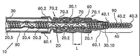
Figure 1 illustrates a cross section of a first
catheter 10 according to the invention. The catheter 10
comprises an outer shaft 80, from which a hollow shaft
30 projects to the right in the longitudinal direction.
The rear end of a catheter tip 40 is anchored in the
hollow shaft 30 at the front end 30.10 of the hollow
shaft 30 and so the catheter tip 40 protrudes out of
the hollow shaft 30 in the longitudinal direction. The
catheter tip 40 consists of a cylindrical helical
spring 40.1, which has a support wire 40.2 in the
center, the support wire running along the entire
length of the helical spring 40.1 for stabilization
purposes. Here, the support wire 40.2 is connected to
the helical spring 40.1 in approximately the center of
the longitudinal direction of the helical spring 40.1
in a cohesive fashion by a braze point 90 made of
silver filler. Additionally, at the front end of the
helical spring 40, there is a rounded end cap 40.3. For
the purposes of anchoring the helical spring 40, the
rearmost windings of the helical spring 40.1 arranged
in the interior of the hollow shaft 30 are pressed into
the front end 30.10 of the hollow shaft 30.
- 14 -
CA 02703926 2010-04-28
Additionally, a wire helix 20 made of platinum is
arranged coaxially in the hollow shaft 30 in a region
behind the helical spring 40.1. The wire helix has e.g.
an external diameter of 0.4 mm, wherein the wire
thickness of the wire helix 20 is 50 m, for example.
In a frontmost or first region 20.1, the wire helix 20
has five abutting windings. The first region 20.1 of
the helical spring 20 is radiopaque due to the material
(platinum) and the density of the windings. In a second
region 20.2 adjoining the first region 20.1, the wire
helix 20 has three windings attached completely without
contact, wherein there is a spacing L in the
longitudinal direction of approximately 160 m between
two windings. The spacing L corresponds to
approximately three times the wire thickness of the
wire helix 20. Due to the low density of the windings,
the second region 20.2 of the helical spring 20 is
radiolucent. The second region 20.2 of the wire helix
20 is adjoined by the third region 20.3, which again
comprises five abutting windings of the helical spring
20 and is therefore likewise radiopaque. The fourth
region 20.4 of the helical spring 20 situated behind
the third region 20.3 comprises a winding arranged
without contact, as result of which this region is
radiolucent. The fourth region 20.4 is adjoined by the
fifth region 20.5 of the helical spring, which fifth
region has substantially the same design as the third
region 20.3 of the helical spring 20. Further regions
of the helical spring 20 not illustrated in figure 1
are arranged behind the fifth region 20.5. In the
process, regions with windings arranged without contact
(analogous to the fourth region 20.4) alternate with
regions with abutting windings (analogous to the third
region 20.3) and form a regular pattern of radiopaque
and radiolucent regions.
- 15 -
CA 02703926 2010-04-28
The hollow shaft 30 is fitted throughout to the outer
shape or contour of the wire helix 20. The outer
surface 30.1 of the hollow shaft 30 therefore has a
screw-like structure, wherein a region with short pitch
and a region with long pitch alternate.
The rear end 60.2 of an actuatable and expandable
balloon 60 is additionally attached to the front end
80.1 of the outer shaft 80 over the entire
circumference of the outer shaft 80. Here, the balloon
60 is designed as a tubular hollow body and arranged
such that its longitudinal axis is coaxial to the
longitudinal axis of the catheter 10. Here, the part of
the balloon 60 situated in front of the outer shaft 80
is situated directly on the hollow shaft 30 with the
screw-like structure and the front end 60.1 of said
part of the balloon 60 is welded to the hollow shaft 30
in the region of the front end 30.10 thereof. Here, the
first two regions 20.1, 20.2 of the wire helix are
completely surrounded by the balloon 60.
Moreover, a stent 70 or a tubular medical implant that
can be expanded by the balloon 60 is attached outside
of the balloon 60 and likewise coaxially to the
longitudinal axis of the catheter. Here, the stent 70
is pressed against the balloon 60 from the outside and
fitted to the outer surface 30.1 of the hollow shaft 30
or to the contour of the wire helix 20 in the first two
regions 20.1, 20.2. Here, the rear end 70.2 of the
stent 70 is situated in front of the rear end 60.2 of
the balloon 60. At the same time, the front end 70.1 of
the stent 70 is situated behind the front end 60.1 of
the balloon 60.
Additionally, the stent 70 is surrounded in the region
of the outer shell surface by a coating 70.3 containing
a medicament.
- 16 -
CA 02703926 2010-04-28
When the balloon 60 is actuated, the fluid used in the
process can expand over the entire length of the
balloon 60 due to the screw-shaped structure of the
outer surface 30.1 of the hollow shaft 30, as a result
of which there is an even actuation of the balloon 60.
This also reduces the risk of the stent 70 being able
to be displaced or even slipping off the catheter 10 in
the longitudinal direction thereof during the actuation
of the balloon 60.
Figure 2 shows the cross section of a second catheter
11 according to the invention. The catheter 11 has a
hollow shaft 31, in which a wire helix 21 made of
stainless steel, which protrudes out of the hollow
shaft 31, is arranged coaxially in a region of the
front end 31.10. The wire helix 21 has an external
diameter of for example 0.5 mm and a wire diameter of
e.g. 100 m. A straight-line support wire 41.2 is
attached in the interior of the wire helix 21. In the
region of the front end 21.1 of the wire helix 21, said
wire helix is brazed to a catheter tip 41 with a first
braze point 91.1 of silver filler. The catheter tip 41
consists of a helical spring 41.1 made of stainless
steel and it has a rounded end cap 41.3 at its front
end.
A support wire 41.2 is arranged in the interior of the
helical spring 41.1, which extends from the end cap
41.3 of the catheter tip 41 and through the first braze
point 91.1 and the wire helix to the rear end 21.2 of
the latter. Just in front of the front end 31.10 of the
hollow shaft 31, there is a second braze point made of
sliver filler, which interconnects the support wire
41.2 and the wire helix. At the rear end of the wire
helix, there is a third braze point which connects the
rear end of the support wire 41.2 to the wire helix 21.
- 17 -
CA 02703926 2010-04-28
Directly in front of the front end 31.10 of the hollow
shaft 31, a cylindrical stent 71 or a tubular medical
implant is arranged around the wire helix 21. The rear
end 71.2 of the stent 71 surrounds the second braze
point 91.2, while the first braze point 91.1 is
surrounded by the front end 71.1 of the stent 71. There
are sufficient amounts of silver filler at the three
braze points 91.1, 91.2, 91.3 in this case and so these
are radiopaque.
The stent 71 is additionally pressed against the wire
helix 21, and so irregularities (not illustrated in
figure 2) of the inner surface 71.3 of the tubular
stent 71 are pressed into the intermediate spaces
between the individual windings of the wire helix 21.
This significantly hinders a displacement of the stent
71 in a longitudinal direction of the catheter 11,
which is particularly advantageous during the insertion
of the catheter 11 because the stent 71 can hardly slip
off the catheter 11 as a result of this.
The stent 71 is designed as a self-expandable hollow
body and is kept in the compressed form illustrated in
figure 2 by an envelope (not illustrated).
A third catheter 12 according to the invention is
illustrated in figure 3. Here, a hollow shaft 32
extends in an outer shaft 82, and the former protrudes
toward the front out of the front end 82.1 of the outer
shaft 82. A radiopaque ring 100, for example made of
platinum, is arranged around the hollow shaft 32 in a
region in front of the front end 82.1 of the outer
shaft 82. The hollow shaft 32 has three ribs 32.2,
32.3, 32.4, completely encircling the hollow shaft 32,
arranged behind one another in a region directly in
front of the radiopaque ring 100. Here, the three ribs
32.2, 32.3, 32.4 protrude outward from a shell surface
of the hollow shaft 32 and consist of compressed
- 18 -
CA 02703926 2010-04-28
material of the hollow shaft 32 made of plastic. Two
further ribs 32.5, 32.6 are arranged behind the
radiopaque ring 100 in the same fashion. Here, the five
ribs 32.2...32.6 of the hollow shaft 32 have an external
diameter corresponding to the external diameter of the
radiopaque ring 100. As a result, the radiopaque ring
100 is embedded between the three front ribs 32.2,
32.3, 32.4 and the two rear ribs 32.5, 32.6. In
particular, the rear edge 102 and the front edge 101 of
the radiopaque ring 100 are thus covered in the
longitudinal direction of the catheter 11 by the ribs
32.2...32.6.
In the region of the front end 32.10 of the hollow
shaft 32, a helical spring 42.1 made of stainless steel
is anchored in the hollow shaft 32 as component of a
catheter tip 42. The helical spring 42.1 has a support
wire 42.2 extending in the longitudinal direction in
the interior and a rounded end cap 42.3 at the front
end.
A wire helix 22 with six windings is arranged in the
hollow shaft 32 behind the helical spring 42.1, the
helix being connected to the rear end of the helical
spring 42.1 by a braze point 92. The wire helix
consists of tungsten and has an external diameter of
for example 0.2 mm. The wire thickness of the wire
helix is e.g. 30 m. Due to the windings of the wire
helix 22 abutting one another, the latter is
radiopaque.
Furthermore, outside of the hollow shaft 32, an
actuatable balloon 62 is arranged around the hollow
shaft 32. Here, the front end 62.1 of the balloon 62 is
welded to the hollow shaft 32 in the region of the
front end 32.10 thereof. The rear end 62.2 of the
balloon 62 is welded to the outside of the outer shaft
82 at the front end 82.1 thereof.
- 19 -
CA 02703926 2010-04-28
During the production of the catheter 12 from figure 3,
a radiopaque ring 100 is pushed onto a hollow shaft 32
in a first step. Subsequently, the hollow shaft 32 is
compressed, for example in the region in front of the
radiopaque ring 100, to form a first rib 32.4 and it is
subsequently stretched slightly again. A second rib
32.3 and a third rib 32.2 are formed in an analogous
fashion. Subsequently, the ribs 32.5, 32.6 are produced
in the same way behind the radiopaque ring 100.
The described exemplary embodiments should be
understood as illustrative examples, which can be
extended or modified arbitrarily within the scope of
the invention.
Thus, for example, instead of the three described
catheter tips 40, 41, 42, it is also possible to
provide catheter tips that have a different suitable
flexible device instead of the helical springs 40.1,
41.1, 42.1. It is likewise possible for the helical
springs 40.1, 41.1, 42.1 of the catheter tips to be
produced from a radiopaque material, such as gold,
platinum or tungsten, and so the entire catheter tip is
radiopaque. A further option consists of forming the
three wire helices 20, 21, 22 and the three helical
springs 40.1, 41.1, 42.1 from a single helical spring,
made in particular from a radiopaque material, which
according to the invention extends as far as the
regions of the catheter encased by the expandable
hollow bodies.
In the case of the first catheter 10 described in
figure 1, the number of windings of the wire helix 20
abutting one another and/or number of windings arranged
without contact can be increased, for example to obtain
better visibility during X-raying. For this, it is also
- 20 -
CA 02703926 2010-04-28
possible to attach additional radiopaque silver filler
in the radiopaque regions of the wire helix 20.
Moreover, the wire helix 20 in the first catheter 10
does not necessarily have to protrude into the region
of the outer shaft 80. It is also possible that
provision is made for a wire helix which, as in the
case of the third catheter 12 in figure 3, merely
protrudes into a front region of the balloon 60 or
stent 70.
It is also possible for the coating 70.3 of the stent
70 to be omitted in the case of the first catheter 10,
or to be replaced by a different coating. By way of
example, coatings of silicone which improve the sliding
properties of the catheter are particularly suitable.
It is also possible for the stent 70 to be omitted in
the first catheter 10 and so the balloon 60 is present
as the outermost expandable hollow body. In this case,
e.g. the balloon can also be provided with a coating
with medicaments or a coating for improving the gliding
properties.
In the case of the second catheter 11 from figure 2,
the wire helix 21 can also be stretched or designed
such that the individual windings of the wire helix'are
arranged without contact in the region of the stent 71.
This possibly affords a further increase in the
friction between the stent 71 and the wire helix 21. In
the case of the second catheter, rather than using a
wire helix made of stainless steel, it is also possible
to use a wire helix made of a radiopaque material such
as gold, platinum or tungsten.
In addition to the radiopaque ring 100 shown in figure
3, it is also possible for further radiopaque rings to
be arranged on a shaft of the catheter.
- 21 -
CA 02703926 2010-04-28
In summary, it should be noted that a catheter with an
expandable casing and a novel design is provided, which
in particular has a very compact design. This is
accompanied with significant advantages when inserting
the catheter because the latter can be guided through
stenoses in a hollow organ or a vessel in the best
possible fashion due to the small external diameter.
Additionally, the catheter according to the invention
can be produced economically.
- 22 -