Note: Descriptions are shown in the official language in which they were submitted.
CA 02703947 2010-04-26
WO 2009/052623
PCT/CA2008/001873
- 1 -
Title: Therapeutic and Diagnostic Methods Using Tim-3
Field of the application
[0001] The
present application relates to methods of treating viral
infections and methods of increasing immune system activity by modulating
Tim-3 activity. In addition, the present application relates to methods of
diagnosing or monitoring immune system activity, acute and chronic viral
infection and inflammatory disease using Tim-3 expression.
Background of the application
[0002] It is
clear from many studies that HIV-1-specific CD8+ and CD4+
T cell responses have a prominent role in controlling viral replication (1-4).
However, in most cases cellular immunity to HIV-1 proves incapable of long-
term control of viremia, and without antiretroviral therapy, progression to
AIDS
occurs. It has become evident that the ultimate failure of the host immune
system to contain HIV-1 is related to the functional impairment of virus-
specific CD8+ and CD4+ T cells which accompanies progressive HIV-1
infection, a phenomenon referred to as T cell exhaustion (5-7).
[0003]
Effective T cell responses are characterized by polyfunctional
cytokine production, cytotoxic potential, and strong proliferation in response
to
antigen (11-14). In the context of chronic infection with HIV-1, the
deterioration of the T cell response follows a characteristic pattern.
Proliferative capacity, cytotoxic potential, and the ability to produce IL-2
are
lost early, while production of IFN-y is more enduring. Ultimately, the
majority
of both CD8+ and CD4+ T cells chronically exposed to antigen lose the ability
to produce IFN-y and enter into a state of peripheral anergy (8-13). This has
been demonstrated by tetramer studies which have observed that only a small
fraction of HIV-1-specific T cells produce cytokine in response to antigen (14-
18). Recently, a step forward has been made in understanding T cell
exhaustion by the identification of a causative contribution of signaling
through PD-1 (5-7). Given the characteristic complexity of T cell regulation,
other mechanisms for dampening effector functions Of chronically activated
cells likely exist.
CA 02703947 2010-04-26
WO 2009/052623
PCT/CA2008/001873
- 2 -
[0004] T
cell immunoglobulin and mucin domain-containing molecule 3
(Tim-3) is an immunoglobulin (Ig) superfamily member. The murine
homologue of Tim-3 was identified as a specific cell surface marker of Thi
CD4+ T cells (19). Interaction of murine Tim-3 with its interferon inducible
ligand galectin-9, has been shown to regulate Thi responses by promoting T
cell aggregation and the death of IFN-y producing Thi cells (20). In mice,
blockade of the Tim-3 pathway prevents the acquisition of transplantation
tolerance induced by costimulatory blockade (21) (22). Furthermore, Tim-3-
deficient mice are refractory to the induction of high dose tolerance in an
experimental autoimmune encephalomyelitis (EAE) model, and anti-Tim-3
rnAbs treatment of SJUJ mice exacerbated EAE (23) (19). Together, these
results show that Tim-3 interactions play a role in suppressing Thi mediated
immune responses in mice through the termination of effector Thi cells.
Summary of the application
[0005] The
inventors have identified a novel population of functionally
impaired T cells in subjects infected with acute and chronic viruses, such as
HIV. This population of cells expresses the glycoprotein Tim-3 on their
surface. In addition, the inventors have identified that the presence of this
population of cells correlates with CD38 expression and with the viral load in
subjects either acutely or chronically infected with viruses, such as HIV, and
that the presence of this population of cells inversely correlates with CD4+ T
cell count. Further, the inventors have shown that blocking Tim-3 activity
improves immune system function. In particular, the inventors have shown
that blocking Tim-3 signaling improves the function of T-cells.
[0006]
Accordingly, the application includes a method of monitoring
immune system activity or function in a subject, comprising the steps:
(a) determining the expression of Tim-3 on the surface of T
cells in a sample from the subject; and
(b) comparing the expression of Tim-3 on the surface of the
T cells from the sample with a control;
CA 02703947 2010-04-26
WO 2009/052623
PCT/CA2008/001873
- 3 -
wherein a difference in expression of Tim-3 on the surface of T
cells in the sample from the subject as compared to the control is indicative
of
immune system activity or function.
[0007]
Another aspect of the application is a method of detecting
functionally impaired T cells in a subject, comprising the steps:
(a) determining the expression of Tim-3 on the surface of T cells
in a sample from the subject; and
(b) comparing the expression of Tim-3 on the surface of the T
cells from the sample with a control;
wherein a difference in expression of Tim-3 on the surface of T
cells in the sample from the subject as compared to the control is indicative
of
the presence of functionally impaired T cells in the subject.
[0008] A
further aspect of the application is a method of monitoring or
assessing viral load in a subject, comprising
(a) determining the expression of Tim-3 on the surface of T cells
in a sample from the subject,
(b) comparing the expression of Tim-3 on the surface of the T
cells from the sample with a control;
wherein a difference in expression of Tim-3 on the surface of T
cells in the sample from the subject as compared to the control is indicative
of
viral load in the subject.
[0009]
Another aspect of the application is a method of monitoring or
assessing disease progression in a subject with a chronic viral infection,
comprising
(a) determining the expression of Tim-3 on the surface of T cells
in a sample from the subject,
(b) comparing the expression of Tim-3 on the surface of the T
cell from the sample with a control;
CA 02703947 2010-04-26
WO 2009/052623
PCT/CA2008/001873
- 4 -
wherein an increase in expression of Tim-3 on the surface of T
cells in the sample as compared to the control is indicative of disease
progression, while a decrease in expression of Tim-3 on the surface of T cells
in the sample is indicative of disease remission. in one embodiment the
control comprises a sample from a previous time-point from the same
individual.
[0010] An additional aspect of the application is a method of
monitoring
or diagnosing viral infection in a subject, comprising the steps:
(a) determining the expression of Tim-3 on the surface of T cells
in a sample from the subject; and
(b) comparing the expression of Tim-3 on the surface of the T
cells from the sample with a control;
wherein a difference in expression of Tim-3 on the surface of T
cells in the sample from the subject as compared to the control is indicative
of
viral infection in a subject. The viral infection can be acute or chronic
viral
infection.
[0011] A further aspect of the application is a method of monitoring
the
efficacy of highly active antiretroviral therapy (HAART), comprising the
steps:
(a) determining the expression of Tim-3 on the surface of T cells
in a subject prior to initiating HAART; and
(b) comparing Tim-3 expression on the surface of T cells from at
least one time point after initiation of HAART;
wherein a decrease in Tim-3 expression is indicative of effective
therapy.
[0012] A further aspect of the application is a method of treating a
subject with a viral infection, comprising administering an effective amount
of
an inhibitor of Tim-3 to the subject afflicted with a viral infection.
[0013] The application also includes the use of an effective amount
of
an inhibitor of Tim-3 for treating a subject afflicted with a viral infection
and the
CA 02703947 2010-04-26
WO 2009/052623
PCT/CA2008/001873
- 5 -
use of an effective amount of an inhibitor of Tim-3 for manufacturing a
medicament for treating a subject afflicted with a viral infection. In
addition,
the application relates to an inhibitor of Tim-3 for use in treating viral
infections. In one embodiment, the viral infection is an acute viral
infection. In
another embodiment, the viral infection is a chronic viral infection.
[0014] Another aspect of the invention is a method of reversing
immune
defects which persist with highly active antiretroviral treatment (HAART)
therapy comprising administering an effective amount of an inhibitor of Tim-3
to the subject in need thereof.
[0015] The application also includes the use of an inhibitor of Tim-3 for
reversing immune defects which persist with HAART therapy and the use of
an inhibitor of Tim-3 for manufacturing a medicament for reversing immune
defects which persist with HAART therapy. In addition, the application relates
to an inhibitor of Tim-3 for use in reversing immune defects which persist
with
HAART therapy.
[0016] A further aspect of the application is a method of improving
the
function of functionally impaired T cells, comprising treating the
functionally
impaired T cells with an inhibitor of Tim-3.
[0017] The application also includes the use of an inhibitor of Tim-3
for
improving the function of functionally impaired T cells and the use of an
inhibitor of Tim-3 for manufacturing a medicament for improving the function
of functionally impaired T cells. In addition, the application relates to an
inhibitor of Tim-3 for use in improving the function of functionally impaired
T
cells.
[0018] In addition, the application includes a method of inducing an
immune response in a subject against a chronic virus, such as HIV-1 or HCV,
comprising co-administering to said subject an effective amount of a chronic
viral antigen, such as an HIV-1 antigen or HCV antigen, and an inhibitor of
Tim-3.
CA 02703947 2010-04-26
WO 2009/052623 PCT/CA2008/001873
- 6 -
[0019] The application also includes the use of an effective amount
of a
chronic viral antigen and an inhibitor of Tim-3 for inducing an immune
response in a subject against a chronic virus and the use of an effective
amount of an chronic antigen and an inhibitor of Tim-3 for manufacturing a
medicament for inducing an immune response in a subject against a chronic
virus. In addition, the application relates to a chronic viral antigen and an
inhibitor of Tim-3 for use in inducing an immune response in a subject against
a chronic virus.
[0020] In addition, the application provides a method of inducing an
immune response in a subject against human endogenous retrovirus (HERV)
or long-interspersed nuclear element (LINE) antigens comprising co-
administering to said subject an effective amount of a LINE or HERV
immunogen, and an inhibitor of Tim-3. In one embodiment, the method is
used to induce an immune response against HIV infected cells which express
HERV or LINE antigens. HERV antigens are described in USSN 11/880,126
incorporated herein by reference.
[0021] The application also includes the use of an effective amount
of a
LINE or HERV immunogen and an inhibitor of Tim-3 for inducing an immune
response in a subject and the use of an effective amount of a LINE or HERV
immunogen and an inhibitor of Tim-3 for manufacturing a medicament for
inducing an immune response in a subject. In addition, the application relates
to a LINE or HERV immunogen and an inhibitor of Tim-3 for use in inducing
an immune response in a subject.
[0022] Further, the application includes a method of treating or
preventing a chronic viral infection, such as an HIV-1 infection or HCV
infection, in a subject comprising co-administering to said subject an
effective
amount of a chronic viral antigen, such as an HIV-1 antigen, an HCV antigen,
a HERV antigen or a LINE antigen, and an inhibitor of Tim-3.
[0023] The application also includes the use of an effective amount
of a
chronic viral antigen and an inhibitor of Tim-3 for treating or preventing a
chronic viral infection in a subject and the use of an effective amount of a
CA 02703947 2010-04-26
WO 2009/052623
PCT/CA2008/001873
- 7 -
chronic viral antigen and an inhibitor of Tim-3 for manufacturing a medicament
for treating or preventing a chronic viral infection in a subject. In
addition, the
application relates to a chronic viral antigen and an inhibitor of Tim-3 for
use
in treating or preventing a chronic viral infection in a subject.
[0024] The application also includes compositions comprising a soluble
form of Tim-3 and methods and uses thereof.
[0025] Other features and advantages of the present application will
become apparent from the following detailed description. It should be
understood, however, that the detailed description and the specific examples
while indicating preferred embodiments of the invention are given by way of
illustration only, since various changes and modifications within the spirit
and
scope of the invention will become apparent to those skilled in the art from
this detailed description.
Brief description of the drawings
[0026] The invention will now be described in relation to the drawings in
which:
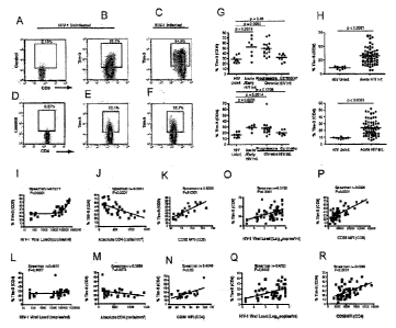
[0027] Figure 1 shows that Tim-3 is upregulated on T cells in HIV-1
infection and its expression correlates with parameters of HIV-1 disease
progression. (A-F) PBMCs from HIV-1 infected individuals and HIV-1
uninfected controls were stained with antibodies against Tim-3, CD4, CD8
and CD3. Shown is data obtained by staining with a biotinylated polyclonal
goat anti-Tim-3 antibody, followed by a secondary streptavidin-APC
conjugate. Confirmatory experiments were performed using PE conjugated
monoclonal anti-Tim-3, and an excellent correlation between the two data sets
was observed, with slightly higher frequencies of Tim-3 expressing cells
observed with polyclonal anti-Tim-3 (see also Figure 12). Representative plots
show events gated on the CD3+ population and subsequently on the CD8+ (A,
B, C) or CD4+ (D, E, F) populations. Staining was performed using
biotinylated normal goat control antisera and streptavidin-APC to control for
potential non-specific binding of polyclonal goat anti-Tim-3 (A, D). Shown are
representative levels of Tim-3 in an HIV-1 uninfected subject (B, E) in
CA 02703947 2010-04-26
WO 2009/052623
PCT/CA2008/001873
- 8 -
comparison to an HIV-1 infected subject (C, F). The percentages of Tim-3+
cells on CD8+ and CD4 + T cells (G, H) are indicated for 31 individuals
separated into the following groups: HIV-1 uninfected, HIV-1-infected
acute/early, HIV-1-infected chronic, and HIV-1-infected controller. Groups
were defined as follows: Acute/early = infected with HIV-1 within the last 4
months; chronic = infected > 1 year with CD4 decline; controller = infected >
1
year, no evidence of CD4 decline, and viral load <5,000 copies/ml bDNA.
Statistical analyses were performed using the Mann-Whitney test. (I-R)
Correlation between Tim-3 expression on CD8+ (I-K, 0,P) and CD4 + (L-N, Q,
R) T cells and viral load (I, L, 0, Q), CD4 T cell counts (J, M) and levels of
CD38 expression (K, N, P, R) are shown. Statistical analyses were performed
using the Spearman's rank correlation test.
[0028] Figure 2 shows PBMC from 8 chronically HIV-1 infected
individuals were stained with pentamers to A2 restricted CMV, EBV, and HIV-
1 epitopes (A-H). (A-D) Shown are representative flow cytometry data from
one individual using tetramer to the CMV pp65 epitope AILVPMVATV' (0), the
EBV epitope `GLCTLVAML' (P), the HIV-1-Pol epitope `ILKEPVHGV (Q), and
the HIV-1-Gag epitope `SLYNTVATL'. The mean fluorescence intensity (MFI)
of pentamer+ cells were compared for all detectable responses to each
epitope. Tim-3 expression was heterogenous amongst HIV-1-specific
responses with some exhibiting very high levels of Tim-3, while others
exhibited only baseline levels (F,H).Statistical analyses were performed using
the Wilcoxon matched pairs T test.
[0029] Figure 3 shows the effect of HAART on levels of Tim-3
expression in Chronic HIV-1 infection. Seven chronically HIV-1-infected
individuals from the CIRC cohort were sampled at baseline and at 1, 2, 3 and
6 months post-initiation of HAART. Shown are (a) compiled Tim-3 expression
on CD8+ T cells versus month post-initiation of HAART (b) Tim-3 and CD38
expression levels as determined by flow cytometry, along with absolute CD4+
T cell count and HIV-1 viral load clinical data. The 6 individuals followed
for 6
months achieved undetectable viral loads (bDNA <50 copies/nil). The chart in
CA 02703947 2010-04-26
WO 2009/052623 PCT/CA2008/001873
- 9 -
panel (b) summarizes the p values obtained from a mixed-effects longitudinal
analysis studying associations between Tim-3 expression on CD8+ T cells
with: HIV-1 viral load, CD8+ T cell activation as measured by CD38
expression (MFI), and absolute CD4+ T cell count.
[0030] Figure 4 shows PBMC from both HIV-1-infected and uninfected
individuals that were sorted for Tim-3+ and Tim-3" populations within both
CD8+ and CD4+ T cell subsets and quantified T-bet (Th1), GATA-3 (Th2), and
IFN-y (TO mRNA by qPCR. For both CD8+ and CD4+ T cell populations,
GATA-3 was expressed at higher levels in the Tim-3" fraction than in the Tim-
3+ fraction, while T-bet was more highly expressed in the Tim-3+ population.
[0031] Figure 5 shows Tim-3 expressing CD8+ and CD4+ T cells
populations hyporesponsive to antigenic stimulation. PBMCs derived from
HIV-1 infected and uninfected individuals were stimulated with pooled
peptides or SEB superantigen for 12 hours, and then stained for IFN-7, TNF-g
and Tim-3 using monoclonal antibodies, and analyzed by multiparametric flow
cytometry. (A-D) Representative plots showing cytokine responses in CD8+
and CD4+ T cells from HIV-1 infected and HIV-1 uninfected individuals. (E-G)
Tetramer analysis was performed on PBMC from a chronically HIV-1 infected
individual using A2*SLYNTVATL (E). PBMC from the same individual were
stimulated with SLYNTVATL (SEQ ID NO: 7) peptide, or with DMSO as a
control, and cytokine production versus Tim-3 expression was analyzed by
flow cytometry (F, G). CD8+ T cells were sorted into purified Tim-3+N CD8+ T
cells and Tim-34 (H, I, J) CD8+ T cells populations and labeled with CFSE.
These two populations were then cultured in the presence of anti-CD3 and
anti-CD28 monoclonal antibodies for 5 days. Cells where then assessed for
the diminution of CFSE as a readout of cell division (K, L). (M-R) Co-stained
ex vivo PBMC from 5 HIV-1-uninfected individuals, and 5 HIV-1-infected
chronic progressors, with Tim-3 and Ki67 antigen. Elevated frequencies of
Ki67+ cells were observed in both the CD4+ and CD8+ T cell subsets of HIV-1-
infected versus uninfected PBMC (M-N). The large majority of Tim-3+ cells
CA 02703947 2010-04-26
WO 2009/052623
PCT/CA2008/001873
- 10 -
were Ki67-, Ki67+ CD8+ and CD4+ T cells were greatly enriched for Tim-3
expressing cells (R).
[0032] Figure 6 shows that blocking the Tim-3 signaling pathway by
the
addition of soluble Tim-3 enhances proliferation and cytokine production of
HIV-1-specific T cells. (A) The addition of sTim-3 enhanced the expansion of
CD8+ T cells specific for the HLA-A*0201 restricted HIV-1-Gag epitope
`SLYNTVATL' (SL9) in HIV-1-infected chronic progressors in a dose-
dependent manner up to 2 pg/ml. (B,C) PBMCs from 6 HIV-1 infected
patients were stained with CFSE and the effect of slim-3 on cytokine
production and proliferation of PBMCs was determined in four individuals over
a 6 day stimulation assay. Shown is representative data from an acutely HIV-
1 infected individual on day 6 of culturing showing IFN-y secretion (y-axis)
by
CFSE (x-axis) in CD8+ (B) and CD4+ (C), T cell populations in response to
DMSO (Upper row), pooled Gag/Nef peptides (middle row) or CEF pooled
peptides (lower row) in the presence or absence of either 1 pg/ml sTim-3 or
an equal volume of expression control. (D) Enhanced proliferation of both
CD8+ and CD4+ T cells was also observed when PBMC from chronic
progressors were stimulated with pooled Gag and Nef peptides. (E) Addition
of 10pg/m1 of mAb 2E2 resulted in a profound rescue of HIV-1-Gag T cell
proliferative responses.
[0033] Figure 7 shows PBMC from 10 individuals with chronic
progressive HIV-1 infection co-stained for Tim-3 and PD-1. Expression was
analyzed by flow cytometry after gating on CD8+ or CD4+ T cells. (A-B)
Demonstrates that in 9/10 subjects, Tim-3 and PD-1 were primarily expressed
by distinct populations of CD8+ T cells. One subject, 0M513, displayed a
frequent Tim-3+PD-1+ population (23.6%), but retained both Tim-3+PD-1- and
Tim-3-PD-1+ populations (23.0% and 16.7% respectively). (C-D)
Demonstrates that 9/10 subjects showed primarily divergent staining for PD-1
and Tim-3 on CD4+ T cells. (E-F) In HIV-1-specific CD8+ T cells, two patterns
of expression were observed: tetramer+ populations were predominantly Tim-
3+PD-1- (E), or they were predominantly Tim-3- and PD-1+ (F). Both patterns
CA 02703947 2010-04-26
WO 2009/052623
PCT/CA2008/001873
-11 -
showed that a minority population co-expressed both Tim-3 and PD-1
demonstrating that Tim-3 and PD-1 expression define primarily distinct
populations.
[0034]
Figure 8 (A-B) shows dual staining for Tim-3 and CD25 on both
CD4+ and CD8+ T cells. Tim-3 and CD25 were primarily expressed by distinct
populations of T cells and demonstrate that Tim-3 expression on CD4+ T cells
does not mark a population of classical regulatory T cells. (C-E) Demonstrates
a phenotypic flow cytometry assessment of Tim-3+ (D) versus Tim-3- (E) CD8+
T cells subpopulations from chronically HIV-1 infected individuals. PBMCs
were stained with monoclonal antibodies against Tim-3, CD3, CD8, 0D28,
CD27, CD45RA, CCR7 and CD57, as well as with a dead cell discriminating
marker. Gating was first performed to include only the viable, CD3+CD8+
population in subsequent analysis. Gating for maturation/differentiation
markers was determined based on fluorescence minus one controls, and
results were analyzed using SPICE software. Shown are the frequencies of
populations with the corresponding combination of phenotypic markers, with
each individual represented by a single bar. These data support that Tim-3
expressing CD8+ T cells from chronically HIV-1-infected individuals were
distributed across a range of phenotypic profiles.
[0035] Figure 9 (A) Demonstrates phospho-flow cytometry analyses of
phosphorylation status of Stat5, p38, and ERK-1/2 in Tim-3+ versus Tim-3-
CD8+ T cells from HIV-1 infected subject. CD8+ T cells were sorted based on
their Tim-3 expression status and stimulated with either rIL-2 or
PMA/Ionomycin in triplicates wells from each sample. Shown is representative
FACS gating for sorting Tim-3+/"1 and Tim-341 PBMCs. Shown is a summary
of data from 4 chronically HIV-1 infected individuals. (B) A representative
time
course from one individual. (C-E) Shown is the compiled data for (C) Stat5,
(D) ERK-1/2, and (E) p38 showing differential levels of change in target
phosphorylation (measured by change in mean fluorescence intensity) in Tim-
3+ versus Tim-3- cells within each of the following CD8+ T cell sub-
CA 02703947 2010-04-26
WO 2009/052623
PCT/CA2008/001873
- 12 -
populations: naïve (CD27+CD45RA+), memory (C D27 CD45RA-), effector
memory (CD27-, CD45RA-), or effector (CD27-, CD45RA+).
[0036] Figure 10 shows (A-E) the expression of Tim-3 in NKT cells and
monocyte subpopulations in PBMCs from a healthy subject. Representative
plots of n=8.
[0037] Figure 11 shows the flow cytometry plots of CD38 versus Tim-3
expression on CD8+ T cells from three subjects: (A) an HIV-1 infected
controller, (B) an HIV-1 infected chronic progressor with a moderate viral
load,
and (C) an HIV-1 infected chronic progressor with advanced disease and a
high viral load.
[0038] Figure 12 shows the correlation of the frequency of surface
Tim-
3 expression on CD8+ T cells from HIV-1 infected individuals as determined
by either a rabbit monoclonal antibody (X-axis) or goat polyclonal antibodies
(Y-axis) against Tim-3.
[0039] Figure 13 shows analogous patterns of cytokine production were
observed for acutely/early infected individuals, chronic progressors, viral
controllers, and HIV-1-uninfected subjects.
[0040] Figure 14 shows TNF-a and CD107a expression in response to
antigen were similarly restricted to Tim-3- cells.
[0041] Figure 15 is a silver-stained SDS PAGE of purified soluble Tim-3
(lane 3) and an expression control (lane 2).
[0042] Figure 16 shows the cells which had undergone proliferation in
vitro exhibited high levels of Tim-3 expression
[0043] Figure 17 demonstrates that in the presence of sTim-3 the
cells
in Figure 16 consistently express higher levels of IFN-y than in the presence
of a control.
[0044] Figure 18 shows the effect of HAART on levels of Tim-3
expression in Chronic HIV-1 infection. Seven chronically HIV-1-infected
individuals from the CIRC cohort were sampled at baseline and at 1, 2, 3 and
CA 02703947 2010-04-26
WO 2009/052623
PCT/CA2008/001873
- 13-
6 months post-initiation of HAART. Shown are Tim-3 and CD38 expression
levels as determined by flow cytometry, along with absolute CD4+ T cell count
and HIV-1 viral load clinical data. Absolute CD4+ T cell count is displayed as
cells/mm3 divided by 10.
Detailed description of the application
[0045] As mentioned above, the inventors have identified a novel
functionally impaired T cell population that expresses Tim-3. This population
of T cells is found in subjects afflicted with acute and chronic viral
infections,
such as HIV infection. The inventors have identified that the presence of this
population in subjects infected with chronic viruses proportionally correlates
with viral load and CD38 expression, and inversely correlates with CD4+ T cell
count. In addition, the inventors have shown that blocking Tim-3 activity
improves immune system function.
[0046] Accordingly, the application includes a method of monitoring
immune system activity or function in a subject, comprising the steps:
(a) determining the expression of Tim-3 on the surface of T
cells in a sample from the subject; and
(b) comparing the expression of Tim-3 on the surface of the
T cells from the sample with a control;
wherein a difference in expression of Tim-3 on the surface of T
cells in the sample from the subject as compared to the control is indicative
of
immune system activity or function.
[0047] The term "Tim-3" as used herein refers to T cell
immunoglobulin
and mucin domain-containing molecule 3. In one embodiment, Tim-3 is of
human origin. In another embodiment, Tim-3 has the sequence:
nnfshlpfdcv IIIIIIIItr sseveyraev gqnaylpcfy tpaapgnlvp vcwgkgacpv
fecgnvvIrt derdvnywts rywIngdfrk gdvsltienv tladsgiycc riqipgimnd
ekfnIklvik pakvtpaptr qrdftaafpr mlttrghgpa etqtlgslpd initqistla
CA 02703947 2010-04-26
WO 2009/052623 PCT/CA2008/001873
- 14 -
nelrdsrlan dlrdsgatir igiyigagic aglalalifg alifkwyshs kekignIsli
slanIppsgl anavaegirs eeniytieen vyeveepney ycyvssrqqp sqpIgcrfamp
(SEQ ID NO:5) or a variant thereof.
[0048] The term "variant" as used herein includes modifications,
substitutions, additions, derivatives, analogs, fragments or chemical
equivalents of the Tim-3 amino acid sequences disclosed herein that perform
substantially the same function as the Tim-3 peptides and peptide inhibitors
disclosed herein in substantially the same way. For instance, the variants of
the Tim-3 peptides would have the same function of being useful in monitoring
immune system activity or function, in detecting functionally impaired cells,
in
monitoring viral load and monitoring or diagnosing chronic viral infection.
Variants of Tim-3 peptide inhibitors would have the same function as being
useful to inhibit Tim-3.
[0049] Variants also include peptides with amino acid sequences that
are substantially or essentially identical to the amino acid sequences of SEQ
ID NO:5, 2 or 6.
[0050] The term "substantially identical" or "essentially identical"
as
used herein means an amino acid sequence that, when optimally aligned, for
example using the methods described herein, share at least 75%, 80%, 85%,
90%, 95%, 96%, 97%, 98%, 99%, or 100% sequence identity with a second
amino acid sequence.
[0051] The term "sequence identity" as used herein refers to the
percentage of sequence identity between two polypeptide and/or nucleotide
sequences.
[0052] To determine the percent identity of two amino acid sequences,
the sequences are aligned for optimal comparison purposes (e.g., gaps can
be introduced in the sequence of a first amino acid or nucleic acid sequence
for optimal alignment with a second amino acid or nucleic acid sequence).
The amino acid residues at corresponding amino acid positions are then
compared. When a position in the first sequence is occupied by the same
CA 02703947 2010-04-26
WO 2009/052623 PCT/CA2008/001873
- 15 -
amino acid residue or nucleotide as the corresponding position in the second
sequence, then the molecules are identical at that position. The percent
identity between the two sequences is a function of the number of identical
positions shared by the sequences (i.e., % identity=number of identical
overlapping positions/total number of positions×100cY0). In one
embodiment, the two sequences are the same length. The determination of
percent identity between two sequences can also be accomplished using a
mathematical algorithm. A preferred, non-limiting example of a mathematical
algorithm utilized for the comparison of two sequences is the algorithm of
Karlin and Altschul, 1990, Proc. Natl. Acad. Sci. U.S.A. 87:2264-2268,
modified as in Karlin and Altschul, 1993, Proc. Natl. Acad. Sci. U.S.A.
90:5873-5877. Such an algorithm is incorporated into the NBLAST and
XBLAST programs of Altschul et al., 1990, J. Mol. Biol. 215:403. BLAST
nucleotide searches can be performed with the NBLAST nucleotide program
parameters set, e.g., for score=100, word1ength=12 to obtain nucleotide
sequences homologous to a nucleic acid molecule of the present application.
BLAST protein searches can be performed with the XBLAST program
parameters set, e.g., to score-50, wordlength=3 to obtain amino acid
sequences homologous to a protein molecule of the present disclosure. To
obtain gapped alignments for comparison purposes, Gapped BLAST can be
utilized as described in Altschul et al., 1997, Nucleic Acids Res. 25:3389-
3402. Alternatively, PSI-BLAST can be used to perform an iterated search
which detects distant relationships between molecules (Id.). When utilizing
BLAST, Gapped BLAST, and PSI-Blast programs, the default parameters of
the respective programs (e.g., of XBLAST and NBLAST) can be used (see,
e.g., the NCB! website). Another preferred, non-limiting example of a
mathematical algorithm utilized for the comparison of sequences is the
algorithm of Myers and Miller, 1988, CABIOS 4:11-17. Such an algorithm is
incorporated in the ALIGN program (version 2.0) which is part of the GCG
sequence alignment software package. When utilizing the ALIGN program for
comparing amino acid sequences, a PAM120 weight residue table, a gap
length penalty of 12, and a gap penalty of 4 can be used. The percent identity
CA 02703947 2010-04-26
WO 2009/052623
PCT/CA2008/001873
- 16 -
between two sequences can be determined using techniques similar to those
described above, with or without allowing gaps. In calculating percent
identity,
typically only exact matches are counted.
[0053] The percentage of identity between two polypeptide sequences,
the amino acid sequences of such two sequences are aligned, for example
using the Clustal W algorithm (Thompson, JD, Higgins DG, Gibson TJ, 1994,
Nucleic Acids Res. 22(22): 4673-4680.), together with BLOSUM 62 scoring
matrix (Henikoff S. and Henikoff J.G., 1992, Proc. Natl. Acad. Sci. USA 89:
10915-10919.) and a gap opening penalty of 10 and gap extension penalty of
0.1, so that the highest order match is obtained between two sequences
wherein at least 50% of the total length of one of the sequences is involved
in
the alignment.
[0054] Other methods that may be used to align sequences are the
alignment method of Needleman and Wunsch (Needleman and Wunsch. J.
MoL Biol., 1970, 48:443), as revised by Smith and Waterman (Smith and
Waterman. Adv. App!. Math. 1981, 2:482) so that the highest order match is
obtained between the two sequences and the number of identical amino acids
is determined between the two sequences. Other methods to calculate the
percentage identity between two amino acid sequences are generally art
recognized and include, for example, those described by Carillo and Lipton
(Carillo and Lipton SIAM J. Applied Math. 1988, 48:1073) and those
described in Computational Molecular Biology (Computational Molecular
Biology, Lesk, e.d. Oxford University Press, New York, 1988, Biocomputing:
Informatics and Genomics Projects). Generally, computer programs will be
employed for such calculations.
[0055] Variants of the Tim-3 peptides and peptide inhibitors
disclosed
herein also include, without limitation, conservative amino acid
substitutions.
A "conservative amino acid substitution" as used herein, is one in which one
amino acid residue is replaced with another amino acid residue without
abolishing the desired function or activity of the peptide inhibitors
disclosed
herein. Conservative substitutions typically include substitutions within the
CA 02703947 2010-04-26
WO 2009/052623 PCT/CA2008/001873
- 17 -
following groups: glycine, alanine; valine, isoleucine, leucine; aspartic
acid,
glutamic acid, asparagine, glutamine; serine, threonine; lysine, arginine; and
phenylalanine, tyrosine. Conserved amino acid substitutions involve replacing
one or more amino acids of the polypeptides of the disclosure with amino
acids of similar charge, size, and/or hydrophobicity characteristics. When
only conserved substitutions are made the resulting variant should be
functionally equivalent. Changes which result in production of a chemically
equivalent or chemically similar amino acid sequence are included within the
scope of the disclosure. If the peptide inhibitors of the present application
are
made using recombinant DNA technology, variants of the peptide inhibitors
may be made by using polypeptide engineering techniques such as site
directed mutagenesis, which are well known in the art for substitution of
amino
acids. For example, a hydrophobic residue, such as glycine can be
substituted for another hydrophobic residue such as alanine. An alanine
residue may be substituted with a more hydrophobic residue such as leucine,
valine or isoleucine. A negatively charged amino acid such as aspartic acid
may be substituted for glutamic acid. A positively charged amino acid such as
lysine may be substituted for another positively charged amino acid such as
arginine. The phrase "conservative substitution" also includes the use of a
chemically derivatized residue in place of a non-derivatized residue provided
that such polypeptide displays the requisite activity.
[0056] Variants of the Tim-3 peptides and peptide inhibitors of the
present application also include additions and deletions to the amino acid
sequences disclosed herein.
[0057] Variants of the Tim-3 peptides and peptide inhibitors of the
present application also include analogs thereof. The term "analog" as used
herein includes any active agent capable of performing the function of the
Tim-3 peptides and peptide inhibitors disclosed herein, and may include
peptide mimetics and the like. The term "active" refers to molecules in a
conformation suitable for performing substantially the same functions as the
peptide inhibitors disclosed herein in substantially the same way. Peptide
CA 02703947 2010-04-26
WO 2009/052623
PCT/CA2008/001873
- 18 -
mimetics include synthetic structures that may serve as substitutes for
peptides in interactions between molecules (see Morgan and Gainor. (1989),
Ann. Reports Med. Chem. 24:243-252 for a review). Peptide mimetics include
synthetic structures which may or may not contain amino acids and/or peptide
bonds but are designed to retain the desired structural and functional
features
and thus may be suitable substitutes of the peptide inhibitor analog disclosed
in the present application.
[0058]
Peptide mimetics also include molecules incorporating peptides
into larger molecules with other functional elements (e.g., as described in WO
99/25044). Peptide mimetics also include peptoids, oligopeptoids (Simon et
al (1972) Proc. Natl. Acad, Sci USA 89:9367), and peptide libraries containing
peptides of a designed length representing all possible sequences of amino
acids corresponding to an isolated peptide of the disclosure. Peptide mimetics
may be designed based on information obtained by systematic replacement of
L-amino acids by D-amino acids, replacement of side chains with groups
having different electronic properties, and by systematic replacement of
peptide bonds with amide bond replacements. Local
conformational
constraints can also be introduced to determine conformational requirements
for activity of a candidate peptide mimetic. The mimetics may include
isosteric amide bonds, or D-amino acids to stabilize or promote reverse turn
conformations and to help stabilize the molecule.
Cyclic amino acid
analogues may be used to constrain amino acid residues to particular
conformational states. The mimetics can also include mimics of inhibitor
peptide secondary structures. These structures can model the 3-dimensional
orientation of amino acid residues into the known secondary conformations of
proteins. Peptoids may also be used which are oligomers of N-substituted
amino acids and can be used as motifs for the generation of chemically
diverse libraries of novel molecules.
[0059]
Variant Tim-3 peptides and peptide inhibitors of the present
application also include derivatives thereof. The term "derivative" refers to
a
peptide having one or more residues chemically derivatized by reaction of a
CA 02703947 2010-04-26
WO 2009/052623
PCT/CA2008/001873
- 19 -
functional side group. Such derivatized molecules include for example, those
molecules in which free amino groups have been derivatized to form amine
hydrochlorides, p-toluene sulfonyl groups, carbobenzoxy groups, t-
butyloxycarbonyl groups, chloroacetyl groups or formyl groups. Free carboxyl
groups may be derivatized to form salts, methyl and ethyl esters or other
types of esters or hydrazides. Free hydroxyl groups may be derivatized to
form 0-acyl or 0-alkyl derivatives. The imidazole nitrogen of histidine may be
derivatized to form N-im-benzylhistidine. Also included as derivatives are
those peptides which contain one or more naturally occurring amino acid
derivatives of the twenty standard amino acids. For examples: 4-
hydroxyproline may be substituted for proline; 5-hydroxylysine may be
substituted for lysine; 3-methylhistidine may be substituted for histidine;
homoserine may be substituted for serine; and ornithine may be substituted
for lysine. A derivative of a polypeptide also optionally includes
polypeptides
comprising forms of amino acids that are oxidized.
[0060]
Variant Tim-3 peptides and peptide inhibitors of the present
application also include fragments thereof. The term "fragment" as used
herein means a portion of a polypeptide that contains, preferably, at least
10%, 20%, 30%, 40%, 50%, 60%, 70%, 80%, 90%, 95%, or more of the entire
length of the reference polypeptide.
[0061] The
phrase "determining expression of Tim-3 on the surface of
T cells" as used herein means assessing the expression of Tim-3, including
qualitative and quantitative expression, on the surface of T cells. This
includes
assessing the frequency or level of Tim-3 expression on individual cells or
populations of cells. This also includes assessing the frequency or number of
Tim-3 expressing cells. A person skilled in the art will appreciate that a
number of methods can be used to detect, determine and/or quantify cell
surface expression of Tim-3 including immunoassays such as Western blots,
immunoprecipitation followed by SDS-PAGE, immunocytochemistry, FACS,
protein arrays, and the like.
CA 02703947 2010-04-26
WO 2009/052623
PCT/CA2008/001873
- 20 -
[0062] For example,
antibodies specific for Tim-3 can be used to
determine the expression of Tim-3 on the surface of T cells.
[0063] The term
"antibody" as used herein is intended to include
monoclonal antibodies, polyclonal antibodies, and chimeric antibodies. The
antibody may be from recombinant sources and/or produced in transgenic
animals. The term "antibody fragment" as used herein is intended to include
without limitations Fab, Fab', F(ab')2, scFv, dsFv, ds-scFv, dimers,
minibodies, diabodies, and multimers thereof, multispecific antibody
fragments and domain antibodies. Antibodies can be fragmented using
conventional techniques. For example, F(ab')2 fragments can be generated
by treating the antibody with pepsin. The resulting F(ab')2 fragment can be
treated to reduce disulfide bridges to produce Fab' fragments. Papain
digestion can lead to the formation of Fab fragments. Fab, Fab' and F(ab')2,
scFv, dsFv, ds-scFv, dimers, minibodies, diabodies, bispecific antibody
fragments and other fragments can also be synthesized by recombinant
techniques.
[0064] Antibodies to
Tim-3 are commercially available (R&D Systems).
However, a person skilled in the art will appreciate that one could produce
other antibodies that are specific for Tim-3.
[0065] To produce monoclonal antibodies, antibody producing cells
(lymphocytes) can be harvested from an immunized animal with the antigen of
interest (e.g. Tim-3) and fused with myeloma cells by standard somatic cell
fusion procedures thus immortalizing these cells and yielding hybridoma cells.
Such techniques are well known in the art, (e.g. the hybridoma technique
originally developed by Kohler and Milstein (Nature 256:495-497 (1975)) as
well as other techniques such as the human B-cell hybridoma technique
(Kozbor et al., Immunol.Today 4:72 (1983)), the EBV-hybridoma technique to
produce human monoclonal antibodies (Cole et al., Methods Enzymol,
121:140-67 (1986)), and screening of combinatorial antibody libraries (Huse
et al., Science
246:1275 (1989)). Hybridoma cells can be screened
CA 02703947 2010-04-26
WO 2009/052623 PCT/CA2008/001873
- 21 -
immunochemically for production of antibodies specifically reactive with the
antigen of interest and the monoclonal antibodies can be isolated.
[0066] The phrase "method of monitoring immune system activity or
function" as used herein refers to a method or process of determining or
assessing the activity or function of the immune system, including the degree
of immune system activity or function. The term also includes determining or
assessing the frequency, function and/or activity of immune cells, including T
cells.
[0067] The term "immune system function" as used herein refers to the
function of the immune system including hunnoral or cell-mediated. Immune
system function can be assessed using assays known to those skilled in the
art including, but not limited to, antibody assays (for example ELISA assays),
proliferation assays, antigen specific cytotoxicity assays and the production
of
cytokines (for example ELISPOT assays), such as IFN-y, TNF-a, IL-2 and/or
IL-17. In one embodiment, immune system function refers to the number of,
proliferation of and/or cytokine production by CD4+ and/or CD8+ T cells.
[0068] The term "immune system activity" as used herein refers to the
activation status of the immune system. For example, activation status can be
assessed using surface markers on T cells, such as CD38.
[0069] A person skilled in the art will appreciate that immune system
activity and immune system function are different. For example, the
functionally impaired T cells identified by the inventors that express Tim-3
have impaired function (e.g. impaired ability to proliferate and produce
cytokines). However, the inventors have also shown that Tim-3 expression
correlates with CD38 expression, which is a predictor of T cell activation.
Thus, without being limited to theory, Tim-3 acts to suppress the effector
functions of activated T cells.
[0070] The term "subject" as used herein refers to any member of the
animal kingdom, preferably a mammal, more preferably a human being. In
one embodiment, the subject has a chronic viral infection such as HIV
CA 02703947 2010-04-26
WO 2009/052623
PCT/CA2008/001873
- 22 -
infection or other chronic viral infection, such as HCV. In another
embodiment,
the subject has an acute viral infection, such as acute HIV infection, acute
HCV infection, influenza infection, SARS infection, hepatitis B infection,
hepatitis C infection, rhinovirus infection, cytomegalovirus infection,
Epstein-
barr virus infection, measles, varicella-zoster virus infection, herpes
simplex
infection, human papillomavirus infection, enterovirus infection, rubella
infection, dengue virus, HTLV-I infection, HTLV-II infection, west nile virus,
infection, and others. In a further embodiment, the subject has a chronic
rheumatologic condition, such as rheumatoid arthritis, systemic lupus
erythematosis, ankylosing spondylitis, or other rheumatologic condition. In an
additional embodiment, the subject has an immunosuppressed condition or is
immunosuppressed, such as after a transplantation.
[0071] The term
"sample" as used herein refers to any fluid, cell or
tissue sample from a subject which contains T cells. For example, the sample
could be from the circulatory system or lymphatic system, such as blood,
serum or lymphatic fluid.
[0072] The term "T
cells" includes CD4+ T cells and/or CD8+ T cells.
For example, Tim-3 expression can be determined on either or both CD4+ or
CD8+ T cells.
[0073] The term "control"
as used herein refers to a sample from a
subject or a group of subjects who are either known as having a particular
condition or trait or as not having a particular condition or trait. The
control can
vary depending on what is being monitored, assessed or diagnosed. For
example, if one is monitoring immune system activity or function, the control
can be from a subject
who is known to have a suppressed immune system or
an activated immune system. In another embodiment, the control is from a
subject or a group of subjects known to express a particular level or amount
of
Tim-3 on the surface of their T cells. The control can also be a predetermined
standard or reference range of values.
[0074] The term
"difference in expression of Tim-3 on the surface of T
cells in the sample from the subject as compared to the control" means that
CA 02703947 2010-04-26
WO 2009/052623 PCT/CA2008/001873
- 23 -
Tim-3 is differentially expressed on the surface of T cells in the sample from
the subject as compared to the control.
[0075] The term "differentially expressed" or "differential
expression" as
used herein refers to a difference in the level of expression of Tim-3. The
term
"difference in the level of expression" refers to an increase or decrease in
the
measurable expression level of Tim-3 as compared with the measurable
expression level of Tim-3 in a second sample or control. The term can also
refer to an increase or decrease in the measurable expression level of Tim-3
in a population of samples as compared with the measurable expression level
of Tim-3 in a second population of samples. In one embodiment, the
differential expression can be compared using the ratio of the level of
expression of Tim-3 as compared with the expression level of the Tim-3 of a
control, wherein the ratio is not equal to 1Ø For example, a protein is
differentially expressed if the ratio of the level of expression in a first
sample
as compared with a second sample is greater than or less than 1Ø For
example, a ratio of greater than 1, 1.2, 1.5, 1.7, 2, 3, 5, 10, 15, 20 or
more, or
a ratio less than 1, 0.8, 0.6, 0.4, 0.2, 0.1, 0.05, 0.001 or less. In another
embodiment the differential expression is measured using p-value. For
instance, when using p-value, Tim-3 is identified as being differentially
expressed as between a first and second population when the p-value is less
than 0.1, preferably less than 0.05, more preferably less than 0.01, even more
preferably less than 0.005, the most preferably less than 0.001.
[0076] The phrase "indicative of immune system activity or function"
as
used herein refers to comparing the expression of Tim-3 on the surface of T
cells from the sample with a control and determining whether there is a
difference of expression and whether the results indicate that the immune
system of the subject has decreased or increased activity or function as
compared to the control. As mentioned above, Tim-3 expression is indicative
of functionally impaired T cells, and thus indicative of impaired immune
system function. Accordingly in one embodiment, if the control is from a
normal subject, known to be healthy and not have a viral infection or
CA 02703947 2010-04-26
WO 2009/052623
PCT/CA2008/001873
- 24 -
inflammatory disease, then increased Tim-3 expression on T cells from the
subject as compared to the control indicates that the subject has decreased
immune system function relative to a normal control. In another example, if
the control is from a normal subject, known to be healthy and not have a viral
infection or inflammatory disease, then decreased Tim-3 expression on T cells
from the subject as compared to the control indicates that the subject has
increased immune system function relative to a normal control. In a further
embodiment, if the control is a reference standard known to be indicative of a
healthy individual not having a viral infection or inflammatory disease, then
increased Tim-3 expression is on T cells from the subject as compared to the
control indicates that the subject has decreased immune system function
relative to the control. If the control is a reference standard known to be
indicative of viral infection or inflammatory disease, then decreased Tim-3
expression from the subject compared to the control indicates that the subject
has increased immune system function relative to the control.
[0077] Higher than normal immune system activity can be an indicator
of an inflammatory disease. Thus, the method can be used to monitor or
diagnose an inflammatory disease. This includes determining whether or not a
subject has an inflammatory disease or the extent or severity of the
inflammatory disease as compared to a control. This method can be used in
combination with other traditional diagnostic techniques for inflammatory
disease.
[0078] In one embodiment, the inflammatory disease is an autoimmune
disease. In one embodiment, the autoimmune disease is multiple sclerosis,
transplant rejection, GVHD, acute disseminated encephalomyelitis, coeliac
disease, Crohn's disease, diabetes mellitus type 1, Graves' disease,
Kawasaki's Disease, myasthenia gravis or a chronic rheumatologic condition.
In a specific embodiment, the rheumatologic condition is rheumatoid arthritis,
systemic lupus erythematosis, or ankylosing spondylitis.
CA 02703947 2010-04-26
WO 2009/052623
PCT/CA2008/001873
- 25 -
[0079] The method can also be used to monitor inflammatory activity
in
immunosuppressed conditions, such as transplantation to monitor organ
rejection.
[0080] Another aspect of the application is a method of detecting
functionally impaired T cells in a subject, comprising the steps:
(a) determining the expression of Tim-3 on the surface of T cells
in a sample from the subject; and
(b) comparing the expression of Tim-3 on the surface of the T
cells from the sample with a control;
wherein a difference in expression of Tim-3 on the surface of T
cells in the sample from the subject as compared to the control is indicative
of
the presence of functionally impaired T cells in the subject.
[0081] The term "functionally impaired T cells" as used herein refers
to
hyporesponsive T cells, which are T cells that no longer mount a response to
an antigen. In one embodiment, the T cells are antigen-specific CD8+ and/or
CD4+ T cells, but no longer produce cytokines (such as IFN-y, TNF-a, IL-2
and/or IL-17), no longer are cytotoxic and/or no longer proliferate in
response
to antigen. In a specific embodiment, the antigen is a viral antigen, such as
an
HIV antigen.
[0082] The phrase "indicative of the presence of functionally impaired T
cells in the subject" as used herein refers to comparing the expression of Tim-
3 on the surface of T cells from the sample with a control and determining
whether there is a difference of expression and whether the results indicate
that the subject has more or fewer functionally impaired T cells as compared
to the control.
[0083] The inventors identified that functionally impaired T cells
express Tim-3. Thus, if the control is from a normal subject, known to be
healthy and not have a viral infection or inflammatory disease, then increased
Tim-3 expression on T cells from the subject as compared to the control
indicates that the subject has more functionally impaired T cells than a
normal
CA 02703947 2010-04-26
WO 2009/052623 PCT/CA2008/001873
- 26 -
control. In another example, if the control is from a normal subject, known to
be healthy and not have a viral infection or inflammatory disease, then
decreased Tim-3 expression on T cells from the subject as compared to the
control indicates that the subject has fewer functionally impaired T cells
than a
normal control. In a further embodiment, if the control is a reference
standard
known to be indicative of a healthy individual not having a viral infection or
inflammatory disease, then increased Tim-3 expression on T cells from the
subject as compared to the control indicates that the subject indicates that
the
subject has more functionally impaired T cells than the normal control. If the
control is a reference standard known to be indicative of viral infection or
inflammatory disease, then decreased Tim-3 expression from the subject
compared to the control indicates that the subject has has fewer functionally
impaired T cells than the normal control.
[0084]
Another aspect of the application is a method of detecting or
isolating functionally impaired T cells by detecting Tim-3 expression. For
example, T cells expressing Tim-3 can be detected or isolated from a sample
or population of cells for further study.
[0085] A
further aspect of the application is a method of monitoring or
assessing viral load in a subject, comprising
(a) determining the expression of Tim-3 on the surface of T cells
in a sample from the subject,
(b) comparing the expression of Tim-3 on the surface of the T
cells from the sample with a control;
wherein a difference in expression of Tim-3 on the surface of T
cells in the sample from the subject as compared to the control is indicative
of
viral load in the subject.
[0086] The
term "viral load" refers to the amount of virus in a subject
infected with a virus. For example, it refers to the amount of virus in the
circulating blood. The method can be used to monitor or assess the viral load
of a number of different types of viral infections, including chronic viral
CA 02703947 2010-04-26
WO 2009/052623
PCT/CA2008/001873
- 27 -
infections, such as HIV infection or hepatitis C viral infection (HCV). In a
specific embodiment, the chronic viral infection is an HIV infection.
[0087] The
term "HIV" as used herein refers to the human
immunodeficiency virus, and includes HIV-1 and HIV-2.
[0088] The
phrase "indicative of viral load in the subject" as used
herein refers to comparing the expression of Tim-3 on the surface of T cells
from the sample with a control and determining whether there is a difference
of expression and whether the results indicate that the subject has a higher
or
lower viral load as compared to the control.
[0089] The
inventors identified that viral load in a subject correlates
with the expression of Tim-3 on T cells in the subject. Thus, if the control
is
from a normal subject, known to be healthy and not have a chronic viral
infection, then increased Tim-3 expression on T cells from the subject as
compared to the control indicates that the subject has a higher viral load
than
a normal control. In another example, if the control is from a normal subject,
known to be healthy and not have a chronic viral infection, then decreased
Tim-3 expression on T cells from the subject as compared to the control
indicates that the subject has lower viral load than a normal control.
[0090] An
additional aspect of the application is a method of monitoring
or diagnosing viral infection in a subject, comprising the steps:
(a) determining the expression of Tim-3 on the surface of T cells
in a sample from the subject; and
(b) comparing the expression of Tim-3 on the surface of the T
cells from the sample with a control;
wherein a difference in expression of Tim-3 on the surface of T
cells in the sample from the subject as compared to the control is indicative
of
viral infection in a subject. In one embodiment, the viral infection is a
chronic
viral infection. In another embodiment, the viral infection is an acute viral
infection.
CA 02703947 2010-04-26
WO 2009/052623 PCT/CA2008/001873
- 28 -
[0091] The term "chronic viral infection" as used herein refers to a
subject afflicted or infected with a chronic virus. In one embodiment, the
chronic viral infection is an HIV infection or a hepatitis C viral infection
(HCV).
In a specific embodiment, the chronic viral infection is an HIV infection.
[0092] The term "acute viral infection" as used herein refers to a subject
afflicted or infected with an acute virus. Acute viral infections include,
without
limitation, acute HIV infection, acute HCV infection, influenza infection,
SARS
infection, hepatitis B infection, hepatitis C infection, rhinovirus infection,
cytomegalovirus infection, Epstein-barr virus infection, measles, varicella-
zoster virus infection, herpes simplex infection, human papillomavirus
infection, enterovirus infection, rubella infection, dengue virus, HTLV-I
infection, HTLV-II infection, west nile virus, infection, and others.
[0093] The phrase "indicative of viral infection in a subject" as
used
herein refers to comparing the expression of Tim-3 on the surface of T cells
from the sample with a control and determining whether there is a difference
of expression and whether the results indicate that the subject has a viral
infection or does not have a viral infection or the extent or severity of the
viral
infection as compared to the control.
[0094] The inventors identified that viral infection in a subject
correlates
with the expression of Tim-3 on T cells in the subject. Thus, if the control
is
from a normal subject, known to be healthy and not have a viral infection,
then
increased Tim-3 expression on T cells from the subject as compared to the
control indicates that the subject has a viral infection, more of a viral
infection
or more severe of a viral infection than a normal control. In another example,
if the control is from a normal subject, known to be healthy and not have a
viral infection, then decreased Tim-3 expression on T cells from the subject
as
compared to the control indicates that the subject has less of a viral
infection
or less severe of a viral infection than a normal control. In a further
embodiment, if the control is a reference standard known to be indicative of a
healthy individual not having a viral infection, then increased Tim-3
expression
on T cells from the subject as compared to the control indicates that the
CA 02703947 2010-04-26
WO 2009/052623
PCT/CA2008/001873
- 29 -
subject has a viral infection, more of a viral infection or more severe of a
viral
infection than the normal control. If the control is a reference standard
known
to be indicative of viral infection, then decreased Tim-3 expression from the
subject compared to the control indicates that the subject has less of a viral
infection or less severe of a viral infection than the normal control.
[0095]
Another aspect of the application is a method of monitoring or
assessing disease progression in a subject with a chronic viral infection,
comprising
(a) determining the expression of Tim-3 on the surface of T cells
in a sample from the subject,
(b) comparing the expression of Tim-3 on the surface of the T
cell from the sample with a control;
[0096]
wherein an increase in expression of Tim-3 on the
surface of T cells in the sample as compared to the control is indicative of
disease progression, while a decrease in expression of Tim-3 on the surface
of T cells in the sample is indicative of disease remission, in one embodiment
the control comprises a sample from a previous time-point from the same
individual.
[0097] A
further aspect of the application is a method of monitoring the
efficacy of highly active antiretroviral therapy (HAART), comprising the
steps:
(c) determining the expression of Tim-3 on the surface of T cells
in an individual prior to initiating HAART; and
(d) comparing Tim-3 expression on the surface of T cells at at
least one time point after initiation of HAART;
wherein a decrease in Tim-3 expression is indicative of effective
therapy.
[0098] A
further aspect of the application is a method of treating a
subject with a viral infection, comprising administering an effective amount
of
an inhibitor of Tim-3 to the subject afflicted with a viral infection. In one
CA 02703947 2010-04-26
WO 2009/052623 PCT/CA2008/001873
- 30 -
embodiment, the subject is afflicted with a chronic viral infection. In
another
embodiment, the subject is afflicted with an acute viral infection.
[0099] The term "afflicted with a chronic viral infection" as used
herein
refers to a subject with a long-term viral infection. In one embodiment, the
viral infection is an HIV infection or a hepatitis C viral infection (HCV). In
a
specific embodiment, the chronic viral infection is an HIV infection.
[00100] The term "afflicted with an acute viral infection" as used
herein
refers to a subject with a short-term viral infection. In one embodiment, the
viral infection is acute HIV infection, acute HCV infection, influenza
infection,
SARS infection, hepatitis B infection, hepatitis C infection, rhinovirus
infection,
cytomegalovirus infection, Epstein-barr virus infection, measles, varicella-
zoster virus infection, herpes simplex infection, human papillomavirus
infection, enterovirus infection, rubella infection, dengue virus, HTLV-I
infection, HTLV-II infection, west nile virus infection.
[00101] A person skilled in the art can readily determine whether an
infection is chronic or acute.
[00102] The phrase "method of treating a subject with a viral
infection"
as used herein includes inhibiting the infection, preventing the infection or
reducing the symptoms associated with the infection.
[00103] The term a "therapeutically effective amount", "effective amount"
or a "sufficient amount" of a compound or composition of the present
application is a quantity sufficient to, when administered to the subject,
including a mammal, for example a human, effect beneficial or desired results,
including clinical results, and, as such, an "effective amount" or synonym
thereto depends upon the context in which it is being applied. For example, in
the context of treating a chronic viral infection, for example, it is an
amount of
the compound or composition sufficient to achieve such a treatment as
compared to the response obtained without administration of the compound or
composition. In the context of disease, therapeutically effective amounts of
the compounds or compositions disclosed in the present application are used
CA 02703947 2010-04-26
WO 2009/052623 PCT/CA2008/001873
- 31 -
to treat, modulate, attenuate, reverse, or affect chronic viral infections in
a
mammal. An "effective amount" is intended to mean that amount of a
compound or composition that is sufficient to treat, prevent or inhibit
chronic
viral infections. In some suitable embodiments, the amount of a given
compound or composition will vary depending upon various factors, such as
the given drug or compound, the pharmaceutical formulation, the route of
administration, the type of disease or disorder, the identity of the subject
or
host being treated, and the like, but can nevertheless be routinely determined
by one skilled in the art. Also, as used herein, a "therapeutically effective
amount" of a compound or composition of the present application is an
amount which prevents, inhibits, suppresses or reduces chronic viral
infections in a subject as compared to a control. As defined herein, a
therapeutically effective amount of a compound or composition of the present
application may be readily determined by one of ordinary skill by routine
methods known in the art.
[00104] The term "inhibitor of Tim-3" or "Tim-3 inhibitor" as used
herein
refers to a compound, substance or composition that can inhibit the function
of Tim-3. For example, the inhibitor can inhibit the expression or activity of
Tim-3, modulate or block the Tim-3 signaling pathway and/or block the
binding of Tim-3 to a ligand. Such inhibitors include peptides, antibodies,
nucleic acid molecules and small molecules. In one embodiment, the inhibitor
binds a Tim-3 ligand. In another embodiment, the inhibitor is an antibody
specific for Tim-3 and/or its ligand. Antibodies to Tim-3 can be prepared as
described previously.
[00105] In an embodiment, the inhibitor is a soluble form of Tim-3. A
soluble form of Tim-3 includes, without limitation, a molecule lacking the
transmembrane and intracellular domains, for example, a molecule
comprising the IgV and/or mucin domains of Tim-3. In one embodiment, the
soluble form of Tim-3 comprises the amino acid sequence of SEQ ID NO:2 or
a variant thereof. In another embodiment, the soluble form of Tim-3 consists
of the amino acid sequence of SEQ ID NO:2. In another embodiment, the
CA 02703947 2010-04-26
WO 2009/052623
PCT/CA2008/001873
- 32 -
soluble form of Tim-3 comprises the amino acid sequence of SEQ ID NO:6 or
a variant thereof. In another embodiment, the soluble form of Tim-3 consists
of the amino acid sequence of SEQ ID NO:6.
[00106] The application also includes an isolated amino acid sequence
comprising the amino acid sequence of SEQ ID NO: 2 or 6 or a variant
thereof. The term variant has been defined previously.
[00107] In another embodiment, the Tim-3 inhibitor is a nucleic acid
molecule. The nucleic acid molecule may be a small interfering RNA (SiRNA)
or antisense molecule that targets and inhibits the expression of the Tim-3
nucleic acid sequence.
[00108] The term "antisense nucleic acid" as used herein means a
nucleotide sequence that is complementary to its target e.g. a Tim-3
transcription product. The nucleic acid can comprise DNA, RNA or a chemical
analog, that binds to the messenger RNA produced by the target gene.
Binding of the antisense nucleic acid prevents translation and thereby
inhibits
or reduces target protein expression. Antisense nucleic acid molecules may
be chemically synthesized using naturally occurring nucleotides or variously
modified nucleotides designed to increase the biological stability of the
molecules or to increase the physical stability of the duplex formed with
mRNA or the native gene e.g. phosphorothioate derivatives and acridine
substituted nucleotides. The antisense sequences may be produced
biologically using an expression vector introduced into cells in the form of a
recombinant plasmid, phagemid or attenuated virus in which antisense
sequences are produced under the control of a high efficiency regulatory
region, the activity of which may be determined by the cell type into which
the
vector is introduced.
[00109] The term "siRNA" refers to a short inhibitory RNA that can be
used to silence gene expression of a specific gene. The siRNA can be a short
RNA hairpin (e.g. shRNA) that activates a cellular degradation pathway
directed at mRNAs corresponding to the siRNA. Methods of designing specific
siRNA molecules and administering them are known to a person skilled in the
CA 02703947 2010-04-26
WO 2009/052623 PCT/CA2008/001873
- 33 -
art. It is known in the art that efficient silencing is obtained with siRNA
duplex
complexes paired to have a two nucleotide 3' overhang. Adding two thymidine
nucleotides is thought to add nuclease resistance. A person skilled in the art
will recognize that other nucleotides can also be added.
[00110] Aptamers are short strands of nucleic acids that can adopt
highly specific 3-dimensional conformations. Aptamers can exhibit high
binding affinity and specificity to a target molecule. These properties allow
such molecules to specifically inhibit the functional activity of proteins.
Thus,
in another embodiment, the Tim-3 inhibitor is an aptamer that binds and
inhibits Tim-3 activity.
[00111] The application also includes compositions comprising an
inhibitor of Tim-3, such as a soluble form of Tim-3. In one embodiment, the
inhibitor of Tim-3, such as a soluble form of Tim-3, is formulated into
pharmaceutical compositions for administration to subjects in a biologically
compatible form suitable for administration in vivo. By "biologically
compatible
form suitable for administration in vivo" is meant a form of the substance to
be
administered in which any toxic effects are outweighed by the therapeutic
effects. Another embodiment is a pharmaceutical composition for treating a
subject with a chronic viral infection comprising an inhibitor of Tim-3, such
as
a soluble form of Tim-3, and a pharmaceutically acceptable carrier, diluent or
excipient.
[00112] The compositions described herein can be prepared by per se
known methods for the preparation of pharmaceutically acceptable
compositions that can be administered to subjects, such that an effective
quantity of the active substance is combined in a mixture with a
pharmaceutically acceptable vehicle. Suitable vehicles are described, for
example, in Remington's Pharmaceutical Sciences (Remington's
Pharmaceutical Sciences, 20th ed., Mack Publishing Company, Easton, Pa.,
USA, 2000). On this basis, the compositions include, albeit not exclusively,
solutions of the substances in association with one or more pharmaceutically
CA 02703947 2010-04-26
WO 2009/052623
PCT/CA2008/001873
- 34 -
acceptable vehicles or diluents, and contained in buffered solutions with a
suitable pH and iso-osmotic with the physiological fluids.
[00113]
Pharmaceutical compositions include, without limitation,
lyophilized powders or aqueous or non-aqueous sterile injectable solutions or
suspensions, which may further contain antioxidants, buffers, bacteriostats
and solutes that render the compositions substantially compatible with the
tissues or the blood of an intended recipient. Other components that may be
present in such compositions include water, surfactants (such as Tween),
alcohols, polyols, glycerin and vegetable oils, for example. Extemporaneous
injection solutions and suspensions may be prepared from sterile powders,
granules, tablets, or concentrated solutions or suspensions. The
pharmaceutical composition may be supplied, for example but not by way of
limitation, as a lyophilized powder which is reconstituted with sterile water
or
saline prior to administration to the patient.
[00114]
Pharmaceutical compositions of the application may comprise a
pharmaceutically acceptable carrier. Suitable pharmaceutically acceptable
carriers include essentially chemically inert and nontoxic compositions that
do
not interfere with the effectiveness of the biological activity of the
pharmaceutical composition. Examples of suitable pharmaceutical carriers
include, but are not limited to, water, saline solutions, glycerol solutions,
ethanol, N-(1(2,3-dioleyloxy)propyl)N,N,N-trimethylammonium
chloride
(DOTMA), diolesylphosphotidyl-ethanolamine (DOPE), and liposomes. Such
compositions should contain a therapeutically effective amount of the
compound, together with a suitable amount of carrier so as to provide the
form for direct administration to the patient.
[00115] The
composition may be in the form of a pharmaceutically
acceptable salt which includes, without limitation, those formed with free
amino groups such as those derived from hydrochloric, phosphoric, acetic,
oxalic, tartaric acids, etc., and those formed with free carboxyl groups such
as
those derived from sodium, potassium, ammonium, calcium, ferric hydroxides,
isopropylamine, triethylamine, 2-ethylarnino ethanol, histidine, procaine, etc
CA 02703947 2010-04-26
WO 2009/052623 PCT/CA2008/001873
- 35 -
[00116] In
accordance with the methods disclosed herein, the inhibitors
of Tim-3, may be administered to a patient in a variety of forms depending on
the selected route of administration, as will be understood by those skilled
in
the art. The inhibitors of Tim-3 may be administered, for example, by oral,
parenteral, buccal, sublingual, nasal, rectal, patch, pump or transdermal
administration and the pharmaceutical compositions formulated accordingly.
Parenteral administration includes intravenous, intraperitoneal, subcutaneous,
intramuscular, transepithelial, nasal, intrapulmonary, intrathecal, rectal and
topical modes of administration.
Parenteral administration may be by
continuous infusion over a selected period of time.
[00117] The
inhibitors of Tim-3 may be orally administered, for example,
with an inert diluent or with an assimilable edible carrier, or it may be
enclosed
in hard or soft shell gelatin capsules, or it may be compressed into tablets,
or
it may be incorporated directly with the food of the diet. For oral
therapeutic
administration, the compound of the invention may be incorporated with
excipient and used in the form of ingestible tablets, buccal tablets, troches,
capsules, elixirs, suspensions, syrups, wafers, and the like.
[00118] The
inhibitors of Tim-3 may also be administered parenterally.
Solutions of the inhibitors of Tim-3 can be prepared in water suitably mixed
with a surfactant such as hydroxypropylcellulose. Dispersions can also be
prepared in glycerol, liquid polyethylene glycols, DMSO and mixtures thereof
with or without alcohol, and in oils. Under ordinary conditions of storage and
use, these preparations contain a preservative to prevent the growth of
microorganisms. A person skilled in the art would know how to prepare
suitable formulations. Conventional procedures and ingredients for the
selection and preparation of suitable formulations are described, for example,
in Remington's Pharmaceutical Sciences (2000 - 20th edition) and in The
United States Pharmacopeia: The National Formulary (USP 24 NF19)
published in 1999.
[00119] The
pharmaceutical forms suitable for injectable use include
sterile aqueous solutions or dispersion and sterile powders for the
CA 02703947 2010-04-26
WO 2009/052623
PCT/CA2008/001873
- 36 -
extemporaneous preparation of sterile injectable solutions or dispersions. In
all cases, the form must be sterile and must be fluid to the extent that easy
syringability exists.
[00120] Compositions for nasal administration may conveniently be
formulated as aerosols, drops, gels and powders. Aerosol formulations
typically comprise a solution or fine suspension of the active substance in a
physiologically acceptable aqueous or non-aqueous solvent and are usually
presented in single or multidose quantities in sterile form in a sealed
container, which can take the form of a cartridge or refill for use with an
atomising device. Alternatively, the sealed container may be a unitary
dispensing device such as a single dose nasal inhaler or an aerosol dispenser
fitted with a metering valve which is intended for disposal after use. Where
the dosage form comprises an aerosol dispenser, it will contain a propellant
which can be a compressed gas such as compressed air or an organic
propellant such as fluorochlorohydrocarbon. The aerosol dosage forms can
also take the form of a pump-atomizer.
[00121] Compositions suitable for buccal or sublingual administration
include tablets, lozenges, and pastilles, wherein the active ingredient is
formulated with a carrier such as sugar, acacia, tragacanth, or gelatin and
glycerine. Compositions for rectal administration are conveniently in the form
of suppositories containing a conventional suppository base such as cocoa
butter.
[00122] The inhibitors of Tim-3 can also be administered in the form
of
liposome delivery systems, such as small unilamellar vesicles, large
unilamellar vesicles and multilamellar vesicles. Liposomes can be formed
from a variety of phospholipids, such as cholesterol, stearylamine or
phosphatidylcholines.
[00123] The inhibitors of Tim-3 may also be delivered by the use of
monoclonal antibodies as individual carriers to which the inhibitors of Tim-3
are coupled. The compounds of the application may also be coupled with
soluble polymers as targetable drug carriers. Such polymers can include
CA 02703947 2010-04-26
WO 2009/052623
PCT/CA2008/001873
- 37 -
polyvinylpyrrolidone, pyran copolymer, polyhydroxypropylmethacrylamide-
phenol, polyhydroxy-ethylaspartamide-phenol, or polyethyleneoxide-
polylysine substituted with palmitoyl residues. Furthermore, compounds of
the application may be coupled to a class of biodegradable polymers useful in
achieving controlled release of a drug, for example, polylactic acid,
polyglycolic acid, copolymers of polyactic and polyglycolic acid, polyepsilon
caprolactone, polyhydroxy butyric acid, polyorthoesters, polyacetals,
polydihydropyrans, polycyanoacrylates and crosslinked or amphipathic block
copolymers of hydrogels.
[00124] The compounds of the application may be used alone or in
combination with other known agents useful for treating or preventing chronic
viral infections.
[00125] When used in combination with other agents useful in treating
chronic viral infections, the inhibitors of Tim-3 are suitably administered
contemporaneously with those agents. As used herein, "contemporaneous
administration" of two substances to an individual means providing each of
the two substances so that they are both biologically active in the individual
at
the same time. The exact details of the administration will depend on the
pharmacokinetics of the two substances in the presence of each other, and
can include administering the two substances within a few hours of each
other, or even administering one substance within 24 hours of administration
of the other, if the pharmacokinetics are suitable. Design of suitable dosing
regimens is routine for one skilled in the art. In particular embodiments, two
substances will be administered substantially simultaneously, i.e., within
minutes of each other, or in a single composition that contains both
substances.
[00126] The compounds of the application may be administered to an
animal alone or also in combination with pharmaceutically acceptable carriers,
as noted above, the proportion of which is determined by the solubility and
chemical nature of the compound, chosen route of administration and
standard pharmaceutical practice.
CA 02703947 2010-04-26
WO 2009/052623
PCT/CA2008/001873
- 38 -
[00127] The
dosage of inhibitors of Tim-3 can vary depending on many
factors such as the pharmacodynamic properties of the compound, the mode
of administration, the age, health and weight of the recipient, the nature and
extent of the symptoms, the frequency of the treatment and the type of
concurrent treatment, if any, and the clearance rate of the compound in the
animal to be treated. One of skill in the art can determine the appropriate
dosage based on the above factors. Compounds of the application may be
administered initially in a suitable dosage that may be adjusted as required,
depending on the clinical response. As a representative example, oral
dosages of a compound of the invention will range between about 1 mg per
day to about 400 mg per day for an adult, suitably about 1 mg per day to
about 200 mg per day, more suitably about 1 mg per day to about 20 mg per
day. When formulated for oral administration, the compounds are suitably in
the form of tablets containing 0.25, 0.5, 0.75, 1.0, 5.0, 10.0, 20.0, 25.0,
30.0,
40.0, 50.0, 60.0, 70.0 75.0, 80.0, 90.0, 100.0 150, 200, 250, 300, 350 or 400
mg of active ingredient per tablet.
Suitably, for oral administration, the
compounds are suitably in the form of tablets containing 0.25, 0.5, 0.75, 1.0,
5.0 or 10.0, mg of active ingredient per tablet. The compounds of the
invention may be administered in a single daily dose or the total daily dose
may be divided into two, three of four daily doses. If the compounds of the
application are to be administered transdernnally, using, for example, those
forms of transdermal skin patches that are well known to those skilled in the
art, the dosage administration will be continuous rather than intermittent
throughout the dosage range.
[00128] The
application also includes the use of an effective amount of
an inhibitor of Tim-3 for treating a subject afflicted with a chronic viral
infection
and the use of an effective amount of an inhibitor of Tim-3 for manufacturing
a
medicament for treating a subject afflicted with a chronic viral infection. In
addition, the application relates to an inhibitor of Tim-3 for use in treating
chronic viral infections.
CA 02703947 2010-04-26
WO 2009/052623
PCT/CA2008/001873
- 39 -
[00129] A further aspect of the application is a method of improving
the
function of functionally impaired T cells, comprising treating the
functionally
impaired T cells with an inhibitor of Tim-3.
[00130] The term "improving the function of functionally impaired T
cells"
as used herein means improving or restoring the function of the functionally
impaired T cells in comparison to functionally impaired T cells that have not
been contacted with an inhibitor of Tim-3. For instance, the functionally
impaired T cells with improved function will have improved or restored ability
to mount a response to an antigen. In one embodiment, the functionally
impaired T cells have improved or restored ability to produce cytokines,
cytotoxic activity and/or proliferation in response to an antigen. In a
specific
embodiment, the antigen is a viral antigen, such as an HIV or HCV antigen.
[00131] The application also includes the use of an inhibitor of Tim-3
for
improving the function of functionally impaired T cells and the use of an
inhibitor of Tim-3 for manufacturing a medicament for improving the function
of functionally impaired T cells. In addition, the application relates to an
inhibitor of Tim-3 for use in improving the function of functionally impaired
T
cells.
[00132] In one embodiment, the method is performed ex vivo. For
example, functionally impaired T cells, which express Tim-3, are obtained
from a subject. These functionally impaired T cells are contacted or treated
with an inhibitor of Tim 3, such as soluble Tim-3 or an antibody specific for
Tim-3 and/or its ligand, for a period of time in vitro so that the function of
the
functionally impaired T cells is restored or improved, and then these T cells
are re-infused back into the subject.
[00133] Another aspect of the invention is a method of reversing
immune
defects which persist with highly active antiretroviral treatment (HAART)
therapy comprising administering an effective amount of an inhibitor of Tim-3
to the subject in need thereof.
CA 02703947 2010-04-26
WO 2009/052623
PCT/CA2008/001873
- 40 -
[00134] The application also includes the use of an inhibitor of Tim-3
for
reversing immune defects which persist with HAART therapy and the use of
an inhibitor of Tim-3 for manufacturing a medicament for reversing immune
defects which persist with HAART therapy. In addition, the application relates
to an inhibitor of Tim-3 for use in reversing immune defects which persist
with
HAART therapy.
[00135] As described above, the inhibitor of Tim-3 is able to improve
the
function of functionally impaired T cells, such as hyporesponsive T cells,
which are T cells that no longer mount a response to antigen. Thus, inhibitors
of Tim-3 can be used in vaccine preparations to induce an immune response
in a subject against a chronic virus, such as HIV-1 or HCV. This includes DNA
vaccine approaches.
[00136] Accordingly, the application includes a method of inducing an
immune response in a subject against a chronic virus, such as HIV-1 or HCV,
comprising co-administering to said subject an effective amount of a chronic
viral antigen, such as an HIV-1 antigen or HCV antigen, and an inhibitor of
Tim-3.
[00137] The term "inducing an immune response" or "eliciting an
immune response" as used herein means initiating, triggering, causing,
enhancing, improving or augmenting any response of the immune system, for
example, of either a humoral or cell-mediated nature. The initiation or
enhancement of an immune response can be assessed using assays known
to those skilled in the art including, but not limited to, antibody assays
(for
example [LISA assays), antigen specific cytotoxicity assays and the
production of cytokines (for example ELISPOT assays).
[00138] The term "co-administering" as used herein means that the
inhibitor of Tim-3 and chronic viral antigen is administered
contemporaneously. As mentioned above, the term "contemporaneous
administration" of two substances to an individual means providing each of
the two substances so that they are both biologically active in the individual
at
the same time. The exact details of the administration will depend on the
CA 02703947 2010-04-26
WO 2009/052623
PCT/CA2008/001873
- 41 -
pharmacokinetics of the two substances in the presence of each other. In one
embodiment, the inhibitor of Tim-3 is administered prior to the chronic viral
antigen, for example, to pre-prime or improve the activity of the functionally
impaired T cells. In another embodiment, the inhibitor of Tim-3 is
administered
at the same time as the chronic viral antigen.
[00139] In one embodiment, the chronic viral antigen is an HIV-1
antigen.
[00140] The term "HIV-1 antigen" as used herein refers to a portion of
HIV that is capable of stimulating or inducing the immune system of a subject
against HIV-1. The term includes, without limitation, HIV peptide-based
vaccines (including gag and nef), recombinant subunit protein-based vaccines
(including gp120, gp140 and gp160), live vector-based vaccines, and DNA
vaccination containing coding sequences for any HIV-1 gene product
(including Gag, Pol, Env, Nef, Tat, Vpu, Vpr, Vif, and Rev). The term also
encompasses antigens not directly encoded by HIV-1, but expressed as a
result of HIV-1 infection, which can be targeted as effective surrogate
markers
of HIV-1 infected cells. This includes some peptides and polypeptides
encoded by human endogenous retroviruses (Garrison and Jones et al, T cell
responses to human endogenous retroviruses in HIV-1 infection. PLoS
Pathog. 2007 Nov;3(11):e165. PMID: 17997601), and human long
interspersed nuclear element sequences.
[00141] In another embodiment, the viral antigen is an HCV antigen.
The
term "HCV antigen" as used herein refers to a portion of HCV that is capable
of stimulating or inducing the immune system of a subject against HCV. The
term includes, without limitation, HCV peptide-based vaccines (including C,
El, E2, NS1 NS2, NS3, NS4, NS5), recombinant subunit protein-based
vaccines (including C, El, E2, NS1 NS2, NS3, NS4, NS5), live vector-based
vaccines, and DNA vaccination containing coding sequences for any HCV
gene product (including Gag, C, El, E2, NS1 NS2, NS3, NS4, NS5).
[00142] In another embodiment, the chronic viral antigen is an HIV-2
antigen. The term "HIV-2 antigen" as used herein refers to a portion of HIV-2
CA 02703947 2010-04-26
WO 2009/052623
PCT/CA2008/001873
- 42 -
that is capable of stimulating or inducing the immune system of a subject
against HIV-2. The term includes, without limitation, HIV-2 peptide-based
vaccines (including gag and nef), recombinant subunit protein-based vaccines
(including gp120, gp140 and gp160), live vector-based vaccines, and DNA
vaccination containing coding sequences for any HIV-1 gene product
(including Gag, Pol, Env, Nef, Tat, Vpr, Vif, and Rev). The term also
encompasses antigens not directly encoded by HIV-2, but expressed as a
result of HIV-2 infection, which can be targeted as effective surrogate
markers
of HIV-2 infected cells. This includes some peptides and polypeptides
encoded by human endogenous retroviruses, and human long interspersed
nuclear element sequences.
[00143] In another embodiment, the chronic viral antigen is an HTLV-I
antigen. The term "HTLV-I antigen" as used herein refers to a portion of
HTLV-I that is capable of stimulating or inducing the immune system of a
subject against HTLV-I. The term includes, without limitation, HTLV-I peptide-
based vaccines, recombinant subunit protein-based vaccines, live vector-
based vaccines, and DNA vaccination containing coding sequences for any
HTLV-I gene product. The term also encompasses antigens not directly
encoded by HTLV-I, but expressed as a result of HTLV-I infection, which can
be targeted as effective surrogate markers of HTLV-I infected cells.
[00144] In another embodiment, the antigen is derived from human
endogenous retroviruses (HERVs). The term "HERV antigen" as used herein
refers to a portion of HERV that is capable of stimulating or inducing the
immune system of a subject against cells expressing HERVs. The term
includes, without limitation, HERV peptide-based vaccines, recombinant
subunit protein-based vaccines, live vector-based vaccines, and DNA
vaccination containing coding sequences for any HERV gene product
(including, but not limited to, HERV-K/HML-2, HERV-L, HERV-H, HERV-R,
HERV-FRD, HERV-E families). This also includes antigens from HERV-
derived open reading frames (ORFs), which do not correspond to full-length
gene products (due to deletions, stop codons, frame-shift mutations).
CA 02703947 2010-04-26
WO 2009/052623
PCT/CA2008/001873
-43 -
[00145] In another embodiment, the antigen is derived from long-
interspersed nuclear elements (LINEs). The term "LINE antigen" as used
herein refers to a portion of LINE-1 or LINE-2 that is capable of stimulating
or
inducing the immune system of a subject against cells expressing LINE-1 or
LINE-2. The term includes, without limitation, LINE peptide-based vaccines,
recombinant subunit protein-based vaccines (ORF1p or ORF2p), live vector-
based vaccines (ORF1 or ORF2), and DNA vaccination containing coding
sequences for any LINE gene product including ORF1p and ORF2p. This also
includes antigens from LINE-derived open reading frames (ORFs), which do
not correspond to full-length gene products (due to deletions, stop codons,
frame-shift mutations)
[00146] lmmunogenicity can be significantly improved if the immunizing
agent (i.e. the chronic viral antigen co-administered with an inhibitor of Tim-
3)
and/or composition is, regardless of administration format, co-immunized with
an adjuvant. Adjuvants enhance the immunogenicity of an immunogen but are
not necessarily immunogenic in and of themselves. Adjuvants may act by
retaining the immunogen locally near the site of administration to produce a
depot effect facilitating a slow, sustained release of immunogen to cells of
the
immune system. Adjuvants can also attract cells of the immune system to an
immunogen depot and stimulate such cells to elicit immune response. As
such, embodiments of this present application encompass pharmaceutical
compositions further comprising adjuvants.
[00147] Adjuvants have been used for many years to improve the host
immune responses to, for example, vaccines. Intrinsic adjuvants (such as
lipopolysaccharides) normally are the components of killed or attenuated
bacteria used as vaccines. Extrinsic adjuvants are immunomodulators which
are typically non-covalently linked to antigens and are formulated to enhance
the host immune responses. Thus, adjuvants have been identified that
enhance the immune response to antigens delivered parenterally. Some of
these adjuvants are toxic, however, and can cause undesirable side-effects
making them unsuitable for use in humans and many animals. Indeed, only
CA 02703947 2010-04-26
WO 2009/052623
PCT/CA2008/001873
-44 -
aluminum hydroxide and aluminum phosphate (collectively commonly referred
to as alum) are routinely used as adjuvants in human and veterinary vaccines.
The efficacy of alum in increasing antibody responses to diphtheria and
tetanus toxoids is well established.
[00148] A wide range of extrinsic adjuvants can provoke potent immune
responses to immunogens. These include saponins complexed to membrane
protein antigens (immune stimulating complexes), pluronic polymers with
mineral oil, killed rnycobacteria and mineral oil, Freund's complete adjuvant,
bacterial products such as muramyl dipeptide (MDP) and lipopolysaccharide
(LPS), as well as lipid A, and liposomes.
[00149] In one aspect of the present application, adjuvants useful in
any
of the embodiments described herein are as follows. Adjuvants for parenteral
immunization include aluminum compounds (such as aluminum hydroxide,
aluminum phosphate, and aluminum hydroxy phosphate). The antigen can
be precipitated with, or adsorbed onto, the aluminum compound according to
standard protocols. Other adjuvants such as RIBI (ImmunoChem, Hamilton,
MT) can also be used in parenteral administration.
[00150] Adjuvants for mucosal immunization include bacterial toxins
(e.g., the cholera toxin (CT), the E. coli heat-labile toxin (LT), the
Clostridium
difficile toxin A and the pertussis toxin (PT), or combinations, subunits,
toxoids, or mutants thereof). For example, a purified preparation of native
cholera toxin subunit B (CTB) can be of use. Fragments, homologs,
derivatives, and fusion to any of these toxins are also suitable, provided
that
they retain adjuvant activity. Preferably, a mutant having reduced toxicity is
used. Suitable mutants have been described (e.g., in WO 95/17211 (Arg-7-
Lys CT mutant), WO 96/6627 (Arg-192-Gly LT mutant), and WO 95/34323
(Arg-9-Lys and Glu-129-Gly PT mutant)). Additional LT mutants that can be
used in the methods and compositions disclosed herein include, for example
Ser-63-Lys, Ala-69-Gly, Glu-110-Asp, and Glu-112-Asp mutants. Other
adjuvants (such as a bacterial monophosphoryl lipid A (MPLA) of various
sources (e.g., E. coli, Salmonella minnesota, Salmonella typhimurium, or
CA 02703947 2010-04-26
WO 2009/052623 PCT/CA2008/001873
-45 -
Shigella flexneri, saponins, or polylactide glycolide (PLGA) microspheres) can
also be used in mucosal administration.
[00151] Adjuvants useful for both mucosal and parenteral immunization
include polyphosphazene (for example, WO 95/2415), DC-chol (3 b-(N-(N',N'-
dimethyl aminomethane)-carbamoyl) cholesterol (for example, U.S. Patent
No. 5,283,185 and WO 96/14831) and QS-21 (for example, WO 88/9336).
[00152] A subject may be immunized with a pharmaceutical composition
comprising the chronic viral antigen, such as a HIV-1 antigen or HCV antigen,
co-administered with an inhibitor of Tim-3 disclosed in the present
application
by any conventional route as is known to one skilled in the art. This may
include, for example, immunization via a mucosal (e.g., ocular, intranasal,
oral, gastric, pulmonary, intestinal, rectal, vaginal, or urinary tract)
surface, via
the parenteral (e.g., subcutaneous, intradermal, intramuscular, intravenous,
or
intraperitoneal) route or intranodally. Preferred routes depend upon the
choice of the immunogen as will be apparent to one skilled in the art. The
administration can be achieved in a single dose or repeated at intervals. The
appropriate dosage depends on various parameters understood by skilled
artisans such as the immunogen itself, the route of administration and the
condition of the animal to be vaccinated (weight, age and the like).
[00153] The application also includes the use of an effective amount of a
chronic viral antigen and an inhibitor of Tim-3 for inducing an immune
response in a subject against a chronic virus and the use of an effective
amount of a chronic viral antigen and an inhibitor of Tim-3 for manufacturing
a
medicament for inducing an immune response in a subject against a chronic
virus. In addition, the application relates to a chronic viral antigen and an
inhibitor of Tim-3 for use in inducing an immune response in a subject against
a chronic virus.
[00154] Further, the application includes a method of treating or
preventing a chronic viral infection, such as HIV-1 or HCV, in a subject
comprising co-administering to said subject an effective amount of a chronic
CA 02703947 2010-04-26
WO 2009/052623 PCT/CA2008/001873
- 46 -
viral antigen, such as an HIV-1 antigen or HCV antigen, and an inhibitor of
Tim-3.
[00155] As used herein, the phrase "treating or preventing a chronic viral
infection" refers to inhibiting a chronic viral infection, preventing a
chronic viral
infection, decreasing the severity of a chronic viral infection, or improving
signs and symptoms related to a chronic viral infection. In one embodiment,
the chronic vial infection is an HIV-1 infection or an HCV infection.
[00156] The application also includes the use of an effective amount of a
chronic viral antigen and an inhibitor of Tim-3 for treating or preventing a
chronic viral infection in a subject and the use of an effective amount of a
chronic viral antigen and an inhibitor of Tim-3 for manufacturing a medicament
for treating or preventing a chronic viral infection in a subject. In
addition, the
application relates to a chronic viral antigen and an inhibitor of Tim-3 for
use
in treating or preventing a chronic viral infection in a subject.
[00157] The application also includes a soluble form of Tim-3 and
methods and uses thereof. In one embodiment, the soluble form of Tim-3
comprises the amino acid sequence of SEQ ID NO:2 or 6. In another
embodiment, the soluble form of Tim-3 consists of the amino acid sequence
of SEQ ID NO:2 or 6.
[00158] A person skilled in the art will appreciate that the proteins of
the
invention, such as the soluble form of Tim-3 or other protein based inhibitors
of Tim-3, may be prepared in any of several ways, but is most preferably
prepared using recombinant methods.
[00159] Accordingly, nucleic acid molecules encoding the soluble form of
Tim-3 or other protein based inhibitors of Tim-3 may be incorporated in a
known manner into an appropriate expression vector which ensures good
expression of the proteins. Possible expression vectors include but are not
limited to cosmids, plasmids, or modified viruses (e.g. replication defective
retroviruses, adenoviruses and adeno-associated viruses), so long as the
vector is compatible with the host cell used. The expression vectors are
CA 02703947 2010-04-26
WO 2009/052623 PCT/CA2008/001873
- 47 -
"suitable for transformation of a host cell", which means that the expression
vectors contain a nucleic acid molecule of the application and regulatory
sequences selected on the basis of the host cells to be used for expression,
which is operatively linked to the nucleic acid molecule. Operatively linked
is
intended to mean that the nucleic acid is linked to regulatory sequences in a
manner which allows expression of the nucleic acid.
[00160] The
application therefore contemplates a recombinant
expression vector of the application containing a nucleic acid molecule
encoding a soluble form of Tim-3 or other protein based inhibitors of Tim-3,
and the necessary regulatory sequences for the transcription and translation
of the inserted protein-sequence.
[00161]
Suitable regulatory sequences may be derived from a variety of
sources, including bacterial, fungal, viral, mammalian, or insect genes (For
example, see the regulatory sequences described in Goeddel, Gene
Expression Technology: Methods in Enzymology 185, Academic Press, San
Diego, CA (1990)).
Selection of appropriate regulatory sequences is
dependent on the host cell chosen as discussed below, and may be readily
accomplished by one of ordinary skill in the art. Examples of such regulatory
sequences include: a transcriptional promoter and enhancer or RNA
polymerase binding sequence, a ribosomal binding sequence, including a
translation initiation signal. Additionally, depending on the host cell chosen
and the vector employed, other sequences, such as an origin of replication,
additional DNA restriction sites, enhancers, and sequences conferring
inducibility of transcription may be incorporated into the expression vector.
[00162] The
recombinant expression vectors of the application may also
contain a selectable marker gene which facilitates the selection of host cells
transformed or transfected with a recombinant molecule of the application.
Examples of selectable marker genes are genes encoding a protein such as
G418 and hygromycin which confer resistance to certain drugs, (3 -
galactosidase, chloramphenicol acetyltransferase, firefly luciferase, or an
immunoglobulin or portion thereof such as the Fc portion of an
CA 02703947 2010-04-26
WO 2009/052623 PCT/CA2008/001873
- 48 -
immunoglobulin preferably IgG. Transcription of the selectable marker gene
is monitored by changes in the concentration of the selectable marker protein
such as p-galactosidase, chloramphenicol acetyltransferase, or firefly
luciferase. If the selectable marker gene encodes a protein conferring
antibiotic resistance such as neomycin resistance transformant cells can be
selected with G418. Cells that have incorporated the selectable marker gene
will survive, while the other cells die. This makes it possible to visualize
and
assay for expression of recombinant expression vectors of the application and
in particular to determine the effect of a mutation on expression and
phenotype. It will be appreciated that selectable markers can be introduced
on a separate vector from the nucleic acid of interest.
[00163] The recombinant expression vectors may also contain genes
which encode a fusion moiety which provides increased expression of the
recombinant protein; increased solubility of the recombinant protein; and aid
in the purification of the target recombinant protein by acting as a ligand in
affinity purification. For example, a proteolytic cleavage site may be added
to
the target recombinant protein to allow separation of the recombinant protein
from the fusion moiety subsequent to purification of the fusion protein.
Typical
fusion expression vectors include pGEX (Amrad Corp., Melbourne, Australia),
pMal (New England Biolabs, Beverly, MA) and pRIT5 (Pharmacia,
Piscataway, NJ) which fuse glutathione S-transferase (GST), maltose E
binding protein, or protein A, respectively, to the recombinant protein.
[00164] Recombinant expression vectors can be introduced into host
cells to produce a transformed host cell. The terms "transformed with",
"transfected with", "transformation" and "transfection" are intended to
encompass introduction of nucleic acid (e.g. a vector) into a cell by one of
many possible techniques known in the art. The term "transformed host cell"
as used herein is intended to also include cells capable of glycosylation that
have been transformed with a recombinant expression vector of the invention.
Prokaryotic cells can be transformed with nucleic acid by, for example,
electroporation or calcium-chloride mediated transformation. For example,
CA 02703947 2010-04-26
WO 2009/052623
PCT/CA2008/001873
- 49 -
nucleic acid can be introduced into mammalian cells via conventional
techniques such as calcium phosphate or calcium chloride co-precipitation,
DEAE-dextran mediated transfection, lipofectin, electroporation or
microinjection. Suitable methods for transforming and transfecting host cells
can be found in Sambrook et al. (Molecular Cloning: A Laboratory Manual, 3rd
Edition, Cold Spring Harbor Laboratory Press, 2001), and other laboratory
textbooks.
[00165] Suitable host cells include a wide variety of eukaryotic host
cells
and prokaryotic cells. For example, the proteins of the application may be
expressed in yeast cells or mammalian cells. Other suitable host cells can be
found in Goeddel, Gene Expression Technology: Methods in Enzymology
185, Academic Press, San Diego, CA (1991). In addition, the proteins of the
application may be expressed in prokaryotic cells, such as Escherichia coli
(Zhang et al., Science 303(5656): 371-3 (2004)). In addition, a Pseudomonas
based expression system such as Pseudomonas fluorescens can be used
(US Patent Application Publication No. US 2005/0186666, Schneider, Jane C
et al.).
[00166] Yeast and fungi host cells suitable for carrying out the
present
application include, but are not limited to Saccharomyces cerevisiae, the
genera Pichia or Kluyveromyces and various species of the genus
Aspergillus. Examples of vectors for expression in yeast S. cerevisiae include
pYepSec1 (Baldari. et al., Embo J. 6:229-234 (1987)), pMFa (Kurjan and
Herskowitz, Cell 30:933-943 (1982)), pJRY88 (Schultz et al., Gene 54:113-
123 (1987)), and pYES2 (Invitrogen Corporation, San Diego, CA). Protocols
for the transformation of yeast and fungi are well known to those of ordinary
skill in the art (see Hinnen et al., Proc. Natl. Acad. Sci. USA 75:1929
(1978);
Itoh et al., J. Bacteriology 153:163 (1983), and Cullen et al. (BiolTechnology
5:369 (1987)).
[00167] Mammalian cells suitable for carrying out the present
application
include, among others: COS (e.g., ATCC No. CRL 1650 or 1651), BHK (e.g.
ATCC No. CRL 6281), CHO (ATCC No. CCL 61), HeLa (e.g., ATCC No. CCL
CA 02703947 2010-04-26
WO 2009/052623 PCT/CA2008/001873
- 50 -
2), 293 (ATCC No. 1573) and NS-1 cells. Suitable expression vectors for
directing expression in mammalian cells generally include a promoter (e.g.,
derived from viral material such as polyoma, Adenovirus 2, cytomegalovirus
and Simian Virus 40), as well as other transcriptional and translational
control
sequences. Examples of mammalian expression vectors include pCDM8
(Seed, B., Nature 329:840 (1987)) and pMT2PC (Kaufman et al., EMBO J.
6:187-195 (1987)).
[00168] Given the teachings provided herein, promoters, terminators,
and methods for introducing expression vectors of an appropriate type into
plant, avian, and insect cells may also be readily accomplished. For example,
within one embodiment, the proteins of the application may be expressed
from plant cells (see Sinkar et al., J. Biosci (Bangalore) 11:47-58 (1987),
which reviews the use of Agrobacterium rhizogenes vectors; see also
Zambryski et al., Genetic Engineering, Principles and Methods, Hollaender
and Setlow (eds.), Vol. VI, pp. 253-278, Plenum Press, New York (1984),
which describes the use of expression vectors for plant cells, including,
among others, PAPS2022, PAPS2023, and PAPS2034).
[00169] Insect cells suitable for carrying out the present application
include cells and cell lines from Bombyx, Trichoplusia or Spodotera species.
Baculovirus vectors available for expression of proteins in cultured insect
cells
(SF 9 cells) include the pAc series (Smith et al., Mol. Cell Biol. 3:2156-2165
(1983)) and the pVL series (Lucklow, V.A., and Summers, M.D., Virology
170:31-39 (1989)). Some baculovirus-insect cell expression systems suitable
for expression of the recombinant proteins of the application are described in
PCT/US/02442.
[00170] Alternatively, the proteins of the application may also be
expressed in non-human transgenic animals such as rats, rabbits, sheep and
pigs (Hammer et at. Nature 315:680-683 (1985); Palmiter et al. Science
222:809-814 (1983); Brinster et at. Proc. Natl. Acad. Sci. USA 82:4438-4442
(1985); Palmiter and Brinster Cell 41:343-345 (1985) and U.S. Patent No.
4,736,866).
CA 02703947 2010-04-26
WO 2009/052623 PCT/CA2008/001873
- 51 -
[00171] The proteins may also be prepared by chemical synthesis using
techniques well known in the chemistry of proteins such as solid phase
synthesis (Merrifield, J. Am. Chem. Assoc. 85:2149-2154 (1964); Frische et
al., J. Pept. Sci. 2(4): 212-22 (1996)) or synthesis in homogenous solution
(Houbenweyl, Methods of Organic Chemistry, ed. E. Wansch, Vol. 15 I and II,
Thieme, Stuttgart (1987)).
[00172] N-terminal or C-terminal fusion proteins comprising the
soluble
form of Tim-3 or other protein based inhibitors of Tim-3 conjugated with other
molecules, such as proteins may be prepared by fusing, through recombinant
techniques. The resultant fusion proteins contain a soluble form of Tim-3 or
other protein based inhibitors of Tim-3 fused to the selected protein or
marker
protein as described herein. The recombinant protein of the application may
also be conjugated to other proteins by known techniques. For example, the
proteins may be coupled using heterobifunctional thiol-containing linkers as
described in WO 90/10457, N-succinimidy1-3-(2-pyridyldithio-proprionate) or
N-succinimidy1-5 thioacetate. Examples of proteins which may be used to
prepare fusion proteins or conjugates include cell binding proteins such as
immunoglobulins, hormones, growth factors, lectins, insulin, low density
lipoprotein, glucagon, endorphins, transferrin, bombesin, asialoglycoprotein
glutathione-S-transferase (GST), hemagglutinin (HA), and truncated myc.
[00173] Accordingly, the application provides a recombinant expression
vector comprising the nucleic acid sequences that encode the soluble form of
Tim-3 or other protein based inhibitors of Tim-3. Further, the application
provides a host cell comprising the nucleic acid sequences or recombinant
expression vectors disclosed herein.
[00174] In one embodiment, the term "isolated amino acid sequence"
refers to an amino acidsubstantially free of cellular material or culture
medium
when produced by recombinant techniques.
[00175] The above disclosure generally describes the present
application. A more complete understanding can be obtained by reference to
the following specific examples. These examples are described solely for the
CA 02703947 2010-04-26
WO 2009/052623 PCT/CA2008/001873
- 52 -
purpose of illustration and are not intended to limit the scope of the
application.
Changes in form and substitution of equivalents are
contemplated as circumstances might suggest or render expedient. Although
specific terms have been employed herein, such terms are intended in a
descriptive sense and not for purposes of limitation.
[00176] The
following non-limiting examples are illustrative of the
present application:
Examples
[00177]
Progressive loss of T cell functionality is a hallmark of chronic
HIV-1 infection with viruses including HIV-1. A novel population of anergic T
cells was identified in HIV-1 infection marked by surface expression of the
glycoprotein Tim-3. The frequency of this population correlated positively
with
HIV-1 viral load, and inversely with CD4+ T cell count. Blocking the Tim-3
signaling pathway using soluble Tim-3 restored proliferation and enhanced
cytokine production in HIV-1-specific T cells. Thus the present inventors have
uncovered a novel mechanism of HIV-1 induced T cell dysfunction, and
presented a powerful opportunity for intervention.
Materials and Methods
[00178]
Subjects. Subjects were selected from participants in the
Canadian Immunodeficiency Research Collaborative (CIRC) Cohort, Toronto,
Canada, and the OPTIONS Cohort, University of California San Francisco
(UCSF). The CIRC cohort represented acutely/early HIV-1 infected subjects,
HIV-1-infected chronic progressors, and HIV-1-infected viral controllers.
Acute/early subjects were defined as individuals infected with HIV-1 within
the
last 4 months. Chronic progressors were defined as individuals infected with
HIV-1 for > 1 year with CD4+ T cell count decline >50 cells/m3/year. Viral
controllers were defined as individuals infected with HIV-1 > 1 year, no
evidence of CD4+ T cell count decline, and viral load <5,000 copies/ml bDNA.
Clinical data for the cohort employed in this study were: acute/early ¨
absolute
CD4+ T cell counts (median, 542; range, 180-1240 cells/mm3) and viral loads
CA 02703947 2010-04-26
WO 2009/052623
PCT/CA2008/001873
- 53 -
(median, 227,567; range, 79,000->500,000 copies/ml); chronic progressors -
absolute CD4+ T cell counts (median, 250; range, 132-660 cells/ml) and viral
loads (median, 50,000; range, 290-500,000 copies/m1) and viral controllers -
absolute CD4+ T cell counts (median, 936; range, 600-1440 cells/mm3) and
viral loads (median, 100; range, 50-250 copies/mm3). The subject with a viral
load of 290 copies/ml, defined as a chronic progressor, was included in this
patient group based on a CD4+ T cell count that had declined to 200
cells/mm3. The next lowest viral load in the chronic progressor group was
11,608 copies/ml. The chronic progressor with a relatively healthy absolute
CD4+ T cell count of 660 cells/mm3 had a viral load of 51,250 copies/ml, and
exhibited CD4+ T cell count decline. The relatively high CD4+ T cell count in
this individual was likely to their relatively recent infection (13 months).
Controls were obtained from HIV-1 uninfected patients in the same
demographic area, with a similar age and sex profile, and were processed in
an identical manner. OPTIONS Cohort: Baseline samples from all recruited
subjects are evaluated to establish their HIV-1 infection status. Screened
subjects must meet one of three criteria to be defined as having acute/early
HIV-1 infection: (1) HIV-1 RNA >5,000 copies/ml with a negative or
indeterminate HIV-1 antibody test, or; (2) a documented negative HIV-1
antibody test within 6 months with current seroconversion, or (3) a history
compatible with acute/early HIV-1 infection with laboratory confirmation based
on a non-reactive less sensitive antibody test. All subjects discuss the
advantages and disadvantages of early antiretroviral therapy with study staff
and arrangements are made for therapy for those who elect to initiate
treatment; slightly over half of participants decline therapy. A total of 60
individuals with acute/early HIV-1 infection from the OPTIONS cohort were
examined in this study; median CD4+ T cell count of 544 (interquartile range
429.5, 721) cells/mm3 and median HIV-1 viral load of 4.7 (interquartile range
3.66, 5.20) logio copies/ml. Controls were obtained from HIV-1 uninfected
patients from both the Stanford Blood Bank and from uninfected individuals
from the cohort demographics. Additional subjects on HAART were recruited
from these cohorts. This study was approved by the University of Toronto
CA 02703947 2010-04-26
WO 2009/052623
PCT/CA2008/001873
- 54 -
Institutional Review Board and by the UCSF Committee on Human Research
and subjects gave written informed consent. Studies were performed on
cryopreserved PBMCs immediately after thawing. At the initiation of this study
a comparison between fresh and frozen PBMCs was performed and it was
found that Tim-3 levels remained proportional after freezing/thawing.
Culturing
the cells overnight does however affect levels of Tim-3 expression, so it is
important to minimize the time between thawing and staining.
[00179] Peptides and stimulation reagents. Overlapping HIV-1 Clade
B Gag and Nef pooled peptides (10g/ml) were obtained from the National
Institutes of Health AIDS Research and Reference Reagent Program
(Rockville, MD). CEF (human Cytomegalovirus, Epstein Barr and Influenza
Virus) pooled peptides (1014m1) (Anaspec), SEB (Sigma), and purified anti-
CD3 and anti-CD28 monoclonal antibodies (BD) were used as additional
reagents.
[00180] Multicolor cytokine flow cytometry. PBMCs from healthy HIV-
1 uninfected and HIV-1-infected individuals were stained with fluorophore-
conjugated monoclonal antibodies to CD4, CD8, CD57, CCR7, CD27,
CD45RA, CD25, Ki67 (BD), CD28, PD-1 (Biolegend), CD3 (Beckman
Coulter), and TIM-3 (R&D Systems) to determine phenotype assessment. An
Aqua amine dye (Invitrogen) was used as a discriminating marker for live and
dead cells. In some experiments cells were stimulated after thawing with an
HIV-1 Gag and Nef peptide pool, a CMV/EBV/influenza (CEF) peptide pool, or
SEB followed by a fixation and permeabilzation step. Intracellular staining
for
cytokines was performed using anti-TNF-a and IFN-y (BD). Cells were fixed in
PBS + 2% paraformaldehyde. Cells were acquired with a modified FACSAria,
modified LSRII system, or FACSCalibur (Becton-Dickinson). A total of
>100000 events were collected and analysed with FlowJo software
(TreeStar). SPICE software (version 3.0, Mario Roederer, Vaccine Research
Center, NIAID, NIH) was used to assist in the organization and presentation of
.. multicolor flow data. (see below for Phospho flow cytometric methods)
CA 02703947 2010-04-26
WO 2009/052623 PCT/CA2008/001873
- 55 -
[00181] Pentamer/Tetramer Analyses. All pentamers were obtained
from Proimmune Ltd, all tetramers were obtained from Beckman Coulter.
Pentamers were used for the experiment displayed in Fig. 2, while tetramers
were used for the experiment summarized in Fig. 7. Cryopreserved PBMC
samples from chronically HIV-1 infected individuals were thawed, and washed
with 2 x 10 ml of 1% FBS PBS with 2mM EDTA. Staining was performed
immediately after thawing with fluorophore conjugated antibodies against CD8
(BD), Tim-3 (R&D Systems), CD3 (BD), and the indicated pentamers
(unlabled), followed by a secondary staining step with APC labeled pentamer
fluorotags. Cells were washed 2 x with 1% FBS PBS, and then fixed in 2%
paraformaldehyde. Analysis was performed using a FACSCalibur instrument
(BD Biosciences).
[00182] Synthesis of recombinant Tim-3. The expression vector, pPA-
TEV, was previously derived from pIRESpuro3 (Clontech), and modified to
incorporate the transin leader sequence and N-terminal Protein A tag. The
Tim-3 insert was obtained from PCR using the following primers Tim-3-extF 5'
TTCGGCCGGCCCTCAGAAGTGGAATACAGAGCGG 3' (SEQ ID NO:3), and
Tim-3-extR 5' TGAGCGGCCGCTCATCATCTGATGGTTGCTCCAGAGTC 3'
(SEQ ID NO:4). For each primer the underlined bases represent the template
annealing sequence. Additional 5' sequences comprise restriction sites and
stop codons. The region amplified by these primers constitutes only the IgV
and mucin domains of Tim-3. The resultant Tim-3 amplicon was cloned into
the Fse I/Not I cloning site of pPA-TEV. 10 1,tg of circular DNA plasmid was
then transfected into HEK293T cells using the calcium phosphate method
(Invitrogen). Expression of Tim-3 was confirmed by Western blot using a
1/5000 dilution of a polyclonal anti-Tim-3 antibody (R&D Systems) and a
1/5000 dilution of HRP-conjugated streptavidin (Pierce). Transfection was
then repeated with linearized pPA-TEV-Tim-3 plasmid to generate stable cell
lines. A parallel transfection was performed with empty linearized pPA-TEV.
Three days after transfection, puromycin drug selection was initiated by
replacing the media with fresh media supplemented with 1 to 5[1g/m1
CA 02703947 2010-04-26
WO 2009/052623 PCT/CA2008/001873
- 56 -
puromycin. The media was exchanged with fresh puromycin containing media
every 2 days. Ten days later 6 colonies from the pPA-TEV-Tim-3
transfection, and 6 from the pPA-TEV transfection were isolated and
expanded into 6 well tissue culture plates. Secreted proteins were detected
by Western blot analysis using an anti-Tim-3 antibody for pPA-TEV-Tim-3,
and an anti-protein-A antibody for pPA-TEV. A Tim-3 secreting clone (pPA-
TEV-Tim-3 transfected), and a control protein A secreting clone (pPA-TEV
transfected) were selected and grown up in 2 L each of CHO-SFM-II media
supplemented with 2% FBS, penicillin, streptomycin, HEPES, L-glutamine,
and 1ug/L apoprotinin (Sigma) in 6, T175 tissue culture flasks. Cells were
plated at 50% confluency, and protein secretion was allowed to continue for 5
days. Supernatants were concentrated from 2 L to 10 ml using centricon plus
70 centrifugal filter units (Millipore). Proteins were purified using IgG
Sepharose 6 Fast Flow beads (GE Healthcare) as per the manufacturer's
instructions. 200 !Al of 0.33 mg/ml His-tagged TEV protease were then added
to the beads, and cleavage was allowed to proceed overnight at 4 C.
Supernatants were removed from beads, the beads were washed 3x with 1 ml
of TST, and supernatants were pooled with wash eluates. This combined
eluate was passed through a 1 ml nickel column (B-PRE 6xHis fusion protein
purification kit, Pierce) to remove TEV protease, and washed with 3 x 2 ml of
wash buffer 2 from the same kit. The eluates were subsequently passed
through detoxi-gel endotoxin removal columns (Pierce) following
manufacturer's instructions, and then concentrated to 0.5 ml using centricon
plus-20 centrifugal filter units (Millipore). Volumes were then adjusted to 15
ml
using sterile PBS, and reconcentrated to 0.5 ml. The purity and identity of
products were confirmed by SDS-PAGE and Western blot analysis. Protein
concentration was determined by a Bradford assay. As expected, only small
amounts of residual protein were detectable in the protein-A control
purification. This sample serves as a control for any effect of contaminant
proteins, or reagents from the purification process on proliferation or
cytokine
production.
CA 02703947 2010-04-26
WO 2009/052623
PCT/CA2008/001873
- 57 -
[00183] Proliferation assay To track cell division, PBMC from
chronically HIV-1 infected individuals were labeled with 1mM of the
fluorescent intracellular dye, 5-(and -6) carboxyfluorescein diacetate,
succinimidyl ester (CFSE; Molecular Probes) in PBS and mixed periodically
for 10 minutes at room temperature. Labeling was quenched by addition of an
equal volume of complete media (15% FBS in RPMI) for 2 minutes. The
labeled cells were then washed twice, counted and resuspended in cell
culture media. CFSE labeled cells were stimulated for 5-6 days with either
DMSO alone, SLYNTVATL peptide (SEQ ID NO:7), pooled HIV-1 derived
Gag and Nef peptides or Cytomegalovirus, Epstein-Barr Virus, and Flu Virus
(CEF) pooled peptides in the presence or absence of either sTim-3 or an
equal volume of expression control. At the end of the culture period, cells
were washed and incubated with a combination of the following conjugated
anti-human monoclonal antibodies: CD4, CD8 (BD Biosciences, San Jose,
CA), and. Intracellular staining for (IFN-y, IL-2 (BD, San Diego, CA) and CD3
(Beckman Coulter, Fullerton, CA) was performed after cells were fixed and
permeablized. Cells were then washed in PBC with 2mM EDTA and 1%
bovine serum albumin and then fixed in 1% paraformaldehyde before being
run on an LSRII flow cytometer (BD Biosciences, San Jose, CA). Data was
analyzed by using Flowjo Software version 6.4 (Treestar Inc, Ashland, OR).
[00184] Signaling Analyses: Prior to analyses of cellular signaling,
archived PBMCs that had been viably frozen were thawed in 15 mL RPM' cell
culture medium (Mediatech) containing 5% FBS (HyClone; RPMI+), washed
in PBS containing 2% FBS (PBS+), and then rested at 5x10⁶ cells/mL in
RPMI+ at 37 C, 5% CO2 over night. The following day, cells were washed
with ice-cold PBS+, transferred to a 96-well V-bottom plate and stained for
cell surface markers with fluorophore-conjugated monoclonal antibodies
against CD3, CD8, CD27, CD45RA and Tim-3, on ice for 40min. An amine-
reactive dye (Invitrogen) was used to stain dead cells. After washing, cells
were transferred to PBS containing IL-2 (SIGMA; final 10Ong/mL), or
combination of phorbol 12-myristate 13-acetate (PMA) and ionomycin (P+I)
CA 02703947 2010-04-26
WO 2009/052623 PCT/CA2008/001873
- 58 -
(SIGMA; final 100 ng/mL and 1 il.g/nril, respectively) at 37 C to induce
signaling. Signaling was arrested after 15, 30 and 45min by immediate
fixation, adding 4% paraformalehyde (final concentration 2%). After 20 min
fixation and subsequent washing, cells were perrneabilized in 70% ice cold
methanol for 20 min on ice. Cells were washed and stained with an antibody
cocktail containing phospho- specific antibodies: p-Erk1/2(pT202/pY204), p-
p38(pT180/pY182) and p-Stat5(pY694) (BD) for 60min on ice. Before
analysis, cells were washed and resuspended in PBS+ with 0.05%
formaldehyde. The unstimulated control cells underwent the same
manipulations. Cells were analyzed on a customized LSR ll Flow Cytometer
(BD). Analysis of data was performed using FlowJo (Tree Star). Fold changes
in phosphorylation were calculated as the ratio of Median Fluorescence
Intensity (MFI) of stimulated cells over unstimulated cells.
[00185]
Quantitative PCR Primer sequences used as follows: TBP-for-
GGGCATTATTTGTGCACTGAGA (SEQ ID NO:8), TBP-rev-
TAGCAGCACGGTATGAGCAACT (SEQ ID NO:9), GATA-3-for-
TGCATGACTCACTGGAGGAC (SEQ ID NO:10), GATA-3-rev-
TCAGGGAGGACATGTGTCTG (SEQ ID NO:11), T-bet-for-
GAGGCTGAGTTTCGAGCAGT (SEQ ID NO:12), T-bet-rev-
CTGGCCTCGGTAGTAGGACA (SEQ ID NO:13), IFN-y-for-
TCCAAGTGATGGCTGAACTG (SEQ ID NO:14), I F N-
y-rev-
CTTCGACCTCGAAACAGCAT (SEQ ID NO:15). Manufacturers protocols
were followed where applicable, unless otherwise noted. RNA was isolated
from samples with Trizol (Invitrogen), resuspended in 44tA,I DEPC water, and
treated with DNAse using DNA-Free (Ambion Inc.). RNA concentrations were
determined by spectrophotometry, and matched to the sample with the lowest
concentration by dilution with DEPC treated water. 4111 of RNA were used for
each Superscript III First-Strand Synthesis SuperMix (Invitrogen) RI reaction
with 1 p,1 50uM oligo(dT)20, Parallel reactions lacking the RT enzyme were
performed and consistently displayed no amplification in subsequent steps.
Real-time PCRs were performed using the ABI Prism 7900H1 (PE Applied
CA 02703947 2010-04-26
WO 2009/052623
PCT/CA2008/001873
- 59 -
Biosystems) in 384 micro-well plates. All samples, including the external
standards, non-template control, and RT- controls were run in triplicate. Each
pl reaction contained lx PCR buffer (Invitrogen), 3 mM MgCl2, 0.2 mM
dNTP (Applied Biosystems), 1 nM forward and reverse primers (Invitrogen),
5 1/50 dilution of ROX reference dye (Sigma¨Aldrich Co.), 3/100 000
dilution of
SYBR Green 1 (Sigma¨Aldrich Co.), 0.05 U of Platinum Taq polymerase
(Invitrogen) and template DNA. Template was either a 7-fold serial dilution of
gDNA for generation of standard curves, 5itAl of cDNA synthesis reactions, or
5111 of matching RT- control. Reaction conditions were: 95 C for 3 min,
10 followed by 36 cycles of: 95 C - 15s, 64 C - 15s, 72 C ¨ 20s. A final
dissociation stage was run to generate a melting curve for verification of
amplification product specificity. Real-time PCR was monitored and analyzed
by the Sequence Detection System version 2.0 (Applied Biosystems).
[00186] Statistical Analyses: Mixed effects longitudinal analyses were
used to determine if CD8+ T cell activation levels independently associated
with Tim3 percentage on CD8+ T cells during anti-retroviral therapy. A
random effect for time and the individual was specified. The models were run
in the SAS System 9.2 under Proc Mixed. Other statistical tests employed are
identified in corresponding figure legends.
Results and Discussion
[00187] Tim-3 expression on PBMC was profiled from 9 HIV-1-
uninfected subjects by flow cytometry using both monoclonal and polyclonal
antibodies. Tim-3 was uniformly expressed on monocytes (Figure 11 A-C),
highly expressed in the CD3- lymphocyte population, and expressed at lower
frequencies on CD8+, CD4f T cells and NKT cells (Figure 1 A, B, E, F, and 11
D, E). In a cohort of HAART naïve, acute/early and chronically infected HIV-1
infected patients, that included both viral controllers (non-progressors) and
progressors, elevated frequencies of Tim-3 expressing CD8+ T cells was
observed in acute/early, and chronic progressive HIV-1 infected individuals,
but not in viral controllers, relative to uninfected individuals (28.5 6.8%
for
HIV-1 uninfected versus 49.0 16.2% for chronic progressors, p=0.0008;
CA 02703947 2010-04-26
WO 2009/052623
PCT/CA2008/001873
- 60 -
52.8 17.5% for acutely/early infected individuals, p=0.0015; and 31.6
4.5%, p=0.48) (Figure 1 A-H). Elevated Tim-3 expression on CD4+ T cells
from chronic-progressive HIV-1-infected individuals were also observed when
compared to both viral controllers and HIV-1 uninfected individuals (Figure 1
A-H).
[00188] Since HIV-viral load and CD4 count predict disease
progression,
the relationship between Tim-3 expression and these surrogate markers was
examined. A significant positive correlation was observed between the
frequency of Tim-3+ CD8+ T cells and HIV-1 viral load (p <0.0001, Figure 11),
and an inverse correlation with absolute CD4+ T cell counts (p = 0.0397, p <
0.05, Figure 1J). Similarly, the frequencies of Tim-3+ CD4+ T cells were also
significantly associated with viral load (p=0.0087) and absolute CD4+ T cell
counts (0.0273) (Figure 1 L, M). The status of T cell activation as reported
by
CD38 expression is an additional strong predictor of disease progression (24).
CD38 expression on CD8+ T cells correlated with levels of Tim-3 expression
on CD8+ T cells (p < 0.0001, Figure 1K), while CD38 expression on CD4+ T
cells correlated with levels of Tim-3 expression on CD4+ T cells (p < 0.05,
Figure 1N). In acute/early and chronic progressive HIV-1 infection, increased
expression of both Tim-3 and CD38 manifested as a frequent dual Tim-3+
CD38 + population of CD8+ T cells (Figure 11). In a separate cohort of 60
treatment naive, acutely/early HIV-1-infected individuals (OPTIONS cohort),
an analogous increase was observed in the frequency of Tim-3+ CD8+ and
CD4+ T cells as assessed with a monoclonal anti-Tim-3 antibody (Figure 1H,
Figure 12A). Similar positive correlations between HIV-1 viremia, CD38 and
Tim-3 expression on T cells were also observed in this acute/early infection
cohort (Figure 1 O-R).
[00189] Next levels of Tim-3 expression was determined on EBV-
GLCTLVAML, CMV-NLVPMVATV, HIV-1-Gag-SLYNTVATL, and HIV-1-Pol-
ILKEPVHGV specific CD8+ T cells in 9 HLA A*0201+, HLA-B*0702+, and HLA-
B*0801+ chronically HIV-1 infected individuals using matched MHC-I
pentamers. Significantly higher levels of Tim-3 were observed on HIV-1-
CA 02703947 2010-04-26
WO 2009/052623 PCT/CA2008/001873
- 61 -
specific versus CMV-specific CD8+ T cells (p=0.0065 by MFI, p=0.0026 by
%Tim-3+) (Figure 2A-H). CMV-specific CD8" T cells exhibited low levels of
Tim-3 expression, with the exception of one response to CMV-pp65-
TPRVTGGGAM, which exhibited high levels of Tim-3 expression as
measured by MFI, observed in cells from an individual with AIDS (Abs CD4
count = 132 cells/pi). Tim-3 expression was heterogeneous amongst HIV-1-
specific responses with some exhibiting very high levels of Tim-3, while
others
exhibited only baseline levels (Figure 2F,H). The heterogeneity observed in
Tim-3 expression levels on HIV-1-specific CD8" T cells cannot be attributed
solely to inter-subject variability, as responses with high levels of Tim-3
expression were frequently observed contemporaneously with responses
exhibiting low levels of Tim-3 expression within the same individual.
[00190] Using PBMC from both HIV-1-infected and uninfected
individuals, Tim-3 populations were sorted from Tim-3" populations within
both CD8+ and CD4" T cell subsets and T-bet (T hi), GATA-3 (Th2), and IFN-y
(Thi) rnRNA was quantified by qPCR. For both CD8+ and CD4+ T cell
populations, GATA-3 was expressed at higher levels in the Tim-3- fraction
than in the Tim-3' fraction, while T-bet was more highly expressed in the Tim-
3+ population (Fig. 4). Despite the Thirrci character of Tim-3+ cells, the
majority of IFN-y mRNA was detected in the Tim-3- CD8+ population. IFN-y
and TNF-a production was then examined in response to stimulation with
pooled HIV-1-Gag peptides, CMV/EBV/Influenza (CEF) peptides, or
staphylococcus enterotoxin B (SEB) in PBMC from 10 acutely/early HIV-1-
infected individuals, 10 chronic progressors, 10 viral controllers, and 5 HIV-
1-
uninfected individuals. In both HIV-1-infected and uninfected subjects, IFN-y
production from CD4+ and CD8+ T cells in response to stimulation was
observed predominately from the Tim-3- population with minimal cytokine
production observed in either the Tim-3I0 or Tim-311i populations (Figure 5 A-
D). Analogous patterns of cytokine production were observed for acutely/early
infected individuals, chronic progressors, viral controllers, and HIV-1-
uninfected subjects (Figure 13 A-B). TNF-a and CD107a expression in
response to antigen were similarly restricted to Tim-3- cells (Figure 13 A-B,
CA 02703947 2010-04-26
WO 2009/052623 PCT/CA2008/001873
- 62 -
Figure 14 A-D). As a corollary, HIV-1-specific CD8+ T cells were identified by
staining with MHC-I tetramers and observed that, in response to cognate
peptide, IFN-y was produced only by the Tim-3-ft fraction, with no IFN-y
production from tetramer+Tim-3h1 cells (Figure 5 E-G). Thus, the lack of
cytokine secretion from the Tinn-3h1 population cannot be attributed to an
absence of antigen specific cells.
[00191] Tim-
3" cells were subsequently sorted from Tim-3-ft cells
using ex vivo PBMC from untreated chronic progressor HIV-1 infected
individuals. Both subsets were stimulated with anti-CD3 and anti-CD28, and
proliferation was assessed by CFSE dilution. Proliferation of the Tim-3-ft
cells
was observed, while minimal proliferation was detected in the Tim-3h'
population (Figure 5H-L). Ex vivo PBMC from 5 HIV-1-uninfected individuals
and 5 HIV-1-infected chronic progressors were costained with Tim-3 and Ki67
antigen. Ki67 antigen is a nuclear protein that is generally expressed only in
cells in late G1, S, G2 and M phases of cell cycle (30), hence it is generally
used as a marker of proliferating cells. In chronic HIV-1 infection, however,
it
has been demonstrated that the large majority (92 5%) of Ki67+ T cells in
peripheral blood are activated cells that are arrested in the Go/G1 phases of
cell cycle (31). A number of studies have noted that Ki67 expression on T
cells from HIV-1-infected individuals is associated with dysfunction or anergy
(32-34). In line with previous studies, elevated frequencies of Ki67 + cells
were
observed in both the CD4+ and CD8+ T cell subsets of HIV-1-infected versus
uninfected PBMC (35) (Figure 5 M-N). While the large majority of Tim-3+ cells
were Ki67-, Ki67 + CD8+ and CD4+ T cells were greatly enriched for Tim-3
expressing cells (Figure 5R, p=0.0159). Expression of Tim-3 on this
population, which has been characterized as activated but arrested in cell-
cycle, is consistent with in vitro data showing a lack of proliferation of Tim-
3
expressing cells. Taken
together, these studies indicate that Tim-3
expression defines a population of activated, but dysfunctional T cells in HIV-
1
infection.
CA 02703947 2010-04-26
WO 2009/052623 PCT/CA2008/001873
- 63 -
[00192] To delineate the causal relationship between Tim-3 expression
and T cell dysfunction, it was tested whether blocking the interaction of Tim-
3
with its ligand(s) would restore proliferation, and cytokine production of Tim-
3+
cells. In mice, galectin-9 has been identified as a carbohydrate dependent
ligand for Tim-3 (10). In humans it has been suggested that Tim-3 may also
have an as of yet unidentified, carbohydrate independent ligand (5). To
ensure a comprehensive block of Tim-3 signaling, a recombinant soluble Tim-
3 glycoprotein was employed to saturate all Tim-3 binding ligands (Figure 15).
Addition of sTim-3 enhanced the expansion of CD8+ T cells specific for the
HLA-A*0201 restricted HIV-1-Gag epitope `SLYNTVATL' (SL9) in HIV-1-
infected chronic progressors in a dose-dependent manner up to 2 pg/ml
(Figure 6A). Enhanced proliferation of both CD8+ and CD4+ T cells was also
observed when PBMC from chronic progressors were stimulated with pooled
Gag and Nef peptides (Figure 6B-D). These data were corroborated by
employing a blocking anti-Tim-3 mAb clone (2E2) to disrupt the Tim-3
pathway in an analogous proliferation assay experiment. Addition of 10pg/m1
of mAb 2E2 resulted in a profound rescue of HIV-1-Gag T cell proliferative
responses (Figure 6E). An additional observation from these experiments is
that cells which had undergone proliferation in vitro exhibited high levels of
Tim-3 expression (Figure 16). Tim-3 upregulation in response to anti-
CD3/anti-CD28 was observed as early as 20 hours after stimulation, and
progressively increased out to at least 120 hours. This is consistent with Tim-
3 acting as a negative immune regulator, where antigen stimulated cells
perform effector functions and then upregulate Tim-3 as a means of
terminating responses. In reconciling the ex vivo data showing a lack of
cytokine production from Tim-3+ cells with published in vitro data
demonstrating an association between IFN-y production and high levels of
Tim-3 expression there is an important distinction to make. Cells expressing
Tim-3 ex vivo have been subjected to chronic stimulation in vivo and are
dysfunctional to further in vitro stimulation. In contrast, when Tim-3"
cultured
cells are stimulated in vitro they perform effector functions, such as produce
IFN-y, and then upregulate Tim-3 to dampen these responses. Thus,
CA 02703947 2010-04-26
WO 2009/052623 PCT/CA2008/001873
- 64 -
depending on when one observes these cultures, high levels of Tim-3 and
IFN-y could be observed in association. This model predicts that in addition
to
restoring functions of exhausted HIV-1-specific T cells, in vitro treatment
with
sTim-3 should prolong effector function in response to other antigens. This is
supported by examining the level of IFN-y production at day 5 of in vitro
stimulation with anti-CD3/CD28. Under these conditions, all cells that have
undergone division express high levels of Tim-3. In the presence of sTim-3
these cells consistently express higher levels of IFN-y than in the presence
of
a control (Figure 17).
[00193] Since PD-
1 has previously been identified as a marker of
exhausted HIV-1-specific T cells, it was determined whether Tim-3 expression
represents a second marker of this same exhausted population, or defines a
distinct population. PBMC from 10 individuals with chronic progressive HIV-1
infection were co-stained for Tim-3 and PD-1. Expression was analyzed by
flow cytometry after gating on CD8+ or CD4+ T cells (Figure 7). In 9/10
subjects, Tim-3 and PD-1 were primarily expressed by distinct populations of
CD8+ T cells. One subject, 0M513, displayed a frequent Tim-3+PD-1+
population (23.6%), but retained both Tim-3+PD-1" and Tim-3"PD-1
populations (23.0% and 16.7% respectively). Similarly 9/10 subjects showed
primarily divergent staining for PD-1 and Tim-3 on CD4+ T cells (Figure 7C,D).
In HIV-1-specific CD8+ T cells, two patterns of expression were observed:
tetramer populations were predominantly Tim-3+PD-1" (Figure 7E), or they
were predominantly Tim-3" and PD-1+ (Figure 7F). In both patterns, a minority
population co-expressed both Tim-3 and PD-1 (Figure 7E,F). Thus Tim-3 and
PD-1 expression define primarily distinct populations. Dual staining for Tim-3
and CD25 was performed on both CD4+ and CD8+ T cells (Figure 8A, B). Tim-
3 and CD25 were primarily expressed by distinct populations of T cells.
These data demonstrate that Tim-3 expression on CD4+ T cells does not mark
a population of classical regulatory T cells. It was then determined if the
Tim-
3hi population could be defined by other cell surface markers that have been
used to define the maturation/differentiation status of T cells, by co-
staining
for CD57, CD45RA, CD27, CD28, and CCR7 (26-28, 36). Tim-3 expressing
CA 02703947 2010-04-26
WO 2009/052623 PCT/CA2008/001873
- 65 -
CD8+ T cells from chronically HIV-1-infected individuals were distributed
across a range of phenotypic profiles (Figure 8C-E).
[00194] The kinetics of STAT5, Erk and p38 phosphorylation (pSTAT5,
pErk and p38 respectively) were assessed after stimulation in Tim-3hi versus
Tim-341 CD8+ T cells in three HIV-1 infected individuals (37). Tim-3h1 CD8+ T
cells had higher levels of basal phosphorylation of STAT5, p38 and ERK1/2
compared to Tim-341 CD8+ T cells, and exhibited lower fold changes in the
phosphorylation of these molecules when stimulated in vitro, with: IL-2 for
the
STAT5 pathway, and PMA/Ionomycin (P+I) for p38 and ERK1/2 (MAP kinase
pathway) (Figure 9A,B). This impaired signaling response was seen in every
stage of differentiation of Tim-3 expressing cells (Figure 9C-E). Thus, Tim-3
expressing CD8+ T cells exhibit a blunted change in phosphorylation of 'pre-
activated' signaling proteins. This is consistent with the model recently
proposed by Schweneker et al in which HIV-1 infection induces chronic
activation of T cells resulting in enhanced basal phosphorylation and
perturbed signaling in response to restimulation (37). The intracellular
domain
of Tim-3 contains 5 conserved tyrosine residues, but does not contain
sequences corresponding to the ITIM consensus, and its downstream
signaling targets remain unknown.
[00195] These data provide evidence that human Tim-3 acts to suppress
effector functions of activated T cells in chronic viral infection. This
complements and integrates previous studies which have identified an
important role for Tim-3 in immunoregulation, and have implicated defective
Tim-3 signaling in the pathogenesis of multiple sclerosis and other
autoimmune diseases (38-40). In HIV-1 infection, the proportion of CD8+ and
CD4+ T cells in peripheral blood that express Tim-3 can reach in excess of
70% and 30% respectively (in contrast to means of 28.5% and 17.6% in HIV-
1-uninfected individuals). As these frequencies exceed the proportion of HIV-
1-specific cells in the periphery, suppression of T cell function by Tim-3
likely
contributes not only to the loss of functional virus-specific responses, but
also
to the impairment of responses to other antigens. This is supported by the
CA 02703947 2010-04-26
WO 2009/052623 PCT/CA2008/001873
- 66 -
present observations that a subset of CMV and EBV-specific CD8+ T cells in
chronic HIV-1-infected individuals express high levels of Tim-3, and is
consistent with observations that HIV-1-infected individuals have reduced
responses to recall antigens and vaccinations (41). The factors leading to
this
generalized expression of Tim-3 are not fully elucidated. The present data
does however show a disproportionately high level of Tim-3 expression on
HIV-1-specific CD8+ T cells, consistent with the preferential dysfunction of
HIV-1-specific CD8+ T cells in chronic HIV-1 infection. Whether the fixation
of
escape mutations results in diminished Tim-3 expression on epitope-specific
T cells and improvement in functionality, as has been described for PD-1 can
be determined (42).
[00196] Reduction of Tim-3 expression upon initiation of HAART is
correlated with levels of ongoing T cell activation (CD38 expression) ¨ The
effect of highly active antiretroviral therapy (HAART) on Tim-3 expression was
studied in 7 chronically HIV-1-infected individuals at baseline and at 1, 2, 3
and 6 months post-initiation of HAART (Fig. 3). Four subjects with chronic
infection demonstrated a steady decline in Tim-3 levels on both CD4+ and
CD8+ T cells with HAART, while three subjects (OM 304, 331, 287)
maintained high levels of Tim-3 expression despite achieving undetectable
HIV-1 viral loads (<50 copies/ml bDNA) (Fig. 3a,b). In a mixed-effects
longitudinal analysis it was observed that CDS+ T cell activation, as measured
by CD38 expression, was found to be significantly associated with Tim-3
expression over the period of HAART. Both the percentage of CD8+ T cells
expressing CD38, and the CD38 median fluorescence intensity on CD8+ T
cells each associated with higher Tim-3 percentages on CD8+ T cells during
therapy (0.38 (SE =0.11) percentage point higher Tim-3 expression on CD8+
per each 1 percent higher CD38 expression on CD8+ T cells (p=0.001; Fig.
3b), and 0.7 (SE=0.19) percentage point higher Tim-3 expression on CD8+
per each 1 unit higher CD38 MFI on CD8+ T cells (p=0.001). These effects
remained unaltered when adjusted for CD4+ T cell count. In contrast, neither
HIV-1 viral load (p=0.25) nor absolute CD4+ T cell count (p=0.07), were
significantly associated with Tim-3 expression post-HAART. Maintenance of
CA 02703947 2010-04-26
WO 2009/052623 PCT/CA2008/001873
- 67 -
high levels of Tim-3 expression in a subset of chronically HIV-1-infected
individuals treated with HAART therapy is thus related to ongoing T cell
activation (CD38 expression).
[00197] Recent data supporting that a dysregulation of the Tim-3
pathway may contribute to the pathology of multiple sclerosis highlights the
importance of Tim-3 in regulating potentially harmful immune responses (38,
43). This situation is analogous to the considerations required in pursuing PD-
1 as a therapeutic target. An important distinction of Tim-3 as a therapeutic
target is its unique association with T cells that are impaired not only in
their
survival and proliferative potential, but also in their ability to produce
cytokine.
Thus, blockade of the Tim-3 pathway carries the novel potential to enhance
not only the numbers of T cells in HIV-1 infection, but also to improve the
functionality of both CD8+ and CD4+ T cells in HIV-1-infected individuals.
Since a subset of subjects maintain high levels of Tim-3 expression despite
seemingly effective HAART regimens, Tim-3 therapeutics may also play a role
in reversing immune defects which persist with HAART.
[00198] The data presented herein clearly demonstrate that Tim-3
expression defines a distinct population of exhausted T cells from that of the
recently identified PD-1 expressing population. This corroborates a recent
study which reported that PD-1 expressing cells comprise only a sub-
population of dysfunctional HIV-1-specific CD8+ T cells in chronic progressors
(44). The mechanisms leading to T cell exhaustion in the context of HIV-1
infection are clearly complex, and cannot be attributed to a single pathway.
Further, it may be that there is an additive, or a synergistic, effect of
simultaneously blocking both the Tim-3 and PD-1 pathways, which may allow
for a more comprehensive reversal of T cell exhaustion, potentially leading to
potent combination therapies.
[00199] While the present invention has been described with reference
to what are presently considered to be the preferred examples, it is to be
understood that the invention is not limited to the disclosed examples. To the
contrary, the invention is intended to cover various modifications and
CA 02703947 2015-02-13
- 68 -
equivalent arrangements included within the spirit and scope of the appended
claims.
CA 02703947 2010-04-26
WO 2009/052623 PCT/CA2008/001873
- 69 -
References
1. X. Jin etal., J Exp Med 189, 991 (Mar 15, 1999).
2. P. Motta at al., Medicina (B Aires) 62, 245 (2002).
3. 0Ø Yang etal., J Virol 71, 3120 (Apr, 1997).
4. J. E. Schmitz etal., Science 283, 857 (Feb 5,1999).
5. C. Petrovas etal., J Exp Med 203, 2281 (Oct 2, 2006).
6. C. L. Day etal., Nature 443, 350 (Sep 21, 2006).
7. L. Trautmann etal., Nat Med 12, 1198 (Oct, 2006).
8. V. Appay at al., J Exp Med 192,63 (Jul 3, 2000).
9. M. A. Ostrowski etal., J Immunol 165, 6133 (Dec 1,2000).
10. M. T. Roos etal., J Infect Dis 182, 451 (Aug, 2000).
11. L. E. Gamadia, I. J. ten Berge, L. J. Picker, R. A. van Lier, Nat
Immunol
3, 203 (Mar, 2002).
12. M. J. Deeths, R. M. Kedl, M. F. Mescher, J Immunol 163, 102 (Jul 1,
1999).
13. E. L. Tham, P. Shrikant, M. F. Mescher, J Immunol 168, 1190 (Feb 1,
2002).
14. D. Scott-Algara at al., J Clin Immunol 25, 57 (Jan, 2005).
15. A. R. Thomsen, 0. Marker, Scand J Immunol 24, 137 (Aug, 1986).
16. P. Shankar etal., Blood 96, 3094 (Nov 1,2000).
17. L. A. Trimble, J. Lieberman, Blood 91, 585 (Jan 15, 1998).
18. S. Kostense etal., Eur J Immunol 31, 677 (Mar, 2001).
19. L. Monney at al., Nature 415, 536 (Jan 31, 2002).
20. C. Zhu etal., Nat Immunol 6, 1245 (Dec, 2005).
21. H. G. Zhu, Z. H. Feng, H. Geng, G. M. Zhang, Xi Bao Yu Fen Zi Mian
Yi Xue Za Zhi 21, 403 (Jul, 2005).
22. A. Sanchez-Fueyo etal., Nat Immunol 4, 1093 (Nov, 2003).
23. C. A. Sabatos at al., Nat Immunol 4, 1102 (Nov, 2003).
24. J. V. Giorgi at al., J Acquir Immune Defic Syndr 6, 904 (Aug, 1993).
25. E. Cao at al., Immunity 26, 311 (Mar, 2007).
26. B. Emu at al., J Virol 79, 14169 (Nov, 2005).
27. V. Appay at al., Nat Med 8, 379 (Apr, 2002).
CA 02703947 2010-04-26
WO 2009/052623 PCT/CA2008/001873
- 70 -
28. P. Champagne etal., Nature 410, 106 (Mar 1,2001).
29. 0. D. Perez, G. P. Nolan, Nat Biotechnol 20, 155 (Feb, 2002).
30. J. Gerdes et al., Immunol 133, 1710 (1984)
31. B. Combadere et al., European Journal of immunology 30, 3598 (2000)
32. M.G. van Oijen et at., American journal of clinical pathology 110, 24
(1998)
33. Z. Grossman, W.E. Paul, Nature medicine 6, 976 (2000)
34. Z. Grossman et al., Proceedings of the National Academy of Sciences
of the United States of America 95, 6314 (1998)
35. N. Sachsenberg et al., The Journal of experimental medicine 187, 1295
(1998)
36. K.E. Garrison et al., PLoS pathogens 3, e165 (2007)
37. M. Schweneker et al., Journal of Immunology 180, 6490 (2008)
38. K. Koguchi et at., The Journal of experimental medicine 203, 1413
(2006)
39. L. Yang et al., Journal of Immunology 180, 4409 (2008)
40. A.C. Anderson , D.E. Anderson, Current opinion in immunology 18, 665
(2006)
41. H.C. Lane et at., New England Journal of Medicine 313, 79(1985)
42. H. Streeck et al., PLoS medicine 5, e100 (2008)
43. A.C. Anderson , et at., Science 318, 1141 (2007)
44. C. Wang, et at., Journal of Immunology 179, 8252 (2007)
30
CA 02703947 2010-04-26
WO 2009/052623 PCT/CA2008/001873
- 71 -
Table 1
Tim-3-external nucleic acid sequence (SEQ ID NO:1)
CC TCAGAAGTGGAATACAGAGC GGAGGTCGGTCAGAATGCC TATC TGCC C TGC
TTCTACACCCCAGCCGCCCCAGGGAACCTCGTGCCCGTCTGCTGGGGCAAAGG
AGCCTGTCCTGTGTTTGAATGTGGCAACGTGGTGCTCAGGACTGATGAAAGGG
ATGTGAATTATTGGACATCCAGATACTGGCTAAATGGGGATTTCCGCAAAGGA
GATGTGTC CC TGAC CATAGAGAATGTGACTC TAGCAGACAGTGGGATCTAC TG
CTGCCGGATCCAAATCCCAGGCATAATGAATGATGAAAAATTTAACCTGAAGT
TGGTCATCAAAC CAGC CAAGGTCACC CC TGCAC CGAC TC GGCAGAGAGAC T TC
AC TGCAGC CTTTC CAAGGATGC TTAC CACCAGGGGACATGGCC CAGCAGAGAC
ACAGACAC TGGGGAGC C T CC CTGATATAAATC TAACACAAATATC CACATTGG
CCAATGAGTTACGGGACTCTAGATTGGCCAATGACTTACGGGACTCTGGAGCA
AC CATCAGA
Tim-3-external-translated amino acid sequence (SEQ ID NO:2)
SEVE YRAEVGQNAYLPCFYTPAAPGNLVPVCWGKGAC PVFE CGNVVLRT DE RD
VNYWTSRYWLNGDFRKGDVSLT I ENVTLADSGI YCCRIQ I PGI MNDEKFNLKL
VI KPAKVTPAPTRQRDFTAAFPRMLT TRGHGPAETQTLGSLPD INLTQI STLA
NELRDSRLANDLRDSGAT IR
Tim-3-external-IgV-domain translated amino acid sequence (SEQ ID
NO:6)
SEVEYRAEVGQNAYLPCFYTPAAPGNLVPVCWGKGACPVFECGNVVLRTD
ERDVNYVVISRYWLNGDFRKGDVSLTIENVTLADSGIYCCRIQIPGIMN