Note: Descriptions are shown in the official language in which they were submitted.
CA 02703953 2013-04-23
V.
VASCULAR CLOSURE DEVICE
BACKGROUND OF THE INVENTION
1. Field of the Invention
The present invention relates to vascular closure devices, and more
particularly to
vascular closure devices formed from bioabsorbabie polymers and structures,
blends of
bioabsorbable polymers and plasticizers, blends of polymers, plasticizers,
antibacterial
agents and therapeutic agents, or any combination thereof. These polymeric
closure
devices may be prepared by different processes.
2. Discussion of the Related Art
Each year, patients undergo a vast number of surgical procedures in the United
States. Current data shows about twenty-seven million procedures are performed
per
year. Post operative or surgical site infections ("SSIs") occur in
approximately two to three
percent of all cases. This amounts to more than 675,000 SSIs each year.
The occurrence of SS's is often associated with bacteria that can colonize on
implantable medical devices used in surgery. During a surgical procedure,
bacteria from
the surrounding atmosphere may enter the surgical site and attach to the
medical device.
Specifically, bacteria can spread by using the implanted medical device as a
pathway to
surrounding tissue. Such bacterial colonization on the medical device may lead
to
1
CA 02703953 2012-08-29
infection and trauma to the patient. Accordingly, SSIs may significantly
increase the cost
of treatment to patients.
From a clinical perspective, it is generally necessary to administer a
chemical
compound that will provide anti-biotic or anti-bacterial, anti-fungal, or anti-
parasitical
activity when a vascular closure device is used in high-risk patients (e.g.,
prior MI, stroke,
diabetes, or additional risk factors). Most infections associated with medical
_device are
caused by bacteria. The primary mode of infection associated with medical
device is
attachment of microorganisms to the device followed by growth and formation of
a biofilm
on the device. Once a biofilm is formed, it is practically impossible to treat
the infection
without actually removing the device.
Implantable medical devices that contain antimicrobial agents applied to or
incorporated within have been disclosed and/or exemplified in the art.
Examples of such
devices are disclosed in European Patent Application No. EP 0761243. Actual
devices
exemplified in the application include French Percuflex catheters. The
catheters were dip-
coated in a coating bath containing 2,4,4'-tricloro-2-hydroxydiphenyl ether
[Ciba Geigy
lrgasanTM; (DP300)] and other additives. The catheters were then sterilized
with ethylene
oxide and stored for thirty days. Catheters coated with such solutions
exhibited
antimicrobial properties, i.e., they produced a zone of inhibition when placed
in a growth
medium and challenged with microorganism, for thirty days after being coated.
There have been efforts to prepare antibacterial surgical devices such as
sutures
as disclosed in US 6,514,517 B2 (Antibacterial Coatings for Medical Devices);
US
6,881,766 B2 (Sutures and Coatings Made from Therapeutic Absorbable Glass) and
WO
2004/032704 A2 (Packaged Antimicrobial Medical Device and Method of Preparing
Same).
There have been several closure devices disclosed in prior art as described in
US
6,090,130 (Hemostatic puncture closure system including blood vessel locator
and
2
CA 02703953 2010-04-27
WO 2009/058990 PCT/US2008/081770
method of use) and US 6,179,863 B1 (Hemostatic puncture closure system and
method
of use) by Kensey Nash Corporation; US 2007/0073345 Al (Vascular sealing
device with
high surface area sealing plug), US 2007/0032824 Al (Tissue puncture closure
device
with track plug), US 2007/0032823 Al (Tissue puncture closure device with
coiled
automatic tamping system), US 2006/0265007 Al (Tissue puncture closure system
with
retractable sheath), US 2006/0058844 Al (Vascular sealing device with locking
system)
and US 2005/0267521 Al (Collagen sponge for arterial sealing) by St. Jude
Medical; and
US 6,969,397 (Guide wire element for positioning vascular closure devices and
method
for use) and US 2005/0267528 Al (Vascular plug having composite construction)
by
Ensure Medical. In these disclosures, bioabsorbable plugs were used for
puncture
closure.
Most implantable medical devices are manufactured, sterilized and contained in
packages until opened for use in a surgical procedure. During surgery, the
opened
package containing the medical device, packaging components contained therein,
and
the medical device, is exposed to the operating room atmosphere, where
bacteria from
the air may be introduced. Incorporating antimicrobial properties into the
closure plug
delivery system, package and/or the packaging components contained therein
substantially prevents bacterial colonization on the package and components
once the
package has been opened. The antimicrobial package and/or packaging components
in
combination with the incorporation of antimicrobial properties onto or into
the medical
device itself would substantially ensure an antimicrobial environment about
the sterilized
medical device.
SUMMARY OF THE INVENTION
The present invention relates to an implantable medical device, and in
particular, a
bioabsorbable vascular closure medical device that may include therapeutic
agent(s) and
methods for preparing such medical devices. In accordance with embodiments of
the
3
CA 02703953 2010-04-27
WO 2009/058990 PCT/US2008/081770
present invention, an agent is disposed on the surfaces, in interstitial
spaces, and/or in
the bulk of the medical device.
In one embodiment of the invention, the implantable medical device comprises
a fibrous structure configured for sealing a wound. The fibrous structure is
formed from
at least one randomly oriented fiber formed from at least one polymer. At
least one
agent, in therapeutic dosage, may be incorporated into at least one of the
fibrous
structure and the at least one randomly oriented fiber, and configured for
controlled
elution therefrom.
Another embodiment of the vascular closure medical device includes an
antimicrobial agent disposed thereon, the antimicrobial agent being selected
from
halogenated hydroxyl ethers, acyloxydiphenyl ethers, and combinations thereof,
silver
containing compounds, chlorhexidine gluconate, methylisothiazolone, terpineol,
thymol,
chloroxylenol, cetylpyridinium chloride, iodine compounds, chlorinated
phenols,
quaternary ammonium compounds, biguanide compounds, and gentian violet
compounds. The amount is sufficient to substantially inhibit bacterial
colonization on the
medical device.
The present invention is also directed to applying and utilizing vascular
closure
devices to minimize the potential for infection at the puncture site.
In accordance with one aspect, the present invention is directed to an
implantable
medical device which comprises a structure formed from at least one polymer,
and at
least one therapeutic agent or antimicrobial agent dispersed throughout the at
least one
polymer.
In accordance with another aspect, the present invention is directed to an
implantable medical device which comprises a structure formed from a first
material, and
4
CA 02703953 2010-04-27
WO 2009/058990 PCT/US2008/081770
a coating layer affixed to the first material, the coating layer including at
least one
therapeutic agent or antimicrobial agent dispersed throughout a polymeric
material.
In accordance with another aspect, the present invention is directed to an
implantable medical device which comprises a fibrous structure formed from at
least one
polymer, and at least one therapeutic agent or antimicrobial agent dispersed
throughout
the at least one polymer.
In accordance with another aspect, the present invention is directed to an
implantable medical device which comprises a porous vascular closure device
formed
from at least one polymer, and at least one therapeutic agent or antimicrobial
agent
dispersed throughout the at least one polymer.
The implantable medical devices of the present invention may be formed out of
any number of biocompatible polymeric materials. In order to achieve the
desired
properties, the polymeric material, whether in the raw state or in the tubular
or sheet or
fibrous or porous state may be physically deformed to achieve a certain degree
of
alignment of the polymer chains.
The medical devices of the present invention may also be formed from blends of
polymeric materials, blends of polymeric materials and plasticizers, blends of
polymeric
materials and therapeutic agents, blends of polymeric materials and
antimicrobial agents,
blends of polymeric materials with both therapeutic and antimicrobial agents,
blends of
polymeric materials with plasticizers and therapeutic agents, blends of
polymeric materials
with plasticizers and antimicrobial agents, blends of polymeric materials with
plasticizers,
therapeutic agents and antimicrobial agents, and/or any combination thereof.
By blending
materials with different properties, a resultant material may have the
beneficial
characteristics of each independent material. In addition, by blending either
or both
therapeutic agents and antimicrobial agents together with the other materials,
higher
concentrations of these materials may be achieved as well as a more
homogeneous
5
CA 02703953 2010-04-27
WO 2009/058990 PCT/US2008/081770
dispersion. Various methods for producing these blends include solvent and
melt
processes and coating techniques.
BRIEF DESCRIPTION OF THE DRAWINGS
The foregoing and other features and advantages of the invention will be
apparent
from the following, more particular description of preferred embodiments of
the invention,
as illustrated in the accompanying drawings.
Figure 1 is an isometric view of a fibrous antimicrobial plug according to one
embodiment of the present invention.
Figure 2A is a schematic representation of a fiber in the vascular closure
plug
showing the dispersion of the antimicrobial agent within the individual fiber
structure
according to one embodiment of the present invention.
Figure 2B is a schematic representation of a fiber in the vascular closure
plug
showing the dispersion of the antimicrobial agent within the outer polymer
layer of the
fiber structure according to one embodiment of the present invention.
Figure 3 is a schematic representation of the fiber in the vascular closure
plug having a
thin coating of spin finish/lubricant plus agent along the outer surface of
the fiber structure
according to one embodiment of the present invention.
Figure 4A is a schematic representation of a non-woven fibrous mat according
to one
embodiment of the present invention.
Figure 4B is a section view of the non-woven mat depicted in Figure 4A taken
along
reference line A-A.
6
CA 02703953 2010-04-27
WO 2009/058990 PCT/US2008/081770
Figure 4C is a schematic representation of a non-woven fibrous mat according
to one
embodiment of the present invention.
Figure 4D is a schematic representation of a fibrous antimicrobial plug having
agent
dispersed between the fiber structure according to one embodiment of the
present
invention.
Figure 4E is a close-up schematic representation of a portion of a fibrous
antimicrobial
plug having agent dispersed between the fiber structure according to one
embodiment of
the present invention.
Figure 5A is a schematic representation of the fiber in the vascular closure
plug having a
thin coating of agent along the outer surface of the fiber structure according
to one
embodiment of the present invention.
Figure 5B is a schematic representation of the fiber in the vascular closure
plug having a
thin coating of agent along a portion of the outer surface of the fiber
structure according to
one embodiment of the present invention.
Figure 6A illustrates a plug that has been dip coated with a
polymer/agent/solvent
solution according to one embodiment of the present invention.
Figure 6B illustrates a plug having agent occupying the interstitial spaces
that has been
dip coated with a polymer/agent/solvent solution according to one embodiment
of the
present invention.
Figure 6C illustrates a plug that has been dip coated with a agent/solvent
solution
according to one embodiment of the present invention.
7
CA 02703953 2010-04-27
WO 2009/058990 PCT/US2008/081770
Figure 6D illustrates a plug having agent occupying the interstitial spaces
that has been
dip coated with a agent/solvent solution according to one embodiment of the
present
invention.
8
CA 02703953 2010-04-27
WO 2009/058990 PCT/US2008/081770
DETAILED DESCRIPTION OF THE PREFERRED EMBODIMENTS
Implantable medical devices may be fabricated from any number of suitable
biocompatible materials, including polymeric materials. The internal structure
of these
polymeric materials may be altered utilizing mechanical and/or chemical
manipulation of
the polymers. These internal structure modifications may be utilized to create
devices
having specific gross characteristics such as crystalline and amorphous
morphology and
orientation as is explained in detail subsequently. Although the present
invention applies
to any number of implantable medical devices, for ease of explanation, the
following
detailed description will focus on an exemplary vascular closure device.
In accordance with the present invention, implantable medical devices may be
fabricated from any number of biocompatible materials, including polymeric
materials.
These polymeric materials may be non-degradable, biodegradable and/or
bioabsorbable.
These polymeric materials may be formed from single polymers, blends of
polymers and
blends of polymers and plasticizers. In addition, other agents such as drugs
and/or
antimicrobial agents may be blended with the materials described above or
affixed or
otherwise added thereto. A number of chemical and/or physical processes may be
utilized to alter the chemical and physical properties of the materials and
ultimately the
final devices.
EXEMPLARY DEVICES
Catheterization and interventional procedures, such as angioplasty and
stenting,
generally are performed by inserting a hollow needle through a patient's skin
and muscle
tissue into the vascular system. This creates a puncture wound in a blood
vessel,
frequently the femoral artery, which, once the interventional procedure has
been
completed, needs to be closed or sealed in a suitable manner.
9
CA 02703953 2010-04-27
WO 2009/058990 PCT/US2008/081770
Procedures and devices have been proposed for accomplishing such closure
which involve the use of an introducer sheath that is placed in the tract of
the puncture
wound following which a closure delivering device is introduced through the
introducer
sheath to deploy a sealing or closing element within the tract. The vascular
closure
device in one embodiment of the present invention is one such device. The
vascular
closure device substantially occludes blood flow from a puncture.
In a preferred embodiment, the vascular closure device is a porous plug
preferably prepared from a bioabsorbable material. There are several
approaches that
can be used to make these plugs with antibacterial additives.
It is generally known to use multilayered fabrics in connection with medical
procedures. For example, multilayered fabrics are used as all purpose pads,
wound
dressings, surgical meshes, including hernia repair meshes, adhesion
prevention meshes
and tissue reinforcement meshes, defect closure devices, and hemostats.
Additionally,
multilayered fabrics are useful for tissue engineering and orthopedic
applications. The
recent emergence of tissue engineering offers numerous approaches to repair
and
regenerate damaged/diseased tissue. Tissue engineering strategies have
explored the
use of biomaterials that ultimately can restore or improve tissue function.
The use of
colonizable and remodelable scaffolding materials has been studied extensively
as tissue
templates, conduits, barriers and reservoirs. In particular, synthetic and
natural materials
in the form of foams, sponges, gels, hydrogels, textiles, and nonwovens have
been used
in vitro and in vivo to reconstruct/regenerate biological tissue, as well as
deliver agents for
inducing tissue growth. The different forms of scaffolds may be laminated to
form a
multilayered tissue engineering scaffold.
As used herein, the term "nonwoven fabric" includes, but is not limited to,
bonded
fabrics, formed fabrics, or engineered fabrics, that are manufactured by
processes other
than spinning, weaving or knitting. More specifically, the term "nonwoven
fabric" refers to
a porous, textile-like material, usually in flat sheet form, composed
primarily or entirely of
CA 02703953 2010-04-27
WO 2009/058990 PCT/US2008/081770
staple fibers assembled in a web, sheet or bats. The structure of the nonwoven
fabric is
based on the arrangement of, for example, staple fibers that are typically
arranged more
or less randomly. The tensile stress-strain and tactile properties of the
nonwoven fabric
ordinarily stem from fiber to fiber friction created by entanglement and
reinforcement of,
for example, staple fibers, and/or from adhesive, chemical or physical
bonding.
Notwithstanding, the raw materials used to manufacture the nonwoven fabric may
be
yarns, scrims, netting, or filaments made by processes that include spinning,
weaving or
knitting.
Preferably, the nonwoven fabric is made by processes other than spinning,
weaving or knitting. For example, the nonwoven fabric may be prepared from
yarn,
scrims, netting or filaments that have been made by processes that include
spinning,
weaving or knitting. The yarn, scrims, netting and/or filaments are crimped to
enhance
entanglement with each other and attachment to the second absorbable woven or
knitted
fabric. Such crimped yarn, scrims, netting and/or filaments may then be cut
into staple
that is long enough to entangle. The staple may be between about 0. 1 and 3.0
inches
long, preferably between about 0.75 and 2.5 inches, and most preferably
between about
1.5 and 2.0 inches. The staple may be carded to create a nonwoven bat, which
may be
then needle-punched or calendared into an absorbable nonwoven fabric.
Additionally, the
staple may be kinked or piled.
Figure 4A is a schematic representation of a non-woven fibrous mat according
to
one embodiment of the present invention. The non-woven mat 105 is formed from
filaments or fibers 101 entangled in random order. In a preferred embodiment,
the non-
woven mat 105 also includes an antibacterial or antimicrobial agent 102
dispersed
throughout the mat, either in, on or between the entangled fibrous structure.
Other methods known for the production of nonwoven fabrics may be utilized and
include such processes as air laying, wet forming and stitch bonding. Such
procedures
are generally discussed in the Encyclopedia of Polymer Science and
Engineering, Vol.
11
CA 02703953 2012-08-29
10, pp: 204-253 (1987) and Introduction to Nonwovens by Albin Turbak (Tappi
Press,
Atlanta GA 1999).
The thickness of the nonwoven fabric may range from about 0.25 to 2 mm. The
basis weight of the nonwoven fabric ranges from about 0.01 to 0. 2 g/in2;
preferably from
about 0.03 to 0.1 g/in2; and most preferably from about 0.04 to 0.08 g/in2.
Additionally, the nonwoven fabric may comprise pharmacologically and
biologically active agents, including but not limited to, wound healing
agents, antibacterial
agents, antimicrobial agents, growth factors, analgesic and anesthetic agents.
When used
as a tissue scaffold, the reinforced absorbable multilayer fabric may be
seeded or cultured
with appropriate cell types prior to implantation for the targeted tissue.
A typical process to make the vascular closure plug according to one
embodiment of the
present invention is as follows:
The desired absorbable polymer resin [e.g., poly (glycolic acid)] is melt
extruded in
to multi-filaments (about 40 to 70 filaments) with different denier (about 120
to 150 denier)
and tenacity (about 3 to 7 grams/denier). During the melt spinning process, a
spin finish
is applied on the fiber surface to prevent excessive fiber breakage. The
fibers are then
crimped and cut in to short staple fibers (for example, 1-2 inches staple
lengths), carded
and needle punched to prepare a non-woven mat with the desired density and
integrity.
The mat is rinsed (scoured) with a solvent (e.g., isopropanol or acetone or
hexane, ethyl
acetate or other co-solvents) to remove the spin finish and dried; and then
cut in to
cylindrical plugs or other desired geometry.
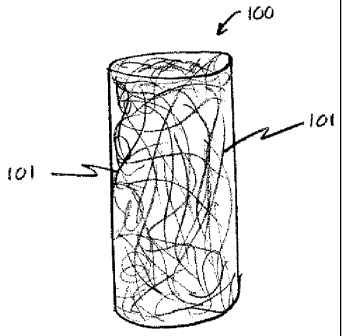
Figure 1 is an isometric view schematically representing a fibrous
antimicrobial
plug according to one embodiment of the present invention. The plug 100
includes
randomly oriented fiber or fibers 101. In a preferred embodiment the plug 100
may also
12
CA 02703953 2010-04-27
WO 2009/058990 PCT/US2008/081770
include an agent, preferably an antibacterial or antimicrobial agent on,
within or in
between the fibers 101. In addition the agent may be coated over the entire
plug 100.
The antibacterial (or any other agents) is added by different ways in the
above-
mentioned manufacturing process as described below in further details.
In one embodiment of the invention, the agent may be added (bulk loaded) in
the fiber matrix during the melt spinning process. Figure 2A shows the
dispersion of
the antimicrobial agent in the fiber matrix forming the plug 100 according to
one
embodiment of the present invention. The bulk loaded fiber 101 is comprised of
an
agent 102 dispersed within a polymer 103. One way this is achieved is by
preparing a
master batch concentrate of the agent 102 and then adding desired amount of
the
concentrate to the polymer 103 during the fiber extrusion process. This allows
uniform
dispersion of large quantity of the agent 102 in the fiber 101 and provides
long-term
diffusion of the agent 102 during the life cycle of the plug 100 in the
vascular
environment. The agent 102 is preferably thermally stable at melt processing
temperatures. Alternatively, the agent 102 can be added on, or incorporated
into a
polymeric layer on the fiber 101 surfaces. Figure 2B is a schematic
representation of a
fiber in the plug 100 showing the dispersion of the agent within the outer
polymer layer
of the fiber structure according to one embodiment of the present invention.
This type
of fiber 101 may be formed by mixing the agent 102 with a low melting polymer
203
(e.g., Polycaprolactone/Polyglycolic acid copolymer) to form a sheath on the
core fiber
(filament) 103 (e.g., PGA) using a bicomponent fiber spinning technology.
Referring
again to Figure 2B, the antimicrobial agent 102 is dispersed within polymer
layer 203,
which is coated on the base polymer 103. Together, this bicomponent fiber 101
forms
the fibrous structure of the plug 100.
In accordance with another embodiment of the invention, the agent 102 may be
mixed with the spin finish that is coated during the melt spinning process.
This
approach allows the agent 102 to disperse uniformly on the fiber 101 surfaces.
Figure
13
CA 02703953 2010-04-27
WO 2009/058990 PCT/US2008/081770
3 is a schematic representation of the fiber 101 comprising the plug 100
having a thin
coating 104 along the outer surface of the fibrous structure 101. The scouring
process
should not be used to remove the surface coatings when using this approach.
Accordingly, the thin outer coating 104 comprises the spin finish/lubricant
plus the
agent 102.
In another embodiment of the invention, the agent may be dip coated on the
scoured non-woven mat 105, which is then cut into plugs 100. The dip coating
solution
404 comprises the agent 102 and a bioabsorbable polymer (e.g.,
Polycaprolactone/Polyglycolic acid ) and may also include a solvent. One
embodiment
of the invention illustrating a non-woven mat that has been dip coated with an
agent
102 and polymer is illustrated in Figure 4A and 4B. As earlier described,
Figure 4A is
an isometric schematic representation of a non-woven fibrous mat the non-woven
mat
105 made up of randomly oriented fibers 101. For clarity, Figure 4B is a
section view
of the mat 105 depicted in Figure 4A taken along section line A-A. In each
view the dip
coating solution 404 is shown encapsulating the outer surfaces of the mat 105.
During the solvent removal process, the agent 102 and the polymer are coated
uniformly on the fiber 101 surfaces. Alternatively, the agent 102 can be added
on the
mat 105 surface in the absence of the bioabsorbable polymer. In this
embodiment, the
agent 102 coating on the non-woven mat 105 may be non-uniform. An isometric
schematic representation of a mat 105 having a non-uniform agent 102 coating
on the
mat 105 surface in illustrated in Figure 4C.
It should be noted that the coating process might also allow the dip coating
solution 404 or agent 102, as the case may be, to penetrate the exterior
surface of the
mat 105 into the interstitial spaces formed between adjacent fibers 101.
Figure 4D is
a schematic representation of a plug 101 wherein the agent 102 has penetrated
the
surface and resides in the interstitial spaces between fibers 101. Figure 4E
is a close
up section view of entangled fibers 101 forming the interstitial spaces
occupied by
agent 102. Although not explicitly depicted, the plug 101 may have agent 102
or
14
CA 02703953 2010-04-27
WO 2009/058990 PCT/US2008/081770
solution 404 covering the top and bottoms ends of the cylindrical plug 101. In
addition,
the coating process may allow some amount of agent or coating solution 404 to
cover
various side sections of the plug 101.
Figures 5A and 5B are schematic representations of another embodiment of the
invention where the extruded filaments 101 are first scoured to remove the
spin finish,
and the scoured filaments 101 are dip coated with the coating solution 404
(polymer,
agent and solvent). During the solvent removal process, the polymer and agent
coating 104 disperses uniformly on the filaments 101 as shown in Figure 5A.
These
filaments are then crimped, carded, needle punched into a mat 105 and then cut
in to
plugs 100. Alternatively, the scoured filaments 101 may be dip coated in an
agent/solvent solution. During the solvent removal process, the agent 102
remains on
the filament 101. The agent may uniformly cover the filament 102, but
generally will
non-uniformly cover the filament 102 as illustrated in Figure 5B. These
filaments are
then crimped, carded, needle punched into a mat 105 and then cut in to plugs
100.
In still another embodiment of the invention, the plugs 100 prepared from the
non-woven mat 105 may be covered with a coating after plug formation. This
coating
may be in the form of a solution or a powder. By way of example a solution of
polymer
and agent; polymer, agent and solvent; or agent and solvent may be applied to
the
formed plug 100. In addition, the coating may be applied to the plug 100 in a
powdered form, such as through an electrostatic coating process.
Figures 6A ¨ 6D are schematic representations illustrating plugs 100 covered
after formation with a coating. In particular, Figure 6A illustrates a plug
101 that has
been dip coated with a polymer/agent/solvent solution. When the solvent is
removed,
the polymer and agent substantially encapsulates the outer surfaces of the
plug 100
with a thin coating 404. In addition, the plug 100 may have been originally
prepared
with the agent occupying the interstitial spaces formed between adjacent
randomly
oriented fibers 101 before coating. Figure 6B illustrates a plug 101 having
agent
CA 02703953 2010-04-27
WO 2009/058990 PCT/US2008/081770
occupying the interstitial spaces that has been dip coated with a
polymer/agent/solvent
solution.
Alternatively, the plug 100 may be dip coated with an agent/solvent solution.
When the solvent is removed, the agent 102 may non-uniformly cover the outer
surfaces of the plug 100. Figure 6C is a schematic representation illustrating
a plug
100 covered by a non-uniform coating of agent 102 according to one embodiment
of
the present invention. In addition, the plug 100 may have been originally
prepared with
the agent 102 occupying the interstitial spaces formed between adjacent
randomly
oriented fibers 101 before coating. Figure 6D illustrates a plug 101 having
agent
occupying the interstitial spaces that has been dip coated with an
agent/solvent
solution.
There are several alternative methods that can be used to have the agent
either
dispersed within the fiber matrix or on the fiber surface to provide the
antimicrobial
properties.
The components of the porous closure device have therapeutic aentsl and
polymer coating combinations that are used to deliver the various agents and
drugs,
i.e. therapeutic and/or pharmaceutical agents including:
antiproliferative/antimitotic
agents including natural products such as vinca alkaloids (i.e. vinblastine,
vincristine,
and vinorelbine), paclitaxel, epidipodophyllotoxins (i.e. etoposide,
teniposide),
antibiotics (dactinomycin (actinomycin D) daunorubicin, doxorubicin and
idarubicin),
anthracyclines, mitoxantrone, bleomycins, plicamycin (mithramycin) and
mitomycin,
enzymes (L-asparaginase which systemically metabolizes L-asparagine and
deprives
cells which do not have the capacity to synthesize their own asparagine);
antiplatelet
agents such as G(GP)11bIlla inhibitors and vitronectin receptor antagonists;
antiproliferative/antimitotic alkylating agents such as nitrogen mustards
(mechlorethamine, cyclophosphamide and analogs, melphalan, chlorambucil),
ethylenimines and methylmelamines (hexamethylmelamine and thiotepa), alkyl
16
CA 02703953 2012-08-29
sulfonates-busulfan, nirtosoureas (carmustine (BCNU) and analogs,
streptozocin),
trazenes dacarbazinine (DTIC); antiproliferative/antimitotic antimetabolites
such as
folic acid analogs (methotrexate), pyrimidine analogs (fluorouracil,
floxuridine, and
cytarabine), purine analogs and related inhibitors (mercaptopurine,
thioguanine,
pentostatin and 2-chlorodeoxyadenosine {cladribine}); platinum coordination
complexes (cisplatin, carboplatin), procarbazine, hydroxyurea, mitotane,
aminoglutethimide; hormones (i.e. estrogen); anticoagulants (heparin,
synthetic
heparin salts and other inhibitors of thrombin); fibrinolytic agents (such as
tissue
plasminogen activator, streptokinase and urokinase), AspirinTM, dipyridamole,
ticlopidine, clopidogrel, abciximab; antimigratory; antisecretory (breveldin);
antiinflammatory: such as adrenocortical steroids (cortisol, cortisone,
fludrocortisone,
prednisone, prednisolone, 6a-methylprednisolone, triamcinolone, betamethasone,
and
dexamethasone), non-steroidal agents (salicylic acid derivatives i.e. aspirin;
para-
aminophenol derivatives i.e. acetominophen; indole and indene acetic acids
(indomethacin, sulindac, and etodalac), heteroaryl acetic acids (tolmetin,
diclofenac,
and ketorolac), arylpropionic acids (ibuprofen and derivatives), anthranilic
acids
(mefenamic acid, and meclofenamic acid), enolic acids (piroxicam, tenoxicam,
phenylbutazone, and oxyphenthatrazone), nabumetone, gold compounds (auranofin,
aurothioglucose, gold sodium thiomalate); immunosuppressives: (cyclosporine,
tacrolimus (FK-506), sirolimus (rapamycin), azathioprine, mycophenolate
mofetil);
angiogenic agents: vascular endothelial growth factor (VEGF), fibroblast
growth factor
(FGF) platelet derived growth factor (PDGF), erythropoetin; angiotensin
receptor
blocker; nitric oxide donors; anti-sense oligionucleotides and combinations
thereof; cell
cycle inhibitors, mTOR inhibitors, and growth factor signal transduction
kinase
inhibitors.
The closure device can be made from biodegradable or bioabsorbable polymer
compositions. The type of polymers used can degrade via different mechanisms
such
as bulk or surface erosion. Bulk erodible polymers include aliphatic
polyesters such
poly (lactic acid); poly (glycolic acid); poly (caprolactone); poly (p-
dioxanone) and poly
17
CA 02703953 2010-04-27
WO 2009/058990 PCT/US2008/081770
(trimethylene carbonate); and their copolymers and blends. Other polymers can
include amino acid derived polymers; phosphorous containing polymers [e.g.,
poly
(phosphoesters)] and poly (ester amide). Surface erodible polymers include
polyanhydrides and polyorthoesters. The closure device can be made from
combinations of bulk and surface erodible polymers to control the degradation
mechanism of the stent. The selection of the polymers will determine the
absorption of
that can be very short (few weeks) and long (weeks to months).
The bioabsorbable compositions to prepare the closure device will also include
drug and other agents such as antibacterial materials. The drug or agent will
release
by diffusion and during degradation of the closure device. The porous
structure to
prepare vascular closure device can be fabricated either by melt or solvent
processing.
The medical devices described herein are generally implantable medical
devices,
including but not limited to mono and multifilament sutures, surgical meshes
such as
hernia repair mesh, hernia plugs, brachy seed spacers, suture clips, suture
anchors,
adhesion prevention meshes and films, and suture knot clips. Also included are
implantable medical devices that are absorbable and non-absorbable. An
absorbable
polymer is defined as a polymer that, when exposed to physiological
conditions, will
degrade and be absorbed by the body over a period of time. Absorbable medical
devices
typically are formed from generally known, conventional absorbable polymers
including,
but not limited to, glycolide, lactide, co-polymers of glycolide, or mixtures
of polymers,
such as polydioxanone, polycaprolactone and equivalents thereof. Examples of
absorbable medical device include mono and multifilament sutures. The
multifilament
suture includes sutures wherein a plurality of filaments is formed into a
braided structure.
Examples of non-absorbable medical devices include mono and multifilament
sutures,
surgical meshes such as hernia repair mesh, hernia plugs and brachy seed
spacers,
which may be polymeric or nonpolymeric.
18
CA 02703953 2013-04-23
Suitable antimicrobial agents may be selected from, but are not limited to,
halogenated 5 hydroxyl ethers, acyloxydiphenyl ethers, or combinations
thereof. In
particular, the antimicrobial agent may be a halogenated 2-hydroxy diphenyl
ether and/or
a halogenated 2-acyloxy diphenyl ether, as described in U.S. Patent No.
3,629,477.
Antimicrobial activity similar to that of the halogen-o-hydroxy-diphenyl
ethers is
also attained using the 0-acyl derivatives thereof which .partially or
completely hydrolyze
under the conditions for use in practice. The esters of acetic acid,
chloroacetic acid,
methyl or dimethyl carbamic acid, benzoic acid, chlorobenzoic acid,
methylsulfonic acid
and chloromethylsulfonic acid are particularly suitable.
One particularly preferred antimicrobial agent within the scope of the above
formula is 2,4,4'-trichloro-2'-hydroxydiphenyl ether, commonly referred to as
triclosan
(manufactured by Ciba Geigy under the trade name Irgasarirm DP300 or
IrgacareTm MP).
Triclosan is a broad-spectrum antimicrobial agent that has been used in a
variety of
products, and is effective against a number of organisms commonly associated
with SSIs.
Such microorganisms include, but are not limited to, genus Staphylococcus,
Staphylococcus epidermidis, Staphylococcus aureus, methicillin-resistant
Staphylococcus
epidermidis, methicillin-resistant Staphylococcus aureus, and combinations
thereof.
It is advantageous to use a coating composition as a vehicle for delivering
the
antimicrobial agent to the surface of the device where such coating already is
used
conventionally in the manufacture of the device, such as, for example,
absorbable and
nonabsorbable vascular closure plug. Examples of medical devices, as well as
coatings
that may be applied thereto, may be found in U.S. Patent Nos. 4,201,216,
4,027,676,
4,105,034, 4,126,221, 4,185,637, 3,839,297, 6,260,699, 5,230,424, 5, 555,976,
5,868,244, 5,972,008 and WO 2004/032704 A2. As disclosed in U.S. Patent No.
4,201,216, the coating composition may include a film-forming polymer and a
substantially water-insoluble salt of a C6 or higher fatty acid. As another
example, an
absorbable coating composition that may be
19
CA 02703953 2010-04-27
WO 2009/058990 PCT/US2008/081770
used for an absorbable medical device may include poly(alkylene oxylates)
wherein the
alkylene moieties are derived from C6 or mixtures of C4 to C12 diols, which is
applied to a
medical device from a solvent solution, as disclosed in U.S. Patent No.
4,105,034. The
coating compositions of the present invention may include a polymer or co-
polymer, which
may include lactide and glycolide, as a binding agent. The compositions may
also include
calcium stearate, as a lubricant, and an antimicrobial agent. Medical devices
not
conventionally employing a coating in the manufacturing process, however, also
may be
coated with a composition comprising an antimicrobial agent. The coating may
be applied
to the device by, for example, dip coating, spray coating, suspended drop
coating, or any
other conventional coating means.
Microorganisms of the genus Staphylococcus are the most prevalent of all of
the
organisms associated with device-related surgical site infection. S.aureus and
S.
epidermidis are commonly present on patients' skin and as such are introduced
easily into
wounds. One of the most efficacious antimicrobial agents against
Staphylococcus is
2,4,4'-trichloro-2' hydroxydiphenyl ether. This compound has a minimum
inhibitory
concentration (MIC) against S. aureus of 0.01 ppm, as measured in a suitable
growth
medium and as described by Bhargava, H. et al in the American Journal of
Infection
Control, June 1996, pages 209-218. The MIC for a particular antimicrobial
agent and a
particular microorganism is defined as the minimum concentration of that
antimicrobial
agent that must be present in an otherwise suitable growth medium for that
microorganism, in order to render the growth medium unsuitable for that
microorganism,
i.e., the minimum concentration to inhibit growth of that microorganism. The
phrase "an
amount sufficient to substantially inhibit bacterial colonization" as used
herein is defined
as the minimum inhibitory concentration for S. aureus or greater.
A demonstration of this MIC is seen in the disk diffusion method of
susceptibility. A
filter paper disk, or other object, impregnated with a particular
antimicrobial agent is
applied to an agar medium that is inoculated with the test organism. Where the
anti-
CA 02703953 2010-04-27
WO 2009/058990 PCT/US2008/081770
microbial agent diffuses through the medium, and as long as the concentration
of the
antimicrobial agent is above the minimum inhibitory concentration (MIC), none
of the
susceptible organism will grow on or around the disk for some distance. This
distance is
called a zone of inhibition. Assuming the antimicrobial agent has a diffusion
rate in the
medium, the presence of a zone of inhibition around a disk impregnated with an
antimicrobial agent indicates that the organism is inhibited by the presence
of the
antimicrobial agent in the otherwise satisfactory growth medium. The diameter
of the zone
of inhibition is inversely proportional to the MIC.
Alternatively, the concentration of triclosan on the surface of a medical
device
such as a coated vascular closure plug may be greater than about 0.01 ppm
(wt./wt.
coating) or between about 30 ppm to 5,000 ppm (wt./wt. plug). The
concentration of
triclosan on the surface of the delivery system or package or containment
compartment
may be between about 5 ppm to 5, 000 ppm (wt./wt. package or compartment). For
other
particular applications, however, higher amounts of antimicrobial agent may be
useful and
should be considered well within the scope of the present invention.
In accordance with various methods of the present invention, a package and
containment compartment that are initially substantially free of an
antimicrobial agent, i.e.,
no antimicrobial agent is intended to be present on the package or containment
compartment surfaces, may be provided. A medical device, which has an
antimicrobial
agent disposed thereon, is positioned within the package or containment
compartment.
Subsequently, the package, the containment compartment if utilized and the
medical
device are subjected to time, temperature and pressure conditions sufficient
to vapor
transfer a portion of the antimicrobial agent from the medical device to the
package and/or
the containment compartment.
The rate of transfer of an antimicrobial agent such as triclosan from the
medical
device to the package and/or containment compartment is substantially
dependent upon
the time, temperature and pressure conditions under which the package with the
21
CA 02703953 2012-08-29
containment compartment and the medical device is processed, stored and
handled. For
example, triclosan is capable of transferring from a vascular plug to a
containment
compartment (in a closed vial at atmospheric pressure) when the temperature is
maintained at 55 C over a period of time. The conditions to effectively vapor
transfer an
antimicrobial agent such as triclosan include a closed environment,
atmospheric pressure,
a temperature of greater than 40 C, for a period of time ranging from 4 to 8
hours. Also
included are any combinations of pressure and temperature to render a partial
pressure
for the antimicrobial agent that is the same as the partial pressure rendered
under the
conditions described above, in combination with a period of time sufficient to
render an
effective amount or concentration of the antimicrobial agent on the package
and/or
containment compartment, i.e., the minimum inhibitory concentration (MIC) or
greater.
Specifically, it is known to one of ordinary skill that if the pressure is
reduced, the
temperature may be reduced to effect the same partial pressure. Alternatively,
if the
pressure is reduced, and the temperature is held constant, the time required
to render an
effective amount or concentration of the antimicrobial agent on the package
and/or
containment compartment may be shortened. While a portion of the antimicrobial
agent is
transferred to the package and/or containment compartment during this process,
a
second portion is retained on the surface of the medical device. Accordingly,
after the
transfer, the medical device and the package and/or the containment
compartment
contain the antimicrobial agent in an amount effective to substantially
inhibit bacterial
colonization thereon and thereabout.
Example 1
Coating experiments were conducted using a PGA plug to evaluate the effect of
triclosan as an antibacterial agent for vascular closure devices. Each plug
was hand
dipped in a coating solution for 10 seconds and then air dried at ambient
temperature for
2 h. Table I summarizes the coating compositions. Samples 1 to 6 were packaged
in
universal folders containing vapor hole without tyvek patches, and samples 7
and 8 were
packaged in universal folders containing the vapor hole and dosed Tyvek
patches. All the
22
CA 02703953 2010-04-27
WO 2009/058990
PCT/US2008/081770
samples were sterilized by ethylene oxide. The sterilized plug samples were
then cut into
two pieces and tested against two strains of bacteria namely, Staphylococcus
aureus and
Escherichia coli, to determine zone of inhibition (Z01). Table 1 summarizes
the results
from this test. The ZO1 results show that all plug samples provide anti
bacterial effects for
S. aureus bacteria exceeding 40 mm; and different levels of inhibition (from
7.7 mm to
greater than 40 mm) for E. coli bacteria.
15
Table!. Summary of coating compositions and zone of inhibition for PGA plugs
2 .00i040400.00
sornpioiniiiisubstratciiiiiiisampteaypoiiiiiiiimmmmammmmgogtiogmonvogiopomgmmgm
gm HimimiAmommg
ummmmmmmmmgmmmmmu]g]g]g]g]g]g]gmummmmmg]g]g]g]E]g]gmm
PGA plug Control No Coating 0
0
2 PGA plug Coated 2% w/w triclosan in ethyl acetate (no
polymer) >40 7.7
3 PGA plug Coated 2% w/w triclosan and 5% w/w PLGA 65/35 in
ethyl acetate >40 14.5
4 PGA plug Coated 2% w/w triclosan and 1% w/w PLGA 65/35 in
ethyl acetate >40 14.5
5 PGA plug Coated 2% w/w
triclosan and 5% w/w PCL/PGA 90/10 in ethyl acetate >40 >40
6 PGA plug Coated 2% w/w
triclosan and 1% w/w PCL/PGA 90/10 in ethyl acetate >40 >40
7 PGA plug Vapor 8 mg
triclosan in tyvek patch by vapor deposition (no polymer) >40 14.5
8 PGA plug Vapor 4 mg
triclosan in tyvek patch by vapor deposition (no polymer) >40 14.5
MATERIAL CHARACTERISTICS
Accordingly, in one exemplary embodiment, a vascular closure device may be
fabricated from a material such as a polymeric material including non-
crosslinked
23
CA 02703953 2010-04-27
WO 2009/058990 PCT/US2008/081770
thermoplastics, cross-linked thermosets, composites and blends thereof. There
are
typically three different forms in which a polymer may display the mechanical
properties associated with solids; namely, as a crystalline structure, as a
semi-
crystalline structure and/or as an amorphous structure. All polymers are not
able to
fully crystallize, as a high degree of molecular regularity within the polymer
chains is
essential for crystallization to occur. Even in polymers that do crystallize,
the degree of
crystallinity is generally less than one hundred percent. Within the continuum
between
fully crystalline and amorphous structures, there are two thermal transitions
possible;
namely, the crystal-liquid transition (i.e. melting point temperature, Tm) and
the glass-
liquid transition (i.e. glass transition temperature, Tg). In the
temperature range
between these two transitions there may be a mixture of orderly arranged
crystals and
chaotic amorphous polymer domains.
Molecular orientation is important as it primarily influences bulk polymer
properties
and therefore will have a strong effect on the final properties that are
essential for different
material applications. Physical and mechanical properties such as
permeability, wear,
refractive index, absorption, degradation rates, tensile strength, yield
stress, tear strength,
modulus and elongation at break are some of the properties that will be
influenced by
orientation. Orientation is not always favorable as it promotes anisotropic
behavior.
Orientation may occur in several directions such as uniaxial, biaxial and
multiaxial. It may
be induced by drawing, rolling, calendaring, spinning, blowing, and any other
suitable
process, and is present in systems including fibers, films, tubes, bottles,
molded and
extruded articles, coatings, and composites. When a polymeric material is
processed,
there will be preferential orientation in a specific direction. Usually it is
in the direction in
which the process is conducted and is called the machine direction (MD). Many
of the
products are purposely oriented to provide improved properties in a particular
direction. If
a product is melt processed, it will have some degree of preferential
orientation. In case of
solvent processed materials, orientation may be induced during processing by
methods
such as shearing the polymer solution followed by immediate precipitation or
quenching to
the desired geometry in order to lock in the orientation during the shearing
process.
24
CA 02703953 2010-04-27
WO 2009/058990 PCT/US2008/081770
Alternately, if the polymers have rigid rod like chemical structure then it
will orient during
processing due to the liquid crystalline morphology in the polymer solution.
The orientation state will depend on the type of deformation and the type of
polymer. Even though a material is highly deformed or drawn, it is not
necessary to
impart high levels of orientation as the polymer chains may relax back to
their original
state. This generally occurs in polymers that are very flexible at the draw
temperature.
Therefore, several factors may influence the state of orientation in a given
polymer
system, including rate of deformation for example, strain rate, shear rate,
frequency, and
the like, amount of deformation or draw ratio, temperature, molecular weight
and its
distribution, chain configuration for example, stereoregularity, geometrical
isomers, and
the like, chain architecture, for example, linear, branched, cross-linked,
dendritic and the
like, chain stiffness, for example, flexible, rigid, semi-rigid, and the like,
polymer blends,
copolymer types, for example, random, block, alternating, and the like, and
the presence
of additives, for example, plasticizers, hard and soft fillers, long and short
fibers,
therapeutic agents and the like.
Since polymers consist of two phases; namely, crystalline and amorphous, the
effect of orientation will differ for these phases, and therefore the final
orientation may not
be the same for these two phases in a semi-crystalline polymer system. This is
because
the flexible amorphous chains will respond differently to the deformation and
the loading
conditions than the hard crystalline phase.
Different phases may be formed after inducing orientation and its behavior
depends on the chemistry of the polymer backbone. A homogenous state such as a
completely amorphous material would have a single orientation behavior.
However, in
polymers that are semi-crystalline, block co-polymers or composites, for
example, fiber
reinforced, filled systems and liquid crystals, the orientation behavior needs
to be
described by more than one parameter. Orientation behavior, in general, is
directly
proportional to the material structure and orientation conditions. There are
several
CA 02703953 2010-04-27
WO 2009/058990 PCT/US2008/081770
common levels of structure that exist in a polymeric system, such as
crystalline unit cell,
lamellar thickness, domain size, spherulitic structures, oriented
superstructures, phase
separated domains in polymer blends and the like.
PROCESSES
According to the systems and methods of the present invention, a vascular
closure device comprised of polymeric, bioabsorbable materials may be made by
any of a
variety of processes. The processes used to prepare the antimicrobial vascular
closure
device are preferably low temperature processes in order to minimize the
degradation of
the agents that are unstable at high temperatures and are incorporated into
the matrix of
bioabsorbable polymeric materials comprising the device. Processing methods
may
comprise forming the device from bioabsorbable polymeric materials via low
temperature,
solution-based processes using solvents as by, for example, fiber spinning,
including dry
and wet spinning, electrostatic fiber spinning, co-mingled fibers, solvent
extraction,
coating, wire-coating, hollow fiber and membrane spinning, spinning disk (thin
films with
uniform thickness), ink-jet printing (three dimensional printing and the
like), lyophilization,
extrusion and co-extrusion, supercritical fluids, solvent cast films, or
solvent cast tubes.
Alternately, the vascular closure devices may also be prepared by more
conventional
polymer processing methods in melt condition for drugs or agents that are
stable at high
temperature as by, for example, fiber spinning, extrusion, co-extrusion,
injection molding,
blow molding, pultrusion and compression molding. Alternately, the agents may
also be
incorporated in the device by diffusion through the polymer matrix. This may
be achieved
by several methods such as swelling the device in a agent-enriched solution
followed by
high-pressure diffusion or by swelling and diffusing the agent in the device
using
supercritical fluids. Alternately, the drugs or agents may be sprayed, dipped,
or coated
onto the device after formation thereof from the bioabsorbable polymers. In
either case,
the polymer matrix, and drug or agent blend when provided, is then converted
into a
structure such as fibers, films, foams, discs/rings or tubes, for example,
that is thereafter
further manipulated into various geometries or configurations as desired.
26
CA 02703953 2010-04-27
WO 2009/058990 PCT/US2008/081770
Different processes may provide different structures, geometries or
configurations
to the bioabsorbable polymer being processed. For example, tubes processed
from rigid
polymers tend to be very stiff, but may be very flexible when processed via
electrostatic
processing or lyophilization. In the former case, the tubes are solid, whereas
in the latter
case, the tubes are porous. Other processes provide additional geometries and
structures that may include fibers, microfibers, thin and thick films, discs,
foams,
microspheres and even more intricate geometries or configurations. The
differences in
structures, geometries or configurations provided by the different processes
are useful for
preparing different devices with desired dimensions, strengths, agent or drug
delivery and
visualization characteristics.
In the case of a vascular closure device comprised of bioabsorbable polymeric
materials formed by supercritical fluids, such as supercritical carbon
dioxide, the
supercritical fluids are used to lower processing temperatures during
extrusion, molding or
otherwise conventional processing techniques. Different structures, such as
fibers, tubes,
films, or foams, may be formed using the supercritical fluids, whereby the
lower
temperature processing that accompanies the supercritical fluids tends to
minimize
degradation of the agents or drugs incorporated into the structures formed.
SOLVENT PROCESSING
In the case of a vascular closure device comprised of bioabsorbable polymeric
materials formed from solution, the viscosity of the polymer solution will
determine the
processing method used to prepare the devices. Viscosity of the polymer
solutions will, in
turn, depend on factors such as the molecular weight of the polymer, polymer
concentration, and the solvent used to prepare the solutions, processing
temperatures
and shear rates.
Another method to prepare tubes or fibers from polymer solutions, for example
in
the range from about 1 percent to 50 percent (wt/wt), is to extrude the
solutions using an
27
CA 02703953 2010-04-27
WO 2009/058990 PCT/US2008/081770
extruder with a tubular or rod die. During extrusion, the viscosity of the
solution may be
raised by gradual removal or multi-stage de-volatilization of solvent from
vents using, for
example, vacuum pumps. Twin screw or vented screw extruders may be used for
this
purpose. Residual solvent may be further removed at temperatures and
conditions that
will not degrade the drug. The polymer solutions may also comprise
antibacterial agent
and other additives such as plasticizers, other polymers and the like.
All the solvent processed devices may be prepared in different shapes,
geometries and configurations. For example, the tube may be co-extruded and/or
wire
coated. Other processing methodologies that are known in the art may be
utilized.
MELT PROCESSING
Vascular closure devices may also be prepared by more conventional polymer
processing methods in melt condition for drugs or agents that are stable at
high
temperature. Polymer compounding may be achieved by using twin-screw extruders
with
different screw elements to achieve desired mixing and dispersion. There are
also
feeders to add additives during the compounding process to from multi-
component blends
or composites. These additives may include pellets, powders of different
sizes, short
fibers or liquids. Polymer and antibacterial agent, for example, 1 percent to
about 50
percent (wt/wt) may be melt-compounded using a twin- screw extruder at low
temperatures under low shear conditions. The compounded material may be
pelletized
and extruded into a tube, fiber or other desired geometry using a single screw
extruder.
Other additives such as plasticizers and other polymers may also be added to
the
polymer formulation during the compounding step.
In the case of a vascular device comprised of bioabsorbable materials formed
by
co-extrusion, different bioabsorbable polymeric materials may be used whereby
the
different polymer tubes or fibers are extruded generally at the same time to
form an outer
layer for tubes or sheaths in case of fibers, and a inner layer for tubes or
core in case of
28
CA 02703953 2010-04-27
WO 2009/058990 PCT/US2008/081770
fibers. Bioabsorbable polymeric materials having low melting points are
extruded to form
the sheath or outside surface, and these low melting point materials will
incorporate the
drugs or other bio-active agents for eventual delivery to the patient.
Materials and their
blends having higher melting points are extruded to form the core or inside
surface that is
surrounded by the sheath. During processing, the temperatures for extruding
the low
melting point drug comprising materials, for example, polycaprolactone,
polydioxanone,
and their copolymers and blends may be as low as 60 degrees C to 100 degrees
C.
Further, because the drugs or other bio-active agents added to the devices
made by this
co-extrusion method tend to be coated onto the device after the device has
been
extruded, the drugs or agents are not exposed to the high temperatures
associated with
such methods. Degradation of the drugs during processing is therefore
minimized.
In the case of a vascular closure device comprised of bioabsorbable polymeric
materials formed by co-mingled fibers, different bioabsorbable polymeric
materials may
also be used. Contrary to the co-extrusion techniques described above, the co-
mingled
fibers technique requires that each fiber be separately extruded and then
later combined
to form a device of a desired geometry. Alternately, different fibers may also
be extruded
using the same spin pack but from different spinning holes thereby combining
them in one
step. The different bioabsorbable polymeric materials include a first fiber
having a low
temperature melting point into which a drug is incorporated, and a second
fiber having a
higher temperature melting point.
There are several different morphological variations that may occur during
melt or
solution processing bioabsorbable materials. When semi-crystalline polymers
are
processed from solution, since the solvent evaporates gradually, the polymers
may get
sufficient time to re-crystallize before it is completely dry. It will also
allow time for phase
separation to occur in case of multi-component blend systems. These changes
are driven
by well-known theories of thermodynamics of polymer crystallization and phase
separation. In order to prepare, for example, amorphous tubes or films or
fibers from
solution, it may be necessary to remove the solvent in a relatively short time
so that
kinetics will prevent crystallization and phase separation from occurring. For
example,
29
CA 02703953 2010-04-27
WO 2009/058990 PCT/US2008/081770
when the PLGA fibers are prepared from dioxane solutions, it may be necessary
to
remove the solvent in a relatively short time, for example, a few minutes to
hours at low
temperatures, for example, below 60 degrees C, after the fiber forming process
to obtain
an almost amorphous material. If the solvent removal process is carried out
over a long
period of time, for example, 6 to 10 h, at elevated temperatures, for example,
60 degrees
C, then PLGA may begin to crystallize (up to 10 to 20 percent crystallinity).
In case of
polymer blends, it is preferred to have an amorphous system to achieve good
compatibility between the amorphous phases of the polymers so that the
physical
properties are not adversely affected. When the polymer solutions are
precipitated or
coagulated, the resulting structure will be almost amorphous (1 to 5 percent
crystallinity),
as the solvent removal process is very fast thereby not allowing the polymer
to crystallize.
In case of melt processed materials, the tubes or films or fibers are quenched
immediately after exiting the extrusion die. Therefore, the polymers, in
general, do not
crystallize if the quenched temperature is below the glass transition
temperature of the
materials. In case of PGA or PLGA, the extruded fiber or tubes have very low
levels of
crystallinity (1 to 5 percent). This also makes it favorable when polymer
blends are
prepared from this process. Annealing the materials between the glass
transition and
melt temperatures for a given period of time will increase the amount of
crystallinity. For
example, PLGA fibers or tubes may be annealed at 110 degrees C for 3 to 10h by
mounting them over a mandrel under tension to prevent any shrinkage or
buckling. The
amount of crystallinity will increase from about 0 percent to about 35 to 45
percent.
Accordingly, this way the properties may be altered to achieve the desired
morphology
and physical properties.
These morphological variations in the precursor material (fibers, tubes,
films, etc)
will dictate to some extent the performance of the devices prepared from these
materials.
Amorphous materials will absorb faster, have higher toughness values, will
physically
age, and may not have sufficient dimensional stability compared to crystalline
material. In
contrast, crystalline material may not form compatible blends, will take a
longer time to
CA 02703953 2010-04-27
WO 2009/058990 PCT/US2008/081770
absorb, are stiffer with lower toughness values, and may have superior
physical device
properties such as low creep, higher strength, etc. For example, a material
that is
mechanically tested from a quenched state (higher amorphous form) and a slow
cooled
state (higher crystalline form) will provide a ductile high deformation
behavior and a brittle
behavior, respectively. This behavior is from the differences in the
crystallinity and
morphological features driven by different thermal treatments and histories.
The
morphological structure of a device surface may be modified by applying energy
treatment (e.g., heat). For example, an amorphous surface morphology can be
converted to a crystalline surface morphology by annealing it under different
conditions
(temperature/time). This may result in the formation of a crystalline skin or
layer on the
device that may provide several benefits such as agent elution control and
surface
toughness to prevent crack formation and propagation. Therefore, it is
important to
balance the structure ¨ property ¨ processing relationship for the materials
that are used
to prepare the devices to obtain optimum performance.
The implantable medical devices of the current invention may be prepared from
pure polymers, blends, and composites and may be used to prepare agent or drug-
loaded
vascular closure devices. The precursor material may be a fiber or a tube or a
film that is
prepared by any of the processes described above. The precursor material may
be used
as prepared or can be modified by quenching, annealing, orienting or relaxing
them under
different conditions. Alternately, the device may be used as prepared or may
be modified
by quenching, annealing, orienting or relaxing them under different
conditions.
MECHANICAL ORIENTATION
Orientation may be imparted to fibers, tubes, films or other geometries that
are
loaded or coated with agents or drugs in the range from about 1 to 50 percent.
For
example, drug loaded PGA tubes prepared by any of the above-mentioned
processes
may be oriented at about 70 degrees C to different amounts (for example, 50
percent to
300 percent) at different draw rates (for example, 100 mm/min to 1000 mm/min).
The
31
CA 02703953 2010-04-27
WO 2009/058990 PCT/US2008/081770
conditions to draw the material is important to prevent excessive fibrillation
and void
formation that may occur due to the presence of drug. If the draw temperature
is
increased to a higher value (for example, 90 degrees C), then the orientation
may not be
retained as the temperature of orientation is much higher than the glass
transition
temperature of PGA (about 45 degrees C) and would cause relaxation of the
polymer
chains upon cooling.
Other methods of orienting the materials may include multi-stage drawing
processes in which the material or device may be drawn at different draw rates
at different
temperatures before or after intermediate controlled annealing and relaxation
steps. This
method allows increasing the total draw ratio for a given material that is not
otherwise
possible in one-step drawing due to limitations of the material to withstand
high draw ratio.
These steps of orientation, annealing and relaxation will improve the overall
strength and
toughness of the material.
POLYMERIC MATERIALS
Polymeric materials may be broadly classified as synthetic, natural and/or
blends
thereof. Within these broad classes, the materials may be defined as biostable
or
biodegradable. Examples of biostable polymers include polyolefins, polyamides,
polyesters, fluoropolymers, and acrylics.
Examples of natural polymers include
polysaccharides and proteins.
Bioabsorobable and/or biodegradable polymers consist of bulk and surface
erodable materials. Surface erosion polymers are typically hydrophobic with
water labile
linkages. Hydrolysis tends to occur fast on the surface of such surface
erosion polymers
with no water penetration in bulk. The initial strength of such surface
erosion polymers
tends to be low however, and often such surface erosion polymers are not
readily
available commercially. Nevertheless, examples of surface erosion polymers
include
polyanhydrides such as poly (carboxyphenoxy hexane-sebacic acid), poly
(fumaric acid-
32
CA 02703953 2010-04-27
WO 2009/058990 PCT/US2008/081770
sebacic acid), poly (carboxyphenoxy hexane-sebacic acid), poly (imide-sebacic
acid)(50-
50), poly (imide-carboxyphenoxy hexane) (33-67), and polyorthoesters (diketene
acetal
based polymers).
Bulk erosion polymers, on the other hand, are typically hydrophilic with water
labile linkages. Hydrolysis of bulk erosion polymers tends to occur at more
uniform
rates across the polymer matrix of the device. Bulk erosion polymers exhibit
superior
initial strength and are readily available commercially.
Examples of bulk erosion polymers include poly (a-hydroxy esters) such as poly
(lactic acid), poly (glycolic acid), poly (caprolactone), poly (p-dioxanone),
poly
(trimethylene carbonate), poly (oxaesters), poly (oxaamides), and their co-
polymers
and blends. Some commercially readily available bulk erosion polymers and
their
commonly associated medical applications include poly (dioxanone) [PDS@ suture
available from Ethicon, Inc., Somerville, NJ], poly (glycolide) [Dexon@
sutures available
from United States Surgical Corporation, North Haven, CT], poly (lactide)-PLLA
[bone
repair], poly (lactide/glycolide) [Vicryl@ (10/90) and Panacryl@ (95/5)
sutures available
from Ethicon, Inc., Somerville, NJ], poly (glycolide/caprolactone (75/25)
[Monocryl@
sutures available from Ethicon, Inc., Somerville, NJ], and poly
(glycolide/trimethylene
carbonate) [Maxon sutures available from United States Surgical Corporation,
North
Haven, CT].
Other bulk erosion polymers are tyrosine derived poly amino acid [examples:
poly (DTH carbonates), poly (arylates), and poly (imino-carbonates)],
phosphorous
containing polymers [examples: poly (phosphoesters) and poly (phosphazenes)],
poly
(ethylene glycol) [PEG] based block co-polymers [PEG-PLA, PEG-poly (propylene
glycol), PEG-poly (butylene terephthalate)], poly (a -malic acid), poly (ester
amide), and
polyalkanoates [examples: poly (hydroxybutyrate (HB) and poly
(hydroxyvalerate) (HV)
co-polymers].
33
CA 02703953 2010-04-27
WO 2009/058990 PCT/US2008/081770
Of course, the devices may be made from combinations of surface and bulk
erosion polymers in order to achieve desired physical properties and to
control the
degradation mechanism. For example, two or more polymers may be blended in
order to achieve desired physical properties and device degradation rate.
Alternately,
the device may be made from a bulk erosion polymer that is coated with a
surface
erosion polymer. The drug delivery device may be made from a bulk erosion
polymer
that is coated with a antibacterial agent containing a surface erosion
polymer. For
example, the coating may be sufficiently thick that high loads may be
achieved, and the
bulk erosion polymer may be made sufficiently thick that the mechanical
properties of the
device are maintained even after all of the drug has been delivered and the
surface
eroded.
Shape memory polymers may also be used. Shape memory polymers are
characterized as phase segregated linear block co-polymers having a hard
segment
and a soft segment. The hard segment is typically crystalline with a defined
melting
point, and the soft segment is typically amorphous with a defined glass
transition
temperature. The transition temperature of the soft segment is substantially
less than
the transition temperature of the hard segment in shape memory polymers. A
shape in
the shape memory polymer is memorized in the hard and soft segments of the
shape
memory polymer by heating and cooling techniques. Shape memory polymers may be
biostable and bioabsorbable. Bioabsorbable shape memory polymers are
relatively
new and comprise thermoplastic and thermoset materials. Shape memory thermoset
materials may include poly (caprolactone) dimethylacrylates, and shape memory
thermoplastic materials may include poly (caprolactone) as the soft segment
and poly
(glycolide) as the hard segment.
The selection of the bioabsorbable polymeric material used to comprise the
device
according to the invention is determined according to many factors including,
for example,
the desired absorption times and physical properties of the bioabsorbable
materials, and
the geometry of the drug delivery device.
34
CA 02703953 2010-04-27
WO 2009/058990 PCT/US2008/081770
The local delivery of the antibacterial agent/therapeutic agent combinations
may
be utilized to treat a wide variety of conditions utilizing any number of
medical devices, or
to enhance the function and/or life of the device. For example, intraocular
lenses, placed
to restore vision after cataract surgery is often compromised by the formation
of a
secondary cataract. The latter is often a result of cellular overgrowth on the
lens surface
and can be potentially minimized by combining a drug or drugs with the device.
Other
medical devices which often fail due to tissue in-growth or accumulation of
proteinaceous
material in, on and around the device, such as shunts for hydrocephalus,
dialysis grafts,
colostomy bag attachment devices, ear drainage tubes, leads for pace makers
and
implantable defibrillators can also benefit from the device-drug combination
approach.
Devices which serve to improve the structure and function of tissue or organ
may also
show benefits when combined with the appropriate agent or agents. For example,
improved osteointegration of orthopedic devices to enhance stabilization of
the implanted
device could potentially be achieved by combining it with agents such as bone-
morphogenic protein. Similarly other surgical devices, sutures, staples,
anastomosis
devices, vertebral disks, bone pins, suture anchors, hemostatic barriers,
clamps, screws,
plates, clips, vascular implants, tissue adhesives and sealants, tissue
scaffolds, various
types of dressings, bone substitutes, intraluminal devices, including stents,
stent-grafts
and other devices for repairing aneurysims, and vascular supports could also
provide
enhanced patient benefit using this drug-device combination approach.
Perivascular
wraps may be particularly advantageous, alone or in combination with other
medical
devices. The perivascular wraps may supply additional drugs to a treatment
site.
Essentially, any other type of medical device may be coated in some fashion
with a drug
or drug combination, which enhances treatment over use of the singular use of
the device
or pharmaceutical agent.
In addition to various medical devices, the coatings on these devices may be
used
to deliver therapeutic and pharmaceutic agents including, all the compounds
described
above and anti-proliferative agents, anti-throrombogenic agents, anti-
restenotic agents,
CA 02703953 2010-04-27
WO 2009/058990 PCT/US2008/081770
anti-infective agents, anti-viral agents, anti-bacterial agents, anti-fungal
agnts, anti-
inflammatory agents, cytostatic agents, cytotoxic agents, immunosuppressive
agents,
anti-microbial agents, anti-calcification agents, anti-encrustation agents,
statins,
hormones, anti-cancer agents, anti-coagulants, anti-migrating agents and
tissue growth
promoting agents.
As described herein, various drugs or agents may be incorporated into the
medical device by a number of mechanisms, including blending it with the
polymeric
materials or affixing it to the surface of the device. Different drugs may be
utilized as
therapeutic agents, including sirolimus, or rapamycin, heparin, everolimus,
tacrolimus,
paclitaxel, cladribine as well as classes of drugs such as statins. These
drugs and/or
agents may be hydrophilic, hydrophobic, lipophilic and/or lipophobic.
Rapamycin is a macroyclic triene antibiotic produced by streptomyces
hygroscopicus as disclosed in U.S. Patent No. 3,929,992. It has been found
that
rapamycin inhibits the proliferation of vascular smooth muscle cells in vivo.
Accordingly, rapamycin may be utilized in treating intimal smooth muscle cell
hyperplasia, restenosis and vascular occlusion in a mammal, particularly
following
either biologically or mechanically mediated vascular injury, or under
conditions that
would predispose a mammal to suffering such a vascular injury. Rapamycin
functions
to inhibit smooth muscle cell proliferation and does not interfere with the re-
endothelialization of the vessel walls.
The drugs, agents or compounds described herein may be utilized in
combination with any number of medical devices, and in particular, with
implantable
medical devices such as stents and stent-grafts. Other devices such as vena
cava
filters and anastomosis devices may be used with coatings having drugs, agents
or
compounds therein or the devices themselves may be fabricated with polymeric
materials that have the drugs contained therein.
36
CA 02703953 2010-04-27
WO 2009/058990 PCT/US2008/081770
Any of the above-described medical devices may be utilized for the local
delivery
of drugs, agents and/or compounds to other areas, not immediately around the
device
itself. In order to avoid the potential complications associated with systemic
drug delivery,
the medical devices of the present invention may be utilized to deliver the
agents to areas
adjacent to the medical device. For example, a triclosan coated vascular
closure plug
may deliver the agent to the tissues surrounding the plug. The degree of
tissue
penetration depends on a number of factors, including the drug, agent or
compound, the
concentrations of the drug and the release rate of the agent.
The amount of agent incorporated within the device according to the systems
and methods of the present invention may range from about 0 to 99 percent
(percent
weight of the device). The drugs or other agents may be incorporated into the
device
in different ways. For example, the drugs or other agents may be coated onto
the
device after the device has been formed, wherein the coating is comprised of
bioabsorbable polymers into which the drugs or other agents are incorporated.
Alternately, the drugs or other agents may be incorporated into the matrix of
bioabsorbable materials comprising the device. The drugs or agents
incorporated into
the matrix of bioabsorbable polymers may be in an amount the same as, or
different
than, the amount of drugs or agents provided in the coating techniques
discussed
earlier if desired. These various techniques of incorporating drugs or other
agents into,
or onto, the device may also be combined to optimize performance of the
device, and
to help control the release of the drugs or other agents from the device.
Where the drug or agent is incorporated into the matrix of bioabsorbable
polymers comprising the device, for example, the drug or agent will release by
diffusion
and during degradation of the device. The amount of drug or agent released by
diffusion will tend to release for a longer period of time than occurs using
coating
techniques, and may often more effectively treat local and diffuse conditions
thereof.
Polymer compositions and their diffusion and absorption characteristics will
control
37
CA 02703953 2010-04-27
WO 2009/058990 PCT/US2008/081770
agent or drug elution profile for these devices. The release kinetics will be
controlled
by diffusion and polymer absorption. Initially, most of the agent or drug will
be released
by diffusion from the device surfaces and bulk and will then gradually
transition to
release due to polymer absorption. There may be other factors that will also
control
drug or agent release. If the polymer composition is from the same monomer
units
(e.g., lactide; glycolide), then the diffusion and absorption characteristics
will be more
uniform compared to polymers prepared from mixed monomers. Also, if there are
layers of different polymers with different drug in each layer, then there
will be more
controlled release of drug from each layer. There is a possibility of agent or
drug
present in the device until the polymer fully absorbs thus providing drug
release
throughout the device life cycle.
The vascular closure device according to the systems and methods of the
present invention preferably retains its integrity during the active drug
delivery phase
of the device. After drug delivery is achieved, the structure of the device
ideally
disappears as a result of the bioabsorption of the materials comprising the
device. The
bioabsorbable materials comprising the drug delivery device are preferably
biocompatible with the tissue in which the device is implanted such that
tissue
interaction with the device is minimized even after the device is deployed
within the
patient. Minimal inflammation of the tissue in which the device is deployed is
likewise
preferred even as degradation of the bioabsorbable materials of the device
occurs. In
order to provide multiple drug therapy, enriched or encapsulated drug
particles or
capsules may be incorporated in the polymer matrix. Some of these actives may
provide different therapeutic benefits such as anti-inflammatory, anti-
thrombotic; etc.
In accordance with another exemplary embodiment, the vascular closure device
described herein may be utilized as antibacterial agents or drug delivery
devices wherein
the agent is affixed to the surface of the device. Typical material properties
for coatings
include flexibility, ductility, tackiness, durability, adhesion and cohesion.
Biostable and
bioabsorbable polymers that exhibit these desired properties include
methacrylates,
38
CA 02703953 2010-04-27
WO 2009/058990 PCT/US2008/081770
polyurethanes, silicones, poly (vinyl acetate), poly (vinyl alcohol), ethylene
vinyl alcohol,
poly (vinylidene fluoride), poly (lactic acid), poly (glycolic acid), poly
(caprolactone), poly
(trimethylene carbonate), poly (dioxanone), polyorthoester, polyanhydrides,
polyphosphoester, polyaminoacids as well as their copolymers and blends
thereof.
In addition to the incorporation of therapeutic agents, the surface coatings
may
also include other additives such as radiopaque constituents, chemical
stabilizers for both
the coating and/or the therapeutic agent, radioactive agents, tracing agents
such as
radioisotopes such as tritium (i.e. heavy water) and ferromagnetic particles,
and
mechanical modifiers such as ceramic microspheres as will be described in
greater detail
subsequently. Alternatively, entrapped gaps may be created between the surface
of the
device and the coating and/or within the coating itself. Examples of these
gaps include air
as well as other gases and the absence of matter (i.e. vacuum environment).
These
entrapped gaps may be created utilizing any number of known techniques such as
the
injection of microencapsulated gaseous matter.
As described above, different agents may be utilized as therapeutic agents,
including sirolimus, heparin, everolimus, tacrolimus, paclitaxel, cladribine
as well as
classes of drugs such as statins. These drugs and/or agents may be
hydrophilic,
hydrophobic, lipophilic and/or lipophobic. The type of agent will play a role
in determining
the type of polymer. The amount of the drug in the coating may be varied
depending on a
number of factors including, the storage capacity of the coating, the drug,
the
concentration of the drug, the elution rate of the drug as well as a number of
additional
factors. The amount of drug may vary from substantially zero percent to
substantially one
hundred percent. Typical ranges may be from about less than one percent to
about forty
percent or higher. Drug distribution in the coating may be varied. The one or
more drugs
may be distributed in a single layer, multiple layers, single layer with a
diffusion barrier or
any combination thereof.
39
CA 02703953 2010-04-27
WO 2009/058990 PCT/US2008/081770
Different solvents may be used to dissolve the drug/polymer blend to prepare
the
coating formulations. Some of the solvents may be good or poor solvents based
on the
desired drug elution profile, drug morphology and drug stability.
There are several ways to coat the device that are disclosed in the prior art.
Some of the commonly used methods include spray coating; dip coating;
electrostatic
coating; fluidized bed coating; and supercritical fluid coatings.
Some of the processes and modifications described herein that may be used will
eliminate the need for polymer to hold the agent on the vascular closure
device. Device
surfaces may be modified to increase the surface area in order to increase
agent or drug
content and tissue-device interactions. Nanotechnology may be applied to
create self-
assembled nanomaterials that can contain tissue specific agent/drug containing
nanoparticles. Microstructures may be formed on surfaces by microetching in
which
these nanoparticles may be incorporated. The microstructures may be formed by
methods such as laser micromachining, lithography, chemical vapor deposition
and
chemical etching. Microstructures may be added to the device surface by vapor
deposition techniques. Microstructures have also been fabricated on polymers
and
metals by leveraging the evolution of micro electro-mechanical systems (MEMS)
and
microfluidics. Examples of nanomaterials include carbon nanotubes and
nanoparticles
formed by sol-gel technology. Therapeutic agents may be chemically or
physically
attached or deposited directly on these surfaces. Combination of these surface
modifications may allow agent or drug release at a desired rate. A top-coat of
a polymer
may be applied to control the initial burst due to immediate exposure of drug
in the
absence of polymer coating.
As described above, vascular closure devices may contain antimicrobial or
therapeutic agents as a coating, e.g. a surface modification. Alternatively,
the agents may
be incorporated into the device structure, e.g. a bulk modification that may
not require a
coating. For devices prepared from biostable and/or bioabsorbable polymers,
the coating,
CA 02703953 2012-08-29
if used, could be either biostable or bioabsorbable. However, as stated above,
no coating
may be necessary because the device itself is fabricated from a delivery
depot. This
embodiment offers a number of advantages. For example, higher concentrations
of the
therapeutic agent or agents may be achievable such as about >50percent by
weight. In
addition, with higher concentrations of therapeutic agent or agents, agent
delivery is
achievable for greater durations of time.
The sterilization process of the present invention is particularly adapted to
the
challenges of sterilizing drug coated medical devices. Specifically, the
sterilization
process is designed to remove all biological contaminants without affecting
the drug,
agent or compound or the polymeric material comprising the device or the
coating.
Medical devices typically are sterilized to render microorganisms located
thereon
non-viable. In particular, sterile is understood in the field of art to mean a
minimum sterility
assurance level of 10-6. Examples of sterilization processes are described in
U.S. Patent
Nos. 3,815,315, 3,068,864, 3,767,362, 5,464, 580, 5,128,101 and 5,868,244.
Specifically,
absorbable medical devices may be sensitive to radiation and heat.
Accordingly, it may be
desirable to sterilize such devices using conventional sterilant gases or
agents, such as,
for example, ethylene oxide gas. Upon completion of the sterilization process,
the
antimicrobial medical device, the delivery system, the package and/or the
containment
compartment have thereon an amount of the antimicrobial agent effective to
substantially
inhibit colonization of bacteria on or adjacent the antimicrobial device, the
package and/or
the containment compartment.
In accordance with one exemplary embodiment, a low temperature sterilization
method may be utilized to sterilize the devices of the present invention. The
method
comprises the steps of positioning at least one packaged, drug coated or drug
containing
medical device in a sterilization chamber, creating a vacuum in the
sterilization chamber,
increasing and maintaining the temperature in the sterilization chamber in the
range from
about twenty-five degrees C to about forty degrees C and the relative humidity
in the
41
CA 02703953 2010-04-27
WO 2009/058990 PCT/US2008/081770
sterilization chamber in the range from about forty percent to about eighty-
five percent for
a first predetermined period, injecting a sterilization agent at a
predetermined
concentration into the sterilization chamber and maintaining the temperature
in the
sterilization chamber in the range from about twenty-five degrees C to about
forty
degrees C and the relative humidity in the range from about forty percent to
about eighty-
five percent for a second predetermined period, and removing the sterilization
agent from
the sterilization chamber through a plurality of vacuum and nitrogen washes
over a third
predetermined period, the temperature in the sterilization chamber being
maintained at a
temperature in the range from about thirty degrees C to about forty degrees C.
In accordance with another exemplary embodiment, a low temperature
sterilization method may be utilized to sterilize the devices of the present
invention. The
method comprising the steps of loading the at least one packaged, drug coated
medical
device in a preconditioning chamber, the preconditioning chamber being
maintained at a
first predetermined temperature and a first predetermined relative humidity
for a first
predetermined time period, positioning at least one packaged, drug coated
medical device
in a sterilization chamber creating a vacuum in the sterilization chamber
increasing and
maintaining the temperature in the sterilization chamber in the range from
about twenty-
five degrees C to about forty degrees C and the relative humidity in the
sterilization
chamber in the range from about forty percent to about eighty-five percent for
a first
predetermined period injecting a sterilization agent at a predetermined
concentration into
the sterilization chamber and maintaining the temperature in the sterilization
chamber in
the range from about twenty-five degrees C to about forty degrees C and the
relative
humidity in the range from about forty percent to about eighty-five percent
for a second
predetermined period, and removing the sterilization agent from the
sterilization chamber
through a plurality of vacuum and nitrogen washes over a third predetermined
period, the
temperature in the sterilization chamber being maintained at a temperature in
the range
from about thirty degrees C to about forty degrees C.
42
CA 02703953 2012-08-29
In each embodiment described above, the sterilization or sterilizing agent may
comprise ethylene oxide or any other suitable agent. The nitrogen washes,
which serve
to remove the ethylene oxide may be replaced with other suitable gases,
including any of
the noble gases.
Other sterilization methods may also be used, such gamma and electron beam
radiations. In these methods the dosage should be low so that agent or drug in
the
devices is not adversely affected. The dosage may range from about one to four
mrad
and more preferably below 2 mrad. Radiation sterilized polymers will absorb
relatively
faster than ethylene oxide sterilized polymers.
43