Note: Descriptions are shown in the official language in which they were submitted.
CA 02704006 2010-04-28
WO 2009/061349 PCT/US2008/011979
METHODS, KITS, AND COMPOSITIONS FOR ADMINISTERING
PHARMACEUTICAL COMPOUNDS
Background of the Invention
The invention relates to methods, kits, and compositions for delivering
compounds to a tissue, and more particularly to methods for treating a skin-
related
condition, comprising disrupting the skin (e.g. by removing one or more layers
of the
skin) and embedding drugs in the skin.
Some existing methods for skin disruption, such as microdermabrasion,
involve removing the most superficial layer of the skin by propulsion of
particles or a
liquid jet. However, conventional microdermabrasion procedures do not result
in
significant embedding of particles. Most particles do not have suffcient
momentum
density (momentum divided by the cross sectional area of the particle) to
penetrate
past the stratum corneum. Accidental embedding of microdermabrasion particles
during procedures is generally considered undesirable, because it can lead to
granuloma formation. Consequently, manufacturers of microdermabrasion devices
adjust the operating parameters (particle size, particle density, suction
pressure,
particle velocity, number of passes) so that embedding of particles is
minimized.
Typical microdermabrasion procedures use relatively large particles, on the
order of 100-150 micron, which are convenient for obtaining substantial skin
disruption, but are not necessarily desirable for applications that require
embedding of
a particle into the skin. embedding of particles in the range of 0.1 to 250
micron is
possible, typical particle sizes for drug delivery may be on the order of 10
micron.
Methods for embedding particles in the skin for transdermal drug delivery
have been described in the prior art. These techniques, referred to as
`velocity-based'
or `needle-free', are based on propelling particles at very high speeds
(generally
supersonic), in order to breach the stratum corneum and penetrate the
underlying
tissues. Use of compressed helium to accelerate solid drug particles through a
Venturi
nozzle at velocities of up to 800 m/s has been reported by Bellhouse et al, US
5,630,796. US 7,207,967 describes a velocity-based method for accelerating
drug-
containing particles with an average size of 10-70 micron at velocities
ranging from
200 to 3000 m/sec. Appropriate pressure to accelerate the particles is
obtained by a
CA 02704006 2010-04-28
WO 2009/061349 PCT/US2008/011979
transient supersonic helium gas jet. US 6,893,664 describes a method that
makes use
of a needleless syringe whereby the penetration depth of the particles
propelled can be
controlled by adjusting the extent to which a gas container is breached, which
restricts
the outflow of gas from the container. Some examples of needle-free devices
under
development include Intraject , Implaject , Jet Syringe , Iject , Mini ject ,
Crossjet , and PowderJect . For example, the PowderJect device consists of a
gas
canister that allows pressurized helium gas to enter a chamber that contains a
cassette
filled with drug. The powdered drug sits between two polycarbonate membranes,
which are instantaneously ruptured when the gas is released; this, in turn,
causes the
gas to expand rapidly, forming a shock wave that progresses down the nozzle at
speeds of 600-900 m/s. The particle velocity is controlled by the nozzle
geometry, the
burst strength, and the gas pressure.
Existing transdermal technologies based on propelling drug particles against
the skin generally have several limitations. First, a fraction of the drug
propelled gets
retained in the stratum corneum. Using the PowderJect device, Lahm and Lee (J
of
Pharm Sci, 95, 7, 2006) have shown that the ratio of drug that remains in the
stratum
corneum to drug that crosses beyond is roughly 10:1. As a result, an important
fraction of the drug administered is wasted. A second problem with existing
technologies is that particles accelerated at supersonic speeds typically
collide
strongly against each other resulting in significant particle attrition; by
the time such
particles collide with the skin, their size distribution has been shifted to a
lower value
(Lahm and Lee, J of Pharm Sci, 95, 7, 2006). As a result, it is difficult to
have precise
control of the size of the particles delivered to the skin, which in turn may
play an
important role in the release pattern of the drug and the depth and retention
of
embedded particles. Other problems that have been associated with velocity-
based
techniques are the lack of reliability and occasional bruising.
Other technologies for transdermal delivery of drugs do not involve propelling
particles at high velocities, but instead rely on removing or modifying the
stratum
corneum, which constitutes the main resistance to drug transport through the
skin, and
then applying a drug topically. These devices rely on techniques widely used
by
dermatologists, such as dermabrasion, or light-based methods. One such device
for
enhancing the delivery of topical drug formulations has been developed by Med
Pharm Ltd and is described in W005058226A1. Another technique currently in
2
CA 02704006 2010-04-28
WO 2009/061349 PCT/US2008/011979
development by Carlisle Scientific, relies on creating microchannels in the
skin by
disrupting the stratum comeum with sharp metal granules (microscissuining). In
the
context of skin abrasion-based techniques for enhancing drug delivery,
microdermabrasion devices have been used solely for the purpose of removing
the
stratum corneum, but not for embedding a drug. Instead, the drug has been
applied in
a topical formulation following the abrasion step.
Skin abrasion-based techniques for enhancement of topical drug delivery can
have several limitations. First, while removing part or all of the stratum
corneum
typically increases the permeability of the skin to agents, some molecules are
too
large to penetrate the remaining layers of the skin. For example, large
proteins, or
drugs that must be formulated into carrier particles (e.g. controlled release
depots),
may not readily diffuse through the epidermis or dermis. In addition, in
existing
approaches, where the drug is applied topically, it will inevitably be
distributed into
the skin through a concentration gradient that develops over time, with the
highest
concentration being at the skin surface. As a result, high concentrations of
drug may
be difficult to obtain in deep layers of the skin over relevant time scales
(e.g. hours,
days, or weeks), or high concentrations of drug may be impossible to avoid in
the
outer layers (e.g. the stratum corneum or the epidermis), which may not be the
desired
target of the treatment. Third, in some cases it may be necessary for the skin
to remain
uncovered after the drug application; a topically applied drug may wash off
through
friction (e.g. with clothes) or contact with water, while embedded drug
particles
would not.
Summary of the Invention
The invention features methods, kits, and compositions for delivering
compounds to a tissue, and more particularly methods for treating a skin-
related
condition comprising disrupting the skin (e.g. by removing a layer of the
skin) and
embedding drugs in the skin.
Given the limitations of both velocity-based and skin abrasion-based drug
delivery methods, a technique that combined the advantages of the two would be
desirable. The methods of the invention include continuous transdermal
delivery of
particles (a) taking place at lower particle velocities, lower particle sizes,
and lower
particle densities than those needed in velocity-based devices, while (b)
maintaining a
3
CA 02704006 2010-04-28
WO 2009/061349 PCT/US2008/011979
high penetration efficiency into the skin by removing the stratum corneum, and
(c)
retaining good control over the depth and distribution of the drug in the
skin. In this
invention, the incorporation of smaller particle sizes can be used to effect
embeddingin the skin without forming granulomas, and for optimizing drug
deliverty.
The compositions of the invention feature microdermabrasion particles
containing
pharmaceutical compounds formulated for controlled release. In certain cases
it may
be desirable to improve permeation by propelling solid particles of these
drugs against
the skin, as opposed to applying them in a topical formulation (e.g. cream,
gel, foam).
Second, in some cases it may be desirable to insert the drug at a specific
depth in the
skin so that it lies near a specific structure of the skin (e.g. the
epidermis, dermis, the
hair bulge, the hair papilla, the sebaceous gland, etc).
In one aspect, the invention features a method of delivering (e.g., using a
transdermal delivery device or microdermabrasion device) a pharmaceutical
compound (e.g., a therapeutic or cosmetic compound) to a tissue (e.g., an
internal or
external tissue) including continually propelling particles onto the tissue
where at
least some of the particles include a therapeutic compound, at least some of
the
particles (e.g., 0.1%, 1%, 5%, 10%, 20%, 30%, 50% or more) embed in the
tissue, and
the therapeutic compound is released into the tissue.
In another aspect, the invention features a method of delivering a
pharmaceutical compound (e.g., a therapeutic or cosmetic compound) to a tissue
including embedding particles into the tissue where at least some of the
particles
include a therapeutic compound by propelling at least some of the particles
(e.g.,
0.1%, 1%, 5%, 10%, 20%, 30%, 50% or more) into the tissue; and releasing the
therapeutic compound into the tissue.
In another aspect, the invention features a microdermabrasion particle
formulated for controlled release of a pharmaceutical compound. The
microdermabrasion particle can be formulated to melt at least in part at
temperatures,
for example, between body temperature and 60 C, melt at least in part at body
temperature, or melt between room temperature and body temperature. Such
microdermabrasion particle may be a mixture of high melting point fats and low
melting point fats. Such microdermabrasion particles may also be formulated to
stick
to the tissue (e.g., skin). The microdermabrasion particle may have at least
one
property selected from the group consisting of. a high surface charge or
polarity,
4
CA 02704006 2010-04-28
WO 2009/061349 PCT/US2008/011979
carboxylic acids, poly(anhydride) groups, high molecular weight polymers, and
polymers with high chain flexibility. The diameter of the microdermabrasion
particle
can be between 0.01 m to 200 m (e.g., 0.01 m, 0.05 m, 0.1 m, 0.5 m, 1
m,
m, 25 m, 50 m, 75 m, 100 m, 150 m, 175 m, and 200 gm in diameter.
5 In another aspect, the invention also features a microdermabrasion device
including a handpiece, a tip, a propellant, and a cartridge selected from the
group
consisting of a cartridge containing therapeutic compound particles formulated
into an
abrasive carrier and a cartridge containing a mixture of abrasive particles
and
therapeutic compound particles.
10 In yet another aspect, the invention features a microdermabrasion device
including a handpiece, a tip, a propellant, a cartridge containing abrasive
particles and
a cartridge containing therapeutic compound particles.
In another aspect, the invention features a microdermabrasion device including
a handpiece, a tip, a propellant, and a cartridge containing therapeutic
compound
particles, wherein the therapeutic compound particles are formulated into an
abrasive
solid carrier.
In yet another aspect, the invention features a microdermabrasion kit for use
with a microdermabrasion device, the kit including a cartridge and a tip,
wherein the
cartridge includes microdermabrasion particles and wherein the
microdermabrasion
particles include a therapeutic compound formulated for controlled release.
This kit
may also feature a recycling unit and/or a collection unit.
In any of the forgoing aspects, the particles can be embedded to a depth of
between 0.01 mm and 7mm (e.g., 10-30 m, 30-100 m, 500 m, 800 m, 2 mm, and
5 mm).
In any of the forgoing aspects, particles can be a mixture of particles
containing a therapeutic compound and particles that do not contain a
therapeutic
compound. In this aspect, particles containing a therapeutic compound may
differ in
size or shape from those that do not contain a therapeutic compound. In a
related
aspect, the above method also features the selective removal of the non-
therapeutic
compounds on the basis of size or shape. Such particles may be collected,
recycled,
and/or purified on the basis of size or shape. In another related aspect, the
invention
features the selective collection, recycling , and/or purification of the
particles
containing the pharmaceutical compound based on, for example, size or shape.
5
CA 02704006 2010-04-28
WO 2009/061349 PCT/US2008/011979
In another related aspect, the particles can be a mixture of particles
containing
differing pharmaceutical compounds. Mixtures of particles can differ based on
size
and shape. Such particles can be propelled simultaneously or in sequence to
different
depths depending on the size and or shape of the differing particles.
In any of the forgoing aspect, methods can further include disrupting the
tissue
(e.g., using a microdermabrasion device). This disruption can be in an amount
to
trigger an embryonic like state and/or reepithelialization in the tissue
(e.g., skin). In
this aspect, the compound can be administered prior to, simultaneous with, or
after
disruption of the tissue. Also in this aspect, the therapeutic compound can be
administered in an amount sufficient to enhance hair follicle neogenesis,
inhibit
follicle neogenesis or hair growth, prevent or treat an aging related skin
condition,
treat a pigmentation disorder, treat a growth, or treat acne.
This disruption can result in removal of tissue to a depth of between 0.01 and
7 mm. For example, the disruption can result in removal of at least one skin
component selected from the group consisting of the stratum comeum, a portion
of
the epidermis, the full epidermis, a portion of the dermis, the full dermis,
the
sebaceous glands, the bulges, and the dermal papillas.
In any of the above methods or compositions, the therapeutic compound can
be formulated for controlled release. For example, the compound can be
formulated
for delayed released (after a period of 1, 2, 3, 4, 5, 6, 7, 8, 9, and 10
days) or sustained
released (over a period of 1, 2, 3, 4, 5, 6, 7, 8, 9, and 10 days). The
controlled release
can be activated by endogenous sources (e.g., temperature, chemicals,
pressure, water,
cell secretions, enzymes, dissolved gases, and reactive oxygen species) or
exogenous
sources (e.g., electromagnetic radiation, electric current, light, heat,
chemicals,
pressure, ultrasound, water, solvents, catalysts, and enzymes).
Also in any of the forgoing methods or compositions, the therapeutic
compound can be a small molecule EGFR inhibitor, or metabolite thereof (e.g.,
a non-
naturally occurring nitrogen-containing heterocycle of less than about 2,000
daltons,
leflunomide, gefitinib, erlotinib, lapatinib, canertinib, vandetanib, CL-
387785,
PKI166, pelitinib, HKI-272, and HKI-357), EGF, an EGFR antibody (zalutumumab,
cetuximab, IMC 11F8, matuzumab, SC 100, ALT 110, PX 1032, BMS599626, MDX
214, and PX 1041), a suppressor of the expression of a Writ protein in the
hair follicle
or an inducer of expression of a Dkkl protein (e.g., from lithium chloride, a
molecule
6
CA 02704006 2010-04-28
WO 2009/061349 PCT/US2008/011979
that synergizes with lithium chloride, the agonists 6-bromoindirubin-3_-oxime,
deoxycholic acid, a pyrimidine derivative, antagonists quercetin, ICG-001, the
purine
derivative QS11, fungal derivatives PKF115-854 and CGP049090, and the organic
molecule NSC668036), a modulator the retinoic acid signaling pathway (trans-
retinoic acid, N-retinoyl-D-glucosamine, and seletinoid G), a modulator of the
estrogen signaling pathway (e.g., 17(3-estradiol and selective estrogen
receptor
modulators), a compound which modulates the ubiquitin-proteasome system, a
compound which modulates cytokine signaling of Imiquimod or IL-Ialpha, a
modulator of melanocortin signaling, tyrosinase activity, apoptosis signaling,
endothelin signaling, nuclear receptor signaling, TGF(3-SMAD signaling, bone
morphogenetic protein signaling, stem cell factor signaling, androgen
signaling,
retinoic acid signaling, peroxisome proliferator-activated response receptor
signaling,
estrogen signaling, cytokine signaling, growth factor signaling, nonandrogenic
hormone signaling, toll-like receptor signaling, and neurotrophin,
neuroendocine
signaling, and cytokine signaling, benzoyl peroxide, a photosenitizer (e.g.,
aminolevulinic acid), an interferon, dacarbazine, interleukin-2, imiquimod, or
a
promoter of the expression of the transcription factor MITF.
By the terms "embed" and "embedding" are meant fixing or setting securely
or deeply, a particle, within or below the surface of the tissue.
By "pharmaceutical compound" is meant any compound that, when contacted
with a tissue, results in therapeutic, cosmetic, or prophylactic activity.
The terms "administration" and "administering" refer to a method of giving a
dosage of a pharmaceutical composition to a patient, where the method is,
e.g.,
topical, oral, intravenous, transdermal, subcutaneous, intraperitoneal, or
intramuscular.
As used herein, "reepithelialization" refers to the process that occurs during
formation of a new epidermis after wounding. Tissue undergoing this process
may be
characterized by the lack of fully developed hair follicles, cells in an
embryonic-like
state, or by lack of a stratum corneum.
As used herein, to "promote differentiation" refers to the act of increasing
the
percentage of cells that will differentiate as indicated or to increase the
number of
cells per unit area of skin that will differentiate.
7
CA 02704006 2010-04-28
WO 2009/061349 PCT/US2008/011979
By "uncommitted epidermal cell" is meant an epidermal stem cell, a bulge
cell, a bulge-derived cell, or any other type of cell known in the art that
can be
induced to differentiate into an HF cell.
By "corticosteroid" is meant any naturally occurring or synthetic compound
characterized by a hydrogenated cyclopentanoperhydrophenanthrene ring system
and
having immunosuppressive and/or antinflammatory activity. Naturally occurring
corticosteriods are generally produced by the adrenal cortex. Synthetic
corticosteroids may be halogenated. Examples corticosteroids are provided
herein.
By "disruption" is meant a sufficient amount of disturbance to existing tissue
(e.g., hair follicles and the surrounding epidermis and/or dermis) to induce
an
"embryonic-like" state. This embryonic-like state includes the activation,
migration,
and differentiation of epithelial stem cells from the bulge region of the hair
follicle or
the interfollicular epidermis. The depth of skin disruption can include in
increasing
amounts: partial removal of the stratum corneum, complete removal of the
stratum
corneum, partial removal of the epidermis, complete removal of the epidermis,
partial
disruption of the dermis and complete removal of the dermis. Skin disruption
can
also include disruption of the mid to lower epidermis and/or dermis without
any
disturbance to the stratum corneum and/or outer epidermis. Different levels of
skin
disruption can be accomplished by chemical, energetic, mechanical, sound,
ultrasound, and/or electromagnetic based methods.
By "controlled release" is meant the regulated spatial and/or temporal release
of a therapeutic compound from a formulation. The term "controlled release" is
meant to include delayed release, sustained release, and release from the
formulation
in pulses or cyclical patterns. The controlled release of the compound may be
activated by an exogenous or endogenous stimulus.
By "delayed release" is meant that the therapeutically active component is not
immediately released from the formulation (e.g., a carrier particle).
By "sustained release" is meant a form of controlled release whereby the
therapeutically active compound is released over an extended period of time.
As used herein, "formulated for topical administration" refers to a
composition
of the invention containing a therapeutic, cosmetic, or prophylactic compound
and
formulated with a pharmaceutically acceptable excipient to form a dispersible
composition. Compositions formulated for topical administration (e.g., as a
cream,
8
CA 02704006 2010-04-28
WO 2009/061349 PCT/US2008/011979
gel, lotion, ointment, microdermabrasion particle, and any other topical
formulation
described herein) are those manufactured or sold in accordance with
governmental
regulations regarding a therapeutic, prophylactic, or cosmetic regimen that
includes
instructions for the topical administration of the composition.
By "microdermabrasion" is meant a technique for skin disruption that uses
propulsion of particles or a liquid jet. The term is also meant to include a
technique
for skin disruption that uses a small, reciprocating, hard tip (e.g., a
diamond).
By "microdermabrasion particle" is meant a composition, that when propelled
onto the skin, results in disruption of the skin. The term "microdermabrasion
particle" is meant to include both compositions comprising a therapeutic
compound
and compositions which themselves have no therapeutically active compounds.
Microdermabrasion particles may include frozen solutions containing a
therapeutic
compound or may include formulations of therapeutic compounds that are solid
at
room temperature.
By "microdermabrasion device" is meant a device for skin disruption that uses
propulsion of particles or a liquid jet. The term is also meant for a device
for skin
disruption that uses a small, reciprocating, hard tip (e.g., a diamond). As
described
herein, microdermabrasion devices may propel frozen particles, or particles
that are
solid at room temperature or at the temperature that the procedure takes
place.
By "recycling unit" is meant a device that separates propelled
microdermabrasion particles, or the therapeutic compound contained therein,
from a
fraction of cellular debris and other byproducts of skin disruption resulting
from
microdermabrasion.
By "collection unit" is meant a device that collects the propelled
microdermabrasion particles, cellular debris, and other byproducts of skin
disruption
resulting from microdermabrasion.
By "small molecule EGFR inhibitor" is meant a molecule that inhibits the
function of one or more EGFR family tyrosine kinases. Tyrosine kinases of the
EGFR family include EGFR, HER-2, and HER-4 (see Raymond et al., Drugs
60(Suppl.1):15 (2000); and Harari et al., Oncogene 19:6102 (2000)). Small
molecule
EGFR inhibitors include, for example, gefitinib (Baselga et at., Drugs
60(Suppl. 1):33
(2000)), erlotinib (Pollack et al., J. Pharm. Exp. Ther. 291:739 (1999)),
lapatinib
(Lackey et al., 92"d AACR Meeting, New Orleans, abstract 4582 (2001)),
canertinib
9
CA 02704006 2010-04-28
WO 2009/061349 PCT/US2008/011979
(Bridges et al., Curr. Med. Chem. 6:825 (1999)), vandetanib (Wedge et al.,
Cancer
Res. 62:4645 (2002)), CL-387785 (Discafani et al., Biochem. Pharmacol. 57:917
(1999)), PKI166 (Takada et al., Drug Metab. Dispos. 32:1272 (2004)), pelitinib
(Torrance et al., Nature Medicine 6:1024 (2000)), HKI-272, HKI-357 (for HKI-
272
and HKI-357 see, for example, Greenberger et al., I1`h NCI-EORTC-AACR
Symposium on New Drugs in Cancer Therapy, Amsterdam, abstract 388 (2000);
Rabindran et al., Cancer Res. 64:3958 (2004); Holbro et al., Ann. Rev. Pharm.
Tox.
44:195 (2004); Tsou et al., J. Med. Chem. 48:1107 (2005); and Tejpar et al.,
J. Clin.
Oncol. ASCO Annual Meeting Proc. 22:3579 (2004)), and leflunomide (Kochhar et
al., FEBS Lett. 334:161 (1993)). The structures for each of these compounds is
provided below in Table 1.
Table 1. EGFR Inhibitors
Drug Structure
leflunomide F F
0 / F
N 'O
N
Gefitinib
0 l HN \ CI
v IN v v O / N
Erlotinib
HN
O~~-- 0 I/JIN
Lapatinib
O \ F
HN CI
C H
^ /N
\il/ v p N
CA 02704006 2010-04-28
WO 2009/061349 PCT/US2008/011979
Canertinib
~/ HN \ CI
HN
N -
Vandetanib
F
N
/ I J~
0 \ N"
/N
CL-387785 /
HN Br l:Zzzz N
PK1166
HN
HO
N N /
Pelitinib /
HN CI
N CN
0
O N
HKI-272 I \
N /
0
N CN
0 \ ~
N
11
CA 02704006 2010-04-28
WO 2009/061349 PCT/US2008/011979
HKI-357 F
O
N CN
\ N / / \
O
O N
Small molecule EGFR inhibitors which can be used in the methods and
compositions
of the invention include anilinoquinazolines, such as gefitinib, erlotinib,
lapatinib,
canertinib, vandetanib, and CL-387785 and the other anilinoquinazolines
disclosed in
PCT Publication No. WO/2005/018677 and U.S. patent Nos. 5,747,498 and
5,457,105; quinoline-3-carbonitriles, such as pelitinib, HKI-272, and HKI-357,
and
the quinoline-3-carbonitriles disclosed in U.S. patent Nos. 6,288,082 and
6,002,008;
pyrrolopyrimidines, such as PK1166, and the pyrrolopyrimidines disclosed in
U.S.
Patent No. 6,713,474 and U.S. Patent Publication Nos. 20060211678,
20060035912,
20050239806,20050187389,20050165029,20050153989,20050037999,
20030187001, and 20010027197; pyridopyrimidines, such as those disclosed in
U.S.
Patent Nos. 5,654,307 and 6,713,484; pyrazolopyrimidines, such as those
disclosed in
U.S. Patent Nos. 6,921,763 and 6,660,744 and U.S. Patent Publication Nos.
20060167020, 20060094706,20050267133, 20050119282, 20040006083, and
20020156081; isoxazoles, such as leflunomide; imidazoloquinazolines,
pyrroloquinazolines, and pyrazoloquinazolines. Preferably, the small molecule
EGFR
inhibitor contains a heterobicyclic or heterotricyclic ring system. Each of
the patent
publications listed above is incorporated herein by reference.
By "A77 7628" is meant the active metabolite of leflunomide having the
structure below.
F
F
O F
N
O CH,FI
Other features and advantages of the invention will be apparent from the
following Detailed Description, the drawings, and the claims.
12
CA 02704006 2010-04-28
WO 2009/061349 PCT/US2008/011979
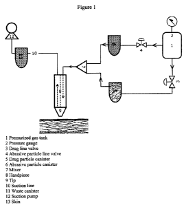
Brief Description of the Drawings
Figure 1 is a schematic view of a microdermabrasion and drug delivery
device.
Figure 2 is a schematic of an alternative micrdermabrasion device in which the
device also includes a recycling unit.
Detailed Description
The invention features compositions, methods, kits, and devices for
administering pharmaceutical compounds to a patient. In general, the invention
features the propulsion of particles containing a pharmaceutical compound
(e.g., in a
controlled release formulation) into a tissue of a patient (e.g., skin). The
particles
may, for example, be propelled into an intact tissue, or they may be propelled
onto a
tissue after one or more layers of tissue have been removed. Futhermore, the
invention features a device that first removes the stratum corneum, or that
continually
circulates drug particles (i.e. by propelling them against the skin, then
vacuuming out
the non-embedded particles, recycling them, and propelling them again) which
has the
advantage over prior art devices of more accurately controlling the depth of
embedding and reducing the amount of drug that is wasted. Further details of
the
methods, kits, and compositions of the invention are provided below.
Methods of Drug Delivery
The invention features methods of delivering a therapeutic compound to a
tissue by continually propelling particles against the tissue at a velocity
sufficient to
breach the interface and penetrate into the tissue. The method may involve the
steps
of (a) removing the most superficial layers of the skin, for example, by
abrading the
skin with microdermabrasion particles, tape stripping, a chemical peel, or
light-based
methods, and (b) propelling drug particles at a velocity sufficient to embed a
significant percentage (e.g. more than 1%, 5%, 10%, 20%, 30%, 40%, 50%, or
more)
of the particles into the skin. These steps could also be combined to occur
simultaneously in the same procedure.
In one embodiment, a microdermabrasion device is used for, in a first step,
removing the stratum corneum of the skin and, in a second step, propelling
drug
particles at a velocity sufficient to embed a significant percentage (e.g.
more than I%,
2%, 5%, 10%, 20%, 25%, 30%, 40%, 50%, or more) of the particles into the skin.
13
CA 02704006 2010-04-28
WO 2009/061349 PCT/US2008/011979
Both steps can be performed using the same device, or optionally with
different
devices.
While typical sizes needed to attain significant disruption of the skin (at
typical propulsion velocities used in microdermabrasion devices) are on the
order of
100 micron, ideal sizes for drug delivery particles may be one or more orders
of
magnitude smaller. Therefore a method of contacting a tissue with particles of
two
different sizes, such that certain particles disrupt the tissue, while other
particles
efficieintly deliver a pharmaceutical compound to the tissue is desirable.
Furthermore, typical dosing needed for skin abrasion is widely different than
dosing
need for drug delivery. While generally a microdermabrasion procedure uses on
the
order of 200 g of particles per treatment, typical drug dosages are on the
order of mg.
Because of the 1,000 to a 100,000-fold difference between the doses needed for
abrasion and for drug delivery, the invention features the combination of
different
doses of drug and abrasive particles. In the first step, any of the types of
particle (also
in some cases "crystals") known in the art (e.g. alumina and other metal
oxides, glass,
salts such as sodium chloride or sodium bicarbonate, ice, or any type of
biocompatible
particle such as the ones described by Weber et al in US 6,764,493, US
6,726,693,
and US 6,306,119) can be propelled against the skin until most or all of the
stratum
corneum has been removed. Optionally, the debris generated can be vacuumed out
to
clean the surface of the skin. If metal oxide particles such as alumina are
used to
perform the abrasion step, the abrasion ideally does not proceed beyond the
stratum
corneum in order to minimize granuloma formation. If salts, ice, or other
biocompatible materials are used, it may be desirable to proceed beyond the
stratum
corneum and remove portions or all of the epidermis, and portions or all of
the
dermis. Particles embedded during this first step can be removed by applying
water
(if, for example, they are particles of salt, ice, or water soluble
compounds), or mildly
warming the skin (if, for example, the particles have melting points near room
temperature). Typically, during the abrasion step, a negligible amount of
particles gets
embedded into the skin, since few particles have enough momentum density to
penetrate the stratum corneum. It has been determined that a momentum density
higher than 2.5 kg/(sec*m) is required in order to breach and cross the
stratum
corneum (Kendall et al, J of Biomechanics, 37, 2004); a typical, 100-micron
alumina
14
CA 02704006 2010-04-28
WO 2009/061349 PCT/US2008/011979
particle, with density of 3.7 g/cm3, propelled at 30 m/sec has a momentum
density of
1.9 kg/(sec*m), which is insufficient to penetrate the full stratum corneum.
In the second step of the above-described method, drug-containing particles
are propelled against the skin using a microdermabrasion device (e.g., the
same
microdermabrasion device used in the first step), and embed at certain desired
depths.
Since the stratum corneum has been removed in the previous step, the remaining
skin
no longer possesses the mechanical cohesion and integrity of normal skin. Well-
controlled penetration of the particles to desired depths can then be ensured
by
altering one or more of the parameters selected from particle size, particle
shape,
particle density, and particle velocity. In addition, particle penetration is
also a
function of the specific characteristics of the skin, which in turn depend on
the age of
the subject, and on the area of the body being treated. These parameters can
be
modified by the doctor or practitioner to ensure consistent and desirable
penetration
for the particular subject and/or tissue being treated. For example, particle
density
can be increased by compacting the pharmaceutical composition using high
pressure
and optionally vacuum, as described in W01997048485. The resulting compacted
materials can be attritioned into small particles using conventional methods.
Particle
velocity can be varied by adjusting the level of vacuum -if a suction pump is
used to
propel the particles- or the positive pressure level - if a source of
compressed air is
used to propel the particles. The specific geometry of the device nozzle has
an effect
on particle velocity, as well. Entrainment of the drug particles by the gas
flow occurs
in the same manner as entrainment of abrasive crystals. For example, in a
compressor-
assisted system, air from a compressor is flown through the particle cartridge
or a
mixing bottle, the air entrains drug particles and the exiting stream is
directed to a
handpiece.
The methods and devices of the invention extend the range of feasible particle
sizes that can be embedded in the skin. Since the resistance to particle
penetration is
greatly reduced after removal of the stratum corneum, smaller particles can be
inserted at a given velocity. For example, 10-micron particles of drug with a
density
of 1 g/cm3 (a typical value for drug formulations), and with a velocity of
1000 m/sec,
would typically not embed because their momentum density is -1.7 kg/(sec*m)
(below the threshold of 2.5 kg/(sec*m) to cross the stratum corneum). However,
after
removing the stratum corneum embedding can be acheived.
CA 02704006 2010-04-28
WO 2009/061349 PCT/US2008/011979
The penetration depth can be adjusted to anywhere between 0.01 mm and 7
mm. The penetration depth of the particles can be predicted by a penetration
model
that accounts for the inertial force of the particle and the static force
required to yield
the skin (Dehn, Int J of Impact Eng, 5, 239-248,1987). This is given by the
relationship:
d = 4Pnrn {ln( 2 p, v, + 3a-, ln(36, )
P,
Where d is the penetration depth, pp is the particle density, pt is the tissue
density
(skin), rp is the particle radius, v; is the particle velocity at impact, and
, at is the yield
stress of the tissue, which in this case corresponds to the skin without
stratum
corneum. The expression above can be used to guide the design of the drug
carrier as
well as the selection of operation conditions of the propelling device.
Drug particles may be non-spherical to facilitate embedding and reduce loss
by vacuuming. Alternatively, the abrasive particles may be spherical while the
drug
particles are non-spherical, which facilitates preferential embedding of the
drug
particles and preferential removal of the abrasive particles.
Any mechanical, chemical, electromagnetic, ultrasound, or light-based method
is used to remove the stratum corneum, following which a device selected from
the
group of a microdermabrasion device and a velocity-based or needle-free
transdermal
delivery device is used to embed particles into the skin. Also, a mixture of
biocompatible abrasive particles and drug particles can be propelled against
the skin
simultaneously so that the treatment consists of a single step.
Different drug-containing particles can be delivered to different depths in
the
skin at which their action is desired. The drugs can be applied simultaneously
by one
single gas jet at a given velocity; in which case their ratio of sizes, their
ratio of
densities, or their ratio of sphericity determine the difference in
penetration depths.
The drugs can also be applied in sequential steps, in which case the particles
can have
different or equal sizes, densities, or shapes, and they can be applied at
different
velocities. The different drugs may be applied with the purpose of treating
different
conditions simultaneously, or with the purpose of treating one single
condition
through a combination therapy of several drugs. A combination therapy with
different
16
CA 02704006 2010-04-28
WO 2009/061349 PCT/US2008/011979
particle-containing drugs may be helpful in cases where the action of the
different
drugs takes place at different locations in the skin. By way of example but
not by way
of limitation, a combination therapy for hair growth could consist of
application of
minoxidil-containing particles and particles containing inhibitors of steroid
metabolism. Minoxidil is thought to work by increasing vascular circulation to
the
hair follicle, while inhibitors of steroid metabolism affect the hair cycle by
stopping
the conversion of testosterone to dihydrotestosterone. While a topically
applied
formulation of minoxidil and an inhibitor of steroid metabolism would
distribute
everywhere in the skin, application through different particles could be
tailored so that
the drugs embed preferentially at different depths where they are most
effective or
where they have the least side effects.
A combination therapy for the treatment of psoriasis could consist of using
particles containing corticosteroids (which have an anti-inflammatory action)
and
particles containing Vitamin D analogues (which reduce lesions by acting on
keratinocytes). A combination therapy for acne could consist of using
particles
containing retinoids (which normalize desquamation of the follicular
epithelium) and
particles containing antibiotics (which inhibit the growth of P. acnes).
Microdermabrasion beads
The compounds of the invention (e.g., EGFR inhibitors) can be formulated
into microdermabrasion particles. These particles, when used in a
microdermabrasion
device, can serve one or more of the following purposes: (1) abrade the skin
to a
precisely defined depth that optimizes a subsequent treatment for a skin-
related
condition such as hair follicle regeneration (e.g., EGFR inhibitors), (2)
deliver a
controlled release formulation of a therapeutic compound, and (3) provide
elimination
of the therapeutic compound carrier by a natural, or an internally or
externally
triggered degradation process after the therapeutic compound has been
released.
The microdermabrasion particles of the invention may be, for example, 0.05
pm to 200 m in diameter (e.g., from 15 m to 150 m, 0.1 m to 10 m, or 1 m
to
2 m). Particles larger than 150 m can be used in combination with
microdermabrasion devices modified to accomodate larger particles. The ideal
average particle diameter and acceptable standard deviation would depend on
the
17
CA 02704006 2010-04-28
WO 2009/061349 PCT/US2008/011979
condition being treated, the specific therapeutic compound being released, and
the
desired timing of the release.
In one aspect of the invention, the particle size distribution (psd) of the
population of particles will be very narrow. In one aspect, the average
particle size is
near 100 pm and 90% in weight of the particle composition can pass through a
200
pm mesh screen (preferably, 95%, more preferably, 99%), In one aspect, the
particle
size distribution is monomodal.
In another aspect of the invention, the microdermabrasion particles provide
controlled release (e.g., delayed, sustained, or modified release) of a
compound (e.g.,
an EGFR inhibitor). In particles formulated for delayed release, therapeutic
compound may not be substantially released prior to the induction of
reepithelialization or prior to a certain phase of reepithelialization, as
described
below. In one embodiment, an exogenous stimulus is administered to trigger
release
or activation of the compound, for example, over a period of several seconds,
several
minutes, several hours, several days (e.g., 2, 3, 4, 5, 6, 7, 8, 9, or 10
days), or several
weeks (e.g., 2 weeks) or months. Some examples of exogenous triggers that can
be
used to stimulate therapeutic compound release include, without limitation,
application of light, heat, electricity, magnetism, ultrasound, or chemicals.
Alternatively, the therapeutic compound may be designed in a way such that the
release is triggered by an endogenous event related to any of the parameters
characteristic of the reepithelialization, as described below. Some examples
of
endogenous triggers are (1) increased expression of a marker that can bind to
or
enzymatically cleave the particle carrier that contains the therapeutic
compound,
thereby causing a change in the structure of the carrier particle which
enables
therapeutic compound release, and (2) increased levels of water in the skin
due to
completion of the reepithelialization process which causes hydrolytic cleavage
of a
crosslinked gel structure, or swelling of a hydrogel, thereby allowing
therapeutic
compound release.
Particles with a specific release window can be designed by manipulating
parameters relating to the physical and chemical properties of the carrier,
and to a
lesser extent by manipulating the concentration of additives such as
emulsifiers. If the
carrier is a polymer, the molecular weight, hydrophilicity, and relative
ratios of the
monomers of the polymer (in the case of a copolymer) can be tailored so that a
18
CA 02704006 2010-04-28
WO 2009/061349 PCT/US2008/011979
specific release window is obtained. Several polymers with different
degradation
kinetics may coexist in one formulation; in this case, the total rate of
release is the
average of the rates of release from each polymer, which can therefore be
tuned by
adjusting the ratio of polymers in the formulation, as described, for example,
in U.S.
Patent No. 4,897,268.
Well-controlled synthesis methods known in the art (e.g., those described
below) may be used to generate particles with the controlled release and
disruption
properties described above. The synthesis methods include, but are not limited
to,
coacervation, emulsion phase separation, spray-drying encapsulation, and
solvent
evaporation in organic or water phase. These methods are well known in the
art, and
have been described in, for example, U.S. Patent No. 6,506,410. In one
embodiment,
synthesis may involve solvent evaporation in water phase. Solvent evaporation
can
follow water/oil/water emulsification, which is used to encapsulate water-
soluble
therapeutic compounds (for example, in a biodegradable carrier), or oil/water
encapsulation, for lipid-soluble therapeutic compounds. A feature of this
method is
that the oil/water technique yields particles that are more porous, allowing a
high
burst of therapeutic compound to be delivered initially (Ibid). Therapeutic
compound-loaded particles may also be produced by dispersing porous carrier
particles into a solution containing the therapeutic compound, whereby the
therapeutic
compound in solution penetrates the pores of the carrier and remains trapped
inside.
In a subsequent step, an additive can be added to facilitate stabilization of
the
therapeutic compound in the carrier. The fluid in the remaining solution may
then be
separated by decantation, drying, lyophilization, vacuum-drying, or other
methods
known to people skilled in the art.
Some examples of polymers that may be synthesized by these methods include
cellulose derivatives (e.g. cellulose acetate, cellulose butyrate, ethyl
cellulose),
poly(urethanes), poly(siloxanes), poly(carbonates), poly(butadienes),
poly(esters),
poly(hydroxybutyric acid), poly(methyl methacrylate), poly(vinyl acetate),
poly(vinyl
alcohol), poly(ethylenes), waxes, proteins, and lipids (Ibid). In certain
embodiments,
the carrier may be chemically inert, non-degradable, and processable by the
synthesis
methods described above. Some materials with such properties that are well-
suited
for controlled release include poly(2-hydroxy ethyl methacrylate), poly(N-
vinyl
pyrrolidone), poly(methyl methacrylate), poly(vinyl alcohol), poly(acrylic
acid),
19
CA 02704006 2010-04-28
WO 2009/061349 PCT/US2008/011979
poly(acrylamide), poly(ethylene-co-vinyl acetate), poly(ethylene glycol), and
poly(methacrylic acid). In certain other embodiments, the carrier may be
designed so
that it degrades within the body, while still being processable by the
synthesis
methods described above. Some materials that are biodegradable and well-suited
for
controlled release include poly(lactides), poly(glycolides), poly(lactide-go-
glycolides), poly(anhydrides), and poly(orthoesters). In certain other
embodiments,
the carrier may be designed so that some important property, such as phase
state or
swelling, changes at or close to body temperature. Materials that are solid
slightly
below body temperature but that melt at or slightly above body temperature
include
low melting fats, and mixtures of low melting and high melting fats. Materials
that
swell around body temperature include temperature-sensitive hydrogels. In
certain
other embodiments, the carrier may be designed so that it adheres strongly to
the skin.
Materials suited for this purpose tend to have high concentration of polar
groups (i.e.
carboxylic acid), high molecular weight, polymer chain flexibility, and
surface
charge. (e.g., poly(anhydride)). In certain other embodiments, the carrier
material
may be designed such that the release of the therapeutic compound may be
triggered
by an exogenous or endogenous event. For example, exposure to UV light can
cause
photorelease of a therapeutic compound in poly(amides); ultrasound can
accelerate
therapeutic compound release from poly(anhydrides); hydrogels can be designed
so
that changes in temperature, pH, ionic strength, or binding of certain
molecules
trigger the therapeutic compound release. In certain other embodiments, the
carrier
may be designed so that the therapeutic compound is released at a constant
rate.
Carriers with such properties include double-walled polymer systems, such as a
mixture of poly(1,3-bis(p-carboxyphenoxypropane)-co-sebacic anhydride and
poly(lactic acid).
Antioxidants
If desired, the small molecule therapeutic compound (e.g, EGFR inhibitor)
formulations of the invention can contain one or more antioxidants. Useful
antioxidants include, without limitation, thiols (e.g., aurothioglucose,
dihydrolipoic
acid, propylthiouracil, thioredoxin, glutathione, cysteine, cystine,
cystamine,
thiodipropionic acid), sulphoximines (e.g., buthionine-sulphoximines, homo-
cysteine-
sulphoximine, buthionine-sulphones, and penta-, hexa- and heptathionine-
CA 02704006 2010-04-28
WO 2009/061349 PCT/US2008/011979
sulphoximine), metal chelators (e.g, a-hydroxy-fatty acids, palmitic acid,
phytic acid,
lactoferrin, citric acid, lactic acid, and malic acid, humic acid, bile acid,
bile extracts,
bilirubin, biliverdin, EDTA, EGTA, and DTPA), vitamins (e.g., vitamin E,
vitamin C,
ascorbyl palmitate, Mg ascorbyl phosphate, and ascorbyl acetate), phenols
(e.g.,
butylhydroxytoluene, butylhydroxyanisole, ubiquinol, nordihydroguaiaretic
acid,
trihydroxybutyrophenone), benzoates (e.g., coniferyl benzoate), uric acid,
mannose,
propyl gallate, selenium (e.g., selenium-methionine), stilbenes (e.g.,
stilbene oxide
and trans-stilbene oxide), and combinations thereof.
Antioxidants that may be incorporated into the formulations of the invention
include natural antioxidants prepared from plant extracts, such as extracts
from aloe
vera; avocado; chamomile; echinacea; ginko biloba; ginseng; green tea;
heather;
jojoba; lavender; lemon grass; licorice; mallow; oats; peppermint; St. John's
wort;
willow; wintergreen; wheat wild yam extract; marine extracts; and mixtures
thereof.
The total amount of antioxidant included in the formulations can be from
0.001% to 3% by weight, preferably 0.01% to 1% by weight, in particular 0.05%
to
0.5% by weight, based on the total weight of the formulation.
Other Biologically Active Ingredients
Other biologically active agents that can be used in the methods, kits, and
compositions of the invention include, without limitation, antihistamines,
anti-
inflammatory agents, anti-cancer agents, retinoids, anti-androgen agents,
immunosuppressants, channel openers, antimicrobials, herbs (e.g., saw
palmetto),
extracts (e.g., Souhakuhi extract), vitamins (e.g., biotin), co-factors,
psoralen,
anthralin, and antibiotics.
Antihistamines
In certain embodiments, an antihistamine can be used in the compositions,
methods, and kits of the invention. Useful antihistamines include, without
limitation,
Ethanolamines (e.g., bromodiphenhydramine, carbinoxamine, clemastine,
dimenhydrinate, diphenhydramine, diphenylpyraline, and doxylamine);
Ethylenediamines (e.g., pheniramine, pyrilamine, tripelennamine, and
triprolidine);
Phenothiazines (e.g., diethazine, ethopropazine, methdilazine, promethazine,
thiethylperazine, and trimeprazine); Alkylamines (e.g., acrivastine,
brompheniramine,
21
CA 02704006 2010-04-28
WO 2009/061349 PCT/US2008/011979
chlorpheniramine, desbrompheniramine, dexchlorpheniramine, pyrrobutamine, and
triprolidine); Piperazines (e.g., buclizine, cetirizine, chlorcyclizine,
cyclizine,
meclizine, hydroxyzine); Piperidines (e.g., astemizole, azatadine,
cyproheptadine,
desloratadine, fexofenadine, loratadine, ketotifen, olopatadine, phenindamine,
and
terfenadine); and Atypical antihistamines (e.g., azelastine, levocabastine,
methapyrilene, and phenyltoxamine). Both non-sedating and sedating
antihistamines
may be employed. Non-sedating antihistamines include loratadine and
desloratadine.
Sedating antihistamines include azatadine, bromodiphenhydramine;
chlorpheniramine; clemizole; cyproheptadine; dimenhydrinate; diphenhydramine;
doxylamine; meclizine; promethazine; pyrilamine; thiethylperazine; and
tripelennamine.
Other antihistamines suitable for use in the compositions, methods, and kits
of
the invention are acrivastine; ahistan; antazoline; astemizole; azelastine;
bamipine;
bepotastine; bietanautine; brompheniramine; carbinoxamine; cetirizine;
cetoxime;
chlorocyclizine; chloropyramine; chlorothen; chlorphenoxamine; cinnarizine;
clemastine; clobenzepam; clobenztropine; clocinizine; cyclizine; deptropine;
dexchlorpheniramine; dexchlorpheniramine maleate; diphenylpyraline; doxepin;
ebastine; embramine; emedastine; epinastine; etymemazine hydrochloride;
fexofenadine; histapyrrodine; hydroxyzine; isopromethazine; isothipendyl;
levocabastine; mebhydroline; mequitazine; methafurylene; methapyrilene;
metron;
mizolastine; olapatadine; orphenadrine; phenindamine; pheniramine;
phenyltoloxamine; p-methyldiphenhydramine; pyrrobutamine; setastine;
talastine;
terfenadine; thenyldiamine; thiazinamium; thonzylamine hydrochloride;
tolpropamine; triprolidine; and tritoqualine.
Antihistamine analogs can be used in the compositions, methods, and kits of
the invention. Antihistamine analogs include 10-
piperazinylpropylphenothiazine; 4-
(3-(2-chlorophenothiazin-10-yl)propyl)-1-piperazineethanol dihydrochloride; 1-
(10-
(3-(4-methyl-l-piperazinyl)propyl)-1OH-phenothiazin-2-yl)-(9CI) 1-propanone; 3-
methoxycyproheptadine; 4-(3-(2-Chloro-1 OH-phenothiazin-10-
yl)propyl)piperazine-
1-ethanol hydrochloride; 10, 11 -dihydro-5 -(3 -(4-ethoxycarbonyl-4-
phenylpiperidino)propylidene)-5H-dibenzo(a,d)cycloheptene; aceprometazine;
acetophenazine; alimemazin (e.g., alimemazin hydrochloride); aminopromazine;
benzimidazole; butaperazine; carfenazine; chlorfenethazine; chlormidazole;
22
CA 02704006 2010-04-28
WO 2009/061349 PCT/US2008/011979
cinprazole; desmethylastemizole; desmethylcyproheptadine; diethazine (e.g.,
diethazine hydrochloride); ethopropazine (e.g., ethopropazine hydrochloride);
2-(p-
bromophenyl-(p'-tolyl)methoxy)-N,N-dimethyl-ethylamine hydrochloride; N,N-
dimethyl-2-(diphenylmethoxy)-ethylamine methylbromide; EX-10-542A;
fenethazine; fuprazole; methyl 10-(3-(4-methyl-l-
piperazinyl)propyl)phenothiazin-2-
yl ketone; lerisetron; medrylamine; mesoridazine; methylpromazine; N-
desmethylpromethazine; nilprazole; northioridazine; perphenazine (e.g.,
perphenazine
enanthate); 10-(3-dimethylaminopropyl)-2-methylthio-phenothiazine; 4-
(dibenzo(b,e)thiepin-6(11 H)-ylidene)-1-methyl-piperidine hydrochloride;
prochlorperazine; promazine; propiomazine (e.g., propiomazine hydrochloride);
rotoxamine; rupatadine; Sch 37370; Sch 434; tecastemizole; thiazinamium;
thiopropazate; thioridazine (e.g., thioridazine hydrochloride); and 3-(10,11-
dihydro-
5 H-dibenzo(a, d)cyc l ohepten-5 -ylidene)-tropane.
Other compounds that are suitable for use in the compositions, methods, and
kits of the invention are AD-0261; AHR-5333; alinastine; arpromidine; ATI-
19000;
bermastine; bilastin; Bron-12; carebastine; chlorphenamine; clofurenadine;
corsym;
DF-1105501; DF-11062; DF-1111301; EL-301; elbanizine; F-7946T; F-9505; HE-
90481; HE-90512; hivenyl; HSR-609; icotidine; KAA-276; KY-234; lamiakast; LAS-
36509; LAS-36674; levocetirizine; levoprotiline; metoclopramide; NIP-53 1;
noberastine; oxatomide; PR-881-884A; quisultazine; rocastine; selenotifen;
SK&F-
94461; SODAS-HC; tagorizine; TAK-427; temelastine; UCB-34742; UCB-35440;
VUF-K-8707; Wy-4905 1; and ZCR-2060.
Still other compounds that can be used in the compositions, methods, and kits
of the invention are described in U.S. Patent Nos. 3,956,296; 4,254,129;
4,254,130;
4,282,233; 4,283,408; 4,362,736; 4,394,508; 4,285,957; 4,285,958; 4,440,933;
4,510,309; 4,550,116; 4,692,456; 4,742,175; 4,833,138; 4,908,372; 5,204,249;
5,375,693; 5,578,610; 5,581,011; 5,589,487; 5,663,412; 5,994,549; 6,201,124;
and
6,458,958.
23
CA 02704006 2010-04-28
WO 2009/061349 PCT/US2008/011979
Antimicrobial agents
In certain embodiments, an antimicrobial agent can be used in the
compositions, methods, and kits of the invention. Useful antimicrobial agents
include, without limitation, benzyl benzoate, benzalkonium chloride, benzoic
acid,
benzyl alcohol, butylparaben, ethylparaben, methylparaben, propylparaben,
camphorated metacresol, camphorated phenol, hexylresorcinol,
methylbenzethonium
chloride, cetrimide, chlorhexidine, chlorobutanol, chlorocresol, cresol,
glycerin,
imidurea, phenol, phenoxyethanol, phenylethylalcohol, phenylmercuric acetate,
phenylmercuric borate, phenylmercuric nitrate, potassium sorbate, sodium
benzoate,
sodium proprionate, sorbic acid, and thiomersal.
The antimicrobial can be from about 0.05% to 0.5% by weight of the total
composition, except for camphorated phenol and camphorated metacresol. For
camphorated phenol, the preferred weight percentages are about 8% to 12%
camphor
and about 3% to 7% phenol. For camphorated metacresol, the preferred weight
percentages are about 3% to 12% camphor and about 1% to 4% metacresol.
Anti-inflammatory agents
In certain embodiments, an antiinflammtory agent can be used in the
compositions, methods, and kits of the invention. Useful antiinflammtory
agents
include, without limitation, Non-Steroidal Anti-Inflammtory Drugs (NSAIDs)
(e.g.,
naproxen sodium, diclofenac sodium, diclofenac potassium, aspirin, sulindac,
diflunisal, piroxicam, indomethacin, ibuprofen, nabumetone, choline magnesium
trisalicylate, sodium salicylate, salicylsalicylic acid (salsalate),
fenoprofen,
flurbiprofen, ketoprofen, meclofenamate sodium, meloxicam, oxaprozin,
sulindac,
and tolmetin), COX-2 inhibitors (e.g., rofecoxib, celecoxib, valdecoxib, and
lumiracoxib), and corticosteroids (e.g., alclometasone dipropionate,
amcinonide,
betamethasone dipropionate, betamethasone valerate, clobetasol propionate,
desonide,
desoximetasone, dexamethasone, diflorasone diacetate, flucinolone acetonide,
flumethasone, fluocinonide, flurandrenolide, halcinonide, halobetasol
propionate,
hydrocortisone butyrate, hydrocortisone valerate, methylprednisolone,
mometasone
furoate, prednisolone, or triamcinolone acetonide).
Immunosuppressants
24
CA 02704006 2010-04-28
WO 2009/061349 PCT/US2008/011979
In certain embodiments, a nonsteroidal immunosuppressant can be used in the
compositions, methods, and kits of the invention. Suitable immunosuppressants
include cyclosporine, tacrolimus, rapamycin, everolimus, and pimecrolimus.
Cyclosporines
The cyclosporines are fungal metabolites that comprise a class of cyclic
oligopeptides that act as immunosuppressants. Cyclosporine A is a hydrophobic
cyclic polypeptide consisting of eleven amino acids. It binds and forms a
complex
with the intracellular receptor cyclophilin. The cyclosporine/cyclophilin
complex
binds to and inhibits calcineurin, a Cat+-calmodulin-dependent serine-
threonine-
specific protein phosphatase. Calcineurin mediates signal transduction events
required for T-cell activation (reviewed in Schreiber et al., Cell 70:365-368,
1991).
Cyclosporines and their functional and structural analogs suppress the T cell-
dependent immune response by inhibiting antigen-triggered signal transduction.
This
inhibition decreases the expression of proinflammatory cytokines, such as IL-
2.
Many different cyclosporines (e.g., cyclosporine A, B, C, D, E, F, G, H, and
I)
are produced by fungi. Cyclosporine A is a commercially available under the
trade
name NEORAL from Novartis. Cyclosporine A structural and functional analogs
include cyclosporines having one or more fluorinated amino acids (described,
e.g., in
U.S. Patent No. 5,227,467); cyclosporines having modified amino acids
(described,
e.g., in U.S. Patent Nos. 5,122,511 and 4,798,823); and deuterated
cyclosporines, such
as ISAtx247 (described in U.S. Patent Application Publication No. 2002/0132763
Al). Additional cyclosporine analogs are described in U.S. Patent Nos.
6,136,357,
4,384,996, 5,284,826, and 5,709,797. Cyclosporine analogs include, but are not
limited to, D-Sar (a-SMe)3 Va12-DH-Cs (209-825), Allo-Thr-2-Cs, Norvaline-2-
Cs,
D-Ala(3-acetylamino)-8-Cs, Thr-2-Cs, and D-MeSer-3-Cs, D-Ser(O-CH2CH2-OH)-8-
Cs, and D-Ser-8-Cs, which are described in Cruz et al., Antimicrob. Agents
Chemother. 44:143 (2000).
Tacrolimus
Tacrolimus and tacrolimus analogs are described by Tanaka et al. (J. Am.
Chem. Soc., 109:5031 (1987)) and in U.S. Patent Nos. 4,894,366, 4,929,611, and
4,956,352. FK506-related compounds, including FR-900520, FR-900523, and FR-
CA 02704006 2010-04-28
WO 2009/061349 PCT/US2008/011979
900525, are described in U.S. Patent No. 5,254,562; O-aryl, O-alkyl, O-
alkenyl, and
O-alkynylmacrolides are described in U.S. Patent Nos. 5,250,678, 532,248,
5,693,648; amino O-aryl macrolides are described in U.S. Patent No. 5,262,533;
alkylidene macrolides are described in U.S. Patent No. 5,284,840; N-
heteroaryl, N-
alkylheteroaryl, N-alkenylheteroaryl, and N-alkynylheteroaryl macrolides are
described in U.S. Patent No. 5,208,241; aminomacrolides and derivatives
thereof are
described in U.S. Patent No. 5,208,228; fluoromacrolides are described in U.S.
Patent
No. 5,189,042; amino O-alkyl, O-alkenyl, and O-alkynylmacrolides are described
in
U.S. Patent No. 5,162,334; and halomacrolides are described in U.S. Patent No.
5,143,918.
Tacrolimus is extensively metabolized by the mixed-function oxidase system,
in particular, by the cytochrome P-450 system. The primary mechanism of
metabolism is demethylation and hydroxylation. While various tacrolimus
metabolites are likely to exhibit immunosuppressive biological activity, the
13-
demethyl metabolite is reported to have the same activity as tacrolimus.
Pimecrolimus
Pimecrolimus is the 33-epi-chloro derivative of the macrolactam ascomyin.
Pimecrolimus structural and functional analogs are described in U.S. Patent
No.
6,384,073.
Rapamycin
Rapamycin structural and functional analogs include mono- and diacylated
rapamycin derivatives (U.S. Patent No. 4,316,885); rapamycin water-soluble
prodrugs
(U.S. Patent No. 4,650,803); carboxylic acid esters (PCT Publication No. WO
92/05179); carbamates (U.S. Patent No. 5,118,678); amide esters (U.S. Patent
No.
5,118,678); biotin esters (U.S. Patent No. 5,504,091); fluorinated esters
(U.S. Patent
No. 5,100,883); acetals (U.S. Patent No. 5,151,413); silyl ethers (U.S. Patent
No.
5,120,842); bicyclic derivatives (U.S. Patent No. 5,120,725); rapamycin dimers
(U.S.
Patent No. 5,120,727); O-aryl, O-alkyl, O-alkyenyl and O-alkynyl derivatives
(U.S.
Patent No. 5,258,389); and deuterated rapamycin (U.S. Patent No. 6,503,921).
Additional rapamycin analogs are described in U.S. Patent Nos. 5,202,332 and
5,169,851.
26
CA 02704006 2010-04-28
WO 2009/061349 PCT/US2008/011979
Retinoids
In certain embodiments, a retinoid can be used in the compositions, methods,
and kits of the invention. Useful retinoids include, without limitation, 13-
cis-retinoic
acid, 9-cis retinoic acid,, all-trans-retinoic acid, etretinate, acitretin,
retinol, retinal,
tretinoin, alitretinoin, isotretinoin, tazarotene, bexarotene, and adapelene.
Channel openers
In certain embodiments, a channel opener can be used in the compositions,
methods, and kits of the invention. Useful channel openers include, without
limitation, minoxidil, diazoxide, and phenytoin.
Anti-androgens
In certain embodiments, an anti-androgen can be used in the compositions,
methods, and kits of the invention. Useful anti-androgens include, without
limitation,
finasteride, flutamide, diazoxide, 11 alpha-hydroxyprogesterone, ketoconazole,
RU58841, dutasteride, fluridil, QLT-7704, and anti-androgen oligonucleotides.
Antibiotics
In certain embodiments, an antibiotic can be used in the compositions,
methods, and kits of the invention. Useful antibiotics include, without
limitation,
penicillin G, penicillin V, methicillin, oxacillin, cloxacillin,
dicloxacillin, nafcillin,
ampicillin, amoxicillin, carbenicillin, ticarcillin, mezlocillin,
piperacillin, azlocillin,
temocillin, cepalothin, cephapirin, cephradine, cephaloridine, cefazolin,
cefamandole,
cefuroxime, cephalexin, cefprozil, cefaclor, loracarbef, cefoxitin,
cefmatozole,
cefotaxime, ceftizoxime, ceftriaxone, cefoperazone, ceftazidime, cefixime,
cefpodoxime, ceftibuten, cefdinir, cefpirome, cefepime, BAL5788, BAL9141,
imipenem, ertapenem, meropenem, astreonam, clavulanate, sulbactam, tazobactam,
streptomycin, neomycin, kanamycin, paromycin, gentamicin, tobramycin,
amikacin,
netilmicin, spectinomycin, sisomicin, dibekalin, isepamicin, tetracycline,
chlortetracycline, demeclocycline, minocycline, oxytetracycline, methacycline,
doxycycline, erythromycin, azithromycin, clarithromycin, telithromycin, ABT-
773,
lincomycin, clindamycin, vancomycin, oritavancin, dalbavancin, teicoplanin,
27
CA 02704006 2010-04-28
WO 2009/061349 PCT/US2008/011979
quinupristin and dalfopristin, sulphanilamide, para-aminobenzoic acid,
sulfadiazine,
sulfisoxazole, sulfamethoxazole, sulfathalidine, linezolid, nalidixic acid,
oxolinic
acid, norfloxacin, perfloxacin, enoxacin, ofloxacin, ciprofloxacin,
temafloxacin,
lomefloxacin, fleroxacin, grepafloxacin, sparfloxacin, trovafloxacin,
clinafloxacin,
gatifloxacin, moxifloxacin, gemifloxacin, sitafloxacin, metronidazole,
daptomycin,
garenoxacin, ramoplanin, faropenem, polymyxin, tigecycline, AZD2563, and
trimethoprim.
Growth Factors
In another embodiment, growth factors, growth factor antagonists, and growth
factor agonists, can also be used in the compounds of the invention.
Reepithelialization
In one aspect of this invention, the compositions of the invention are
administered and released into a subject's skin (without limitation examples
of the
skin location are the head, for example, the scalp, the face, the eyebrow, or
a scarred
region) while the skin is in a state of reepithelialization.
Reepithelialization is the
process that occurs during formation of a new epidermis and can be
characterized for
the purposes of this invention by the lack of fully formed hair follicles
(e.g., if within
the tissue some cells are in the pre-placode stage of hair follicle
formation), an
embryonic-like state, in which the follicle regenerates, or by lack of a
stratum
corneum.
State of Reepithelialization
Reepithelialization can be detected through inspection of the new epidermis
where covering of the wound area by keratinocytes indicates
reepithelialization. The
presence of keratinocytes can be seen with the naked eye as a white, glossy,
shiny
surface that gradually covers the open wound. Using a confocal microscope,
keratinocytes can be visualized as a sheet of "cobblestone"-looking cells.
Reepithelialization can also be detected through the measurement of
transepidermal
water loss (TEWL). TEWL decreases when the epithelial barrier is restored.
Confocal scanning laser microscopy and/or optical coherence tomography can
also be
28
CA 02704006 2010-04-28
WO 2009/061349 PCT/US2008/011979
used to detect the state of reepithelialization, where the presence of
keratinocytes
indicates reepithelialization.
The presence of a stratum corneum can be determined though visual
inspection, direct observation of papillary blood vessels using a capillary
microscope,
or through a colorimetric redox reaction of a compound that reacts in the
presence of
live cells. For example, 0.01% nitrazine yellow applied to the skin will
remain yellow
if a stratum corneum is present, and will turn greenish brown if not. In
another
example 0.01% bromcresol purple applied to the skin will stay yellow if the
stratum
corneum is present and will turn purple if the stratum corneum is not present.
The area of reepithelialization can be, for example, between 0-2 centimeters
(cm) in width (e.g., 1 cm, 1.5 cm, and 2.0 cm) or the area may be greater.
Optionally,
the area of reepithelialization can be interfollicular (e.g., the area of
disruption leading
to reepithelialization can be limited to the area immediately surrounding the
previously existing or new follicle).
In some aspects of the invention, it is desirable to release the compounds of
the invention at a particular phase of reepithelialization. Stages at which
compounds
of the invention may preferably be administered and/or activated include
periods:
^ prior to disruption,
^ simultaneous with disruption,
^ after completion of the reepithelialization process (e.g., 3-12 days, or 9-
11
days after having disrupted the skin),
^ after or during the establishment of a stem cell population that will
develop
into a regenerated hair follicle (Ito et al, Nature 447, 316-320, May 2007),
^ prior to the expression of hair follicle differentiation markers KRT17 and
Lefl
for several days after wound closure (Ito et al, Nature 447, 316-320, May
2007),
^ after or during expression of one or more proteins including KRT 17, Lef- 1,
alkaline phosphatase, WntlOb, and Shh (Ito et al, Nature 447, 316-320, May
2007),
characterized by the absence of K10 expression (which is expressed in normal
epidermis) and/or induction of expression of K16 and K17 (which are not
expressed in normal epidermis) (Patel et al, Journal of Investigative
Dermatology, 126, 2006),
29
CA 02704006 2010-04-28
WO 2009/061349 PCT/US2008/011979
^ charactized by the elevation of one or more transcription factors including
AP-
1 and NF-KB, primary cytokines IL-10 and TNF-a, and matrix
metalloproteases (Karimipour et al, Journal of the American Academy of
Dermatology, 52, Issue 2, 2005),
^ characterized by histologic changes (Freedman et al, Dermatologic Surgery,
27 Issue 12, December 2001), including, for example:
o thickening of the epidermis and dermis,
o flattening of rete pegs,
o vascular ectasia,
o perivascular inflammation,
o hyalinization of the papillary dermis with newly deposited collagen
and elastic fibers,
o change in orientation, density, or packing of collagen and other
structures,
^ characterized by detachment of the scab. Depending on the depth of the
abrasion process, it may be desirable for the compounds of the invention to be
administered or activated prior to or after the detachment of a scab.
Alternatively, hair follicles may start to form before the scab falls off, in
the
case of, for example, dermabrasion.
Alternatively the compounds of the invention can be administered prior to
epidermal disruption. In such embodiments, the compound may be formulated for
controlled release such that the therapeutically active compound is released
during
reepithelialization or during a particular phase of reepithelialization (e.g.,
as described
above). The compound may also be formulated such that it becomes activated by
an
endogenous or exogenous stimulus (e.g., as described below).
Induction of reepithelialization
The state of reepithelialization can be induced. Methods of inducing this
state
include the disruption of the subject's skin at the location where the
compounds of the
invention are going to be administered. Disruption can be achieved through
abrasion
(e.g., the rubbing or wearing away of skin), or through any method that
results in
disturbing the intactness of the epidermis or epidermal layer including
burning (e.g.,
by inducing a sunburn) or perforating the epidermis or epidermal layer. The
CA 02704006 2010-04-28
WO 2009/061349 PCT/US2008/011979
disruption can either result in partial or complete removal of the epidermal
layer at the
intended location.
The disruption of the epithelial layer can be accomplished, for example,
through mechanical, chemical, electromagnetic, or electrical means. Mechanical
means can be achieved through the use of, for example, sandpaper, a felt
wheel,
ultrasound, supersonically accelerated mixture of saline and oxygen, tape-
stripping,
microdermabrasion, or application of chemical compounds (e.g. peels)
Microdermabrasion provides a way of disrupting (e.g., abrading) skin to an
optimal depth simultaneous with, or followed by, application of particles (or
lotion,
gel, cream, or foam) that can release a therapeutic compound in a sustained
and/or
controlled release manner over a window that is relevant to hair follicle
regeneration.
In one particular example, the particles are suspended in a fluid (e.g. liquid
or
gas) and projected through a tip to the tissue being treated, for example, as
described
previously (e.g., U.S. Patent No. 5,037,432). The particles projected on the
skin first
disrupt the superficial tissue to a certain depth (e.g., as described below).
The
particles either stay on the surface of the disrupted skin (and may
subsequently be
removed) or become inserted into the skin. Immediately after wounding or after
a
certain delay as described above, the therapeutic compound contained in the
particles
is released either immediately or in a controlled manner over a period of
hours to
weeks. If a carrier was used to deliver the therapeutic compound, the carrier
becomes
biodegraded and cleared from the skin naturally or by a degradation process
triggered
exogenously or endogenously. The skin debris and the abrading particles left
on the
surface of the skin after or during the initial wounding step may be removed
with a
vacuum.
The depth of the abrasion performed on the skin may be optimized to achieve
maximum hair follicle regeneration or another therapeutic benefit. The
particles
described in this invention may be used to abrade the skin to narrowly defined
depths,
from a minimum of 5 m, which only removes partially the stratum corneum, to a
maximum of about 5mm, which completely removes the dermis. A given depth may
be achieved by (i) varying the particle velocity and flow rate (e.g., by
adjusting the
level of the suction pressure that draws the particles out of the cartridge in
the
microdermabrasion device), and/or (ii) adjusting the number of times the
device is
passed over the skin.
31
CA 02704006 2010-04-28
WO 2009/061349 PCT/US2008/011979
Any of the above described methods may be used to remove a precise amount
of epidermal tissue. For example, the methods of abrasion described herein may
be
used to achieve:
= Removal of the stratum corneum through removal of the first 10-30 m of
dead skin cells.
= Removal of the stratum corneum and part or all of the epidermis by removing
the first 30-100 m of the skin. This is not deep enough to remove the
sebaceous gland, bulge, or hair papilla of existing follicle structures.
= Removal of the stratum corneum, the full epidermis, and part of the dermis
down to approximately 500 m. This process removes most of the sebaceous
glands, which are at a depth beneath 500 m.
= Removal of the stratum corneum, the full epidermis, and part of the dermis
down to approximately 800 m. This process removes most of the sebaceous
glands, and the bulge regions, which are at a depth beneath 800 m: (Dunkin
et al.,Plastic Reconstructive Surgery, 119 (6), May 2007)
= Removal of the stratum corneum, the full epidermis, and part of the dermis
down to approximately 2000-4000 m. This process removes the sebaceous
glands, the bulge regions, and most of the hair papillas, which are at a depth
beneath 2000 m.
= Removal of the stratum corneum, the full epidermis, and the full dermis
resulting in removal of up to 5 to 7 mm of skin. This process removes all the
structures of the follicles, including the sebaceous gland, bulge, and
papilla.
In any of the above methods, the disruption can be localized to a region
approximately the size of a hair follicle (e.g., the disruption may cover an
area of the
skin of 0.00001 mm2, 0.001 mm2, 0.01 mm2, 0.05 mm2, 0.1 mm2, 0.5 mm2, 1 mm2, 2
mm2, 3 mm2, 4 mm2, or 5 mm2). In such cases, the areas of disruption may be
separated from each other. Limiting the area of the disruption may allow
deeper
disruption without resulting in scar formation. This could be accomplished
manually
or by, for example, placing a patterned template on the surface of the skin
prior to the
disruption step, whereby the skin beneath the solid portion of the template is
not
disrupted and the skin beneath the void portion of the template is exposed to
the
microdermabrasion and disrupted. For example a mesh or checkerboard template
comprised of a thin and flexible but microdermabrasion-resistant material with
a
32
CA 02704006 2010-04-28
WO 2009/061349 PCT/US2008/011979
series of holes or gaps could be placed on the skin during the
microdermabrasion
process.
The invention also may feature the recycling of compounds administered as
microdermabrasion particles. Not all of the particles that are projected onto
the skin
will become embedded in it. A significant portion of the particles may remain
mixed
with the skin debris that may be removed by vacuum. The method to allow
recycling
of the therapeutic compound or therapeutic compound particles involves the
addition
of at least one collection or separation operation to the existing
microdermabrasion
devices. The purpose of this separation operation would be to separate out a
fraction
of the therapeutic compound or the therapeutic compound and its carrier bead
from
the remaining materials. Several differential properties between the
therapeutic
compound (or the therapeutic compound carrier bead) and the other byproducts
may
be exploited to achieve this separation, including, but not limited to: size,
density,
solubility, ignition points, vaporization point, melting points, freezing
points, ionic
properties, magnetic properties, and phase state. The specific separation
techniques
that would exploit these differential properties include, without limitation,
sieving or
membrane separation, centrifugation, sedimentation or decantation, burning,
vaporization, any type of ionic or affinity separation, magnetic separation,
melting,
freezing, crystallization, or flocculation. The purpose of a collection step
would be to
allow the later separation of the therapeutic compound or therapeutic compound
particles from the debris, either on or off site.
The invention also features devices for administering the microdermabrasion
particles and abrading the skin. Such a device includes a propulsion unit, a
handpiece, a tip, and a cartridge or pair of cartridges. The cartridge or
cartridges are
selected, for example, from a cartridge containing a mixture of abrasive
particles and
therapeutic compound particles, a cartridge containing therapeutic compound
particles
formulated into an abrasive solid carrier, and the combination of a cartridge
containing abrasive particles and a cartridge containing therapeutic compound
particles. Such a device would also optionally include a vacuum source to
remove the
abraded skin debris, and a recycling unit to separate vacuumed therapeutic
compound
particles from skin debris and other particles not containing recoverable
therapeutic
compound, or a collection unit to collect vacuumed therapeutic compound
particles
and skin particles.
33
CA 02704006 2010-04-28
WO 2009/061349 PCT/US2008/011979
The invention also features a kit for use with a standard microdermabrasion
device or with a microdermabrasion device of the invention (e.g., as described
above).
This kit contains a cartridge of the therapeutic compound containing
microdermabrasion particles of the invention, a tip, and, optionally, a
recycling unit to
separate therapeutic compound from other byproducts of the abrasion step or a
collection unit to collect vacuumed therapeutic compound particles and debris
for
return to the manufacturer and later separation. In one embodiment, the
recycling unit
can be part of the tip. (e.g. a tip with a sieve incorporated on the section
that vacuums
the byproducts of the abrasion, so that only certain sizes are allowed back
into the
device).
Other means of disruption include chemical which can be achieved, for
example, using phenol, trichloracetic acid, or ascorbic acid.
Electromagnetic means of disruption of the epidermis can be achieved, for
example, by the use of a laser capable of inducing trans-epithelial injury
(e.g., a
Fraxel laser, a CO2 laser, or an excimer laser). Disruption can also be
achieved
through, for example, the use of visible, infrared, ultraviolet, radio, or X-
ray
irradiation.
Electrical means of disruption of the epidermis can be achieved, for example,
through the application of an electrical current or through electroporation.
Any of the previously mentioned means of disruption can be used to induce
for example, a burn, excision, or microdermabrasion.
Optionally, the skin, following the epidermal disruption, is not contacted for
a
period of time with any substance (e.g., ointment, a bandage, or a device)
that is
normally administered to an abrasion or wound to prevent infection. Here the
skin is
not contacted with any substance until, for example, the epidermal disruption
has
healed (e.g., any time between 2 days and 3 weeks). Alternatively, the skin
can be
contacted with a cast or bandage (e.g., resulting in increased blood flow to
the
disrupted skin or decreased transdermal water loss or decreased mass transfer
of gases
into the skin and from the skin (e.g. oxygen, carbon dioxide, water vapor) or
decreased heat transfer from the skin (e.g. resulting in an increased
temperature of the
skin surface).
34
CA 02704006 2010-04-28
WO 2009/061349 PCT/US2008/011979
Prior to disruption, the skin can be depilated or epilated. The depilation or
epilation can be accomplished through, for example, waxing, plucking, an
abrasive
material, a laser, electrolosis, a mechanical device, or thioglycolic acid.
The disruption of the epidermis can be induced between simultaneous with, or
1-12 days (e.g., 4-12, 5-12, 4-11, 6-11, 6-10, 6-9, 7-8, 5-11, 5-10, or 7-10
days) prior
to the addition of the compositions of the invention. In another embodiment,
the
compositions of the invention can be embedded into the skin prior to the
disruption of
the skin.
The following examples are put forth so as to provide those of ordinary skill
in
the art with a complete disclosure and description of how the methods and
compounds
claimed herein are performed, made, and evaluated, and are intended to be
purely
exemplary of the invention and are not intended to limit the scope of what the
inventors regard as their invention.
Example 1: Composition for skin abrasion, strong adhesion to skin, controlled
drug release, biodegradability, and use in a conventional microdermabrasion
device.
One or more of the compounds of the invention (e.g., EGFR inhibitors as
described above) are formulated into a polyanhydride polymer synthesized by
established methods (Mathiowitz et al, Biomaterials, 24, 2003). This method
comprises a first step in which fumaric anhydride oligomer and sebacic
anhydride
oligomer are blended in a melt polycondensation process, and a second step in
which
microspheres of this polymer are obtained through a holt melt technique
(Mathiowitz
et al, J Control Rel, 5, 1987). The spheres obtained are sieved to a certain
desired
size range (e.g., from 100 to 125 m). The desired size range may vary
depending on
(1) the desired release duration (larger particles take longer to degrade and
therefore
release drug for a longer period), and (2) the desired abrasive power (larger
particles
are more abrasive).
The poly(anhydride) carrier obtained by this synthesis method has the
convenient properties of being rigid, eroding in a biological environment, and
adhering strongly to the skin. Rigidity is convenient for the carrier to be
abrasive.
Erosion in a biological environment allows controlled release of the drug
contained in
the carrier and clearance of the carrier from the skin after the release is
complete.
CA 02704006 2010-04-28
WO 2009/061349 PCT/US2008/011979
Strong adhesion to the skin makes the carrier less likely to be removed by
vacuum
than the remaining byproducts of the skin abrasion step (e.g. skin debris).
Strong
adhesion also ensures that the carrier will remain in contact with the tissue
where the
drug must be delivered for the desired period of time. The surface roughness
and the
strength of adhesion of the carrier can be increased by increasing the
percentage of
fumaric anhydride oligomers in the initial blend.
The carrier particles are packaged in a cartridge that can be slotted into a
conventional microdermabrasion device. The cartridge is connected to a suction
line
that aspirates the particles and propels them at high velocity through a
handheld piece.
The handheld piece has a tip at its end and includes an outlet through which
the
propelled particles exit and impact the skin and an inlet through which a
vacuum is
applied to remove the products of the abrasion step. In another embodiment,
the tip
may have an outlet for exiting particles and an adjustable inlet to control
the strength
of the vacuum. In yet another embodiment, the tip may only have an outlet for
exiting
particles but no inlet for vacuuming. In another embodiment, the tip may have
several
outlets that allow several types of particles to be propelled against the skin
simultaneously. The mixture of products generated by the abrasion step (skin
debris
and particles) may be removed by applying a vacuum. The vacuumed products may
be directed to a separation unit where the particle carrier containing the
drug or only
the drug contained in the particle carrier are recovered by one of the methods
described in herein. In another embodiment, the mixture of products generated
by the
abrasion step is not vacuumed and remains on the surface of the skin.
In a further embodiment, the pressure is high enough to propel the particles,
or
a liquid jet containing suspended particles, at velocities that ensure
insertion into the
epidermis or dermis. Insertion depths on the order of hundreds of m can be
obtained
(Mitragorti et al, PNAS, 104(11), 2007). The penetration depth of the
particles into
the epidermis and dermis can be precisely adjusted so that the highest
concentration
of particles is at the level of a relevant structure. Relevant insertion
depths are, for
example, between 10-30 pm (up to or past the stratum corneum), around 100 m
(past
the epidermis), between 300-500 m (past the sebaceous gland), between 500-800
m
(past the bulge), and between 2000-4000 m (past the papilla). In a preferred
embodiment, particles are propelled against the skin after the stratum corneum
has
been removed (e.g., by conventional microdermabrasion with alumina particles).
36
CA 02704006 2010-04-28
WO 2009/061349 PCT/US2008/011979
Inserting the particles into the skin ensures that the majority are not
removed by
vacuum or mechanical friction. In a preferred embodiment, the average particle
diameter is less than 100 m. In another embodiment, the particles are
inserted into
the skin by other methods such as ultrasound or injection with microneedles.
A desired depth of abrasion is obtained by adjusting (1) the number of
abrasion passes performed with the handholdable piece, (2) the pressure head
used to
propel the particles at a given velocity and flowrate, and (3) the particle
size. In
another embodiment, a desired depth of abrasion is obtained by propelling
against the
skin common abrasives used in microdermabrasion (e.g. alumina particles)
simultaneously with particles of drug carrier, prior to the application of
particles of
drug carrier, or after the application of particles of drug carrier. In
another
embodiment, a desired depth of abrasion is obtained by propelling against the
skin
common abrasive particles, such as alumina, which are formulated to contain a
drug
or a drug and a carrier.
A precise desired duration of drug release can be obtained by using particles
with a narrow size distribution. Poly(anhydride) copolymers have been shown to
display nearly constant degradation rates and drug release rates at relevant
time scales
(2 to 15 days) under physiological conditions (Domb et al, Journal of Polymer
Science Part A- Polymer Chemistry, 29 (4), 1991).
A constant (or zero order) release of the drug can be obtained by using
particles with a double-wall structure. Such a structure consists of an inner
core of a
first material which contains the drug, surrounded by a shell of a second
material
which controls the rate of release of the drug. The outer shell does not
rapidly
degrade, and therefore its thickness remains constant; and as a result, the
diffusion
rate of the drug is constant as long as there is drug left within the shell.
Methods for
synthesizing double-walled spheres of poly(anhydride) and poly(lactic acid)
have
been reported (see, for example, Matthiowitz et al, Nature, 367 (6460), 1994).
Such
particles can be synthesized by introducing a two-polymer solution of poly(1,3-
bis(p-
carboxyphenoxypropane)-co-sebacic anhydride and poly(lactic acid) into a
continuous phase. A stable emulsion is created, in which phase separation
occurs
within each drop so that one polymer engulfs the other, thereby forming a
double-
walled microsphere. Spheres from 20 to 1000 pm with external layers of
poly(lactic
acid) have been obtained using this method. These can later be sieved so that
the
37
CA 02704006 2010-04-28
WO 2009/061349 PCT/US2008/011979
range of sizes fall within given acceptable limits. Other synthesis methods,
such as
solvent evaporation, have been presented elsewhere (Matthiowitz et al,
Advanced
Materials, 6 (9), 1994). The specific duration of the release may be adjusted
by
manipulating the sizes of the core and shell.
In another embodiment, the degradation rate of the polyanhydride particles is
increased by using ultrasound to a level that does not compromise the
integrity of
cells. Optional additional features of this embodiment include a permeation
enhancer,
the combination of the anhydride oligomers with a second polymer (e.g., a
poly(styrene), which confers a desired property (e.g., slower drug release), a
carrier
polymer with favorable bioadhesion properties such as a high concentration of
polar
groups (e.g. carboxylic acid), high molecular weight, and high surface charge.
Examples of polymers with such properties include hydrogels, and hydrophilic
polymers containing carboxylic groups such as poly(acrylic acid).
Example 2. Composition sensitive to exogenous or endogenous stimuli for
controlled drug release with an initial delay, biodegradability, and use in a
conventional microdermabrasion device.
One or more of the pharmaceutical compounds used in the invention (e.g., a
small molecule EGFR inhibitor) may also be formulated into a hydrogel using
methods known in the art (see, for example, N Peppas et al, J Biomater Sci
Polymer
Edn, 15, 2, 2004). This hydrogel can swell without dissolving when placed in a
biological tissue. The hydrogel carrier has the convenient properties of being
degradable in a biological environment, and most notably, the ability to swell
in
response to changes in the surrounding environment, which in turn may allow
the
pharmaceutical compound release in a controlled manner. Depending on the
specific
type of hydrogel, the environmental change that causes the swelling may
include a
change in pH (acidic or basic hydrogels), temperature (thermoresponsive
hydrogel),
or ionic strength (ionic hydrogel), recognition of a chemical or biological
species such
as an enzyme (hydrogel containing immobilized enzymes), an applied magnetic
field
(magnetic particles dispersed in alginate microspheres), an applied electric
field
(polyelectrolyte hydrogel), applied UV light (photoresponsive hydrogel), or
the
application of ultrasound (Ethylene-vinyl alcohol hydrogel).
38
CA 02704006 2010-04-28
WO 2009/061349 PCT/US2008/011979
In one embodiment, the swelling of the gel and concomitant release of the
pharmaceutical compound is triggered by a temperature change. The temperature-
sensitive hydrogel can exhibit positive thermosensitivity (experience swelling
at
higher temperature due to the presence of hydrophilic monomers) and negative
thermosensitivity (experience swelling at lower temperature due to the
presence of
hydrophobic monomers). In one embodiment, the temperature-sensitive hydrogel
is
prepared from a crosslinked poly(N-isopropylacrylamide) which experiences a
conformational change above 32 C (N Peppas et al, JBiomater Sci Polymer Edn,
15,
2, 2004). The swelling temperature can be adjusted by co-polymerization with
small
amounts of ionic copolymers so that it falls at, slightly below, or slightly
above 37 C.
In one embodiment, the swelling of the gel and concomitant release of the
pharmaceutical compound is triggered by an increase in the water levels in the
epidermis and dermis following completion of the reepithelialization process
(removal of the stratum corneum causes loss of water and decreased average
water
concentrations near the skin's outer surface; when the skin reepithelializes,
water
levels go back to normal). The water-sensitive hydrogel may be a gel that
experiences
hydrolysis reactions. In another embodiment, high levels of water cause
hydrophobic
groups in the hydrogel to aggregate, causing a collapse of the structure
thereby
releasing the pharmaceutical compound by a "squeezing" process (N Peppas et
al, J
Biomater Sci Polymer Edn, 15, 2, 2004).
In another embodiment, recognition of a physiological marker differentially
expressed during the neogenic window triggers a conformational change in the
carrier
or causes the cross-linkages of the gel network to break, which in turn causes
release
of the pharmaceutical compound. For example, high concentrations of matrix
metalloproteases caused by abrasion of the skin (Karimipour et al, Journal of
the
American Academy of Dermatology, 52, Issue 2, 2005) can cause the
pharmaceutical
compound to be released from the hydrogel matrix in an active form, if the
hydrogel
is so designed. In the absence of matrix metalloproteases, or in presence of
low levels
thereof, the pharmaceutical compound is not released, or is released at a much
lower
rate.
In another embodiment, the carrier is a polyamide microcapsule that can
release its pharmaceutical compound contents by photorelease when exposed to
UV
radiation.
39
CA 02704006 2010-04-28
WO 2009/061349 PCT/US2008/011979
In another embodiment, the particles described in this example can be used in
conjunction with the particles described in Example 18. The two types of
particles
may be supplied in two separate cartridges and applied on the skin at
different times
or at the same time (e.g. by drawing from both cartridges at the same time and
mixing
them before they impact the skin), or they can be supplied mixed in one single
cartridge and applied on the skin at the same time.
Example 3. Composition that melts at body temperature, is biodegradable, and
can be used in a conventional microdermabrasion device.
One or more of the compounds used in the invention (e.g., EGFR inhibitors,
retinoids, anti-inflammatories, etc.) are formulated into a low melting fat
(e.g., sal fat
olein, cocoa butter, palm super olein, and olive oil) or a mixture of low
melting and
high melting fats (e.g., fully hydrogenated rapeseed oil with a high amount of
behenic
acid, fully hydrogenated rapeseed oil with a high amount of stearic acid,
tristearoyl-
glycerol, triarachidonoyl-glycerol, and tribehenoyl-glycerol) by any of
several well-
established methods, such as disk spinning (Geary et al, Journal of Controlled
Release, 23, Issue 1, 1993) or rapid cooling and heating cycles Higaki K et
al,
Journal of the American Oil Chemists Society, (3), 2003. The fat or mixture of
fats
has the desirable property of being a solid below body temperature and melting
at or
near body temperature. The carrier in its solid form (or a mixture of carrier
particles
and conventional abrasion particles such as alumina) can be propelled against
the skin
to abrade it. The carrier in its liquid form can dissolve when in contact with
a
biological tissue, which allows pharmaceutical compound release.
In one embodiment, the fat is second-stage solid fraction (stearin) from palm
oil, which melts between 34 C and 38 C (Higaki et alõ Food Research
International,
37 (8), 2004). These particles can be used alone, or in combination with any
of the
other microdermabrasion particles described herein.
In one embodiment, the fat is a high melting fat which melts at a temperature
higher than body temperature but not high enough to damage the skin. One
example
would be tripalmitin of more than 85% purity, which melts between 61 C and 65
C.
In this embodiment, the skin is heated to between 61 C and 65 C in order to
cause
release of the pharmaceutical compound.
CA 02704006 2010-04-28
WO 2009/061349 PCT/US2008/011979
Example 4. Microdermabrasion devices
The device in Figure 1 includes a pressurized gas tank 1 with a pressure gauge
2. Tank 1 supplies pressurized gas through valves 3 and 4. Valve 3 regulates
the
pressure of the gas entering drug cartridge 5, and can therefore be used to
control the
speed of the drug particles entrained by the gas. Valve 4 regulates the
pressure of the
gas entering abrasive particle cartridge 6, and can therefore be used to
control the
speed of the abrasive particles entrained by the gas. The relative aperture of
valves 3,4
determines whether the gas flow entrains abrasive particles only, drug
particles only,
or a mixture of both, as well as the relative proportions of a mixture. By way
of
example but not by way of limitation, drug particle cartridge 5 may contain
particles
made of a polymer carrier that can release a drug in a controlled manner. By
way of
example but not by way of limitation, abrasive particle cartridge 6 may
contain
abrasives such as alumina particles or other metal oxides, sodium chloride, or
sodium
bicarbonate. Alternatively (not shown), the device can include on single
cartridge
which contains a mixture of abrasive and drug particles. The gas with
entrained
particles from cartridge 5, 6, or both, enters mixer 7, where the flow simply
passes by
if only one of the lines is in use, or the flows from cartridges 5, 6 mix
forming a
homogeneus gas mixture, if both lines are in use. The gas with entrained
particles
flows from mixer 7 to handpiece 8, which directs it, through tip 9, against
skin 13.
The handpiece 8 is contacted with the skin of a patient by moving it
horizontally on
the surface of the skin. The handpiece also has a waste recovery line 10,
through
which skin debris fragments and leftover particles are removed from the
surface of the
skin. This conduit is coupled to a suction pump 12 which regulates the level
of the
vacuum. The suction directs the waste to a waste canister 11. A mode of use
may
consist of first opening valve 4 while keeping valve 3 closed, so that gas
entrains only
abrasive particles but not drug particles. On a first pass, abrasive particles
from
cartridge 6 are used to abrade the superficial layers of the skin. The
abrasive power of
the gas stream is determined by the size and hardness of the abrasive
particles and the
aperture of valve 4, which regulates the pressure and therefore the velocity
of the gas
stream. The aperture of valve 4 is chosen so that abrasive particles have
enough
momentum to remove the superficial layers of the skin but not enough momentum
to
penetrate them and embed deep into the skin. In order to ensure thorough
removal of
the waste generated (skin debris and leftover abrasive particles), the suction
in line 10
41
CA 02704006 2010-04-28
WO 2009/061349 PCT/US2008/011979
is kept at a high level by suction pump 12. When this first step is completed,
valve 4
is closed and the level of vacuum in suction pump 12 is reduced or eliminated
altogether by shutting down the pump. On a second pass, valve 3 is opened and
the
gas stream entrains drug particles from cartridge 7. These particles are meant
to be
propelled against the skin so that they penetrate it and embed in it. The
penetration
depth of the particles is determined by the properties of the skin and by the
particle
size, shape, velocity, and density. The practitioner implementing the
treatment will
know in advance the properties of the skin and the characteristics of the
particles, and
will use aperture of valve 3 as a means for regulating the particle velocity
and
penetration depth. During this step, little or no vacuum is applied by suction
pump 12
in order to minimize the losses of drug particles due to suction.
The device set forth in figure 2 includes a recycling unit 14 downstream of
cartridge 11. In recycling unit 14 drug particles are separated from skin
debris and,
optionally from other abrasive particles. By way of example but not by way of
limitation, one separation method can consist of a sieve that allows small
drug
particles to cross through but retains larger abrasive particles or skin
fragments. The
fraction of waste retained exits the recycling unit through waste line 15, and
is
subsequently discarded, or recirculated back to waste cartridge 11 for further
rounds
of purification (not shown). The drug particles that cross the sieve are
recovered and
recirculated to drug cartridge 5 for re-use.
Other Embodiments
All publications, patents, and patent applications mentioned in this
specification are herein incorporated by reference to the same extent as if
each
independent publication or patent application was specifically and
individually
indicated to be incorporated by reference.
While the invention has been described in connection with specific
embodiments thereof, it will be understood that it is capable of further
modifications
and this application is intended to cover any variations, uses, or adaptations
of the
invention following, in general, the principles of the invention and including
such
departures from the present disclosure that come within known or customary
practice
within the art to which the invention pertains and may be applied to the
essential
features hereinbefore set forth, and follows in the scope of the claims.
42
CA 02704006 2010-04-28
WO 2009/061349 PCT/US2008/011979
Other embodiments are within the claims.
What is claimed is:
43