Note: Descriptions are shown in the official language in which they were submitted.
CA 02704234 2010-04-29
WO 2009/062100 PCT/US2008/082888
MODIFIED RECOMBINANT FACTOR VIII AND VON WILLEBRAND
FACTOR AND METHODS OF USE
CROSS-REFERENCES TO RELATED APPLICATIONS
[0001] The present application claims priority to USSN60/986,975, filed
November 9,
2007, herein incorporated by reference in its entirety.
STATEMENT AS TO RIGHTS TO INVENTIONS MADE UNDER
FEDERALLY SPONSORED RESEARCH OR DEVELOPMENT
[0002] NOT APPLICABLE
REFERENCE TO A "SEQUENCE LISTING," A TABLE, OR A COMPUTER
PROGRAM LISTING APPENDIX SUBMITTED ON A COMPACT DISK.
[0003] NOT APPLICABLE
BACKGROUND OF THE INVENTION
[0004] Hemostasis involves the interaction of various hemostatic reaction
routes finally
leading to thrombus formation. Thrombi are deposits of blood components on the
surface of
the vascular wall that mainly consist of aggregated blood platelets and
insoluble cross-linked
fibrin. Fibrin formation is the result of the restricted proteolysis of
fibrinogen by thrombin, a
coagulation enzyme. Thrombin is the end product of the coagulation cascade, a
succession of
zymogen activations occurring on the surfaces of activated blood platelets and
leucocytes,
and a variety of vascular cells (for a survey, cf. K. G. Mann et al., Blood,
1990, Vol. 76, pp.
1-16).
[0005] A key function in the coagulation cascade resides in the activation of
Factor X by
the complex of activated Factor IX (Factor IXa) and activated Factor VIII
(Factor VIIIa). A
deficiency or a dysfunction of the components of this complex is associated
with the blood
disease known as hemophilia (J. E. Sadler & E. W. Davie: Hemophilia A,
Hemophilia B, and
von Willebrand's Disease, in G. Stamatoyannopoulos et al., (Eds.): The
molecular basis of
blood diseases. W.B. Saunders Co., Philadelphia, 1987, pp. 576-602).
Hemophilia A is
r a
CA 02704234 2010-04-29
WO 2009/062100 PCT/US2008/082888
related to a deficiency of Factor VIII activity, whereas Hemophilia B is
related to a Factor IX
deficiency. Current treatment consists of a replacement therapy using
pharmaceutical
preparations comprised of the normal coagulation factor. Of these
thrombopathies,
Hemophilia A occurs more frequently, affecting approximately one out of 10,000
men.
Replacement therapy in Hemophilia A patients involves the repeated
administration of
preparations containing normal Factor VIII by intravenous infusion. The
interval between
the infusions is a function of the degradation of the Factor VIII activity in
blood circulation.
The half-life of the Factor VIII activity after an infusion differs from one
individual to
another, ranging from 10 to 30 hours. Thus, a prophylactic therapy requires an
infusion every
two to three days. This constitutes a heavy load on the life of hemophilic
patients, in
particular, if the venous access has become difficult due to local
citratization following
frequent needle punctures for intravenous infusions.
[0006] It would be particularly advantageous if the frequency of infusions
could be lowered
by using Factor VIII having extended half-lives. The half-life of Factor VIII
may be
extended by interfering with the mechanism of Factor VIII degradation
(clearance), for
instance, by reducing the affinity of Factor VIII to receptors that are
essential to its clearance,
either directly by modifying Factor VIII on its binding site(s) for the
clearance receptors
concerned, or indirectly by using compounds interfering with the interaction
of Factor VIII
with those receptors. However, the design of such agents has so far been
impeded by not
knowing the Factor VIII clearance mechanism, the cell receptors involved in
this process, and
the molecular sites involved in the Factor VIII receptor interaction.
[0007] There is limited knowledge in the molecular field as to the clearance
mechanism of
Factor VIII. The Factor VIII protein is synthesized as a single chain
polypeptide comprising
2332 amino acids and having the typical domain structure Al-A2-B-A3-C1-C2 (G.
A. Vehar
et al., Nature, Vol. 312, 1984, pp 337-342; J. J. Toole et al., Nature, Vol.,
312, 1984, 342-
347). Factor VIII enters the blood circulation as a heterodimeric complex of
heavy and light
chains as a result of intracellular endoproteolytic processing. The light
chain comprises the
amino acid residues 1649-2332 and contains the A3-C1-C2 domains. The heavy
chain
contains the domains Al-A2-B (residues 1-1648) and is heterogenic due to the
limited
proteolysis in a number of positions within the B domain. The Factor VIII
heterodimer has
no biological activity, but the heterodimer becomes active as a cofactor of
the enzyme Factor
IXa after proteolytic activation by thrombin or Factor Xa. Proteolysis affects
both the heavy
chain and the light chain of Factor VIII (M. J. S. H. Donath et al., J. Biol.
Chem., Vol. 270,
2
CA 02704234 2010-04-29
WO 20091062100 PCT/US2008/082888
1995, pp. 3648-3655), leading to the cleavage of an amino-terminal fragment
from the light
chain and a break of domain connection sites within the heavy chain (between
domains AI-
A2 and A2-B). The activated cofactor, Factor VIIIa, is a heterotrimer
comprised of the Al
domain, the A2 domain and the light chain including domains A3-C1-C2.
[00081 It is well known in the art that the half-life of the non-activated
Factor VIII
heterodimer strongly depends on the presence of von Willebrand Factor, which
exhibits a
strong affinity to Factor VIII (yet not to Factor VIIIa) and serves as a
carrier protein (J. E.
Sadler and E. W. Davie: Hemophilia A, Hemophilia B and von Willebrand's
disease, in G.
Stamatoynnopoulos et at. (Eds.): The molecular basis of blood diseases. W.B.
Saunders Co.,
Philadelphia, 1987, pp. 576-602). It is known that patients suffering from von
Willebrand's
disease type 3, who do not have a detectable von Willebrand Factor in their
blood circulation,
also suffer from a secondary Factor VIII deficiency. In addition, the half-
life of
intravenously administered Factor VIII in those patients is 2 to 4 hours,
which is considerably
shorter than the 10 to 30 hours observed in Hemophilia A patients.
[0009] From these findings results that Factor VIII tends to a rapid clearance
from the
blood circulation and that this process is to some extent inhibited by
complexation with its
natural carrier, von Willebrand Factor. Nevertheless, its half-life remains
undesirably short.
[00101 Recently, it has been indicated in a preliminary report that Factor
VIII activated by
thrombin binds to low density lipoprotein receptor protein ("LRP") (A.
Yakhyaev et at.,
Blood, Vol. 90 (Suppl. 1), 1997, 126-I (Abstract). This abstract describes the
cell absorption
and the degradation of Factor VIII fragments activated by thrombin and reports
that the A2
domain, unlike the two other subunits of the Factor Villa heterotrimer,
interacts with cell-
bound LRP. The authors have suggested that binding of the A2 domain to LRP
further
destabilizes the loose interaction of the A2 domain in the Factor Villa
heterotrimer and
thereby downwardly regulating Factor VIIIa activity.
[00111 It is known that LRP is one of the receptors that are involved in the
clearance of
various proteins. LRP in this field is also known as the alpha2-macroglobulin
receptor,
belonging to the family of low density lipoprotein (LDL) receptors. It is
comprised of two
non-covalently connected polypeptide chains: an alpha chain (515 kd) and a
beta.-chain (85
kd) [for a review refer to D. K. Strickland et at., FASEB J Vol. 9, 1995, pp.
890-898]. LRP is
a multi-ligand receptor for lipoprotein and proteinase catabolism. The (3-
chain includes a
transmembrane domain and a short cytoplasmatic tail which is essential to
endocytosis. The
3
A a
CA 02704234 2010-04-29
WO 2009/062100 PCT/US2008/082888
alpha chain functions as a large ectodomain and includes three types of
repeats: epidermal
growth factor-like domains, Tyr-Trp-Thr-Asp (SEQ ID NO:1) sequences and LDL
receptor
class A domains. These class A domains are present in four separate clusters,
clusters I (2
domains), I1(8 domains), III (20 domains) and IV (11 domains). It has been
shown that these
clusters are involved in ligand binding. LRP is expressed in a plurality of
tissues such as the
placenta, lungs, brain, and liver. In the liver, LRP is present on parenchyma
cells and
Kupffer cells. Moreover, LRP is expressed in a plurality of cell types such as
fibroblasts,
smooth muscle cells, Leydig cells, Sertoli cells, and monocytes. The
differentiation from
monocytes to macrophages is associated with a drastic increase in LRP
expression. Finally,
LRP is expressed also in cell types such as ape kidney cells (COS) or Chinese
hamster ovary
cells (CHO) (D. J. FitzGerald et al., J. Cell Biol. Vol. 129, 1995, pp. 1533-
1541), which are
both frequently used to express mammalian proteins including Factor VIII (R.
J. Kaufman et
al., Blood Coag. Fibrinol. Vol. 8 (Suppl. 2), 1997, pp. 3-14).
[00121 LRP is involved in the clearance of a diversity of ligands including
proteases,
inhibitors of the Kunitz type, protease serpin complexes, lipases and
lipoproteins, which
suggests that LRP plays an essential role in various physiological and
pathophysiological
clearance processes (Narita et al., Blood, Vol. 2, pp. 555-560, 1998; Orth et
at., Proc. Natl.
Acad. Sci., Vol. 89, pp. 7422-7426, 1992; Kounnas et al., J. Biol. Chem., Vol.
271, pp. 6523-
6529, 1996). LRP's physiological importance goes back to the finding that LRP
knock-out
mice do not survive the embryonic stage (Herz, J. Curr. Opin. Lipidol Vol. 4,
1993, pp. 107-
113). LRP secretion may be complicated by LRP interacting with multiple
ligands. Within
the cell, LRP is, however, associated with its chaperone protein, the receptor-
associated
protein (RAP). If bound to RAP, LRP cannot interact with any of its known
ligands (Herz et
al., J. Biol. Chem., Vol. 266, pp. 21232-21238, 1991).
[00131 The interaction of LRP with its natural ligands may be effectively
blocked by
soluble LRP fragments. These fragments may be obtained by various methods
known in the
art, including recombinant techniques, and as such provide access to effective
LRP
antagonists (I. R. Horn, J. Biol. Chem., Vol. 272, 1997, pp. 13608-13613; B.
Vash et al.,
Blood, Vol. 92, 1998, pp. 3277-3285).
[00141 In view of the typical role of LRP in the clearance of proteases,
inhibitors and
protease inhibitor complexes, it is to be noted that LRP also binds the
activated non-
enzymatic cofactor Factor Villa (A. Yakhyaev et al., Blood Vol. 90 (Suppl. 1),
1997, 126-I
4
CA 02704234 2010-04-29
WO 2009/062100 PCT/US2008/082888
(Abstract)). While that disclosure suggests LRP's role in the regulation of
Factor VIIIa, it
does not give any hint as to its role in the regulation of non-activated
heterodimeric Factor
VIII, although this would be of potential interest for the clearance of Factor
VIII from the
blood circulation, and hence the half-life of Factor VIII.
[00151 Accordingly, it was further shown in Lentig et al. (JBC 274(34):23734-9
(1999))
and U.S. Patent No. 6,919,311, that the light chain, but not the heavy chain,
of Factor VIII
bound to surface exposed LRP1 receptor protein. Further experimentation led to
the
identification of several exosites in both the C2 and A3-C1 regions of the
light chain, that are
responsible for the LRPI binding activity. This led to the discovery that
specific mutations in
this region weaken the interaction between the proteins.
[00161 Von Willebrand factor (vWF) is a glycoprotein circulating in plasma as
a series of
multimers ranging in size from about 500 to 20,000 kD. Multimeric forms of vWF
are
composed of 250 kD polypeptide subunits linked together by disulfide bonds.
vWF mediates
the initial platelet adhesion to the sub-endothelium of the damaged vessel
wall, only the
larger multimers also exhibiting hemostatic activity. Multimerized VWF binds
to the platelet
surface glycoprotein Gp 1 ba, through an interaction in the Al domain of VWF,
in order to
facilitate platelet adhesion. It is assumed that endothelial cells secret
large polymeric forms
of vWF and that those forms of vWF which have a low molecular weight (low
molecular
weight vWF) have arisen from proteolytic cleavage. The multimers having large
molecular
masses are stored in the Weibel-Pallade bodies of the endothelial cells and
liberated upon
stimulation. The full length of cDNA of vWF was cloned; the propolypeptide
corresponds to
amino acid residues 23 to 764 of the full length prepro-vWF (Eikenboom et at
(1995)
Haemophilia 1, 77 90).
[00171 Moreover, monomeric vWF functions as a molecular carrier of Factor VIII
(FVIII)
in plasma, stabilizing the coagulation factor. Reduction of FVIII binding
activity, due to
either reduced vWF protein levels or lowered FVIII binding affinity, results
in one of three
types of von Willebrand's Disease. In addition to, or alternatively, certain
types of von
Wildebrand's disease are characterized by an increase or decrease in the level
of Gplba-
mediated platelet association, namely in Types 2A, 2B, and 2M (summarized in
Castaman et
al., Disorders of Hemostasis 88(1):94-108 (2003)). As such, the modulation of
vWF
interactions with both FVIII and Gplba is a viable strategy for the treatment
of both
Haemophlia and von Willebrand's Disease.
5
CA 02704234 2010-04-29
WO 2009/062100 PCT/US2008/082888
[0018] There have been several prior art attempts to enhance the
pharmacokinetic profile of
Factor VIII, including modifications in various regions of Factor VIII
polypeptides:
[0019] WO 87/07144 describes various modifications of proteolytic interfaces
comprising
arginine and lysine residues, reducing the instability of the molecules for a
specific protease-
catalyzed cleavage, for instance the Factor Villa interface between Arg 1721
and Ala 1722.
[0020] WO 95/18827, WO 95/18828 and WO 95/18829 describe Factor VIII
derivatives
with modifications in the A2 region of the heavy chain.
[0021] WO 97/03193 discloses Factor VIII polypeptide analogs in which the
modifications
comprise alterations of the metal binding properties of the molecule.
[0022] WO 97/03195 describes Factor VIII:C polypeptide analogs in which
modifications
are provided on one or several amino acid residues adjacent an Arg residue.
[0023] EP-0 808 901 describes the construction of Factor VIII variants
including at least
one mutation in at least one immunodominant region of Factor VIII and the use
of these
Factor VIII variants in the treatment of patients with Factor VIII inhibitors.
Those
modifications do not result in an extended half-life or enhanced stability of
the Factor VIII
variant, neither in vivo nor in vitro.
[0024] U.S. Patent No. 6,919,311 describes the construction of mutant Factor
VIII variants
with reduced affinity for LRP 1 in vitro, further suggesting that these
protein variants will
have an increased half-life when administered in vivo.
[0025] In light of the prior art, none of the documents suggest that chemical
conjugates of
Factor VIII or von Willebrand Factor will display modified binding affinity
for cellular
clearance receptors, resulting in a reduced clearance rate of the Factor VIII
protein and,
consequently, an extended half-life and enhanced stability of Factor VIII. The
present
invention fulfills a need in the art for conjugated coagulation proteins with
reduced clearance
and increased half-lives in viva.
BRIEF SUMMARY OF THE INVENTION
[0026] The present invention provides methods of increasing the survival of a
coagulation
protein by inhibiting the interaction with a clearance receptor. In one
embodiment, the
methods of the present invention comprise modifying a coagulation protein with
a water
6
CA 02704234 2010-04-29
WO 2009/062100 PCT/US2008/082888
soluble polymer and administering to a mammal in need thereof a
therapeutically effective
amount of a composition comprising the modified coagulation factor.
[00271 In one embodiment, the modified coagulation proteins of the invention
are selected
from those involved in the coagulation cascade. In particular embodiments, the
proteins may
be Factor VIII (FVIII) or Von Willebrand Factor (V P). In one embodiment, the
clearance
receptor may be selected from the class of LFP receptors, vLDL receptors, LDL
receptor
related proteins, megalin receptors, and macrophage mannose receptors. In a
particular
embodiment, the clearance receptor is LRP 1.
[00281 In certain embodiments, the coagulation proteins of the present
invention are
modified by conjugation of a water soluble polymer, such as a PEG, a PEO, a
polypropylene
glycol, a polyoxyalkylene, a starch, a poly-carbohydrate, a polysialic acid,
and the like. In
particular embodiments, the polymer may be conjugated to the coagulation
protein through a
linker. In other embodiments, the polymer may be linked directly to the
protein. In certain
embodiments, the polymer may be stably conjugated to the coagulation protein,
or
alternatively, conjugated to the coagulation protein through a releasable
linker.
[00291 In one embodiment, the present invention provides methods of increasing
the
survival of Factor VIII by inhibiting the interaction with a clearance
receptor. In certain
embodiments, the method comprises the steps o(a) modifying a binding protein
of Factor
VIII with a water soluble polymer, and (b) administering to a mammal in need
thereof a
therapeutically effective amount of a composition comprising the modified
binding protein.
In particular embodiments, the binding protein is von Willebrand Factor. In
certain
embodiments, the clearance receptor is LRP1. In particular embodiments, the
water soluble
polymer is selected from a PEG, a PEO, a polypropylene glycol, a
polyoxyalkylene, a poly-
carbohydrate, a polysialic acid, and the like. In still another embodiment,
both Factor VIII
and a binding protein of FVIII are modified with a water soluble polymer.
[00301 In another embodiment, the present invention provides methods of
preparing a
composition that inhibits coagulation protein clearance receptors. In certain
embodiments,
the methods comprise the step of modifying a coagulation protein with a water
soluble
polymer, wherein the modification increases the survival of the protein in
blood circulation of
a mammal by inhibiting coagulation protein clearance receptors. In certain
embodiments of
the invention, the coagulation protein is FVIII or VWF.
7
CA 02704234 2010-04-29
WO 2009/062100 PCT/US2008/082888
[0031] In one embodiment, the present invention provides modified coagulation
proteins
that demonstrate reduced binding to a clearance receptor and have increased
half-lives in
vivo. In certain embodiments, the coagulation proteins of the present
invention are
conjugated to water soluble polymers or carbohydrate moieties. In one
embodiment, the
modified coagulation proteins of the invention are selected from VWF and
FVIII. In
particular embodiments, the modified coagulation proteins of the invention may
be plasmatic
(plasma-derived) VWF or FVIII, recombinant VWF or FVIII, or a biologically
active
derivative of VWF or FVIII.
[0032] The present invention also provides compositions comprising modified
coagulation
proteins with increased survival in vivo, wherein said coagulation proteins
are modified with
a water soluble polymer. In certain embodiments, the modified coagulation
proteins of the
invention have reduced binding affinity for their clearance receptors. The
present invention
also provides pharmaceutical formulations of modified coagulation proteins for
administration to an individual with a blood clotting disease. In certain
embodiments, the
pharmaceutical compositions of the present invention comprise modified FVIII,
modified
VWF, or both.
[0033] In another embodiment, the present invention provides methods of
treating an
individual with a blood clotting disease, the method comprising the step of
administering to a
patient in need thereof a modified coagulation protein, wherein said
coagulation protein has
an increased survival in vivo. In certain embodiments, the coagulation protein
is FVIII,
VWF, or both. In other embodiments, the coagulation protein is modified with a
water
soluble polymer, such as a PEG, a PEO, a polypropylene glycol, a
polyoxyalkylene, a poly-
carbohydrate, a polysialic acid, and the like. In one embodiment, the modified
coagulation
protein has a reduced binding affinity for its clearance receptor. In one
particular
embodiment, the clearance receptor is LRPI.
BRIEF DESCRIPTION OF THE DRAWINGS
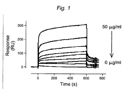
[0034] Figure 1. SPR analysis of wt-rFVIII (0-50 g) binding to immobilized
LRPI .
[0035] Figure 2. Comparison of wt-FVIII and hPEGylated-FVIII binding to
immobilized
LRP1 as determined by SPR analysis.
[0036] Figure 3. Comparison of wt-FVIII and PSA-FVIII binding to immobilized
LRPI as
determined by SPR analysis.
8
CA 02704234 2010-04-29
WO 2009/062100 PCT/US2008/082888
[0037] Figure 4. Comparison of the inhibitory binding effect of vWF and
PEGylated-vWF
on FVIII binding to LRPI as determined by SPR analysis. Similar inhibition is
seen for both
vWF constructs.
[0038] Figure 5. Comparison of the inhibitory binding effect of vWF and PSA-
vWF on
FVIII binding to LRP1 as determined by SPR analysis. PSA-vWF appears to be
slightly less
efficient than wt-vWF in interfering with the FVIII-LRP1 interaction.
[0039] Figure 6. Comparison of rVWF, natural VWF, and conjugated VWF binding
to
Gplba in the presence of botrocetin, as determined by SPR analysis. Binding of
PEGylated-
VWF to Gplba is reduced by approximately 50%, whereas PSA-VWF virtually lacks
the
ability to bind Gplba in a botrocetin-dependent manner.
[0040] Figure 7. Comparison of rVWF (wt), natural VWF (NPP), mutant rVWF (2B),
and
conjugated VWF binding to nanobody AU/vWFa-11. The binding of conjugated VWF
to the
nanobody is greatly reduced.
[0041] Figure 8. Survival assays of recombinant FVIII administered to patients
suffering
from Haemophilia A and Von Willebrand Disease type 3. Administered rFVIII
appears to be
stabilized in Haemophilia A patients as compared to VWD type 3 patients
presumably by the
presence of functional vWF in the former.
[0042] Figure 9. Correlation between the calculated half-life of VWF and the
half-life of
administered rFVIII in Haemophilia A patients. It is noted that in 33 of 38
patients, the half-
life of VWF is equal to or greater than the half-life of administered rFVIII.
[0043] Figure 10. Diagram of the equilibrium between free and VWF-bound FVIII
in vivo.
[0044] Figure 11. SPR analysis of FVIII (0-50 g) binding to immobilized LRP1.
[0045] Figure 12. SPR analysis of hPEGylated-FVIII (50 g) binding to
immobilized
LRP 1.
[0046] Figure 13. (Top Panel) Comparison of wt-rFVIII and hPEGylated-rFVIII
binding
(0-50 g) to LRP1 as determined by SPR analysis. (Bottom Panel) Chemical
structure of the
hydrolysable PEG moiety conjugated to rFVIII.
[0047] Figure 14. SPR results comparing wt-rFVIII and hPEGylated-rFVIII
binding
(50 g) to immobilized LRP1.
9
CA 02704234 2010-04-29
WO 2009/062100 PCT/US2008/082888
[0048] Figure 15. (Top Panel) Comparison of wt-rFVIII and sPEGylated-rFVIII
binding
(0-50 g) to LRP1 as determined by SPR analysis. (Bottom Panel) Chemical
structure of the
stable PEG moiety conjugated to rFVIII.
[0049] Figure 16. Comparison of equilibrium LRP1 binding responses for wt and
conjugated FVIII protein activated by thrombin cleavage. Results indicate that
hPEGylated-,
but not sPEGylated-, FVIII is induced to bind LRP1 upon thrombin activation
(compare bars
7 and 8 to bars 3 and 4).
[0050] Figure.17. (Top Panel) Comparison of the effect of wt-VWF and
sPEGylated-VWF
(0-100 g) on FVIII binding to LRPI as determined by SPR analysis. (Middle
Panel)
Chemical structure of the stable PEG moiety conjugated to rFVIII. (Bottom
Panel) IC50
values for the effect of wt-VWF and sPEG-VWF on FVIII-LRP1 binding.
[0051] Figure 18. (Top Panel) Comparison of the effect of wt-VWF and
hPEGylated-VWF
(0-100 g) on FVIII binding to LRP1 as determined by SPR analysis. (Middle
Panel)
Chemical structure of the hydrolysable PEG moiety conjugated to rFVIII.
(Bottom Panel)
IC50 values for the effect of wt-VWF and hPEG-VWF on FVIII-LRPI binding.
[0052] Figure 19. SPR analysis of wt-VWF, hPEG-VWF, and sPEG-VWF binding to
immobilized heparin.
[0053] Figure 20. Images of PMN static adhesion to immobilized wt and
conjugated VWF.
[0054] Figure 21. SPR analysis of FVIII and sPEG-FVIII binding to immobilized
LRP1.
[0055] Figure 22. Comparison of FVIII and sPEG-FVIII (0-150 nM) binding to
immobilized LRPI as determined by SPR analysis.
[0056] Figure 23. SPR analysis of hPEG-FVIII binding to immobilized LRPl.
[0057] Figure 24. SPR analysis of wt and conjugated VWF binding to immobilized
LRP1.
[0058] Figure 25. Comparison of wt and conjugated VWF (0-1000 nM) binding to
immobilized LRP 1 as determined by SPR analysis.
[0059] Figure 26. Results of ELISA experiments comparing wt-FVIII and
conjugated
FVIII binding to cluster 11 of LRPI.
[0060] Figure 27. Results of ELISA experiments comparing wt-FVIII and
conjugated
FVIII binding to cluster IV of LRP1.
CA 02704234 2010-04-29
WO 2009/062100 PCT/US2008/082888
[0061] Figure 28. Results of ELISA experiments comparing wt-VWF and conjugated
VWF binding to cluster II of LRP1.
[0062] Figure 29. Results of ELISA experiments comparing wt-VWF and conjugated
VWF binding to cluster IV of LRP1.
[0063] Figure 30. Non-limiting examples of water soluble polymer moieties that
are well
suited for conjugation to the coagulation proteins of the present invention.
DETAILED DESCRIPTION OF THE INVENTION
[0064] The present invention provides methods of increasing the survival or
half-life of a
coagulation protein by inhibiting or reducing the interaction with a clearance
receptor. In one
embodiment, the methods of the present invention comprise modifying a
coagulation protein
with a water soluble polymer and administering to a mammal in need thereof a
therapeutically effective amount of a composition comprising the modified
coagulation
factor. Coagulation proteins embraced by the present invention include those
that participate
in or assist in the regulation of a pathway involved in the coagulation
cascade.
[0065] The coagulation proteins of the present invention may be purified from
endogenous
sources, such as pooled human plasma, or may be produced by recombinant means.
In one
embodiment, the coagulation proteins modified in the methods of the invention
are selected
from Factor VIII (FVIII) and Von Willebrand Factor (VWF). In a particular
embodiment, the
coagulation proteins are selected from recombinant Factor VIII (rFVIII) and
recombinant von
Willebrand Factor (rVWF).
[0066] One known clearance receptor for FVIII is LRPI. In certain embodiments,
the
invention provides methods of increasing the survival or half-life of FVIII by
inhibiting or
reducing the binding affinity of FVIII for LRP 1. In some embodiments, the
methods
comprise administering to a mammal in need thereof a therapeutically effective
amount of a
modified or conjugated FVIII molecule with a reduced binding affinity for
LRP1. In
particular embodiments, the modified FVIII may be administered simultaneously
with VWF,
or in a preformed FVIIUVWF complex.
[0067] Ina related embodiment, the methods of the present invention comprise
the steps
of:(a) modifying a binding protein of a coagulation protein with a water
soluble polymer, and
(b) administering to a mammal in need thereof a therapeutically effective
amount of a
11
CA 02704234 2010-04-29
WO 2009/062100 PCT/US2008/082888
composition comprising the modified binding protein. In one embodiment, the
interaction
between FVIII and the clearance receptor LRPI is inhibited by administering to
a mammal a
modified VWF protein. In certain embodiments, the modified VWF is administered
simultaneously with FVIII or in a preformed FVIII/VWF complex. In yet other
embodiments, both FVIII and VWF are modified.
[0068] The present invention also provides methods of preparing a composition
that
inhibits the interaction between a coagulation protein and a clearance
receptor. In certain
embodiments, the methods comprise modifying a coagulation protein with a water
soluble
polymer, wherein the modification increases the survival of the protein in
blood circulation of
a mammal by inhibiting coagulation protein clearance receptors. In specific
embodiments,
the coagulation protein composition comprises FVIII, VWF, or a preformed
FVIII/VWF
complex.
[0069] In one embodiment, the present invention also provides modified or
conjugated
coagulation proteins that demonstrate reduced binding to their clearance
receptor and have
increased half-lives in vivo. In other embodiments, the invention provides
pharmaceutical
formulations of modified or conjugated coagulation proteins for administration
to a mammal
in need thereof. In particular embodiments, the formulations comprise modified
FVIII,
VWF, or a preformed FVIII/VWF complex.
[0070] In another embodiment, the present invention provides methods of
treating an
individual with a blood clotting disease, the method comprising administering
to a patient in
need thereof a modified coagulation protein, wherein said coagulation protein
has an
increased survival in vivo. The methods of the present invention may be
practiced with any
of the modified coagulation proteins, compositions, or formulations thereof
presented
herewith. In a particular embodiment, the clotting disease is Haemophilia or
von
Willebrand's Disease.
[0071] In one embodiment, the invention provides a method of treating an
individual
suffering from a disease characterized by a FVIII deficiency, by administering
a modified
VWF to the individual, wherein said VWF is conjugated to a water soluble
polymer. In
certain embodiments, the method further comprises administering FVIII to the
individual. In
some embodiments, the FVIII may also be modified by a water soluble polymer.
In other
embodiments, the FVIII is not modified by a water soluble polymer. In
particular
embodiments, the patient is administered a preformed VWF/FVIII complex,
wherein the
12
CA 02704234 2010-04-29 1
WO 2009/062100 PCT/US2008/082888
VWF is conjugated to a water soluble polymer. In certain embodiments, the
disease may be
Haemophilia or von Willebrand's Disease.
Definitions
[00721 As used herein, a "coagulation protein" refers to a protein that
functions in or has a
regulatory role in a pathway of the coagulation cascade that results in the
cross-linking of
fibrin molecules. Coagulation proteins embraced by the present invention may
participate in
or regulate, for example, the tissue factor or extrinsic coagulation pathway,
the contact
activation or intrinsic pathway, or the common final coagulation pathway. Non-
limiting
examples of coagulation proteins include; Factor I (fibrinogen), Factor II
(prothrombin),
Factor IIa (thrombin), Factor III (Tissue Factor), Factor V, Factor VI, Factor
VII, Factor VIII,
Factor IX, Factor X, Factor XI, Factor XII, Factor XIII, VWF, Prekallikrein,
High Molecular
Weight Kininogen (HMWK), Fibronectin, Antithrombin III, Heparin cofactor II,
Protein C,
Protein S, Protein Z, Protein Z-related Protease Inibitor (ZPI), Plasminogen,
alpha 2-
antiplasmin, tissue Plasminogen Activator (tPA), Urokinase, Plasminogen
Activator
Inhibitor-1 (PAI1), Plasminogen Activator Inhibitor-2 (PAI2), Cancer
Procoagulant, and the
like. The coagulation proteins of the present invention include full-length
proteins as well as
matured polypeptides, activated polypeptides, precursor polypeptides,
partially proteolysed
polypeptides, and the like. It is understood that the coagulation proteins of
the present
invention include alternatively spliced forms, conservatively modified
proteins, substantially
identical proteins, homologues, and the like.
[00731 As used herein, a "clearance receptor" refers to a class of proteins
which bind to and
remove coagulation proteins from the blood or plasma of an individual, thereby
reducing the
effective concentration of a given coagulation protein. Generally, a clearance
receptor is a
membrane protein comprising at least an extracellular domain and a membrane
attachment
domain. In certain embodiments, a membrane protein may be a transmembrane
protein, an
integral membrane protein, or a peripheral membrane protein. Exemplary
clearance receptors
embraced by the present invention include LFP receptors, vLDL receptors, LDL
receptor
related proteins, megalin receptors, and macrophage mannose receptors. For
example, LRP1
binds to and removes Factor VIII in vivo. One of skill in the art will know of
many clearance
receptors well suited for use in the present invention.
[00741 The term "water-soluble" refers to moieties that have some detectable
degree of
solubility in water. Methods to detect and/or quantify water solubility are
well known in the
13
CA 02704234 2010-04-29
WO 2009/062100 PCT/US2008/082888
art. Exemplary water-soluble polymers include peptides, saccharides,
poly(vinyls),
poly(ethers), poly(amines), poly(carboxylic acids) and the like. Peptides can
have mixed
sequences or be composed of a single amino acid, e.g., poly(lysine). An
exemplary
polysaccharide is poly(sialic acid) or hydroxyl ethyl starch. An exemplary
poly(ether) is
poly(ethylene glycol), e.g., m-PEG. Poly(ethylene imine) is an exemplary
polyamine, and
poly(acrylic) acid is a representative poly(carboxylic acid). Other water-
soluble polymers
that are suited for use in the present invention include polyelkylenes such as
polyoxyethylene,
polyoxypropylene, and block copolymers of polyoxyethylene and polyoxypropylene
(Pluronics); polymethacrylates; and carbomers. One of skill in the art will
know of other
water-soluble polymers well suited for use in the present invention.
[0075] The polymer backbone of the water-soluble polymer can be poly(ethylene
glycol)
(i.e. PEG). However, it should be understood that other related polymers are
also suitable for
use in the practice of this invention and that the use of the term PEG or
poly(ethylene glycol)
is intended to be inclusive and not exclusive in this respect. The term PEG
includes
poly(ethylene glycol) in any of its forms, including alkoxy PEG, difunctional
PEG,
multiarmed PEG, forked PEG, branched PEG, pendent PEG (i.e. PEG or related
polymers
having one or more functional groups pendent to the polymer backbone), or PEG
with
degradable linkages therein.
[0076] The polymer backbone can be linear or branched. Branched polymer
backbones are
generally known in the art. Typically, a branched polymer has a central branch
core moiety
and a plurality of linear polymer chains linked to the central branch core.
PEG is commonly
used in branched forms that can be prepared by addition of ethylene oxide to
various polyols,
such as glycerol, pentaerythritol and sorbitol. The central branch moiety can
also be derived
from several amino acids, such as lysine. The branched poly(ethylene glycol)
can be
represented in general form as R(-PEG-OH) in which R represents the core
moiety, such as
glycerol or pentaerythritol, and in represents the number of arms. Multi-armed
PEG
molecules, such as those described in U.S. Pat. No. 5,932,462, which is
incorporated by
reference herein in its entirety, can also be used as the polymer backbone.
[0077] Many other polymers are also suitable for the invention. Polymer
backbones that
are non-peptidic and water-soluble, with from 2 to about 300 termini, are
particularly useful
in the invention. Examples of suitable polymers include, but are not limited
to, other
poly(alkylene glycols), such as poly(propylene glycol) ("PPG"), copolymers of
ethylene
14
CA 02704234 2010-04-29
WO 2009/062100 PCT/US2008/082888
glycol and propylene glycol and the like, poly(oxyethylated polyol),
poly(olefinic alcohol),
poly(vinylpyrrolidone), poly(hydroxypropylmethacrylamide), poly(-hydroxy
acid),
poly(vinyl alcohol), polyphosphazene, polyoxazoline, poly(N-
acryloylmorpholine), such as
described in U.S. Pat. No. 5,629,384, which is incorporated by reference
herein in its
entirety, and copolymers, terpolymers, and mixtures thereof. Although the
molecular weight
of each chain of the polymer backbone can vary, it is typically in the range
of from about 100
Da to about 100,000 Da, often from about 6,000 Da to about 80,000 Da.
[0078] The term "glycoconjugation," as used herein, refers to the
enzymatically mediated
conjugation of a modified sugar moiety to an amino acid or glycosyl residue of
a polypeptide,
e.g., a coagulation protein of the present invention. A subgenus of
"glycoconjugation" is
"glycol-PEGylation," in which the modifying group of the modified sugar is
poly(ethylene
glycol), and alkyl derivative (e.g., m-PEG) or reactive derivative (e.g., H2N-
PEG, HOOC-
PEG) thereof.
[0079] The term, "glycosyl linking group," as used herein refers to a glycosyl
residue to
which a modifying group (e.g., PEG moiety or other water-soluble polymer) is
covalently
attached; the glycosyl linking group joins the modifying group to the
remainder of the
conjugate. In the methods of the invention, the "glycosyl linking group"
becomes covalently
attached to a glycosylated or unglycosylated coagulation protein, thereby
linking the agent to
an amino acid and/or glycosyl residue on the peptide. A "glycosyl linking
group" is
generally derived from a "modified sugar" by the enzymatic attachment of the
"modified
sugar" to an amino acid and/or glycosyl residue of the coagulation protein.
The glycosyl
linking group can be a saccharide-derived structure that is degraded during
formation of
modifying group-modified sugar cassette (e.g., oxidation Schiff base formation
reduction), or
the glycosyl linking group may be intact. An "intact glycosyl linking group"
refers to a
linking group that is derived from a glycosyl moiety in which the saccharide
monomer that
links the modifying group and to the remainder of the conjugate is not
degraded, e.g.,
oxidized, e.g., by sodium metaperiodate. "Intact glycosyl linking groups" of
the invention
may be derived from a naturally occurring oligosaccharide by addition of
glycosyl unit(s) or
removal of one or more glycosyl unit from a parent saccharide structure.
[0080] A "physiologically cleavable" as well as a "hydrolyzable" bond is a
relatively weak
bond that reacts with water (i.e., is hydrolyzed) under physiological
conditions. The
tendency of a bond to hydrolyze in water will depend not only on the general
type of linkage
CA 02704234 2010-04-29
WO 2009/062100 PCT/US2008/082888
connecting two central atoms but also on the substituents attached to these
central atoms.
Exemplary hydrolyzable bonds include, but are not limited to, carboxylate
ester, phosphate
ester, anhydride, acetal, ketal, acyloxyalkyl ether, imine, and ortho esters.
[0081] A "releasable linkage", or "hydrolysable linkage", or "releasable
linkage" includes,
but is not limited to, a physiologically cleavable bond, a hydrolyzable bond,
and an
enzymatically degradable linkage. Thus, a "releasable linkage" is a linkage
that may undergo
either hydrolysis or cleavage by some other mechanism (e.g., enzyme-catalyzed,
acid-
catalyzed, base-catalyzed, and so forth) under physiological conditions. For
example, a
"releasable linkage" can involve an elimination reaction that has a base
abstraction of a
proton, (e.g., an ionizable hydrogen atom, Ha), as the driving force. For
purposes herein, a
"releasable linkage" is synonymous with a "degradable linkage." Thus, a
releasable moiety
has one or more groups (e.g., a linker) that is releasable, degradable, or
capable of being
removed or cleaved under physiological and/or laboratory conditions, thus
releasing, e.g., the
water soluble polymer from the protein, or a protecting group linked to the
conjugation
moiety.
[0082] An "enzymatically releasable linkage" means a linkage that is subject
to degradation
by one or more enzymes.
[0083] A "hydrolytically stable" linkage or bond refers to a chemical bond,
typically a
covalent bond, that is substantially stable in water, that is to say, does not
undergo hydrolysis
under physiological conditions to any appreciable extent over an extended
period of time.
Examples of hydrolytically stable linkages include but are not limited to the
following:
carbon-carbon bonds (e.g., in aliphatic chains), ethers, amides, and the like.
Generally, a
hydrolytically stable linkage is one that exhibits a rate of hydrolysis of
less than about 1-5%
per day under physiological conditions.
[0084] As used herein, a protein having a "reduced binding affinity" for a
receptor refers to
a modified or recombinant protein that displays partially or totally
inhibited, decreased,
reduced, or down-regulated interactions with a particular receptor. In the
context of the
present invention, a modified or recombinant coagulation protein is said to
inhibit the
interaction with its clearance receptor if it binds with a lower binding
affinity or does not bind
at all. The reduced binding of the coagulation protein may be from about a 5%
to about a
100% or more reduction in the interaction with the clearance receptor. For
example, the
reduction may be about 5%, 10%, 20%, 30%, 40%, 50%, 60%, 70%, 80%, 90%, 100%,
or
16
CA 02704234 2010-04-29
WO 2009/062100 PCT/US2008/082888
more. Similarly, the inhibition of the binding between the modified or
recombinant
coagulation protein and clearance receptor maybe about a 5%, 10%, 20%, 30%,
40%, 50%,
60%, 70%, 80%, 90%, 95%, or 100% inhibition of the interaction. In certain
embodiments,
the reduced interaction may be from about 1-fold to about 10-fold reduced, for
example, 1-
fold, 2-fold, 3-fold, 4-fold, 5-fold, 6-fold, 7-fold, 8-fold, 9-fold, 10-fold,
or more reduced
binding in comparison to the interaction between the wild type coagulation
protein and
clearance receptor. In other embodiments, the reduced interaction may be from
about 101 -
fold to about 105-fold reduced, for example 101-fold, 102-fold, 103-fold, 104-
fold, 105-fold,
or more reduced as compared to the wild type protein. Quantitative means for
determining
the affinity of an interaction are well known in the art and include without
limitation, Surface
Plasmon Resonance (SPR) analysis, Isothermal Titration Calorimetry, affinity
chromatography, Fluorescence Polarization (FP) and Anisotropy (FA) assays, and
the like.
[0085] , As used herein, a "conjugation moiety" refers to a chemical structure
comprising a
water soluble polymer that is covalently attached to a protein, such as a
coagulation protein as
in the present invention. Conjugation moieties may further comprise one or
more linking
groups as well as one or more branching groups.
[0086] As used herein, "treatment" refers to clinical intervention in an
attempt to alter the
natural course of the individual or condition being treated, for example a
blood coagulation
disorder such as Haemophilia or von Willebrand's Disease, and may be performed
either for
prophylaxis or during the course of clinical pathology. Desirable effects
include preventing
occurrence or recurrence of symptoms of the disease, alleviation of symptoms,
diminishment
of any direct or indirect pathological consequences of the disease, lowering
the rate of disease
progression, amelioration or palliation of the disease state, and remission or
improved
prognosis.
[0087] An "effective amount" or a "therapeutically effective amount" is an
amount
sufficient to effect a beneficial or desired clinical result, for example, in
the treatment of a
disease state such as Haemophilia, von Willebrand's Disease, or a related
coagulapathy. In
terms of clinical response for subjects bearing a disease, an effective amount
is an amount
sufficient to palliate, ameliorate, stabilize, reverse, or slow progression of
the disease, or
otherwise reduce pathological consequences of the disease. An effective amount
may be
given in single or divided doses.
17
CA 02704234 2010-04-29
WO 2009/062100 PCT/US2008/082888
[0088] Non-limiting examples of coagulapathies that may be treated with the
methods and
compositions of the present invention include hypercoagulability diseases,
such as
Antithrombin III deficiency, Protein C deficiency, Activated protein C
resistance, Protein S
deficiency, Factor V Leiden, Hyperprothrombinemia; essential thrombocytosis;
hyopcoagulability diseases, such as Hemophilia, including Types A, B, and C,
Von
Willebrand's disease, Hypoprothrombinemia/Factor II deficiency,
Hypofibrinogenemia,
Factor XIII deficiency, and the like; purpura, such as Henoch-Schonlein,
idiopathic
thrombocytopenic purpura (ITP), Evans syndrome, and thrombotic
thrombocytopenic
purpura (TTP); and thrombocytopenia, including heparin-induced
thrombocytopenia.
[0089] As used herein, the terms "Hemophilia" or "Haemophilia" refer to a
group of
disease states broadly characterized by reduced blood clotting or coagulation.
Haemophilia
may refer to Type A, Type B, or Type C Haemophilia, or to the composite of all
three
diseases types. Type A Haemophilia (Haemophilia A) is caused by a reduction or
loss of
Favtor VIII (FVIII) activity and is the most prominent of the Haemophilia
subtypes. Type B
Haemophilia (Haemophilia B) results from the loss or reduction of Factor IX
(FIX) clotting
function. Type C Haemophilia (Haemophilia C) is a consequence of the loss or
reduction in
Factor XI (FXI) clotting activity. Haemophilia A and B are X-linked diseases,
while
Haemophilia C is autosomal. Common treatments for Haemophilia include both
prophylactic
and on-demand administration of clotting factors, such as FVIII, FIX,
including Bebulin VH,
and FXI, as well as FEIBA-VH, desmopressin, and plasma infusions.
[0090] As used herein, "Von Willebrand Disease" or "Von Willebrand's disease"
(vWD),
refers to a class of diseases characterized by a defect in the normal activity
of von Willebrand
Factor (vWF). The defect in vWF may include loss or reduction of function, as
in Type 1,
Type 3, and some Type 2 Von Willebrand Diseases, or alternatively may result
from a gain of
function, as in Type 2B and platelet-type vWD. In the context of the present
invention, vWD
may refer to any type of the disease, including Type 1, Type 2, Type 3, and
platelet type
vWD, any subtype of the disease, such as Type 2A, Type 2B, Type 2M, or Type
2N, or to the
group of diseases as a whole.
[0091] Common treatments for VWD include administration of VWF, FVIII, and
FVIII/VWF compositions and equivalents, such as Advate , Hemophil M, MONARC-
MTM,
and Recombinate. Other treatments include desmopressin, which can be
administered orally
or intravenously (DDAVP), subcutaneously (octostim), or nasally (octostim
spray);
18
.......................
CA 02704234 2010-04-29
WO 2009/062100 PCT/US2008/082888
cyklokapron and amicar, which help to stabilize established clots; thrombin,
which can be
applied directly to a site of bleeding, and general plasma infusions.
[0092] Factor VIII (FVIII) exists naturally and in therapeutic preparations as
a
heterogeneous distribution of polypeptides arising from a single gene product
(see, e.g.,
Andersson et al., Proc. Natl. Acad. Sci. USA, 83, 2979-2983, May 1986). The
term "Factor
VIII" as used herein refers to all such polypeptides, whether derived from
blood plasma or
produced through the use of recombinant DNA techniques. Commercially available
examples of therapeutic preparations containing Factor VIII include those sold
under the
trade names of HEMOFIL M and RECOMBINATE (available from Baxter Healthcare
Corporation, Deerfield, Ill., U.S.A.). Other preparations currently in
development comprise
primarily a single subpopulation of Factor VIII molecules which lack the B
domain portion of
the molecule. In the context of the present invention, FVIII may be post-
translationally
modified, either in vivo, or in vitro, and/or conjugated to a water soluble
polymer, e.g. a
polyether such as a PEG, PEO, POE, and the like. In certain embodiments, the
FVIII
molecules of the present invention may be polysialylated, PEGylated, or
otherwise post-
translationally modified.
[0093] VWF and FVIII molecules particularly well suited for use in the present
invention
include full-length protein constructs, precursor protein constructs,
biologically active
fragments, subunits, or derivatives thereof, plasmonic polypeptides,
recombinant
polypeptides, and the like.
[0094] In certain embodiments, VWF proteins of the invention may comprise a
construct,
for example, prepared as in WO 1986/06096 published on Oct. 23, 1986 and U.S.
patent
application Ser. No. 07/559,509, filed on Jul. 23, 1990, in the name of
Ginsburg et al., which
is incorporated herein by reference with respect to the methods of producing
recombinant
VWF. The VWF useful for the present invention includes all potential forms,
including the
monomeric and multimeric forms. One particularly useful form of VWF are homo-
multimers
of at least two VWFs. The VWF proteins may be either a biologically active
derivative, or
when to be used solely as a stabilizer for FVIII the V\VF may be of a form not
biologically
active. It should also be understood that the present invention encompasses
different forms of
VWF to be used in combination. For example, a composition useful for the
present invention
may include different multimers, different derivatives and both biologically
active derivatives
and derivatives not biologically active. In primary hemostasis VWF serves as a
bridge
19
CA 02704234 2010-04-29
WO 2009/062100 PCT/US2008/082888
between platelets and specific components of the extracellular matrix, such as
collagen. The
biological activity of VWF in this process can be measured by two different in
vitro assays
(Turecek et at., Semin. Thromb. Hemost. 28: 149-160, 2002). The ristocetin
cofactor assay is
based on the agglutination of fresh or formalin-fixed platelets induced by the
antibiotic
ristocetin in the presence of VWF. The degree of platelet agglutination
depends on the VWF
concentration and can be measured by the turbidimetric method, e.g. by use of
an
aggregometer (Weiss et al., J. Clin. Invest. 52: 2708-2716, 1973; Macfarlane
et al., Thromb.
Diath. Haemorrh. 34: 306-308, 1975). The second method is the collagen binding
assay,
which is based on ELISA technology (Brown et Bosak, Thromb. Res. 43: 303-311,
1986;
Favaloro, Thromb. Haemost. 83: 127-135, 2000). A microtiter plate is coated
with type I or
III collagen. Then the VWF is bound to the collagen surface and subsequently
detected. with
an enzyme-labeled polyclonal antibody. The last step is the substrate
reaction, which can be
photometrically monitored with an ELISA reader.
[00951 As used herein, "plasma-derived VWF (pdVWF)" includes all forms of the
protein
found in blood including the mature VWF obtained from a mammal having the
property of in
vivo-stabilizing, e.g. binding, of at least one FVIII molecule. However, the
invention is not
limited to the mature VWF. One, biologically active derivative of said pVWF is
pro-VWF
which contains the pro-peptide. Other forms of VWF useful for the present
invention include
the proteinaceous construct comprises immature VWF including the precursor VWF
molecule (pre-pro-VWF) synthesized by endothelial cells and megakaryocytes,
and/or the
VWF propeptide (pro-VWF) and/or mature pdVWF obtained upon cleavage of the
signal
peptide and pro-peptide, respectively of the precursor molecule. Further
examples of
biologically active derivatives of pdVWF include pro-drugs which are processed
or converted
into the biologically active form, or is biologically active as such,
truncated forms, forms
having deletions, forms having substitutions, forms having additions other
than pro-forms,
fragments of the mature form, chimeric forms, and forms having post-
translational
modifications as compared to the natural form. PdVWF useful for the present
invention also
includes those forms not biologically active. This may be accomplished by
modification of
the mature VWF or other naturally occurring forms found in blood. The source
for VWF
useful for the invention is mammalian, including porcine and human versions.
[0096] As used herein, "recombinant VWF (rVWF)" includes VWF obtained via
recombinant DNA technology. One form of useful rVWF has at least the property
of in vivo-
stabilizing, e.g. binding, of at least one FVIII molecule and having
optionally a glycosylation
4 .
CA 02704234 2010-04-29
WO 2009/062100 PCT/US2008/082888
pattern which is pharmacologically acceptable. Specific examples thereof
include VWF
without A2 domain thus resistant to proteolysis (Lankhof et al., Thromb.
Haemost. 77: 1008-
13, 1997), the VWF fragment from Val 449 to Asn 730 including the glycoprotein
lb-
binding domain and binding sites for collagen and heparin (Pietu et al.,
Biochem. Biophys.
5 Res. Commun. 164: 1339-1347, 1989). The determination of stabilizing at
least one FVIII
molecule can be carried out in VWF-deficient mammals according to methods
known in the
state in the art. The level of FVIII activity can be measured by, for
instance, a chromogenic
assay such as published in the European Pharmacopoeia (Ph. Eur., 3rd Ed.
1997:2.7.4).
[0097] In certain embodiments, FVIII proteins of the invention may comprise a
construct,
10 for example, prepared as in any of U.S. Patent Nos. 4,757,006; 5,733,873;
5,250,421; and
5,919,766, or as in EP 306 968. Generally, a FVIII protein of the invention
may comprise
any FVIII molecule having at least a portion of the B domain intact, and which
has biological
activity that is associated with wild type FVIII. For example, the construct
may be a full
length FVIII, a construct encoded by a nucleotide capable of hybridizing to a
nucleic acid
encoding Factor VIII:C. Such a protein may contain amino acid deletions at
various sites
between or within the domains A1-A2-B-A3-C1-C2 (U.S. Patent No. 4,868,112).
The FVIII
molecule may also be an analog of native FVIII wherein one or more amino acid
residues
have been replaced by site-directed mutagenesis. Non-limiting example of
constructs well
suited for use in the methods of the present invention include, for example,
those described in
WO 2007/126808.
[0098] The production of rVWF or rFVIII may include any method known in the
art for (i)
the production of recombinant DNA by genetic engineering, e.g. via reverse
transcription of
RNA and/or amplification of DNA, (ii) introducing recombinant DNA into
prokaryotic or
eukaryotic cells by transfection, e.g. via electroporation or microinjection,
(iii) cultivating
said transformed cells, e.g. in a continuous or batchwise manner, (iv)
expressing rVWF or
rFVIII, e.g. constitutively or inducibly, and (v) isolating said rVWF or
rFVIII, e.g. from the
culture medium or by harvesting the transformed cells, in order to (vi) obtain
purified rVWF
or rFVIII, e.g. via anion or cation exchange chromatography, affinity
chromatography, size
exclusion chromatography, and the like.
[0099] The rVWF or rFVIII can be produced by expression in a suitable
prokaryotic or
eukaryotic host system characterized by producing a pharmacologically
acceptable rVWF or
rFVIII molecule. Examples of eukaryotic cells are mammalian cells, such as
CHO, COS,
21
CA 02704234 2010-04-29
WO 2009/062100 PCT/US2008/082888
HEK 293, BHK, SK-Hep, and HepG2. There is no particular limitation to the
reagents or
conditions used for producing or isolating rVWF or rFVIII according to the
present invention
and any system known in the art or commercially available can be employed.
[0100] A wide variety of vectors can be used for the preparation of the rVWF
or rFVIII and
can be selected from eukaryotic and prokaryotic expression vectors. Examples
of vectors for
prokaryotic expression include plasmids such as pRSET, pET, pBAD, etc.,
wherein the
promoters used in prokaryotic expression vectors include lac, tre, tip, recA,
araBAD, etc.
Examples of vectors for eukaryotic expression include: (i) for expression in
yeast, vectors
such as pAO, pPIC, pYES, pMET, using promoters such as AOX1, GAP, GAL I, AUG1,
etc;
(ii) for expression in insect cells, vectors such as pMT, pAc5, pIB, pMIB,
pBAC, etc., using
promoters such as PH, p 10, MT, Acs, OpIE2, gp64, polh, etc., and (iii) for
expression in
mammalian cells, vectors such as pSVL, pCMV, pRc/RSV, pcDNA3, pBPV, etc., and
vectors derived from viral systems such as vaccinia virus, adeno-associated
viruses, herpes
viruses, retroviruses, etc., using promoters such as CMV, SV40, EF-1, UbC.
RSV, ADV,
BPV, and actin.
[0101] The terms "polypeptide," "peptide," and "protein" are used
interchangeably herein
to refer to a polymer of amino acid residues. The terms apply to amino acid
polymers in
which one or more amino acid residue is an artificial chemical mimetic of a
corresponding
naturally occurring amino acid, as well as to naturally occurring amino acid
polymers and
non-naturally occurring amino acid polymer.
[0102] The term "amino acid" refers to naturally occurring and synthetic amino
acids, as
well as amino acid analogs and amino acid mimetics that function in a manner
similar to the
naturally occurring amino acids. Naturally occurring amino acids are those
encoded by the
genetic code, as well as those amino acids that are later modified, e.g.,
hydroxyproline, y-
carboxyglutamate, and O-phosphoserine. Amino acid analogs refers to compounds
that have
the same basic chemical structure as a naturally occurring amino acid, i.e.,
an a carbon that is
bound to a hydrogen, a carboxyl group, an amino group, and an R group, e.g.,
homoserine,
norleucine, methionine sulfoxide, methionine methyl sulfonium. Such analogs
have modified
R groups (e.g., norleucine) or modified peptide backbones, but retain the same
basic chemical
structure as a naturally occurring amino acid. Amino acid mimetics refers to
chemical
compounds that have a structure that is different from the general chemical
structure of an
amino acid, but that functions in a manner similar to a naturally occurring
amino acid.
22
CA 02704234 2010-04-29
WO 2009/062100 PCT/US2008/082888
[0103] Amino acids maybe referred to herein by either their commonly known
three letter
symbols or by the one-letter symbols recommended by the IUPAC-IUB Biochemical
Nomenclature Commission. Nucleotides, likewise, may be referred to by their
commonly
accepted single-letter codes.
[0104] "Conservatively modified variants" applies to both amino acid and
nucleic acid
sequences. With respect to particular nucleic acid sequences, conservatively
modified
variants refers to those nucleic acids which encode identical or essentially
identical amino
acid sequences, or where the nucleic acid does not encode an amino acid
sequence, to
essentially identical sequences. Because of the degeneracy of the genetic
code, a large
number of functionally identical nucleic acids encode any given protein. For
instance, the
codons GCA, GCC, GCG and GCU all encode the amino acid alanine. Thus, at every
position where an alanine is specified by a codon, the codon can be altered to
any of the
corresponding codons described without altering the encoded polypeptide. Such
nucleic acid
variations are "silent variations," which are one species of conservatively
modified
variations. Every nucleic acid sequence herein which encodes a polypeptide
also describes
every possible silent variation of the nucleic acid. One of skill will
recognize that each codon
in a nucleic acid (except AUG, which is ordinarily the only codon for
methionine, and TGG,
which is ordinarily the only codon for tryptophan) can be modified to yield a
functionally
identical molecule. Accordingly, each silent variation of a nucleic acid which
encodes a
polypeptide is implicit in each described sequence with respect to the
expression product.
[0105] As to amino acid sequences, one of skill will recognize that individual
substitutions,
deletions or additions to a nucleic acid, peptide, polypeptide, or protein
sequence which
alters, adds or deletes a single amino acid or a small percentage of amino
acids in the encoded
sequence is a "conservatively modified variant" where the alteration results
in the substitution
of an amino acid with a chemically similar amino acid. Conservative
substitution tables
providing functionally similar amino acids are well known in the art. Such
conservatively
modified variants are in addition to and do not exclude polymorphic variants,
interspecies
homologs, and alleles of the invention. One of skill in the art will also
recognize that
conservative substitutions to a protein embraced by the present invention will
be well
tolerated, especially when made in residues not involved in active sites or
required for a
particular catalytic function. One of skill in the art will recognize that a
plethora of
conservative mutations, as well as non-conservative mutations made in regions
with low
homology or distal to an active site or protein binding interface, may be well
tolerated and
23
CA 02704234 2010-04-29
WO 2009/062100 PCT/US2008/082888
can be designed by inspection of high resolution structural information
readily available in
the art.
[01061 The following eight groups each contain amino acids that are
conservative
substitutions for one another: 1) Alanine (A), Glycine (G); 2) Aspartic acid
(D), Glutamic
acid (E); 3) Asparagine (N), Glutamine (Q); 4) Arginine (R), Lysine (K); 5)
Isoleucine (1),
Leucine (L), Methionine (M), Valine (V); 6) Phenylalanine (F), Tyrosine (Y),
Tryptophan
(W); 7) Serine (S), Threonine (T); and 8) Cysteine (C), Methionine (M) (see,
e.g., Creighton,
Proteins (1984)).
[01071 The term "recombinant" when used with reference, e.g., to a cell, or
nucleic acid,
protein, or vector, indicates that the cell, nucleic acid, protein or vector,
has been modified by
the introduction of a heterologous nucleic acid or protein or the alteration
of a native nucleic
acid or protein, or that the cell is derived from a cell so modified. Thus,
for example,
recombinant cells express genes that are not found within the native (non-
recombinant) form
of the cell or express native genes that are otherwise abnormally expressed,
under expressed
or not expressed at all.
[01081 The terms "isolated," "purified," or "biologically pure" refer to
material that is
substantially or essentially free from components that normally accompany it
as found in its
native state. Purity and homogeneity are typically determined using analytical
chemistry
techniques such as polyacrylamide gel electrophoresis or high performance
liquid
chromatography. A coagulation protein or complex of coagulation proteins, for
example,
FVIII, VWF, or FVIIUVWF, that is the predominant species present in a
preparation is
substantially purified. The term "purified" in some embodiments denotes that a
nucleic acid
or protein gives rise to essentially one band in an electrophoretic gel. In
other embodiments,
it means that the nucleic acid or protein is at least 50% pure, more
preferably at least 60%,
65%, 70%, 75%, 80%,85%,90%,95%, 96%, 97%, 98%, 99% or more pure. "Purify" or
"purification" in other embodiments means removing at least one contaminant
from the
composition to be purified. In this sense, purification does not require that
the purified
compound be homogenous, e.g., 100% pure.
[01091 The terms "identical" or percent "identity," in the context of two or
more nucleic
acids or polypeptide sequences, refer to two or more sequences or subsequences
that are the
same or have a specified percentage of amino acid residues or nucleotides that
are the same
(i.e., about 60% identity, preferably 65%, 70%, 75%, 80%, 85%, 90%, 91%, 92%,
93%, 94%,
24
CA 02704234 2010-04-29
WO 2009/062100 PCT/US2008/082888
95%, 96%, 97%, 98%, 99%, or higher identity over a specified region, when
compared and
aligned for maximum correspondence over a comparison window or designated
region) as
measured using a BLAST or BLAST 2.0 sequence comparison algorithms with
default
parameters described below, or by manual alignment and visual inspection (see,
e.g., NCBI
web site http://www.ncbi.nlm.nih.gov/BLAST/ or the like). Such sequences are
then said to
be "substantially identical." This definition also refers to, or may be
applied to, the
compliment of a test sequence. The definition also includes sequences that
have deletions
and/or additions, as well as those that have substitutions. As described
below, the preferred
algorithms can account for gaps and the like. Preferably, identity exists over
a region that is
at least about 25 amino acids or nucleotides in length, or more preferably
over a region that is
50-100, 200, 300, 400, 500, or more amino acids or nucleotides in length.
[0110] For sequence comparison, typically one sequence acts as a reference
sequence, to
which test sequences are compared. When using a sequence comparison algorithm,
test and
reference sequences are entered into a computer, subsequence coordinates are
designated, if
necessary, and sequence algorithm program parameters are designated.
Preferably, default
program parameters can be used, or alternative parameters can be designated.
The sequence
comparison algorithm then calculates the percent sequence identities for the
test sequences
relative to the reference sequence, based on the program parameters.
[0111] A "comparison window", as used herein, includes reference to a segment
of anyone
of the number of contiguous positions selected from the group consisting of
from 20 to 600,
usually about 50 to about 200, more usually about 100 to about 150 in which a
sequence may
be compared to a reference sequence of the same number of contiguous positions
after the
two sequences are optimally aligned. Methods of alignment of sequences for
comparison are
well-known in the art. Optimal alignment of sequences for comparison can be
conducted,
e.g., by the local homology algorithm of Smith & Waterman, Adv. Appl. Math.
2:482 (1981),
by the homology alignment algorithm of Needleman & Wunsch, J. Mol. Biol.
48:443 (1970),
by the search for similarity method of Pearson & Lipman, Proc. Nat'l. Acad.
Sci. USA
85:2444 (1988), by computerized implementations of these algorithms (GAP,
BESTFIT,
FASTA, and TFASTA in the Wisconsin Genetics Software Package, Genetics
Computer
Group, 575 Science Dr., Madison, WI), or by manual alignment and visual
inspection (see,
e.g., Current Protocols in Molecular Biology (Ausubel et al., eds. 1987-2005,
Wiley
Interscience)).
CA 02704234 2010-04-29
WO 2009/062100 PCT/US2008/082888
[0112] An example of algorithms that are suitable for determining percent
sequence
identity and sequence similarity include the BLAST and BLAST 2.0 algorithms,
which are
described in Altschul et al., Nuc. Acids Res. 25:3389-3402 (1977) and Altschul
et al., J. Mol.
Biol. 215:403-410 (1990), respectively. BLAST and BLAST 2.0 are used, with the
parameters described herein, to determine percent sequence identity for the
nucleic acids and
proteins of the invention. Software for performing BLAST analyses is publicly
available
through the National Center for Biotechnology Information
(http://www.ncbi.nlm.nih.gov/).
This algorithm involves first identifying high scoring sequence pairs (HSPs)
by identifying
short words of length W in the query sequence, which either match or satisfy
some positive-
valued threshold score T when aligned with a word of the same length in a
database
sequence. T is referred to as the neighborhood word score threshold (Altschul
et al., supra).
These initial neighborhood word hits act as seeds for initiating searches to
find longer HSPs
containing them. The word hits are extended in both directions along each
sequence for as
far as the cumulative alignment score can be increased. Cumulative scores are
calculated
using, for nucleotide sequences, the parameters M (reward score for a pair of
matching
residues; always > 0) and N (penalty score for mismatching residues; always <
0). For amino
acid sequences, a scoring matrix is used to calculate the cumulative score.
Extension of the
word hits in each direction are halted when: the cumulative alignment score
falls off by the
quantity X from its maximum achieved value; the cumulative score goes to zero
or below,
due to the accumulation of one or more negative-scoring residue alignments; or
the end of
either sequence is reached. The BLAST algorithm parameters W, T, and X
determine the
sensitivity and speed of the alignment. The BLASTN program (for nucleotide
sequences)
uses as defaults a wordlength (W) of 11, an expectation (E) of 10, M=5, N=-4
and a
comparison of both strands. For amino acid sequences, the BLASTP program uses
as
defaults a wordlength of 3, and expectation (E) of 10, and the BLOSUM62
scoring matrix
(see Henikoff & Henikoff, Proc. Natl. Acad. Sci. USA 89:10915 (1989))
alignments (B) of
50, expectation (E) of 10, M=5, N=-4, and a comparison of both strands.
CONJUGATES AND POST-TRANSLATIONAL MODIFICATIONS
[0113] Generally, the conjugation, post-translation modification, or covalent
modification
of the coagulation proteins of the invention include modifications of the N-
or C- terminal
residues as well as modifications of selected side chains, for example, at
free sulfhydryl-
groups, primary amines, and hydroxyl-groups. In one embodiment, the water
soluble
polymer is linked to the protein (directly or via a linker) by a lysine group
or other primary
26
CA 02704234 2010-04-29
WO 2009/062100 PCT/US2008/082888
amine. In one embodiment, the coagulation proteins of the present invention
may be
modified by conjugation of a water soluble polymer, including without
limitation, a
polyethylene glycol (PEG), a polypropylene glycol, a polyoxyalkylene, a
polysialic acid,
hydroxyl ethyl starch, a poly-carbohydrate moiety, and the like.
[01141 Water soluble polymers that maybe used to modify the coagulation
proteins of the
present invention include linear and branched structures. The conjugated
polymers may be
attached directly to the coagulation proteins of the invention, or
alternatively may be attached
through a linking moiety. Non-limiting examples of protein conjugation with
water soluble
polymers can be found in U.S. Patent Nos. 4,640,835; 4,496,689; 4,301,144;
4,670,417;
4,791,192, and 4,179,337, as well as in Abuchowski and Davis "Enzymes as
Drugs,"
Holcenberg and Roberts, Eds., pp. 367 383, John Wiley and Sons, New York
(1981), and
Hermanson G., Bioconjugate Techniques 2nd Ed., Academic Press, he. 2008.
[01151 Protein conjugation maybe performed by a number of well known
techniques in the
art, for example, see Hermanson G., Bioconjugate Techniques 2nd Ed., Academic
Press, Inc.
2008. Examples include linkage through the peptide bond between a carboxyl
group on one
of either the coagulation protein or water-soluble polymer moiety and an amine
group of the
other, or an ester linkage between a carboxyl group of one and a hydroxyl
group of the other.
Another linkage by which a coagulation protein of the invention could be
conjugated to a
water-soluble polymer compound is via a Schiff base, between a free amino
group on the
polymer moiety being reacted with an aldehyde group formed at the non-reducing
end of the
polymer by periodate oxidation (Jennings and Lugowski, J. Immunol. 1981;
127:1011-8;
Femandes and Gregonradis, Biochim Biophys Acta. 1997; 1341; 26-34). The
generated
Schiff Base can be stabilized by specific reduction with NaCNBH3 to form a
secondary
amine. An alternative approach is the generation of terminal free amino groups
on the
polymer by reductive amination with NH4CI after prior oxidation. Bifunctional
reagents can
be used for linking two amino or two hydroxyl groups. For example a polymer
containing an
amino group can be coupled to an amino group of the coagulation protein with
reagents like
BS3 (Bis(sulfosuccinimidyl)suberate/Pierce, Rockford, I11.). In addition
heterobifunctional
cross linking reagents like Sulfo-EMCS (N-s-Maleimidocaproyloxy)
sulfosuccinimide
ester/Pierce) can be used for instance to link amine and thiol groups. In
other embodiments,
an aldehyde reactive group, such as PEG alkoxide plus diethyl acetal of
bromoacetaldehyde;
PEG plus DMSO and acetic anhydride, and PEG chloride plus the phenoxide of 4-
hydroxybenzaldehyde, succinimidyl active esters, activated dithiocarbonate
PEG, 2,4,5-
27
CA 02704234 2010-04-29
WO 2009/062100 PCT/US2008/082888
trichlorophenylcloroformate and P-nitrophenylcloroformate activated PEG, may
be used in
the conjugation of a coagulation protein.
[0116] In yet other embodiments of the invention, various reactive groups
maybe used to
conjugate a water-soluble polymers to a coagulation protein of the invention,
for example, an
imidoester, a hydroxymethyl phosphine, a carbodiimide, a N-hydroxysuccinimide
ester
(NHS-ester), a pentafluorophenyl ester (PFP-ester), a psoralen group, an aryl
azide, a
hydrazide, an isocynate, a maleimide, a pyridyl disulfide, a vinyl sulfone,
and the like.
[0117] The term "sialic acid" refers to any member of a family of nine-carbon
carboxylated
sugars. The most common member of the sialic acid family is N-acetyl-
neuraminic acid (2-
keto-5-acetamido-3,5-dideoxy-D-glycero-D-galactononulopyranos-l-onic acid
(often
abbreviated as Neu5Ac, NeuAc, or NANA). A second member of the family is N-
glycolyl-
neuraminic acid (Neu5Gc or NeuGc), in which the N-acetyl group of NeuAc is
hydroxylated.
A third sialic acid family member is 2-keto-3-deoxy-nonulosonic acid (KDN)
(Nadano et al.
(1986) J. Biol. Chem. 261: 11550-11557; Kanamori et al., J. Biol. Chem. 265:
21811-21819
(1990)). Also included are 9-substituted sialic acids such as a 9-0-C1-C6 acyl-
Neu5Ac like
9-O-lactyl-Neu5Ac or 9-O-acetyl-Neu5Ac, 9-deoxy-9-fluoro-Neu5Ac and 9-azido-9-
deoxy-
Neu5Ac. For review of the sialic acid family, see, e.g., Varki, Glycobiology
2: 25-40 (1992);
Sialic Acids: Chemistry, Metabolism and Function, R. Schauer, Ed. (Springer-
Verlag, New
York (1992)). Poly-sialic acid moieties can be conjugated to the coagulation
proteins of the
invention for example by the method described in U.S. Pat. No. 4,356,170,
which is herein
incorporated by reference. In one embodiment of the invention, the
polysaccharide
compound may be a naturally occurring polysaccharide, a derivative of a
naturally occurring
polysaccharide, or a naturally occurring polysaccharide derivative.
[0118] One type of covalent modification included within the scope of this
invention
comprises altering the native glycosylation pattern of the coagulation
protein. Generally,
altering the native glycosylation pattern of a protein refers to removing
and/or adding one or
more glycosylation sites to the coagulation protein such that the interaction
between the
coagulation protein and its clearance receptor is reduced or inhibited.
Additionally, the
native glycosylation pattern of a coagulation protein may be altered by
quantitatively or
qualitatively changing the various carbohydrate moieties present, i.e. the
amount of
glycosylation per molecule may be increased, or the identity of the
carbohydrate moieties
may be changed. In one embodiment, the coagulation proteins of the present
invention may
28
CA 02704234 2010-04-29
WO 2009/062100 PCT/US2008/082888
be chemically or enzymatically coupled to glycosides, for example, as in WO
87/05330 or
Aplin and Wriston, CRC Crit. Rev. Biochem., pp. 259 306 (1981).
[0119] In one embodiment, the present invention provides O-linked glycosylated
coagulation proteins, conjugates of these species, and methods for forming O-
linked
glycosylated peptides that include a selected amino acid sequence ("an O-
linked
glycosylation site"). Of particular interest are mutant coagulation proteins
that include an 0-
linked glycosylation site that is not present in the corresponding wild type
coagulation
protein. The O-linked glycosylation site is a locus for attachment of a
glycosyl residue that
bears a modifying group.
[0120] In one embodiment, a polymer conjugated to a coagulation factor of the
invention
comprises a polysaccharide, which may be branched or unbranched. Monomer units
of the
polysaccharides used for conjugation include without limitation, D-mannose, D-
and L-
galactose, fucose, fructose, D-xylose, L-arabinose, D-glucuronic acid, sialic
acid, D-
galacturonic acid, D-mannuronic acid, D-glucosamine, D-galactosamine, D-
glucose and
neuraminic acid, and the like. Non-limiting examples of polysaccharides the
may be used
include homopolysaccharides and heteropolysaccharides such as lactose,
amylopectin, starch,
hydroxyethyl starch, amylose, dextrane sulfate, dextran, dextrins, glycogen,
or the
polysaccharide subunit of acid mucopolysaccharides, e.g. hyaluronic acid;
polymers of sugar
alcohols such as polysorbitol and polymannitol; heparin, heparin, and the
like.
[0121] In one particular embodiment, a coagulation protein of the present
invention may be
conjugated to a water soluble polymer selected from those shown in Figure 30.
[0122] Polymers used for conjugation of the coagulation proteins of the
invention may
have an average molecular weight of about 100 Da to about 500,000 Da. In
certain
embodiments, the polymers may have an average molecular weight of about 1,000
Da to
about 20,000 Da. In other embodiments, the average molecular weight of the
polymers may
be about I kDa, or about 2 kDa, 3 kDa, 3.5 kDa, 4 kDa, 5 kDa, 6 kDa, 7 kDa, 8
kDa, 9 kDa,
10 kDa, 11 kDa, 12 kDa, 13 kDa, 14 kDa, 15 kDa, 16 kDa, 17 kDa, 18 kDa, 19
kDa, 20 kDa,
kDa, 40 kDa, 50 kDa, 75 kDa, 100 kDa, 125 kDa, 150 kDa, 200 kDa, 250 kDa, 300
kDa,
400 kDa, 500 kDa, or higher. The average molecular weight of the polymers used
to
30 conjugate the coagulation proteins of the invention will depend upon many
factors, including
the nature of the coagulation protein, the nature of the polymer, the degree
of conjugation,
and the like.
29
CA 02704234 2010-04-29
WO 2009/062100 PCT/US2008/082888
101231 In certain embodiments, the invention comprises a coagulation protein
linked to a
conjugation moiety comprising a first linker group, a first branching group,
and one or more
water soluble polymers attached to said branching group. In other embodiments,
the
conjugation moiety may further comprise at least a second linking group
connecting said
branching group and said water soluble polymers. Suitable linking groups
include, without
limitation, a urethane, an amide, urea, an ester, a thioether, and the like.
One of skill in the
art will know of other linking groups particularly well suited for use in the
present invention.
In yet other embodiments, the conjugation moiety may comprise a copolymer, for
example,
an alternating copolymer, a periodic copolymer, a random copolymer, a block
copolymer,
such as a diblock or triblock, or a branched copolymer. Many conjugation
moieties well
suited for use with the present invention are known in the art and can be
found, for example,
in Hermanson G., Bioconjugate Techniques 2d Ed., Academic Press, Inc. 2008.
[01241 In one embodiment, the coagulation proteins of the invention may be
conjugated to
a water soluble polymer through a reversible or hydrolysable linkage. U. S.
Patent
Application Publication No. 2005/0079155 describes conjugates using reversible
linkages.
As described in this publication, reversible linkages can be effected through
the use of an
enzyme substrate moiety. Another approach for reversible PEGylation is
described in U.S.
Patent No. 7,060,259, which describes water-soluble prodrugs in which a
biologically active
agent is linked to a water-soluble non-immunogenic polymer by a hydrolyzable
carbamate
bond. As described therein, the biologically active agent can be readily
released by the
hydrolysis of the carbmate bond in vivo without the need for adding enzymes or
catalytic
materials. A different approach for the conjugation of reversible moieties is
described by
Peleg-Schulman (2004) J. Med. Chem. 47:4897-4904, WO 2004/089280 and U.S.
Patent
Application Publication No. 2006/0171920. Although this approach has been
applied to a
limited number of active agents, these references ignore other active agents
for which
reversible PEGylation would be particularly suited. Yet another releasable
approach is
described in U.S. Patent Application Publication No. 2006/0293499.
[01251 In one embodiment, the present invention provides modified coagulation
proteins
with the general structure:
R L1-X1
wherein R is a coagulation protein, L1 is a linking group, and X1 is a water
soluble polymer.
CA 02704234 2010-04-29
WO 2009/062100 PCT/US2008/082888
[01261 Examples of coagulation moieties that are well suited for use in the
present
invention can be found, for example, in U.S. Patent Application No.
20060293499, which
describes the use of conjugation moieties with degradable linkages; WO
2004/089280, which
describes the use of reversible PEGylation moieties, including PEGylation
moieties coupled
to proteins through a 9-hydroxymethyl-7-sulfofluorene-N-hydroxysuccinimide
linkage (PEG-
FMS), U.S. Patent Application No. 20050009988, which describes the use of
various linking
moieties that may be used to couple a polymer with a coagulation protein of
the present
invention; U.S. Patent No. 5,672,662, which describes the use of
monosubstituted
Poly(ethylene glycol) and related moieties for protein conjugation; U.S.
Patent Application
No. 20060171920, which describes the use of modifying groups that are
sensitive to mild
basic conditions, such as Fmoc and 2-sulfo-9-fluorenylmethoxycarbonyl (FMS),
for
conjugation of polymers to proteins; U.S. Patent Application Nos. 20040235723
and
20080058504, which describe polymer conjugates of FVIII; and U.S. Patent
Application No.
20060160948, which describes VWF and FVIII polymer conjugates; all of which
are herein
incorporated by reference in their entirety for all purposes. In certain
embodiments, the
methods of the invention may be practiced using the modified coagulation
proteins described
in, for example, WO 2008/082669 or WO 2007/126808, which are herein
incorporated by
reference in their entirety for all purposes.
[01271 The conjugates of the present invention may comprise a variety of
formulae. In one
embodiment, a conjugate of the invention may comprise the general formula;
/L2-X1
R L1-B1\
L3-X2
wherein R is a coagulation protein, L1, L2, and L3 are linker moieties, B 1 is
a first branching
moiety, and X1 and X2 are water soluble polymer groups. In certain
embodiments, L1, L2,
and L3 are optional. In particular embodiments, the conjugation moiety may be
chemically
stable. In other embodiments, the conjugation moieties used in the present
invention may be
hydrolysable. In certain embodiments, L1 or B1 may comprise a protecting
group, such as an
alcohol protecting group, an amine protecting group, a carbonyl protecting
group, a
carboxylic protecting group, and the like.
[0128] In a particular embodiment, the protecting group is a substituted Fmoc
group. In
one embodiment, a conjugate of the invention may comprise the formula;
31
CA 02704234 2010-04-29
WO 2009/062100 PCT/US2008/082888
POLY2 XZ X1-- POLY'
[Rflb a V11.
RZ R'
Y1
R__Y2
wherein, POLY' is a first water-soluble polymer; POLY2 is a second water-
soluble polymer;
X' is a first spacer moiety; X2 is a second spacer moiety; Ha is an ionizable
hydrogen atom;
R' is H or an organic radical; R2 is H or an organic radical; (a) is either
zero or one; (b) is
either zero or one; Re', when present, is a first electron altering group;
Reg, when present, is a
second electron altering group; and Y' is 0 or S; Y2 is 0 or S; and R is a
coagulation protein.
[0129] In one specific embodiment, the present invention provides a conjugate
of a
coagulation protein comprising the structure;
L2 -X2
X1-_ L,
O
O_;~_,_ R
wherein R is a coagulation protein, L, and L2 are linkers, and X, and X2 are
water soluble
polymers. In other embodiments, the conjugation moieties of the present
invention may be
one of those shown in figure 13 or 15. In certain embodiments, the conjugation
moiety may
be conjugated at an amino acid side, at the carboxy-terminus of the protein,
or at the amino-
terminus of the protein. In certain embodiments, the water soluble moieties
are conjugated to
a side chain at a free sulthydryl-group, a primary amine, a hydroxyl-group, or
a combination
thereof. In a particular embodiment, the water soluble protein is VWF or
FVIII.
[0130] In one embodiment, the present invention provides a conjugate of a
coagulation
protein comprising the structure;
32
CA 02704234 2010-04-29
WO 2009/062100 PCT/US2008/082888
PEG-m
NH
m-PEGO 0 0
H 7 \", NH
N
H
O
OR
wherein R is a coagulation protein and m-PEGO is a PEG moiety, or other water
soluble
polymer, connected to the remainder of the structure through an ether linkage.
In certain
embodiments, the conjugation moiety may be conjugated at an amino acid side,
at the
carboxy-terminus of the protein, or at the amino-terminus of the protein. In
certain
embodiments, the water soluble moieties are conjugated to a side chain at a
free sulfhydryl-
group, a primary amine, a hydroxyl-group, or a combination thereof. In a
particular
embodiment, the water soluble protein is VWF or FVIII.
[01311 In one specific embodiment, the present invention provides a conjugate
of a
coagulation protein comprising the structure;
ACH2CH2-
O (OCH2CH2)1-
NH OCH3
CH30-(CH2CH2O)õ-CH2CH2-O O O
H NH
N
H
O
O`__1_ R
wherein R is a coagulation protein. In certain embodiments, the conjugation
moiety may be
conjugated at an amino acid side, at the carboxy-terminus of the protein, or
at the amino-
terminus of the protein. In certain embodiments, the water soluble moieties
are conjugated to
a side chain at a free sulthydryl-group, a primary amine, a hydroxyl-group, or
a combination
thereof. In a particular embodiment, the water soluble protein is VWF or
FVIII.
[01321 In one specific embodiment, the present invention provides a conjugate
of a
coagulation protein comprising the structure;
33
CA 02704234 2010-04-29
WO 2009/062100 PCT/US2008/082888
O OII
R O 0)~N4CH2CH20)m CH3
H
O~_N-(CH2CH20)m CH3
H
wherein R is a coagulation protein. In certain embodiments, the conjugation
moiety may be
conjugated at an amino acid side, at the carboxy-terminus of the protein, or
at the amino-
terminus of the protein. In certain embodiments, the water soluble moieties
are conjugated to
a side chain at a free sulfhydryl-group, a primary amine, a hydroxyl-group, or
a combination
thereof. In a particular embodiment, the water soluble protein is VWF or
FVIII.
[0133] In yet another embodiment, the present invention provides a conjugate
of a
coagulation protein comprising the structure;
O
03H
Y-S NN
O H
O
O_:__1_ R
wherein R is a coagulation protein and Y is a water soluble polymer, such as a
PEG. In
certain embodiments, the conjugation moiety may be conjugated at an amino acid
side, at the
carboxy-terminus of the protein, or at the amino-terminus of the protein. In
certain
embodiments, the water soluble moieties are conjugated to a side chain at a
free sulfhydryl-
group, a primary amine, a hydroxyl-group, or a combination thereof. In a
particular
embodiment, the water soluble protein is VWF or FVIII.
[0134] In yet another embodiment, the present invention provides a conjugate
of a
coagulation protein comprising the formula;
R-X'-X2-Y
wherein R is a coagulation protein, X1 is selected from the group consisting
of NH, S, CO,
COO, CH2, SO2, SO3, P02, and P03, X2 is a bond or linker which connects the
water soluble
moiety to X', and Y is a water soluble moiety. In a specific embodiment, the
water soluble
moiety is a PEG. In certain embodiments, the conjugation moiety may be
conjugated at an
amino acid side, at the carboxy-terminus of the protein, or at the amino-
terminus of the
34
CA 02704234 2010-04-29
WO 2009/062100 PCT/US2008/082888
protein. In certain embodiments, the water soluble moieties are conjugated to
a side chain at
a free sulfhydryl-group, a primary amine, a hydroxyl-group, or a combination
thereof. In a
particular embodiment, the water soluble protein is VWF or FVIII.
[0135] The modified or conjugated coagulation proteins of the present
invention are
considered to be "substantially uniformly modified" generally if at least
about 40% of the
proteins in a given batch or solution are modified to the same extent. In
other embodiments,
the coagulation proteins in a uniformly modified batch or solution may be
about 50%, 60%,
70%, 80%, 90%, 95%, 99%, or higher modified to the same extent. In certain
embodiments,
the extent of protein modification may be determined in terms of number of
moles of
modifying polymer per number of moles of protein. For example, a modified
coagulation
protein of the invention may be conjugated by about 1 to about 30 water
soluble polymers. In
certain embodiments, a coagulation protein of the invention may comprise about
1, or about
2, 3, 4, 5, 6, 7, 8, 9, 10, 11, 12, 13, 14, 15, 16, 17, 18, 19, 20, 25, 30, or
more moles of
conjugated polymer per mol protein. Typically, the extent of modification
occurring in a
conjugation reaction can be controlled. For example, an average reaction may
result in a
variation in the extent of conjugation of less than about 50%. In other
embodiments, the
variation in the extent of conjugation may be less than about 40%, or less
than about 35%,
30%, 25%, 20%, 15%, 10%, or 5%. Alternatively, the variation in the extent of
conjugation
for a particular batch of coagulation protein, may further be controlled by
fractionation after
the conjugation reaction, for example by size exclusion chromatography. Thus,
small
amounts of variation in the extent of conjugation may be achieved. In other
embodiments,
the extent of modification may be expressed in terms of the percent of
potential conjugation
sites that are modified. For example, a coagulation protein may be from about
I% to about
100% modified. In certain embodiments, the conjugates of the present invention
may be
about 1%, 2%,3%,4%,5%,10%,15%,20%,25%,30%,40%,50%,60%,70%,80%,90%,
95%, 98%, 99%, or more modified.
COMPOSITIONS AND FORMULATIONS
[0136] In certain embodiments, the coagulation protein compositions of the
present
invention, i.e. FVIII, vWF, or FVIII/vWF, further comprise bulking agents,
stabilizing
agents, buffering agents, sodium chloride, calcium salts, and, advantageously,
other
excipients. These excipients have been chosen in order to maximize the
stability of Factor
CA 02704234 2010-04-29
WO 2009/062100 PCT/US2008/082888
VIII in lyophilized preparations. However, the blood factor compositions of
the present
invention exhibit stability in the liquid state as well.
101371 As used herein, a "bulking agent" refers to a chemical entity which
provides
structure to the "cake" or residual solid mass of a pharmaceutical preparation
after it has been
lyophilized and which protect it against collapse. A "crystallizable bulking
agent" shall mean
a bulking agent as described herein which can be crystallized during
lyophilization, other
than sodium chloride. Particularly well suited bulking agents for use in the
present invention
include, without limitation, mannitol, glycine, alanine, and hydroxyethyl
starch (HES).
[0138] The bulking agents used in the present formulations, which form the
crystalline
portion of a lyophilized product (except in the case of HES), are selected
from the group
consisting of mannitol, glycine, alanine, hydroxyethyl starch (HES), and the
like. One of
ordinary skill will know of other bulking agents particularly well suited for
use with the
present invention. Mannitol, glycine, or alanine are present in an amount of
about 1% to
about 15%. In certain embodiments, the amount is about 1%,
2%,3%,4%,5%,6%,7%,8%,
9%, 10%, I 1%, 12%, 13%, 14%, 15%, or more. When HES is used as a bulking
agent, it is
present in an amount of about 1% to about 10%. In certain embodiments, the
amount is
about 1%, 2%,3%,4%,5%,6%,7%,8%,9%, 10%, or more HES.
[0139] In some embodiments, the stabilizing agents used in the formulations of
the present
invention are selected from the group consisting of sucrose, trehalose,
raffinose, sorbitol,
glycerol, arginine, or the like. One of ordinary skill will know of other
stabilizers particularly
well suited for use with the present invention. These agents are present in
the formulations of
the present invention in an amount of about I% to about 10%. In certain
embodiments, the
amount is about 1%, 2%, 3%, 4%, 5%, 6%, 7%, 8%, 9%, 10%, or more stabilizing
agent.
[01401 Various physiologically compatible salts may also be used in the
formulations of the
present invention. In one embodiment of the invention, sodium chloride is
included in the
present formulations in an amount of about 100 to about 300 mM, or from about
150 to about
250 mM, or about 225 mM. In one embodiment of the present invention, sodium
chloride
itself can be used without any of the aforementioned bulking agents, in which
case it would
be included in the formulation in an amount of between about 300 mm and about
500 mm
NaC1. In certain embodiments of the invention, a physiologically compatible
salt may be
used at about 50 to about 1000 mM. In other embodiments, the concentration of
salt in the
formulation may be about 50 mM, or about 100, 150, 200, 250, 300, 350, 400,
450, 500, 600,
36
CA 02704234 2010-04-29
WO 2009/062100 PCT/US2008/082888
700, 800, 900, 1000 mM or higher. It is understood that the pharmaceutically
acceptable
salts are non-toxic. Additional information on suitable pharmaceutically
acceptable salts can
be found in Remington's Pharmaceutical Sciences, 17th ed., Mack Publishing
Company,
Easton, Pa., 1985, which is incorporated herein by reference.
[0141] In some embodiments, the compositions of the present invention will
include a
buffer at concentration of from about 10 mM to about 200 mM. In other
embodiments, the
buffer concentration will be from about 10 mM to about 50 mM. In yet other
embodiments,
the compositions of the present invention may comprise about 10 mM, 20 mM, 30
mM, 40
mM, 50 mM, 75 mM, 100 mM, 125 mM, 150 mM, 175 mM, 200 mm, or more buffer.
Buffers well suited for use in the present invention include, without
limitation, histidine, Tris,
BIS-Tris Propane, PIPES, MOPS, HEPES, MES, ACES, and the like. One of skill in
the art
will know of other buffers that are particularly well suited for use in the
compositions of the
present invention. The compositions of the present invention may further
comprise an
antioxidant.
[0142] As used herein, "administering" means oral administration,
administration as a
suppository, topical contact, intravenous, intraperitoneal, intramuscular,
intralesional,
intranasal or subcutaneous administration, or the implantation of a slow-
release device e.g., a
mini-osmotic pump, to a subject. Administration is by any route including
parenteral, and
transmucosal (e.g., oral, nasal, vaginal, rectal, or transdermal). Parenteral
administration
includes, e.g., intravenous, intramuscular, intra-arteriole, intradermal,
subcutaneous,
intraperitoneal, intraventricular, and intracranial. Other modes of delivery
include, but are
not limited to, the use of liposomal formulations, intravenous infusion,
transdermal patches,
etc.
[0143] Formulations suitable for parenteral administration, such as, for
example, by
intraarticular (in the joints), intravenous, intramuscular, intratumoral,
intradermal,
intraperitoneal, and subcutaneous routes, include aqueous and non-aqueous,
isotonic sterile
injection solutions, which can contain antioxidants, buffers, bacteriostats,
and solutes that
render the formulation isotonic with the blood of the intended recipient, and
aqueous and
non-aqueous sterile suspensions that can include suspending agents,
solubilizers, thickening
agents, stabilizers, and preservatives. In the practice of this invention,
compositions can be
administered, for example, by intravenous infusion, orally, topically,
intraperitoneally,
intravesically or intrathecally. The formulations of compounds can be
presented in unit-dose
37
CA 02704234 2010-04-29
WO 2009/062100 PCT/US2008/082888
or multi-dose sealed containers, such as ampules and vials. Injection
solutions and
suspensions can be prepared from sterile powders, granules, and tablets.
[01441 In certain embodiments, the pharmaceutical preparation is in unit
dosage form. In
such form the preparation is subdivided into unit doses containing appropriate
quantities of
the active component. The unit dosage form can be a packaged preparation, the
package
containing discrete quantities of preparation, such as packeted tablets,
capsules, and powders
in vials or ampoules. Also, the unit dosage form can be a capsule, tablet,
cachet, or lozenge
itself, or it can be the appropriate number of any of these in packaged form.
The composition
can, if desired, also contain other compatible therapeutic agents.
[0145] Pharmaceutically acceptable carriers are determined in part by the
particular
composition being administered, as well as by the particular method used to
administer the
composition. Accordingly, there are a wide variety of suitable formulations of
pharmaceutical compositions of the present invention (see, e.g., Remington's
Pharmaceutical
Sciences, 20th ed., 2003, supra). As used herein, "pharmaceutically acceptable
carrier"
includes any material, which when combined with the conjugate retains the
conjugates'
activity and is non-reactive with the subject's immune systems. Examples
include, but are not
limited to, any of the standard pharmaceutical carriers such as a phosphate
buffered saline
solution, water, emulsions such as oil/water emulsion, and various types of
wetting agents.
Other carriers may also include sterile solutions, tablets including coated
tablets and capsules.
Typically such carriers contain excipients such as starch, milk, sugar,
certain types of clay,
gelatin, stearic acid or salts thereof, magnesium or calcium stearate, talc,
vegetable fats or
oils, gums, glycols, or other known excipients. Such carriers may also include
flavor and
color additives or other ingredients. Compositions comprising such carriers
are formulated
by well known conventional methods.
[0146] Effective dosage forms, modes of administration and dosage amounts of
the
composition of the invention may be determined empirically, and making such
determinations is within the skill of the art. It is understood by those
skilled in the art that the
dosage amount will vary with the particular composition employed, the
condition being
treated, the severity of the condition, the route of administration, the rate
of excretion, the
duration of the treatment, the identity of any other drugs being administered
to the mammal,
the age, size and species of the mammal, and like factors well known in the
medical and
veterinary arts. In general, a suitable daily dose of a compound of the
present invention will
38
CA 02704234 2010-04-29
WO 2009/062100 PCT/US2008/082888
be that amount which is the lowest dose effective to produce a therapeutic
effect. However,
the total daily dose will be determined by an attending physician or
veterinarian within the
scope of sound medical judgment. If desired, the daily dose may be
administered in multiple
sub-doses, administered separately at appropriate intervals throughout the
day.
EXAMPLES
[0147] Example 1
[0148] It has been shown that LRPI contributes to the regulation of FVIII
plasma levels.
LRP 1 is a cellular receptor that is able to bind and transport FVIII to
intracellular degradation
pathways. The present example, demonstrates that PEGylation or polysialylation
of FVIII
disrupts in vitro binding to LRP1.
[0149] Components: Purified recombinant wild-type FVIII (batch MOQ-Hepes-08E;
2.28
mg/ml; 12117 IU/ml); PEGylated FVIII (batch hydrolysable PEG-rFVIII
ORHLUFBO7001PHR; 1.76 mg/ml; 2498 IU/ml); polysialylated FVIII (batch PSA-
rFVIII-
11.OKD NHS; 0.613 mg/ml; 268 lU/ml). Purified LRPI was obtained from Biomac
(Leipzig;
Cat no. #04-03).
[0150] Experimental design: Binding of FVIII or its derivatives to LRPI was
assessed using
surface plasmon resonance (SPR) analysis using Biacore2000 equipment.
Specifications:
LRPI was immobilized on a standard CM5-biosensorchip (Biacore). The flow rate
was set at
gl/min to avoid potential rebinding due to mass-transfer limitations. Samples
were run in
20 buffer containing 0.005% Tween-20, 150 mM NaCl, mM CaC12, and 20 mM Hepes
(pH
7.4) at 25 C. Proteins were diluted on basis of protein content. Proteins were
injected for 10
minutes to allow equilibrium to be reached, and dissociation was followed for
an additional
2.5 minutes. Data analysis involved plotting of the response at equilibrium
versus protein
concentration. Since the experimental system is sensitive to changes in buffer
composition
during the analysis (buffer-refraction index), all preparations to be injected
were designed to
contain similar buffer compositions. Thus, for comparison between wt-FVIII and
PEG-
FVIII, equal amounts of the respective buffers were present in the final
preparations. An
example of an experimental set-up for wt-rFVIII and hydrolysable PEG-FVIII is
provided in
Table 1.
Table 1. Dillution series for a typical LPR1 binding assay.
rFVII pre-dillution l Sample gl rFVIII l PEG- l system total volume
39
CA 02704234 2010-04-29
WO 2009/062100 PCT/US2008/082888
( g/ml) buffer FVIII buffer buffer ( l)
0 - 0.0 12.28 22.84 244.88 280
0.1 1/1000 12.28 0.0 22.84 244.88 280
0.5 1/100 6.14 6.14 22.84 244.88 280
1.0 1/10 12.28 0.0 22.84 244.88 280
2.5 1/10 3.07 9.21 22.84 244.88 280
5.0 1/10 6.14 6.14 22.84 244.88 280
1/10 12.28 0.0 22.84 244.88 280
25 - 3.07 9.21 22.84 244.88 280
50 - 6.14 6.14 22.84 244.88 280
PEG-FVIII l rFVI1I l PEG- l system total volume
pre-dillution l Sample
( g/ml) buffer FVIII buffer buffer ( l)
0 - 0.0 12.28 22.84 244.88 280
0.1 1/100 4.57 12.28 18.27 244.88 280
0.5 1/10 2.28 12.28 20.56 244.88 280
1.0 1/10 4.57 12.28 18.27 244.88 280
2.5 1/10 11.42 12.28 11.42 244.88 280
5.0 - 2.28 12.28 20.56 244.88 280
10 - 4.57 12.28 18.27 244.88 280
25 - 11.42 12.28 11.42 244.88 280
50 - 22.84 12.28 0.0 244.88 280
[0151] As an example, raw sensorgram-data for the binding of wt-rFVIII to
immobilized
LRPI is depicted in Figure 1. In agreement with published data (eg. Lenting et
al 1999, JBC
274:23734), FVIII binds efficiently to LRPI in a dose-dependent manner. It
appeared that
5 10-min injections are indeed sufficient to allow equilibrium to be reached.
In Figures 2 and
CA 02704234 2010-04-29
WO 2009/062100 PCT/US2008/082888
3, typical equilibrium-responses are depicted for PEG-FVIII and PSA-FVIII,
respectively.
Strikingly, no binding of PEG-FVIII or PSA-FVIII to immobilized LRP1 could be
detected,
even at concentrations of 50 gg/ml (corresponding to 50-fold the normal plasma
concentration).
[0152] Example 2
[0153] Binding of FVIII to LRP1 is inhibited in the presence of VWF, because
LRPI
interaction sites within the FVIII light chain are inaccessible when FVIII is
bound to VWF.
The present example demonstrates that PEGylation or polysialylation of vWF
does not
interfere with the vWF-mediated inhibition of LRP 1 binding by FVIII.
[0154] Components: Purified recombinant wild-type FVIII (batch MOQ-Hepes-08E;
2.28
mg/ml; 12117 IU/ml); recombinant-wt-vWF (batch ORWSECO6006FIHL; 0.464 mg/ml;
72.1
IU Ag/ml; 20.6 IU RCo/ml), stable PEG-vWF (batch NTT - VWF-600-S2 I; 1.021
mg/ml;
61.4 IU Ag/ml; 41.9 IU RCo/ml); and stable PSA-vWF (batch PSA-RVWF-19.3KD CAO
batch2 (06.10.2006); 0.0087 mg/ml; 11.3 IU Ag/ml; 0.2 IU RCo/ml).
[0155] Wild-type recombinant FVIII (40 nM) was pre-incubated with various
concentrations of vWF (0-400 nM for wt-vWF and PEG-vWF and 0-200 nM for PSA-
vWF).
Concentrations of vWF were based on protein concentrations and a molecular
weight of 250
kDa per vWF monomer. Again, since the various proteins were in different
buffers, dilution-
schemes were designed in such a way that buffer-compositions remained equal
throughout
the analysis. Mixtures of FVIII/vWF were applied to LRP1 (immobilized on a CM5
-
biosensorchip) at a flow rate of 20 l/min in system-buffer containing 0.005%
Tween-20, 150
mM NaCl, 2 mM CaC12, 20 mM Hepes (pH 7.4) at 25 C.
[0156] In the absence of vWF, efficient binding of FVIII to LRPI was observed.
However,
increasing concentrations of vWF resulted in a concordant decrease in binding
of FVIII to
LRP1. As for PEG-vWF, this modulated protein appeared to be as efficient as wt-
vWF in
interfering with the FVIII-LRP1 interaction (Figure 4). Although PSA-vWF also
interfered
with LRPI-binding (Figure 5), this conjugated protein seemed slightly less
efficient than wt-
vWF. However, these data are based on a single range of concentrations, and
additional
experiments need to be performed to determine whether this difference is
within the
experimental range, or whether it represents a true decrease in capacity to
inhibit the
FVIII/LRPI interaction. Nevertheless, since both conjugations affected the
capacity of vWF
41
CA 02704234 2010-04-29
WO 2009/062100 PCT/US2008/082888
to inhibit binding of FVIII to LRP1 to only a minor extent, if any, these data
also indicate that
conjugating vWF by either PEG or PSA does not affect the ability of vWF to
interact with
FVIII to a major extent, if at all.
[0157] Apart from functioning as a carrier-protein for FVIII, vWF also plays a
critical role
in the recruitment of platelets to sites of vascular injury. VWF acts as a
molecular bridge
between the subendothelial matrix and the platelet Glycoprotein (Gp)-Ib/IX/V
receptor
complex. In order to interact with Gplba, vWF needs to be converted from a
cryptic into an
active conformation. Since chemical modulation of vWF may affect the exposure
of the
Gplba binding site, we have tested binding of wt-vWF and its conjugated
derivatives to
Gplba (both in the absence and presence of Botrocetin) and to nanobody AU/vWFa-
11, an
antibody fragment which displays preferential binding to vWF in its active
conformation.
[0158] Components: recombinant-wt-vWF (batch ORWSECO6006F1HL; 0.464 mg/ml;
72.1 IU Ag/ml; 20.6 IU RCo/ml), stable PEG-vWF (batch NTT-vWF-600-S2 I; 1.021
mg/ml;
61.4 IU Ag/ml; 41.9 IU RCo/ml); and stable PSA-vWF (batch PSA-RvWF-19.3KD CAO
batch2 (06.10.2006); 0.0087 mg/ml; 11.3 IU Ag/ml; 0.2 IU RCo/ml). Recombinant
Gplba
(comprising residues 1-290) has been described previously (Huizinga et al
(2002) Science
2973176). Botrocetin was obtained from Kordia BV (Leiden, the Netherlands).
Anti-Gplba
antibody 2D4 was kindly provided by Dr. H. Deckmyn (Kortrijk, Belgium).
Nanobody
AUIvWFa-11 has been described previously (Hulstein et al 2005 Blood 106:3035).
HRP-
conjugated Polyclonal anti-vWF antibodies were purchased from Dako (Glostrup,
Denmark).
[0159] Binding to Gplba and AU/vWFa-11 were performed in an immunosorbent
assay,
essentially as described (van Schooten et al 2005, JTH 3:2228 and Hulstein et
al 2005 Blood
106:3035, respectively). In the absence of botrocetin, no binding of wt-vWF or
its
conjugated derivatives (up to 1 g/ml) to Gplba could be observed. Thus, in
none of the
preparations tested (including wt-vWF), vWF molecules appear to be presence
that are able
to spontaneously interact with Gplba. The presence of botrocetin, however, is
sufficient to
induce efficient binding to Gplba in the case of wt-vWF (figure 6). In fact,
it seems that wt-
vWF provided by Baxter is slightly more efficient than VWF present in normal
pooled
plasma, or recombinant VWF that is produced in a university laboratory
setting. Conjugation
of vWF diminishes botrocetin-induced Gplba binding as both PEG-vWF and PSA-vWF
bind
with lower affinity to GplBa as compared to non-conjugated vWF. PEG-vWF bound
with
approximately 2-fold less affinity than wt-vWF, whereas PSA-vWF was virtually
unable to
42
CA 02704234 2010-04-29
WO 2009/062100 PCT/US2008/082888
bind to Gplba in the presence of botrocetin. The fact that PSA-vWF displayed
strongly
impaired botrocetin-binding to Gplba corresponds to the low Ristocetin-
cofactor activity that
is reported for this protein (2.3 IU/mg protein). In contrast, PEG-vWF has a
similar
Ristocetin-cofactor activity compared to wt-vWF (41.0 IU/mg compared to 44.4
IU/mg,
respectively). It is of importance to mention, however, that both assays are
dependent on the
activation of vWF by ristocetin or botrocetin. It cannot be excluded that
pegylation and/or
polysialylation of vWF affects the interaction with these activators.
Conclusive data
regarding the interaction between VWF and Gplba may be obtained from in vitro
perfusion
experiments.
[01601 The results of AU/vWFa-11 binding experiments are given in figure 7.
Normal
pooled plasma (NPP) is used as a binding control reference, the slope of which
is arbitrarily
assigned as 1. As positive control, we have included recombinant vWF carrying
an
Arg1306G1n mutation (type 2B) which induces a platelet-binding conformation.
The relative
activity of this preparation (defined as the ratio's between the slopes of the
preparation over
the slope of normal pooled plasma) is 6.9. Recombinant wt-vWF from the
University
Medical Center Utrecht preparation displayed slightly enhanced binding to
nanobody
AU/vWFa- 11 (relative activity 1.6), whereas recombinant wt-vWF provided by
Baxter was
similar to normal pooled plasma (relative activity 1.1). In contrast, both PEG-
vWF and PSA-
vWF displayed reduced binding to AU/vWFa-11 (relative activities 0.04 and 0.3,
respectively). Usually, these types of reduced relative activities are found
in vWF molecules
carrying a type 2A mutation. Reduced biding to AU/vWFa-11 indicates that
conjugation of
vWF with PEG or PSA does not convert the molecule into an active, platelet
binding
conformation. Alternatively, conjugation of VWF may alter access of the
nanobody to its
binding site within the vWF Al domain.
[01611 Example 3
[01621 The relationship between the half-life survival rate of FVIII in the
presence and
absence of VWF was studied in patients with Haemophilia A and Von Willebrand
Disease
type 3. Haemophilia A patients have a deficiency in FVIII levels, but
typically display
normal VWF expression. Conversely, patients with Von Willebrand Disease (VWD)
type 3
are homozygous for deficient VWF, but show normal FVIII expression. However,
despite
the normal expression of FVIII in patients with Von Willebrand Disease type 3,
plasma levels
of the clotting factor are greatly reduced, presumably due to a lack of
protection normally
43
CA 02704234 2010-04-29
WO 2009/062100 PCT/US2008/082888
provided by stable binding of VWF to FVIIL Thus, it is predicted that
administration of a
FVIII concentrate to Haemophilia A patients will result in a longer half life
of the clotting
factor than will administration to patients with Von Willebrand Disease type
3.
[0163] The hypothesis presented above was tested by determining the survival
rate of
administered FVIII in Haemophilia A patients and in patients with VWD type 3.
Briefly, 30
IU/kg b.w. of a Factor VIII concentrate (Advate, Baxter Healthcare Corp.) was
administered
to patients with Haemophilia A and VWD type 3. Blood samples were drawn,
citrated
plasma was prepared and FVIII levels were measured by ELISA (Asserachrom,
Stago;
Asnieres sur Seine, France), using a monoclonal antibody specific for FVIII,
at different time
points after administration. As can be seen in Figure 8, the half life of
administered FVIII in
Haemophilia A patients is roughly 20 hours, while the half-life in patients
with Von
Willebrand Disease type 3 is only 1 to 2 hours. Thus, FVIII clearance in vivo
occurs at a
much greater rate in the absence of wild type VWF, as indicated by the lower
half-life of
FVIII administered to patients suffering from Von Willebrand Disease Type 3.
[0164] Next, the relationship between the half-life of administered FVIII and
VWF in
Haemophilia A patients was determined. The half-life of VWF in the patients
was calculated
as proposed by Nossent et al. (Journal of Thrombosis and Haemostasis,
4(12):2556-62
(2006)). Specifically, it was assumed that the half-life, in hours, of VWF is
equal to twice the
ratio of the concentration of steady state VWF to the steady state
concentration of the
propeptide. In this fashion, the half-life was calculated in 38 severe
Haemophilia A patients
after administration of 30 IU FVIIIIkg b.w. of a Factor VIII concentrate, by
determining the
plasma levels of VWF-antigen by ELISA using a polyclonal antibody specific for
VWF
(DAKO Cytomation, Glostrup, Denmark) and VWF propeptide levels by a specific
monoclonal antibody (Sanquin Research; Amsterdam, NL). The determined half-
lives of
FVIII and VWF from the sample cohort were plotted and a Pearson-rank
coefficient of 0.6 (P
= 0.0001) was found, indicating that the two half-lives are strongly
correlated (Figure F).
Notably, in 33 of the 38 patients, the half-life of VWF was greater than the
half-life of FVIII.
A proposed equilibrium for FVIII clearance in vivo is shown in Figure 10.
[0165] Example 4
[01661 Several receptors have been described as being putatively responsible
for FVIII
clearance in vivo, including LDL-receptor related protein (LRP 1, CD9 1) /
megalin (Lenting
et al., JBC 274:23734-9 (1999); Saenko et al., JBC 274:37685-92)), LDL-
receptor / vLDL-
44
CA 02704234 2010-04-29
WO 2009/062100 PCT/US2008/082888
receptor Bovenschen et al., Blood 106:906-12 (2005)), Asialoglycoprotein-
receptor(
Bovenschen et al., J Thromb Haemost 3:1257-65 (2005)), and CD206 (macrophage
mannose-
receptor) (Lenting et at., J Thromb Haemost 5:1353-60 (2007)). It is known
that FVIII
binding to these receptors is prevented or reduced in the presence of VWF.
However, only
LRP1 has been shown to be physiologically relevant for FVIII clearance
(Bovenschen et al.,
Blood 101(10):3933-9 (2003)). Therefore, kinetic studies of the interaction
between FVIII
and LRP1 were undertaken in an attempt to determine conditions that result in
reduced FVIII
clearance via LRP 1.
[0167] Surface Plasmon Resonance (SPR) experiments were carried out on the
Biacore
2000 system to determine the effect that chemical conjugation and the presence
of additional
blood factors have on the kinetics of FVIII - LRP1 binding. Briefly, LRP1,
purified as
described by Huizinga et al. (J Thromb Haemost 3:2228-37 (2005), was
immobilized onto a
CM5 biosensor chip at 4000 RU / mm2, which was determined to be approximately
6.7 finol /
mm2 LRP1. Various concentrations of recombinant FVIII, PEGylated (Figure 11)
and non-
conjugated (Figure 12), were flowed over the chips at 20 l / min, and steady
state kinetics of
the interaction were determined using a Biacore 2000 system and the
BlAevaluation
software. hPEGylated-FVIII was modified with a hydrolysable PEG moiety shown
in Figure
13, which was attached as described in US 2008/0234193 Al. As can be seen in
Figure 13,
hPEGylated FVIII failed to bind LRP 1, even at concentrations as high as 50
ug/ml. The
experiments were then repeated with sPEGylated-FVIII, which was modified by a
stable PEG
moiety as shown in Figure L. As seen in Figures 14 and 15, sPEGylated FVIII
(open circles)
bound to LRPI at substantially reduced levels as compared to unmodified FVIII
(closed
circles). These experiments show that PEGylation of FVIII inhibits binding to
LRP1.
[0168] It is known that LRP1 interacts with FVIII light chain, but not to full-
length FVIII
heavy chain (Lenting et al., JBC 274(34):23734-9 (1999)). However, after
partial proteolysis
by thrombin, FVIII heavy chain becomes competent to bind to LRP1 (Bovenschen
et al., J
Thromb Haemost. 4(7):1487-93 (2006)). Thus, the effect of PEGylation on
thrombin
activated FVIII binding to LRP1 was investigated. Briefly, unmodified FVIII,
hPEGylated-
FVIII, and sPEGylated-FVIII at 250 ug / ml was incubated with 2 nM thrombin
for 5 minutes
at 37 C. To stop proteolysis, the reaction mixture was diluted 10-fold in 50
nM aqueous
solution of a thrombin specific inhibitor (PPACK, Biomol Int., Germany). LRP1
binding of
the activated FVIII solutions were than analyzed by SPR using a Biacore 2000
system as
described before. As can be seen by results of the SPR FVIII binding
experiments, shown in
CA 02704234 2010-04-29
WO 2009/062100 PCT/US2008/082888
Figure 16, thrombin cleavage induces LRP-binding of unmodified and hPEGylated-
FVIII,
but not sPEGylated-FVIII.
[0169] The effect of VWF on FVIII-LRP1 binding was next examined by SPR
analysis. 40
nM unmodified FVIII was preincubated with 0 to 400 nM VWF, either unmodified
or
PEGylated, for 25 minutes at 37 C. VWF/FVIII complexes were then subjected to
SPR
analysis as above. Both sPEGylated (Figure 17) and hPEGylated (Figure 18) VWF
further
inhibited FVIII binding to LRP1 with respect to unmodified VWF. These data
suggest that
PEGylation of VWF can further reduce FVIII clearance via the LRP 1 receptor.
[0170] In summary, the FVIII-LRP1 binding experiments show that PEGylation of
FVIII
strongly reduces the interaction between FVIII and its clearance receptor LRP
1. Further,
thrombin cleavage of hPEGylated-FVIII, but not sPEGylated-FVIII, induces
binding to
LRP 1, although not to the same level as unmodified FVIII. Finally, PEGylation
of VWF
does not interfere with VWF mediated inhibition of the interaction between
FVIII and its
clearance receptor LRP1. Conversely, PEGylation actually increases VWF's
inhibitory effect
on FVIII binding to LRP1, as demonstrated. These experiments suggest that
PEGylation of
either or both of recombinant VWF and FVIII may have beneficial effects for
administration
in patients with blood clotting disorders such as Haemophilia and Von
Willebrand Disease.
[0171] Example 5
[0172] To further characterize the effects of VWF PEGylation, SPR experiments
were
employed on a Biacore 2000 system to test the binding of conjugated VWF to
heparin.
Briefly, biotinylated heparin was conjugated. to streptavidin coated
sensorchips (GE
Healthcare) at an RU / mm2 of 70. 25 .ig / ml non-conjugated, sPEGylated, and
hPEGylated
VWF, in buffer containing 20 mM HEPES (pH 7.4) and 100 mM NaCl, was then
flowed over
the chips at a flow rate of 10 l / ml. As before data was collected and
analyzed on a Biacore
2000 system (GE Healthcare) using the BlAevaluation software package. As seen
in Figure
19, non-conjugated VWF bound heparin with low affinity. This is in contrast to
PEGylated
VWF, which failed to bind heprin at all. These data suggest that PEGylation of
VWF reduces
or eliminates the proteins capacity to bind heparin.
[0173] Example 6
[0174] ELISA experiments were performed to determine the effect PEGylation has
on
VWF binding to GpIba. Briefly, recombinant Gplba was immobilized on antibody-
46
CA 02704234 2010-04-29
WO 2009/062100 PCT/US2008/082888
conjugated microtiter plates and blocked with PBS-buffer, pH 7.4 containing 3%
Bovine
Serum Albumin (BSA) and 0.1% Tween 20. 0 to 500 ng / ml VWF from various
sources was
dialyzed into PBS-buffer containing 3% BSA and 0.1% Tween 20, and then
incubated in the
microtiter wells for 120minutes at 37 C. Unbound protein was then removed and
the wells
were washed 3 times with washing buffer (PBS-buffer, pH 7.4 containing 0.1 %
Tween 20).
VWF was detected using Horseradish Peroxidase (HRP) labeled polyclonal anti-
VWF
antibody (DAKO Cytomation). As seen in Figure 6, PEGylated VWF bound Gplba
with
slightly reduced affinity as compared to non-conjugated VWF. These results
indicate that
PEGylated-VWF is still competent for Gplba-mediated platelet binding.
[0175] Example 7
[0176] VWF binds leukocytes under both perfusion and static conditions (Pendu
et al.,
Blood 108(12):3746-52 (2006)). Static binding assays were performed to
investigate the
effect that PEGylation has on these interactions. Briefly, unmodified,
hPEGylated, and
sPEGylated VWF was immobilized in microtiter wells, PMN cells were then pre-
treated,
subsequently added to the protein coated wells, and incubated for 60 minutes
at 37 C (Pendu
et al., Blood 108(12):3746-52 (2006))Unbound cells were then removed by gentle
washing
of the wells with PBS - buffer, pH 7.4. As can be seen in Figure 20, both
sPEGylated and
hPEGylated-VWF stably bound PMN cells at similar levels as non-conjugated VWF,
under
static conditions. This data indicates that conjugation of VWF does not affect
specific
interactions with leukocytes.
[0177] Example 9
[0178] To characterize the effect that PEGylation has on interactions between
FVIII / VWF
and LRP1, SPR experiments were performed. Briefly, LRP1 was conjugated to CM5 -
biosensor chips (Biacore Life Sciences) as before, and the conjugation was
measured at
approximately 8 fmol / mm2 LPR1. Various concentrations of both conjugated and
non-
conjugated VWF and FVIII were diluted with 20 mM Hepes buffer (pH 6.5)
containing 150
mM NaCl, 2 mM CaC12 and 0.005% (v/v) Tween 20. Protein samples were then
flowed over
the chips at 20 gL / min and equilibrium kinetics determined using a Biacore
2000 system
employing the BlAevaluation software. As seen previously, sPEGylated-FVIII
bound to
LRP 1 with reduced affinity as compared to non-conjugated FVIII (Figures 21
and 22), and
hPEGylated-FVIII failed to bind LRP1 at all (Figure 23). Surprisingly, it was
found that both
non-conjugated and PEGylated-VWF also bound to LRP1, although at a much lower
affinity
47
CA 02704234 2010-04-29
WO 2009/062100 PCT/US2008/082888
than FVIII (Figures 24 and 25). PEGylated-VWF bound to LRP1 with roughly half
the
affinity of the non-conjugated protein. These data show that the affinity of
both FVIII and
VWF for the clearance receptor LRPI is reduced by conjugation of PEG to the
blood clotting
factors. Taken together, this suggests that PEGylation of FVIII and / or VWF
will raise the in
vivo half-live of said proteins administered to patients suffering from a
blood clotting
disorder, such as Haemophilia and Von Willebrand Disease, as these modified
factors
demonstrate reduced affinity for the LRP 1 clearance receptor.
[0179] Example 10
[0180] It is widely accepted that different clusters in the extracellular
domain of LRP 1 bind
with different affinities to different substrates (Willnow et al., JBC
269(22):15827-32
(1994)). Particularly, it is though that cluster II and IV demonstrate the
highest binding
promiscuity and therefore likely contribute to the binding affinity for blood
factors such as
FVIII and VWF. In order to characterize the interactions between FVIII / VWF
and LRP 1,
with respect to individual LRP1 clusters, ELISA experiments were performed
comparing the
binding affinities of conjugated and non-conjugated blood factors to clusters
II and IV of
LRP I. Briefly, recombinant cluster II or cluster IV peptides were immobilized
in microtiter
wells and blocked with Tris/NaCI buffer, pH 7.4 (50 mM Tris, 150 mM NaCl, 5 MM
CaCl2,
1% HSA, 0.1% Tween 20). Conjugated and non-conjugated FVIII and VWF was
dialyzed
into the same Tris/NaCI buffer, pH 7.4. The blood factors were then incubated
in the
microtiter wells for 120 minutes at 37 C. Unbound protein was removed and the
wells were
washed 3 times with Tris/NaCI buffer, pH 7.4. Bound FVIII or VWF was detected
with
HRP-labeled FVIII monoclonal antibody (Lenting et al., JBC 269:7150-5 (1994).
or HRP-
labeled VWF polyclonal antibody (DAKO Cytomation). As seen in Figures 26 and
27,
conjugated and non-conjugated FVIII, bound to both LRP1 clusters II and IV.
Consistent
with the SPR data, conjugated FVIII consistently bound to the LRP1 clusters
with reduced
affinity as compared to the non-conjugated protein. Similarly, conjugated and
non-
conjugated VWF also bound to both LRPI clusters. Again, consistent with the
SPR results,
PEGylated-VWF reproducibly bound with significantly reduced affinity to both
clusters II
and IV.
[0181] Example 11
[0182] The present example demonstrates conjugation of rFVIII with PSA using
the MAL-
FMS-OSU-linker. For preparation of rFVIII-PSA conjugate, 6 ml of a solution of
48
CA 02704234 2010-04-29
WO 2009/062100 PCT/US2008/082888
recombinant FVIII (4.5 mg/ml) derived from the Advate manufacturing process in
20 mM
Hepes buffer, pH 7.4 the bifunctional linker MAL-FMS-OSU (prepared as outlined
by
Tsubery et al., JBC 2004;279:38118-24) was added (concentration: 0.315 mg/mg
protein) and
incubated at R.T. for 30 min. Then derivatized PSA containing a terminal SH
group was
prepared. The PSA derivative was added to the mixture (concentration: 27.8 mg
PSA-SH /
mg protein - 450 fold molar excess) and incubated for additional 2 hours at
R.T. The
reaction was stopped by adding an aqueous solution of 0.1 M glycine (final
concentration 10
mM) and 5 mM cysteine (end concentration 0.5 mM). The free reagents were
separated from
the rFIX-PSA conjugate by Hydrophobic Interaction Chromatography using a
prepacked
Butyl Sepharose column (HiTrap Butyl FF 5 ml, GE Healthcare). A buffer
containing 5 M
NaCI (50 mM Hepes-buffer, 5M NaCl, 0.01% Tween 80, 6.7 mM CaC12, pH 6.9) was
added
to the PSA-rFIX containing solution to give a final concentration of 3M NaCl.
Then this
mixture is applied to the column, which was subsequently washed with 10 CV
equilibration
buffer (50 mM Hepes-buffer, 3M NaCl, 0.1% Tween 80, 5 mM CaC12, pH 6.9) and
the
elution of the rFIX-PSA conjugate was carried out with Citrate buffer, pH 7.4
(13.6 mM
Na3Citrate, 20 mM CaC12, 20 mM Histidine, 0.01% Tween 80). After elution of
the
conjugate the pH was adjusted to pH 6.9. The eluate contained 2.5 mg/ml
protein (BCA
assay).
[0183] It is understood that the examples and embodiments described herein are
for
illustrative purposes only and that various modifications or changes in light
thereof will be
suggested to persons skilled in the art and are to be included within the
spirit and purview of
this application and scope of the appended claims. All publications, patents,
and patent
applications cited herein are hereby incorporated by reference in their
entirety for all
purposes.
49