Note: Descriptions are shown in the official language in which they were submitted.
CA 02704674 2010-05-04
WO 2009/061481 -1- PCT/US2008/012592
SMALL MAGNET AND RF COIL FOR
MAGNETIC RESONANCE RELAXOMETRY
RELATED APPLICATIONS
This application claims priority to and the benefit of U.S. Provisional
Application No. 61/002,022, filed November 6, 2007; and this application
claims
priority to and the benefit of U.S. Provisional Application No. 61/008,991,
filed
December 21, 2007. The entire contents of the prior applications are
incorporated
herein by reference in their entirety.
BACKGROUND OF THE INVENTION
Nuclear magnetic resonance (NMR) systems make use of nuclear magnetic
resonance of atomic nuclei contained in a sample and are known to be able to
provide a large variety of information characterizing the sample and
corresponding
sample components. Systems include, for example, magnetic resonance imaging
(MRI) devices, magnet resonance spectrometers and magnetic resonance
relaxometers. The nature of the nuclear magnetic resonance phenomenon requires
the presence of a magnetic field upon excitation with a radiofrequency
electromagnetic wave. Thus, generally, NMR systems include a magnet and a
radiofrequency coil, either as separate system components or combined in a
probehead.
Magnets that are preferred in magnetic resonance systems provide magnetic
fields with high magnetic field strength and high homogeneity. Magnets known
to
satisfy these requirements are typically large and/or expensive. They are
therefore
not suitable for portable devices and/or implantation devices, and/or not
suitable as
part of disposable probeheads. Thus, a need exists for small, inexpensive
probeheads
for use in magnetic resonance systems, allowing portability, implantation
and/or
one-time use applications.
CA 02704674 2010-05-04
WO 2009/061481 PCT/US2008/012592
-2-
SUMMARY OF THE INVENTION
Provided probeheads and methods of preparing the same solve the problems
of the current MR systems relating to portability, potential implantation
and/or
disposability of probeheads for use in MR systems. Probeheads provided in the
present invention are particularly suitable , though not limited to, magnetic
resonance relaxation measurements.
One embodiment is a small probehead for use in a magnetic resonance
relaxometer. The probehead comprises (a) at least one magnet or magnetic field
generator providing a magnetic field, (b) a space capable of accommodating a
sample volume having an associated excitable volume, and (c) a radiofrequency
coil
having an associated detection volume, the radiofrequency coil being adapted
and
positioned such that its detection volume overlaps at least partly with an
excitable
volume. The provided magnetic field is inhomogeneous, and the space
accommodating the sample volume and the radiofrequency coil are adapted and
positioned according to a radiofrequency pulse optimized for the magnetic
field
distribution corresponding to the position of the sample volume. A provided
probehead is optimized to obtain relaxometry parameters from a sample
contained in
the detection volume..
Another embodiment of is a probehead for magnetic resonance relaxometry.
A small probehead comprises (a) two magnets or two magnetic field generators
attached to a yoke, the south pole surface of one of the magnets or magnetic
field
generators opposing the north pole surface of the other magnet or magnetic
field
generator to form a gap between the magnets or magnetic field generators and
to
provide a magnetic field in the gap, (b) a space capable of accommodating a
sample
volume having an associated excitable volume, and (c) a radiofrequency coil
within
the gap, the radiofrequency coil having an associated detection volume and
being
adapted to emit a radiofrequency pulse with a pulse length, the radiofrequency
coil
being positioned and designed to have the detection volume partly overlap with
an
excitable volume within the gap. The provided magnetic field is inhomogeneous.
Additionally, the space accommodating the sample volume and the radiofrequency
CA 02704674 2010-05-04
WO 2009/061481 PCT/US2008/012592
-3-
coil are adapted and positioned according to a radiofrequency pulse bandwidth
optimized for the magnetic field distribution corresponding to the position of
the
sample volume. The probehead is thus optimized to obtain relaxometry
parameters
from a sample contained in the sample volume.
Additionally provided are methods for preparing probeheads for use in a
magnetic resonance relaxometry. In one embodiment is a method for preparing a
probehead for use in a magnetic resonance relaxometer. The method comprises
the
steps of (a) providing at least one magnet or magnetic field generator
providing a
magnetic field, (b) providing a radiofrequency coil, (c) positioning the
radiofrequency coil to have its associated detection volume overlap at least
partly
with an excitable volume, (d) positioning a space capable of accommodating a
sample volume having an associated excitable volume; and (e) adapting the
space
for the sample volume and the radiofrequency coil according to a
radiofrequency
pulse optimized for the magnetic field distribution corresponding to the
position of
the detection volume. The probehead is thus optimized to obtain relaxometry
parameters from a sample contained in the sample volume.
A further embodiment is a method of preparing a small probehead for use in
portable magnetic resonance relaxometry. The method comprises the steps of (a)
attaching two magnets or two magnetic field generators to a yoke such that the
south
pole surface of one of the magnets or magnetic field generators opposes the
north
pole surface of the other magnet or magnetic field generator to form a gap
between
the magnets or magnetic field generators and to provide a magnetic field in
the gap,
(b) positioning a space capable of accommodating a sample volume having an
associated excitable volume, and (c) positioning a radiofrequency coil within
the
gap, the radiofrequency coil having an associated detection volume and being
adapted to emit a radiofrequency pulse with a pulse length, the radiofrequency
coil
being positioned and designed to have the detection volume at least partly
overlap
with an excitable volume within the gap. The probehead is thus optimized to
obtain
relaxometry parameters from a sample contained in the sample volume.
The foregoing will be apparent from the following more particular
description of example embodiments of the invention, as illustrated in the
accompanying drawings in which like reference characters refer to the same
parts
CA 02704674 2010-05-04
WO 2009/061481 PCT/US2008/012592
-4-
throughout the different views. The drawings are not necessarily to scale,
emphasis
instead being placed upon illustrating embodiments of the present invention.
BRIEF DESCRIPTION OF THE DRAWINGS
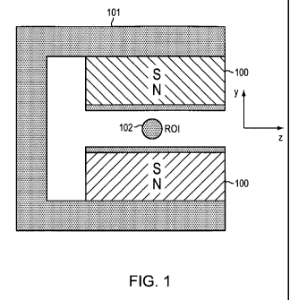
FIG. I provides a schematic representation of a probehead including a c-
shaped yoke with magnets attached thereto and a radiofrequency coil placed
between the magnets.
FIG. 2 shows a probehead employing two NdFeB permanent magnets and a
ten-turn radiofrequency coil from two sides.
FIG. 3 shows the "T2-yoke" made from a steel yoke and 1 "x 1"x 0.5" NdFeB
magnets.
FIG. 4 shows a Halbach magnet positioned on a field mapping apparatus with a
gaussmeter probe positioned within the center gap.
FIG. 5 provides a schematic representation (side view and front view) of a
probehead including a c-shaped yoke with magnets attached thereto and a
radiofrequency coil placed between the magnets.
FIG. 6 shows the measured dependency of the magnetic field strength along
three
directions (along the gap, across the gap, and from bottom to top) within the
gap
between the magnets of the probehead shown in FIG. 2.
FIG. 7 provides a shaded field map within the center gap of a theoretical
model
magnet.
FIG. 8 provides a relaxation decay curve of a nanoparticle assay measured
using the
magnet and probehead in FIG. 2. A solution of magnetic relaxation switch
nanoparticles sensitized to the protein R-hCG was used to detect a
concentration of
65 nM (or 1 microgram/mL) hCG in 0.4 microliters of pH 7.4 PBS buffer.
FIG. 9 shows the measured dependency of the magnetic field strength along
three
directions (along the gap, across the gap, and from bottom to top) within the
gap
between the magnets or within the center gap of the T2-yoke magnet (shown in
FIG.
3) and the Halbach magnet (shown in Figure 4) respectively. Examples of
determining the region that can be excited for each dimension are shown with
the
dashed-line boxes.
CA 02704674 2010-05-04
WO 2009/061481 PCT/US2008/012592
-5-
FIG. 10 shows the radiofrequency coil resonant circuit for the Halbach magnet.
FIG. 11 shows the radiofrequency coil resonant circuit for the T2-yoke.
DETAILED DESCRIPTION OF CERTAIN EMBODIMENTS OF THE
INVENTION
Probeheads of the present invention include a magnet and/or magnetic field
generator and at least one radiofrequency coil, and are much smaller and much
less
expensive than conventional combinations of magnet(s) and radiofrequency
coil(s).
Weight and size of a probehead are critical factors for portable MR
instruments. For example, weight and size reduction has implications in
regards to
system development and manufacturing, cost, and placement. Small probes may
be,
e.g., implantable in-vivo devices, embedded sensors for material testing, and
sensors
for on-line process monitoring. Additionally, because they are inexpensive
provided
small probeheads may be used in applications that benefit from disposable
probeheads.
One aspect of the present invention is the scalability of a Magnetic
Resonance (MR) probehead comprising a magnet and a radio-frequency (RF) coil.
In particular, the present invention addresses the issue of significantly
reducing size
of probehead components while allowing measurement of magnetic resonance
signal
level(s), and, in particular, magnetic resonance relaxation parameter(s) and
time(s).
Designing a probehead specifically for relaxometry instead of conventional MR
spectroscopy, allows for a dramatic reduction in its size and cost.
Magnet configuration and yoke design, if desired, can be accomplished
initially by a theoretical prediction of what magnet and yoke configuration
will lead
to in terms of magnetic field strength. Suitable magnetic field strength will
be
discussed below. This can be done using standard analytical methods known in
the
art.
In one embodiment at least one magnet or magnetic field generator is shaped
and/or configured to provide the magnetic field in a gap. In certain
embodiments, a
radiofrequency coil is positioned, either partly or completely within the gap
of such
a configuration.
CA 02704674 2010-05-04
WO 2009/061481 PCT/US2008/012592
-6-
For a given magnet configuration, for example, two opposing permanent
magnets as presented schematically in Figure 1 and shown in Figures 2 and 3,
and a
Halbach magnet as shown in Figure 4, the magnetic field in the x, y, and z
directions
can be determined using standard methods known in the art, for example, by
fixing a
gaussmeter probe relative to the magnet and moving the magnet in incremental
steps
with a three axis stage while recording the field strength as a function of
position to
obtain a field map.
Given knowledge of the magnetic field, for example, in terms of a calculated
or measured magnetic field map and a pulse length of a radiofrequency pulse to
be
used in relaxation measurements, a radiofrequency coil or radiofrequency coil
array
can be designed and concurrently a proper position for the same be determined.
Pulse length and excitation bandwidth are inversely related. For example, a 2
s
pulse corresponds to a 500 kHz excitation bandwidth (see below for more
details).
The excitation bandwidth can be used to calculate: 1) for a given sample
volume, the
necessary magnetic field homogeneity to be able to excite part of or an entire
sample
volume, and/or 2) for a given magnet or magnet array, the volume that is
excitable
with a radiofrequency pulse of a given pulse length in the presence of the
magnetic
field of the given magnet or magnet array.
Typically, a given excitation bandwidth dictates a requisite magnetic field
homogeneity. Once a magnet is designed to create limited homogeneity of a
volume
that is suitable or desirable for a sample (e.g., which may be dictated by
fluidics or
specimen size of a sample), a coil is designed to excite a complete volume of
excitable spins of a sample volume. Thus, according to the present invention,
an
excitation bandwidth appropriate for a magnet configuration guides the magnet
and
coil design as well as the probehead configuration design.
A probehead of the present invention includes (a) at least one magnet or
magnetic field generator providing a magnetic field; (b) a space capable of
accommodating a sample volume having an associated excitable volume; and (c) a
radiofrequency coil having an associated detection volume, the radiofrequency
coil
being adapted and positioned such that its detection volume overlaps at least
partly
with the excitable volume. The magnetic field provided by the magnet or
magnetic
field generator is inhomogenous. The space accommodating the sample volume and
CA 02704674 2010-05-04
WO 2009/061481 PCT/US2008/012592
-7-
the radiofrequency coil are adapted and positioned according to a
radiofrequency
pulse bandwidth optimized for a magnetic field distribution corresponding to
the
position of the sample volume. The probehead is thus optimized to obtain
relaxometry parameters from a sample contained in the detection volume.
An "excitable volume" as used herein is a volume of hydrogen nuclei of
water within a sample volume which are transitioned to a higher energy state
by a
radiofrequency pulse of a given pulse length in the presence of a magnetic
field
provided by a magnet and/or magnet field generator.
All atomic nuclei with an odd atomic mass or an odd atomic number (like
hydrogen nuclei of water for example) possess an intrinsic nuclear magnetic
momentum. When such atomic nuclei are placed in a static magnetic field, this
momentum can take at least two different orientations. For spin ''/z nuclei,
such as 'H
the momentum may take either a parallel or anti-parallel orientation relative
to the
magnetic field. Considering a population of hydrogen nuclei immersed in the
same
static magnetic field, the number of nuclei having a parallel orientation is
slightly
greater than the number of nuclei having an anti parallel orientation (a ratio
of
1,000,003: 1,000,000 at fields of 0.5 T and room temperature). This is due the
fact
that the parallel orientation is only slightly more energetically favorable.
Transitions
from a parallel state to an anti-parallel state occur when nuclei absorb
electromagnetic energy at a given frequency called a resonance frequency,
which is
dictated by the strength of the magnetic field. Typically, hydrogen nuclei in
different locations in a magnetic field experience different magnetic field
strengths
and therefore have different resonance frequencies required for excitement.
Therefore, in prior systems, a range of frequencies were necessary to
sufficiently
excite a significant portion of hydrogen nuclei in a sample and generate
effective
relaxation readings. A given pulse length produces a corresponding excitation
bandwidth that, at a given magnetic field, excites a volume of hydrogen nuclei
with
a radiofrequency pulse. The resulting signal after excitation can be detected
via
typical methods known in the art.
In one embodiment, a RF coil included in a probehead of the present
invention is adapted to provide pulse lengths between about 0.4 s and about
10 s.
Typically, a pulse length of between about 0.5 s and about 4 s is used. More
CA 02704674 2010-05-04
WO 2009/061481 PCT/US2008/012592
-8-
typically, a pulse length of between about I s and about 4 s is used. Even
more
typically, a pulse length of between about 1 s and about 3 s is used.
A "probehead" as used herein is a sensing or probing device of a nuclear
magnetic resonance system. A probehead may be implanted, partially or
completely, in a mammal's body. Typically, a probehead of the present
invention
includes (a) at least one magnet and/or magnetic field generator providing a
magnetic field, and (b) a radiofrequency coil having an associated detection
volume, and the radiofrequency coil being positioned such that its detection
volume
overlaps at least partly with an excitable volume..
In one embodiment, a probehead comprises a space capable of
accommodating a sample volume and/or a port. In certain embodiments, a space
capable of accommodating a sample volume and port can be, for example, a
radiofrequency coil (as part of a radiofrequency circuit) wound to enclose a
sample
volume while providing an opening (i.e., space capable of accommodating a
sample
volume) to allow a sample volume to be placed within the opening. In other
embodiments, a space capable of accommodating a sample volume and/or port is
distinct from the opening of a radiofrequency coil but adapted to a given
radiofrequency coil, for example, formed to enclose part or all of a detection
volume
of the radiofrequency coil. For example, a glass capillary within a
radiofrequency
coil.
In some embodiments a radiofrequency coil is wound to enclose a volume of
less than about 500 l. In certain embodiment a radiofrequency coil is wound
to
enclose a volumes of less than about 100 l. In still other embodiments a
radiofrequency coil is wound to enclose a volume of less than about 10 gl are
used.
In still further embodiment a radiofrequency coil is wound to enclose a volume
of
less than about 5 l. In particular embodiments a radiofrequency coil is wound
to
enclose a volume of less than about 1.6 l. In still further particular
embodiments a
radiofrequency coil is wound to enclose a volume of less than about 0.4 l.
Also, for implantable probeheads, typically, material used to form a sample
volume, and, in particular, any material that may be in contact with a
biological
sample or tissue is typically biocompatible, that is constructed of materials
that
allow for proper function of both the device and a host animal's biological
functions
CA 02704674 2010-05-04
WO 2009/061481 PCT/US2008/012592
-9-
and/or coated with a physiologically acceptable coating as known in the art to
render
the implantable bioinert, biomimetic, or bioactive, as desired. Suitable
materials
include titanium, inert silicone elastomers, ceramics, glass, polymeric
materials,
poly-(3-hydroxybutyrate (PHB) and the like. One or more sample volumes and
corresponding ports can be fabricated using methods known in the art. Suitable
methods include form or injection molding methods, and microfabrication
methods
for sample containers smaller than a few millimeter, for example, two-photon
three-
dimensional lithography. A probehead may contain a "housing" that encloses the
components of the probehead such as, for example, a radiofrequency coil and
magnet. In certain embodiments at least one component of a probehead (e.g., a
magnet, a magnetic field generator, a radiofrequency coil) is attached to the
housing.
A "port" as used herein, refers in the simplest case to an opening as provided
above, but can also be a structure or device that is adapted to selectively
allow
analytes or reagents to enter and/or exit the sample volume.
In certain embodiments, a probehead includes one or more separate sample
volumes. In some embodiments a probehead includes between about 1 and about
100 sample volumes. In some embodiments a probehead includes between about 1
and about 10 sample volumes. In some embodiments a probehead includes two
sample volumes. In certain embodiments a probehead includes one sample volume.
A probehead containing more than one sample volume may comprise a
radiofrequency coil with an associated detection volume encompassing at least
part
of each sample volume. Alternatively, a probehead may have more than one
radiofrequency coil and/or radiofrequency circuit, one for each sample volume
or a
subgroup of the sample volumes. In certain embodiments, a probehead comprises
at
least two radiofrequency coils. Also, a probehead of the systems of the
present
invention can include a magnet or magnetic, field generator as discussed
above.
For probeheads that include a plurality of separate sample volumes but only
one radiofrequency coil that is employed to probe the plurality of sample
volumes
simulatenously, multiplexing methods may be used to distinguish the magnetic
resonance signal or information from the separate sample chambers. For
example,
one multiplexing method that may be used is based on extracting decay constant
CA 02704674 2010-05-04
WO 2009/061481 PCT/US2008/012592
- 10-
values, for example, values of spin-spin relaxation constant T2 from multi-
exponential relaxation curves (see T.J. Lowery et al., Anal. Chem. (2008), 80,
1118-
1123.). Relaxation data obtained using a probehead of the present invention
may be
fit to a decaying exponential curve defined by the following equation:
f(t)=SE A.exp( ~(.)J
wheref(t) is the signal intensity as a function of time, t, A; is the
amplitude
coefficient for the ith component, and (T); the decay constant (such as T2)
for the ith
component. For relaxation phenomenon discussed here the detected signal is the
sum of a discrete number of components (i=1,2,3,4...n). Such functions are
called
mono-, bi-, tri-, tetra- or multi-exponential, respectively. Due to the
widespread
need for analyzing multi-exponential processes in science and engineering,
there are
several established mathematical methods for rapidly obtaining estimates of A,
and
(T); for each coefficient (Istratov, A. A. & Vyvenko, 0. F. 1999. Exponential
analysis in physical phenomena. Rev. Sci. Inst. 70 (2): 1233-1257).
A "magnet" as used herein can be any material or combination of materials
that provides a magnetic field in at least some volume around the material.
Typically, the magnet is a permanent magnet. Suitable, materials include but
are not
limited to NdFeB, FeCo, and the like. Magnets can be configured to form new
magnets, that is, magnet arrays, for example, a permanent magnet with a c-
shaped
yoke, a Halbach magnet (cylinder and other configurations), u-magnet,
torroidal
magnet and the like.
The magnets, magnet configurations and magnetic field generators of the
present systems can be weak and/or provide magnetic fields that are
inhomogeneous. Typically, maximum magnetic field strength values provided by
the magnets and/or magnet configurations of the present invention are between
about 0.2 Tesla and about 2 Tesla. More typically, they are between about 0.3
and
about 1.5 Tesla. Even more typically, they are between about 0.4 and about 1.1
Tesla. Even more typically, they are between about 0.2 and about 1.1 Tesla.
Even
more typically, they are between about 0.2 and about 0.8 Tesla. Most
typically, they
are between about 0.45 and 0.85 Tesla. In some embodiments the magnetic field
strength is less than about 2 Tesla. In certain embodiments the magnetic field
CA 02704674 2010-05-04
WO 2009/061481 PCT/US2008/012592
-11-
strength is less than about 1.1 Tesla. In certain embodiments the magnetic
field
strength is less than about 0.8 Tesla.
The term "inhomogeneous" refers to magnetic fields that are lower in
uniformity than those required for spectroscopy. Homogeneity is dependent on
the
space in which the measurement is defined. For the instant applications,
homogeneities of the magnetic fields can range between about 10000 ppm and
about
ppm. in some embodiments homogeneities can range between about 50 ppm and
5000 ppm. In particular embodiments homogeneities can range betweenabout 100
ppm and about 1000 ppm.
10 Also, typically, magnetic fields employed in the present systems are
effectively static, that is, they do not change substantially over time.
Changes in
magnetic field such as due to temperature fluctuations are considered to be
not
substantial.
Small probeheads of the present invention can be used for, but are not
limited to in-vivo magnetic resonance measurements. Small probeheads for
complete implantation within a mammal's body, preferably have small magnets to
lessen the invasiveness of the implantation. Typically, magnets for
implantation are
smaller than about 2 inches in any dimension. More typically, magnets for
implantation are smaller than about 1 inch in any dimension. Most typically,
magnets for implantation are smaller than about 0.5 inches in any dimension.
The probeheads of the present invention can also be used in-vitro, for
example, as part of small and/or portable magnetic resonance systems.
Typically,
magnets in probeheads for these systems are smaller than about 2 inches in any
dimension. More typically, they are smaller than about 1 inch in any
dimension.
Most typically, they are smaller than about 0.5 inches in any dimension. Each
dimension may be independently determined.
A "magnetic field generator" as used herein, is a device that provides a
magnetic field in at least some volume around the device. Typically, a
magnetic
field generator requires a power supply and provides the targeted magnetic
field
only when powered. Examples of magnetic field generators include but are not
limited electromagnets with and without a metal pole (see Cardot et al Sensors
and
Actuators 1994).
CA 02704674 2010-05-04
WO 2009/061481 PCT/US2008/012592
- 12-
Probeheads using magnetic field generators can be implanted in a mammal's
body. However, because magnetic field generators tend to be larger than
magnets,
and they are more complex, for example, require a power supply, more
typically,
probeheads using magnetic field generators are used for disposition outside a
mammal's body.
The magnet(s) and magnetic field generator(s) in the present systems are
selected and positioned to provide a magnetic field of sufficient strength in
the
sample volume to allow measuring magnetic resonance signals. The magnetic
field
strength of a given magnet or magnetic field generator in a given volume, for
example, a sample volume can be calculated and/or approximated using methods
known in the art. Typically, the magnetic field strength depends on the nature
of the
magnet or magnetic field generator and the position of the magnet or magnetic
field
generator relative to the sample volume. Also, magnetic field strength of a
given
magnet or magnetic field generator in a sample volume can be measured using
methods and devices known in the art, for example, gaussmeters, teslameters,
hall
effect probes, and the like. Typically, magnetic field strengths within a
sample
volume of between about 0.2 and about 2 Tesla are sufficient to allow
measuring
magnetic resonance signals. More typically, magnetic field strengths within
the
sample volume of between about 0.2 and about 1 Tesla are sufficient to allow
measuring magnetic resonance signals. Even more typically, magnetic field
strengths within the sample volume of between about 0.2 and about 0.8 Tesla
are
sufficient to allow measuring magnetic resonance signals. Most typically,
magnetic
field strengths within the sample volume of between about 0.3 and about 0.65
Tesla
are sufficient to allow measuring magnetic resonance signals.
The magnets and magnetic field generators suitable for the probeheads of the
present invention are not limited to any particular size. However, in
particular, for
implantable and handheld probeheads small magnets are desired. Typically, each
of
the at least one magnet or magnetic field generator of the probeheads of the
present
invention is in any dimension less than about one two inches. More typically,
each
of the at least one magnet or magnetic field generator is in any dimension
less than
about 1 inch. Most typically, each of the at least one magnet or magnetic
field
generator is in any dimension less than about 0.5 inch.
CA 02704674 2010-05-04
WO 2009/061481 PCT/US2008/012592
- 13 -
Probeheads of the present invention may be used to sense/measure magnetic
resonance signals as part of a magnetic resonance system with sensing reagents
enclosed within the probehead, and, in particular, within one or more sample
volume.
A "sensing agent" as used herein is an agent that senses, responds to or is
influenced by a sample characteristic to correlate the presence and/or extent
of the
sample characteristic with the presence, change or magnitude of the magnetic
resonance signals associated with the sample. The term "sample characteristic"
as
used herein refers to any chemical and/or physical property of a given sample.
Suitable sample characteristics can be, but are not limited to concentration
of an
analyte (that is, a molecule, ion, or radical of interest in the sample), pH-
value, ionic
strength, hydration state (e.g., of tissue or biofluids, that is,
concentration of water in
tissue or biofluids), temperature, and the like.
Suitable sensing agents can be, but are not limited to dry reagent
compositions, magnetic particles, responsive polymers, magnetic resonance
contrast
agents, and the like.
Dried reagent compositions that are suitable include, for example, dried
biotinylated coated nanoparticles (see T.J. Lowery et al., Anal. Chem. (2008),
80,
1118-1123), for example, based on the following formulation (216 L, 0.083 mM
Fe, 10 mM PBS, 20 mg/ml dextran, pH 7.4). Dried reagent compositions can be
prepared by placing a magnetic particle solution, for example, biotinylated
coated
nanoparticle solution into a container, for example, a container such as a
glass tube,
and freezing the container in a freeze dryer (e.g., VirTis freeze dryer
(Gardiner,
NY)), for example, at -80 C for 24h. Each of the one or more separate volumes
of
the sample containers may be filled by transfer of the dried reagent
composition
from the container that was used during freeze drying.
"Magnetic particles" as used herein, are particles that respond to or are
influenced by a sample characteristic to correlate the presence and/or extent
of the
sample characteristic with the presence, change or magnitude of the magnetic
resonance signals associated with the sample. Typically, the magnetic
particles
respond by aggregating. Also, typically, magnetic particles have an average
particle
size of between about 1 nm and 5 m. Magnetic particles may be paramagnetic or
CA 02704674 2010-05-04
WO 2009/061481 PCT/US2008/012592
-14-
superparamagnetic. They can have binding moieties on their surface. The
binding
moieties are preferably operative to alter the aggregation of the magnetic
particles as
a function of the presence or concentration of the analyte. The magnetic
particles
may include an oxide and/or a hydroxide of Fe, Si, Sn, An, Ti, Bi, Zr, and/or
Zn.
The magnetic particles are preferably superparamagnetic and have crystallite
size
from about I nm to about 100 nm. The magnetic nanoparticles preferably have a
metal oxide core of about I to about 25 nm, from about 3 to about 10 rim, or
about 5
nm in diameter. The binding moieties may include one or more species of one or
more of the following: an amino acid, a nucleic acid, an oligonucleotide, a
therapeutic agent, a metabolite of a therapeutic agent, a peptide, a
polypeptide, a
protein, a carbohydrate, a polysaccharide, a virus, and/or bacteria. For
example, in
one embodiment, the binding moieties may include one, two, or more types of
oligonucleotides and/or one, two, or more types of proteins. The binding
moieties
may be a polymer, or may be part of a polymer that is linked to, or otherwise
associated with one or more of the magnetic particles. The binding moieties
preferably include functional groups, for example, the binding moieties may
include
one or more species of one or more of the following: an amino group, a
carboxyl
group, a sulfhydryl group, an amine group, an imine group, an epoxy group, a
hydroxyl group, a thiol group, an acrylate group, and/or an isocyano group.
The analyte may include one or more species of one or more of the
following: a protein, a peptide, a polypeptide, an amino acid, a nucleic acid,
an
oligonucleotide, a therapeutic agent, a metabolite of a therapeutic agent,
RNA,
DNA, an antibody, an organism, a virus, bacteria, a carbohydrate, a
polysaccharide,
and glucose. The analyte may also include, for example, a lipid, a gas (e.g.,
oxygen,
carbon dioxide), an electrolyte (e.g., sodium, potassium, chloride,
bicarbonate,
BUN, creatinine, glucose, magnesium, phosphate, calcium, ammonia, lactate), a
lipoprotein, cholesterol, a fatty acid, a glycoprotein, a proteoglycan, and/or
a
lipopolysaccharide.
For example, magnetic particles can be adapted to respond to glycated
hemoglobin. For example, amino-CLIO nanoparticles, that is, iron oxide
nanoparticles coated with amino-functionalized cross-linked dextran, may be
decorated with boronate compounds by standard solution-phase chemistries. The
CA 02704674 2010-05-04
WO 2009/061481 PCT/US2008/012592
- 15-
boronate compounds such as boronic acid, phenylboronic, boric acid and
boronate,
etc. have an affinity for HbAlc, a specific type glycated hemoglobin
designated
based on its separation from other species of glycated hemoglobin.. Hemoglobin
is
composed of four subunits, two a chains and two 0 chains therefore HbA 1 c is
divalent. The divalency allows HbAlc to facilitate the boronic acid
functionalized
superparamagnetic iron oxide partcle agglomeration. Boronate reacts with HbAlc
in a sample through the cis-diol moiety of glucose bound to hemoglobin,
forming a
five-membered ring structure. A boronate group can be attached to a solid
phase
covalently or electrostactically by a variety of chemistries. Solid phases
such as
amino-CLIO nanoparticles can be decorated with boronate compounds by standard
solution-phase chemistries. Amino-CLIO are iron oxide nanoparticles coated
with
amino-functionalized cross-linked dextran. The dextran polymer coating endows
these nanoparticles with solubility and enabled solution-phase chemistries.
Suitable
boronate compounds include but are not limited to 4-carboxyphenylboronic acid,
3-
nitro-5-carboxyphenylboronic acid, and m-aminophenylboronic acid (APBA).
"Nanosensors" are paramagnetic or superparamagnetic magnetic particles,
typically of nanometer scale, that comprise a polymer matrix layer about a
magnetic
core and/or are derivatized/functionalized with binding moieties or affinity
groups
for a target compound or analyte. Suitable nanosensors include responsive
polymer-
coated magnetic nanoparticles. These nanosensors can exploit the ability of
magnetic nanoparticles to dephase nuclear spins detectable by nuclear magnetic
resonance (NMR), hereinafter generally exemplified as the protons of water
molecules, for detection without aggregation of nanoparticles. Each
nanoparticle
has a polymer matrix layer which expands or contracts when exposed to an
analyte
and/or condition to be detected. The resulting change in nanoparticle size
affects the
dephasing of freely-diffusing water molecules in the vicinity of the
nanoparticles,
which affects one or more NMR-detectable properties. By calibrating the NMR-
detected properties with known reference samples, the existence of the
condition
and/or analyte of interest may be detected in test samples via NMR techniques
using
the probeheads of the present invention.
In the case where the detected nuclei are water protons, the polymer matrix
preferably takes the form of a stimuli or molecule sensitive hydrogel
comprising a
CA 02704674 2010-05-04
WO 2009/061481 PCT/US2008/012592
-16-
polymer "mesh" that is cross-linked by binding moieties that affects the
volume,
permeability and the proton content of the matrix as a function of a physical
or
chemical stimulus or a physical parameter of the analyte under study. This is
accomplished by design of the matrix as a hydrophilic polymer network
comprising
(as pendent groups or as part of the polymer backbone) binding moieties that
influence water permeability (and/or permeability of other molecules in the
environment) through formation of one or more covalent or hydrogen bonds, van
der
Waals interactions, or physical entanglement with a component of the analyte.
The
presence of analyte induces a change in the crosslink density of the polymer,
which
leads to a change in the volume fraction of the solution occupied by the
polymer.
The change in cross link density also leads to a change in the diameter of the
nanoparticles, which leads to a change in their diffusion time. Both diffusion
time
and specific volume are proportional to the T2 relaxivity observed for a
solution, as
shown in the proportionality:
1/T2 a (Vp)(R2/D)
where VP is the specific volume fraction of the particles in solution, R the
radius of
the particles, and D the diffusion constant of water. The term R2/D is equal
to the
diffusion time, 'rd. This is the time necessary for a water molecule to
diffuse past a
particle, and is proportional to the extent of T2 relaxation that occurs.
The binding moiety may be a chemical binder, an electroactive mediator, an
electron-pair donor, and/or an electron-pair acceptor. It may contain an
amino,
carboxyl, sulfhydryl, amine, imine, epoxy, hydroxyl, thiol, acrylate, or
isocyano
group, or a mixture thereof. For example, the binding moiety may be an acetic
acid
moiety such as in poly(acrylic acid) for sensing pH, or phenylboronic acid for
sensing the presence of diols, such as glucose Alternatively, the binding
moieties
are binding pairs, or binding pendants, such as antibodies that serve as cross-
linkers
in the presence of their cognate antigen, or antigens that serve as cross-
linkers in the
presence of their cognate antibodies, and which mediate the water proton flux
in and
out of the matrix and change in specific volume by competitive affinity
reactions.
This typically is accomplished as the extent of cross-linking of matrix
polymer is
mediated as a function of the physical parameter under study so as to control
the
permeability of water, including its amount and rate of translational
diffusion in an
CA 02704674 2010-05-04
WO 2009/061481 PCT/US2008/012592
-17-
out of the matrix and within the matrix volume in proximity to the magnetic
particle(s). For example, the binding pairs may be a ligand binding protein
such as
concanavalin A bound to a low-affinity ligand such as a carbohydrate. Addition
of
glucose to this system would displace the low affinity ligand and change the
crosslinking of the matrix. Another example is a matrix-immobilized antibody,
antibody fragment, or peptide that crosslinks the matrix by binding to its
matrix-
immobilized antigen or target. The presence of a higher affinity analyte would
lead
to disruption of the cross-linked matrix and a swelling of the matrix.
The responsive matrix may comprise a matrix of material which includes one
or more monomers and/or polymers. The one or more monomers and/or polymers
contains functional groups that enable the binding moiety to be attached to or
otherwise in stable association with the nanoparticle to form the conjugate.
The
polymer can be a natural polymer, a synthetic polymer, a combination of
natural and
synthetic polymers, shape memory polymers, block co-polymers (PEO, PPO), or
derivatives of each type. For example, the matrix polymer may be poly (N-
isopropylacrylamide). The matrix polymer may also be (or include), for
example,
Poly(N-isopropylacrylamide) (PNIAAm), Poly(N,N-diethyacrylamide) (PDEAAm),
P(NIAAm-co-BMA), PEO-PPO-PEO (e.g., Pluronic ), N,N-diethylaminoethyl
methacrylate (DEA), 2-hydroxypropyl methacrylate (HPMA), Poly-(methacrylic
acid-g-ethylen glycol), Poly(2-glucosyloxyethyl methacrylate), Poly(N-vinyl-
2pyrrolidone - co - 3-(acrylamido)phenylboronic acid), and/or N-(S)-sec-
butylacrylamide. The functional groups can be any appropriate chemical
functional
group, e.g. carboxy, amino, or sulfhydryl groups. A specific moiety or
moieties may
be attached to the nanoparticle via conjugation to these groups, or by
physical
adsorption and/or through hydrogen bonds or van der Waals interactions. The
responsive polymer matrix, through physical and/or chemical stimuli, mediates
the
specific volume of the polymer layer, leading to a detectable change in NMR-
measurable properties such as T2 relaxivity.
"Responsive polymers" (also referred to herein as "smart polymers") are
polymers that are, for example, sensitive to pH, ionic strength, and specific
molecular and biomolelar analytes. In these cases the hydration level, cross-
link
density, or other characteristic of the polymer changes in response to a
changes in
CA 02704674 2010-05-04
WO 2009/061481 PCT/US2008/012592
- 18-
the sample, for example, biofluid. This change in polymer state leads to
changes in
the magnetic resonance signals that can be detected by an implanted
radiofrequency
coil. Suitable smart polymers are known in the art, and described, for
example, in
Gemeinhart, RA, Chen, J, Park, H, Park, K. 2000. pH-sensitivity of fast
responsive
superporous hydrogels. J. Biomater. Sci. Polym. Ed. 11: 1371-1380; Murakami,
Y,
Maeda, M. 2005. DNA-responsive hydrogels that can shrink or swell.
Biomacromolecules, 6: 2927-2929; Miyata, T, Uragami, T, Nakamae, K. 2002.
Biomolecule-sensitive hydrogels. Adv Drug Deliv Rev, 54: 79-98; and Zhang, R,
Bowyer, A, Eisenthal, R, Hubble, J. 2006. A smart membrane based on an antigen-
responsive hydrogel. Biotechnol Bioeng.
Probeheads of the present invention include a radiofrequency coil.
A "radiofrequency coil" as used herein is a is a coil that is suited to sense
and/or detect magnetic resonance signals in an associated detection volume,
and,
optionally, also allows to apply/emit radiofrequency pulses with associated
pulse
length(s) to a sample under investigation with the probehead as part of a
magnetic
resonance system. Suitable radiofrequency coil types include planar coils and
"whole volume" coils such as might be constructed of opposed saddle coils,
solenoids, Helmholtz coils and the like. Typically, the probeheads employed in
the
systems of the present invention include solenoids.
"Detection volume" as used herein refers to a volume associated with a given
radiofrequency coil from which magnetic resonance signals, in principle, are
detectable with the given radiofrequency coil as part of a given magnetic
resonance
system. "Detectable" as used herein refers to distinguishable from the
background
noise level, that is, a magnetic resonance signal is detectable if a signal
can be
distinguished from background noise level with a given radiofrequency coil as
part
of a given magnetic resonance system. The detection volume for a given
radiofrequency coil-magnetic resonance system combination can be calculated,
approximated and/or measured using methods known in the art. Typically,
however, it is sufficient to approximate the detection volume. For example,
for a
solenoid coil, typically, the detection volume is effectively, the volume
enclosed
within the coil, which, typically, is of about cylindrical shape. In certain
embodiments a radiofrequency coil is a cylinder shape. Thus, for a solenoid a
good
CA 02704674 2010-05-04
WO 2009/061481 PCT/US2008/012592
-19-
approximation of the detection volume is the volume of the enclosed cylinder,
which
can be calculated very easily. Similar approximations are known in the art for
other
types of radiofrequency coils (see, e.g., Mispelter, J., Lupu, M., Briquet, A.
"NMR
Probeheads for biophysical and biomedical experiments" 2006 Imperial College
Press, London.). In certain embodiments a radiofrequency coil is wound to
enclose
a coil volume having a shape of about cylindrical shape and the associated
detection
volume is effectively the volume of the cylindrical shape. In some embodiments
a
radiofrequency coil is positioned to have the coil volume include between
about 80
percent and about 100% of the excitable volume. In stillother embodiments a
radiofrequency coil is positioned to have the coil volume include effectively
all of
the excitable volume.
"Sensitive volume" as used herein refers to the overlap volume between the
excitable volume and the detection volume, and is the volume from which
magnetic
resonance signals can be detected with the radiofrequency coil. A sensitive
volume
is determined by a fill factor (i.e., a fraction of the detection volume of an
RF coil
which is filled with a sample volume).
In some embodiments, a fill factor is between about 10 percent and about
100 percent. In certain embodiments a fill factor is between about 50 percent
and
about 100 percent. In some embodiments the fill factor is about 80 percent. In
certain embodiments the fill factor is effectively 100 percent. In some
embodiments
a fill factor is at least about 0.1, at least about 0.5, at least about 0.75,
at least about
0.9, and or about 1.
Typically, a detection volume includes between about 10 percent and about
100 percent of the excitable volume. More typically, a detection volume of a
given
radiofrequency coil within the probehead includes between about 50 percent and
about 100 percent of the excitable volume. Even more typically, a detection
volume
includes about 80 percent of the excitable volume. Most typically, a detection
volume includes effectively all of the excitable volume.
Also, typically, an excitable volume includes between about 10 percent and
about 100 percent of the detection volume. More typically, the excitable
volume
includes between about 50 percent and about 100 percent of the detection
volume.
Even more typically, the excitable volume includes between about 80 percent
and
CA 02704674 2010-05-04
WO 2009/061481 PCT/US2008/012592
-20-
about 100 percent of the detection volume. Most typically, the excitable
volume
includes effectively all of the detection volume.
Further, for a given sample volume within the probehead, typically, the
sample volume includes between about 10 percent and about 100 percent of the
excitable volume. More typically, the sample volume includes between about 50
percent and about 100 percent of the excitable volume. Even more typically,
the
sample volume includes between about 80 percent and about 100 percent of the
excitable volume. Even more typically, the sample volume includes effectively
all of
the excitable volume. Most typically, the sample volume includes effectively
all of
the excitable volume and the detection volume includes effectively all of the
sample
volume.
In some embodiments a sample volume includes effectively all of the
excitable volume and a detection volume includes effectively all of the sample
volume. In still further embodiments a sample volume includes between about 10
and about 100 percent of the sensitive volume.
Typically, for a magnetic field of between about 0.2 Tesla and 1.1 Tesla,
radiofrequency coils with associated detection volumes of less than about 500
l are
used. More typically, radiofrequency coils with associated detection volumes
of less
than about 100 l are used. Even more typically, radiofrequency coils with
associated detection volumes of less than about 10 l are used. Even more
typically,
radiofrequency coils with associated detection volumes of less than about 5 l
are
used. Most typically, radiofrequency coils with associated detection volumes
of less
than about 1.6 l are used. In some embodiments a radiofrequency coil with
associated detection volume of about 1.6 l is used, and a sample volume of
about
0.4 l is used.
A radiofrequency coil of a given probehead of the present invention senses
and/or detects magnetic resonance signals of a sample in the presence of a
magnetic
field and provides the sensed signals to a processing unit. The processing
unit can
be included within a probehead; but does not have to be included in a
probehead. In
any case, a probehead contains any parts, for example, circuitry, logic
circuitry,
power sources and other parts such as capacitors and the like, as known in the
art, to
allow the sensed signals to be provided to the processing unit. For example, a
CA 02704674 2010-05-04
WO 2009/061481 PCT/US2008/012592
-21-
probehead of the present invention that is to be used in a magnetic resonance
system
with a radiofrequency coil of the probehead being inductively coupled to the
processing unit via an external pickup coil, typically, includes the
radiofrequency
coil as part of a radiofrequency circuit with one or more tuning capacitors
included
in the circuit. In one embodiment a probehead further comprises at least one
capacitor, wherein a radiofrequency coil and at least one capacitor are part
of a
radiofrequency circuit.
One embodiment of the present invention is a probehead for magnetic
resonance relaxometry that includes (a) at least one magnet providing a
magnetic
field, and (b) a radiofrequency coil having an associated detection volume,
the
radiofrequency coil being adapted and positioned such that its detection
volume
includes between about 80 percent and about 100 percent of an excitable
volume,
wherein the magnetic field has a magnetic field strengths of less than about
1.1
Tesla.
Another embodiment of the present invention is a probehead for magnetic
resonance relaxometry that includes (a) at least one permanent magnet
providing a
magnetic field, and (b) a radiofrequency coil having an associated detection
volume,
the radiofrequency coil being adapted and positioned such that its detection
volume
includes effectively all of an excitable volume, wherein the magnetic field
has
magnetic field strengths of less than about 1.1 Tesla.
Another embodiment of the present invention is a probehead for magnetic
resonance relaxometry that includes (a) at least one permanent magnet
providing a
magnetic field, and (b) a radiofrequency coil having an associated detection
volume,
the radiofrequency coil being adapted and positioned such that its detection
volume
includes effectively all of an excitable volume, wherein the magnetic field
having
CA 02704674 2010-05-04
WO 2009/061481 PCT/US2008/012592
-22-
magnetic field strengths of less than about 1.1 Tesla, the probehead further
comprising a sample volume and the sample volume including between about 10
and about 100 percent of the excitable volume.
Another embodiment of the present invention is a probehead for magnetic
resonance relaxometry that includes (a) at least one permanent magnet
providing a
magnetic field, and (b) a radiofrequency coil having an associated detection
volume,
the radiofrequency coil being adapted and positioned such that its detection
volume
includes effectively all of an excitable volume, wherein the magnetic field
having
magnetic field strengths of less than about 1.1 Tesla, the probehead further
comprising a sample volume, and the sample volume including between about 50
and about 100 percent of the excitable volume.
Another embodiment of the present invention is a probehead for magnetic
resonance relaxometry that includes (a) at least one permanent magnet
providing a
magnetic field, and (b) a radiofrequency coil having an associated detection
volume,
the radiofrequency coil being adapted and positioned such that its detection
volume
overlaps at least partly with an excitable volume, wherein the magnetic field
having
magnetic field strengths of less than about 1.1 Tesla, the probehead further
comprising a sample volume, the sample volume including effectively all of the
excitable volume and the detection volume including effectively all of the
sample
volume.
Another embodiment of the present invention is a probehead for magnetic
resonance relaxometry that includes (a) at least one permanent magnet
providing a
magnetic field, and (b) a radiofrequency coil having an associated detection
volume,
the radiofrequency coil being adapted and positioned such that its detection
volume
overlaps at least partly with an excitable volume, wherein the magnetic field
has a
magnetic field strengths of less than about 1.1 Tesla, and wherein the
probehead
further comprises a sample volume, the excitable volume and the detection
volume
overlapping in a sensitive volume, and wherein the sample volume includes
between
about 10 and 100 percent of the sensitive volume.
Another embodiment of the present invention is a probehead for magnetic
resonance relaxometry that includes (a) at least one permanent magnet
providing a
magnetic field, and (b) a radiofrequency coil having an associated detection
volume,
CA 02704674 2010-05-04
WO 2009/061481 PCT/US2008/012592
-23-
the radiofrequency coil being adapted and positioned such that its detection
volume
overlaps at least partly with an excitable volume, wherein the magnetic field
having
magnetic field strengths of less than about 1.1 Tesla, wherein the probehead
further
comprises a structure defining a sample volume, the excitable volume and the
detection volume overlapping in a sensitive volume, and wherein the sample
volume
includes between about 50 and 100 percent of the sensitive volume.
Another embodiment of the present invention is a probehead for magnetic
resonance relaxometry that includes (a) at least one permanent magnet
providing a
magnetic field, and (b) a radiofrequency coil having an associated detection
volume,
the radiofrequency coil being adapted and positioned such that its detection
volume
overlaps at least partly with an excitable volume, wherein the magnetic field
has a
magnetic field strengths of less than about 1.1 Tesla, the probehead further
comprising a sample volume, the excitable volume and the detection volume
overlapping in a sensitive volume, and wherein the sample volume includes
effectively all of the sensitive volume.
Another embodiment of the present invention is a probehead for magnetic
resonance relaxometry that includes (a) two permanent magnet providing a
magnetic
field, and (b) a radiofrequency coil having an associated detection volume,
the
radiofrequency coil being adapted and positioned such that its detection
volume
overlaps at least partly with an excitable volume, wherein the magnetic field
has
magnetic field strengths of less than about 1.1 Tesla, wherein the
radiofrequency
coil is wound around a sample tube or capillary to enclose, the radiofrequency
coil
and the sample tube or capillary enclosing a sample volume within the sample
tube
or capillary. In certain embodiments, the excitable volume and the detection
volume
are overlapping in a sensitive volume, and the sample volume includes between
about 50 and about 100 percent of the sensitive volume.
Another embodiment of the present invention is a probehead for magnetic
resonance relaxometry that includes (a) two permanent magnet providing a
magnetic
field, and (b) a radiofrequency circuit comprising (1) a radiofrequency coil
and (2) a
capacitor, the radiofrequency coil having an associated detection volume and
the
radiofrequency coil being adapted and positioned such that its detection
volume
overlaps at least partly with an excitable volume, wherein the magnetic field
has
CA 02704674 2010-05-04
WO 2009/061481 PCT/US2008/012592
-24-
magnetic field strengths of less than about 1.1 Tesla, the radiofrequency coil
being
wound around a sample tube or capillary to enclose, the radiofrequency coil
and the
sample tube or capillary enclosing a sample volume within the sample tube or
capillary, the excitable volume and the detection volume overlapping in a
sensitive
volume, and the sample volume includes between about 50 and about 100 percent
of
the sensitive volume.
Another embodiment of the present invention is a probehead for magnetic
resonance relaxometry that includes (a) two permanent magnet attached to a
yoke
providing a magnetic field, and (b) a radiofrequency circuit comprising (1) a
radiofrequency coil and (2) a capacitor, the radiofrequency coil having an
associated
detection volume and the radiofrequency coil being adapted and positioned such
that
its detection volume overlaps at least partly with an excitable volume,
wherein the
magnetic field has magnetic field strengths of less than about 1.1 Tesla, the
radiofrequency coil being wound around a sample tube or capillary to enclose,
the
radiofrequency coil and the sample tube or capillary enclosing a sample volume
within the sample tube or capillary, the excitable volume and the detection
volume
overlapping in a sensitive volume, and the sample volume including between
about
50 and about 100 percent of the sensitive volume.Other specific embodiments of
the
present invention are the probeheads as described in the preceding paragraphs,
wherein a probehead is adapted for a pulse length of between about 0.4
microseconds and about 10 microseconds, between about 1 microsecond and about
4
microseconds, or between about 1.5 microseconds and 2.5 microseconds, and,
independently, each magnet is independently in any dimension less than about
two
inches, less than about 1 inch, or less than about 0.5 inch.Other embodiments
of the
present invention include methods of preparing probeheads provided and
described
in the preceding paragraphs, and the examples which follow.In one embodiment
is a
method for preparing a probehead for use in a magnetic resonance relaxometer.
The
method comprises the steps of (a) providing at least one magnet or magnetic
field
generator providing a magnetic field, (b) providing a radiofrequency coil, (c)
positioning the radiofrequency coil to have its associated detection volume
overlap
at least partly with an excitable volume, (d) positioning a space capable of
accommodating a sample volume having an associated excitable volume; and (e)
CA 02704674 2010-05-04
WO 2009/061481 PCT/US2008/012592
-25-
adapting the space for the sample volume and the radiofrequency coil according
to a
radiofrequency pulse optimized for the magnetic field distribution
corresponding to
the position of the detection volume. The probehead is thus optimized to
obtain
relaxometry parameters from a sample contained in the sample volume.
Another embodiment is a method of preparing a small probehead for use in
portable magnetic resonance relaxometry. The method comprises the steps of (a)
attaching two magnets or two magnetic field generators to a yoke such that the
south
pole surface of one of the magnets or magnetic field generators opposes the
north
pole surface of the other magnet or magnetic field generator to form a gap
between
the magnets or magnetic field generators and to provide a magnetic field in
the gap,
(b) positioning a space capable of accommodating a sample volume having an
associated excitable volume, and (c) positioning a radiofrequency coil within
the
gap, the radiofrequency coil having an associated detection volume and being
adapted to emit a radiofrequency pulse with a pulse length, the radiofrequency
coil
being positioned and designed to have the detection volume at least partly
overlap
with an excitable volume within the gap. The probehead is thus optimized to
obtain
relaxometry parameters from a sample contained in the sample volume.
In a further embodiment a method of preparing a small probehead for use in
a portable magnetic resonance relaxometer further comprises the step of
providing,
calculating, and/or measuring a magnetic field map of the at least one magnet
or
magnetic field generator, and further wherein the step of providing the
radiofrequency coil comprises selecting or manufacturing a radiofrequency coil
dimensioned based on the magnetic field map to optimize its associated
detection
volume be at least as large as the excitable volume.
In another further embodiment a method of preparing a small probehead for
use in a portable magnetic resonance relaxometer further comprises the step of
providing, calculating or measuring a magnetic field map of the at least one
magnet
or magnetic field generator, and further wherein the step of positioning the
radiofrequency coil is based on the magnetic field map to optimize its
associated
detection volume overlap at least partly with the excitable volume.
In yet another further embodiment a method of preparing a small probehead
for use in a portable magnetic resonance relaxometer further comprises the
step of
CA 02704674 2010-05-04
WO 2009/061481 PCT/US2008/012592
-26-
providing, calculating or measuring a magnetic field map of the at least one
magnet
or magnetic field generator, and further wherein providing the radiofrequency
coil
comprises selecting or manufacturing a radiofrequency coil dimensioned based
on
the magnetic field map to have its associated detection volume be at least as
large as
the excitable volume, and positioning the radiofrequency coil is based on the
magnetic field map to have the associated detection volume overlap at least
partly
with the excitable volume.
EXEMPLIFICATION
Example I
The probehead of Figure 2 (also referred herein as "Abe" probehead) was
fabricated from a custom machined c-shaped yoke 208 (0.688" x 0.5" x 0.438",
steel), custom machined electronics enclosure 201 (1" x 0.5" x 0.5",
aluminum), coil
holders 202 (0.19" x 0.19", teflon), magnet positioner (0.438" x 0.5" x
0.063",
Teflon), sample tube 203 (1mm O.D., 0.5 mm I.D., 0.6" long, Teflon), and ten-
turn
RF coil 204 (32 gage enamel coated copper wire, hand-wound, fastened to the
sample tube by lock-tite instant adhesive). The resonant circuit was
constructed by a
10-120 pF variable matching capacitor 206, and a combination of a 10-120 pF
variable capacitor 206 and two fixed capacitors 205, and a bulkhead SMA
connector
207.
Two inexpensive, off-the-shelf permanent magnets made of NdFeB 200 were
attached to the steel yoke 208 as shown schematically in Figures 1 and 5.
(Figures
I and 5 show two magnets 100 attached to a c-shaped yoke 101 and a
radiofrequency coil 102 positioned between the magnets). The size of the
magnets
was 1/4" x 1/8" x 1/2", magnetized along the 1/8" axis. The largest dimension
of
the array, including the supporting yoke was 1/2". It is believed that the
steel yoke
helps driving the magnetic flux between the magnet blocks, following the path
of
high magnetic permeance.
The magnetic field was mapped by measuring along the three axis around the
geometrical center of the gap between the magnet pieces. Figures 6 and 7 show
the
magnetic field distribution.
CA 02704674 2010-05-04
WO 2009/061481 PCT/US2008/012592
-27-
A solenoid radiofrequency coil 204 was designed and positioned based on
the magnetic field map with the goal of maximizing the sensitive volume,
(e.g., the
overlap volume of excitable volume and detection volume to be excited by short
duration RF pulses). This was achieved by winding the radiofrequency coil to
enclose a volume determined by the bandwidth of the RF pulses, provided in
Figure
6 as box shaped area 600, that is, approximately 1 mm diameter by about 2 mm
length. The radiofrequency coil was fabricated using these dimensions. In this
manner, a coil taking up about 20% of the gap between the magnets with a
microliter sensitive volume was achieved. It is contemplated that this concept
can
be further exploited by, e.g., using more than one block at each side of the
magnet.
This allows for higher control on the field distribution and therefore a
further size
reduction keeping the same sensitive volume.
The Abe probehead was attached to a KEA spectrometer (not shown;
Magritek, Wellington, New Zeeland) outfitted with a Tomco pulse amplifier and
controlled by Prospa softare (Magritek, Wellington, New Zeeland). The resonant
circuit was tuned at the so-called Larmor frequency by using the "wobble"
macro
provided by the Prospa software, that is, using standard procedures known in
the art
and signal was acquired utilizing a conventional CPMG pulse sequence as
controlled
by the "cpmgadd" and "cpmgint" macros of the Prospa software, that is, by
using
standard procedures known in the art. Figure 8 shows the signal decay
utilizing a
conventional CPMG pulse sequence.
The sample was loaded by means of a syringe outfitted with a fused silica
glass capillary. A series of CuSO4 samples were analyzed as well as
nanoparticle
assay solutions. The nanoparticle assay solutions consisted of antibody
functionalized nanoparticles that bind to the beta subunit of the hCG protein
(Kim,
G. Y., Josephson, L., Langer, R., Cima, M. J. "Magnetic Relaxation Switch
Detection of Human Chorionic Gonadotrophin". 2007, Bioconjugate Chemistry
18(6), 2024-2028.). Two solutions were prepared that contained 0.14 mM
nanoparticle iron and 0 and 1 g/mL beta subunit of hCG in PBS pH 7.4 buffer.
The T2 values of these solutions were measured in a volume of 300 L on a
Bruker
minispec and in a volume of 0.4 L on the Abe probehead. Both measurements
showed a decrease in T2 upon addition of hCG. However, the absolute T2 values
on
CA 02704674 2010-05-04
WO 2009/061481 PCT/US2008/012592
-28-
the Abe magnet were lower than those for the Bruker minispec. It is believed
that
that the reason for lower values is that the T2 value measured with the Abe
probehead is an effective T2 value that includes effects from diffusion,
temperature,
and stimulated echoes. However, the most important result is that addition of
hCG
leads to a change in the measured T2 and this information is successfully
provided
using the many orders of magnitude less expensive and smaller Abe probehead.
Example 2
Magnet configuration and yoke design can be accomplished initially by a
theoretical prediction of what magnet and yoke configuration will lead to in
terms of
magnetic field strength. This was done by using standard analytical methods. A
magnet assembly including two NdFeB permanent magnets (1" x 1" x 0.5") 301 was
fabricated according to this method (see "T2-yoke" in Figure 3). The yoke 300
was
fabricated from steel stock using standard machining methods. The magnetic
field in
the x, y, and z directions was determined by fixing a gaussmeter probe
relative to the
magnet and moving the magnet in incremental steps with a three axis stage
while
recording the field strength as a function of position. The strength along the
x, y, and
z axes was measured by fixing two of the three directions to zero while
incrementing
the other. The same process was conducted for a pre-fabricated Halbach magnet
(Figure 4; the figure shows the Halbach magnet 400 while the magnetic field is
being measured with a Gaussmeter probe 401).
Values obtained for a field map of each of the T2-yoke and the Halbach
magnet configurations were plotted as function of position and magnetic field
strength as shown in Figure 9. Data were fitted with a quadratic function (y =
ax 2 +
bx + c). The information of these plots was used to design a radiofrequency
coil by
determining what length in each dimension corresponds to a region that can be
excited by a 2 s RF pulse. For example, a 2 s pulse excites a bandwidth of
500
kHz (bandwidth = (pulselength)-'). For the T2-yoke shown in Figure 3, the
homogeneous region has a field strength of 535 millitesla. The resonant
frequency
of hydrogen nuclei at this field can be calculated from
f = 21r (1)
rB
CA 02704674 2010-05-04
WO 2009/061481 PCT/US2008/012592
-29-
where f is the resonant frequency in Hz, ythe gyromagnetic ratio for 'H nuclei
(267.522 x 10⁶ rad s"' T-'), and B. the magnetic field in Tesla. Accordingly,
the
resonant frequency for the T2-yoke is 22.8 MHz. Equation I can also be used to
determine the range of magnetic field over which a sample can be excited.
Solving
for Bo and substituting 500 kHz for f, a AB, of 11 millitesla is calculated.
This value
was used to determine the length in each dimension for the volume of sample
that
can be excited by a 2 s pulse. Figure 9 shows a graphical representation as
to how
this can be determined for each magnet. A box with a height corresponding to
11
mT is positioned on the plot such that one edge is at the minimum of the curve
fit for
"along the gap" and the other edge is used to determine the appropriate width
of the
box such that the two corners are traversed by the curve fit. The width of
this box
(-5 mm) corresponds to the length in this dimension that an excitation coil
would
enclose to maximize the sensitive volume. A similar box is shown for the
Halbach
magnet for the "bottom to top" dimension. Other analytical methods can be used
to
determine this, but the general idea is taking the OBo and using the field map
to
translate that into a distance for each dimension.
Figures 10 and 11 show radiofrequency circuits that were fabricated for the
Halbach
magnet and the T2-yoke respectively to form a complete probehead. The coils
1000
were custom made from inductors that are commercially available (inductors can
be
hand wound as for the previous example), enclose a sample volume to which
sample
can be delivered through a sample tube 1001, are part of a radiofrequency
circuit
that includes capacitors 1003 and a bulkhead SMA connector 1002, and are
supported by a support plate 1004. Magnetic resonance signal was successfully
measured using these probeheads (data not shown).