Note: Descriptions are shown in the official language in which they were submitted.
CA 02705099 2010-05-05
WO 2009/073672 PCT/US2008/085295
Image analysis
Field
The present invention relates to image analysis. More particularly, the
present
invention relates to an improved image analysis technique for medical
diagnostics, for
example, using positron emission tomography (PET) image data.
Background
Various medical imaging techniques exist to aid clinicians in the diagnosis of
pathological conditions caused, for example, by anatomic or functional
manifestations
of a disease. Many such techniques produce a sequence of image frames that can
be
used to highlight to the clinician various temporal variations in anatomical
and/or
functional properties of a patient.
For example, a magnetic resonance imaging (MRI) scan, a functional magnetic
resonance imaging (fMRI) scan, a computed tomography (CT) scan, or a single
photon emission computed tomography (SPECT) scan can be performed that
provides
a sequence of image frames showing how a patient's anatomy, such as, for
example,
the heart or brain, varies over time. Any such temporal variations might be
detected
during a single scan and/or between multiple scans performed during successive
hospital visits, for example.
As another example, PET imaging can be used to obtain a sequence of image
frames
showing, for example, how the physiological functional properties of a
patient's
organ, such as, for example, the brain, vary over time.
PET is a known imaging technique that uses tomography to computer-generate a
three-dimensional image or map of a functional process in the body as a result
of
detecting gamma rays when artificially introduced radionuclides incorporated
into
biochemical substances decay and release positrons. Analysis of the photons
detected
from the annihilation of these positrons is used to generate the tomographic
image
frames which may be quantified using a colour scale to show the diffusion of
the
biochemical substances in the tissue thereby indicating localization of
metabolic
and/or physiological processes.
1
CA 02705099 2010-05-05
WO 2009/073672 PCT/US2008/085295
For example, radionuclides used in PET may be a short-lived radioactive
isotopes
such as flourine-18, oxygen-15, nitrogen-13, and carbon-11 (with half-lives
ranging
from about 110 minutes to about 20 minutes). The radionuclides may be
incorporated
into biochemical substances such as compounds normally used by the body that
may
include, for example, sugars, water, and/or ammonia. The biochemical
substances
may then be injected or inhaled into the body (e.g. into the blood stream)
where the
substance (e.g. a sugar) becomes concentrated in the tissue of interest, and
where the
radionuclides decay by emitting positrons. These positrons collide with nearby
electrons producing gamma ray photons which can be detected and recorded
thereby
indicating where the radionuclide was taken up by the body. This set of data
may be
used to explore and depict one or more of anatomical, physiological, and
metabolic
information in the human body.
Due to limitations in the amount of radioactivity that can be administered to
the
subject, a generally short half-life of the radionuclide, and limited
sensitivity of
certain recording systems, dynamic PET data is typically characterized by a
rather
high level of noise. A further limiting factor is the metabolic decomposition
of the
molecule of interest, which lowers the signal and increases the noise. This
together
with a high level of non-specific binding to the target and the sometimes
small
differences in target expression between healthy and pathological areas are
factors
which can make the analysis of dynamic PET data difficult, regardless of the
radionuclide used or type of scan conducted. This means that the individual
image
frames are generally not optimal for the analysis and visualization of anatomy
and
pathology by a clinician.
Accordingly, various techniques have been developed to try and improve the
image
quality of the dynamic temporally sequential image frames produced from a PET
scan.
One of the standard methods used for the reduction of the noise and
quantitative
estimation in dynamic PET data is to take the sum, average (or mean) of the
image
frames of the whole or part of the sequence. However, though sum, average/mean
processed images may be effective in reducing noise, this approach results in
the
dampening of the differences detected between regions with different kinetic
2
CA 02705099 2010-05-05
WO 2009/073672 PCT/US2008/085295
behaviour and hence to a reduction in the contrast between non-significant and
potentially significant clinical features.
Another method used for analysis of dynamic PET data uses kinetic modelling,
with
the generation of parametric images, aiming to extract areas with specific
kinetic
properties that can enhance the discrimination between normal and pathological
regions.
One such kinetic modelling method, used for parameter estimation, is known as
the
Patlak method (or sometimes Gjedde method) [1]. The ratio of target region to
reference radioactivity concentration is plotted against a modified time,
obtained as
the time integral of the reference radioactivity concentration up to the
selected time
divided by the radioactivity concentration at this time. In cases where the
tracer
accumulation can be described as irreversible, the Patlak graphical
representation of
tracer kinetics becomes a straight line with a slope proportional to the
accumulation
rate. This method can readily be applied to each pixel separately in a dynamic
imaging frame sequence and allows the generation of parametric images
representative of the accumulation rate.
However, one problem when using kinetic modelling is that the generated
parametric
images suffer from poor quality, while the images are rather noisy. In this
regard,
kinetic modelling methods such as the Patlak method [1] do not optimise the
signal-
to-noise ratio (SNR) during the measurement of physiological parameters from
the
dynamic data.
Alternative methods for the generation of parametric images also exist, based
on other
types of modelling. For example, Logan plots, compartment modelling, or
extraction
of components such as in factor analysis or spectral analysis [2]. Other
alternative
techniques, such as population based approaches where an iterative two stage
(ITS)
method is utilized, have also been proposed [3].
Dynamic PET data can also be analyzed using various different multivariate
statistical
techniques such as principal component analysis (PCA) [4,5,6]. PCA is employed
in
order simultaneously to find variance-covariance structures in data in order
to reduce
the dimensionality of the data set. The results of PCA can be used for
different
3
CA 02705099 2010-05-05
WO 2009/073672 PCT/US2008/085295
purposes, e.g. factor analysis, regression analysis, performing pre-processing
of the
input/raw image frame data, etc.
However, in summary, interpretation of sequential image frames remains
difficult and
there still exists a need for a technique in which clinically significant
features can be
more reliably extracted for highlighting to clinicians.
Summary of the invention
Various aspects and embodiments of the present invention have thus been
devised
whilst bearing in mind the aforementioned problems and disadvantages
associated
with conventional techniques.
According to a first aspect of the present invention, there is provided a
method for
extracting low-intensity features from an image data set comprising data
corresponding to a sequence of original image frames. The method comprises
determining a plurality of principal components (PCs) from the image data set
corresponding to the original image frames, applying a principal component
analysis
(PCA) filter to the plurality of principal components to determine a filtered
data set by
discarding at least one principal component (PC) from the plurality of
principal
components, and transforming the filtered data set to create a plurality of
filtered
image frames having enhanced low-intensity features.
The method according to this aspect of the present invention provides various
improvements over conventional techniques. For example, it not only enhances
low
intensity features in the image that might be of clinical significance, but
also reduces
noise and enables better temporal image visualisation to be provided, for
example,
when viewing a temporal sequence of image frames such as those depicting
tracer
uptake during a PET scan where image frame data can be particularly noisy.
According to a second aspect of the present invention, there is provided a
computer
program product comprising computer code for configuring a data processing
apparatus to implement the method according to the first aspect of the present
invention.
4
CA 02705099 2010-05-05
WO 2009/073672 PCT/US2008/085295
Such a computer program product may be used, for example, to upgrade the
functionality of conventional medical imaging systems to allow them to provide
improved image sequences having enhanced low-intensity features.
According to a third aspect of the present invention, there is provided a
system for
displaying low-intensity features from an image data set comprising data
corresponding to a sequence of original image frames. The system comprises an
image acquisition module that is operable to acquire the sequence of original
image
frames. The system also comprises an image analyser that is operable to: a)
determine a plurality of principal components from the image data set
corresponding
to the original image frames, b) apply a principal component analysis (PCA)
filter to
the plurality of principal components to determine a filtered data set by
discarding at
least one principal component from the plurality of principal components, and
c)
transform the filtered data set to create a plurality of filtered image frames
having
enhanced low-intensity features. The system further comprises a display that
is
operable to display the filtered image frames, for example, to a clinician for
their
subsequent interpretation.
It is understood that the elements of such a system may be remotely located
from one
another, and are not necessarily to be found together in the same physical or
geographical location.
5
CA 02705099 2010-05-05
WO 2009/073672 PCT/US2008/085295
Brief description of the drawings
Figure 1 shows a system for clinical diagnosis of a subject according to an
embodiment of the present invention;
Figure 2 shows a method for extracting low-intensity features from a dynamic
sequential image data set according to various embodiments of the present
invention;
Figure 3 shows principal component analysis images derived from an
experimental 17
frame dynamic PET brain imaging study conducted in accordance with one aspect
of
the present invention;
Figure 4 shows filtered frames 11 to 15 from the experimental 17 frame dynamic
PET
brain imaging study; and
Figure 5 shows original unfiltered frames 11 to 15 from the experimental 17
frame
dynamic PET brain imaging study;
Figure 6 shows a first screen shot obtained from a graphical user interface
(GUI) for
use with various embodiments of the present invention;
Figure 7 shows a further screen shot obtained using the same GUI as Figure 6
but
with different operating parameters set; and
Figure 8 shows a screen shot of a residual image obtained using the same GUI
as
Figure 6 but with different operating parameters set.
6
CA 02705099 2010-05-05
WO 2009/073672 PCT/US2008/085295
Detailed description
Figure 1 shows a system 100 for clinical diagnosis of a subject according to
an
embodiment of the present invention. The system 100 includes a data processing
apparatus 120 that is configured to provide various interfaces 123,126, an
image
acquisition module 122 and an image analyser 124. The interfaces 123,126,
image
acquisition module 122 and image analyser 124 can be logically coupled
together by
way of a data bus 125 under the control of a central processing unit (not
shown).
The data processing apparatus 120 provides a first general purpose interface
126 for
interfacing the data processing apparatus 120 to external components. In this
embodiment the external components include: an input data link 127 coupled to
at
least one user input device 128 (e.g. a mouse/keyboard/etc.), a network data
link 143
coupled to the Internet 142, and a display data link 129 coupled to a display
130.
Additionally, the general purpose interface 126 also provides a GUI 123
through
which a user of the system 100 can input data, commands etc., and receive
visual
information by viewing the display 130.
The GUI 123 may be operable to generate a two- and/or three-dimensional
representation of various anatomical portions of the subject. Such
representations
may, for example, include colour coding of regions according to uptake or use
of a
substance in respective of those regions. This provides ease of visualisation
for users
of the system 100. In addition, in various embodiments, a user can also rotate
images
and/or slice 3D images by manipulating the GUI 123 using the input device 128
.
In various embodiments, the data processing apparatus 120 can be provided by a
general purpose computer, such as, for example, a personal computer (PC). Such
a
general purpose computer can use software modules to provide both the image
acquisition module 122 and the image analyser 124, and hence can be
implemented
by upgrading the functional capability of existing equipment using software
upgrades.
For example, a computer program product 144, comprising computer code, may be
transmitted from a remote server (not shown) via the Internet 142 to the data
processing apparatus 120 through the network data link 143 or may be provided
on a
7
CA 02705099 2010-05-05
WO 2009/073672 PCT/US2008/085295
physical medium, such as, for example, a CD, DVD, magnetic disk, ROM, flash
memory device, etc.
The system 100 also comprises an optional positron emission tomography (PET)
scanner 140 coupled to the data processing apparatus 120 by a data link 139,
and an
optional data store 132 coupled to the data processing apparatus 120 by a data
link
131. The PET scanner 140 and/or the data store 132 may be configured to
provide
image data to the image acquisition module 122. For example, where no PET
scanner
is provided, image data could be provided from the data store 132 that may
contain
previously generated image data stored therein. Such previously generated
image
data could be generated remotely from the system 100 (e.g. in a remote
hospital, etc.
where suitable image data generation facilities are available), and
subsequently
transferred to the data store 132 from where it can be retrieved by the image
acquisition module 122. The image acquisition module 122 is further operable
to
transfer image data generated by the PET scanner 140 to the data store 132 for
archiving purposes.
The image analyser 124 is operable to perform image analysis on image data.
Such
image data can be provided in the form of a sequence of image frames,
corresponding,
for example, to a temporal sequence of images derived from a certain portion
of a
subject's anatomy. For example, the image frames may correspond to a time
sequence of images showing the uptake of a radio-isotope tagged molecule in a
subject's brain, heart, etc. derived from a PET scan. Alternatively, or in
addition, the
image frames may be derived from magnetic resonance imaging (MRI) (e.g. from
different scan sequences, dynamic studies, and/or functional imaging), optical
imaging (e.g. at different wavelengths) and/or X-ray imaging (e.g. when
performing a
dynamic study, CT-scan etc.).
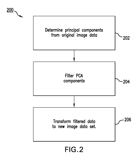
Figure 2 shows a method 200 for extracting low-intensity features from a
dynamic
sequential image data set according to various embodiments of the present
invention.
The image data can be in the form of a dynamic sequence of single slices (two-
dimensional images) or a dynamic sequence of volumes (three-dimensional,
stacks of
images handled as one entity). The description below assumes the two-
dimensional
image sequence. The filtering of the image sequences may be performed for each
and
8
CA 02705099 2010-05-05
WO 2009/073672 PCT/US2008/085295
every slice location. The method 200 may, for example, be performed using an
image
analyser 124 as shown in Figure 1, and as described above.
The method comprises a first step 202 of determining the principal components
from
the data corresponding to each image forming a frame in the sequential image
data
set.
Pixel value data from each image frame is normalised and put into an n-
dimensional
vector form. The vector is then normalised to a zero mean by subtracting the
average
data value (e.g. x, y, etc.) across each dimension from the data values in
that
dimension (e.g. xi - xi- X, yi-> yj - y, etc.) and the data is also normalised
so that the
variance (var) is set to unity; wherein the variance (var) is defined
according to:
N(Xi -X)
var l ~ = - (1)
N
where Xi is the ith data point in the X dimension, X is the mean value of all
the data in
the X dimension, and N is the total number of data points in the X dimension.
Similarly, the data may be normalised by dividing by the standard deviation.
Other normalisation techniques, for example those described by Razifar [6],
may
prove useful, but the aforementioned method is know to be reliable as it is
robust and
weights data from all frames equally.
Having normalised the data, including normalising the data sets such that they
have a
zero mean, principal component analysis (PCA) is applied. In one method for
applying PCA, for example as described by Smith [5], a covariance matrix Can
for a
data set having n dimensions is calculated, as follows:
IN (X. -M ~Y -Y)
cov(X, Y) _ `-~ N - (2)
where covariance is measured between two dimensions, and cov(X,Y) is the
covariance measured between the X and Y dimensions. Using equation (2) a
9
CA 02705099 2010-05-05
WO 2009/073672 PCT/US2008/085295
covariance matrix can be built up using pairs of data in two dimensions to
define the
covariance matrix C for a set of data with n dimensions as:
Cxtz = (ci,j, ci 1= cov(Dimi ,Dim1)) - (3)
with Dim, being the xth dimension. For example, where a three dimensional data
set is
provided, having dimensions x, y and z, n = 3 and the covariance matrix C has
three
rows and three columns, and is defined as:
cov(x,x) cov(x,y) cov(x,z)
C = cov(y,x) cov(y,y) cov(y,z) - (4)
cov(z,x~ cov(z,y) cov(z,z)
Having determined the covariance matrix C, unit eigenvectors for that
covariance
matrix C are then determined in a conventional manner [4,5].
The eigenvectors thus determined are ordered according to their respective
eigenvalues, starting from the eigenvector having the highest eigenvalue (i.e.
the most
significant component, PC1) and moving to the eigenvector having the lowest
eigenvalue (i.e. the least significant component, PCn). The eigenvectors thus
ordered
PC1-PCn therefore provide a set of n eigenvectors corresponding to the
principal
components of the image data set for the respective image frame.
Having determined the principal components, the next step in the method 200 is
that
of applying a PCA filter to the principal components to determine a filtered
data set at
step 204.
Various techniques can be used to apply the PCA filter so as to discard at
least one
principal component, and several of these are described further below. By way
of
definition, as used herein expressions relating to discarding are understood
to mean
reducing the magnitude of one or more principal components, and as such
discarding
includes multiplication of one or more principal components by a weighting
factor a,
such that 0<a<1.
CA 02705099 2010-05-05
WO 2009/073672 PCT/US2008/085295
The principal component for discarding may be a higher order or lower order
principal component, the higher order principal components being those grouped
towards and including the most significant component PC1, and the lower order
principal components being those grouped towards and including the least
significant
component PCn. Removal of such lower order noise components provides for noise
reduction.
In various embodiments, at least one higher order principal component for
discarding
is determined by merely removing the most significant principal component PC
I. For
various imaging techniques, PC I will identify the dynamic behaviour of blood.
The
removal of one or more higher order principal components may thus be used to
remove real features (i.e. not noise) which might otherwise mask fainter
features with
different kinetic behaviour.
Additionally, or alternatively, at least one principal component for
discarding may be
determined by dynamically setting one or more variance contribution thresholds
and
discarding principal components whose percentage variance contribution, e.g.
based
upon a corresponding eigenvalue, is less than or more than a respective
variance
contribution threshold.
Various embodiments of the present invention may further use a scree plot
analysis,
for example, in order to determine one or more lower order principal
components for
discarding.
A scree plot is a graphical analysis technique with the principal components
plotted
on the x-axis and a corresponding percentage variance value for each of the
principal
components plotted on the y-axis. Generally, the scree plot decreases rapidly
from the
higher order principal components, reaches a "knee", and then levels off.
The scree plot is applied in order to determine where the variance
contributions of the
principal components (e.g. as defined by respective eigenvalues for the
principal
components) level off into a noise floor, and discarding those components that
are
below the noise floor. The noise floor can be determined in various ways, it
being
data dependent. For example, a lower variance contribution threshold can be
set at a
11
CA 02705099 2010-05-05
WO 2009/073672 PCT/US2008/085295
value just below the knee of the scree plot with all principal components
having a
variance value below that threshold being discarded.
Additionally, or alternatively, at least one principal component for
discarding may be
determined by dynamically setting one or more principal components to discard,
and
analyzing the residual of the filtered image. That is, the difference between
the
filtered and the original image is analyzed, visually or by computer
algorithm. The
residual image may be calculated using the PCA filter, selecting the lower-
order
principal components that are discarded (for filtering). That is, the
principal
components used for creating an image are discarded for the residual image.
Figure 8
shows an example of a residual image of the filtered image from Figure 6.
The original image can comprise raw data obtained from a PET scan that
corresponds
to coincidences between detector pairs. The counts of recorded events for each
detector pair is raw data, which can be histogrammed to yield a sinogram. The
sinogram can be considered to be an image of counts for each detector pair,
and the
sinogram can be transformed (e.g. reconstructed) to provide images. In various
embodiments, the raw data can thus be filtered prior to transforming, or
reconstructing, images.
Various embodiments of methods according to the present invention may further
comprise filtering of background pixels from the image data set prior to
determining
the plurality of principal components.
This is particularly useful for pharmaco-kinetic modelling that uses the input
from
image frames to calculate significant physiological properties, since it
addresses the
existing problem that, under certain circumstances, the algorithm used to
calculate
these properties pixel by pixel is not robust, but instead adds noise to
create poor
quality images. By performing a filtering step prior to use of such modelling
algorithms various embodiments of the present invention address this problem.
For example parametric images may be created using a Patlak model [7].
However,
this generates very noisy images. So in order to address this problem,
application of a
PCA filter according to various embodiments of the present invention is used
to
12
CA 02705099 2010-05-05
WO 2009/073672 PCT/US2008/085295
remove noise prior to the creation of images using the Patlak technique [7],
which in
turn results in greatly improved images.
Typically, pharmacological studies require multiple injections of radioactive
materials
into a subject. Therefore, multiple scans on the same subject (e.g. under
different
conditions) are presently hampered by a limitation on the maximum permitted
radioactivity dose. Hence by using the noise filtering technique of various
embodiments of the present invention (e.g. and by removing less significant
components only) the number of possible studies on a single person can be
usefully
extended.
The latter noise filtering technique is also useful when PET scanning is used.
For
example, it can enable the extraction of better quantative and qualitative
information
from sequential images acquired by PET scans performed on different parts of
the
body, without the need to wait for residual radioactivity to decay (which,
e.g. with a
half-life of almost two hours for flourine-18, would otherwise make this
extremely
impractical). Such a technique conventionally requires a stepping up of the
amount of
injected radioactive material, so that the residual signal from each preceding
injected
dose is masked by the larger signal generated by a subsequently injected dose.
Having applied PCA filter to the principal components to determine a filtered
data set
at step 204, the next step 206 in the method 200 is that of transforming the
filtered
data set to a new image data set, NewDataSet. The new image data set can be
used to
provide a plurality of filtered image frames having enhanced low intensity
features,
that might be of clinical significance, as well as reduced image noise for
better
visualisation.
The filtered data set is calculated using a feature vector, Feature Vector.
The feature
vector is a matrix of vectors comprising a selection of principal components
(eigenvectors), such that:
Feature Vector =(a~PC1a2PC2...a,,PCn) - (5)
13
CA 02705099 2010-05-05
WO 2009/073672 PCT/US2008/085295
where a is the respective weighting factor applied by the PCA filter to the
i`h
eigenvector. A description on how the data is treated for a. values being 0 or
1
follows. Other a. values may also be used.
The original frame image data is modified in the following way:
First, image data is rotated to the coordinate system of the principal
components
(eigenvectors) and the dimensions are truncated to the number of components
selected:
FinalData = FeatureVectorT X DataAdjustT - (6a)
where FeatureVectorT is the transpose of Feature Vector, and DataAdjustT is
the
transpose of the normalized original image data vectors. Thus, FinalData
represents
PCA-images.
Secondly, the truncated data is transformed back to the original dimensions
using the
following operation:
NewDataSetT = Feature Vector X FinalData -
(6b)
Thirdly, the normalizations of NewDataSet are undone. This is typically done
by
multiplying by the standard deviation and adding the mean value; e.g. using
the
standard deviation and mean values being the same values as used above for
normalization.
Finally, NewDataSet is reformatted to provide images, giving a filtered image
set,
with the same number of frames as the original image set. This reformatting
process
can recreate two-dimensional images from the one-dimensional data vectors in
NewDataSet.
Figure 3 shows principal component analysis images 300 that were derived from
an
experimental seventeen frame dynamic PET brain imaging study, the eleventh to
fifteenth original frames of which are shown in Figure 5.
14
CA 02705099 2010-05-05
WO 2009/073672 PCT/US2008/085295
For this experiment, a General Electric Discovery ST PET/CT camera was used
and
the tracer was an experimental tracer provided for studying brain damage. The
scan
times were set to 10 seconds for frames acquired during the first few minutes,
with
gradually longer acquisition times being used for the frames of up to 15
minutes for
the frames acquired after 90 minutes. All the frames were then used in the PCA
filtering analysis.
The most significant principal component PC1 contributes 83% to the variance
of the
principal components. The next highest order principal component PC2
contributes
8% to the variance of the principal components. The third principal component
PC3
contributes 4% to the variance of the principal components. The fourth
principal
component PC4 contributes 0.9% to the variance of the principal components.
The
fifth principal component PC5 contributes 0.8% to the variance of the
principal
components. The sixth principal component PC6 contributes 0.6% to the variance
of
the principal components.
Figure 4 shows filtered frames eleven to fifteen from the experimental
seventeen
frame dynamic PET brain imaging study. The filtered frames are obtained by
applying the method of Figure 2 to the original image frames shown in Figure
5.
In this case, the PCA filter was applied to remove completely the principal
components PC1, PC2 and PC7 to PC17 (i.e. a1=a2=(X7=...a17=0). A feature
vector
was then created in accordance with equation (5) above, and then a new image
data
set was created from the feature vector in accordance with equations (6a) and
(6b).
The displayed subset of the new image data includes the eleventh filtered
image frame
402, the twelfth filtered image frame 404, the thirteenth filtered image frame
406, the
fourteenth filtered image frame 408, and the fifteenth filtered image frame
410, as
shown in Figure 4.
Figure 5 shows original image frames eleven to fifteen from the experimental
seventeen frame dynamic PET brain imaging study. Figure 5 is shown adjacent
Figure 4 for ease of comparison.
CA 02705099 2010-05-05
WO 2009/073672 PCT/US2008/085295
Comparing Figures 4 and 5, it is apparent that a feature of clinical interest
is clearly
visible in the upper right hand quadrant of the images of filtered image
frames twelve
to fifteen. This is a low intensity feature that is not readily apparent from
a viewing of
Figure 5, but which is very clearly delineated with little or no interference
being
provided as a result of tissue dynamic artefacts. Further studies by the
inventors
confirmed that the low intensity feature detected as a result of this
technique
corresponded well with that found using alternative techniques, such as X-ray
computer tomography.
Figure 6 shows a first screen shot 600 provided by a GUI in accordance with an
embodiment of the present invention. The screen shot 600 may be obtained, for
example, from operation of the GUI 123 shown schematically in Figure 1.
The screen shot 600 shows a filtered image frame 610 derived by applying a
method
in accordance with various aspects of the present invention. The screen shot
600 also
shows a PCA filter control section 602 that includes first and second user
operable
sliders 604, 606.
The first slider 604 can be used to set a first variance contribution
threshold for
determining which higher order principal components are discarded when
creating a
filtered data set. In the example case shown, the variance contribution
threshold is set
to "3" thereby ensuring that the first and second principal components (PC1
and PC2)
are discarded when creating the filtered data set.
The second slider 606 can be used to set a second variance contribution
threshold for
determining which lower order principal components are discarded when creating
the
filtered data set. In the case shown, the variance contribution threshold is
set to "5"
ensuring that the third to fifth principal components (PC3 to PC5) are
selected for
creating the filtered data set with all the other lower order components (in
this case
PC6 to PC17) being discarded.
The filtered image frame 610 shows an image in which low-intensity features
have
been enhanced by application of the present invention.
16
CA 02705099 2010-05-05
WO 2009/073672 PCT/US2008/085295
Figure 7 shows a further screen shot 700 obtained from operation of the same
GUI as
Figure 6 but with different operating parameters having been set.
The screen shot 700 shows an unfiltered image frame 710 derived using all of
the
principal components determined from a sequence of original image frames.
The screen shot 700 also shows the PCA filter control section 602 including
the first
and second user operable sliders 604, 606. However, in this case the first
slider 604
has been set to "1" thus ensuring that no higher order principal components
are
discarded when creating a data set for transformation. Additionally, the
second slider
606 is set to "17" thereby ensuring that all of the principal components (PC1
to PC17)
are selected when creating the data set.
Comparing image frames 610 and 710 from Figures 6 and 7, it is again apparent
that
features of clinical interest are made more clearly visible by the application
of various
aspects of the present invention.
It is therefore considered apparent from the inventors' investigations that
the
hereinmentioned image analysis techniques as provided by various aspects and
embodiments of the present invention provide useful improvements over
conventional
image analysis techniques.
Figure 8 shows a screen shot of a residual image 810 obtained using the same
GUI
800 as Figure 6, with different operating parameters set. The residual image
810 is
calculated using the PCA filter described above by selecting the lower-order
principal
components that are discarded (for filtering).
Whilst the present invention has been described in accordance with various
aspects
and preferred embodiments, it is to be understood that the scope of the
invention is
not considered to be limited solely thereto and that it is the applicant's
intention that
all variants and equivalents thereof also fall within the scope of the
appended claims.
17
CA 02705099 2010-05-05
WO 2009/073672 PCT/US2008/085295
References
1. A. M. Peters, Graphical Analysis of Dynamic Data: The Patlak-Rutland Plot,
Nuclear Medicine Communications, 15:669-672, 1994
2. J. Logan, J. S. Fowler, N. D. Volkow, G-J. Wang, Y-S. Ding, D. L. Alexoff,
Distribution Volume Ratios without Blood Sampling from Graphical Analysis of
PET
Data, Journal of Cerebral Blood Flow Metabolism, 16:834-840, 1986
3. A. Bertoldo, G. Sparacino, C. Cobelli, Population Approach Improves
Parameter Estimation of Kinetic Models from Dynamic PET Data, IEEE
Transactions
on Medical Imaging, vol. 23, no3, pp. 297-306, 2004, ISSN 0278-0062
4. R. C. Gonzalez and R. E. Woods, Digital Image Processing, Second Edition,
Chapter 11, Prentice Hall, New Jersey, USA
5. Lindsay I Smith, A tutorial on Principal Components Analysis, 26 February
2002, http://www.cs.otago.ac,nz/cosc453/studeiit tutorials/principal
components . df
6. Pasha Razifar, Novel Approaches for Application of Principal Component
Analysis on PET Images for Improvement of Image Quality and Clinical
Diagnosis,
PhD thesis, Uppsala University, ISSN 1651-6214, ISBN 91-554-6387-8
7. C. S. Patlak and R. G. Blasberg, Graphical Evaluation of Blood-to-Brain
Transfer Constants from Multiple-Time Uptake Data, Journal of Cerebral Blood
Flow
Metabolism, 5:584-590, 1985
Where permitted, the contents of the above-mentioned references are hereby
also
incorporated into this application by reference in their entirety.
18