Note: Descriptions are shown in the official language in which they were submitted.
CA 02705349 2013-08-21
DESC RIP TION
BREAST IMPLANT ASSEMBLY TO REDUCE
CAPSULAR CONTRACTURE
TECHNICAL FIELD
100011 This invention relates
generally to medical implants. More
particularly, this invention relates to implantable prostheses that resist
capsular contracture. The implant in its preferred form is a mammary
prosthesis which is well known in the art. Other applications include
adjustable mammary prostheses and mammary tissue expanders. A
preferred method of assembling the present invention allows a surgeon to
efficiently and accurately assemble the implantable prosthesis immediately
prior to insertion into the human body.
BACKGROUND ART
[0002] The. use of
implantable breast prostheses has become an
acceptable and popular practice to enhance the aesthetic breast form
whether for augmentation, reconstruction. revision needs. These
devices
generally comprise a nonreactive, flexiblc outer surface or shell which
contains a gel or liquid filler.
[0003] Undesirably-, when
inserted into the host, the iniplant is
recognized as a foreign body by the host's immune system and is walled off,
or encapsulated, from the rest of the host's body. Encapsulation can result in
many unwanted effects. To combat encapsulation, surgical correction is
often required. Despite documented high patient satisfaction rates and
enhancement of quality of life, surgical corrcticn or re-operation rates can
te unacceptably high. In fact, recently published FDA PMA (pre- and post-
market approval) studies on the silicone gel breast implants document the
severity of the public need. Within four years of the initial operation, over
twenty-three percent of all primary augmentation patients had to undergo a
re-operation. Approximately forty percent of these re-operations were to
correct capsular contracture. Thirty-five percent of these revision patients
had to undergo another operation, and the leading cause was again capsular
contracture. Patients undergoing
primary breast reconstruction with
silicone gel breast implants (following mast,tctomy for cancer) have an even
CA 02705349 2010-05-10
WO 2009/065013
PCT/US2008/083595
2
greater public need for help. Twenty-three-point-five percent of these women
must undergo a re-operation, and the leading cause was capsular contracture
or implant malposition (usually due to capsular contracture). Thirty-three
percent of these revision patients need another revision. The re-operation
rates for women with saline implants are similar, and again, capsular
contracture is the leading culprit.
[0004] The
inability to control abhorrent scarring or encapsulation
process leads to spherical capsular contracture (often accompanied by
implant displacement, distortion and pain and discomfort). Spherical
capsular contracture is the number one cause of the aforementioned
excessive re-operation rates. Other causes of re-operation include implant
displacement and palpability of the implant through the skin.
[0005]
Spherical capsular contracture has remained a particularly
vexing problem for scientists, surgeons, and patients for almost 50 years.
Although silicone elastomers (often comprising the outer surface of the
implant) are considered inert materials, the host nonetheless reacts to their
in-vivo implantation by treating the implant as a "foreign body" by walling
the implant off from the surrounding host tissue by the formation of a fibrous
sheath surrounding the implant's peripheral surface. This naturally
occurring process is harmless, unless the degree of linear scar formation
becomes excessive, and the capsule tightens or contracts around the
implanted silicone device, causing shape distortion, implant displacement,
implant palpability, and patient pain and discomfort. These specific adverse
affects are the leading cause of the FDA's documented excessive re-operation
rates. Breast implant patients endure these adverse affects due to the
inability to control device-host tissue reaction.
[0006] Intra-
operative tissue manipulations, which have been
advocated as possible remedies to the capsule contracture problem, include
the creation of large surgical pockets in which the implant is placed,
atraumatic surgical technique, use of sub-muscular surgical pockets for
implant placement, and pocket irrigation with steroid and/or antibiotic
containing liquid. Post
surgical exercises or implant displacement
manipulations have been advised, as have arm movements and body position
CA 02705349 2013-08-21
3
maneuvers. (See Maxwell, GP; Hartley, RW; "Breast Augmentation",
Mathers: Plastic Surgery, Second Edition. (Ed) Saunders Philadelphia, Vol 6.
pl, 2006).
[00071 Improvements and alterations to the design of breast implants
have also been initiated in an effort to reduce spherical capsular contracture
and visibility and palpation. For example. -U.S. Patent No. 4,889,744
advocates that texturization of the outer surface of the implant will minimize
capsule contracture around an implant. U.S. Pat. No. 4,648,880 utilizes an
outer polymeric covering of a woven mesh draped over the implant to reduce
scar formation. Further, U.S. Patent No. 6,913,626, submits that capsule
contracture can be reduced by covering the elastomeric shell of the implant
with a bio-absorbable covering.
rE90081 For unrelated uses in the human body, biologically-derived
materials have been developed from allograft and xenograft (such as porcine
or bovine) source and treated in a wav (biotechnologically prepared) to serve
as dermal graft tissue matrixes. These biologically-derived materials
(generally acellular dermis in composition) are thought to serve as a non-
absorbable collagen scaffold, to pronacte the organization of the healing
process, thereby promoting re-generative repair rather than scar formation.
These materials have been used primarily to correct large wounds, hernias,
and other defects caused by trauma or surgical extirpation for cancer.
Examples 3f this type of biological material, specifically allograft or
xenograft
acellular dermal grafts or matrixes, include (but are not limited to)
AllodermTM and
Strattice'm from Life Cell Corporation, Cosmatrix[m/Surgimend'm from TEI
Biosciences, NeofonnTM from Tutogen Medical, and DermamatrixTM from MT-F. It
has not, however, been anticipated in any of these applications that the
materials
become an interfaced component of a medical implant.
[00091 The main functional use of these acellular dermal materials in
the prior art has been as a tissue extension or tissue replacement (tissue
supplement) of the abdominal musculature anclior facial defects in repairing
abdominal wall hernias, ventral hernia repair. In these situations the
abdominal musculature is stretched, weakened, or rendered inadequate for
repair, and, thus, the need for the supplemental tissue substitute.
CA 02705349 2010-05-10
WO 2009/065013
PCT/US2008/083595
4
[0010] Another use of these materials has been as a tissue extension,
supplement, or replacement following cancer extirpation of the breast. Here
the pectoralis major muscle is partially removed, stretched, or inadequate to
provide tissue coverage of the underlying reconstruction. Thus the dermal
graft is used "to simulate total muscle coverage using tissue like materials
over the lower lateral aspect" of the underlying reconstruction ("an alloderm
sling"). (See Gamboa-Bobadilla, G.M.; Implant Breast Reconstruction using
Acellular Dermal Matrix, Annals of Plastic Surgery, 56; p.22, 2006; Salzberg,
C.A.; Nonexpansive immediate breast reconstruction using human acellular
tissue matrix graft, Annals of Plastic Surgery, 57, p.1, 2006). In these
various applications, the acellular dermal graft "serves the function of
native
tissue." (Spear, S.; Use of Regenerative Human Acellular Tissue to
Reconstruct the Abdominal Wall following Pedicle TRAM Flap Breast
Reconstruction; Plastic Reconstructive Surgery 118, p.8, 2006. Spear, S.L.,
Pelletiere, C.V., and Lockwood, M. Immediate Breast Reconstruction with
Tissue Expanders and Alloderm, Plastic Reconstructive Surgery of the
Breast, p.489, 2006).
[0011] In addition, prior art acellular dermal grafts have been used
for
soft tissue deficient patients with "pectoral muscle denervation." (See
Duncan, D.I. Correction of Implant rippling using allograft dermis. Aesthetic
Surgery Journal 21, p.81, 2001). In these applications, the native tissue was
inadequate because of "very thin skin flaps." Id. In this prior use the graft
was also secured "into the vascularized recipient site" of the host tissue to
serve as an extension of the pectoral muscle. Id. The purpose was "soft
tissue augmentation" to cover externally visible "rippling' of an underlying
device ("rippling" can only be seen or present when capsule contracture is not
present around a breast implant). Id. Another way to describe this prior art
is that the dermal graft is used as a replacement, extension, or supplement of
the native tissue, regardless of that which it covers.
[0012] Although the prior art has proffered myriad solutions to reduce
spherical capsular contracture associated with implantable prostheses, all
have proved to be less than optimal. Thus, what is needed is an implant
having an integral interfaced component comprised of an acellular dermal
CA 02705349 2010-05-10
WO 2009/065013
PCT/US2008/083595
graft material (the effectiveness of the interfaced implant being neither
dependent on the texture of the implant's surface nor the dissolution of a
covering) to reduce capsular contracture, implant displacement, and/or
implant palpability.
DISCLOSURE OF THE INVENTION
[0013] The present invention relates generally to implantable
prostheses and more particularly to implantable prostheses that prevent
and/or reduce capsular contracture. The present invention includes a
medical implant and a biological interface. The medical implant may have a
textured or smooth outer shell surface and may have a filler of liquid as
saline, gel as non-form stable silicone gel or enhanced cohesive form-stable
silicone gel, or a more solid material. Moreover, the medical implant may be
that of a fixed volume, adjustable volume, or a temporary tissue expander.
[0014] The biological interface is affixed to the exterior surface of
the
implant. The biological interface may come pre-attached to the medical
implant (in fact the biological interface may be considered a coating on the
implant), may be wedged into the space or pocket created for receipt of the
implant, or may be attached to the implant at time of its insertion into the
host.
[0015] The biological interface is comprised of a dermal material with
capsular contracture inhibiting properties. The dermal material may be an
acellular dermal graft or matrix, which may be of an allograft or xenograft
(such as porcine or bovine). Additionally, the dermal material may be
developed in the form of a sheet, a pouch, a strip, a gel, a liquid, or
particles.
[0016] Importantly, the biological interface and the implant are in
intimate contact and positioned so that the biological material is between the
implant and the tissue of the host. The biological material may be attached
to the implant by various methods including but not limited to sutures,
adhesives, or by engaging recipient flaps or other appendages located on the
outer surface of the implant. Further, the biological material may
encompass the entire implant or only a portion thereof.
[0017] Because the biological material is situated between the implant
and the tissue of the host (and the biological material's ability to promote
re-
CA 02705349 2010-05-10
WO 2009/065013
PCT/US2008/083595
6
generative repair rather than scar formation), the host does not treat the
biological material, and hence the implant, as a foreign body¨thereby
preventing/reducing capsular contracture. As such, the present invention
serves to reduce and/or eliminate capsular contracture associated with
implantable prostheses.
BRIEF DESCRIPTION OF THE DRAWINGS
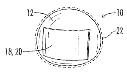
[0018] Fig. 1 is a frontal view of the medical implant of the present
invention wherein the biological interface covers a portion of the exterior
surface of the medical implant.
[0019] Fig. 2 is a cross-sectional view of the medical implant of the
present invention wherein the biological interface covers the entire exterior
surface of the medical implant.
[0020] Fig. 3 is a cross-sectional view of the medical implant of the
present invention wherein the biological interface covers a portion of the
anterior and the inferior portion of the medical implant.
[0021] Fig. 4 is a cross-sectional view of the medical implant of the
present invention wherein the biological interface covers the entire anterior
and inferior surface of the medical implant.
[0022] Fig. 5 is a cross-sectional view of the medical implant of the
present invention wherein the biological interface is secured to the medical
implant except at distal and/or peripheral portions which may allow
attachment for positional maintenance of the biological interface of the
present invention itself.
[0023] Fig. 6 is a cross sectional view of the medical implant of the
present invention wherein the biological interface covers a relatively small
anterior and posterior surface of the medical implant.
[0024] Fig. 7 is a cross-sectional view of the medical implant of the
present invention wherein the biological interface has varied thicknesses.
[0025] Fig. 8 depicts the biological interface fused at its periphery
into
a pouch as a means of covering the medical implant.
[0026] Fig. 9 is a cross-sectional view showing the medical implant
positioned in the biological interface pouch, of Fig. 8, to create the present
invention.
CA 02705349 2010-05-10
WO 2009/065013
PCT/US2008/083595
7
[0027] Fig. 10 is an anterior view of the medical implant of the
present
invention showing a portion of the biological interface scored, or altered, in
a
way that may be more economically or clinically functional.
[0028] Fig. 11 is an anterior view showing the biological interface in
a
meshed form and applied to the medical implant.
[0029] Figs. 12a-d illustrate the interaction between the tissue
pocket,
the implant, and the biological interface.
[0030] Figs. 13a-b are anterior and side views of one embodiment of
the
medical implant of the present invention showing a thickened shell portion
located on the outer surface and a round injection site.
[0031] Fig. 14 is a posterior view of a particular embodiment of the
medical implant of the present invention showing a plurality of attachment
flaps attached to the outer surface of the implant.
[0032] Fig. 15 is the posterior view of the medical implant of Fig. 14
showing the biological interface hooked across each of the attachment flaps.
[0033] Fig. 16 is an anterior view of the medical implant of Fig. 15
wherein the biological interface covers the entire surface.
[0034] Fig. 17 illustrates a cross-sectional view of an attachment
flap
from Fig. 14.
[0035] Fig. 18 illustrates the attachment flap from Fig. 17 in an open
position.
[0036] Fig. 19 illustrates the attachment flap from Fig. 17 with a
portion of the biological interface hooked across the flap.
[0037] Fig. 20 is the anterior view of an alternative embodiment of
the
medical implant of the present invention having a plurality of attachment
flaps attached to the outer surface.
[0038] Fig. 21 is the anterior view of the medical implant of Fig. 20
showing the biological interface covering a portion of the medical implant.
[0039] Figs. 22 and 23 illustrate a plurality of potential
configurations
for the attachment flaps of the present invention.
[0040] Fig. 24 shows a plurality of embodiments of biological
interfaces
having a variety of perforation openings or attachment openings.
CA 02705349 2010-05-10
WO 2009/065013
PCT/US2008/083595
8
[0041] Fig. 25 is an anterior view of another embodiment of the
medical implant of the present invention wherein the biological interface is
attached to the thickened shell portion of the medical implant by sutures.
[0042] Fig. 26 is a posterior view of the medical implant of Fig. 25
wherein the biological interface covers a portion of the medical implant and
is attached to the implant by sutures.
BEST MODE FOR CARRYING OUT THE INVENTION
[0043] The present invention relates generally to a medical implant
assembly 10 and more particularly to a medical implant assembly 10 that
prevents and/or reduces capsular contracture. Although the assembly 10 can
be any implantable prosthesis, a preferred embodiment of the present
invention concerns implants used primarily for breast augmentation,
revision, and reconstruction. Now referring to Figs. 1-26, the assembly 10
includes a medical implant 12 and a biological interface 18. Although the
implant 12 may be relatively non-compliant or have a firm pre-defined
shape, a preferred embodiment has a medical implant 12 with a flexible
silicone elastomeric shell 16 or exterior surface 16. The resilient shell 16
allows the implant to be readily deformed without compromising the
integrity of the implant 12. Such a property facilitates positioning the
prosthesis 12 into a host (or implant recipient). The shell 16 may be textured
or smooth.
[0044] To complement the resilient shell 16, the core of the implant
12
may be filled with a gel (preferably a cohesive silicone gel) or liquid, such
as
saline. Referring generally to Figs. 13-26, in certain embodiments an
adjustable medical implant 12 is employed into which the liquid may be
injected after insertion of the prosthesis 12 into the human body. An
injection dome 42 through which the liquid may be injected is attached to the
exterior surface 16 of the implant 12. As shown in Figs. 13a and 13b, the
injection dome 42 may where desirable be positioned within a thickened shell
portion 44 of the exterior surface 16 of the implant 12.
[0045] The assembly 10 also includes a biological interface 18 (or a
non-bioabsorbable dermal interface 18). The biological interface 18 is affixed
to the shell 16 of the implant 12. In one embodiment, the interface 18 is a
CA 02705349 2010-05-10
WO 2009/065013
PCT/US2008/083595
9
biologically harvested dermal material 20 or biotechnically prepared
material 20, whether cellular or acellular, xenograft (as bovine or porcine)
or
allograft. However, regardless of the precise composition of the dermal
material 20, its defining characteristic is that the material 20 has capsular
contracture inhibiting properties. Further, in one embodiment, the interface
18 is not bio-absorbable.
[0046] The interface 18 in one embodiment is attached to the implant
12 at the time the assembly 10 or implant 12 is inserted into the host, as
will
be described more fully below. In other embodiments the interface 18 may
come pre-attached to the medical implant 12, may be attached to the tissue
of the host which interfaces (comes in contact) with the implant 12, or be
wedged (but not connected) into the space between the implant 12 and the
surrounding tissue pocket of the host. The interface 18 may be affixed to the
implant 12 by suturing, surgical adhesive, staples, or any other method
known to those skilled in the art. Further, the present invention also
envisages that the shell 16 and the interface 18 may be formed in a unitary
process or that the interface 18 functions as the shell 16 of the implant 12.
As shown in Fig. 8, the interface 18 may also be formed into a pocket or
receptacle to receive the implant 12. The pocket may cover a portion or all of
the implant 12.
[0047] The interaction/engagement between the implant 12 and the
interface 18 may alternatively be described as follows: the shell 16 has a
contour 22, and the interface 18 is intimately engaged to the implant 12 such
that the interface 18, or more specifically the dermal material 20, follows
the
contour 22 of the shell 16.
[0048] The dermal material 20 may be particulated, diced, meshed,
shredded (as shown in Figs. 10 and 11), applied in strips or segments, and/or
have varying thickness (as shown in Fig. 7). By allowing the dermal
material 20 to have various configurations/forms, multiple objectives can be
satisfied. For instance, if cost is a central concern the dermal material 20
may be meshed and only cover a portion of the implant 12. However, if the
focus is on optimal performance, the dermal material 20 may be a continuous
sheet enveloping the entire implant 12, as shown in Fig. 2.
CA 02705349 2010-05-10
WO 2009/065013
PCT/US2008/083595
[0049] Irrespective of which embodiment is selected, the purpose of
the
interface 18 is to facilitate the healing of the host tissue around and in
proximity to the foreign body device (e.g. implant 12) in a more natural
manner, or an immunologically benign manner, which does not cause the
formation of excessive scar tissue (capsule contracture), device displacement,
or device visibility or palpation from external evaluation. The assembly 10,
thus, exerts a regenerative and compatible tissue response from the host,
rather than a "foreign-body" scar response.
[0050] While the description of the assembly 10 has already been
detailed herein above, a closer analysis of the biological interface 18 and
more specifically the dermal material 20 and its prior art uses is
appropriate.
[0051] It has been shown that biologically obtained material, such as
the dermal material 20, containing the dermis or deeper layer of skin can be
altered in various ways to allow its use in another living host to be
immunologically accepted, rather than eliciting an immunological rejection
("graft versus host" reaction). Thus, it is said to be biotechnologically
prepared. The material source may be animal or, more specifically,
mammalian, and is usually technically altered in a manner to make it
acellular such that, when re-implanted in a separate host, it does not elicit
a
foreign body reaction, but rather serves as a matrix or foundation for a
tissue¨regenerative process that creates a pliable healing milieu, rather than
an undesirable reactive sclerosis. The material must therefore allow
revascularization and not become infected. Various processes are known in
the art for the former, such as rendering the material acellular and the
latter, such as terminal sterilization or irradiation.
[0052] The non-cellular materials, comprising the dermal material 20
in the preferred embodiment, are generally rich in collagen, and may be
further comprised of proteins, proteinaceous materials, enzymes, antigens,
amino acids, peptides, sugars, and carbohydrates. Current art includes
Cosmatrix/surgimend (TEI) derived from the dermis of fetal calves; Alloderm
and Strattice (Life Cell) derived from human and porcine dermis,
respectively; Neoform (Tutogen) from human dermis; and Dermamatrix
(MTF) from human dermis.
CA 02705349 2010-05-10
WO 2009/065013
PCT/US2008/083595
11
[0053] For exemplary purposes, consider the following application of
the present invention in the field of breast augmentation. Initially, a
surgical pocket is created to accommodate the assembly 10, under the skin,
breast parenchyma, or pectoral muscle. In one embodiment, the biological
interface 18 comes pre-attached to the exterior surface of the silicone
elastomer 16. However, in another embodiment the assembly 10 can also be
"created" during the operative procedure by procuring the respective
components separately (biological interface 18 and prosthesis 12 or implant
12) and placing one in contact with the other, thereby "fused" as a "hybrid"
or
interfaced implant, within the surgical pocket. In this manner the assembly
is created efficiently and accurately under sterile conditions in the
operating
room immediately prior to insertion into the human body.
[0054] In the embodiment of the invention as shown generally in Figs.
14-23, a plurality of appendages 40 are located on the exterior surface 16 of
the implant 12 for the purpose of facilitating attachment of the biological
interface 18. The appendages 40 may comprise recipient attachment flaps,
tabs, loops, or various equivalent alternative conventions, and may be
attached to the exterior surface 16 of the implant 12 or may be formed
integral to the implant 12. The appendages 40 of the present invention are
distinguished from suture tabs as widely known in the industry, as these
suture tabs are typically flexible, floppy or otherwise non-rigid in
composition. Alternatively, the appendages 40 of the present invention will
generally comprise a more rigid composition operable to permit stable
positioning of material over the exterior surface 16 of the medical implant
12.
Various embodiments of shapes or configurations are possible for the
appendages 40 of the present invention, examples of which are shown in
Figs. 22, 23.
[0055] The appendages 40 may be located on the posterior surface, the
anterior surface, or generally on the peripheral of the implant 12. It is
contemplated that the appendages 40 may be created in the non-flexible
outer covering 16 of the implant 12. There may be specific thickened areas
44 in the exterior shell 16 of the implant 12 wherein the appendages 40 are
created.
CA 02705349 2010-05-10
WO 2009/065013
PCT/US2008/083595
12
[0056] Referring to Fig. 14, the appendages 40 here comprise
attachment flaps 40 located on the posterior surface of the implant 12 and
opening toward the periphery of the implant 12. While the attachment flaps
40 here are arranged separate and evenly spaced from each other, it is
anticipated that in alternative embodiments the flaps 40 may be designed in
a random or continuous pattern or formation as desired. The biological
interface 18 will be designed with slots 46 or openings 46 within its
substance, or along its periphery, to facilitate affixation of the interfaced
material 20 to the implant 12 by draping over, around or into the plurality of
attachment flaps 40, as shown in Fig. 15. Fig. 16 shows the anterior view of
the implant 12 where the biological interface 18 has been attached in this
manner.
[0057] In particular embodiments of the present invention, the implant
12 will be at least partially injected with liquid such as saline after
insertion
into the human body. It is contemplated that the attachment of the
biological interface 18 to the appendages 40 located on the implant 12 may
not remain secure upon expansion of the implant 12. This is not problematic
however, as the objective of the method of the present invention specifically
relating to the appendages 40 is primarily to provide a secure assembly prior
to insertion. The biological interface 18 will flexibly remain securely
positioned around or about the implant 12 upon expansion, regardless of the
attachment to the appendages 40.
[0058] Fig. 17 displays a cross-sectional view of one of the
attachment
flaps 40 in a standard closed position. Fig. 18 illustrates the flap 40 in an
open position so as to receive the biological interface 18. Fig. 19 shows a
portion of the biological interface 18 affixed to the implant 12 by draping an
opening 46 into the attachment flap 40, which has now returned to a closed
position so as to hold the biological interface 18 firmly in place. In this
manner slippage or premature displacement of components within the
assembly 10 as a whole may be substantially precluded.
[0059] An alternative arrangement of attachment flaps 40 is shown in
Fig. 20. The flaps are here located in the thickened shell portion 44 of the
anterior surface of the implant 12, rather than the posterior surface. The
CA 02705349 2010-05-10
WO 2009/065013
PCT/US2008/083595
13
biological interface 18 may in this embodiment be securely draped over only
a portion of the anterior surface of the implant 12, as displayed in Fig. 21.
In
this manner the injection dome 42 remains available where the injection of a
liquid into adjustable implants 12 are to be utilized without the necessity of
removing the biological interface 18 prior to performing the operation.
[0060] Referring now to Fig. 25, the thickened shell portion 44 of the
anterior surface of the implant 12 may facilitate suture attachments 48 of
the biological interface 18 and further generally stabilize the attachment to
or engagement of the prosthesis 12. The suture attachments 48 may also be
considered as an alternative method of stabilizing attachment of the
biological interface 18 to the prosthesis 12 where appendages 40 are not
utilized. Referring now to Fig. 26, further suture attachments 48 are made
along a reinforced portion 44 of the posterior surface of the implant 12.
[0061] Another method of achieving this intra-operative assembly is to
affix the biological interface 18 or dermal material 20 to the implant 12 by
tissue adhesive. The dermal material 20 may be diced, shredded or
otherwise particulated and subsequently adhered to the implant 12 in strips
or as a layer or film of coating.
[0062] Another alternative assembly option would be to wedge the
biological interface 18 into the contiguous space created for, and adherent
to,
the implant 12. It should be noted that this manipulation creates a
component of the implant 12, not a tissue cover over the peri-prosthetic space
wherein an implant may be separated by fluid from its enhanced tissue
cover. This described manipulation would maintain its device continuity,
while creating in-vivo the assembly 10.
[0063] Alternatively described and referring to Figs. 12a-b, the
implant
12 could be positioned in a surgically created tissue pocket 24, the tissue
pocket 24 having a pocket surface 26 defining a pocket geometry 28.
Similarly to the tissue pocket 24, the implant 12 has an implant surface 30
defining an implant geometry 32. After the implant 12 has been positioned
in the tissue pocket 24, the interface 18 (having inner and outer interfaces
surfaces 34 and 36 defining an interface geometry 38) is fit into the tissue
pocket 24 between the pocket surface 26 and the implant surface 30.
CA 02705349 2010-05-10
WO 2009/065013
PCT/US2008/083595
14
Further, the pocket geometry 28, the interface geometry 38, and the implant
geometry 32 are selected so that after the interface 18 and the implant 12 are
both in the tissue pocket 24, the interface 18 is engaged to the implant 12 to
optimize the contracture inhibiting properties of the interface 18, more
particularly of the dermal material 20; i.e. the interface 18 and the implant
12 are snuggly engaged. This engagement ensures an intimate coupling
between the implant 12 and interface 18. Although Figs. 12a-b depict the
interface 18 covering only a portion of the implant 12, it is also envisioned
that the interface 18 completely encases the implant 12. Moreover, the scope
of the present invention includes inserting the interface 18 into the tissue
pocket 24 before the implant 12.
[0064] This embodiment may be facilitated by temporary
percutaneous, pull-out sutures useful in re-draping of the wedged material
for adequate secured proximity in the (tight) space, thus creating the
interfaced outer cover of the implant, contiguous with the soft tissue pocket.
[0065] In all of these potential applications, the desired affect of
the
assembly 10 is achieved ¨ promoting, via a tissue regenerative process, the
acceptance of the implant 12 within the host, and minimizing that which
frequently occurs in the current art - an overactive foreign-body, sclerotic
reaction to the presence of the implant 12.
[0066] Whether the interface 18 is affixed to the implant 12 prior to
the
assembly 10 being inserted into the host or the implant 12 and interface 18
are pressure fit into the tissue pocket 24, there is no requirement to suture
the interface 18 to the tissue of the host as a muscle extension or cover over
the implant 12. Specifically, in the context of breast implants, it is
anticipated that the present invention will simplify surgery, operative time,
and patient morbidity (not to mention reduce re-operation rates) by removing
the need of suturing a dermal material 20 (or interface 18 more generally)
into a weakened muscle cover, lessening the need for fascial and lattisimus
flaps. Further, and again with reference to breast prostheses, it will not
require lower pole "muscle-extension" cover, but can simply be under the
skin flap. Likewise it may not require additional upper pole cover, which will
CA 02705349 2010-05-10
WO 2009/065013
PCT/US2008/083595
lead to a major reduction in operative time, post-op pain, morbidity, and a
lessened recovery time.
[0067] The present invention also allows prostheses to be employed
where they could not be utilized in the past. For example, as breast cancer
treatment today consists of increasing numbers of segmented mastectomies
or lumpectomies, which cannot be actually re-constructed with available
implants (due to capsular contracture ¨ especially in the face of post-
operative irradiation), the use of a small flexible prosthesis 12 covered with
dermal material 20, (as taught by the present invention) simply inserted into
the lumpectomy cavity will, again, provide a novel answer to a previously
unmet need, and again, enhancing outcomes, reducing morbidity, and cutting
healthcare costs.
[0068] Thus, although there have been described particular
embodiments of the present invention of an interfaced medical implant, it is
not intended that such references be construed as limitations upon the scope
of this invention except as set forth in the following claims.