Note: Descriptions are shown in the official language in which they were submitted.
CA 02705488 2015-09-17
CA 2705488
THERAPEUTIC APPLICATIONS OF p53 ISOFORMS IN
REGENERATIVE MEDICINE, AGING AND CANCER
BACKGROUND
[0001] Cellular senescence was first described by Hayflick and Moorhead (1961)
when they
observed that normal human fibroblasts entered a state of itTeversible growth
arrest after serial
passage in vitro. In contrast, cancer cells did not enter this growth arrested
state and
proliferated indefinitely. The maximum number of cell divisions that a cell
can undergo,
termed the Hayflick limit, varies from cell type to cell type and organism. In
fibroblasts, this
number is about 50 divisions, after which cell division ceases.
100021 The process of cellular senescence can be triggered by multiple
mechanisms,
including telomere shortening, derepression of the INK4a/ARF locus, and DNA
damage. As
discussed below, all three of these mechanisms implicate thc function of the
tumor suppressor
protein p53.
[0003] Telomcre shortening provides a mechanism capable of counting cell
divisions.
Telomeres consist of repetitive DNA elements at the end of linear chromosomes
that protect
chromosome ends from degradation and recombination. Due to the intrinsic
inability of the
DNA replication machinery to copy the ends of linear molecules, telomeres
become
progressively shorter with each round of replication, thus providing a
counting mechanism for
keeping track of the number of cell divisions that have occurred in a
population of cells. As
increasing numbers of cell division occur, the telomeres reach a critically
short length, which
present as double-stranded DNA breaks that activate the p53 tumor suppressor
protein resulting
in telomere-initiated senescence or apoptosis.
[0004] Derepression of the INK4a/ARF locus can also serve as a cell division
counting
mechanism. The 1NK4a/ARF locus is normally expressed at very low levels in
most tissues of
young organisms but progressively becomes derepressed with aging. Thus, a cell
division
counting mechanism is provided by a progressively increased level of
repression of the
INK4a/ARF locus. The pl 6INK4a protein functions as an inhibitor of cyclin-
dependent
kinases CDK4 and CDK6, thus providing a GI cell cycle arrest. ARF regulates
p53 stability
through inactivation of the p53-degrading ubiquitin ligase MDM2.
1
CA 02705488 2015-09-17
CA 2705488
[0005] The accumulation of DNA damage over time can also serve as a trigger
for cell
senescence. As an organism ages, increases in DNA mutations, DNA oxidation,
and
chromosome losses are observed. These observations have prompted investigators
to consider
DNA damage as contributing to cellular senescence and organismal aging. As a
guardian of
cell cycle progression after DNA daniage, p53 plays a role here too, as p53
induces the
expression of the cell cycle inhibitor p21 when a cell has undergone DNA
damage.
[0006] Given the direct impact that cell senescence has on cell division and
cell cycle arrest,
one would expect this process to play a central role in such diverse processes
as aging, cancer,
and tissue regeneration.
BRIEF SUMMARY
[0007] The present disclosure is based in part on the discovery that the
switching of
expression from one p53 isoform (A133p53) to another (p533) results in
replicative cellular
senescence in normal human fibroblasts. Specifically, the present inventors
have discovered
that p5313 and A133p53 promotes and inhibits, respectively, cellular
senescence when
overexpressed. siRNA-mediated knockdown of endogenous A133p53 induced cellular
senescence. A133p53 counteracted wild-type (wt) p53 to repress its
transcriptional targets
(p21wAPI and miR-34a) and inhibit wt p53-mediated degradation of TRF2,
allowing cell
proliferation beyond the normal senescence setpoint of telomeres. Accordingly,
the present
disclosure takes advantage of a novel telomere-mediated mechanism by which p53
regulates
cellular senescence through inhibition of p53 activity by its own natural
isoforms.
[0008] Accordingly, in one aspect, the present disclosure provides a method of
promoting
senescence in a cell by contacting the cell with an agent that inhibits the
function or expression
of A133p53, thereby promoting cell senescence. This disclosure therefore also
provides a use
of such an inhibitory agent of A133p53 for manufacturing a medicament for
treating a disease
in which cell senescence is inadequate.
[0009] In another aspect, the present disclosure provides a method of treating
or preventing
cancer cell growth by promoting cell senescence by contacting the cancer cell
with an agent
that inhibits the function or expression of A133p53, thereby inhibiting cancer
cell growth.
Similarly, this disclosure provides a method for treating cancer by contacting
cancer cells with
2
CA 02705488 2015-09-17
CA 2705488
an agent that inhibits the function or expression of A133p53 in order to
promote cancer cell
senescence and therefore treat cancer. This disclosure therefore provides a
use of such an
inhibitory agent of A133p53 for manufacturing a medicament for treating or
preventing a
disease or condition involving undesirable cellular proliferation such as
various types of cancer.
[0010] In this disclosure, an inhibitory agent of A133p53 may be an antisense
oligonucleotide, an siRNA (e.g., shRNA), a ribozyme, or a small organic
molecule. Preferably,
such an inhibitor is effective specifically for this particular isoform of p53
protein and not other
isoforms.
[00111 In another aspect, the present disclosure provides a method of
extending the
replicative lifespan of a cell by inhibiting cell senescence by contacting the
cell with an agent
that activates the function or expression of A133p53, thereby inhibiting cell
senescence and
extending the replicative lifespan of the cell. This disclosure therefore
provides a use of an
activator of A133p53 for manufacturing a medicament for treating a condition
where cell
replicative lifespan is inadequate.
[0012] In another aspect, the present disclosure provides a method of
generating a population
of cells for tissue regeneration by inhibiting cell senescence by: (a)
contacting a cell suitable for
tissue regeneration that has a finite number of cell divisions with an agent
that activates the
function or expression of A133p53, thereby inhibiting cell senescence and
increasing the
number of cell divisions undergone by the cell, and (b) culturing the cell to
obtain a cell
population, thereby generating a population of cells for tissue regeneration.
In some aspects of
this embodiment, the agent comprises a nucleic acid for the overexpression of
A133p53, such
as a polynucleotide sequence (e.g., a DNA sequence) encoding 4133p53 or an
expression
cassette capable of overexpressing the protein. The method for producing cell
populations for
tissue regeneration can be useful for treating or preventing degenerative
diseases including
various age-related conditions such as osteoporosis, osteoarthritis, macular
degeneration, and
atherosclerosis.
100131 In another aspect, the present disclosure provides a method of
promoting senescence
in a cell by contacting the cell with an agent that activates the function or
expression of p5313,
thereby promoting cell senescence. In some embodiments, the agent comprises a
nucleic acid
3
CA 02705488 2015-09-17
CA 2705488
encoding p53l3, such as a polynucleotide sequence (e.g., a DNA sequence) or
expression
cassette encoding and capable of overexpressing p533 protein.
[0014] In another aspect, the present disclosure provides a method of treating
or preventing
cancer cell growth by promoting cell senescence by contacting the cancer cell
with an agent
that activates the function or expression of p5313, thereby inhibiting cancer
cell growth. In
sotne embodiments, the agent comprises a nucleic acid encoding p5313, such as
a
polynucleotide sequence (e.g., DNA) or expression cassette encoding and
capable of
overexprcssing p5313 protein.
[0015] In another aspect, the present disclosure provides a method of
extending the
replicative lifespan of a cell by inhibiting cell senescence, the method
comprising the step of
contacting the cell with an agent that inhibits the function or expression of
p5313, thereby
inhibiting cell senescence and extending the replicative lifespan of the cell.
Similarly, this
disclosure provides a method of preventing or treating a degenerative disease
by inhibiting cell
senescence. Degenerative diseases include various age-related conditions such
as osteoporosis,
osteoarthritis, macular degeneration, and atherosclerosis. For instance, the
method includes the
step of contacting cells or tissues that are susceptible of the degenerative
disease or involved in
the disease with an agent that inhibits the function or expression of p530,
therefore inhibits cell
senescence and prevents or treats the degenerative disease.
[0016] In another aspect, the present disclosure provides a method of
extending the
replicative lifespan of a cell by inhibiting cell senescence by way of
contacting the cell with an
agent that inhibits the function or expression of miR-34a, thereby inhibiting
cell senescence and
extending the rcplicative lifespan of the cell. An exemplary agent useful for
this purpose is an
antisense oligonucleotide that specifically inactivates miR-34a.
[0017] In another aspect, this disclosure provides a method for enhancing or
restoring
immune functions by extending T cell lifespan. The method includes the step of
contacting the
T cell with an agent that activates the function or expression of 6.133p53,
thereby extending the
lifespan of the T cell and enhancing or restoring immune functions. The agent
may comprise a
polynucleotide sequence encoding A133p53, or comprise an expression cassette
comprising a
polynucleotide sequence encoding Al 33p53. In contrast, this disclosure also
provides a
method for enhancing or restoring immune functions by extending T cell
lifespan by the means
4
CA 02705488 2015-09-17
CA 2705488
of contacting the T cell with an agent that inhibits the fimction or
expression of p53. Such an
agent may be an siRNA, e.g, an shRNA, or a ribozyme. Furthermore, a method is
provided for
enhancing or restoring immune functions by extending T cell lifespan, the
method comprising
the step of contacting the T cell with an agent that inhibits the function or
expression of miR-
34a (such as an antisense oligonucleotide capable of inactivating miR-34a),
thereby extending
the lifespan of the T cell and enhancing or restoring immune functions.
100181 In another aspect, the present disclosure provides a method of
generating a population
of cells for tissue regeneration by inhibiting cell senescence by: (a)
contacting a cell suitable for
tissue regeneration that has a finite number of cell divisions with an agent
that inhibits the
function or expression of p5313, thereby inhibiting cell senescence and
increasing the number of
cell divisions undergone by the cell, and (b) culturing the cell to obtain a
cell population,
thereby generating a population of cells for tissue regeneration.
[0019] In this disclosure, an inhibitory agent of p53p may be an antisensc
oligonueleotide, an
siRNA (e.g., shRNA), a ribozyme, or a small organic molecule. Preferably, such
an inhibitor is
effective specifically to one isoform of the p53 protein and not to other
isoforms.
100201 In yet another aspect, the present disclosure provides a composition
for promoting cell
senescence comprising an siRNA directed to A133p53. In some cases, the siRNA
is an
shRNA. In an aspect of this embodiment, the siRNA comprises or consists of the
sequence 5'-
UGU UCA CUU GUG CCC UGA CUU UCA A-3' (SEQ ID NO:1) or 5'-CUU GUG CCC
UGA CUU UCA A[dl][cIT]-3' (SEQ ID NO:2). Optionally, a physiologically
acceptable
excipient is also present in this composition. In one example, this
composition may be used for
promoting senescence and inhibiting cellular proliferation by suppressing
A133p53 activity,
and therefore for use in treating conditions relevant to undesired cell
proliferation, such as
various types of cancer.
[0021] In another aspect, the present disclosure provides a method for
identifying a
compound that modulates cell senescence via its effect on A133p53 or p53 0. In
general, the
method includes these steps: (a) contacting a candidate compound with a sample
that comprises
A133p53 or 0313, and (b) determining the functional effect of the compound
(such as increased
or decreased cell proliferation, cell cycle arrest, or apoptosis), based on
which one may
determine whether the compound is a modulator (e.g., an activator or
inhibitor) of the
5
CA 02705488 2016-11-03
CA 2705488
respective p53 isoform. For instance, increased cell proliferation would
indicate a test
compound's role as a suppressor of senescence; whereas decreased cell
proliferation, cell cycle
arrest, or increased apoptosis would indicate a test compound's role as a
promoter of the
senescence. Accordingly, the identified modulator may be useful for preventing
or treating
cancer, or for extending a cell's replicative lifespan, depending on the
specific effect of the
modulator on 4133p53 or p53f3 and therefore on senescence. A candidate
compound can be of
any chemical nature: a small molecule or a macromolecule such as protein,
lipid, polysaccharide,
polynucleotide, etc., synthetic or naturally occurring.
[0022] In various aspects disclosed herein, the agent useful for suppressing
the effects of a p53
isoform by inhibiting or inactivating the expression or function of the
isoform can be an
antisense oligonucleotide, an siRNA (such as a shRNA), a ribozyme, or a small
organic
molecule. In further aspects, the cell whose growth is to be suppressed can be
a cancer cell. In
some aspects of the above embodiments, the agent useful for enhancing the
effects of a p53
isoform comprises a DNA for the overexpression of 4133p53 or p5313.
[0023] The claimed invention relates to use of an inhibitory nucleic acid that
inhibits the
expression of human 6.133p53, but not wildtype p53, for promoting senescence
in a cell. The
use may be in preparation of an agent for promoting cell senescence.
10023A1 The claimed invention also relates to use of an inhibitory nucleic
acid that inhibits the
expression of human 4133p53, but not wildtype p53, for inhibiting cancer cell
growth. The use
may be in preparation of an agent for such inhibiting.
[0023B] The claimed invention also relates to use of an inhibitory nucleic
acid that inhibits the
expression of human 4133p53, but not wildtype p53, for treatment of a cancer.
The use may be
in preparation of a medicament for such treating.
[0023C] The claimed invention also relates to use of a polynucleotide encoding
p53[3, for
promoting senescence in a cell. The use may be in preparation of an agent for
contacting a cell
to promote senescence in the cell.
[0023D] The claimed invention also relates to use of a polynucleotide encoding
human p53f3, for
inhibiting cancer cell growth. The use may be in preparation of an agent for
contacting cancer
cells to inhibit growth thereof.
6
CA 02705488 2016-11-03
CA 2705488
[0023E] The claimed invention also relates to use of a polynucleotide encoding
human
A133p53, for extending the replicative lifespan of a cell. The use may be in
preparation of an
agent for such extending of lifespan.
[0023F] The claimed invention also relates to use of an inhibitory nucleic
acid that inhibits
expression of human p53(3, but not wildtype p53, to extend the replicative
lifespan of a cell. The
use may be in preparation of an agent for such extending of lifespan.
[0023G] The claimed invention also relates to use of an inhibitory nucleic
acid that inhibits
expression of human p5313, but not wildtype p53, for preventing or treating a
degenerative
disease by inhibiting cell senescence. The use may be in preparation of a
medicament for such
preventing or treating.
10023111 The claimed invention also relates to use of an antisense
oligonucleotide that
specifically inactivates miR-34a, for extending the replicative lifespan of a
cell. The use may be
in preparation of an agent for such extending of lifespan.
[00231] The claimed invention also relates to use of a polynucleotide encoding
human
A133p53, for extending T cell lifespan. The use may be in preparation of an
agent for such
extending of lifespan.
10023J] The claimed invention also relates to use of an inhibitory nucleic
acid that inhibits the
expression of human p53I3, but not wildtype p53, for extending T cell
lifespan. The use may be
in preparation of an agent for such extending of lifespan.
10023K] The claimed invention also relates to use of an antisense
oligonucleotide that
specifically inactivates miR-34a, for extending T cell lifespan. The use may
be in preparation of
an agent for such extending of lifespan.
[002314 The claimed invention also relates to a method of generating a
population of cells for
tissue regeneration by inhibiting cell senescence, the method comprising the
steps of: (a)
contacting a cell suitable for tissue regeneration that has a finite number of
cell divisions with a
polynucleotide sequence encoding human A133p53 in vitro, thereby inhibiting
cell senescence
and increasing the number of cell divisions the cell undergoes; and (b)
culturing the cell to obtain
a cell population; thereby generating the population of cells for tissue
regeneration.
10023M] The claimed invention also relates to a method of generating a
population of cells for
tissue regeneration by inhibiting cell senescence, the method comprising the
steps of: (a)
6a
CA 02705488 2016-11-03
CA 2705488
contacting a cell suitable for tissue regeneration that has a finite number of
cell divisions with an
inhibitory nucleic acid that inhibits the expression of human p5313, but not
wildtype p53, in vitro,
thereby inhibiting cell senescence and increasing the number of cell divisions
undergone by the
cell; and (b) culturing the cell to obtain a cell population, thereby
generating a population of cells
for tissue regeneration.
[0023N] The claimed invention also relates to a composition comprising an
siRNA directed to
human A133p53 and a carrier, wherein the siRNA comprises or consists of the
sequence of SEQ
ID NO:1 or 2.
BRIEF DESCRIPTION OF THE DRAWINGS
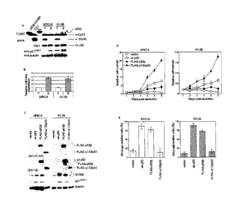
[0024] Fig. 1. p5313 and A133p53 are involved in cellular senescence and
proliferation: (A)
Induction of p5313 and repression of A133p53 at replicative senescence. The
immunoblot
analyses were performed in early-passage (Y) and senescent (S) human
fibroblast strains MRC-5
and WI-38. The examined passage numbers were 30 (Y) and 65 (S) for MRC-5; and
30 (Y) and
58 (S) for WI-38. TLQ40, an antibody detecting p5313 isoforms; MAP4, an
antibody detecting
A133p53; CM1, an antibody detecting wt p53. A40p5313 (Ghosh, A. et al., Mol.
Cell. Biol.
24:7987 (2004)) was a predominant form detected by TLQ40 and was
constitutively expressed in
both early-passage and senescent cells. p21WAF1 expression was also examined.
13-actin was a
loading control. H1299 cells overexpressing p5313 and CC1 cells (Horikawa, I.
et al., Hum. Mol.
Genet. 4:313 (1995)) were used as the positive controls for p5313 and A133p53,
respectively. (B)
miR-34a expression during replicative senescence. The same set of MRC-5 and WI-
38
fibroblasts as used in (A) were examined for miR-34a expression by real-time
ciRT-PCR. The
data were normalized with control RNU66 expression and shown as the relative
values. Three
independent experiments were carried out and the reproducible results were
obtained. (C)
Retroviral overexpression of p5313 and 4133p53 in human fibroblasts. The
retroviral vectors
driving wt p53, FLAG-tagged p5313 and FLAG-tagged A133p53 were transduced to
human
fibroblasts at early passage (at
6b
CA 02705488 2010-08-09
passage number 30 for both strains) and the immunoblot analyses of the
overexpressed p53
isoforms, MDM2 and p21wAF1 were performed. Protein samples were prepared from
the cells
at 8 days after retroviral transduction. The anti FLAG antibody detected FLAG-
tagged p533
and FLAG-tagged A133p53, and the DO-12 antibody detected all the three p53
isoforms. 13-
actin was a loading control. (D) Effects of p5313 and A133p53 on cell
proliferation. The cells
were plated at 8 days after retroviral transduction and the cell numbers were
counted daily.
Vector (open squares), wt p53 (open diamonds), FLAG-p533 (closed circles), and
FLAG-
A133p53 (closed triangles). The data (mean standard error) were from three
independent
experiments. (E) Senescence-associated 13-ga1actosidase (SA-13-ga1) assay. The
cells were
examined at 8 days after retroviral transduction. The data (mean standard
error) were from
three independent experiments.
[0025] Fig. 2. Overexpression of A133p53 extends replicative lifespan. (A)
Examination
of cellular replicative lifespan. The FLAG-A133p53 retroviral vector (open
circles) or the
control vector (open squares) was transduced to human fibroblasts at late
passage (MRC-5 at
passage 53 and WI-38 at passage 51). The cumulative population doublings (PDL)
were
calculated and plotted to days after G418 selection. (B) Telomere length and
telomeric 3'
overhang in A133p53-overexpressing cells. Genomic DNA samples from MRC-5 with
FLAG-A133p53 or control vector were used in the in-gel hybridization with 32P-
[CCCTAA]4
(SEQ ID NO:5) probe under denatured (for telomere length) and native (for
telomeric 3'
overhang) conditions. Lane 1, MRC-5 before transduction; lanes 2-3, vector
control (days 4
and 35 post selection); lanes 4-6, FLAG-A133p53 (days 4, 35 and 96 post
selection). The
telomere lengths were measured as peak TRF (terminal restriction fragment)
lengths. The
amounts of telomeric 3' overhang were normalized with loaded DNA amounts
(EtBr) and
shown as percent signals to the cells before transduction. (C) Repression of
miR-34a
expression by A133p53. RNA samples from MRC-5 (at passage 53) before
transduction (day
0), MRC-5 with control vector and MRC-5 overexpressing A133p53 (at days 20, 36
and 96
post selection) were analyzed as in Fig. 1B. The value before transduction was
defined as 1.0
and the expression levels in the other samples were expressed as the relative
values. (D)
Extension of cellular replicative lifespan by inhibition of miR-34a
expression. The late-
passage MRC-5 fibroblasts were transfected with the antisense oligonucleotide
against miR-
34a and the control oligonucleotide (EGFP) every four days and the cumulative
PDL were
examined as in (A) (left panel). The downregulation of miR-34a expression was
confirmed
by the real-time qRT-PCR (right panel).
7
CA 02705488 2010-05-12
WO 2009/064590
PCT/US2008/080648
[0026] Fig. 3. Knockdown of endogenous 4133p53 expression induces cellular
senescence. Early-passage MRC-5 fibroblasts (at passage 32) were transfected
with siRNAs
targeting 4133p53 (4133si-1 and 4133si-2) and a control oligonucleotide twice
(at day 1 and
day 4), and at day 7 were used for immunoblot analyses (A) and examined for SA-
(3-Gal
activity (B and C) and bromo-deoxyuridine (BrdU) incorporation (D). (A) siRNA-
mediated
repression of 4133p53. Expressions of wt p53 (D0-1 antibody), 4133p53 (MAP4
antibody)
and p21wAF1 were examined. P.-actin was a loading control. (B) Representative
pictures of
SA-I3-Gal staining. (C) Summary of SA-I3-Gal assay. The data (mean standard
error) were
from two independent experiments. (D) Summary of BrdU incorporation assay. The
number
of BrdU-positive cells/the total number of cells examined (at least 100 cells
for each sample)
was recorded.
[0027] Fig. 4. 4133p53 inhibits wt p53-mediated degradation of TRF2. (A)
Immunoblot
analysis of TRF2 expression in 4133p53-overexpressing cells. MRC-5 and WI-38
fibroblasts
with FLAG-4133p53 or control vector at indicated days after G418 selection
(see Fig. 2A)
were examined for TRF2 expression. The expression of 4133p53 was confirmed by
anti-
FLAG antibody. (B) Immunoblot analysis of TRF2 in p53-knocked down cells. A
telomerase-immortalized fibroblast cell line (hTERT/NHF) was transduced with
the retroviral
shRNA vector targeting p53. (C) p53 regulation of miR-34a expression.
hTERT/NHF cells
transduced with p53 shRNA (left) and treated with 10 [IM of Nutlin-3a for 36 h
(right) were
examined for miR-34a expression, as in Fig. 1B. The data is shown as the
relative expression
level to control cells (-). (D) TRF2 expression in fibroblasts from Li-
Fraumeni syndrome
patients. MDAH041 has a p53 frame-shift mutation (-), and MDAH087 and MDAH172
have
p53 missense mutations (mt) (Yin, Y. et al.,. Cell 70:937 (1992)). p53-
heterozygous (wt/-
and wt/mt) and homozygous (-/- and mt/mt) fibroblasts were examined in
parallel. (E)
4133p53 abrogates wt p53-mediated downregulation of TRF2. Cells (293T) were
retrovirally transduced with Myc-tagged TRF2, wt p53 and FLAG-tagged 4133p53
as
indicated. Anti-Myc, anti-FLAG and DO-1 antibodies were used in immunoblot
analyses.
(F) Effects of a proteasome inhibitor (MG-132) on TRF2 expression. Control
hTERT/NHF,
4133p53-overexpressing hTERT/NHF and p53-knocked down hTERT/NHF were cultured
in
the presence (+) or absence (-) of 10 [IM of MG-132 (Sigma-Aldrich) for 5 hrs
and examined
for TRF2 expression. (G) TRF2 accumulation by the inhibition of Siah-1A
activity. The
FLAG-tagged, dominant-negative mutant of Siah-1A (FLAG-Siahl-ARING) was
expressed
in MDAH041 fibroblasts (arrow). P-catenin, known to be degraded by Siah-1A
(Matsuzawa,
8
CA 02705488 2010-08-09
S. I. et al., Mol. Cell 7:915 (2001)), was examined to confirrn the activity
of FLAG-Siahl-
ARING. 13-actin was a loading control in (A), (B), (D), (E), (F) and (G). (H)
Overexpression of
TRF2 extends replicative lifespan. MRC-5 fibroblasts at passage 39 were
transduced with the
retroviral vector driving A 1 33p53 or control vector (G418 resistant) and
selected with G418 for 7
-- days. These cells were then transduced with the retroviral vector driving
TRF2 or control vector
(puromycin resistant), selected with puromycin, and examined for cellular
replicative lifespan as
in Fig. 2A. For each combination of retroviral transductions, the cumulative
PDL at days post
puromycin selection were recorded.
[0028] Fig. 5. MAP4 specifically recognizes A133p53. H1299 cells (p53-null)
transfected
-- with the expression vector for wild-type (wt) p53, p53[3 or A133p53 were
analyzed in Western
blot using MAP4 (left) and DO-1 (right) antibodies. MAP4 detects A133p53, but
not wt p53 or
p5313.
[0029] Fig. 6. mRNA expression analysis of p53 isoforms in human fibroblasts.
The same
sets of cells as in Fig. lA were analyzed by RT-PCR. In contrast to protein
levels, mRNA of
-- p5313 was decreased in senescent cells and A133p53 was primarily unchanged.
The primers to
amplify wt p53 were: 5'-CTC ACC ATC ATC ACA CTG GAA-3' (SEQ ID NO:6) and 5'-
TCA
TTC AGC TCT CGG AAC ATC-3' (SEQ ID NO:7). The primers specifically detecting
the
alternative splicing for p5313 were: 5'-CTT TGA GGT GCG TGT TTG TGC-3' (SEQ ID
NO:8)
and 5'-TTG AAA GCT GGT CTG GTC CTG A-3' (SEQ ID NO:9). The primers
specifically
-- amplifying A133p53 mRNA transcribed from the promoter in intron 4 were: 5'-
TGG GTT GCA
GGA GGT GCT TAC-3' (SEQ ID NO:10) and 5'-CCA CTC GGA TAA GAT GCT GAG G-3'
(SEQ ID NO:11). The lower bands correspond to the reported A133p53 sequences
(GenBank
DQ186650). The upper bands are from mRNA with intron 5 unspliced. GAPDH was
amplified
as a control as previously described (Horikawa, I. et al., Mol. Carcinog.
22:65 (1998)).
-- [0030] Fig. 7. Senescence-associated (SA)-13-galactosidase (gal) staining
of MRC-5
fibroblasts overexpressing wt p53, FLAG-tagged p53f1 and FLAG-tagged A133p53.
MRC-5 with
control vector is also shown.
[0031] Fig. 8. p5313 overexpression induces cellular senescence in human
fibroblasts with
ectopically expressed telomerase. (A) Effects of p5313 on cell proliferation.
hTERT (human
-- telomerase reverse transcriptase) -immortalized human fibroblasts
(hTERT/NHF) were
transduced with the retroviral vector driving FLAG-tagged p5313 or control
vector (a zeocin-
9
CA 02705488 2010-05-12
WO 2009/064590
PCT/US2008/080648
resistant version). Cell proliferation assay was carried out as in Fig. 2B.
(B) Upregulation of
p2, WAF1
by p53r3 overexpression in hTERT/NHF cells. (C) Representative pictures of
SAT.-
gal staining. (D) Summary of SA-f3-gal staining. The data were mean standard
error from
three independent experiments.
[0032] Fig. 9. 4133p53 overexpression delays replicative senescence in late-
passage
human fibroblasts. MRC-5 fibroblasts with control vector or FLAG-tagged
4133p53 (same
cells as in Fig. 3A) were stained for SA-13-gal activity at 10 days post G418
selection. (A)
Representative pictures. (B) Data summary.
[0033] Fig. 10. Knockdown of endogenous 4133p53 induces cellular senescence.
Early-
passage WI-38 fibroblasts (at passage 30) were transfected with siRNAs
targeting 4133p53
(4133si-1 and 4133si-2) and a control oligonucleotide and examined in
immunoblot analyses
(A), SA-13-Gal assay (B) and BrdU incorporation assay (C), as performed in
Fig. 3.
[0034] Fig. 11. Nutlin-3A downregulates TRF2 protein in a p53-dependent
manner.
hTERT/NHF cells with (+) or without (-) p53 shRNA were treated with 10 [IM of
Nutlin-3A
(Cayman Chemical) for the indicated time period and examined for TRF2, p53 and
MDM2
amounts in immunoblot analyses. r3-actin was a loading control.
[0035] Fig. 12. The p53 knockdown-induced increase in TRF2 protein is not due
to an
increase in TRF2 mRNA. hTERT/NHF cells with (+) and without (-) p53 shRNA were
examined for TRF2 mRNA expression by the real-time qRT-PCR (cat. no.
04689038001,
Roche Applied Science).
[0036] Fig. 13. 4133p53 does not affect TRF2 expression in the absence of wt
p53. (A)
wt p53, FLAG-tagged p53r3 and FLAG-tagged 4133p53 were retrovirally expressed
in
MDAH041 (p53-/-) fibroblasts. Neither p53r3 nor 4133p53 changed TRF2
expression in
these cells, while a significant decrease in TRF2 was observed with wt p53.
The expression
of Siah-1A was also examined and shown to be induced by wt p53. The expression
of p53
isoforms was confirmed with anti-FLAG antibody and/or anti-p53 antibody (D0-
1). (B)
Downregulation of TRF2 by Siah-1A overexpression. Cells (293T) were
retrovirally
transduced with Myc-tagged TRF2, FLAG-tagged Siahl-A6 (a stable form of Siah-
1A)
(Tanikawa, J. et al.,1 Biol. Chem. 279:55393 (2004)) and wt 53 as indicated.
Anti-Myc,
anti-FLAG and DO-1 antibodies were used in immunoblot analyses. (C) 4133p53
was
knocked down by p53 shRNA in CC1 cells, which express 4133p53 but not wt p53
due to a
CA 02705488 2010-05-12
WO 2009/064590
PCT/US2008/080648
genomic rearrangement (Horikawa, I. et al., Hum. Mol. Genet. 4:313 (1995)). No
change in
TRF2 expression was observed with a remarkable decrease in 4133p53 (confirmed
by
immunoblot using MAP4). 13-actin was a loading control in (A), (B) and (C).
[0037] Fig. 14. Replicative senescence-associated changes in expression of
endogenous
p53 isoforms and p53-regulated microRNA-34a. a, Induction of p53r3 and
repression of
4133p53 at replicative senescence. The immunoblot analyses were performed in
early-
passage (Y) and senescent (S) human fibroblast strains MRC-5 and WI-38. The
examined
passage numbers were 30 (Y) and 65 (S) for MRC-5; and 30 (Y) and 58 (S) for WI-
38.
TLQ40, an antibody detecting p53r3 isoforms; MAP4, an antibody detecting
4133p53; DO-
12, an antibody used to detect full-length p53; CM1, an antibody used to
simultaneously
detect full-length p53, p53r3 and 4133p53. 440p53r3 was a predominant form
detected by
TLQ40 and was constitutively expressed in both early-passage and senescent
cells. p21wAF1
expression was also examined. r3-actin was a loading control. H1299 cells
overexpressing
p53r3 and CC1 cells were used as the positive controls for p53r3 and 4133p53,
respectively. b,
miR-34a expression during replicative senescence. The same set of MRC-5 and WI-
38
fibroblasts as used in a were examined for miR-34a expression by real-time qRT-
PCR. The
data were normalized with control RNU66 expression and shown as the relative
values (mean
s.d. from triplicate sample). Three independent experiments were carried out
and the
reproducible results were obtained. *, p < 0.001. **, p < 0.01. c and d,
Extension of cellular
replicative lifespan by the inhibition of miR-34a expression. Late-passage MRC-
5 fibroblasts
(at passage 58) were transfected with the antisense oligonucleotide against
miR-34a and the
control oligonucleotide (EGFP). The effectiveness of the antisense miR-34a was
confirmed
by the real-time qRT-PCR (error bars represent s.d. from triplicate sample)
(c). The
transfection was repeated every 4 days and the cumulative population doublings
(PDL) were
examined (d). e, Knockdown of miR-34a expression partially inhibits Nutlin-3A-
induced
senescence. hTERT-immortalized human fibroblasts (hTERT/NHF) were transfected
with the
antisense miR-34a or control oligonucleotide, and then induced to senesce by
treatment with
10 uM of Nutlin-3A for 72 h. Summary of senescence-associated P-galactosidase
(SA-I3-gal)
assay is shown. The data (mean s.d.) were from three independent
experiments. *, p < 0.05.
[0038] Fig. 15. Knockdown of endogenous 4133p53 induces cellular senescence.
Early-passage WI-38 fibroblasts (at passage 30) were transfected with siRNAs
targeting
4133p53 (4133si-1 and 4133si-2) and a control oligonucleotide twice (at day 1
and day 4),
11
CA 02705488 2010-05-12
WO 2009/064590
PCT/US2008/080648
and at day 7 were used for immunoblot analyses (a) and examined for SA-P-gal
activity (b
and c), bromo-deoxyuridine (BrdU) incorporation (d) and PAI-1 (plasminogen
activator
inhibitor-1) expression (e). a, siRNA-mediated repression of 4133p53.
Expressions of full-
length p53 (D0-1 antibody), 4133p53 (MAP4 antibody), p5313 (TLQ40 antibody)
and
p2iwAFi
were examined. The expression levels of full-length p53 and 4133p53 were also
confirmed by the CM1 antibody. P.-actin was a loading control. H1299
expressing p53I3 was
the positive control for TLQ40. b, Representative pictures of SA-I3-gal
staining. c, Summary
of SA-I3-gal assay. The data (mean s.d.) were from three independent
experiments. *, p <
0.01. d, BrdU incorporation assay. The number of BrdU-positive cells/the total
number of
cells examined (at least 100 cells for each well) was recorded. Data are mean
s.d. from
triplicate wells. *, p < 0.05. **, p < 0.01. e, The real-time qRT-PCR assay of
PAI-1. The
relative expression levels of PAI-1 mRNA are shown. Error bars represent s.d.
from triplicate
sample. *, p < 0.05. **, p < 0.01.
[0039] Fig. 16. Overexpression of p5313 induces senescence and overexpression
of
4133p53 extends replicative lifespan. Effects of retrovirally overexpressed
p53P and
4133p53 on cell proliferation and senescence. a, Early-passage MRC-5 and WI-38
fibroblasts (both at passage 32) were retrovirally transduced with vector
alone (open squares),
full-length p53 (open diamonds), FLAG-tagged p53P (closed circles) and FLAG-
tagged
4133p53 (closed triangles), and used in cell proliferation assay at 8 days
after retroviral
transduction. The cell numbers were counted daily and the data (mean s.d.)
were from three
independent experiments. b, Summary of SA-I3-gal assay. The same set of cells
as in (a) were
examined at 8 days after retroviral transduction. The data (mean s.d.) were
from three
independent experiments. *, p < 0.01. c, Extension of cellular replicative
lifespan by
4133p53. The FLAG-4133p53 retroviral vector (open circles) or the control
vector (open
squares) was transduced to human fibroblasts at late passage (MRC-5 at passage
53 and WI-
38 at passage 51). The cumulative PDL were calculated and plotted to days
after G418
selection. d, SA-I3-gal staining of control and 4133p53-overexpressing MRC-5
fibroblasts.
The pictures at 36 days post-selection are shown. e, Repression of miR-34a
expression by
4133p53. RNA samples from MRC-5 (at passage 53) before transduction (day 0),
MRC-5
with control vector and MRC-5 overexpressing 4133p53 (at days 20, 36 and 96
post-
selection) were analyzed as in Fig. 14b. The value before transduction was
defined as 1.0 and
the expression levels in the other samples were expressed as the relative
values (mean s.d.
12
CA 02705488 2010-08-09
from triplicate sample). f, Telomere length and telomeric 3' overhang in
A133p53-
overexpressing cells. Genomic DNA samples from MRC-5 with FLAG-A133p53 or
control
vector were used in the in-gel hybridization with 32
vector ID NO:5) probe
under denatured (for telomere length) and native (for telomeric 3' overhang)
conditions. Lane
1, MRC-5 before transduction; lanes 2-3, vector control (days 4 and 35 post-
selection); lanes
4-6, FLAG-A133p53 (days 4, 35 and 96 post-selection). The telomere lengths
were measured
as peak TRF (terminal restriction fragment) lengths. The amounts of telomeric
3' overhang
were normalized with loaded DNA amounts (EtBr) and shown as percent signals to
the cells
before transduction.
100401 Fig. 17. p53 isoform expression profiles in colon carcinogenesis in
vivo. Elevated
expression of p53r3 and reduced expression of A133p53 in colon adenomas with
senescent
phenotypes, but not in colon carcinomas. (a) SA-13-gal staining of non-adenoma
and adenoma
tissues. The results of case 7 are shown. The rectangular areas are enlarged
in the right
panels. Bars, 500 pm. (b) The expression levels of p5313 and A133p53 were
quantitatively
examined in 9 normal colon tissues obtained from immediate autopsy2I (Table
1), 8 matched
pairs of non-adenoma and adenoma tissues (Table 2) and 29 matched pairs of non-
carcinoma
and carcinoma tissues (Table 3). The data (mean and s.d.) are shown in a
logarithmic scale as
the relative values to normal colon samples. *, p( 0.05 compared with normal
colon. (c) The
expression levels of p5313 and A133p53 in colon carcinomas were analyzed
according to
tumour stage. The data of normal colon and adenoma samples are same as those
in (b). The
expression levels (mean and s.d.) in adenomas, stage I (n = 8), stage II (n =
11) and stage III
(n = 10) carcinomas are shown as relative log2 values to normal colon (defined
as 0, not
shown). *, p < 0.05.
[00411 Fig. 18. SA-13-gal staining in replicative senescence and oncogene-
induced
premature senescence. a, MRC-5 and WI-38 fibroblasts at early passage (upper
panels) and
at replicative senescence (lower panels). b, MRC-5 and WI-38 retrovirally
transduced with
vector control (upper panels) and pBabe-Puro ras (H-RasV12) (Serrano et al.
Cell 88, 593-
602 (1997)) (lower panels). Note that premature senescence by POT1 knockdown
was
induced and confirmed by SA-J3-gal staining as described in our previous study
(Yang et al.
Cancer Res. 67, 11677-11686 (2007)). The dominant-negative TRF2-induced
senescence was
also as previously described by the present inventors (Yang et al. MoL Cell.
Biol. 25, 1070-
1080 (2005)) and others (van Steensel et al. Cell 92, 401-413 (1998)).
13
CA 02705488 2010-05-12
WO 2009/064590
PCT/US2008/080648
[0042] Fig. 19. MAP4 specifically recognizes A133p53. H1299 cells (p53-null)
transfected with the expression vector for wild-type (wt) p53, p5313 or
4133p53 were
analyzed in immunoblot using MAP4 (left) and DO-1 (right) antibodies. MAP4
detects
4133p53, but not wt p53 or p53P.
[0043] Fig. 20. p53 isoform switching does not occur with premature
senescence.
4133p53 and p5313 expression in oncogene-induced senescence (overexpression of
H-
RasV12) (Serrano et al. Cell 88, 593-602 (1997)) (a) and premature senescence
with acute
telomere dysfunction induced by shRNA knockdown of POT1 (Yang et al. Cancer
Res. 67,
11677-11686 (2007)) (b) or overexpression of a dominant-negative TRF2 mutant
(Yang et al.
Mol. Cell. Biol. 25, 1070-1080 (2005); van Steensel et al. Cell 92, 401-413
(1998)) (c). Early-
passage MRC-5 and WI-38 (at passage 32) were used. H1299 cells overexpressing
p5313 was
the positive control for p5313. P.-actin was a loading control.
[0044] Fig. 21. miR-34a expression is p53-dependent. hTERT-immortalized human
fibroblasts (hTERT/NHF) (Sengupta et al. EIVIBO 1 22, 1210-1222 (2003))
transduced with
the shRNA knockdown vector targeting p53 (Brummelkamp and Agami Science 296,
550-
553 (2002)) (left) or treated with 10 JIM of Nutlin-3a for 36 h (Kumamoto et
al. Cancer Res.
68, 3193-3203 (2008)) (right) were examined for miR-34a expression, as in Fig.
14b. The
data (mean s.d. from triplicate sample) is shown as the relative expression
level to control
cells (-).
[0045] Fig. 22. Knockdown of endogenous A133p53 induces cellular senescence.
Early-
passage MRC-S fibroblasts (at passage 32) were transfected with the siRNAs
targeting
4133p53 (4133si-1 and 4133si-2) and a control oligonucleotide and examined in
immunoblot analyses (a), SA-13-gal assay (b) and BrdU incorporation assay (c),
as performed
in Fig. 2. *, p < 0.001.
[0046] Fig. 23. A133p53 knockdown does not induce apoptosis in human
fibroblasts.
MRC-S and WI-38 transfected with control, 4133si-1 and 4133si-2
oligonucleotides were
examined for caspase-3 (top) and PARP (middle, short and long exposure) in
immunoblot.
RKO cells treated with doxorubicin (DOX) were included as the positive control
showing
apoptosis. 13-actin was a loading control (bottom). No cleaved caspase-3 or
PARP was
observed in 4133p53-knocked-down fibroblasts.
14
CA 02705488 2010-05-12
WO 2009/064590
PCT/US2008/080648
[0047] Fig. 24. mir-34a is not upregulated at A133p53 knockdown-induced
senescence. MRC-S and WI-38 transfected with control, 4133si-1 and 4133si-2
oligonucleotides were examined for miR-34a expression, as in Fig. 14b,
together with
untransfected early-passage (Y) and replicatively senescent (R.S.) cells. The
data (mean s.d.
from triplicate sample) are shown as the relative expression levels to
untransfected early-
passage cells (Y, -).
[0048] Fig. 25. Retroviral overexpression of p53 isoforms in human
fibroblasts. The
retroviral vectors driving full-length p53, FLAG-tagged p53P. and FLAG-tagged
4133p53
were transduced to human fibroblasts at early passage (at passage number 30
for both strains)
and the immunoblot analyses of the overexpressed full-length p53 and p53
isoforms, MDM2
and p21wAF1 were performed. Protein samples were prepared from the cells at 8
days after
retroviral transduction. The anti-FLAG antibody detected FLAG-tagged p5313 and
FLAG-
tagged 4133p53. The DO-12 antibody detected full-length p53, FLAG-tagged p5313
and
FLAG-tagged 4133p53. P.-actin was a loading control.
[0049] Fig. 26. p5313 overexpression induces cellular senescence in human
fibroblasts
with ectopically expressed telomerase. a, Effects of p53P. on cell
proliferation.
hTERT/NHF cells (Sengupta et al. EMBO 1 22, 1210-1222 (2003)) were transduced
with the
retroviral vector driving FLAG-tagged p53I3 or control vector (a zeocin-
resistant version).
Cell proliferation assay was carried out as in Fig. 16a. b, Upregulation of
p21wAF1 by p53P.
overexpression in hTERT/NHF cells. c, Representative pictures of SA-I3-gal
staining. d,
Summary of SA-I3-gal staining. The data were mean s.d. from three
independent
experiments. *, p < 0.01.
[0050] Fig. 27. A133p53 overexpression extends the replicative lifespan in
human
fibroblasts. Late-passage MRC-S (at passage 55) and WI-38 (at passage 53) were
transduced with the FLAG-4133p53 retroviral vector or the control vector and
examined for
the cumulative PDL, as in Fig. 16c.
[0051] Fig. 28. Immunoblot analyses of p16INK4A, A133p53 and p5313 in human
colon
adenomas. Eight cases of matched non-adenoma (N) and adenoma (A) tissues were
examined for p16INK4A, 4133p53 and p5313. I3-actin was the control for
quantitation. The data
shown in Fig. 34e and 4f were from the quantitative analysis of these results.
CA 02705488 2010-05-12
WO 2009/064590
PCT/US2008/080648
[0052] Fig. 29. Increased plea' expression in colon adenomas. The expression
levels
of pl6INK4A, an in vivo senescence marker, were examined in 9 normal colon
tissues (Table 1)
and 8 pairs of non-adenoma and adenoma tissues (Table 2) and quantitatively
analyzed. The
data (mean s.d.) are shown as the relative values to normal colon samples.
*, p < 0.0001.
[0053] Fig. 30. Paired t-test analyses of plea', A133p53 and p533 expression
in
matched colon adenoma and non-adenoma tissues. The same data as in Fig. 17b
and Fig.
29 from 8 pairs of non-adenoma (Non-ad) and adenoma tissues were analyzed by
paired t-
test. The vertical axes are the expression levels normalized with 13-actin.
The p-values for
p16INK4A,
4133p53 and p53r3 are 0.0004, 0.024 and 0.03, respectively, and the
corresponding
Bonferroni corrected p-values are 0.001, 0.07 and 0.09, respectively. Case 1,
aqua; case 2,
blue; case 3, cyan; case 4, yellow; case 5, lavender; case 6, navy; case 7,
purple; and case 8,
brown.
[0054] Fig. 31. Immunoblot analyses of A133p53 and p53P expression in matched
colon carcinoma and non-carcinoma tissues. Twenty-nine cases of matched colon
carcinoma (T) and non-carcinoma (N) tissues (Table 3) were examined for
4133p53 and
p53r3. r3-actin was the control for normalization. Each of the six SDS-PAGE
gels included 5
pairs of carcinoma/non-carcinoma tissues, as well as the same set of normal
colon, non-
adenoma and adenoma samples, which allowed quantitative comparisons among
different
blots and different histopathological types, as in Figs 17b and c. One case
(12375) was
duplicated. The data shown in Fig. 17b (Non-ca and Ca), 4c (Carcinoma, stage
I, II and III)
and Fig. 32 were from the quantitative analysis of these results.
[0055] Fig. 32. Paired t-test analyses of A133p53 and p53p expression in p53
'wild-
type' versus 'mutant' cases of colon carcinomas. Twenty-eight cases of colon
carcinomas
were divided into two subgroups assumedly with 'wild-type' or 'mutant' p53,
based on the
immunohistochemical staining of p53 and MDM2 (Costa et al., The Journal of
pathology
176, 45-53 (1995); Nenutil et al., The Journal of pathology 207, 251-259
(2005)). In each
subgroup, the expression levels of 4133p53 (a) and p53r3 (b) were compared
between non-
carcinoma (Non-ca) and carcinoma tissues by paired t-test. The vertical axes
are the
expression levels normalized with r3-actin. The p-values are in the
parentheses. The p53
'wild-type' carcinomas, but not "mutant" carcinomas, expressed significantly
higher levels of
4133p53. p53r3 was significantly less abundant in carcinoma tissues in both
subgroups
16
CA 02705488 2010-05-12
WO 2009/064590
PCT/US2008/080648
because of the marked increase in non-carcinoma tissues (Fig. 17b). The actual
values in each
of the 28 cases are shown in Table 4.
[0056] Fig. 33. IL-8 and IL-8R expression in colon adenoma and carcinoma
tissues.
The mRNA expression levels of IL-8 (upper panel) and IL-8R (lower panel) were
examined
by qRT-PCR in 8 matched pairs of non-adenoma and adenoma tissues (Table 2) and
29
matched pairs of non-carcinoma and carcinoma tissues (Table 3). The expression
levels
(mean and s.d.) in non-carcinoma, adenoma and carcinoma samples are shown as
relative
log2 values to non-adenoma (defined as 0). *, p < 0.05 compared with non-
adenoma or non-
carcinoma. **, p < 0.001 compared with non-adenoma or non-carcinoma.
[0057] Fig. 34. p53 isoform switching in vivo. a-c, Increased p5313 and
decreased
4133p53 expression during CD8+ T lymphocyte senescence in vivo. a, CD8+ T
lymphocytes
were purified from blood samples freshly isolated from healthy donors of age
50 years old,
and sorted by flow cytometry using anti-CD28 and anti-CD57 antibodies. The
result of 50-
year-old male is shown. b, Representative immnunoblot of p533 and 4133p53. The
results of
65-year-old male are shown. HP1-y was examined as a senescence marker. 13-
actin was a
loading control for quantitation. c, The expression levels of p53P and 4133p53
in each of the
quadrants were quantitated in immunoblot analyses and shown as the relative
values to the
CD28-CD57+ quadrant (p53P) or CD28 CD57- quadrant (A133p53). The data (mean
s.d.)
were from three donors (60-year-old female, 65-year-old male and 50-year-old
male). The p-
values from ANOVA trend analysis are shown. d-f, Elevated expression of p53P
and reduced
expression of 4133p53 in colon adenomas with senescent phenotypes. d, SA-I3-
gal staining
of non-adenoma and adenoma tissues. The results of case 7 are shown. The
rectangular areas
are enlarged in the right panels. Bars, 500 e, The expression levels of
p53P and 4133p53,
as well as a senescence marker p16'NK4A, were examined in 9 normal colon
tissues obtained
from immediate autopsy (Table 1) and 8 matched pairs of non-adenoma and
adenoma tissues
surgically resected (Table 2) and quantitatively analyzed. The data (mean
s.d.) are shown in
a logarithmic scale as the relative values to normal colon samples. *, p <
0.05. **, p < 0.0005.
***, p < 0.00005. f, The same data as in (e) from 8 matched pairs of non-
adenoma (Non-ad)
and adenoma tissues were analyzed by paired t-test. The vertical axes are the
expression
levels normalized with I3-actin. The p-values for pl6INK4A, 4133p53 and p5313
are 0.0004,
0.024 and 0.03, respectively, and the corresponding Bonferroni corrected p-
values are 0.001,
17
CA 02705488 2010-08-09
0.07 and 0.09, respectively. Case 1, aqua; case 2, blue; case 3, cyan; case 4,
yellow; case 5,
lavender; case 6, navy; case 7, purple; and case 8, brown.
[0058] Fig. 35. mRNA expression analysis of p53 isoforms in human fibroblasts.
The
same sets of cells as in Fig. 14a were analyzed by RT-PCR. The primers to
amplify wt p53
were: 5'-CTC ACC ATC ATC ACA CTG GAA-3' (SEQ ID NO:6) and 5'-TCA TTC AGC
TCT CGG AAC ATC-3' (SEQ ID NO:7). The primers specifically detecting the
alternative
splicing for p5313 were: 5'-CTT TGA GGT GCG TGT TTG TGC-3' (SEQ ID NO:8) and
5'-
TTG AAA GCT GGT CTG GTC CTG A-3' (SEQ ID NO:9). The primers specifically
amplifying A133p53 mRNA transcribed from the promoter in intron 4 were: 5'-TGG
GTT
GCA GGA GGT GCT TAC-3' (SEQ ID NO:10) and 5'-CCA CTC GGA TAA GAT GCT
GAG G-3' (SEQ ID NO:11). The lower bands correspond to the reported A133p53
sequences
(GenBank DQ186650). The upper bands are from mRNA with intron 5 unspliced.
GAPDH
was amplified as a control as previously described (Horikawa and Barrett Mol.
Carcinog. 22,
65-72 (1998)).
[0059] Fig. 36. FACS (Fluorescence-activated cell sorting) of human CD8+ T
lymphocytes. a, Summary of the sorted fractions from three donors. b, The
purity of sorted
fractions was checked by FACS reanalysis. The result of 50-year-old male is
shown. c,
Immunoblot analysis of the sorted fractions for HP1-y as a senescence marker
(Collado et al.
Nature 436, 642 (2005); Narita et al. Cell 113, 703-716 (2003); Zhang et al.
J. Cell Science
120, 1572-1583 (2007)). The expression levels of HP1-y were quantitated and
expressed as
the relative values to CD28+CD57- fraction. The data (mean s.d.) were from
three donors.
The difference between CD28 CD57- and CD28-CD57+ fractions is statistically
significant (p
( 0.05).
[0060] Fig. 37. A133p53 and p53[3 expression in human CD8+ T lymphocytes.
Immunoblot analysis as shown in Fig. 14b. a, 60-year-old female. b, 50-year-
old male.
[0061] Fig. 38. A133p53 is not subject to proteasomal degradation. Early-
passage (Y)
and replicatively senescent (S) MRC-5 and WI-38 (the same set of cells as in
Fig. 14a) were
maintained in the presence (+) or absence (-) of 15 M of the proteasome
inhibitor MG-132
for 8 hrs and examined in immunoblot.
18
CA 02705488 2010-05-12
WO 2009/064590
PCT/US2008/080648
DETAILED DESCRIPTION OF THE INVENTION
I. Introduction
[0062] The finite division potential of normal human cells leads to cellular
senescence,
which functions as a barrier to human cell transformation and carcinogenesis
(Collado, M. ,
et al., Cell 130:223 (2007)). The induction and prevention of cellular
senescence in human
cells involve the regulation of the specific chromosome end structure,
telomeres (Verdun, R.
E. et al., Nature 447:924 (2007)). The tumor suppressor protein p53 plays a
central role in
sensing and signaling a variety of intrinsic stresses (e.g., telomere
dysfunction) and
environmental cues that induce cellular senescence (Collado, M. , et al., Cell
130:223
(2007); Herbig, U. et al. , Mol. Cell 14:501 (2004)). p53 and Arf can also
cooperate to have
anti-oxidative and anti-aging activities (Matheu, A. et al., Nature 448:375
(2007)). Many of
the mutant p53 proteins observed in human cancers inhibit the tumor
suppressive functions of
full-length, wild-type p53 (wt p53) in a dominant-negative manner (Rozan, L.
M. et al., Cell
Death Differ. 14:3 (2007)). It is suggested that some p53 mutants also gain a
tumor-
promoting function independent of the inhibition of wt p53 (Rozan, L. M. et
al., Cell Death
Differ. 14:3 (2007); Kastan, M. B. et al. , Nat. Cell Biol. 9:489 (2007)). The
human p53
gene encodes, in addition to wt p53, several N-terminally, internally and C-
terminally
truncated isoforms due to alternative promoter usage and RNA splicing (Chan,
W. M. et al.,
Cancer Res. 67:1959 (2007), Bourdon, J. C. et al., Genes Dev. 19:2122 (2005)).
A plausible
hypothesis is that these p53 isoforms cooperate or compete with wt p53 to
modulate the p53's
multiple functions. To test this hypothesis, we examine here the roles of two
major isoforms,
p53r3 (lacking the C-terminal oligomerization domain due to an alternative
splicing) and
4133p53 (transcribed from the alternative promoter in intron 4 and lacking the
N-terminal
transactivation and proline-rich domains) (Bourdon, J. C. et al., Genes Dev.
19:2122 (2005)),
in the regulation of cellular senescence and their functional interplay with
wt p53. Our data
provide novel insights into the p53 regulation of cellular replicative
lifespan.
11. p53 proteins
[0063] p53 is a protein of apparent molecular 53 kDa on SDS PAGE that
functions as a
transcription factor that, among other functions, regulates the cell cycle and
functions as a
tumor suppressor. p53 has been described as "the guardian of the genome",
referring to its
role in providing stability by preventing genome mutation. Among p53 's anti-
cancer
activities include: activation of DNA repair proteins when DNA has sustained
damage; cell
cycle arrest at the Gl/S regulation point when a cell has sustained DNA
damage, thus
19
CA 02705488 2010-05-12
WO 2009/064590
PCT/US2008/080648
allowing DNA repair proteins time to fix the damage before allowing
continuation of the cell
cycle; and the initiation of apoptosis or the programmed cell death, if the
DNA damage
proves to be irreparable.
[0064] Accordingly, p53 can induce growth arrest, apoptosis, and cell
senescence. In
normal cells, p53 is generally held in an inactive form, bound to the protein
MDM2 (HDM2
in humans), which prevents p53 activity and promotes p53 degradation by acting
as a
ubiquitin ligase. Active p53 is induced in response to various cancer-causing
agents such as
UV radiation, oncogenes, and some DNA-damaging drugs. DNA damage is sensed by
'checkpoints' in a cell's cycle, and causes proteins such as ATM, CHK1 and
CHK2 to
phosphorylate p53 at sites that are close to or within the MDM2-binding region
and p300-
binding region of the protein. Oncogenes also stimulate p53 activation,
mediated by the
protein pl4ARF. Some oncogenes can also stimulate the transcription of
proteins which bind
to MDM2 and inhibit its activity. Once activated, p53 activates expression of
several genes
including one encoding for p21, a cell cycle inhibitor. p21 binds to G1-S-
phase and S-phase
cyclin CDK complexes inhibiting their activity. See, e.g., Mills, Genes &
Development, 19:
2091-2099 (2005) for a review.
[0065] Other isoforms or variants of p53 have been identified (see Bourdon,
Brit. 1
Cancer, 97: 277-282 (2007)). For example, two isoforms of p53, p63 and p73,
which are
encoded by distinct genes, have been identified (Kaghad et al., Cell 90: 809-
819 (1997); and
Yang et al. Mol. Cell (1998)). Human p53 isoforms may also arise due to
alternative
promoter usage and alternative splicing. Alternative promoter usage, for
example, can give
rise to the expression of an N-terminally truncated p53 protein initiated at
codon 133
(4133p53). Adding to the complexity of p53 isoforms is the alternative
splicing of intron 9
of the p53 gene to provide the isoforms p5313 and p53y. Combined with
alternative promoter
usage, this gives rise to the p53 isoforms: p53, p5313, p53y, 4133p53,
A133p5313, and
4133p53y. The use of an alternative promoter in intron 2 gives rise to the
additional
isoforms, 440p53, A40p5313, and 440p53y. While the presence of these multiple
p53
isoforms has been established, the biological function of these isoforms
remains obscure.
The present invention invention is based in part on an elucidation of the role
for two of these
isoforms, 4133p53 and p5313, in the opposing functions of cell senescence and
cell
proliferation.
CA 02705488 2010-08-09
111. Definitions
[0066] The term "p53" refers generally to a protein of apparent molecular
weight of 55kDa
on SDS PAGE that functions as a tumor suppressor as described herein. The
protein and
nucleic sequences of the p53 protein from a variety of organisms from humans
to Drosophila
are known and are available in public databases, such as in accession numbers,
NM_000546,
NP 000537, NM 011640, and NP 035770, for the human and mouse sequences.
[0067] The term "A133p53" refers generally to the isoform of p53 that arises
from
initiation of transcription of the p53 gene from codon 133, which results in
an N-terminally
truncated p53 protein. This isoform comprises the following p53 protein
domains: the
majority of the DNA binding domain, the NLS, and the C-terminal sequence
DQTSFQKENC
(SEQ ID NO:12) (see Bourdon, Brit. J. Cancer, 97: 277-282 (2007)).
[0068] The term "p530" refers generally to the isoform of p53 that arises from
alternative
splicing of intron 9 to provide a p53 isoform comprising the following p53
protein domains:
TAD1, TAD2, prD, the DNA binding domain, the NLS, and the C-terminal sequence
DQTSFQKENC (SEQ ID NO:12) (see Bourdon, Brit. J. Cancer, 97: 277-282 (2007)).
[0069] The term "cell senescence" refers generally to the phenomenon where
normal
diploid differentiated cells lose the ability to divide after undergoing a
finite number of cell
divisions characteristic of a particular type of cell.
[0070] The term "replicative lifespan" refers generally to the finite number
of cell divisions
undergone by a particular cell type before undergoing cell senescence and
losing the ability to
further divide.
[0071] The term "extending replicative lifespan" refers generally to the
continuation of cell
division in a normal diploid cell beyond the finite number of cell divisions
at which cell
senescence would occur.
[0072] The term "siRNA" refers to a nucleic acid that forms a double stranded
RNA, which
double stranded RNA has the ability to reduce or inhibit expression of a gene
or target gene
when the siRNA expressed in the same cell as the gene or target gene. "siRNA"
thus refers
to the double stranded RNA formed by the complementary strands. The
complementary
portions of the siRNA that hybridize to form the double stranded molecule
typically have
substantial or complete identity. In one embodiment, an siRNA refers to a
nucleic acid that
has substantial or complete identity to a target gene and forms a double
stranded siRNA. The
21
CA 02705488 2015-09-17
CA 2705488
sequence of the siRNA can correspond to the full length target gene, or a
subsequence thereof.
Typically, the siRNA is at least about 15-50 nucleotides in length (e,g., each
complementary
sequence of the double stranded siRNA is 15-50 nucleotides in length, and the
double stranded
siRNA is about 15-50 base pairs in length, preferable about preferably about
20-30 base
nucleotides, preferably about 20-25 nucleotides in length, e.g., 20, 21, 22,
23, 24, 25, 26, 27,
28, 29, or 30 nucleotides in length.
[0073] The tertn "shRNA" refers generally to an siRNA that is introduced into
a cell as part
of a larger DNA construct. Typically, such constructs allow stable expression
of the siRNA in
cells after introduction, e.g., by integration of the construct into the host
genome.
[0074] An "antisense" oligonucleotide or polynueleotide is a nucleotide
sequence that is
substantially complementary to a target polynuelcotide or a portion thereof
and has the ability
to specifically hybridize to the target polynucleotide.
[0075] Ribozymes are enzymatic RNA molecules capable of catalyzing specific
cleavage of
RNA. The composition of ribozyme molecules preferably includes one or more
sequences
complementary to a target mRNA, and the well known catalytic sequence
responsible for
mRNA cleavage or a functionally equivalent sequence (see, e.g., U.S. Pat. No.
5,093,246).
Ribozyme molecules designed to catalytically cleave target mRNA transcripts
can also be used
to prevent translation of subject target mRNAs.
[0076] The term "promoting" as used, for example in the context of "promoting
senescence,"
refers generally to conditions or agents which increase, induce, open,
activate, facilitate,
enhance activation, sensitize, agonize, or up regulate cell senescence.
[0077] The phrase "functional effects" in the context of assays for testing
compounds that
modulate a protein of the invention includes the determination of a parameter
that is indirectly
or directly under the influence of a protein of the invention, e.g., a
chemical or phenotypic
effect such as altered transcriptional activity of p53 isoforms and the
downstream effects of
such proteins on cellular metabolism and proliferation or growth. A functional
effect therefore
includes transcriptional activation or rcpression, the ability of cells to
proliferate or undergo
apoptosis, whether and at what point cells undergo senescence, among others.
"Functional
effects" include in vitro, in vivo, and ex vivo activities.
22
CA 02705488 2010-05-12
WO 2009/064590
PCT/US2008/080648
[0078] By "determining the functional effect" is meant assaying for a compound
that
increases or decreases a parameter that is indirectly or directly under the
influence of a p53
isoform of the invention, e.g., measuring physical and chemical or phenotypic
effects. Such
functional effects can be measured by any means known to those skilled in the
art, e.g.,
changes in spectroscopic characteristics (e.g., fluorescence, absorbance,
refractive index);
hydrodynamic (e.g., shape), chromatographic; or solubility properties for the
protein; ligand
binding assays, e.g., binding to antibodies; measuring inducible markers or
transcriptional
activation of the marker; measuring changes in enzymatic activity; the ability
to increase or
decrease cellular proliferation, senescence, apoptosis, cell cycle arrest,
measuring changes in
cell surface markers. The functional effects can be evaluated by many means
known to those
skilled in the art, e.g., microscopy for quantitative or qualitative measures
of alterations in
morphological features, measurement of changes in RNA or protein levels for
other genes
expressed in a cell, measurement of RNA stability, identification of
downstream or reporter
gene expression (CAT, luciferase, GFP and the like), e.g., via
chemiluminescence,
fluorescence, colorimetric reactions, antibody binding, inducible markers,
etc.
[0079] "Inhibitors," "activators," and "modulators" of the proteins of the
invention are used
to refer to activating, inhibitory, or modulating molecules identified using
in vitro and in vivo
assays of p53 isoforms. Inhibitors are compounds that, e.g., bind to,
partially or totally block
activity, decrease, prevent, delay activation, inactivate, desensitize, or
down regulate the
activity or expression of p53 isoforms. "Activators" are compounds that
increase, open,
activate, facilitate, enhance activation, sensitize, agonize, or up regulate
activity of p53
isoforms, e.g., agonists. Inhibitors, activators, or modulators also include
genetically
modified versions of p53 isoforms, e.g., versions with altered activity, as
well as naturally
occurring and synthetic ligands, antagonists, agonists, antibodies, peptides,
cyclic peptides,
nucleic acids, antisense molecules, ribozymes, RNAi molecules, small organic
molecules and
the like. Such assays for inhibitors and activators include, e.g., expressing
p53 isoforms in
vitro, in cells, or cell extracts, applying putative modulator compounds, and
then determining
the functional effects on activity, as described above.
[0080] Samples or assays comprising p53 isoforms that are treated with a
potential
activator, inhibitor, or modulator are compared to control samples without the
inhibitor,
activator, or modulator to examine the extent of inhibition. Control samples
(untreated with
inhibitors) are assigned a relative protein activity value of 100%. Inhibition
of p53 isoforms
is achieved when the activity value relative to the control is about 80%,
preferably 50%, more
23
CA 02705488 2010-05-12
WO 2009/064590
PCT/US2008/080648
preferably 25-0%. Activation of p53 isoforms is achieved when the activity
value relative to
the control (untreated with activators) is 110%, more preferably 150%, more
preferably 200-
500% (i.e., two to five fold higher relative to the control), more preferably
1000-3000%
higher.
[0081] The term "test compound" or "drug candidate" or "modulator" or
grammatical
equivalents as used herein describes any molecule, either naturally occurring
or synthetic,
e.g., protein, oligopeptide (e.g., from about 5 to about 25 amino acids in
length, preferably
from about 10 to 20 or 12 to 18 amino acids in length, preferably 12, 15, or
18 amino acids in
length), small organic molecule, polysaccharide, peptide, circular peptide,
lipid, fatty acid,
siRNA, polynucleotide, oligonucleotide, etc., to be tested for the capacity to
directly or
indirectly modulate p53 isoforms. The test compound can be in the form of a
library of test
compounds, such as a combinatorial or randomized library that provides a
sufficient range of
diversity. Test compounds are optionally linked to a fusion partner, e.g.,
targeting
compounds, rescue compounds, dimerization compounds, stabilizing compounds,
addressable compounds, and other functional moieties. Conventionally, new
chemical
entities with useful properties are generated by identifying a test compound
(called a "lead
compound") with some desirable property or activity, e.g., inhibiting
activity, creating
variants of the lead compound, and evaluating the property and activity of
those variant
compounds. Often, high throughput screening (HTS) methods are employed for
such an
analysis.
[0082] A "small organic molecule" refers to an organic molecule, either
naturally occurring
or synthetic, that has a molecular weight of more than about 50 daltons and
less than about
2500 daltons, preferably less than about 2000 daltons, preferably between
about 100 to about
1000 daltons, more preferably between about 200 to about 500 daltons.
IV. Nucleic acids and proteins of the invention
A. General Recombinant DNA Methods
[0083] This invention relies on routine techniques in the field of recombinant
genetics.
Generally, the nomenclature and the laboratory procedures in recombinant DNA
technology
described below are those well known and commonly employed in the art.
Standard
techniques are used for cloning, DNA and RNA isolation, amplification and
purification.
Generally enzymatic reactions involving DNA ligase, DNA polymerase,
restriction
endonucleases and the like are performed according to the manufacturer's
specifications.
24
CA 02705488 2010-05-12
WO 2009/064590
PCT/US2008/080648
Basic texts disclosing the general methods of use in this invention include
Sambrook et al.,
Molecular Cloning, A Laboratory Manual (2nd ed. 1989); Kriegler, Gene Transfer
and
Expression: A Laboratory Manual (1990); and Current Protocols in Molecular
Biology
(Ausubel et al., eds., 1994)).
[0084] For nucleic acids, sizes are given in either kilobases (kb) or base
pairs (bp). These
are estimates derived from agarose or acrylamide gel electrophoresis, from
sequenced nucleic
acids, or from published DNA sequences. For proteins, sizes are given in
kilodaltons (kDa)
or amino acid residue numbers. Proteins sizes are estimated from gel
electrophoresis, from
sequenced proteins, from derived amino acid sequences, or from published
protein sequences.
[0085] Oligonucleotides that are not commercially available can be chemically
synthesized
according to the solid phase phosphoramidite triester method first described
by Beaucage &
Caruthers, Tetrahedron Letts. 22:1859-1862 (1981), using an automated
synthesizer, as
described in Van Devanter et. al., Nucleic Acids Res. 12:6159-6168 (1984).
Purification of
oligonucleotides is by either native acrylamide gel electrophoresis or by
anion-exchange
HPLC as described in Pearson & Reanier, 1 Chrom. 255:137-149 (1983).
[0086] The sequence of the cloned genes and synthetic oligonucleotides can be
verified
after cloning using, e.g., the chain termination method for sequencing double-
stranded
templates of Wallace et al., Gene 16:21-26 (1981).
B. Methods for isolating nucleotide sequences encoding 4133p53 or
p53I3
[0087] In general, the nucleic acid sequences encoding 4133p53 or p53r3 and
related
nucleic acid sequence homologues can be cloned from cDNA libraries or isolated
using
amplification techniques with oligonucleotide primers. Nucleic acids encoding
4133p53 or
p53r3 can also be isolated from expression libraries using antibodies as
probes.
[0088] Advantageously, the cloning of 4133p53 or p53I3 or other p53 isoforms
can employ
the use of synthetic oligonucleotide primers and amplification of an RNA or
DNA template
(see U.S. Patents 4,683,195 and 4,683,202; PCR Protocols: A Guide to Methods
and
Applications (Innis et al., eds, 1990)). Methods such as polymerase chain
reaction (PCR) and
ligase chain reaction (LCR) can be used to amplify nucleic acid sequences of
4133p53 or
p53I3 directly from mRNA, from cDNA, from genomic libraries or cDNA libraries.
Degenerate oligonucleotides can be designed to amplify 4133p53 or p53I3
homologues for
other species using known sequences. Restriction endonuclease sites can be
incorporated
CA 02705488 2010-05-12
WO 2009/064590
PCT/US2008/080648
into the primers. Genes amplified by the PCR reaction can be purified from
agarose gels and
cloned into an appropriate vector.
[0089] The nucleic acids encoding 4133p53 or p53I3 or other p53 isoforms are
typically
cloned into intermediate vectors before transformation into prokaryotic or
eukaryotic cells for
replication and/or expression. These intermediate vectors are typically
prokaryote vectors,
e.g., plasmids, or shuttle vectors. The isolated nucleic acids encoding
4133p53 or p53I3 or
other p53 isoforms comprise nucleic acid sequences these proteins and
subsequences,
interspecies homologues, alleles and polymorphic variants thereof
C. Expression of 4133p53 or p53I3 in prokaryotes and eukaryotes
[0090] To obtain high level expression of a cloned gene, such as those cDNAs
encoding
4133p53 or p5313, one typically subclones 4133p53 or p53 nucleic acids into an
expression
vector that contains a strong promoter to direct transcription, a
transcription/translation
terminator, and if for a nucleic acid encoding a protein, a ribosome binding
site for
translational initiation. Suitable bacterial promoters are well known in the
art and described,
e.g., in Sambrook et al. and Ausubel et al. Bacterial expression systems for
expressing
4133p53 or p53I3 proteins are available in, e.g., E. coli, Bacillus sp., and
Salmonella (Palva et
al., Gene 22:229-235 (1983); Mosbach et al., Nature 302:543-545 (1983). Kits
for such
expression systems are commercially available. Eukaryotic expression systems
for
mammalian cells, yeast, and insect cells are well known in the art and are
also commercially
available.
[0091] The promoter used to direct expression of a heterologous nucleic acid
depends on
the particular application. The promoter is preferably positioned about the
same distance
from the heterologous transcription start site as it is from the transcription
start site in its
natural setting. As is known in the art, however, some variation in this
distance can be
accommodated without loss of promoter function.
[0092] In addition to the promoter, the expression vector typically contains a
transcription
unit or expression cassette that contains all the additional elements required
for the
expression of 4133p53 or p53I3 encoding nucleic acid in host cells. A typical
expression
cassette thus contains a promoter operably linked to the nucleic acid sequence
encoding
4133p53 or p53I3 proteins and signals required for efficient polyadenylation
of the transcript,
ribosome binding sites, and translation termination. Additional elements of
the construct may
26
CA 02705488 2010-05-12
WO 2009/064590
PCT/US2008/080648
include enhancers and, if genomic DNA is used as the structural gene, introns
with functional
splice donor and acceptor sites.
[0093] In addition to a promoter sequence, the expression cassette should also
contain a
transcription termination region downstream of the structural gene to provide
for efficient
termination. The termination region may be obtained from the same gene as the
promoter
sequence or may be obtained from different genes.
[0094] Many conventional vectors for transport of genetic information into a
cell may be
used for expression in eukaryotic or prokaryotic cells may be used. Standard
bacterial
expression vectors include plasmids such as pBR322 based plasmids, pSKF,
pET23D, and
fusion expression systems such as GST and LacZ. Epitope tags can also be added
to
recombinant proteins to provide convenient methods of isolation and detection,
e.g., c-myc.
[0095] Expression vectors containing regulatory elements from eukaryotic
viruses are
typically used in eukaryotic expression vectors, e.g., 5V40 vectors, papilloma
virus vectors,
and vectors derived from Epstein-Barr virus. Other exemplary eukaryotic
vectors include
pMSG, pAV009/A+, pMT010/A+, pMAMneo-5, baculovirus pDSVE, and any other vector
allowing expression of proteins under the direction of the 5V40 early
promoter, 5V40 later
promoter, metallothionein promoter, murine mammary tumor virus promoter, Rous
sarcoma
virus promoter, polyhedrin promoter, or other promoters shown effective for
expression in
eukaryotic cells.
[0096] Standard transfection methods may be used to introduce the nucleic acid
constructs
of the invention into bacterial, mammalian, yeast or insect cell lines.
Transformation of
eukaryotic and prokaryotic cells are performed according to standard
techniques (see, e.g.,
Morrison, 1 Bact 132:349-351 (1977); Clark-Curtiss & Curtiss, Methods in
Enzymology
101:347-362 (Wu et al., eds, 1983). These include the use of calcium phosphate
transfection,
polybrene, protoplast fusion, electroporation, liposomes, microinjection,
plasma vectors, viral
vectors and any of the other well known methods for introducing cloned genomic
DNA,
cDNA, synthetic DNA or other foreign genetic material into a host cell (see,
e.g., Sambrook
et al., supra).
[0097] An advantageous expression system involves the use of retroviral
expression
vectors to express the constructs of the invention. After the cloning of a
suitable nucleic acid
encoding 4133p53 or p53I3 or an inhibitory nucleic acid into an appropriate
retroviral vector,
the nucleic acid constructs are transfected into an appropriate retroviral
packaging cell such
27
CA 02705488 2010-05-12
WO 2009/064590
PCT/US2008/080648
as PE 501, BOSC, CRE, GP+E-86, PA317, CRIP, GP+envAm12, and Phoenix, among
others, depending on the cell type to be ultimately infected with the
resulting retrovirus (see,
e.g., Recombinant Gene Expression Protocols in Methods in Molecular Biol.,
vol. 62, ed. R.
Tuan, Humana Press (1997)).
V. Inhibition of p53 isoforms using nucleic acids
[0098] Inhibitory nucleic acids to 4133p53 or p5313, such as siRNA, shRNA,
ribozymes, or
antisense molecules, can be synthesized and introduced into cells using
methods known in the
art. Molecules can be synthesized chemically or enzymatically in vitro
(Micura, Agnes
Chem. Int. Ed. Emgl. 41: 2265-9 (2002); Paddison et al., Proc. Natl. Acad.
Sci. USA, 99:
1443-8 2002) or endogenously expressed inside the cells in the form of shRNAs
(Yu et al.,
Proc. Natl. Acad. Sci. USA, 99: 6047-52 (2002); McManus et al., RNA 8, 842-50
(2002)).
Plasmid-based expression systems using RNA polymerase III U6 or H1, or RNA
polymerase
II Ul, small nuclear RNA promoters, have been used for endogenous expression
of shRNAs
(Brummelkamp et al., Science, 296: 550-3 (2002); Sui et al., Proc. Natl. Acad.
Sci. USA, 99:
5515-20 (2002); Novarino et al.,1 Neurosci., 24: 5322-30 (2004)). Synthetic
siRNAs can
be delivered by electroporation or by using lipophilic agents (McManus et al.,
RNA 8, 842-
50 (2002); Kishida et al.,1 Gene Med., 6: 105-10 (2004)). Alternatively,
plasmid systems
can be used to stably express small hairpin RNAs for the suppression of target
genes
(Dykxhoorn et al., Nat. Rev. Mol. Biol., 4: 457-67 (2003)). Various viral
delivery systems
have been developed to deliver shRNA-expressing cassettes into cells that are
difficult to
transfect (Brummelkamp et al., Cancer Cell, 2: 243-7 (2002); Rubinson et al.,
Nat. Genet.,
33: 401-6 2003). Furthermore, siRNAs can also be delivered into live animals.
(Hasuwa et
al., FEBS Lett., 532, 227-30 (2002); Carmell et al. , Nat. Struct. Biol., 10:
91-2 (2003);
Kobayashi et al.,1 Pharmacol. Exp. Ther., 308: 688-93 (2004)).
[0099] Methods for the design of siRNA or shRNA target sequences have been
described
in the art. Among the factors to be considered include: siRNA target sequences
should be
specific to the gene of interest and have ¨20-50% GC content (Henshel et al.,
Nucl. Acids
Res., 32: 113-20 (2004); G/C at the 5' end of the sense strand; A/U at the 5'
end of the
antisense strand; at least 5 A/U residues in the first 7 bases of the 5'
terminal of the antisense
strand; and no runs of more than 9 G/C residues (Ui-Tei et al., Nucl. Acids
Res., 3: 936-48
(2004)). Additionally, primer design rules specific to the RNA polymerase will
apply. For
example, for RNA polymerase III, the polymerase that transcribes from the U6
promoter, the
28
CA 02705488 2010-05-12
WO 2009/064590
PCT/US2008/080648
preferred target sequence is 5'-GN18-3'. Runs of 4 or more Ts (or As on the
other strand)
will serve as terminator sequences for RNA polymerase III and should be
avoided. In
addition, regions with a run of any single base should be avoided (Czauderna
et al.,Nucl.
Acids Res., 31: 2705-16 (2003)). It has also been generally recommended that
the mRNA
target site be at least 50-200 bases downstream of the start codon (Sui et
al., Proc. Natl.
Acad. Sci. USA, 99: 5515-20 (2002); Elbashir et al., Methods, 26: 199-213
(2002); Duxbury
and Whang, 1 Surg. Res., 117: 339-44 (2004) to avoid regions in which
regulatory proteins
might bind. Additionally, a number of computer programs are available to aid
in the design
of suitable siRNA and shRNAs for use in the practice of this invention.
[0100] Ribozymes that cleave mRNA at site-specific recognition sequences can
be used to
destroy target mRNAs, particularly through the use of hammerhead ribozymes.
Hammerhead
ribozymes cleave mRNAs at locations dictated by flanking regions that form
complementary
base pairs with the target mRNA. Preferably, the target mRNA has the following
sequence
of two bases: 5'-UG-3'. The construction and production of hammerhead
ribozymes is well
known in the art.
[0101] Gene targeting ribozymes necessarily contain a hybridizing region
complementary
to two regions, each of at least 5 and preferably each 6, 7, 8, 9, 10, 11, 12,
13, 14, 15, 16, 17,
18, 19 or 20 contiguous nucleotides in length of a target mRNA. In addition,
ribozymes
possess highly specific endoribonuclease activity, which autocatalytically
cleaves the target
sense mRNA.
[0102] With regard to antisense, siRNA or ribozyme oligonucleotides,
phosphorothioate
oligonucleotides can be used. Modifications of the phosphodiester linkage as
well as of the
heterocycle or the sugar may provide an increase in efficiency.
Phophorothioate is used to
modify the phosphodiester linkage. An N3'-P5' phosphoramidate linkage has been
described
as stabilizing oligonucleotides to nucleases and increasing the binding to
RNA. Peptide
nucleic acid (PNA) linkage is a complete replacement of the ribose and
phosphodiester
backbone and is stable to nucleases, increases the binding affinity to RNA,
and does not
allow cleavage by RNAse H. Its basic structure is also amenable to
modifications that may
allow its optimization as an antisense component. With respect to
modifications of the
heterocycle, certain heterocycle modifications have proven to augment
antisense effects
without interfering with RNAse H activity. An example of such modification is
C-5 thiazole
modification. Finally, modification of the sugar may also be considered. 2'-0-
propyl and 2'-
29
CA 02705488 2015-09-17
CA 2705488
methoxyethoxy ribose modifications stabilize oligonucicotides to nucleases in
cell culture and in
vivo.
[0103] Inhibitory oligonucleotides can be delivered to a cell by direct
transfection or transfection
and expression via an expression vector. Appropriate expression vectors
include mammalian
expression vectors and viral vectors, into which has been cloned an inhibitory
oligonucleotide with
the appropriate regulatory sequences including a promoter to result in
expression of the antisense
RNA in a host cell. Suitable promoters can be constitutive or development-
specific promoters.
Transfection delivery can be achieved by liposomal transfection reagents,
known in the art (e.g.,
Xtreme transfection reagent, Roche, Alameda, CA; Lipofectamine formulations,
Invitrogen,
Carlsbad, CA). Delivery mediated by cationic liposomes, by retroviral vectors
and direct delivery
are efficient. Another possible delivery mode is targeting using antibody to
cell surface markers for
the target cells.
[0104] For transfection, a composition comprising one or more nucleic acid
molecules (within or
without vectors) can comprise a delivery vehicle, including liposomes, for
administration to a
subject, carriers and diluents and their salts, and/or can be present in
pharmaceutically acceptable
formulations. Methods for the delivery of nucleic acid molecules are
described, for example, in
Gilmore, et al., Curr Drug Delivery (2006) 3:147-5 and Patil, et al., AAP5'
Journal (2005) 7:E61-
E77. Delivery of siRNA molecules is also described in several U.S. Patent
Publications, including
for example, 2006/0019912; 2006/0014289; 2005/0239687; 2005/0222064; and
2004/0204377.
Nucleic acid molecules can be administered to cells by a variety of methods
known to those of skill
in the art, including, but not restricted to, encapsulation in liposomes, by
iontophoresis, by
eleetroporation, or by incorporation into other vehicles, including
biodegradable polymers,
hydrogels, cyclodextrins (see, for example Gonzalez et al., 1999, Bioconfugate
Chem., 10, 1068-
1074; Wang et al., International PCT publication Nos. WO 03/47518 and WO
03/46185),
poly(lactic,-co-glycolic)acid (PLGA) and PLCA microspheres (see for example
U.S. Pat. No.
6,447,796 and US Patent Application Publication No. 2002/130430),
biodegradable nanocapsules,
and bioadhesive microspheres, or by proteinaceous vectors (O'Hare and Normand,
International
PCT Publication No. WO 00/53722). In another embodiment, the nucleic acid
molecules of the
invention can also be formulated or complexed with polyethyleneimine and
derivatives thereof,
such as polyethyleneimine-polyethyleneglyeol-N-acetylgalactosamine (PE1-PEG-
GAL) or
CA 02705488 2010-05-12
WO 2009/064590
PCT/US2008/080648
polyethyleneimine-polyethyleneglycol-tri-N-acetylgalactosamine (PEI-PEG-
triGAL)
derivatives.
[0105] Examples of liposomal transfection reagents of use with this invention
include, for
example: CellFectin, 1:1.5 (M/M) liposome formulation of the cationic lipid
N,NI,NII,NIII-
tetramethyl-N,NI,NII,NIII-tetrapalmit-y-spermine and dioleoyl
phosphatidylethanolamine
(DOPE) (GIBCO BRL); Cytofectin GSV, 2:1 (M/M) liposome formulation of a
cationic lipid
and DOPE (Glen Research); DOTAP (N-[1-(2,3-dioleoyloxy)-N,N,N-tri-methyl-
ammoniummethylsulfate) (Boehringer Manheim); Lipofectamine, 3:1 (M/M) liposome
formulation of the polycationic lipid DOSPA and the neutral lipid DOPE (GIBCO
BRL); and
(5) siPORT (Ambion); HiPerfect (Qiagen); X-treme GENE (Roche); RNAicarrier
(Epoch
Biolabs) and TransPass (New England Biolabs).
[0106] In some embodiments, antisense, siRNA, or ribozyme sequences are
delivered into
the cell via a mammalian expression vector. For example, mammalian expression
vectors
suitable for siRNA expression are commercially available, for example, from
Ambion (e.g.,
pSilencer vectors), Austin, TX; Promega (e.g., GeneClip, siSTRIKE,
SiLentGene), Madison,
WI; Invitrogen, Carlsbad, CA; InvivoGen, San Diego, CA; and Imgenex, San
Diego, CA.
Typically, expression vectors for transcribing siRNA molecules will have a U6
promoter.
[0107] In some embodiments, antisense, siRNA, or ribozyme sequences are
delivered into
cells via a viral expression vector. Viral vectors suitable for delivering
such molecules to
cells include adenoviral vectors, adeno-associated vectors, and retroviral
vectors (including
lentiviral vectors). For example, viral vectors developed for delivering and
expressing
siRNA oligonucleotides are commercially available from, for example,
GeneDetect,
Bradenton, FL; Ambion, Austin, TX; Invitrogen, Carlsbad, CA; Open BioSystems,
Huntsville, AL; and Imgenex, San Diego, CA.
VI. Assays of cell senescence, cell proliferation, and apoptosis
[0108] Any of a number of known methods for the determination and measurement
of cell
senescence, cell proliferation, and apoptosis may be used in the practice of
this invention.
Direct measurements of cell proliferation include direct counting of cells
using, e.g., a
hematocytometer, measurement of the incorporation of labeled DNA precursors
such as 3H-
thymidine and BrdU, or through the measurement of cell markers that are
expressed in
proliferating cells, such PCNA, or by measurement of a marker for cellular
metabolism such
as MTT (see, e.g., Hughes, D., Cell proliferation and apoptosis, Taylor &
Francis Ltd, UK
31
CA 02705488 2010-05-12
WO 2009/064590
PCT/US2008/080648
(2003). Other methods such as soft agar growth or colony formation in
suspension, contact
inhibition and density limitation of growth, or growth factor or serum
dependence of growth,
among others, may be used to assess cell growth, especially of cancer cells as
compared to
normal cells (see, e.g., Freshney, Culture of Animal Cells a Manual of Basic
Technique, 3rd
ed., Wiley-Liss, New York (1994)).
[0109] A number of markers for cell senescence may be used to monitor this
process in the
practice of this invention. The most common of these markers is senescence-
associated-r3-
galactoside (Dimri, G. P. et al., Proc. Natl. Acad. Sci. USA 92:9363 (1995)),
although others
such, as the direct measurement of telomere length by in situ hybridization,
and age-
dependent cellular accumulation of lipofucin in cells (Coates, 1 Pathol., 196:
371-3 (2002)),
are also known.
[0110] Typical assays used to detect and measure apoptosis include microscopic
examination of cellular morphology, TUNEL assays for DNA fragmentation,
caspase
activity assays, annexin-V externalization assays, and DNA laddering assays,
among others
(see, e.g., Hughes, D., Cell proliferation and apoptosis, Taylor & Francis
Ltd, UK (2003)).
VII. Methods to identify modulators
[0111] A variety of methods may be used to identify compounds that modulate
p53
isoforms. Typically, an assay that provides a readily measured parameter is
adapted to be
performed in the wells of multi-well plates in order to facilitate the
screening of members of a
library of test compounds as described herein. Thus, in one embodiment, an
appropriate
number of cells or other suitable preparation can be plated into the cells of
a multi-well plate,
and the effect of a test compound on a p53 isoform can be determined.
[0112] The compounds to be tested can be any small chemical compound, or a
macromolecule, such as a protein, sugar, nucleic acid or lipid. Typically,
test compounds will
be small chemical molecules and peptides. Essentially any chemical compound
can be used
as a test compound in this aspect of the invention, although most often
compounds that can
be dissolved in aqueous or organic (especially DMSO-based) solutions are used.
The assays
are designed to screen large chemical libraries by automating the assay steps
and providing
compounds from any convenient source to assays, which are typically run in
parallel (e.g., in
microtiter formats on microtiter plates in robotic assays). It will be
appreciated that there are
many suppliers of chemical compounds, including Sigma (St. Louis, MO), Aldrich
(St. Louis,
32
CA 02705488 2010-05-12
WO 2009/064590
PCT/US2008/080648
MO), Sigma-Aldrich (St. Louis, MO), Fluka Chemika-Biochemica Analytika (Buchs
Switzerland) and the like.
[0113] In one preferred embodiment, high throughput screening methods are used
which
involve providing a combinatorial chemical or peptide library containing a
large number of
potential therapeutic compounds. Such "combinatorial chemical libraries" or
"ligand
libraries" are then screened in one or more assays, as described herein, to
identify those
library members (particular chemical species or subclasses) that display a
desired
characteristic activity. In this instance, such compounds are screened for
their ability to
reduce or increase the function or expression of the p53 isoforms of the
invention.
[0114] A combinatorial chemical library is a collection of diverse chemical
compounds
generated by either chemical synthesis or biological synthesis, by combining a
number of
chemical "building blocks" such as reagents. For example, a linear
combinatorial chemical
library such as a polypeptide library is formed by combining a set of chemical
building
blocks (amino acids) in every possible way for a given compound length (i.e.,
the number of
amino acids in a polypeptide compound). Millions of chemical compounds can be
synthesized through such combinatorial mixing of chemical building blocks.
[0115] Preparation and screening of combinatorial chemical libraries are well
known to
those of skill in the art. Such combinatorial chemical libraries include, but
are not limited to,
peptide libraries (see, e.g., U.S. Patent 5,010,175, Furka, Int. 1 Pept. Prot.
Res., 37:487-493
(1991) and Houghton et al., Nature, 354:84-88 (1991)). Other chemistries for
generating
chemical diversity libraries can also be used. Such chemistries include, but
are not limited to:
peptoids (e.g., PCT Publication No. WO 91/19735), encoded peptides (e.g., PCT
Publication
No. WO 93/20242), random bio-oligomers (e.g., PCT Publication No. WO
92/00091),
benzodiazepines (e.g., U.S. Patent No. 5,288,514), diversomers such as
hydantoins,
benzodiazepines and dipeptides (Hobbs et al., PNAS USA, 90:6909-6913 (1993)),
vinylogous
polypeptides (Hagihara et al., 1 Amer. Chem. Soc., 114:6568 (1992)),
nonpeptidal
peptidomimetics with glucose scaffolding (Hirschmann et al.,1 Amer. Chem.
Soc.,
114:9217-9218 (1992)), analogous organic syntheses of small compound libraries
(Chen et
al.,1 Amer. Chem. Soc., 116:2661 (1994)), oligocarbamates (Cho et al.,
Science, 261:1303
(1993)), and/or peptidyl phosphonates (Campbell et al., 1 Org. Chem., 59:658
(1994)),
nucleic acid libraries (see Ausubel, Berger and Sambrook, all supra), peptide
nucleic acid
libraries (see, e.g., U.S. Patent No. 5,539,083), antibody libraries (see,
e.g., Vaughn et al.,
Nature Biotechnology, 14(3):309-314 (1996) and PCT/U596/10287), carbohydrate
libraries
33
CA 02705488 2015-09-17
CA 2705488
(see, e.g., Liang et al., Science, 274:1520-1522 (1996) and U.S. Patent No.
5,593,853), small
organic molecule libraries (see, e.g., benzodiazepincs, Baum C&EN, Jan 18,
page 33 (1993);
isoprenoids, U.S. Patent No. 5,569,58R; thiazolidinones and metathiazanoncs,
U.S. Patent No.
5,549,974; pynolidines, U.S. Patent Nos. 5,525,735 and 5,519,134; morpholino
compounds,
U.S. Patent No. 5,506,337; benzodiazepines, 5,288,514, and the like).
[0116] Devices for the preparation of combinatorial libraries are commercially
available (see,
e.g., 357 MPS, 390 MPS, Advanced Chem Tech, Louisville KY, Symphony, Rainin,
Woburn,
MA, 433A Applied Biosystems, Foster City, CA, 9050 Plus, Millipore, Bedford,
MA). In
addition, numerous combinatorial libraries are themselves commercially
available (see, e.g.,
ComGenex, Princeton, N.J., Asinex, Moscow, Ru, Tripos, Inc., St. Louis, MO,
ChemStar, Ltd,
Moscow, RU, 3D Pharmaceuticals, Exton, PA, Martek Biosciences, Columbia, MD,
etc.).
[0117] In the high throughput assays of the invention, it is possible to
screen up to several
thousand different modulators or ligands in a single day. In particular, each
well of a microtiter
plate can be used to run a separate assay against a selected potential
modulator, or, if
concentration or incubation time effects are to be observed, every 5-10 wells
can test a single
modulator. Thus, a single standard microtiter plate can assay about 96
modulators. If 1536
well plates are used, then a single plate can easily assay from about 100-
about 1500 different
compounds. It is possible to assay many plates per day; assay screens for up
to about 6,000,
20,000, 50,000, or 100,000 or more different compounds is possible using the
integrated
systems of the invention.
EXAMPLES
The following examples are offered to illustrate, but not to limit the claimed
invention.
F,xample 1: Methods and Materials
Cells
[0118] CC1, a human choriocarcinoma cell line expressing A 133p53 due to the
gcnotnic
reart-angemcnt deleting the exons 2, 3 and 4 (Horikawa, I. et al., Ikon. MoL
Genet. 4:313
(1995)), was a gift from Dr. Mitsuo Oshimura (Tottori University, Japan).
Fibroblasts from Li-
Fraumeni syndrome patients (MDAH041, MDAH087 and MDAH172) (Bischoff, F. Z. et
al.,
Cancer Res. 50:7979 (1990)) were kindly provided by Dr. Michael Tainsky (MD
Anderson
Cancer Center, Houston, TX). Normal human fibroblast strains (MRC-5 and WI-
38), HI299
34
CA 02705488 2015-09-17
CA 2705488
and 293T were obtained from American Type Culture Collection (Manassas, VA).
hTERT/NHF, an hTERT (human telomerase reverse transcriptase)-immortalized
human
fibroblast cell line, was previously described (Sengupta, S. et al., EMBO J
22:1210 (2003)).
Plastnid constructs
[0119] To generate the retroviral expression vectors of human p53 isoforms,
full-length p53,
FLAG-tagged p53p and FLAG-tagged A133p53 were PCR-amplified using pSVrp53,
pSVp5313 and pSVDNp53 (Bourdon, J. C. et al., Genes Dev. 19:2122 (2005)),
respectively, as
the templates, and then inserted into Not I and Eco RI sites of pQCXIN vector
(BD
Biosciences). A retroviral shRNA construct for p53 knockdown, targeting
nucleotide positions
1026 to 1044 in NM_000546 (Bnimmelkamp, T. R. et al., Science 296:550 (2002)),
was
derived from pSUPERretro vector carrying a ptuomycin-resistant gene
(Oligoenginc, Seattle,
WA). To generate a retroviral vector driving the dominant-negative mutant of
Siah-1A
(FLAG-Siahl-ARING), the human Siah-1A cDNA fragment (nucleotide positions 325
to 966
in NM_003031.3) was PCR-amplificd using a 5' primer with FLAG tag sequence and
cloned
into pBABE-puro. The resulting construct drives an N-terminally deleted Siah-
1A protein
(residues 70 to 282) missing the RING finger domain (Hu, G. el al., Mol. Cell.
Biol. 19:724
(1999)). For a retroviral vector driving FLAG-tagged Siahl-A6 (a stable form
of Siah-IA,
consisting of residues 6 to 282) (Tanikawa, J. et al., J. Biol. Chem.
279:55393 (2004)), the
human Siah-IA cDNA fragment (nucleotide positions 133 to 966 in NM_003031.3)
was
amplified and processed in the same way. These constructs were verified by DNA
sequencing.
The retroviral construct pLPC-Myc-TRF2 was a gift from Dr. Titia de Lange
(Rockefeller
University, NY).
Retroviral vector production and transduction
[0120] The retroviral constructs were transfected into Phoenix packaging cells
(Orbigen,
Inc.) using Lipofectamin 2000 (Invitrogen). Vector supernatants were collected
48 h after
transfection and used to infect cells in the presence of 8 fig/m1polybrene
(Sigma-Aldrich).
Two days after infection, the infected cells were selected with 600 ng/m1 of
G418 (Sigma-
Aldrich), 2 ptg/m1 of puromycin (Sigma-Aldrich) or 1 ing/m1 of zeocin
(Invitrogen).
CA 02705488 2010-05-12
WO 2009/064590
PCT/US2008/080648
siRNA and antisense oligonucleotides
[0121] A stealth siRNA duplex oligoribonucleotide targeting 4133p53 mRNA
(4133si-1,
5'-UGU UCA CUU GUG CCC UGA CUU UCA A-3', SEQ ID NO:1), its scrambled control,
and a standard siRNA duplex oligoribonucleotide targeting 4133p53 mRNA (4133si-
2, 5'-
CUU GUG CCC UGA CUU UCA A[dT][dT]-3', SEQ ID NO:2) were synthesized at
Invitrogen. The following antisense 2'-0-methyl oligonucleotides were
purchased from
Integrated DNA Technologies (Coralville, IA): 5'-AAC AAC CAG CUA AGA CAC UGC
CA-3' (SEQ ID NO:3) for inhibiting miR-34a; and 5'-AAG GCA AGC UGA CCC UGA
AGU-3' (SEQ ID NO:4) as a control, which is complementary to the enhanced
green
fluorescence protein (EGFP). These siRNA and antisense oligonucleotides were
transfected
at the final concentration of 12 nM and 40 nM, respectively, into MRC-5 and WI-
38
fibroblasts by using the Lipofectamine RNAiMAX transfection reagent
(Invitrogen)
according to the supplier's protocol.
Cell proliferation assay, senescence-associated-fl-galactosidase (SA-Agal)
staining,
examination of cellular replicative lifespan, and bromo-deoxyuridine (BrdU)
incorporation
assay
[0122] For cell proliferation assay, 2.4 x 10⁵ cells per well were plated into
12-well plates.
These cells were collected and counted daily for a week using a
hematocytometer. The
experiments were performed at least twice and data at each time point were in
triplicate. For
examining cellular replicative lifespan, the number of cells was counted at
each passage, and
the number of population doublings (PDL) achieved between passages was
determined by
10g2 (number of cells obtained/number of cells inoculated) (Michishita, E. et
al., Mol. Biol.
Cell 16:4623 (2005); Pereira-Smith, O. M. et al., Somatic Cell Genet. 7:411
(1981)). SA-13-
gal staining was performed as previously described (Dimri, G. P. et al., Proc.
Natl. Acad. Sci.
USA 92:9363 (1995)). For BrdU incorporation assay, cells were incubated with
10 [IM of
BrdU for 24 h. The incorporated BrdU was detected using an anti-BrdU
monoclonal
antibody (Amersham Biosciences) and observed with a fluorescent microscope.
The nuclei
were counterstained with 4',6-diamidino-2-phenylindole (DAPI).
Immunoblot analysis and immunoprecipitation
[0123] Cells were lysed in RIPA buffer [10 mM Tris-HCI, pH 7.5, 150 mM NaC1,
0.1 %
SDS, 0.1 % sodium deoxycholate, 1 mM EDTA, 1 % NP-40, complete protease
inhibitors
(Roche), phosphatase inhibitor cocktail 1 and 2 (Sigma)]. Lysates were
separated by SDS-
36
CA 02705488 2015-09-17
CA 2705488
PAGE and transferred to nitrocellulose membranes (1310-RAD). Immunoblot
analysis was
accomplished according to standard procedures using ECL detection (Amersham
Bioscience) or
SupersignalTM West Dura Extended Duration system (PIERCE),
[0124] A polyelonal antibody specifically recognizing A133p53 (MAP4) was
raised at Moravian
Biotechnology (Brno, Czech Republic) in rabbits injected with a mixture of
peptides
MFCQLAKTC (SEQ ID NO:13) and FCQLAKTCP (SEQ ID NO:14), which were synthesized
as
Multiple Antigenic Peptide by G. Bloomberg (University of Bristol,
Bristol,UK). The other
primary antibodies used were: TLQ40 (Bourdon, J. C. et al., Genes Dev, 19:2122
(2005; Murray-
Zmijewski, F. et al., Cell Death Differ. 13:962 (2006)) for p5313; CMI
(Bourdon, J. C. et al., Genes
Dev. 19:2122 (2005); Murray-Zmijewski, F. et al., Cell Death Wel.. 13:962
(2006)), DO-12
(Chemicon) and DO-1 (Santa Cruz) for p53; 11-164 (Santa Cruz) for p21wAFI;
SMPI4 (Santa Cruz)
for IVIDM2; 4A794 (Upstate) for TRF2; M2 monoclonal antibody (Sigma) for FLAG
tag; AC-15
(Sigma) for (3-actin; anti-Myc tag antibody (Invitrogen); anti-ubiquitin
ligase Siah-IA (Aviva
Systems Biology); and anti-f3-catenin mouse monoclonal antibody (BD
Biosciences). Horseradish
peroxidase-conjugated goat anti-mouse or anti-rabbit antibodies (Santa Cruz)
were used as
secondary antibodies.
Real-titne qRT-PCR for quantification of microRNA (rniRNA) expression
[0125] RNA samples were prepared by using Trizol (Invitrogen). Reverse
transcriptase reactions
were performed using TagMan miRNA reverse transcription kit (Applied
Biosystems, cat. no.
4366596) and a miR-34a-specific primer. The TagMan miRNA assay kit for miR-34a
(Applied
Biosystems, cat. no. 4373278) was used according to the supplier's protocol.
Real-time PCR
reactions were performed in triplicate. RNU66 (Applied Biosystetns, cat. no.
4373382) was used as
a control for quantification. Based on Ct (cycle threshold) values from miR-
34a and RNU66
detections, normalized miR-34a expression was calculated by using the AACt
method according to
the supplier's protocol (protocol no. 4310255B and User Bulletin no. 4303859B
at
http://www.appliedhiosystems.com/index.cfm).
Measurement of telonteric 3' overhang and telomere length
[0126] Genomic DNA samples were digested with Hinfl and electrophoresed
through 0.7%
agarose gel. After drying at 25 C for 30 min in a Bio-Rad model 583 gel dryer,
the gel was
hybridized with 32P-labeled [CCCTAA14(SEQ ID NO:5) oligonucleotide as
previously described
(Miura,
37
CA 02705488 2010-05-12
WO 2009/064590
PCT/US2008/080648
N. et al., Cancer Genet. Cytogenet. 93:56 (1997)), followed by washing and
signal detection
using the Typhoon 8600 system (Molecular Dynamics, Sunnyvale, CA). The amounts
of
telomeric 3' overhangs, normalized with loaded DNA amounts detected with
ethidium
bromide (EtBr) staining of the gel, were quantitated by using the ImageQuant
version 5.2
software (Molecular Dynamics). After alkali denaturation (0.5M NaOH/1.5M NaC1)
and
neutralization (2.5M NaC1/0.5M Tris-HC1, pH 7.5) of the dried gel, the same
procedures were
repeated to examine telomere length, which was indicated as a peak TRF
(terminal restriction
fragment) length.
Example 2: Expression of p5313 and 4133p53 and p53 target genes
[0127] The antibodies specific to p5313 (TLQ40) (Bourdon, J. C. et al., Genes
Dev. 19:2122
(2005); Murray-Zmijewski, F. et al., Cell Death Differ. 13:962 (2006)) and
4133p53 (MAP4;
see Materials and Methods and Fig. 5) were raised and used to examine the
endogenous
expression of p53r3 and 4133p53 in normal human fibroblast strains (MRC-5 and
WI-38) at
early passage and at replicative senescence (Y and S, respectively, in Fig.
1A). While the
expression of wt p53 (detected by CM1) showed no changes during replicative
senescence in
these fibroblasts, p53r3 was specifically detected when the cells became
senescent. In
remarkable contrast, the expression of 4133p53 was diminished in the senescent
cells. The
RT-PCR analyses showed neither an increase in the alternative RNA splicing
producing p53r3
nor a decrease in the usage of the intron 4 promoter driving 4133p53 in
senescent fibroblasts
(Fig. 6), suggesting that the induction of p53r3 and the repression of 4133p53
occur at the
posttranscriptional levels during replicative senescence. The senescence-
associated changes
in p53r3 and 4133p53 coincided with the upregulation of p21WAF1 (Fig. 1A), an
effector of
p53-mediated cellular senescence (Herbig, U. et al.,Mol. Cell 14:501 (2004);
Brown, J. P. et
al., Science 277:831 (1997)). An increased expression of miR-34a, a microRNA
(miRNA)
that is transcriptionally activated by wt p53 and has an ability to induce
cellular senescence
when overexpressed (Chang, T. C. et al.,Mol. Cell 26:745 (2007); He, L. et
al.,Nature
447:1130 (2007); Raver-Shapira, N. et al.,Mol. Cell 26:731 (2007)), was
observed in MRC-S
and WI-38 when they entered into senescence (Fig. 1B), suggesting that the
endogenous
expression of miR-34a is involved in the p53-mediated regulation of
replicative senescence.
38
CA 02705488 2010-05-12
WO 2009/064590
PCT/US2008/080648
Example 3: Effect of overexpression of p53I3 and 4133p53 on cell proliferation
and
senescence
[0128] The senescence-specific changes in the endogenous p53r3 and 4133p53
expression
prompted us to examine the effects of overexpression of these p53 isoforms on
cell
proliferation and senescence. The FLAG-tagged p53r3 and 4133p53, as well as wt
p53, were
retrovirally expressed in the early-passage human fibroblast strains (Fig.
1C). Similar to wt
p53, p53r3 inhibited cell proliferation (Fig. 1D) and induced cellular
senescence,
characterized by the senescence-associated r3-galactosidase (SA-f3-Gal)
activity (Fig. lE and
Fig. 7). The senescence induction by p53r3 overexpression was associated with
the
upregulation of the wt p53 transcriptional targets, p21W1F1 and MDM2 (Rozan,
L. M. et al.,
Cell Death Differ. 14:3 (2007)) (Fig. 1C), confirming that p53r3 enhances the
intrinsic
transcriptional activity of p53 as previously described (Bourdon, J. C. et
al., Genes Dev.
19:2122 (2005)). p53r3 also inhibited cell proliferation and induced cellular
senescence in a
telomerase-immortalized fibroblast cell line (Fig. 8). However, p53r3 had no
effects on cell
proliferation, cellular senescence or the expression of p21wAF1 and MDM2 in
p53-null
MDAH041 fibroblasts (Yin, Y. et al., Cell 70:937 (1992)) (data not shown; also
see Fig. 13A
below), indicating that p53r3 co-operates with wt p53 to enhance its
senescence-inducing
activity. In marked contrast to wt p53 and p53r3, the overexpression of
4133p53 accelerated
cell proliferation of the normal human fibroblasts (Fig. 1D) without inducing
cellular
senescence (Fig. lE and Fig. 7), and repressed the expression of p21wAF1 and
MDM2 (Fig.
1C).
[0129] The biological effects of 4133p53 were more evident when it was
overexpressed in
the late-passage human fibroblasts, just before the senescent stage (Fig. 2).
In the fibroblast
strains MRC-5 and WI-38, whereas the vector control cells underwent senescent
growth
arrest at only three or five population doublings (PDLs) after retroviral
transduction, the
4133p53-overexpressing cells bypassed this normal senescence point and
continued to
proliferate for an additional 10 or 15 PDLs (Fig. 2A and Fig. 9). The analysis
of telomeres
revealed that the 4133p53-induced extension of the replicative lifespan was
not due to
telomere stabilization: both the overall length of telomeres and the amount of
telomeric 3'
overhangs continued to be reduced in the 4133p53-overexpressing cells (Fig.
2B). The peak
length of telomere terminal restriction fragments (TRF) in the 4133p53-
overexpressing
MRC-5 at the end of the replicative lifespan was reduced down to 4.3 Kbs,
which was 1.8-Kb
39
CA 02705488 2010-05-12
WO 2009/064590
PCT/US2008/080648
shorter than that in the senescent vector control cells. The relative amount
of 3' overhangs at
the end of the replicative lifespan was also less in the A133p53-
overexpressing cells than in
the vector control cells (37% in the former versus 71% in the latter). These
data suggest that
the A133p53 expression allowed normal human cells to continue proliferating
beyond the
checkpoint defined by a certain level of telomere length and 3' overhang
amount, which
would otherwise lead to cellular senescence (Stewart, S. A. et al., Nat.
Genet. 33:492 (2003)).
As shown in Fig. 2C, the expression of miR-34a in A133p53-overexpressing MRC-5
fibroblasts remained restricted to low levels throughout their replicative
lifespan. The
inhibition of miR-34a expression by an antisense oligonucleotide extended the
replicative
lifespan in MRC-5 fibroblasts (Fig. 2D). These results suggest that the
impaired induction of
this p53 target miRNA contributes to the extension of replicative lifespan by
A133p53.
Example 4: Effect of inhibition of 4133p53 on cell senescence
[0130] To examine the physiological role of 4133p53 in the regulation of
cellular
senescence, the endogenous expression of 4133p53 was knocked down by RNA
interference
in early-passage MRC-5 (Fig. 3) and WI-38 fibroblasts (Fig. 10). Two small
interfering
RNA (siRNA) oligonucleotides (A133si-1 and A133 si-2), which target the
sequences that are
present in A133p53 mRNA as 5' untranslated region but spliced out of wt p53
mRNA as
intron 4, efficiently downregulated the endogenous A133p53 without affecting
wt p53
expression (Fig. 3A and Fig. 10A). The cells transfected with A133si-1 and
A133si-2, but not
those with a control scrambled oligonucleotide, underwent a senescent growth
arrest
uniformly and rapidly (within 7 days), showing the flattened cell morphology
(Fig. 3B and
Fig. 10B), the induction of SA-13-Gal activity (Fig. 3B, 3C and Fig. 10B), the
attenuation of
BrdU incorporation (Fig. 3D and Fig. 10C) and the upregulation of p21wAF1
(Fig. 3A and Fig.
10A). These results indicate that the endogenous expression of 4133p53 is
critical to the
replicative potential of normal human fibroblasts, and that the downregulation
of A133p53
plays a causative role in a physiological induction of cellular senescence.
[0131] The continuous cell proliferation beyond the normal senescence
checkpoint with a
progressive erosion of telomeres was previously observed in various human cell
culture
systems, including SV40 large T- and HPV E6/E7-expressing fibroblasts (Harley,
C. B. et al.,
Gerontol. 27:375 (1992)). Similar to these precedents, the lifespan extension
by A133p53
was associated with the impaired expression of p21WAF1 (Fig. 1C), a cyclin-
dependent kinase
inhibitor responsible for p53-mediated cellular senescence (Herbig, U. et al.,
Mol. Cell
CA 02705488 2010-05-12
WO 2009/064590
PCT/US2008/080648
14:501 (2004); Brown, J. P. et al., Science 277:831 (1997)). The repression of
miR-34a by
4133p53 (Fig. 2C) may allow a group of genes for cell cycle progression to
remain expressed
(Chang, T. C. et al., Mol. Cell 26:745 (2007)). It is thus likely that the
dysregulated cell
cycle progression, even in the presence of short telomeres, underlay the
lifespan extension by
4133p53. Our results indicate that 4133p53 might also have an effect on
telomeres.
Example 5: Effect of inhibition of 4133p53 on proteins involved in telomere
length
[0132] The overexpression of the telomere binding protein, TRF2, in cells with
attenuated
p53 function reset the senescence setpoint of telomeres to a shorter length
and delayed the
onset of cellular senescence (Karlseder, J. et al., Science 295:2446 (2002)).
Therefore, we
investigated whether 4133p53 functions in part through the regulation of TRF2
expression.
The upregulation of TRF2 was observed at all of the time points examined in
the two
fibroblast strains overexpressing 4133p53 (Fig. 4A). We also found that shRNA
knockdown
of p53 induced the expression of TRF2 protein (Fig. 4B). Consistently, the
small-molecule
inhibitor of MDM2 (Nutlin-3a) (Buolamwini, J. K. et al., Curr. Cancer Drug
Targets 5:57
(2005)), which increases p53 stability and transcriptional activity, resulted
in a p53-
dependent decrease in TRF2 expression (Fig. 11). These p53-dependent changes
in TRF2
protein expression were inversely correlated with the changes in expression of
the p53-
regulated miR-34a: the shRNA knockdown of p53 and the treatment with Nutlin-3a
reduced
and induced miR-34a expression, respectively (Fig. 4C). However, a direct, miR-
34a-
targeted downregulation of TRF2 protein was unlikely because the retroviral
overexpression
of miR-34a resulted in no change in TRF2 expression (data not shown).
[0133] In three fibroblast strains derived from Li-Fraumeni syndrome patients
(Fig. 4D;
MDAH041, MDAH087 and MDAH178) (Yin, Y. et al.,. Cell 70:937 (1992)), the loss
of wt
p53 allele (-/- and mt/mt) induced or enhanced the TRF2 expression, providing
further
evidence for the repression of TRF2 by wt p53. As shown in Fig. 4E, although
4133p53 by
itself did not affect the TRF2 expression, it had the ability to cancel the wt
p53-mediated
downregulation of TRF2. The treatment of human fibroblasts with a proteasome
inhibitor
MG-132 resulted in an increased TRF2 protein amount comparable with that in
the p53-
knocked-down and 4133p53-expressing cells, while the same treatment did not
lead to an
additional increase in these cells (Fig. 4F). The increase in TRF2 protein
expression by p53
knockdown occurred without a change in TRF2 mRNA expression (Fig. 12). These
findings
indicate that wt p53 negatively regulates TRF2 through a proteasomal
degradation pathway
41
CA 02705488 2010-05-12
WO 2009/064590
PCT/US2008/080648
and that the inhibition of wt p53 by shRNA knockdown and 4133p53 expression
stabilizes
TRF2, providing a mechanistic, causative link between the p53 inactivation and
the TRF2
upregulation in human cancers (Nakanishi, K. et al., Clin. Cancer Res. 9:1105
(2003); Nijjar,
T. et al., Oncogene 24:3369 (2005)).
101341 We did not find a physical interaction between TRF2 and wt p53 in an
immunoprecipitation (IP)-Western blot analysis (data not shown) and assumed
that wt p53
regulates the TRF2 degradation through its transcriptional target involved in
the proteasome
pathway, rather than through a direct association with TRF2. MDM2, an E3
ubiquitin ligase
well known to be transcriptionally upregulated by wt p53 (Rozan, L. M. et al.,
Cell Death
Diller. 14:3 (2007)), was unlikely to be responsible for the wt p53-mediated
TRF2
degradation, because the p53-dependent decrease in TRF2 expression occurred
even when
MDM2 was inactivated by its specific inhibitor Nutlin-3a (Fig. 11). Because
TRF2 contains
a Myb DNA-binding domain at the C-terminus (van Steensel, B. et al., Cell
92:401 (1998)),
we focused on another E3 ubiquitin ligase transcriptionally induced by wt p53,
Siah-1A
(Matsuzawa, S. et al., EIVIBO 1 17:2736 (1998)) (Fig. 13A), which causes
protein
degradation through a Myb DNA-binding domain (Tanikawa, J. et al., i Biol.
Chem.
279:55393 (2004)). As shown in Fig. 4G, the accumulation of endogenous TRF2
was
observed with the inhibition of endogenous Siah-1A activity by the dominant-
negative
mutant lacking the RING finger domain (Hu, G. et al.,Mol. Cell. Biol. 19:724
(1999)). It
was also shown that the overexpression of Siah-1A activity resulted in the
downregulation of
TRF2 (Fig. 13B). These results suggest that Siah-1A is responsible for the wt
p53-induced
TRF2 degradation.
101351 The present data and our previous results (Bourdon, J. C. et al., Genes
Dev. 19:2122
(2005)) suggest that 4133p53 can inhibit the transcriptional activity of wt
p53. The effects of
4133p53 in the presence of wt p53, an in vivo physical interaction of wt p53
and 4133p53
shown by an IP-Western experiment (data not shown), and the lack of the N-
terminal
transactivation domain in A 133p53 all suggest the dominant-negative
regulation of the wt p53
function by 4133p53. By analogy to the gain-of-function activity of some
mutant p53
proteins (Kastan, M. B. et al., Nat. Cell Biol. 9:489 (2007)), we investigated
whether
4133p53 also functions independently of wt p53. When 4133p53 was overexpressed
in the
Li-Fraumeni syndrome fibroblast MDAH041 null forp53 (homozygous for a
frameshift
mutation at codon 184) (Yin, Y. et al., Cell 70:937 (1992)), no significant
change in TRF2
expression was observed (Fig. 13A). The shRNA knockdown of 4133p53 in CC1
cells,
42
CA 02705488 2010-05-12
WO 2009/064590
PCT/US2008/080648
which express 4133p53, but not wt p53, due to the homozygous genomic deletion
(Horikawa,
I. et al.,Hum. Mol. Genet. 4:313 (1995)), resulted in no change in TRF2
expression (Fig.
13C) or cell proliferation (data not shown). Thus, there is no evidence for a
gain-of-function
activity of 4133p53.
Discussion
[0136] This study provides a novel mechanistic link among p53, telomeres and
cellular
senescence in humans by establishing the causative role of p53 in the
regulation of TRF2, a
key component of the telomere-binding protein complex (Verdun, R. E. et al.,
Nature
447:924 (2007)). Our data show that, in addition to the involvement of p53 in
the ATM-p53-
p21wAF1 pathway (Herbig, U. et al.,Mol. Cell 14:501 (2004)) and the miR-34a-
mediated
pathway (Chang, T. C. et al.,Mol. Cell 26:745 (2007); He, L. et al., Nature
447:1130 (2007);
Raver-Shapira, N. et al.,Mol. Cell 26:731 (2007)), which could signal DNA
damage at
telomeres to the cell cycle control, p53 functions to directly regulate the
structure and
function of telomeres through the TRF2 regulation. This represents a novel p53
regulation
mode of cellular proliferation and senescence. In agreement with this notion,
the TRF2
overexpression extended the cellular replicative lifespan, as previously
reported (Karlseder, J.
et al., Science 295:2446 (2002)), but to a lesser extent than the 4133p53
overexpression, and
the co-overexpression of TRF2 and 4133p53 had a minimal additional effect to
the
overexpression of 4133p53 alone (Fig. 4H). In summary, this study indicates
that 4133p53
functions as a physiological regulator of cellular senescence by modulating
multiple, wt p53-
regulated signaling pathways to cellular senescence. Given that TRF2 inhibits
the ATM-
initiated DNA damage signaling at telomeres (Denchi, E. L. et al., Nature
448:1068 (2007)),
our data also suggest that a feedback loop involving ATM, p53 and TRF2 may
amplify the
p53-mediated and DNA damage-induced cellular responses.
[0137] A novel mechanism for inactivating the tumor suppressor functions of wt
p53 was
characterized in this work: the inhibition by its own natural isoform. Similar
to the viral
oncoproteins, such as HPV E6 and SV40 T antigen, in in vitro cell
transformation models
(Harley, C. B. et al., Gerontol. 27:375 (1992)), 4133p53 inhibits the wt 53
activity to extend
in vitro replicative lifespan of normal human cells. Our preliminary findings
showed high
levels of 4133p53 expression in some cancer cell lines with wild-type p53
retained (Fujita, K.
et al., unpublished observations), suggesting that 4133p53 may also play a
critical role in
human carcinogenesis. Even in the absence ofp53 gene mutations, the
upregulation of
43
CA 02705488 2010-05-12
WO 2009/064590
PCT/US2008/080648
A133p53 could counteract the wt p53 activity, allowing the premalignant cells
to bypass the
senescence checkpoint and acquire oncogenic mutations during the extended
replicative
lifespan.
[0138] The senescence-associated p53 isoform switching revealed herein provide
the basis
for a new strategy for the p53-based manipulation of aging and carcinogenesis.
The detection
of highly expressed 4133p53 in the absence of a p53 mutation in cancer
diagnosis identifies
cases in which the specific inhibition of 4133p53 can activate p53-dependent
cellular
senescence, and therefore, will be of great therapeutic value.
Example 6: A133p53, p5313, and a p53-regulated microRNA, miR-34a are
regulators of
replicative cellular senescence
[0139] The finite replicative potential of normal human cells leads to an
irreversible
proliferation arrest called replicative cellular senescence, which constitutes
a critical
mechanism for tumour suppression in vivo and may contribute to organismal
ageing. p53
plays a central role in the regulation of replicative senescence. The human
p53 gene encodes,
in addition to full-length p53, several truncated p53 isoforms5, whose roles
are poorly
understood. Here, the inventors show that the p53 isoforms (4133p53 and p533)
and a p53-
induced microRNA (miR-34a)6 are involved in p53-mediated replicative
senescence. A
switching in endogenous protein expression from 4133p53 to p533 was associated
with
replicative senescence, but not premature senescence induced by either
oncogene expression
or acute telomere dysfunction, in normal human fibroblasts. The endogenous
expression of
miR-34a was also upregulated at replicative senescence. The siRNA-mediated
knockdown of
endogenous 4133p53 induced cellular senescence, which was associated with the
upregulation of p53 transcriptional targets p21wAF1 and PAI-17. Conversely,
the antisense
inhibition of endogenous miR-34a delayed the onset of replicative senescence.
In the
overexpression experiments, p53r3 cooperated with full-length p53 to
accelerate cellular
senescence, while 4133p53 extended the cellular replicative lifespan with the
repression of
miR-34a, further supporting the roles of the p53 isoforms and miR-34a in
cellular
senescence. It is also discovered that freshly isolated, human senescent T
lymphocytes
(CD8+, CD28- and CD57+ 8' 9; and with an increased senescence marker HP1-710,
11) and colon
adenoma tissues with senescence markers, p16IFIK4A and senescence-associated
P.-
galactosidase12' 13, expressed elevated levels of p53r3 and reduced levels of
4133p53.
44
CA 02705488 2010-05-12
WO 2009/064590
PCT/US2008/080648
Senescence-associated p53 isoform switching occurs during both physiologically
and
pathologically induced senescence, and in various human cell types in vitro
and in vivo.
[0140] Our recent progress provides evidence for the p53 isoform switching in
humans in
vivo. We examined senescent CD8+ T lymphocytes, which are marked by CD28-
/CD57+ and
accumulate as humans age and in HIV (human immunodeficiency virus)-infected
persons
(Effros et al., Immunol. Rev. 205: 147-57, 2005; Brenchley et al., Blood 101:
2711-20, 2003)
and observed elevated levels of p53r3 and reduced levels of 4133p53 in these
senescent T
cells. Human colon adenomas, premalignant tumors associated with accelerated
senescence
(Kuilman et al., Cell 133: 1019-31, 2008; Collado et al., Nature 436; 642,
2005), also had
elevated levels of p533 and reduced levels of 4133p53. These in vivo results
reproduce the
findings from cultured fibroblasts in vitro and indicate a therapeutic
application of the p53
isoforms.
[0141] Normal human somatic cells can undergo only a limited number of cell
divisions,
eventually reaching an irreversible proliferation arrest called replicative
cellular senescence2'
9. Various cellular stresses (e.g., oncogene activation, oxidative stress and
DNA damage) can
also induce cellular senescence'''. Whether replicatively induced or
prematurely stress-
induced, cellular senescence constitutes a critical mechanism for tumour
suppression in vivo
and may contribute to organismal ageing". The p53 signalling pathway plays a
central role
in the regulation of cellular senescence2'3. Although the alternative mRNA
splicings and the
use of an alternative promoter in the human p53 gene produce several truncated
p53
isoforms4, their regulation and function are poorly understood. Here, we
examine the
expression profiles of two p53 isoforms (p533 and 4133p53, for which the
specific
antibodies were raised; see below) during cellular senescence in vitro and in
vivo, their
biological activities in regulating cellular senescence, and the role of miR-
34a5 as a
downstream effector of p53-mediated senescence.
[0142] The endogenous expression of two major p53 isoforms, p533 and 4133p53,
was
examined in normal human fibroblast strains (MRC-5 and WI-38) at early passage
(both
strains at passage number 30, Y in Fig. 14a) and at replicative senescence
(MRC-S at passage
65 and WI-38 at passage 58, S in Fig. 14a) (Fig. 18a). p53r3, detected by the
TLQ40
antibody5, lacks the C-terminal oligomerisation domain due to an alternative
RNA splicing5;
and 4133p53, detected by the MAP4 antibody (Fig. 19), lacks the N-terminal
transactivation
and proline-rich domains due to the transcription from an alternative promoter
in intron 45.
CA 02705488 2010-05-12
WO 2009/064590
PCT/US2008/080648
While the expression of full-length p53 (detected by DO-12) showed no changes
during
replicative senescence, p53r3 was specifically detected when the cells became
senescent. In
remarkable contrast, the expression of 4133p53 was markedly diminished in the
senescent
cells. The immunoblot analysis using CM1 antibody showed that p533 and 4133p53
were
expressed less abundantly than full-length p53 but still at readily detectable
levels (Fig. 14a).
Premature senescence induced by oncogenic Ras (Fig. 18b) or acute telomere
dysfunction (by
knockdown of POT114 or overexpression of a dominant-negative TRF2 mutant15)
was not
associated with either induced p5313 or diminished 4133p53 (Fig. 20),
suggesting that the p53
isoform switching is specific to replicative cellular senescence. The RT-PCR
analyses
showed that the p53 isoform switching at replicative senescence was not due to
a change in
mRNA expression (Fig. 35).
[0143] In addition to the upregulation of p21WAF1 (Fig. 14a), which mediates
p53-induced
senescence16' 17, the replicatively senescent MRC-5 and WI-38 fibroblasts
expressed
increased amounts of miR-34a (Fig. 14b), a microRNA that is transcriptionally
activated by
full-length p536, 18 (-=g.
_Pi 212) and has an ability to induce cellular senescence
when
overexpressed6' 19. To examine the role of endogenous miR-34a in cellular
senescence, an
antisense oligonucleotide was developed to knockdown the endogenous expression
of miR-
34a (Fig. 14c). The antisense inhibition of miR-34a in late-passage human
fibroblasts (MRC-
S at passage 58) extended their replicative lifespan by approximately five
population
doublings (PDLs) (Fig. 14d). The Nutlin-3A-induced senescence, which is
dependent on the
accumulation and activation of endogenous p5320, was significantly but
partially (by
approximately 50%) inhibited by the antisense knockdown of endogenous miR-34a
(Fig.
14e). These findings provide the first evidence that the endogenous levels of
miR-34a, as one
of the downstream effectors of p53, plays a physiological role in the
regulation of cellular
senescence.
[0144] The endogenous expression of 4133p53 was knocked down by RNA
interference in
early-passage WI-38 (Fig. 15) and MRC-5 (Fig. 22). Two small interfering RNA
(siRNA)
oligonucleotides (4133si-1 and 4133si-2), which target the sequences that are
present in
4133p53 mRNA as 5' untranslated region, but spliced out of full-length p53
mRNA as intron
4, efficiently downregulated the endogenous 4133p53 with a minimal effect on
full-length
p53 and no induction of p53r3 (Fig. 15a and Fig. 22a). The cells transfected
with 4133si-1
and 4133si-2, but not those with a control scrambled oligonucleotide,
underwent a senescent
46
CA 02705488 2010-05-12
WO 2009/064590
PCT/US2008/080648
growth arrest uniformly and rapidly (within 7 days), showing the flattened
cell morphology
(Fig. 15b, Fig. 22b), the induction of SA-fl-gal activity (Fig. 15b, 15c,
22b), and the
attenuation of BrdU incorporation (Fig. 15d, 22c). These results indicate that
the endogenous
expression of 4133p53 is critical to the replicative potential of normal human
fibroblasts, and
that the downregulation of 4133p53 can play a physiological role in the
induction of cellular
senescence. The 4133p53 knockdown-induced senescence was accompanied by the
upregulation of p21WAF1 (Fig. 15a, 22a) and PAI-1 (plasminogen activator
inhibitor-1) (Fig.
15e), another p53 target gene responsible for the induction of replicative
senescence',
consistent with the activation of the full-length p53 function upon a relief
from the dominant-
negative inhibition by A133p535. Immunoblot analyses of PARP [poly(ADP-ribose)
polymerase] or caspase-3 did not show a sign of apoptosis in these siRNA-
transfected
fibroblasts (Fig. 23). Unlike at replicative senescence, miR-34a was not
upregulated at
4133p53 knockdown-induced senescence (Fig. 24), suggesting that some (e.g.,
p21WAF1 and
PAI-1), but not all (e.g., miR-34a), p53 target genes respond to an acute
inhibition of
4133p53 and are sufficient for the full induction of cellular senescence.
[0145] To further examine the effects of the p53 isoforms on cell
proliferation and
senescence, the FLAG-tagged p53fl and 4133p53, as well as full-length p53,
were
retrovirally expressed in the early-passage human fibroblast strains (Fig. 16,
25). Similar to
full-length p53, p533 inhibited cell proliferation (Fig. 16a) and induced
cellular senescence,
characterized by the senescence-associated fl-galactosidase (SA-fl-gal)
activity (Fig. 16b).
The senescence induction by p53fl overexpression was associated with the
upregulation of
the full-length p53 transcriptional targets, p21wAF1 and MDM221 (Fig. 25),
confirming that
p53fl enhances the intrinsic transcriptional activity of p53 as previously
described5. p53fl also
inhibited cell proliferation and induced cellular senescence in a telomerase-
immortalized
fibroblast cell line (Fig. 26). However, the overexpression of p53fl had no
effect on cell
proliferation, cellular senescence or the expression of p21wAF1 and MDM2 in
p53-null
MDAH041 fibroblasts (homozygous for a frameshift mutation at codon 184)22
(data not
shown), indicating that p53fl co-operates with full-length p53 to enhance its
senescence-
inducing activity. In contrast to full-length p53 and p53fl, the
overexpression of 4133p53 in
MRC-5 and WI-38 fibroblasts accelerated cell proliferation (Fig. 16a) without
inducing
cellular senescence (Fig. 16b), and repressed the expression of p21wAF1 and
MDM2 (Fig. 25).
47
CA 02705488 2010-05-12
WO 2009/064590
PCT/US2008/080648
[0146] The biological effects of 4133p53 were more evident when overexpressed
in the
late-passage human fibroblasts, just before the senescent stage. In MRC-5 and
WI-38,
whereas the vector control cells underwent senescent growth arrest at only one
to five PDLs
after retroviral transduction, the 4133p53-overexpressing cells reproducibly
bypassed this
normal senescence point and continued to proliferate for six to 15 more PDLs
(Fig. 16c, 16d,
27). As shown in Fig. 16e, the expression of miR-34a in 4133p53-overexpressing
MRC-5
remained restricted to low levels throughout the replicative lifespan. The
4133p53-induced
extension of the replicative lifespan was not due to telomere stabilization;
in the 4133p53-
overexpressing cells, both the overall length of telomeres and the amount of
telomeric 3'
overhangs continued to be reduced beyond those in the senescent vector control
cells (Fig.
16f; compare 4133p53 at day 96 and vector at day 35). Similar to other human
cell cultures
showing the lifespan extension with a progressive erosion of telomeres,
including SV40 large
T- and HPV E6/E7-expressing fibroblasts23, the extension of replicative
lifespan by 4133p53
is thus attributed to the repression of p21wAF1 (Fig. 25), which results in
the failure to arrest
the cell cycle16, and the repression of miR-34a (Fig. 16e), which can allow a
group of genes
for cell cycle progression to remain expressed".
[0147] To investigate whether the p53 isoforms are also involved in cellular
senescence in
vivo, CD8+ T lymphocytes were freshly isolated from healthy donors of age 50
yrs and
fractionated by flow cytometry using the CD28 and CD57 antibodies (Fig. 34a,
36a and 36b),
where CD28- and CD57+ were the surface markers specific to replicative
senescence of CD8+
T lymphocytes8' 9. The senescent state of these CD8+ T lymphocytes was
confirmed by
increased levels of a senescence marker HP1-'10, 11 (Fig. 34b, Fig. 36c). The
results from
three independent donors showed that 4133p53 and p533 were down- and up-
regulated,
respectively, in the order from CD28 CD57- (non-senescent), CD28 CD57+, CD28-
CD57- to
CD28-CD57+ (most senescent) fractions (Fig. 35b, 35c, 37), in vivo reproducing
the p53
isoform switching as observed in human fibroblasts in cell culture in vitro.
We also examined
human colon adenomas, which are premalignant tumours associated with telomere
shortening-induced replicative senescence24' 25 and oncogene-induced,
interleukin-regulated
premature senescence10, 12, 13. Consistently, we observed positive SA-fl-gal
staining in
adenoma tissues (Fig. 34d). When normal colon tissues obtained from immediate
autopsy26 (n
= 9) (Table 1) and 8 pairs of surgically resected, matched non-adenoma and
adenoma tissues
(Table 2) were compared (Fig. 34e, Fig. 28), the expression of pl6INK4A, an in
vivo
48
CA 02705488 2010-05-12
WO 2009/064590
PCT/US2008/080648
senescence marker27, was significantly more abundant in colon adenomas than in
non-
adenomas or normal colon tissues, as reported previously12, 28. As expected
from this increase
in senescence, colon adenoma tissues expressed elevated levels of p533 and
reduced levels of
4133p53 compared with non-adenoma and normal colon tissues. The increased
expression of
p 6INK4A
and p5313 and the decreased expression of 4133p53 in adenoma tissues were also
observed in the paired analysis of the 8 cases of matched non-adenoma and
adenoma tissues
(Fig. 34f). These results suggest that the p53 isoform switching occurs not
only in cultured
cells in vitro but also in humans in vivo, during both physiologically and
pathologically
induced senescence (T lymphocytes in the elderly and colon adenomas,
respectively), and in
cells of different origins (mesenchymal, hematopoietic and epithelial
origins).
[0148] We also examined 29 cases of matched colon carcinoma and non-carcinoma
tissues
(Table 3) for 4133p53 and p53r3 expression (Fig. 31). In contrast to colon
adenomas, colon
carcinoma tissues (Fig. 17b, bars "Ca") did not show the senescence-associated
p53 isoform
expression signature, with 4133p53 increased and p5313 decreased back to
similar levels to
those in normal colons and non-adenoma tissues. Although the adjacent non-
carcinoma
tissues (Fig. 17b, bars "Non-ca") expressed significantly elevated levels of
p53r3, which were
comparable to those in adenomas, its biological importance is currently
unknown. When
colon carcinoma tissues were compared among clinical stages (Fig. 17c), the
stage I
carcinomas already failed to maintain the characteristics of adenomas, showing
significantly
increased 4133p53 and decreased p5313 compared with adenomas. These results
show that
the senescence-associated p53 isoform expression signature becomes lost at an
early stage of
malignant progression. The loss of the signature may signal an escape from the
senescence
barrier observed in premalignant tumors" 3' 8' 20' 23. A further significant
increase in 4133p53
from stage I to II and a further decrease in p5313 from stage II to III (Fig.
17c) suggest that
these p53 isoforms may also play a role during stage progression of colon
carcinoma. The
biological relevance of the function of 4133p53 in colon carcinogenesis was
further
substantiated by the subgroup analysis based on p53 status, which was
determined by p53
and MDM2 immunohistochemical staining of carcinoma tissues24' 25 (Table 3).
The
expression levels of 4133p53 were significantly higher in carcinoma tissues
than in non-
carcinoma tissues in the cases assumed to be 'wild-type' p53, but not in the
cases assumed to
be 'mutant' p53 (Fig. 32 and Table 4). This finding agrees with our in vitro
data showing the
ability of 4133p53 to inhibit the wild-type p53 function (Figs 15a, 15e, 16e,
and 25) and
49
CA 02705488 2010-05-12
WO 2009/064590
PCT/US2008/080648
suggests that elevated levels of 4133p53 may replace p5 3 gene mutations in
colon
carcinogenesis in vivo.
[0149] Interleukin-8 (IL-8) was upregulated in colon adenoma tissues compared
with
adjacent non-adenoma tissues (Fig. 33), reproducing the recent report by
Kuilman et al.8 and
further confirming the senescence status of the adenoma samples used in this
study. The IL-8
signalling pathway seems involved in both replicative senescence and oncogene-
induced
senescence in a p53-dependent manner23, which are observed in colon adenomas8'
18-20
However, it is unlikely that this cytokine-mediated mechanism for senescence
primarily
regulates, or is regulated by, the senescence-associated expression signature
of the p53
isoforms, because colon carcinoma tissues without such signature (Fig. 17b)
still expressed
remarkably increased levels of IL-8 (Fig. 33), as reported26, and adjacent non-
carcinoma
tissues with elevated p53r3 (Fig. 17b) showed no increase in IL-8 expression
(Fig. 33).
Considering our in vitro observation that the senescence-associated p53
isoform expression
signature is specific to replicative senescence (Fig. 14a and Fig. 20), a full
malignant
conversion from adenoma to carcinoma may require overcoming the senescence
barriers by
both p53 isoform-dependent (i.e., replicative senescence) and -independent
(e.g., oncogene-
induced, interleukin-regulated senescence) mechanisms.
[0150] In summary, based on the expression and functional analyses of
endogenous
proteins, which were supported by the overexpression experiments, this study
provides the
first evidence for the physiological regulation of replicative cellular
senescence by natural
p53 isoforms. The data also establish the endogenous miR-34a as a downstream
effector in
the p53-regulated signalling pathways to cellular senescence. Although the
exact mechanism
of the senescence-associated p53 isoform switching still remains to be
determined, we found
that 4133p53, unlike p53r3 and full-length p53, does not accumulate in the
presence of a
proteasome inhibitor MG-132 (Fig. 38), suggesting that the differential
dynamics of protein
turnover may be involved. With the evidence for the p53 isoform switching in
vivo in both
healthy and disease conditions, this study provides a new p53-based,
senescence-mediated
strategy for the manipulation of ageing and carcinogenesis processes in vivo2-
4
METHODS
[0151] Retroviral vector transduction was performed essentially as previously
described"'
29. Transfection of siRNA and antisense oligonucleotides used the
Lipofectamine RNAiMAX
transfection reagent (Invitrogen, Carlsbad, CA). Cell proliferation,
replicative lifespan and
CA 02705488 2010-08-09
senescence assays were essentially as described14'20'29. For immunoblot
analyses, preparation
of protein lysates from cells or tissues, SDS-PAGE, transfer to nitrocellulose
or PVDF
membranes, incubation with antibodies, and signal detection followed the
standard
procedures. The real-time qRT-PCR for miR-34a expression was performed using
the
reagents from Applied Biosystems (Foster City, CA), essentially as described6.
To analyze
telomeric 3' overhang and telomere length, in-gel Southern hybridization with
32P-labeled
[CCCTAA.]4(SEQ ID NO:5) oligonucleotide, under native and denatured
conditions, was
performed as previously described14. Fluorescence-activated cell sorting
(FACS) of human
CD8+ T lymphocytes based on CD28 and CD57 expression patterns essentially
followed
Brenchley et al.8. Human tissues were collected with approval from the
Institutional Review
Board of the National Institutes of Health.
[01521 Cells. CC1, a human choriocarcinoma cell line expressing A133p53 due to
the
genomic rearrangement deleting the exons 2, 3 and 431, was a gift from Dr.
Mitsuo Oshimura
(Tottori University, Japan). Normal human fibroblast strains (MRC-5 and WI-
38), H1299,
RKO and 293T were obtained from American Type Culture Collection (Manassas,
VA).
hTERT/NHF, an hTERT (human telomerase reverse transcriptase)-immortalized
human
fibroblast cell line, was previously described32. MDAH041 was kindly provided
by Dr.
Michael Tainsky (Case Western Reserve University, Cleveland, OH). The
treatment with
Nutlin-3A was as described20
.
[0153] Plasmid constructs. To generate the retroviral expression vectors of
human p53
isoforms, full-length p53, FLAG-tagged p53f3 and FLAG-tagged A133p53 were PCR-
amplified using pSVrp53, pSVp5313 and pSVDNp535, respectively, as the
templates, and then
inserted into Not I and Eco RI sites of pQCXIN vector (BD Biosciences, San
Jose, CA).
These constructs were verified by DNA sequencing. The retroviral construct
pLPC-Myc-
TRF2ABAN4 was a gift from Dr. Titia de Lange (Rockefeller University, NY). The
retroviral
expression vector for H-RasV12 was a gift from Dr. Manuel Serrano (Spanish
National
Cancer Research Center). The shRNA knockdown vectors targeting p53 and POT1
were
previously described14.
[0154] Retroviral vector transduction. The retroviral constructs were
transfected into
Phoenix packaging cells (Orbigen, Inc., San Diego, CA) using Lipofectamin 2000
(Invitrogen, Carlsbad, CA). Vector supernatants were collected 48 h after
transfection and
used to infect cells in the presence of 8 tig/m1polybrene (Sigma-Aldrich, St.
Louis, MO).
51
CA 02705488 2010-05-12
WO 2009/064590
PCT/US2008/080648
Two days after infection, the infected cells were selected with 600 pg/m1 of
G418 (Sigma-
Aldrich), 2 pg/m1 of puromycin (Sigma-Aldrich) or 1 mg/ml of zeocin
(Invitrogen).
[0155] siRNA and antisense oligonucleotides. A stealth siRNA duplex
oligoribonucleotide targeting 4133p53 mRNA (4133si-1, 5'-UGU UCA CUU GUG CCC
UGA CUU UCA A-3', SEQ ID NO:1), its scrambled control, and a standard siRNA
duplex
oligoribonucleotide targeting 4133p53 mRNA (4133si-2, 5'-CUU GUG CCC UGA CUU
UCA A[dT][dT]-3', SEQ ID NO:2) were synthesized at Invitrogen. The following
antisense
2'-0-methyl oligonucleotides were purchased from Integrated DNA Technologies
(Coralville,
IA): 5'-AAC AAC CAG CUA AGA CAC UGC CA-3', SEQ ID NO:3, for inhibiting miR-
34a; and 5'-AAG GCA AGC UGA CCC UGA AGU-3', SEQ ID NO:4, as a control, which
is
complementary to the enhanced green fluorescence protein (EGFP). The siRNA and
antisense oligonucleotides were transfected at the final concentration of 12
nM and 40 nM,
respectively, into MRC-5 and WI-38 fibroblasts by using the Lipofectamine
RNAiMAX
transfection reagent (Invitrogen) according to the supplier's protocol.
[0156] Cell proliferation assay, senescence-associated-B-galactosidase (SA-I3-
gal)
staining, examination of cellular replicative lifespan, and bromo-deoxyuridine
(BrdU)
incorporation assay. For cell proliferation assay, 2.4 x 10⁵ cells per well
were plated into
12-well plates. These cells were collected and counted daily for a week using
a
hematocytometer. The experiments were performed at least twice and data at
each time point
were in triplicate. For examining cellular replicative lifespan, the number of
cells was
counted at each passage, and the number of population doublings (PDL) achieved
between
passages was determined by 10g2 (number of cells obtained/number of cells
inoculated)29. SA-
P-gal staining was performed as previously described'. For BrdU incorporation
assay, cells
were incubated with 10 JIM of BrdU for 24 h. The incorporated BrdU was
detected using an
anti-BrdU monoclonal antibody (Amersham Biosciences) and observed with a
fluorescent
microscope. The nuclei were counterstained with 4',6-diamidino-2-phenylindole
(DAPI).
[0157] Immunoblot analysis. Cells or tissues were lysed in RIPA buffer [10 mM
Tris-
HC1, pH 7.5, 150 mM NaC1, 0.1% SDS, 0.1% sodium deoxycholate, 1 mM EDTA, 1% NP-
40, complete protease inhibitors (Roche, Indianapolis, IN), phoshatase
inhibitor cocktail 1
and 2 (Sigma-Aldrich)]. SDS-PAGE, transfer to nitrocellulose or PVDF membranes
(Bio-
Rad, Hercules, CA), incubation with antibodies, and signal detection followed
the standard
procedures using ECL detection (Amersham Biosciences, Piscataway, NJ) or
SuperSignal
52
CA 02705488 2015-09-17
CA 2705488
West Dura Extended Duration system (Pierce Biotechnology, Rockford, IL). The
quantitative
analysis of the immunoblot data was performed using the ImageJ 1.40g software
(http://rsb.info.nih.gov/ij/).
[0158] Antibodies. A polyclonal antibody specifically recognizing A133p53
(MAP4) was raised
at Moravian Biotechnology (Brno, Czech Republic) in rabbits injected with a
mixture of peptides,
MECQLAKTC (SEQ ID NO:13) and FCQLAKTCP (SEQ ID NO:14), which were synthesized
as
Multiple Antigenic Peptide by Dr. G. Bloomberg (University of Bristol,
Bristol, UK). The other
primary antibodies used were: TLQ405 for p533; CM15, DO-12 (Millipore,
Billerica, MA) and DO-
1 (Santa Cruz Biotechnology, Santa Cruz, CA) for p53; EA10 (Carbiochem, San
Diego, CA) for
p21wAFI; SMP14 (Santa Cruz Biotechnology) for MDM2; 8G10 (Cell Signaling
Technology,
Danvers, MA) for caspase-3; M2 monoclonal antibody (Sigma-Aldrich) for FLAG
tag; AC-15
(Sigma-Aldrich) for 3-actin; MAB3450 (Chernicon International, Temecula, CA)
for HP1-y; anti-
phospho-p53 (Ser15) (Cell Signaling Technology); anti-PARP (Cell Signaling
Technology); FITC-
conjugated anti-CD8 (BD Bioseiences, Franklin Lakes, NJ); APC-conjugated anti-
CD28 (I3D
Bioseiences); and PE-conjugated anti-CD57 (Abeam, Cambridge, MA). Horseradish
peroxidase-
conjugated goat anti-mouse or anti-rabbit antibodies (Santa Cruz
Biotechnology) were used as
secondary antibodies in immunoblots.
[0159] Real-time qRT-PCR for quantification of microRNA expression. RNA
samples were
prepared by using 'Frizol (Invitrogen). Reverse transcriptase reactions were
performed using
TaqManTm microRNA reverse transcription kit (Applied Biosystems, cat. no.
4366596) and a miR-
34a-specific printer. The TaqMan microRNA assay kit for miR-34a (Applied
Biosystems, cat. no.
4373278) was used according to the supplier's protocol. Real-time PCR
reactions were performed
in triplicate. RNU66 (Applied Biosystems, cat. no. 4373382) was used as a
control for
quantification. Based on Ct (cycle (hreshold) values from miR-34a and RNU66
detections,
normalized miR-34a expression was calculated by using the AACt method
according to the
supplier's protocol (protocol no, 4310255B and User Bulletin no. 4303859B at
http://www.appliedbiosystems.condindex.cfin).
[0160] Real-time qRT-PCR for PAM, IL-8 and IL-8R. For quantitative measurement
of
PAI-1 mRNA, the SYBR Green PCR Master Mix (Applied Biosystems) was used with
the
following primers: 5'-CTC CTG GTT CTG CCC AAG T-3' (SEQ ID NO:15) and 5'-CAG
GTT
CTC TAG GGG Cfl CC-3' (SEQ ID NO:16) for PAI-1; and 5'-'I'TC TGG CCT GGA GGC
TAT
53
CA 02705488 2015-09-17
CA 2705488
C-3' (SEQ ID NO:17) and 5'-TCA GGA AAT TFG ACT TIC CAT TC-3' (SEQ ID NO:18)
for
2 microglobulin as an internal control. For IL-8 and IL-8R, the Taqman
Universal PCR Master Mix
(Applied Biosystems) was used with the following sets of probe and primers
purchased from
Applied Biosystems:IL-8 (catalog # 4331182, Hs00174103_m1); IL-8R (catalog #
4331182,
Hs001174304_m1); and 18S endogenous control (catalog # 4319413).
[0161] Measurement of telomerie 3' overhang and telornere length. Genomic DNA
samples
were digested with Flinf 1 and electrophoresed through 0.7% agarose gel. After
drying at 25 C for
30 min in a Bio-Rad model 583 gel dryer, the gel was hybridized with 32P-
labeled [CCCTAA]4
(SEQ ID NO:5) oligonueleotide as previously describec134, followed by washing
and signal
detection using the Typhoon 8600 system (Molecular Dynamics, Sunnyvale, CA).
The amounts of
telomeric 3' overhangs, normalized with loaded DNA amounts detected with
ethidium bromide
(EtBr) staining of thc gel, were quantitated by using the ImageQuant version
5.2 software
(Molecular Dynamics). After alkali denaturation (0.5M Na0H/1.5M NaCI) and
neutralization
(2.5M NaC1/0.5M Tris-IIC1, pH 7.5) of the dried gel, the same procedures were
repeated to
examine telomere length, which was indicated as a peak TRF (terminal
restriction fragment) length.
[0162] T Cell Sorting. Peripheral blood mononuclear cells from healthy
volunteers were
isolated using Histopaque-1077Tm (Sigma-Aldrich), The anti-CD57 (PE-
conjugated), anti-CD8
(FITC-conjugated) and anti-CD28 (APC-conjugated) monoclonal antibodies were
added at
saturating concentrations and the cells were incubated for 30 min on ice and
washed twice, then
resuspended at a concentration of 20 X106 cells/ml. The following cell
fractions were sorted using
the FACSAria cell-sorting system (BD Bioseiences): CD8+/CD28+/CD57-,
CD8+/CD28+/CD57+,
CD8+/CD28-/CD57-, CD8+/CD28-/CD57+. Viability (>99%) was determined by gating
on 7-
AAD-negative cells. Purities of sorted cells were determined on at least 5000
events and analyzed
using Flow.10 software (Tree Star, Ashland, OR).
[0163] Human colon tissues. Normal colon tissues were obtained from immediate
autopsy at
Baltimore area hospitals in Maryland26. Pairs of colon adenotna and adjacent
non-adenoma tissues
were from the University of Maryland Medical Center and the Cooperative Human
Tissue Network.
All tissues were flash frozen immediately after resected. Tumor histopathology
was classified
according to the World Health Organization Classification of Tumor system,
This study was
approved by the Institutional Review Board of the National
54
CA 02705488 2010-05-12
WO 2009/064590
PCT/US2008/080648
Institutes of Health. Tables 1 and 2 summarize information on tissue samples
used in this
study.
REFERENCES
1. Bartkova, J. et al. Oncogene-induced senescence is part of the
tumorigenesis barrier
imposed by DNA damage checkpoints. Nature 444, 633-637 (2006).
2. Collado, M., Blasco, M.A. & Serrano, M. Cellular senescence in cancer
and aging.
Cell 130, 223-233 (2007).
3. Xue, W. et al. Senescence and tumour clearance is triggered by p53
restoration in
murine liver carcinomas. Nature 445, 656-660 (2007).
4. Campisi, J. & d'Adda di Fagagna, F. Cellular senescence: when bad things
happen to
good cells. Nat. Rev. Mol. Cell Biol. 8, 729-740 (2007).
5. Bourdon, J.C. et al. p53 isoforms can regulate p53 transcriptional
activity. Genes Dev.
19, 2122-2137 (2005).
6. He, L. et al. A microRNA component of the p53 tumour suppressor network.
Nature
447, 1130-1134(2007).
7. Kortlever, R.M., Higgins, P.J. & Bernards, R. Plasminogen activator
inhibitor-1 is a
critical downstream target of p53 in the induction of replicative senescence.
Nat. Cell
Biol. 8, 877-884 (2006).
8. Brenchley, J.M. et al. Expression of CD57 defines replicative senescence
and antigen-
induced apoptotic death of CD8+ T cells. Blood 101, 2711-2720 (2003).
9. Effros, R.B., Dagarag, M., Spaulding, C. & Man, J. The role of CD8+ T-
cell
replicative senescence in human aging. Immunol. Rev. 205, 147-157 (2005).
10. Collado, M. et al. Tumour biology: senescence in premalignant tumours.
Nature 436,
642 (2005).
11. Zhang, X., Sejas, D.P., Qiu, Y., Williams, D.A. & Pang, Q. Inflammatory
ROS
promote and cooperate with the Fanconi anemia mutation for hematopoietic
senescence. 1 Cell Science 120, 1572-1583 (2007).
12. Dai, C.Y. et al. p16(INK4a) expression begins early in human colon
neoplasia and
correlates inversely with markers of cell proliferation. Gastroenterology 119,
929-942
(2000).
13. Kuilman, T. et al. Oncogene-induced senescence relayed by an
interleukin-dependent
inflammatory network. Cell 133, 1019-1031 (2008).
14. Yang, Q. et al. Functional diversity of human protection of telomeres 1
isoforms in
telomere protection and cellular senescence. Cancer Res. 67, 11677-11686
(2007).
15. van Steensel, B., Smogorzewska, A. & de Lange, T. TRF2 protects human
telomeres
from end-to-end fusions. Cell 92, 401-413 (1998).
16. Brown, J.P., Wei, W. & Sedivy, J.M. Bypass of senescence after
disruption of
p21CIP1/WAF1 gene in normal diploid human fibroblasts. Science 277, 831-834
(1997).
17. Herbig, U., Jobling, W.A., Chen, B.P., Chen, D.J. & Sedivy, J.M.
Telomere
shortening triggers senescence of human cells through a pathway involving ATM,
p53, and p21(CIP1), but not p16(INK4a). Mol. Cell 14, 501-513 (2004).
18. Chang, T.C. et al. Transactivation of miR-34a by p53 Broadly
Influences Gene
Expression and Promotes Apoptosis. Mol. Cell 26, 745-752 (2007).
19. Tazawa, H., Tsuchiya, N., Izumiya, M. & Nakagama, H. Tumor-suppressive
miR-34a
induces senescence-like growth arrest through modulation of the E2F pathway in
human colon cancer cells. Proc. Natl. Acad. Sci. USA. 104, 15472-15477 (2007).
CA 02705488 2010-05-12
WO 2009/064590
PCT/US2008/080648
20. Kumamoto, K. et al. Nutlin-3a activates p53 to both down-regulate
inhibitor of
growth 2 and up-regulate mir-34a, mir-34b, and mir-34c expression, and induce
senescence. Cancer Res. 68, 3193-3203 (2008).
21. Rozan, L.M. & El-Deiry, W.S. p53 downstream target genes and tumor
suppression: a
classical view in evolution. Cell Death Differ. 14, 3-9 (2007).
22. Yin, Y., Tainsky, M.A., Bischoff, F.Z., Strong, L.C. & Wahl, G.M. Wild-
type p53
restores cell cycle control and inhibits gene amplification in cells with
mutant p53
alleles. Cell 70, 937-948 (1992).
23. Harley, C.B., Vaziri, H., Counter, C.M. & Allsopp, R.C. The telomere
hypothesis of
cellular aging. Exp. Gerontol. 27, 375-382 (1992).
24. Hastie, N.D. et al. Telomere reduction in human colorectal carcinoma
and with
ageing. Nature 346, 866-868 (1990).
25. O'Sullivan, J. et al. Telomere length in the colon declines with age: a
relation to
colorectal cancer? Cancer Epidemiol. Biomarkers Prev. 15, 573-577 (2006).
26. Autrup, H., Harris, C.C., Schwartz, R.D., Trump, B.F. & Smith, L.
Metabolism of
1,2-dimethylhydrazine by cultured human colon. Carcinogenesis 1, 375-380
(1980).
27. Krishnamurthy, J. et al. Ink4a/Arf expression is a biomarker of aging.
1 Clin. Invest.
114, 1299-1307 (2004).
28. Tada, T. et al. Reduced p16 expression correlates with lymphatic
invasion in
colorectal cancers. Hepato-gastroenterology 50, 1756-1760 (2003).
29. Michishita, E., Park, J.Y., Burneskis, J.M., Barrett, J.C. & Horikawa,
I. Evolutionarily
Conserved and Nonconserved Cellular Localizations and Functions of Human SIRT
Proteins. Mol. Biol. Cell 16, 4623-4635 (2005).
30. Ghosh, A., Stewart, D. & Matlashewski, G. Regulation of human p53
activity and cell
localization by alternative splicing. Mol. Cell. Biol. 24, 7987-7997 (2004).
31. Horikawa, I., Suzuki, M. & Oshimura, M. An amino-terminally truncated
p53 protein
expressed in a human choriocarcinoma cell line, CC1. Hum. Mol. Genet. 4, 3 13-
3 14
(1995).
32. Sengupta, S. et al. BLM helicase-dependent transport of p53 to sites of
stalled DNA
replication forks modulates homologous recombination. EIVIBO 1 22, 1210-1222
(2003).
33. Dimri, G.P. et al. A biomarker that identifies senescent human cells in
culture and in
aging skin in vivo. Proc. Natl. Acad. Sci. USA. 92, 9363-9367 (1995).
34. Miura, N. et al. Progressive telomere shortening and telomerase
reactivation during
hepatocellular carcinogenesis. Cancer Genet. Cytogenet. 93, 56-62 (1997).
56
CA 02705488 2010-05-12
WO 2009/064590
PCT/US2008/080648
Table 1
Information on normal colon samples obtained from immediate autopsy.
Case number Age Gender Cause of death
1 25 Male Gun shot wound
2 29 Male Gun shot wound
3 16 Female Motor vehicle accident (closed head injury)
4 28 Male Closed head injury
23 Female Motor vehicle accident (closed head injury)
6 52 Female Motor vehicle accident
7 76 Female Motor vehicle accident
8 20 Male Motor vehicle accident
9 19 Female Gun shot wound
Table 2
Information on 8 pairs of colon adenoma and non-adenoma samples.
________________________________________________________ ¨
Case number Age Gender Histopathological diagnosis
1 62 Male Tubular adenoma
2 M Female Tubular adenoma
3 87 Female Villous adenoma
4 84 Male Villous adenoma
5 78 Male Tubulavillous adenoma
6 66 Male Tubular adenoma
7 79 Male Villous adenoma
8 78 Male Tubulovillous adenoma
57
CA 02705488 2010-05-12
WO 2009/064590 PCT/US2008/080648
Information on 29 cases of colon carcinoma.
.::::-::::::::-::::::::-::::::::-:::: '...*i'..*:.=''' -- -- -- -- --
Survival
Case* ';00Ot Age Stage ==== p53 status** Histology
(months)
10167 ;;i;',':;',;;i;',fir:',;O:T 55 I wild-type adeno 154.0
10186 !':::::::::::.....iiii...Pii::..:::':,:',',;', 70 III mutant
adeno 153.6
10212 *P.. F ..::::::::::',': 66 II *.: wild-type
mucinous 144.3
10515 M .i.i.i.iii 53 III .: mutant adeno
61.9
11148 F ....iii..:: 63 II wild-type adeno 26.6
11157 M 73 II wild typo adeno 130.1
11275 M ::::::..ii 76 II . wild-type adeno
90.4
11692 M i.ii.i.i':ii 58 III . mutant adeno
112.3
11731 M iiiiiiii 59 III mutant mucinous 18.4
11854 M iiiiiiii 70 III n.d",",' adeno 18.8
11873 M ....fai; 72 II ::. wild-type adeno 106.7
11918 M ifii...i.iii;; 59 II *: wild type
adeno 104.9
12004 M 51 III mutant adeno 102.1 T
12031 M iiii.i.iii 50 11 wild-type adeno 38.9
- -
12051 M .i.i.i.ilii. 70 II wild-type
adeno 79.1
:
12076 M :::.::::::::..! 76 II mutant adeno
100.1
12124 M 60 III mutant adeno 98.6
12158 M 70 III mutant mucinous 97.9
12163 M 53 III Mutant adeno 5.9
12169 M 67 II . Wild-type adeno 97.2
12375 ..Fi...i......i...ii...i...... 66 III wild-type
mucinous 92.2
12879 ..i.:!4.: 80 I mutant adeno 62.8
12892 M...:.:::..i:: 69 I : wild-type
adeno 79.9
13201 F ...ia 60 I .. mutant adeno 72.4
..........::: ..
13547 M ..i...i......i!ii 69 I , wild-type
adeno 55.5
13799 :......W.ii.ie: 44 11 mutant adeno
61.2
14278 ....*::Ii':Ii!:::Ii'i 59 I mutant
mucinous 54.1
........
14554 M 59 I mutant adeno 50.1
15059 ... M 67 I .......vilidtype adeno 43.5
............
* Schetter er al., JAMA 299: 425-436, 2008.
,6* p53 status was assumed to be 'wild-type' or 'mutant' by
immunohistochemical staining of p53
and MDM2 (Costa et al., J. Pathol. 176: 45-53, 1995; Nenutil et al., J.
Pathol. 207: 251-259, 2005).
*4.* Not determined.
58
CA 02705488 2015-09-17
CA 2705488
Table 4.
A133p53 and p53l3 expression in p53 'wild-type' and 'mutant' cases of colon
carcinoma.
A133p5 p53
Case-
Non-caCarcinoma -- Non-caCarcinoma
p53 'wild-type' 1 ,
1
10167 -1 . 0.02851 0.2276 0.3299 0.00041
12892 -1 0.1376 0.0892 0.1432 0.01901
13547 -1 0.4270 0.1329 0.0146 0.06041
15059 -I- 0.0816 0.2083 0.1946 0.0398
10212-11 0.0302 0.1529 0.1059 0.00041
11148-11 0.1458 0.1007 0.0809 0.15961
11157 -11 0.3105 0.9175 0.0453 0.02691
.1
- 11275-11 0.0986 0.4103 0.0968 0.00611
11873 -11 0.3557 0.7519 0.0035 0.0077,
11918-11 0.0885 0.4647 0.1268 0.00041
12031 -11 0.4436 0.5961 0.0004 0.0004
12051 -11 0.2774 = 0.0122 0.1213 O,0004J.
12169 -11 0.1679 0.6279 0.0154 0.00041
1
12375 -111 0.0944 0.2126 0.0897 0.0776i
p53 'mutant' i
12879-1 a 2421 0.0033 0.01561 0.00041
13201 -1 0.1807 0.3560 0.11601 0.03041
14278 -1 0.3356 0.2461 0.03951
1 0.0779
14554-1 0.1567 0.2301 0.12261 0.04851
12076 -11 0.3786 0.3812 0.0932 0.11731
13799 -11 0.0033 0.3206 0.0308 0.03881
10186 -111 0.4134 0.0396 0.3002 0.00041
10515-111 0.1003 0.0033 0.0215 0.00041
11692 -111 0.6377 0.4520 0.0146 0.00881
11731-11! 0.1403 0.0737 0.0166 0.0249
112004 -111 0.2440 0.2460 0.0744 0.0019.
112124 -111 0.3315 1
0.51391 0.0769 0.0004,
12158 -111 0.2558 . 0.5446! j. 0.4359 0.02671
12163 -111 a23771 0.52891. ' .1. . 0.1512 0.00041
. ._ _ . . . .. .
[0164] It is understood that the examples and embodiments described herein are
for
illustrative purposes only and that various modifications or changes in light
thereof will be
suggested to persons skilled in the art and are to be included within the
purview of this
application.
101651 This description contains a sequence listing in electronic form in
ASCII text format.
A copy of the sequence listing in electronic form is available from the
Canadian Intellectual
Property Office.
59
,