Note: Descriptions are shown in the official language in which they were submitted.
CA 02705862 2016-06-14
COMPOSITIONS AND METHOD FOR MANIPULATING PIM-1
ACTIVITY IN CIRCULATORY SYSTEM CELLS
10
TECHNICAL FIELD
This invention generally relates to cell and molecular biology, treatment or
prevention of
cardiac disease or injury, and regenerative medicine. Disclosed are
compositions (e.g.,
pharmaceutical compositions) comprising nucleic acids encoding the
serine/threonine kinase
PIM-1 (and related PIM enzymes), and medical uses and methods relating to
alteration of PIM
availability or availability in cardiac or vascular system cells or tissues;
including inducing or
enhancing differentiation, implantation, survival, and function of stem cells,
progenitor cells, or
adult cells in a cardiac or vascular tissue or environment. Also disclosed are
compositions
comprising nucleic acids encoding NM, and methods for enhancing the
regenerative potential of
stem cells and progenitor cells in a vascular or cardiac environment.
BACKGROUND
Intracellular molecular signaling networks communicate via kinases that
phosphorylate
target substrates to regulate critical aspects of growth and survival. PIM-1,
a proto-oncogenic
serine/threonine kinase, was originally discovered as the proviral integration
site for Moloney
murine Leukemia virus. PIM-1 is up-regulated in prostate cancer. The gene is
highly expressed
in the liver and spleen during fetal hematopoiesis and primarily in B-lymphoid
and myeloid cell
lines.
1
CA 02705862 2010-05-14
WO 2009/065080
PCT/US2008/083693
PIM-1 exists in two isoforms with molecular weights of 34 and 44kDa. The 34kDa
isoform is cytosolic and nuclear localized, while the 44kDa isoform was
recently found to be
membrane bound. PIM-1 may be a relatively promiscuous kinase based upon
minimal target
substrate recognition sequence requirements and capacity for
autophosphorylation. Two
additional family members, PIM-2 and PIM-3, may exhibit functional redundancy
with PIM-1.
Induction of PIM-1 expression is mediated by cytokines and growth factors
including
LIF, GM-CSF, EGF, and most interleukins, consistent with a role for PIM-1 in
proliferation and
survival of hematopoeitic cells. PIM-1 mediates proliferative actions through
phosphorylation
of multiple target substrates, resulting in cell cycle transition, as well as
protective effects via
phosphorylation of multiple targets. Induction of PIM-1 expression has been
linked to AKT (a
serine/threonine kinase) in hematopoeitic cells.
SUMMARY
The invention provides compositions, such as pharmaceutical compositions,
comprising
nucleic acids encoding a serine/threonine kinase PIM, and methods for making
and using them;
including methods for inducing cardiac or vascular cellular proliferation, and
protecting cardiac
or vascular cells from hypoxia and cellular apoptosis. In one aspect, the
compositions and
methods of the invention are used to express PIM-1 (e.g., by upregulating PIM
kinase
expression and/or activity) to protect cardiomyocytes from hypertrophy and
inhibit myocardial
apoptosis induced by infarction, reducing infarct size. In another embodiment,
the compositions
and methods of the invention are used to express PIM to induce cardiac or
vascular cellular
dedifferentiation and re-expression of stem cell markers; and in one aspect,
to overexpress PIM
to enhance the regenerative potential of stem cells, including stem cell
ability to engraft in the
heart after a myocardial infarction (post-MI).
One aspect of the disclosure relates to a method, comprising identifying a
patient in need
of enhanced PIM activity in a vascular system tissue, and enhancing levels of
PIM in vascular
system tissue of the patient to alter a functional characteristic of cells in
that tissue. In one
embodiment, the patient has experienced cardiac injury and the enhanced PIM
levels facilitate
cardiac regeneration to repair that injury. The enhancing step may
advantageously comprise
enhancing endogenous production of PIM in the vascular system tissue.
Alternatively, it may
comprise administering to the patient an exogenous PIM. The exogenous PIM may
comprise
PIM-1, for example, or another material sharing that same function, and may
comprise a PIM
enzyme in association with a cellular delivery moiety, such as a translocation
domain that is
attached to the PIM enzyme. In yet another embodiment, the enhancing step
comprises
administering cells to the patient that produce enhanced levels of PIM. As
examples, the
2
CA 02705862 2010-05-14
WO 2009/065080
PCT/US2008/083693
administered cells may be stem cells or vascular system progenitor cells.
Advantageously, the
administered cells comprise a PIM-encoding polynucleotide operatively linked
to a non-PIM
promoter.
In one embodiment, the enhancing step comprises administering cells to
vascular tissue
of the patient, and expressing enhanced levels of PIM from the administered
cells.
A different embodiment comprises PIM-delivering or enhancing material for
treatment
of vascular system disease or injury. This material can be, for example, a PIM
enzyme linked to
a cellular delivery agent, or a cell for introduction into a human or animal,
wherein the cell has
been altered to permit enhanced production of PIM. In some cases, the cell is
a progenitor cell
or a stem cell, and the alteration comprises a PIM-encoding polynucleotide
under control of a
non-PIM promoter. Advantageously, the promoter may be a cardiac-specific
promoter, an
inducible promoter, an endogenous promoter, an exogenous promoter, or a
constituitive
promoter. Alternatively, the PIM-enhancer may be an inducer of endogenous PIM
expression.
Yet another embodiment is use of a PIM-delivering or enhancing material in the
preparation of a medicament for treating vascular system disease or injury.
Still another embodiment is a composition, comprising a vascular system cell
or a cell
that is differentiatable into a vascular system cell, where the cell comprises
a PIM-encoding
polynucleotide sequence operably linked to a non-PIM promoter. The cell may
be, for example,
a stem cell or cardiac progenitor cell. Various types of stem cells that are
contemplated include
mesenchymal stem cells, cardiac stem cells, adipose-derived stem cells,
embryonic stem cells,
and hematopoietic stem cells. Advantageously, the promoter is an inducible
promoter or a
cardiac-specific promoter.
Yet another embodiment is a method for treating cardiac disease or injury,
comprising
enhancing levels of PIM within diseased or injured cardiac tissue. The cardiac
disease or injury
may include ischemic injury, hypoxic injury, myocardial infarction, traumatic
cardiac injury,
cardiac hypertrophy, overpressure injury, congestive heart failure, apoptosis-
inducing injury or
disease, bacterial infection, viral infection, and conditions that create an
enhanced risk of any of
the foregoing.
Another embodiment provides pharmaceutical composition formulated for
administration to heart muscle comprising:
(i) (a) a PIM-1 encoding nucleic acid;
(b) a PIM-1 encoding nucleic acid inserted in an expression construct or
expression
vehicle, or a naked PIM-1 encoding nucleic acid operably linked to a promoter;
3
CA 02705862 2010-05-14
WO 2009/065080
PCT/US2008/083693
(c) the pharmaceutical composition of (b), wherein the expression construct or
expression vehicle comprises or consists of a vector, a plasmid, a recombinant
virus or an
artificial chromosome;
(d) the pharmaceutical composition of (c), wherein the expression construct or
expression vehicle comprises or consists of a recombinant adeno-associated
viral vector; an
adenovirus vector, a retroviral vector; or a lentiviral vector;
(e) the pharmaceutical composition of (d), wherein the expression construct or
expression vehicle comprises or consists of an immunodeficiency virus derived
vector;
(f) the pharmaceutical composition of (e), wherein the immunodeficiency virus
derived
vector comprises or consists of a human immunodeficiency virus (HIV) derived
vector; or
(g) the pharmaceutical composition of (f), wherein the human immunodeficiency
virus
(HIV) derived vector comprises or consists of a human immunodeficiency virus-1
(HIV-1)
derived vector;
(h) the pharmaceutical composition of any of (a) to (g), wherein the PIM-1
encoding
nucleic acid is operably linked to a promoter;
(i) the pharmaceutical composition of (h), wherein the promoter is a
constitutive or an
inducible promoter; or
(j) the pharmaceutical composition of (i), wherein the promoter is
constitutively or
inducibly active in a heart cell (a myocyte); and,
(ii) a pharmaceutically acceptable excipient.
wherein the pharmaceutical composition formulated for administration to heart
muscle.
Also contemplated are liposomes comprising a pharmaceutical compound of the
invention; and/or nanoparticles comprising a pharmaceutical compound of the
invention.
Still other embodiments include uses of a pharmaceutical compound of the
invention, a
lipo some of the invention, or a nanoparticle of the invention, for the
manufacture of a
medicament for:
(a) the amelioration, treatment or prevention of cellular apoptosis and/or
damage in a
cardiac or vascular cell, tissue or organ subsequent to cellular, tissue
and/or organ hypoxia,
hypoxaemia or anoxia, or subsequent to pressure-overload induced hypertrophy
or heart failure,
by increasing PIM-1 kinase activity in the cardiac or vascular cell, tissue or
organ;
(b) the use of (a), wherein the hypoxia, hypoxaemia or anoxia is caused by an
infarction,
trauma, surgery, reimplantation, transplantation or a toxin;
(c) inducing cellular dedifferentiation and/or re-expression of a stem cell
marker in a
cardiac or vascular cell, tissue or organ;
4
CA 02705862 2010-05-14
WO 2009/065080
PCT/US2008/083693
(d) enhancing the retention of engrafted or transplanted cells, tissues or
organs by
overexpressing or expressing PIM-1 in the cells, tissues or organs;
(e) increasing the expression of bc1-2, bcl-XL and/or phosphorylation of Bad
protein in a
cardiac or vascular cell, tissue or organ;
(f) the amelioration, treatment or prevention of ischemia reperfusion injury
in a cardiac
or vascular cell, tissue or organ;
(g) the use of any of (a) to (f), wherein the cardiac or vascular cell, tissue
or organ is or is
contained in: a heart cell (a myocyte), a heart tissue or a heart or other
organ;
(h) overexpressing or expressing PIM-1 in a stem cell or a pluripotent cell to
enhance the
regenerative potential and/or induce proliferation of the stem cell or
pluripotent cell;
(i) overexpressing or expressing PIM-1 in a heart cell (a myocyte) or heart
tissue to
increase Bel-XL expression in the heart cell (myocyte) or heart tissue to
induce cardioprotective
anti-apoptotic signaling and/or to increase myocardial survival signaling;
(j) the use of any of (a) to (i), wherein the cell is a stem cell, an adult
stem cell, a
hematopoietic stem cell, an adipose-derived stem cell, a mesenchymal stem
cell, a c-kit+ stem
cell, a human stem cell, an autologous or allogeneic stem cell, an embryonic
cell, a tissue-
specific resident stem cell, an allogeneic or autologous cell, a progenitor
cell, a placental and/or
cord blood cell, a Sca-1+ cell, or a CD34+ cell; or
(k) the use of any of (a) to (j), wherein the use is for the amelioration,
treatment or
prevention of cellular apoptosis and/or damage in a cardiac or vascular cell,
tissue or organ
subsequent to cellular, tissue and/or organ hypoxia, hypoxaemia or anoxia, or
subsequent to
pressure-overload induced hypertrophy or heart failure; or because of a
hypertrophic
myocardium, an aged myocardium, a failing myocardium, an ischemic myocardium,
a
remodeled myocardium, a myocardium damaged by inflammation, infection, chronic
stress,
disease, diabetes or alcoholism; and/or oxidative damage.
Also included are methods for inducing, upregulating or inserting a PIM-1
nucleic acid
or a PIM-1 kinase activity in a cardiac or vascular cell, tissue or organ,
comprising:
(a) (i) providing a PIM-1 encoding nucleic acid; and inserting the PIM-1
encoding
nucleic acid into the cardiac or vascular cell, tissue or organ; (ii)
providing a cell expressing
and/or secreting a PIM-1 kinase; (iii) administering PIM-1 kinase or a PIM-1
expressing nucleic
acid to the cardiac or vascular cell, tissue or organ; or, (iv) providing a
compound that induces or
upregulates PIM-1 nucleic acid or a PIM-1 kinase activity in a cardiac or
vascular cell, tissue or
organ;
5
CA 02705862 2010-05-14
WO 2009/065080
PCT/US2008/083693
(b) the method of (a), wherein the PIM-1 encoding nucleic acid comprises or
consists of
a PIM-1 encoding message (a PIM-1 encoding mRNA), or a PIM-1 gene;
(c) the method of (a) or (b), wherein the PIM-1 encoding nucleic acid
comprises or
consists of a human PIM-1 encoding nucleic acid, or a human PIM-1 encoding
message
(mRNA), or a human PIM-1 gene, or a human PIM-1 gene locus;
(d) the method of any of (a) to (c), wherein the cell is a human cell, a stem
cell, an adult
stem cell, a hematopoietic stem cell, an adipose-derived stem cell, a
mesenchymal stem cell, a c-
kit+ stem cell, a human stem cell, an autologous or allogeneic stem cell, an
embryonic cell, a
tissue-specific resident stem cell, an allogeneic or autologous cell, a
progenitor cell, a placental
and/or cord blood cell, a Sca-1+ cell, or a CD34+ cell;
(e) the method of any of (a) to (d), wherein the PIM-1 encoding nucleic acid
is inserted
into a cardiac or vascular cell, tissue or organ ex vivo or in vivo;
(f) the method of any of (a) to (e), wherein a PIM-1 encoding nucleic acid is
inserted in
an expression construct or expression vehicle;
(g) the method of any of (f), wherein the expression construct or expression
vehicle
comprises or consists of a vector, a plasmid, a recombinant virus or an
artificial chromosome;
(h) the method of any of (g), wherein the expression construct or expression
vehicle
comprises or consists of a recombinant adeno-associated viral vector; an
adenovirus vector, a
retroviral vector; or a lentiviral vector;
(i) the method of any of (h), wherein the expression construct or expression
vehicle
comprises or consists of an immunodeficiency virus derived vector;
(j) the method of any of (i), wherein the immunodeficiency virus derived
vector
comprises or consists of a human immunodeficiency virus (HIV) derived vector;
(k) the method of any of (j), wherein the human immunodeficiency virus (HIV)
derived
vector comprises or consists of a human immunodeficiency virus-1 (HIV-1)
derived vector;
(1) the method of any of (a) to (k), wherein the PIM-1 encoding nucleic acid
is inserted
into a cell that does not express wild type (normal) levels of PIM-1 protein;
(m) the method of (1), wherein the PIM-1 encoding nucleic acid is inserted
into a cell that
does not express wild type (normal) levels of PIM-1 protein-encoding message
(mRNA);
(n) the method of (m), wherein the PIM-1 encoding nucleic acid is inserted
into a cell
that does not comprise a wild type (normal) PIM-1 gene or genomic PIM-1
encoding nucleic
acid;
6
CA 02705862 2010-05-14
WO 2009/065080
PCT/US2008/083693
(o) the method of any of (a) to (n), wherein the PIM-1 encoding nucleic acid
is inserted
into a cardiac or vascular cell, tissue or organ ex vivo and the cardiac or
vascular cell, tissue or
organ is implanted into an individual in need thereof;
(p) the method of any of (a) to (o), wherein the PIM-1 encoding nucleic acid
is inserted
into a heart cell, cardiac or vascular tissue or cardiac or vascular organ or
a myocyte cell ex vivo
and the cell is implanted into a cardiac or vascular cell, tissue or organ or
a myocardium (a
heart) in need thereof;
(q) the method of any of (a) to (n), wherein the PIM-1 encoding nucleic acid
is in vivo
inserted into a cardiac or vascular cell, tissue or organ in an individual in
need thereof;
(r) the method of (q), wherein the PIM-1 encoding nucleic acid is inserted
into a cardiac
or vascular cell, tissue or organ or a heart cell or a myocyte cell or a heart
in vivo;
(s) the method of (r), wherein the individual has congestive heart failure, or
has had a
myocardial infarction, or heart muscle damage;
(t) the method of any of (a) to (s), wherein the cardiac or vascular cell,
tissue or organ is
or is contained in: a heart cell (a myocyte), a heart tissue or a heart or
other organ;
(u) the method of (a), wherein the compound that induces or upregulates PIM-1
nucleic
acid or a PIM-1 kinase activity in a cardiac or vascular cell, tissue or organ
comprises: an
interleukin, a cytokine and/or a paracrine factor involved in survival and/or
proliferative
signaling; an up-regulator of AKT serine/threonine kinase; insulin-like growth
factor-1 (IGF-1);
insulin; leukemia inhibitory factor (LIF); granulocyte-macrophage colony-
stimulating factor
(GM-CSF); or epidermal growth factor (EGF);
(v) the method of any of (a) to (u), wherein the wherein PIM-1 activity in the
cardiac or
vascular cell, tissue or organ is increased by administering an exogenous PIM-
1 kinase to the
population of cells;
(w) the method of (v), wherein PIM-1 activity is increased by contacting a
population of
cells with a transfected cell that expresses an exogenous PIM-1 gene;
(x) the method of (v), wherein the population of cells comprises stem cells;
or
(y) the method of any of (a) to (y), wherein the PIM-1 kinase activity is
increased and/or
upregulated in the cardiac or vascular cell, tissue or organ by administering
a pharmaceutical
compound of the invention, a liposome of the invention, or a nanoparticle of
the invention, or
any combination thereof.
Still other aspects include methods for treating, preventing or ameliorating a
disease or
condition comprising administering to an individual in need thereof a
pharmaceutical compound
of the invention, a liposome of the invention, or a nanoparticle of the
invention, or any
7
CA 02705862 2010-05-14
WO 2009/065080
PCT/US2008/083693
combination thereof, wherein the treatment, prevention and/or amelioration of
the disease or
condition comprises:
(a) the amelioration, treatment or prevention of cellular apoptosis and/or
damage in a
cardiac or vascular cell, tissue or organ subsequent to cellular, tissue
and/or organ hypoxia,
hypoxaemia or anoxia, or subsequent to pressure-overload induced hypertrophy
or heart failure;
or because of a hypertrophic myocardium, an aged myocardium, a failing
myocardium, an
ischemic myocardium, a remodeled myocardium, a myocardium damaged by
inflammation,
infection, chronic stress, disease, diabetes or alcoholism; and/or oxidative
damage, by increasing
or upregulating PIM-1 kinase activity in the cardiac or vascular cell, tissue
or organ;
(b) the method of (a), wherein the cellular apoptosis and/or damage, or the
hypoxia,
hypoxaemia or anoxia, is caused by an infarction, trauma, surgery,
reimplantation,
transplantation or a toxin, or by inflammation, infection, chronic stress,
diabetes or alcoholism;
and/or oxidative damage;
(c) inducing cellular dedifferentiation and/or re-expression of a stem cell
marker in a
cardiac or vascular cell, tissue or organ;
(d) enhancing the retention of engrafted or transplanted cells, tissues or
organs by
overexpressing or expressing PIM-1 in the cells, tissues or organs;
(e) increasing the expression of bc1-2, bcl-XL and/or phosphorylation of Bad
protein in a
cardiac or vascular cell, tissue or organ;
(f) the amelioration, treatment or prevention of ischemia reperfusion injury
in a cardiac
or vascular cell, tissue or organ;
(g) the method of any of (a) to (f), wherein the cardiac or vascular cell,
tissue or organ is
or is contained in: a heart cell (a myocyte), a heart tissue or a heart or
other organ;
(h) overexpressing or expressing PIM-1 in a stem cell or a pluripotent cell to
enhance the
regenerative potential and/or induce proliferation of the stem cell or
pluripotent cell; or
(i) overexpressing or expressing PIM-1 in a heart cell (a myocyte) or heart
tissue to
increase Bcl-XL expression in the heart cell (myocyte) or heart tissue to
induce cardioprotective
anti-apoptotic signaling and/or to increase myocardial survival signaling.
(j) the method of any of (a) to (i), wherein the cell is a stem cell, an adult
stem cell, a
hematopoietic stem cell, an adipose-derived stem cell, a mesenchymal stem
cell, a c-kit+ stem
cell, a human stem cell, an autologous or allogeneic stem cell, an embryonic
cell, a tissue-
specific resident stem cell, an allogeneic or autologous cell, a progenitor
cell, a placental and/or
cord blood cell, a Sca-1+ cell, or a CD34+ cell.
8
CA 02705862 2016-06-14
The details of one or more embodiments of the invention are set forth in the
accompanying drawings and the description below. Other features, objects, and
advantages of
the invention will be apparent from the description and drawings, and from the
claims.
DESCRIPTION OF DRAWINGS
Figure 1 illustrates immunoblots demonstrating that cardioprotective stimuli
induces
Pim-1 expression, as described in detail in Example 2, below.
Figure 2 illustrates confocal micrographs showing that cardiomyopathic stimuli
induce
Pim-1 expression in surviving myocardium: a widefield view is shown in the
micrographs of the
upper row, with selected regions is shown in higher magnification to reveal
cellular detail is
shown in the micrographs of the lower row, as described in detail in Example
2, below.
Figure 3 graphically illustrates data showing that Pim-1 preserves hemodynamic
function
in ischemia-reperfusion injury, as described in detail in Example 2, below.
Figure 4 illustrates immunoblots demonstrating Pim-1 expression is highest in
postnatal
hearts and decreases with age, as described in detail in Example 2, below.
Figure 5 illustrates immunoblots demonstrating Pim-1 expression in
eardiomyocytes
from recombinant adenoviral vectors, as described in detail in Example 2,
below.
Figures 6 and 7 show how Pim-1 inhibits apoptosis in cardiomyocytes: Figure 6
graphically summarizes that non-infected cells (NI) or GFP-expressing cells
show comparable
TUNEL labeling following doxorubicin treatment, whereas Pimwt expressing cells
show
significant reductions of TUNEL positive nuclei (p<0.05); and Figure 7
illustrates a micrograph
demonstrating that cells expressing the DN construct show enhanced TUNEL
labeling; while
Figure 6 shows quantitative results, the Figure 7 panels illustrate
representative fields of infected
eardiomyoeytes showing GFP fluorescence (green) overlay with actin filaments
revealed by
phalloidin (red) in GFP only, GFP-Pim-wt and GFP-Pim-DN samples, as described
in detail in
Example 2, below.
Figures 8A and 8B illustrate that nuclear accumulation of Akt induces
expression of
Pim-1 kinase in the myocardium: Immunoblot (Figure 8A) and confocal microscopy
(Figure
8B) of sections from 6 month old normal (NTG) and transgenic mice expressing
cardiac-specific
nuclear-targeted Akt; a separated grayscale images in scans correspond to pim-
1, actin, and
nuclei that correspond to the overlay colors of green, red, and blue
respectively, as described in
detail in Example 2, below.
9
CA 02705862 2016-06-14
Figure 9 illustrates that nuclear accumulation of Akt induces Pim-1
expression: Figure
9(A) illustrates a confocal microscopy of cultured cardiomyocytes infected
with adenoviruses
expressing nuclear-targeted I3-galactosidase (B-gal), Akt wild-type (Akt wt),
or nuclear targeted
Akt (Akt-nuc) detected with myc-tag antibody (Tag); Figure 9(B) illustrates an
immunoblot blot
showing increased Pim-1 expression in cardiomyocyte cells infected with
adenovirus encoding
nuclear-targeted Akt (Akt-nuc), as described in detail in Example 2, below.
Figure 10 illustrates an immunoblot blot showing expression of dominant
negative Pim-1
prompts Akt accumulation in cardiomyocytes; immunoblot shows infection of
neonatal rat
cardiomyocytes with adenoviruses expressing Pim-1 in either wild type (wt) or
dominant-
negative (DN) forms, as described in detail in Example 2, below.
Figure 11 illustrates data characterizing founder lines and protein expression
in Pim-1
transgenic mice: Figure 11 left panel illustrates a PCR of genomic DNA samples
from Pim-1
transgenic mice, and Figure 11 right panel illustrates an immunoblot of
cardiac lysates, as
described in detail in Example 2, below.
Figure 12 graphically illustrates data showing that inactivation of Pim-1 in
the
myocardium increases apoptosis and fibrosis: Figure 12a and Figure 12b
graphically illustrate
echocardio graphic measurement of posterior (12a) and anterior (12b) wall
dimension (PWD and
AWD respectively) in NTG and Pim-DN animals at two week intervals; Figure 12c
graphically
illustrates heart weight to body weight ratios in NTG and Pim-DN animals at 10
and 22 weeks
of age; Figure 12d graphically illustrates histogram data representing counts
of TUNEL positive
myocytes per mm2 in 17-22 week old NTG and Pim-DN transgenics, as described in
detail in
Example 3, below.
Figure 13 shows individual cell surface area measurements from uninfected
control,
EGFP, and Pim-wt infected neonatal rat cardiomyocyte cultures treated and
untreated with
endothelin-1, as described in detail in Example 3, below.
Figure 14 graphically illustrates data showing Pim-wt transgenic animals are
resistant to
pressure overload induced hypertrophy: Figures 14a to 14f illustrate line
graphs representing
weekly echo-cardiographic assessment of NTG and Pim-wt sham and TAC banded
hearts for
anterior wall dimension (AWD 14d, 14a), posterior wall dimension (PWD 14d,
14b), end
diastolic dimension (EDD, 14c), end-systolic dimension (ESD, 14d), percent
fractional
shortening (FS, 14e), and ejection fraction (EF, 14f), as described in detail
in Example 3, below.
Figure 15 graphically illustrates data showing that Pim-1 enhances cardiac
function:
Figure 15a, Figure 15b and Figure 15c show in vivo hemodynamic assessment of
NTG and Pim-
wt hearts 4 and 10 weeks after sham or TAC operation, as described in detail
in Example 3,
CA 02705862 2010-05-14
WO 2009/065080
PCT/US2008/083693
below.
Figure 16 graphically illustrates data demonstrating that Pim-1 protects
against infarction
injury: Figure 16a graphically illustrates a histogram representing infarct
size 7 days post-MI as
a percent of left-ventricular free wall in Pim-KO hearts; Figure 16b
graphically illustrates data
showing the number of TUNEL positive myocytes per mm2 7 days post-MI in Pim-KO
hearts;
Figure 16c graphically illustrates in vivo hemodynamic measurements of NTG and
Pim-KO
mice 5 days following MI; Figure 16e graphically illustrates immunoblot
quantitation of
survival protein levels 7 days post-infarction in Pim-KO and NTG control
hearts; Figure 16f
graphically illustrates infarct size measurements 10 days post-infarction;
Figure 16g graphically
illustrates the number of TUNEL-labeled CM/m2 in LV 10 days after MI, as
described in detail
in Example 3, below.
Figure 17 illustrates data showing increased proliferative rate of Pim-1
engineered CSCs:
Figure 17A illustrates a cell growth assessment using trypan blue assay of
control, CGW, and
CGW-Pim-wt transduced CPCs; Figure 17B illustrates an MTT assay on control,
CGW, CGW-
Pim-wt transduced CPCs; Figure 17C illustrates the proliferation rate of Pim-1
expressing
CPC's treated with or without 10uM of Quercetagentin, a specific Pim-1
activity inhibitor, as
described in detail in Example 4, below.
Figure 18 graphically illustrates data showing that intra-myocardial injection
of Pim-1
expressing CPCs improves cardiac function: Figures 18A-C graphically
illustrate
electrocardiographic assessment of AWD (Figure 18A), EF (Figure 18B), and FS
(Figure 18C),
in sham (II), PBS injected (*), CGW (A), and CGW-Pim-WT (*) cardiac progenitor
cells;
Figure 18 D-F graphically illustrates in vivo hemodynamic measurements of left
ventricular
developed pressure (LVDP) (Figure 18D), left ventricular end diastolic
pressure (LVEDP)
(Figure 18E), and dP/dT maximum and minimum (Figure 18F) were used to assess
cardiac
function 12 weeks post-intramyocardial injection of PBS, eGFP, and Pim-1
expressing CPCs, as
described in detail in Example 4, below.
Figure 19 graphically illustrates data showing that CGW-Pim-wt CPCs form
myocytes
and vasculature in infarcted heart tissue reducing infarction area; and shows
a quantitation of
infarction area 12 weeks post CPC injection, as described in detail in Example
4, below.
Figure 20 graphically illustrates that long term cardiac functional recovery
is afforded by
CGW-Pim-wt expressing CPCs 32 weeks after intra-myocardial injection: Figure
20A-C
illustrates electrocardiographic assessment of FS (Figure 20A), EF (Figure
20B), and AWD
(Figure 20C), in sham (N), PBS injected (*), CGW (=), and CGW-Pim-WT (*)
cardiac
progenitor cells 32 weeks post CPC transplantation, as described in detail in
Example 4, below.
11
CA 02705862 2010-05-14
WO 2009/065080
PCT/US2008/083693
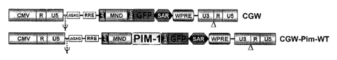
Figure 21 illustrates an exemplary lentiviral constructs of the invention, as
described in
detail in Example 4, below.
Like reference symbols in the various drawings indicate like elements.
DETAILED DESCRIPTION
The present disclosure includes the discovery of new roles for PIM-1, its
isoforms, and
other PIM enzymes having equivalent or overlapping targets and substrates.
Specifically, these
enzymes have a role in cardiac and other circulatory system protection,
survival, repair,
regeneration, and recovery, and in the implantation, differentiation,
function, and survival of
stem cells, progenitor cells, or differentiated cells introduced into
circulatory system tissues.
These discoveries form the basis for new cardiac therapies, including repair
of damaged heart
tissue and implantation, expansion, and survival of implanted stem cells or
progenitor cells that
differentiate into functional heart tissue. Prior to this invention,
enhancement of PIM activity
was not known to have any prophylactic or therapeutic utility in heart tissue,
heart cells, or in
other circulatory system cells or tissues.
We show that circulatory system disease or injury can be attenuated, halted,
prevented,
or reversed, and that damaged circulatory system tissue can be replaced,
repaired, or
regenerated, by enhancement of PIM activity in that tissue. Ways in which PIM
activity can be
enhanced are described in more detail below, but include upregulation of
endogenous PIM
production, direct introduction of materials having PIM activity into tissues
or cells, introduction
of polynucleotide encoding a PIM material into existing cells of a human or
animal, removing
cells from a subject and altering those cells to express enhanced levels of
PIM, then
reintroducing the cells into the subject, introducing exogenous cells into the
subject that have
been engineered to produce enhanced levels of PIM, including stem cells or
progenitor cells that
include a PIM-encoding polynucleotide under the control of a non-PIM promoter,
including an
inducible promoter, a constituitive promoter, or a cardiac-specific promoter.
The term "PIM" is used herein to refer to a serine or threonine kinase,
including the
various PIM enzymes, e.g., PIM-1, PIM-2, and PIM-3, further including any
isoforms thereof
For example, the serine/threonine kinase PIM-1 is known to exist in two
isoforms, and
references to PIM and PIM-1 herein are intended to encompass both isoforms,
unless otherwise
specified. In addition, although certain cells, constructs, polynucleotides,
techniques, uses, and
methods are described in connection with one particular PIM, such as PIM-1,
such descriptions
are exemplary, and should be taken as also including the other PIM enzymes
having similar
activity.
12
CA 02705862 2010-05-14
WO 2009/065080
PCT/US2008/083693
The term "PIM activity" and "PIM kinase activity" refer to the enzymatic or
physiological activity of the PIM enzymes, e.g., the activity of a PIM-1, and
encompasses use of
other materials having similar activity. The discoveries set forth herein
relate to altering
characteristics of living cells by enhancing a particular kinase activity in
the cells. Of course, as
is well known, enzyme variants exist or can be readily constructed, having
conservative amino
acid substitutions, cross-linking, cross-species domain substitutions,
truncations, and the like,
while preserving a physiologically-effective level of enzymatic activity (in
this case, kinase
activity for the PIM-1 target). The present discoveries are not focused only
on a particular
kinase, but include the discovery of an entirely new role for PIM kinase
activity in vascular
system cells and tissues. Thus, the results discussed herein flow from
alteration of PIM kinase
activity, regardless of the exact modality by which that is achieved.
The term "vascular system" is used herein to refer to the blood vessels and
the heart, and
all the tissues and cells of which they are comprised, including cardiac
smooth muscle,
cardiomyocytes, cardiomyoblasts, vascular wall , endothelium, vascular smooth
muscle,
vascular connective tissue, and other known cells and tissues of the vascular
system.
The term "stem cell" is used broadly to include totipotent, pluripotent, and
multipotent
cells that can differentiate into vascular system cells, including cardiac
cells. "Progenitor cells"
overlaps somewhat with multipotent stem cells, and includes cells that are at
least partially
differentiated but that are multipotent or unipotent, in that they have the
ability to differentiate
into at least one type of vascular system cells.
The terms "treat" and "treatment" are used broadly, to include both
prophylactic and
therapeutic treatments. Similarly, when referring to disease or injury of
circulatory system
tissues, those terms are used broadly to include fully developed disease or
injury, as well as
incipient or threatened disease or injury. Thus, a patient at risk of or
beginning to develop a
particular condition, is considered to have that condition "treated" when
methods as disclosed
herein are used to reduce the risk of development or progression of that
condition, as well as
when an already-developed condition is reversed, inhibited, cured, or
ameliorated, and when the
rate of development of a condition is halted or slowed.
Those being treated are referred to variously as patients, individuals,
subjects, humans,
or animals. Treatments identified as useful for one category are also useful
for other categories,
and selection of a particular term (other than "human") is not intended to be
limiting, but rather
just a use of an alternative expression.
13
CA 02705862 2010-05-14
WO 2009/065080
PCT/US2008/083693
The disclosure includes compositions, such as pharmaceutical compositions,
comprising
nucleic acids encoding a PIM serine/threonine kinase, such as PIM-1, and
methods for making
and using them; including methods for inducing cardiac or vascular cellular
proliferation, and
protecting cardiac or vascular cells from hypoxia and cellular apoptosis. In
one aspect, the
compositions and methods of the invention are used to express PIM-1 to protect
cardiomyocytes
from hypertrophy and inhibit myocardial apoptosis induced by infarction,
reducing infarct size.
In another embodiment, the compositions and methods of the invention are used
to express PIM-
1 to induce cardiac or vascular cellular dedifferentiation and re-expression
of stem cell markers;
and in one aspect, to overexpress PIM-1 to enhance the regenerative potential
of stem cells,
including stem cell ability to engraft in the heart after a myocardial
infarction (post-MI). In
another embodiment, the compositions and methods of the invention are used to
express PIM-1
to increase Bel-XL expression to induce cardioprotective anti-apoptotic
signaling, thus
increasing myocardial survival signaling.
Also disclosed are compositions, such as pharmaceutical compositions,
comprising
nucleic acids encoding the serine/threonine kinase PIM-1 and methods for
preventing or
inhibiting cell or tissue damage, e.g., cardiomyocyte cell death or inhibiting
an ischemic or
reperfusion related injury; including preventing or inhibiting the
irreversible cellular and tissue
damage and cell death caused by ischemia, e.g., ischemia subsequent to
reperfusion (which can
exacerbates ischemic damage by activating inflammatory response and oxidative
stress).
The disclosure further provides compositions, such as pharmaceutical
compositions,
comprising nucleic acids encoding a serine/threonine kinase PIM and methods
for regulating
cardiac or vascular cellular proliferation and survival.
Using human and murine myocardial samples, we have demonstrated that both
human
and murine myocardial cells show elevated PIM-1 expression in failing hearts;
where the
elevated PIM-1 has predominantly a nuclear localization. We have also shown
that acute
cardiomyopathic challenge also induces PIM-1 expression with nuclear and
perinuclear
distribution in mouse myocardium.
Expression of PIM-1 in postnatal mouse myocardium decreases with aging, and
cardioprotective stimuli associated with AKT activation and nuclear-targeted
AKT in particular
increase PIM-1 expression. We disclose that cardiomyocyte apoptosis is
inhibited by PIM-1 via
increased expression of bc1-2, bcl-XL, and phosphorylation of Bads112.
Ischemia reperfusion
injury is enhanced in PIM-1 knockout mice. Since loss of PIM-1 expression or
activity leads to
increased AKT expression without associated cardioprotective effects, PIM-1
represents a
critical and novel facet of survival signaling downstream of AKT in the
myocardium.
14
CA 02705862 2010-05-14
WO 2009/065080
PCT/US2008/083693
Treatments and Medical Uses
Detailed strategies for enhancing PIM activity within circulatory tissues are
provided
below. Regardless of the method by which PIM activity is increased, we have
discovered that
enhancement of PIM activity has multiple beneficial effects in cardiac and
other circulatory
system tissues.
Initially, the care provider may wish to perform a patient selection step.
This may
include, for example, assessing whether a patient is in need of one or more of
the various
treatments, or identifying a patient in need of such treatment. Two
significant categories of need
warrant some discussion.
First, there are individuals with readily-diagnosable existing conditions,
including known
disease or injury to cardiac or other circulatory tissue that is treatable
with the compositions,
methods, or techniques contemplated herein. In those cases, diagnosis or
identification of the
disease or injury would constitute diagnosis, selection, or identification of
an individual in need
of the specified treatment.
Second, there are individuals in need of treatment that is more prophylactic,
for example,
treatment that takes advantage of the powerful cardioprotective properties
exerted by PIM. In
some cases, identification can take place by recognizing an inchoate disease
or injury that would
otherwise progress, for example, injury or other factors that have or will
initiate apoptosis, or
conditions or factors that enhance the risk of developing a particular
condition. Identification
and treatment of those individuals may be desirable to prevent development of
a disease or
injury or to slow its development.
In between these two alternatives are individuals with existing disease or
injury, which
disease or injury is likely to progress. Identification and treatment of those
individuals is also
contemplated.
One significant condition lending itself to treatment through enhancement of
PIM
activity in cardiac tissue is myocardial infarction or other ischemic injury.
Prophylactic
treatment is desirable, when high risk of ischemic injury can be identified.
However, in many
cases, the patient will be treated after the injury has occurred. Treatment
should be commenced
as soon as is practicable after the injury.
Similarly, PIM-activity enhancement can be used to treat a number of other
conditions
and to create desired physiological effects, by treating a subject to enhance
PIM activity in
vascular, cardiac, or other circulatory system cells or tissues. These include
prevention,
reduction, or reversal of cardiac hypertrophy, including but not limited to
maladaptive
CA 02705862 2010-05-14
WO 2009/065080
PCT/US2008/083693
hypertrophic remodeling; promoting cardiac cell survival and inhibiting
apoptosis of those cells;
enhancing cardiac contractility; improving cardiac ejection fraction;
enhancing vascular growth
and repair; and promoting differentiation of stem cells and progenitor cells
toward cardiac or
vascular tissue.
In another aspect, the methods contemplated herein include, but are not
limited to,
inhibition of cardiac apoptosis; inhibition of cardiac fibrosis; inhibition of
cardiac remodeling;
inhibition of cardiac hypertrophy; preservation or reduced loss of ejection
fraction in damaged
hearts; enhanced preservation of contractile function; decrease in cardiac
necrosis; reduction in
lesion size following ischemic injury; and increasing cardiac cellularity and
decreasing myocyte
volume. All of these methods can be practiced prophylactically (to prevent or
reduce a
particular condition that would otherwise be likely to occur) and
therapeutically (to treat a
condition that is already in existence, including treatment to slow
progression of a condition).
From another perspective, conditions that may lead to treatment by enhancement
of PIM
activity include (but are not limited to) congenital heart conditions;
ischemic injury of any kind
to heart tissue; damage from infarction; cardiac reperfusion injury; traumatic
cardiac injury;
congestive heart failure injury; and injury relating to cardiac infection with
a pathogen, including
viral, bacterial, and parasitic pathogens.
In addition to identifying an individual having a condition for which PIM
treatment is desirable,
methods of treating the individual can include a step of increasing the level
of PIM activity in a
target tissue, such as vascular tissue or cardiac tissue. This step can be
practiced in the various
ways disclosed herein. By way of example, and not limitation, those include
administering
factors or drugs to the patient, systemically or locally, that upregulate
endogenous PIM
expression; administering PIM protein, preferably in combination with a
delivery modality, such
as a linked transduction domain, a liposome, an antibody, or the like;
administering PIM-
encoding polynucleotide to the patient, including naked DNA administration,
administration of
the polynucleotide in a viral vector, liposome, or other delivery modality;
electroporation of
cells of a subject to deliver DNA; and administering an autologous cell to the
subject (e.g., into
the heart) that has been altered to enhance PIM expression, including
cardiomyocytes, cardiac
progenitor cells, cardiac stem cells, mesenchymal stem cells, hematopoietic
stem cells, adipose-
derived stem cells, and the like.
When administering cells to a human patient, for example, to treat a cardiac
condition,
the number of cells can be any amount effective to enhance cardiac function or
structure or treat
a target condition. Exemplary, non-limiting amounts include 105 to 1010
cells, more typically 106 to109cells.
16
CA 02705862 2010-05-14
WO 2009/065080
PCT/US2008/083693
Exemplary, non-limiting amounts of PIM protein administered to an adult human
heart
can be, for example, from about 10-4 g to about 10-10 g, calculated as the
pure PIM protein.
Exemplary, non-limiting amounts of DNA include about 0.05 to 500 ug/kg, or 0.5
to 50 ug/kg
body weight, and in the case of viral particles, formulated at a titer of
about at least 1010, 1011 ,
1012,
1013 , 1014 , 1015 , 1016 , or 1017 physical particles per milliliter. In one
aspect, the PIM-1
encoding nucleic acid is administered in about 10, 20, 30, 40, 50, 60, 70, 80,
90, 100, 110, 120,
130, 140 or 150 or more microliter ( 1) injections. Doses and dosage regimens
can be
determined by conventional range-finding techniques known to those of ordinary
skill in the art.
For example, in alternative embodiments, about 106, 107, 108, 109, 1010, 1011,
1012,
1013,1014,
1015, 1016 or 10' viral(e.g., lentiviral) particles are delivered to the
individual (e.g., a human
patient) in one or multiple doses.
In other embodiments, an intracardiac single administration (e.g., a single
dose)
comprises from about 0.1 I to 1.0 p1, 10 IA or to about 100 IA of a
pharmaceutical composition
of the invention. Alternatively, dosage ranges from about 0.5 ng or 1.0 ng to
about 10 g, 100
g to 1000 g of PIM-1 expressing nucleic acid is administered (either the
amount in an
expression construct, or as in one embodiment, naked DNA is injected).
PIM sequences
Some embodiments include nucleic acid constructs comprising a PIM-encoding
sequence, e.g., a PIM-1 expressing message or a PIM-1 gene. In one aspect, PIM-
expressing nucleic acids used to practice this invention include PIM-1 genomic
sequences,
or fragments thereof, including coding or non-coding sequences, e.g.,
including introns, 5'
or 3' non-coding sequences, and the like. Also encompassed are PIM-encoding
mRNA
sequences.
In one aspect, the PIM-1 expressing, or PIM-1 inducing or upregulating,
composition is a
nucleic acid, including a vector, recombinant virus, and the like; and a
recombinant PIM-1 is
expressed in a cell in vitro, ex vivo and/or in vivo.
In one aspect, a PIM-1 expressing nucleic acid encodes a human PIM-1, such as
Genbank accession no. AAA36447 (see also, e.g., Domen (1987) Oncogene Res. 1
(1):103-112),
SEQ ID NO:l.
In another aspect, a PIM-1 expressing nucleic acid encodes a human PIM-1
kinase 44
kDa isoform, see e.g., Genbank accession no. AAY87461 (see also, e.g., Xie
(2006) Oncogene
25 (1), 70-78), SEQ ID NO:2.
17
CA 02705862 2010-05-14
WO 2009/065080
PCT/US2008/083693
In a further aspect, a PIM-1 expressing nucleic acid comprises a human PIM-1
kinase
message (mRNA), see e.g., Genbank accession no. NM 002648 (see also, e.g.,
Zhang (2007)
Mol. Cancer Res. 5 (9), 909-922), SEQ ID NO:3.
Also disclosed are human DNA sequences of PIM-2 (SEQ ID NO:4) and PIM-3 (SEQ
ID NO:5).
In alternative embodiments, nucleic acids of this invention are operatively
linked to a
transcriptional regulatory sequence, e.g., a promoter and/or an enhancer,
e.g., cardiac-specific,
promoters to drive (e.g., regulate) expression of Pim-1. Promoters and
enhancers used to
practice this invention can be of any type and/or origin, an in one embodiment
promoters
specific to the species receiving the Pim-1 construct are used; e.g., humans
can receive human
promoters, mice receive murine promoters, etc. In other embodiments, promoters
from
heterologous species can be used; e.g., mammals or vertebrates receiving
promoters that
originate from other mammals or vertebrates, or viral or synthetic promoters
active in the
appropriate specie and/or cell type also can be used. These promoters can
comprise, for
example, a a-myo sin heavy chain promoter; a cardiac troponin-T promoter; a
MLC-2v
promoter; and any other promoter that drives expression in cardiac tissue but
does not drive
significant expression in other tissues. In one embodiment, promoters and
enhancers active in
primordial cells or stem cells, e.g., myocardial stem cells, can be
operatively linked to drive
expression of Pim-1.
Nucleic Acid Delivery - Gene Therapy Vehicles
In one aspect, this disclosure provides constructs or expression vehicles,
e.g., expression
cassettes, vectors, viruses (e.g., lentiviral expression vectors, e.g., see
SEQ ID NO:4), and the
like, comprising a PIM- encoding sequence, e.g., a PIM-1 encoding message or a
PIM-la gene,
for use as ex vivo or in vitro gene therapy vehicles, or for expression of PIM-
1 in heart tissue, a
cardiac or vascular cell, tissue or organ to practice the methods of this
invention, and for
research, drug discovery or transplantation.
In one aspect, an expression vehicle used to practice the invention can
comprise a
promoter operably linked to a nucleic acid encoding a PIM protein (or
functional subsequence
thereof). For example, the invention provides expression cassettes comprising
nucleic acid
encoding a PIM-1 protein operably linked to a transcriptional regulatory
element, e.g., a
promoter.
In one aspect, an expression vehicle used to practice the invention is
designed to deliver
a PIM-1 encoding sequence, e.g., a PIM-1 gene or any functional portion
thereof to a cardiac
tissue or cell of an individual. Expression vehicles, e.g., vectors, used to
practice the invention
18
CA 02705862 2010-05-14
WO 2009/065080
PCT/US2008/083693
can be non-viral or viral vectors or combinations thereof The invention can
use any viral vector
or viral delivery system known in the art, e.g., adenoviral vectors, adeno-
associated viral (AAV)
vectors, herpes viral vectors (e.g., herpes simplex virus (HSV)-based
vectors), retroviral vectors,
lentiviral vectors and baculoviral vectors.
In one aspect of the invention, an expression vehicle, e.g., a vector or a
virus, is capable
of accommodating a full-length PIM-1 gene or a message, e.g., a cDNA. In one
aspect, the
invention provides a retroviral, e.g., a lentiviral, vector capable of
delivering the nucleotide
sequence encoding full-length human PIM-1 in vitro, ex vivo and/or in vivo. An
exemplary
lentiviral expression vector backbone (no "payload" included, e.g., no PIM-1
sequence included)
that can be used to practice this invention is set forth in SEQ ID NO:4.
In one embodiment, a lentiviral vector used to practice this invention is a
"minimal"
lentiviral production system lacking one or more viral accessory (or
auxiliary) gene. Exemplary
lentiviral vectors for use in the invention can have enhanced safety profiles
in that they are
replication defective and self-inactivating (SIN) lentiviral vectors.
Lentiviral vectors and
production systems that can be used to practice this invention include e.g.,
those described in
U.S. Patent Nos. (USPNs) 6,277,633; 6,312,682; 6,312,683; 6,521,457;
6,669,936; 6,924,123;
7,056,699; and 7,198,784; any combination of these are exemplary vectors that
can be employed
in the practice of the invention. In an alternative embodiment, non-
integrating lentiviral vectors
can be employed in the practice of the invention. For example, non-integrating
lentiviral vectors
and production systems that can be employed in the practice of the invention
include those
described in USPN 6,808,923.
The expression vehicle can be designed from any vehicle known in the art,
e.g., a
recombinant adeno-associated viral vector as described, e.g., in U.S. Pat.
App. Pub. No.
20020194630, Manning, et al.; or a lentiviral gene therapy vector, e.g., as
described by e.g.,
Dull, et al. (1998) J. Virol. 72:8463-8471; or a viral vector particle, e.g.,
a modified retrovirus
having a modified proviral RNA genome, as described, e.g., in U.S. Pat. App.
Pub. No.
20030003582; or an adeno-associated viral vector as described e.g., in USPN
6,943,153,
describing recombinant adeno-associated viral vectors for use in the eye; or a
retroviral or a
lentiviral vector as described in USPNs 7,198,950; 7,160,727; 7,122,181
(describing using a
retrovirus to inhibit intraocular neovascularization in an individual having
an age-related
macular degeneration); or 6,555,107.
Any viral vector can be used to practice this invention, and the concept of
using viral
vectors for gene therapy is well known; see e.g., Verma and Somia (1997)
Nature 389:239-242;
and Coffin et al ("Retroviruses" 1997 Cold Spring Harbour Laboratory Press
Eds: JM Coffin,
19
CA 02705862 2010-05-14
WO 2009/065080
PCT/US2008/083693
SM Hughes, HE Varmus pp 758-763) having a detailed list of retroviruses. Any
lentiviruses
belonging to the retrovirus family can be used for infecting both dividing and
non-dividing cells
with a PIM-1-encoding nucleic acid, see e.g., Lewis et al (1992) EMBO J. 3053-
3058.
Viruses from lentivirus groups from "primate" and/or "non-primate" can be
used; e.g.,
any primate lentivirus can be used, including the human immunodeficiency virus
(HIV), the
causative agent of human acquired immunodeficiency syndrome (AIDS), and the
simian
immunodeficiency virus (Sly); or a non-primate lentiviral group member, e.g.,
including "slow
viruses" such as a visna/maedi virus (VMV), as well as the related caprine
arthritis-encephalitis
virus (CAEV), equine infectious anemia virus (EIAV) and/or a feline
immunodeficiency virus
(FIV) or a bovine immunodeficiency virus (BIV).
In alternative embodiments, lentiviral vectors used to practice this invention
are
pseudotyped lentiviral vectors. In one aspect, pseudotyping used to practice
this invention
incorporates in at least a part of, or substituting a part of, or replacing
all of, an env gene of a
viral genome with a heterologous env gene, for example an env gene from
another virus. In
alternative embodiments, the lentiviral vector of the invention is pseudotyped
with VSV-G. In
an alternative embodiment, the lentiviral vector of the invention is
pseudotyped with Rabies-G.
Lentiviral vectors used to practice this invention may be codon optimized for
enhanced
safety purposes. Different cells differ in their usage of particular codons.
This codon bias
corresponds to a bias in the relative abundance of particular tRNAs in the
cell type. By altering
the codons in the sequence so that they are tailored to match with the
relative abundance of
corresponding tRNAs, it is possible to increase expression. By the same token,
it is possible to
decrease expression by deliberately choosing codons for which the
corresponding tRNAs are
known to be rare in the particular cell type. Thus, an additional degree of
translational control is
available. Many viruses, including HIV and other lentiviruses, use a large
number of rare
codons and by changing these to correspond to commonly used mammalian codons,
increased
expression of the packaging components in mammalian producer cells can be
achieved. Codon
usage tables are known in the art for mammalian cells, as well as for a
variety of other
organisms. Codon optimization has a number of other advantages. By virtue of
alterations in
their sequences, the nucleotide sequences encoding the packaging components of
the viral
particles required for assembly of viral particles in the producer
cells/packaging cells have RNA
instability sequences (INS) eliminated from them. At the same time, the amino
acid sequence
coding sequence for the packaging components is retained so that the viral
components encoded
by the sequences remain the same, or at least sufficiently similar that the
function of the
packaging components is not compromised. Codon optimization also overcomes the
Rev/RRE
CA 02705862 2010-05-14
WO 2009/065080
PCT/US2008/083693
requirement for export, rendering optimized sequences Rev independent. Codon
optimization
also reduces homologous recombination between different constructs within the
vector system
(for example between the regions of overlap in the gag-pol and env open
reading frames). The
overall effect of codon optimization is therefore a notable increase in viral
titer and improved
safety. The strategy for codon optimized gag-pol sequences can be used in
relation to any
retrovirus.
Vectors, recombinant viruses, and other expression systems used to practice
this
invention can comprise any nucleic acid which can infect, transfect,
transiently or permanently
transduce a cell. In one aspect, a vector used to practice this invention can
be a naked nucleic
acid, or a nucleic acid complexed with protein or lipid. In one aspect, a
vector used to practice
this invention comprises viral or bacterial nucleic acids and/or proteins,
and/or membranes (e.g.,
a cell membrane, a viral lipid envelope, etc.). In one aspect, expression
systems used to practice
this invention comprise replicons (e.g., RNA replicons, bacteriophages) to
which fragments
of DNA may be attached and become replicated. In one aspect, expression
systems used to
practice this invention include, but are not limited to RNA, autonomous self-
replicating circular
or linear DNA or RNA (e.g., plasmids, viruses, and the like, see, e.g., U.S.
Patent No.
5,217,879), and include both the expression and non-expression plasmids.
In one aspect, a recombinant microorganism or cell culture used to practice
this
invention can comprise "expression vector" including both (or either) extra-
chromosomal
circular and/or linear nucleic acid (DNA or RNA) that has been incorporated
into the host
chromosome(s). In one aspect, where a vector is being maintained by a host
cell, the vector
may either be stably replicated by the cells during mitosis as an autonomous
structure, or is
incorporated within the host's genome.
In one aspect, an expression system used to practice this invention can
comprise any
plasmid, which are commercially available, publicly available on an
unrestricted basis, or can be
constructed from available plasmids in accord with published procedures.
Plasmids that can be
used to practice this invention are well known in the art.
In alternative aspects, a vector used to make or practice the invention can be
chosen from
any number of suitable vectors known to those skilled in the art, including
cosmids, YACs
(Yeast Artificial Chromosomes), megaYACS, BACs (Bacterial Artificial
Chromosomes), PACs
(P1 Artificial Chromosome), MACs (Mammalian Artificial Chromosomes), a whole
chromosome, or a small whole genome. The vector also can be in the form of a
plasmid, a
viral particle, or a phage. Other vectors include chromosomal, non-chromosomal
and synthetic
DNA sequences, derivatives of SV40; bacterial plasmids, phage DNA,
baculovirus, yeast
21
CA 02705862 2010-05-14
WO 2009/065080
PCT/US2008/083693
plasmids, vectors derived from combinations of plasmids and phage DNA, viral
DNA such as
vaccinia, adenovirus, fowl pox virus, and pseudorabies. A variety of cloning
and expression
vectors for use with prokaryotic and eukaryotic hosts are described by, e.g.,
Sambrook.
Bacterial vectors which can be used include commercially available plasmids
comprising
genetic elements of known cloning vectors.
Pharmaceutical compositions
The invention provides compositions, including pharmaceutical compositions,
and
methods for expressing PIM; e.g., for expressing PIM-1 or another functionally-
equivalent
kinase to protect cardiomyocytes from hypertrophy and to inhibit myocardial
apoptosis induced
by infarction, and to reduce infarct size. (Functional equivalence is
considered to exist based on
ability to act on the same substrate and produce the same product, and does
not require identical
kinetics.) In another embodiment, the pharmaceutical compositions of the
invention are used to
express PIM-1 to induce cardiac or vascular cellular dedifferentiation and re-
expression of stem
cell markers; and in one aspect, to overexpress PIM-1 to enhance the
regenerative potential of
stem cells, including stem cell ability to engraft in the heart after a
myocardial infarction (post-
MI).
In one aspect, the PIM-1 expressing, or PIM-1 inducing or upregulating,
composition is a
nucleic acid, including a vector, recombinant virus, and the like; and a
recombinant PIM-1 is
expressed in a cell in vitro, ex vivo and/or in vivo.
In alternative embodiments, in practicing use of the pharmaceutical
compositions and
methods of this invention, compounds that induce or upregulate PIM nucleic
acid or a PIM
kinase activity in the heart or a cardiac or vascular cell, tissue or organ
are administered. For
example, compounds that can be administered in practicing use of the
pharmaceutical
compositions and methods of this invention can comprise: an interleukin, a
cytokine and/or a
paracrine factor involved in survival and/or proliferative signaling; an up-
regulator of AKT
serine/threonine kinase; insulin-like growth factor-1 (IGF-1); insulin;
leukemia inhibitory factor
(LIF); granulocyte-macrophage colony-stimulating factor (GM-CSF); or epidermal
growth
factor (EGF). Okadaic acid and SV40 small T antigen inhibit or block negative
regulation of
PIM-1 by protein phosphatase 2A, and can thus be used to increase PIM-1
levels. See Maj, et
al., Oncogene 26(35):5145-53 (2007).
In alternative embodiments, the PIM-expressing, or PIM-inducing or
upregulating,
compositions of the invention are formulated with a pharmaceutically
acceptable carrier. In
alternative embodiments, the pharmaceutical compositions of the invention can
be administered
parenterally, topically, orally or by local administration, such as by aerosol
or transdermally.
22
CA 02705862 2010-05-14
WO 2009/065080
PCT/US2008/083693
The pharmaceutical compositions can be formulated in any way and can be
administered in a
variety of unit dosage forms depending upon the condition or disease and the
degree of illness,
the general medical condition of each patient, the resulting preferred method
of administration
and the like. Details on techniques for formulation and administration are
well described in the
scientific and patent literature, see, e.g., the latest edition of Remington's
Pharmaceutical
Sciences, Maack Publishing Co, Easton PA ("Remington's").
Therapeutic agents of the invention can be administered alone or as a
component of a
pharmaceutical formulation (composition). The compounds may be formulated for
administration in any convenient way for use in human or veterinary medicine.
Wetting agents,
emulsifiers and lubricants, such as sodium lauryl sulfate and magnesium
stearate, as well as
coloring agents, release agents, coating agents, sweetening, flavoring and
perfuming agents,
preservatives and antioxidants can also be present in the compositions.
Formulations of the PIM-expressing, or inducing or upregulating, compositions
of the
invention include those suitable for systemic administration, direct local
vascular or cardiac
administration, or alternatively oral/ nasal, topical, parenteral, rectal,
and/or intravaginal
administration. The formulations may conveniently be presented in unit dosage
form and may
be prepared by any methods well known in the art of pharmacy. The amount of
active
ingredient which can be combined with a carrier material to produce a single
dosage form will
vary depending upon the host being treated, the particular mode of
administration. The amount
of active ingredient which can be combined with a carrier material to produce
a single dosage
form will generally be that amount of the compound which produces a
therapeutic effect.
Pharmaceutical formulations of this invention may comprise one or more
diluents,
emulsifiers, preservatives, buffers, excipients, etc. and may be provided in
such forms as liquids,
powders, emulsions, lyophilized powders, sprays, creams, lotions, controlled
release
formulations, tablets, pills, gels, on patches, in implants, etc.
Pharmaceutical formulations for oral administration can be formulated using
pharmaceutically acceptable carriers well known in the art in appropriate and
suitable dosages.
Such carriers enable the pharmaceuticals to be formulated in unit dosage forms
as tablets, pills,
powder, dragees, capsules, liquids, lozenges, gels, syrups, slurries,
suspensions, etc., suitable for
ingestion by the patient. Pharmaceutical preparations for oral use can be
formulated as a solid
excipient, optionally grinding a resulting mixture, and processing the mixture
of granules, after
adding suitable additional compounds, if desired, to obtain tablets or dragee
cores. Suitable
solid excipients are carbohydrate or protein fillers include, e.g., sugars,
including lactose,
sucrose, mannitol, or sorbitol; starch from corn, wheat, rice, potato, or
other plants; cellulose
23
CA 02705862 2010-05-14
WO 2009/065080
PCT/US2008/083693
such as methyl cellulose, hydroxypropylmethyl-cellulose, or sodium carboxy-
methylcellulose;
and gums including arabic and tragacanth; and proteins, e.g., gelatin and
collagen.
Disintegrating or solubilizing agents may be added, such as the cross-linked
polyvinyl
pyrrolidone, agar, alginic acid, or a salt thereof, such as sodium alginate.
Dragee cores are provided with suitable coatings such as concentrated sugar
solutions,
which may also contain gum arabic, talc, polyvinylpyrrolidone, carbopol gel,
polyethylene
glycol, and/or titanium dioxide, lacquer solutions, and suitable organic
solvents or solvent
mixtures. Dyestuffs or pigments may be added to the tablets or dragee coatings
for product
identification or to characterize the quantity of active compound (i.e.,
dosage). Pharmaceutical
preparations of the invention can also be used orally using, e.g., push-fit
capsules made of
gelatin, as well as soft, sealed capsules made of gelatin and a coating such
as glycerol or
sorbitol. Push-fit capsules can contain active agents mixed with a filler or
binders such as
lactose or starches, lubricants such as talc or magnesium stearate, and,
optionally, stabilizers. In
soft capsules, the active agents can be dissolved or suspended in suitable
liquids, such as fatty
oils, liquid paraffin, or liquid polyethylene glycol with or without
stabilizers.
Aqueous suspensions can contain an active agent (e.g., a chimeric polypeptide
or
peptidomimetic of the invention) in admixture with excipients suitable for the
manufacture of
aqueous suspensions. Such excipients include a suspending agent, such as
sodium
carboxymethylcellulose, methylcellulose, hydroxypropylmethylcellulose, sodium
alginate,
polyvinylpyrrolidone, gum tragacanth and gum acacia, and dispersing or wetting
agents such as
a naturally occurring phosphatide (e.g., lecithin), a condensation product of
an alkylene oxide
with a fatty acid (e.g., polyoxyethylene stearate), a condensation product of
ethylene oxide with
a long chain aliphatic alcohol (e.g., heptadecaethylene oxycetanol), a
condensation product of
ethylene oxide with a partial ester derived from a fatty acid and a hexitol
(e.g., polyoxyethylene
sorbitol mono-oleate), or a condensation product of ethylene oxide with a
partial ester derived
from fatty acid and a hexitol anhydride (e.g., polyoxyethylene sorbitan mono-
oleate). The
aqueous suspension can also contain one or more preservatives such as ethyl or
n-propyl p-
hydroxybenzoate, one or more coloring agents, one or more flavoring agents and
one or more
sweetening agents, such as sucrose, aspartame or saccharin. Formulations can
be adjusted for
osmolarity.
Oil-based pharmaceuticals can be used to deliver PIM-1 expressing, or PIM-1
inducing
or upregulating, compositions of the invention. Oil-based suspensions can be
formulated by
suspending an active agent in a vegetable oil, such as arachis oil, olive oil,
sesame oil or coconut
oil, or in a mineral oil such as liquid paraffin; or a mixture of these. See
e.g., U.S. Patent No.
24
CA 02705862 2010-05-14
WO 2009/065080
PCT/US2008/083693
5,716,928 describing using essential oils or essential oil components for
increasing
bioavailability and reducing inter- and intra-individual variability of orally
administered
hydrophobic pharmaceutical compounds (see also U.S. Patent No. 5,858,401). The
oil
suspensions can contain a thickening agent, such as beeswax, hard paraffin or
cetyl alcohol.
Sweetening agents can be added to provide a palatable oral preparation, such
as glycerol,
sorbitol or sucrose. These formulations can be preserved by the addition of an
antioxidant such
as ascorbic acid. As an example of an injectable oil vehicle, see Minto (1997)
J. Pharmacol.
Exp. Ther. 281:93-102. The pharmaceutical formulations of the invention can
also be in the
form of oil-in-water emulsions. The oily phase can be a vegetable oil or a
mineral oil, described
above, or a mixture of these. Suitable emulsifying agents include naturally-
occurring gums,
such as gum acacia and gum tragacanth, naturally occurring phosphatides, such
as soybean
lecithin, esters or partial esters derived from fatty acids and hexitol
anhydrides, such as sorbitan
mono-oleate, and condensation products of these partial esters with ethylene
oxide, such as
polyoxyethylene sorbitan mono-oleate. The emulsion can also contain sweetening
agents and
flavoring agents, as in the formulation of syrups and elixirs. Such
formulations can also contain
a demulcent, a preservative, or a coloring agent.
In practicing this invention, the pharmaceutical compounds can also be
administered by
in intranasal, intraocular and intravaginal routes including suppositories,
insufflation, powders
and aerosol formulations (for examples of steroid inhalants, see Rohatagi
(1995) J. Clin.
Pharmacol. 35:1187-1193; Tjwa (1995) Ann. Allergy Asthma Immunol. 75:107-111).
Suppositories formulations can be prepared by mixing the drug with a suitable
non-irritating
excipient which is solid at ordinary temperatures but liquid at body
temperatures and will
therefore melt in the body to release the drug. Such materials are cocoa
butter and polyethylene
glycols.
In practicing this invention, the pharmaceutical compounds can be delivered by
transdermally, by a topical route, formulated as applicator sticks, solutions,
suspensions,
emulsions, gels, creams, ointments, pastes, jellies, paints, powders, and
aerosols.
In practicing this invention, the pharmaceutical compounds can also be
delivered as
microspheres for slow release in the body. For example, microspheres can be
administered via
intradermal injection of drug which slowly release subcutaneously; see Rao
(1995) J. Biomater
Sci. Polym. Ed. 7:623-645; as biodegradable and injectable gel formulations,
see, e.g., Gao
(1995) Pharm. Res. 12:857-863 (1995); or, as microspheres for oral
administration, see, e.g.,
Eyles (1997) J. Pharm. Pharmacol. 49:669-674.
CA 02705862 2010-05-14
WO 2009/065080
PCT/US2008/083693
In practicing this invention, the pharmaceutical compounds can be parenterally
administered, such as by intravenous (IV) administration or administration
into a body cavity or
lumen of the heart. Use of catheters that temporarily block flow of blood from
the heart while
incubating the stem cells or a viral construct in heart tissue can be used, as
well as recirculation
systems of well-known type that isolate the circulation in all or a part of
the heart to increase the
dwell time of an introduced agent (e.g., stem cell, construct, naked DNA, PIM
protein, viral or
other vector) in the heart. These formulations can comprise a solution of
active agent dissolved
in a pharmaceutically acceptable carrier. Acceptable vehicles and solvents
that can be employed
are water and Ringer's solution, an isotonic sodium chloride. In addition,
sterile fixed oils can
be employed as a solvent or suspending medium. For this purpose any bland
fixed oil can be
employed including synthetic mono- or diglycerides. In addition, fatty acids
such as oleic acid
can likewise be used in the preparation of injectables. These solutions are
sterile and generally
free of undesirable matter. These formulations may be sterilized by
conventional, well known
sterilization techniques. The formulations may contain pharmaceutically
acceptable auxiliary
substances as required to approximate physiological conditions such as pH
adjusting and
buffering agents, toxicity adjusting agents, e.g., sodium acetate, sodium
chloride, potassium
chloride, calcium chloride, sodium lactate and the like. The concentration of
active agent in
these formulations can vary widely, and will be selected primarily based on
fluid volumes,
viscosities, body weight, and the like, in accordance with the particular mode
of administration
selected and the patient's needs. For IV administration, the formulation can
be a sterile
injectable preparation, such as a sterile injectable aqueous or oleaginous
suspension. This
suspension can be formulated using those suitable dispersing or wetting agents
and suspending
agents. The sterile injectable preparation can also be a suspension in a
nontoxic parenterally-
acceptable diluent or solvent, such as a solution of 1,3-butanediol. The
administration can be by
bolus or continuous infusion (e.g., substantially uninterrupted introduction
into a blood vessel
for a specified period of time).
The pharmaceutical compounds and formulations of the invention can be
lyophilized.
The invention provides a stable lyophilized formulation comprising a
composition of the
invention, which can be made by lyophilizing a solution comprising a
pharmaceutical of the
invention and a bulking agent, e.g., mannitol, trehalose, raffinose, and
sucrose or mixtures
thereof. A process for preparing a stable lyophilized formulation can include
lyophilizing a
solution about 2.5 mg/mL protein, about 15 mg/mL sucrose, about 19 mg/mL NaC1,
and a
sodium citrate buffer having a pH greater than 5.5 but less than 6.5. See,
e.g., U.S. patent app.
no. 20040028670.
26
CA 02705862 2010-05-14
WO 2009/065080
PCT/US2008/083693
The compositions and formulations of the invention can be delivered by the use
of
liposomes (see also discussion, below). By using liposomes, particularly where
the liposome
surface carries ligands specific for target cells, or are otherwise
preferentially directed to a
specific organ, one can focus the delivery of the active agent into target
cells of the heart or
other part of the circulatory system in vivo. See, e.g., U.S. Patent Nos.
6,063,400; 6,007,839;
Al-Muhammed (1996) J. Microencapsul. 13:293-306; Chonn (1995) Curr. Opin.
Biotechnol.
6:698-708; Ostro (1989) Am. J. Hosp. Pharm. 46:1576-1587.
The formulations of the invention can be administered for prophylactic and/or
therapeutic treatments. In therapeutic applications, compositions are
administered to a subject
already suffering from a condition, infection or disease in an amount
sufficient to cure, alleviate
or partially arrest the clinical manifestations of the condition, infection or
disease and its
complications (a "therapeutically effective amount"). For example, in
alternative embodiments,
pharmaceutical compositions of the invention are administered in an amount
sufficient to treat,
prevent and/or ameliorate the deleterious effects on the heart of a myocardial
infarction (post-
MI); to protect cardiomyocytes from hypertrophy and to inhibit myocardial
apoptosis induced
by infarction, and to reduce infarct size. In another embodiment, the
pharmaceutical
compositions of the invention are used to express PIM-1 to induce cellular
dedifferentiation and
re-expression of stem cell markers; and in one aspect, to overexpress PIM-1 to
enhance the
regenerative potential of stem cells, including stem cell ability to engraft
in the heart post-MI.
The amount of pharmaceutical composition adequate to accomplish this is
defined as a
"therapeutically effective dose." The dosage schedule and amounts effective
for this use, i.e.,
the "dosing regimen," will depend upon a variety of factors, including the
stage of the disease or
condition, the severity of the disease or condition, the general state of the
patient's health, the
patient's physical status, age and the like. In calculating the dosage regimen
for a patient, the
mode of administration also is taken into consideration.
The dosage regimen also takes into consideration pharmacokinetics parameters
well
known in the art, i.e., the active agents' rate of absorption,
bioavailability, metabolism,
clearance, and the like (see, e.g., Hidalgo-Aragones (1996) J. Steroid
Biochem. Mol. Biol.
58:611-617; Groning (1996) Pharmazie 51:337-341; Fotherby (1996) Contraception
54:59-69;
Johnson (1995) J. Pharm. Sci. 84:1144-1146; Rohatagi (1995) Pharmazie 50:610-
613; Brophy
(1983) Eur. J. Clin. Pharmacol. 24:103-108; the latest Remington's, supra).
The state of the art
allows the clinician to determine the dosage regimen for each individual
patient, active agent
and disease or condition treated. Guidelines provided for similar compositions
used as
pharmaceuticals can be used as guidance to determine the dosage regiment,
i.e., dose schedule
27
CA 02705862 2010-05-14
WO 2009/065080
PCT/US2008/083693
and dosage levels, administered practicing the methods of the invention are
correct and
appropriate.
Single or multiple administrations of formulations can be given depending on
the dosage
and frequency as required and tolerated by the patient. The formulations
should provide a
sufficient quantity of active agent to effectively treat, prevent or
ameliorate a conditions,
diseases or symptoms as described herein. Methods for preparing parenterally
or non-
parenterally administrable formulations are known or apparent to those skilled
in the art and are
described in more detail in such publications as Remington's.
The methods of the invention can further comprise co-administration with other
drugs or
pharmaceuticals, e.g., compositions for treating heart attacks, congestive
heart failure and related
symptoms or conditions. For example, the methods and/or compositions and
formulations of the
invention can be co-formulated with and/or co-administered with antibiotics
(e.g., antibacterial
or bacteriostatic peptides or proteins), particularly those effective against
gram negative bacteria,
fluids, cytokines, immunoregulatory agents, anti-inflammatory agents,
complement activating
agents, such as peptides or proteins comprising collagen-like domains or
fibrinogen-like
domains (e.g., a ficolin), carbohydrate-binding domains, and the like and
combinations thereof
Nanoparticles and Liposomes
The invention also provides nanoparticles and liposomal membranes comprising
the
PIM-1-expressing compounds of this invention which target specific molecules,
including
biologic molecules, such as polypeptide, including cardiac or vascular or stem
cell surface
polypeptides, including heart cell (e.g., myocyte) cell surface polypeptides.
In alternative
embodiments, the invention provides nanoparticles and liposomal membranes
targeting diseased
and/or injured heart cells, or stem cells, such as any pluripotent cell.
In alternative embodiments, the invention provides nanoparticles and liposomal
membranes comprising (in addition to comprising compounds of this invention)
molecules, e.g.,
peptides or antibodies, that selectively target diseased and/or injured heart
cells, or stem cells. In
alternative embodiments, the invention provides nanoparticles and liposomal
membranes using
interleukin receptors and/or other receptors to target receptors on cells,
e.g., diseased and/or
injured heart cells, or stem cells. See, e.g., U.S. patent application
publication no. 20060239968.
Thus, in one aspect, the compositions of the invention are specifically
targeted to stem
cells or heart cells, such as myocytes.
The invention also provides nanocells to allow the sequential delivery of two
different
therapeutic agents with different modes of action or different
pharmacokinetics, at least one of
which comprises a composition of this invention. A nanocell is formed by
encapsulating a
28
CA 02705862 2010-05-14
WO 2009/065080
PCT/US2008/083693
nanocore with a first agent inside a lipid vesicle containing a second agent;
see, e.g., Sengupta,
et al., U.S. Pat. Pub. No. 20050266067. The agent in the outer lipid
compartment is released
first and may exert its effect before the agent in the nanocore is released.
The nanocell delivery
system may be formulated in any pharmaceutical composition for delivery to
patients suffering
from any disease or condition as described herein, e.g., such as congestive
heart failure or heart
attack (myocardial infarction). For example, in treating myocardial
infarction, an antibody
and/or angiogenic agent can be contained in the outer lipid vesicle of the
nanocell, and a
composition of this invention is loaded into the nanocore. This arrangement
allows the antibody
and/or angiogenic agent to be released first and delivered to the injured
tissue.
The invention also provides multilayered liposomes comprising compounds of
this
invention, e.g., for transdermal absorption, e.g., as described in Park, et
al., U.S. Pat. Pub. No.
20070082042. The multilayered liposomes can be prepared using a mixture of oil-
phase
components comprising squalane, sterols, ceramides, neutral lipids or oils,
fatty acids and
lecithins, to about 200 to 5000 nm in particle size, to entrap a composition
of this invention.
A multilayered liposome of the invention may further include an antiseptic, an
antioxidant, a stabilizer, a thickener, and the like to improve stability.
Synthetic and natural
antiseptics can be used, e.g., in an amount of 0.01% to 20%. Antioxidants can
be used, e.g.,
BHT, erysorbate, tocopherol, astaxanthin, vegetable flavonoid, and derivatives
thereof, or a
plant-derived antioxidizing substance. A stabilizer can be used to stabilize
liposome structure,
e.g., polyols and sugars. Exemplary polyols include butylene glycol,
polyethylene glycol,
propylene glycol, dipropylene glycol and ethyl carbitol; examples of sugars
are trehalose,
sucrose, mannitol, sorbitol and chitosan, or a monosaccharide or an
oligosaccharide, or a high
molecular weight starch. A thickener can be used for improving the dispersion
stability of
constructed liposomes in water, e.g., a natural thickener or an acrylamide, or
a synthetic
polymeric thickener. Exemplary thickeners include natural polymers, such as
acacia gum,
xanthan gum, gellan gum, locust bean gum and starch, cellulose derivatives,
such as hydroxy
ethylcellulose, hydroxypropyl cellulose and carboxymethyl cellulose, synthetic
polymers, such
as polyacrylic acid, poly-acrylamide or polyvinylpyrollidone and
polyvinylalcohol, and
copolymers thereof or cross-linked materials.
Liposomes can be made using any method, e.g., as described in Park, et al.,
U.S. Pat.
Pub. No. 20070042031, including method of producing a liposome by
encapsulating a
therapeutic product comprising providing an aqueous solution in a first
reservoir; providing an
organic lipid solution in a second reservoir, wherein one of the aqueous
solution and the organic
lipid solution includes a therapeutic product; mixing the aqueous solution
with said organic lipid
29
CA 02705862 2010-05-14
WO 2009/065080
PCT/US2008/083693
solution in a first mixing region to produce a liposome solution, wherein the
organic lipid
solution mixes with said aqueous solution so as to substantially
instantaneously produce a
liposome encapsulating the therapeutic product; and immediately thereafter
mixing the liposome
solution with a buffer solution to produce a diluted liposome solution.
The invention also provides nanoparticles comprising compounds of this
invention to
deliver a composition of the invention as a drug-containing nanoparticles
(e.g., a secondary
nanoparticle), as described, e.g., in U.S. Pat. Pub. No. 20070077286. In one
embodiment, the
invention provides nanoparticles comprising a fat-soluble drug of this
invention or a fat-
solubilized water-soluble drug to act with a bivalent or trivalent metal salt.
Gene Therapy Delivery Methods
The PIM-1 expressing nucleic acid compositions of the invention can be
delivered for ex
vivo or in vivo gene therapy to deliver a PIM-1 encoding nucleic acid. In one
aspect, PIM-1
expressing nucleic acid compositions of the invention, including non-
reproducing viral
constructs expressing high levels of the human PIM-1 protein, are delivered ex
vivo or for in
vivo gene therapy.
The PIM-1 expressing nucleic acid compositions of the invention can be
delivered to and
expressed in a variety of cardiac or vascular cells to induce cellular
proliferation, and/or to
protect cardiac or vascular cells from hypoxia and cellular apoptosis. PIM-1
so expressed (by
practicing the composition and methods of this invention) can protect
cardiomyocytes from
hypertrophy and inhibit cell death induced by myocardial infarction (e.g.
heart attack), reducing
the amount of muscle affected. In addition, PIM-1 overexpression (by
practicing the
composition and methods of this invention) in cardiac or vascular cells, e.g.,
in heart cells,
results in cellular reversion; the cardiac or vascular cells become stem cell
like; complete with
re-expression of stem cell markers (such as cardiac stem cell markers).
In one aspect, overexpression of PIM-1 (by practicing the compositions and
methods of
this invention) enhances the regenerative potential of stem cells and their
ability to repair a
damaged or injured organ (e.g., an injured heart after a heart attack). In one
aspect, the
invention provides compositions and methods for overexpressing PIM-1 using a
controlled
system using cultured stem cells prior to reintroduction in the adult human to
enhance their
ability to repair the organ following injury.
The invention provides use of PIM-1 for a clinical therapy for repair of a
number of
tissues damaged by low oxygen or other means through use of a conditional
control element that
allows control of PIM-1 expression. For example, PIM-1 expressing nucleic acid
delivery
vehicles, e.g., expression constructs, such as vectors or recombinant viruses,
can be injected
CA 02705862 2010-05-14
WO 2009/065080
PCT/US2008/083693
directly into the organ (e.g., a heart) to protect it from immediate injury.
Expression of the
protein can be then activated through an oral prescription drug (formulations
for which are
discussed above).
In one embodiment vectors used to practice this invention, e.g., to generate a
PIM-
expressing cell, are bicistronic. In one embodiment, a MND (or,
myeloproliferative sarcoma
virus LTR-negative control region deleted) promoter is used to drive Pim-1
expression. In one
embodiment, a reporter is also used, e.g., an enhanced green florescent
protein (eGFP) reporter,
which can be driven off a viral internal ribosomal entry site (vIRES). In
alternative
embodiments, all constructs are third generation self-inactivating (SIN)
lentiviral vectors and
incorporate several elements to ensure long-term expression of the transgene.
For example, a
MND promoter allows for high expression of the transgene, while the LTR allows
for long-term
expression after repeated passage. In alternative embodiments, the vectors
also include (IFN)-I3-
scaffold attachment region (SAR) element; SAR elements have been shown to be
important in
keeping the vector transcriptionally active by inhibiting methylation and
protecting the transgene
from being silenced.
In alternative embodiments, as a secondary course of therapy, PIM-1 expressing
nucleic
acid delivery vehicles, e.g., expression constructs, such as vectors or
recombinant viruses, can be
used to enhance proliferation during culture of adult stem cells extracted
from the patient's
damaged heart or other organ. In alternative embodiments, blood, fat, or
marrow-derived stem
cells can also be used. PIM-1 expression can be activated through addition of
the drug to culture
media. After a number of days in culture, the expression of PIM-1 can be then
turned off
through removal of the drug; and, in one aspect, the increased number of cells
produced in
culture are reintroduced into the damaged area contributing to an enhanced
repair process.
The invention can incorporate use of any non-viral delivery or non-viral
vector systems
are known in the art, e.g., including lipid mediated transfection, liposomes,
immunoliposomes,
lipofectin, cationic facial amphiphiles (CFAs) and combinations thereof.
In one aspect, expression vehicles, e.g., vectors or recombinant viruses, used
to practice
the invention are injected directly into the heart muscle. In one aspect, the
PIM-1 encoding
nucleic acid is administered to the individual by direct injection. Thus, in
one embodiment, the
invention provides sterile injectable formulations comprising expression
vehicles, e.g., vectors
or recombinant viruses, used to practice the invention.
In alternative embodiments, it may be appropriate to administer multiple
applications
and employ multiple routes, e.g., directly into the heart muscle and
intravenously, to ensure
sufficient exposure of target cells (e.g., myocytes or stem cells) to the
expression construct.
31
CA 02705862 2010-05-14
WO 2009/065080
PCT/US2008/083693
Multiple applications of the expression construct may also be required to
achieve the desired
effect.
One particular embodiment of the invention is the ex vivo modification of stem
cells of
any origin or any pluripotent cell to enhance PIM-1 expression, followed by
administration of
the stem cells to a human or other mammalian host, or to any vertebrate. The
stem cells may be
directly or locally administered, for example, into cardiac tissue in the same
manner as in
existing stem cell therapy for cardiac injury or insufficiency. Alternatively,
systemic
administration is also contemplated. The stem cells may be autologous stem
cells or
heterologous stem cells. They may be derived from embryonic sources or from
infant or adult
organisms. The enhancement of PIM-1 expression may for example be the result
of
upregulation of the expression of existing chromosomal PIM-1-encoding sequence
in the stem
cells, or may be the result of insertion of an exogenous polynucleotide
operably encoding PIM-
1. As discussed in other contexts herein, a PIM-1-encoding insert in such stem
cells may
advantageously be under inducible expression control. In addition, the use of
a "suicide
sequence" of known type
In alternative embodiments, one or more "suicide sequences" are also
administered,
either separately or in conjunction with a nucleic acid construct of this
invention, e.g.,
incorporated within the same nucleic acid construct (such as a vector,
recombinant virus, and the
like. See, e.g., Marktel S, et al., Immunologic potential of donor lymphocytes
expressing a
suicide gene for early immune reconstitution after hematopoietic T-cell-
depleted stem cell
transplantation. Blood 101:1290-1298(2003). Suicide sequences used to practice
this invention
can be of known type, e.g., sequences to induce apoptosis or otherwise cause
cell death, e.g., in
one aspect, to induce apoptosis or otherwise cause cell death upon
administration of an
exogenous trigger compound or exposure to another type of trigger, including
but not limited to
light or other electromagnetic radiation exposure.
In one aspect, a PIM-1 encoding nucleic acid-comprising expression construct
or vehicle
of the invention is formulated at an effective amount of ranging from about
0.05 to 500 ug/kg, or
0.5 to 50 ug/kg body weight, and can be administered in a single dose or in
divided doses.
However, it should be understood that the amount of a PIM-1 encoding nucleic
acid of the
invention, or other the active ingredient (e.g., a PIM-1 inducing or
upregulating agent) actually
administered ought to be determined in light of various relevant factors
including the condition
to be treated, the age and weight of the individual patient, and the severity
of the patient's
symptom; and, therefore, the above dose should not be intended to limit the
scope of the
invention in any way.
32
CA 02705862 2010-05-14
WO 2009/065080
PCT/US2008/083693
In one aspect, a PIM-1 encoding nucleic acid-comprising expression construct
or vehicle
of the invention is formulated at a titer of about at least 1010, 1011 , 1012,
1013, 1014, 1015, 1016
, or 1017 physical particles per milliliter. In one aspect, the PIM-1 encoding
nucleic acid is
administered in about 10, 20, 30, 40, 50, 60, 70, 80, 90, 100, 110, 120, 130,
140 or 150 or more
microliter (11) injections. Doses and dosage regimens can be determined by
conventional range-
finding techniques known to those of ordinary skill in the art. For example,
in alternative
embodiments, about 10 10,
6, 107, 108, 109, 10 1011, 1012, 1013, 1014, 1015, 1016 or
1017 viral(e.g.,
lentiviral) particles are delivered to the individual (e.g., a human patient)
in one or multiple
doses.
In other embodiments, an intracardiac single administration (e.g., a single
dose)
comprises from about 0.11.11 to 1.0 [d, 10 Ill or to about 100 1 of a
pharmaceutical composition
of the invention. Alternatively, dosage ranges from about 0.5 ng or 1.0 ng to
about 10 lag, 100
jig to 1000 jig of PIM-1 expressing nucleic acid is administered (either the
amount in an
expression construct, or as in one embodiment, naked DNA is injected). Any
necessary
variations in dosages and routes of administration can be determined by the
ordinarily skilled
artisan using routine techniques known in the art.
In one embodiment, a PIM-1 expressing nucleic acid is delivered in vivo
directly to a
heart using a viral stock in the form of an injectable preparation containing
pharmaceutically
acceptable carrier such as saline. The final titer of the vector in the
injectable preparation can be
in the range of between about 108 to 1014, or between about 1010 to 1012,
viral particles; these
ranges can be effective for gene transfer.
In one aspect, PIM-1 expressing nucleic acids (e.g., vector, transgene)
constructs are
delivered to the myocardium by direct intracoronary injection, e.g., using a
standard
percutaneous catheter based methods under fluoroscopic guidance.
Alternatively, PIM-1
expressing nucleic acids (e.g., vector, transgene) constructs are delivered to
organs and tissues,
e.g., the heart, directly into both coronary and/or peripheral arteries, e.g.,
using a lipid-
mediated gene transfer.
In these aspects, including direct intracoronary injection, or directly into
both coronary
and/or peripheral arteries, can be at an amount sufficient for the PIM-1
expressing nucleic acids
(e.g., vector, transgene) to be expressed to a degree which allows for
sufficiently effective; e.g.,
the amount of the PIM-1 expressing nucleic acid (e.g., vector, transgene)
injected can be in the
range of between about 108 to 1014, or between about 1010 to 1012, viral
particles. The injection
can be made deeply (such as 1 cm within the arterial lumen) into the lumen of
the coronary
arteries, and can be made in both coronary arteries, as the growth of
collateral blood vessels is
33
CA 02705862 2010-05-14
WO 2009/065080
PCT/US2008/083693
highly variable within individual patients. By injecting the material directly
into the lumen of
the coronary artery by coronary catheters, it is possible to target the PIM-1
expressing nucleic
acid (e.g., vector, transgene) rather effectively, and to minimize loss of the
recombinant vectors
to the proximal aorta during injection. Any variety of coronary catheter, or
Stack perfusion
catheters, and the like can be used. See, e.g., U.S. Patent App. Pub. No.
20040132190.
In one aspect, the invention combines a therapeutic PIM-1 nucleic acid with a
genetic
"sensor" that recognizes and responds to the oxygen deprivation that follows
the reduced blood
flow, or ischemia, from coronary artery disease and heart attack. As soon as
the oxygen
declines, the sensor turns on the therapeutic gene, thereby protecting the
heart. In addition to its
potential for patients with heart disease, the aspect of this invention is
useful for any condition in
which circulatory system tissues are susceptible to loss of blood supply,
including stroke, shock,
trauma and sepsis.
Direct PIM delivery
In addition to cellular and nucleic acid approaches, PIM proteins can also be
delivered
directly to the affected cardiac or other circulatory tissues. Because PIM
acts intracellularly, it is
preferred to utilize a delivery strategy to facilitate intracellular delivery
of PIM.
One technique that can be used is to provide the PIM in a vehicle that in
taken up by or
that fuses with a target cell. Thus, for example, PIM can be encapsulated
within a liposome or
other vesicle, as described in more detail above in connection with
polynucleotide delivery to
cells.
Alternatively, the PIM may be linked to a transduction domain, such as TAT
protein. In
some embodiments, PIM enzyme can be operably linked to a transduction moiety,
such as a
synthetic or non-synthetic peptide transduction domain (PTD), Cell penetrating
peptide (CPP), a
cationic polymer, an antibody, a cholesterol or cholesterol derivative, a
Vitamin E compound, a
tocol, a tocotrienol, a tocopherol, glucose, receptor ligand or the like, to
further facilitate the
uptake of the PIM by cells.
A number of protein transduction domains/peptides are known in the art and
facilitate
uptake of heterologous molecules linked to the transduction domains (e.g.,
cargo molecules).
Such peptide transduction domains (PTD's) facilitate uptake through a process
referred to as
macropinocytosis. Macropinocytosis is a nonselective form of endocytosis that
all cells perform.
Exemplary peptide transduction domains (PTD's) are derived from the Drosophila
homeoprotein antennapedia transcription protein (AntHD) (Joliot et al., New
Biol. 3:1121-34,
1991; Joliot et al., Proc. Natl. Acad. Sci. USA, 88:1864-8, 1991; Le Roux et
al., Proc. Natl.
34
CA 02705862 2010-05-14
WO 2009/065080
PCT/US2008/083693
Acad. Sci. USA, 90:9120-4, 1993), the herpes simplex virus structural protein
VP22 (Elliott and
O'Hare, Cell 88:223-33, 1997), the HIV-1 transcriptional activator TAT protein
(Green and
Loewenstein, Cell 55:1179-1188, 1988; Frankel and Pabo, Cell 55:1189-1193,
1988), and more
recently the cationic N-terminal domain of prion proteins. Preferably, the
peptide transduction
domain increases uptake of the biomolecule to which it is fused in a receptor
independent
fashion, is capable of transducing a wide range of cell types, and exhibits
minimal or no toxicity
(Nagahara et al., Nat. Med. 4:1449-52, 1998). Peptide transduction domains
have been shown
to facilitate uptake of DNA (Abu-Amer, supra), antisense oligonucleotides
(Astriab-Fisher et al.,
Pharm. Res, 19:744-54, 2002), small molecules (Polyakov et al., Bioconjug.
Chem. 11:762- 71,
2000) and even inorganic 40 nanometer iron particles (Dodd et al., J. Immunol.
Methods
256:89-105, 2001; Wunderbaldinger et al., Bioconjug. Chem. 13:264-8, 2002;
Lewin et al., Nat.
Biotechnol. 18:410-4, 2000; Josephson et al., Bioconjug., Chem. 10:186-91,
1999).
Fusion proteins with such trans-cellular delivery proteins can be readily
constructed
using known molecular biology techniques.
In addition, any of the polynucleotides encoding PIM molecules can be linked
to the
foregoing domains to facilitate transduction of those polynucleotides into
target cells, in vivo or
in vitro.
Kits and Libraries
The invention provides kits comprising compositions of this invention and
methods of
the invention, including PIM-expressing, or PIM-inducing or upregulating
compositions and/or
nucleic acids of the invention, including vectors, recombinant viruses and the
like, transfecting
agents, transducing agents, cardiac or vascular cells and/or cell lines,
instructions (regarding the
methods of the invention), or any combination thereof As such, kits, cells,
vectors and the like
are provided herein.
The invention will be further described with reference to the following
examples;
however, it is to be understood that the invention is not limited to such
examples.
EXAMPLE 1: Demonstrating the therapeutic efficacy of upregulating PIM-1
This example demonstrates that the compositions of the invention comprising
nucleic
acids encoding the serine/threonine kinase PIM-1, and the methods of this
invention, are
effective for inducing cellular proliferation, and protecting cells from
hypoxia and cellular
apoptosis; and to express PIM-1 kinase to protect cardiomyocytes from
hypertrophy and/or
inhibit myocardial apoptosis induced by infarction, reducing infarct size; and
to express PIM-1
CA 02705862 2010-05-14
WO 2009/065080
PCT/US2008/083693
to induce cellular dedifferentiation and re-expression of stem cell markers;
and to overexpress
PIM-1 to enhance the regenerative potential of stem cells, including stem cell
ability to engraft
in the heart after a myocardial infarction (post-MI). These data demonstrate
that in using
compositions and methods described herein, PIM-1 functions as a defense
against apoptotic
stimuli induced during ischemia/ reperfusion injury resulting from myocardial
infarction,
pressure-overload induced hypertrophy, and heart failure.
Results
PIM-1 is expressed in the human myocardium and upregulated in failure.
Immunohistochemistry of normal and failing human heart samples indicates that
PIM-1
expression is distributed throughout the cytoplasm in normal adult human
myocardium. In
contrast, in failing human heart samples, PIM-1 becomes mostly nuclear.
Immunoblotting of
human heart lysates demonstrates that PIM-1 expression increases 2.65-fold in
the failing human
myocardium when compared to normal controls. A similar pattern is seen in
tropomodulin
overexpressing transgenic (TOT) mice, a DCM model. Though PIM-1 is expressed
at low
levels in the 6 month old wildtype (NTG) mouse, expression in the TOT mouse is
increased 5.9-
fold and was mostly nuclear.
PIM-1 expressed in the mouse myocardium is developmentally regulated.
Immunoblot analysis of myocardial lysates from mice at various time points
after birth
demonstrates decreasing PIM-1 expression with age. Neonatal heart samples
exhibit 6.3-fold
more PIM-1 than 30 week old mice. Postnatal expression levels decline, but
remain
significantly elevated, until 8 weeks of age when they became comparable to 30
week old hearts.
Confocal microscopy of mouse hearts at various developmental time points show
PIM-1
expression is predominantly nuclear in neonates, becomes increasingly
cytosolic in early
adulthood, and is virtually absent in the 30 week old adult. This is
corroborated by
immunoblotting of subcellular fractionated myocardium for PIM-1. PIM-1
expression is 10.5-
fold and 5.2-fold more nuclear and 5.0 and 4.6-fold less cytosolic in neonatal
hearts and 8 week
old hearts respectively when compared to 30 week old mouse myocardium.
PIM-1 exhibits cardioprotective effects in vivo.
Using an art-accepted animal model, these data demonstrate that expression of
PIM-1 in
vivo has a cardioprotective effect. PIM-1 localization and expression were
examined in hearts
from 3-month old normal mice processed four days after sham or cardio-
myopathic injury
resulting from infarction (MI) or pressure overload (TAC). Four days following
TAC banding
to induce pressure-overload hypertrophy, a marked pen-nuclear increase in PIM-
1
immunoreactivity is observed in cardiomyocytes surrounding major vessels.
Similarly, peri-
36
CA 02705862 2010-05-14
WO 2009/065080
PCT/US2008/083693
nuclear PIM-1 immunoreactivity is increased in border zone cardiomyocytes, but
is unaffected
in healthy regions of remote myocardium. PIM-1 positive border zone
cardiomyocytes are
negative for "terminal deoxynucleotidyl transferase-mediated dUTP-biotin nick
end labeling"
(TUNEL) labeling and exhibit increased Bel-XL expression indicative of
cardioprotective anti-
apoptotic signaling, demonstrating a role for PIM-1 in myocardial survival
signaling, also
demonstrating the compositions and methods of the invention can be effective
in myocardial
survival signaling by expressing and/or upregulating PIM-1 kinase expression
and/or activity.
A protective role for PIM-1 was confirmed using hearts of mice deficient for
PIM-1 by
genetic deletion subjected to ex vivo ischemia/reperfusion injury together
with age and sex
matched controls. Hearts of PIM-1 knockout mice exhibited statistically
significant decreases in
functional recovery following 45 minutes of reperfusion, as measured by left-
ventricular
developed pressure. TUNEL staining of paraffin embedded sections from hearts
subjected to ex
vivo ischemia reveals a 2.4-fold increase in the number of TUNEL positive
cardiomyocytes in
the PIM-1 knockout mice versus wildtype controls.
PIM-1 induces anti-apoptotic protein expression and protects cardiomyocytes in
vitro.
GFP-tagged cDNAs for wild-type 34kDa PIM-1 (PIM-wt) or a kinase dead (K67M)
mutant
(PIM-DN) as previously describedl were used to generate recombinant
adenoviruses used for
infection of neonatal rat cardiomyocyte cultures. Immunoblotting of lysates
from cultures
expressing GFP-PIM-wt or GFP-PIM-DN accumulate 64IcDa GFP-PIM-1 fusion
proteins
recognized by either GFP or PIM-1 antibodies. Cardiomyocytes overexpressing
GFP-PIM-wt
exhibit a statistically significant decrease in TUNEL labeling compared to
EGFP infected
controls. In comparison, GFP-PIM-DN overexpression induced a 30.8% increase in
apoptotic
cardiomyocytes (*p<0.05). Cultured cardiomyocytes were protected from
apoptotic challenge
with doxorubicin or deoxyglucose by GFP-PIM-wt overexpression (**p<0.01 for
both groups),
whereas GFP-PIM-DN overexpression exacerbated apoptotic effects (**p<0.01, and
*p<0.05
respectively). Consistent with these results, GFP-PIM-DN induced a 3.6-fold
increase in
caspase3 cleavage and an 80% increase in cleaved poly (ADP-ribose) polymerase
(PARP). In
comparison, GFP-PIM-wt produced significant increases in bcl-XL and bc1-2
expression (2.2-
fold and 25.4-fold, respectively) when compared to control (*p<0.01). GFP-PIM-
wt also
increased phosphorylation of Bad at the serine 112 residue (S112) by 16.7-fold
versus
uninfected control while levels of total Bad remained unchanged (*p<0.01).
PIM-1 is induced by cardioprotective stimuli.
These data demonstrate that the compositions and methods of the invention can
be used
to increase the expression of PIM-1 to provide a cardioprotective effect,
e.g., after a myocardial
37
CA 02705862 2010-05-14
WO 2009/065080
PCT/US2008/083693
infarction. Treatment of neonatal rat cardiomyocyte cultures with
cardioprotective agents
including Leukemia Inhibitory Factor (LIF), Insulin-like Growth Factor (IGF-
1),
dexamethasone, and PMA, for 2 hours prior to assay induced PIM-1
immunoreactivity
compared to control cells as visualized by confocal microscopy. PIM-1
immunoreactivity was
not induced by phenylephrine, endothelin-1, forskolin, or estradiol (Fig. 4A).
LIF, IGF, PMA,
and dexamethasone induced PIM-1 expression by 2.8, 2.7, 2.3, and 2.0-fold
respectively
(*p<0.05, **p<0.01). The adenylate cyclase activator, forskolin, reduced PIM-1
expression by
45% versus control cultures (**p<0.01).
IGF-1 induction of PIM-1 expression is AKT-dependent.
PIM-1 expression in response to IGF-1 treatment is significantly reduced in
the presence
of the PI3 kinase inhibitor wortmannin or inactivated AKT (AKT-DN) by 4.0-fold
and 9.1-fold
respectively (**p<0.001). A role for nuclear accumulation of AKT resulting
following IGF
stimulation" was confirmed, as overexpression of nuclear-targeted AKT 12
increased PIM-1
expression in cultured cardiomyocytes by 2.1-fold compared to uninfected
controls (*p<0.05).
In contrast, overexpression of wildtype AKT (AKT-wt) decreased expression of
PIM-1 1.4-fold
versus uninfected control (*p<0.05). Confocal micrographs of cultured
cardiomyocytes
demonstrate that expression of nuclear-targeted AKT induces increased nuclear
localization of
PIM-1 (Fig. 5C). Consistent with in vitro results, immunohistochemistry of
hearts from six-
month-old cardiac-specific nuclear-targeted AKT transgenics exhibit increased
PIM-1
immunoreactivity and nuclear localization compared to controls and a
representative
immunoblot corroborates increased PIM-1 expression.
PIM-1 and AKT exhibit feedback relationships.
AKT expression and phosphorylation (S473) increase in response to
overexpression of
GFP-PIM-DN in cultured cardiomyocytes (Fig. 6A). Comparable findings of
increased
phospho-AKT473 were observed with confocal microscopy of immunolabeled
myocardial
sections from 2-month old PIM-KO mice. Increased phospho-AKT473 and total AKT
in
immunoblots of whole heart lysates from PIM-KO mice correlated with the
immunostaining,
demonstrating that regulation of Akt expression and activity depends, in part,
upon PIM-1
levels.
Discussion
Using an art-accepted animal model, these data demonstrate that the
compositions and
methods of the invention can be used to increase the expression of PIM-1 to
provide a
cardioprotective effect (a myocardium protective effect), e.g., after a
myocardial infarction.
Molecular regulation of cardioprotection endures as a highly significant
research avenue for
38
CA 02705862 2010-05-14
WO 2009/065080
PCT/US2008/083693
therapeutic interventional strategies in the treatment of myocardial injury
and heart failure. With
this invention's discovery of a central role for PIM-1 in cardioprotection, as
demonstrated by the
data presented herein, a new facet of signaling has been uncovered with
profound implications
for regulation of cardiomyocyte survival and AKT-mediated effects. Taken
together, data
presented here provide the first evidence of PIM-1 expression and protective
effects in the
myocardium and demonstrate a reciprocal feedback mechanism between PIM-1 and
AKT. The
codependent interrelationship between AKT and PIM-1 previously documented in
the
hematopoeitic system7 indicates both molecules work in concert.
PIM-I functions as a defense against apoptotic stimuli induced during
ischemia/reperfusion
injury
PIM-1 has not previously been studied in the myocardial context studies. In
non-cardiac
cells is has been demonstrated that PIM-1 as a critical regulator of
proliferation and cell survival
signaling, e.g., as reviewed in ref 1 (see below). Using the compositions and
methods of this
invention, PIM-1 functions as a defense against apoptotic stimuli induced
during
ischemia/reperfiision injury resulting from myocardial infarction, pressure-
overload induced
hypertrophy, and heart failure. Although PIM-1 level is developmentally down-
regulated,
expression reappears in cardiomyocytes following cardiac injury by pressure-
overload induced
by TAC banding and myocardial infarction. PIM-1 is one of several
protoncogenes participating
in the "immediate early response" gene profile expressed following cardiac
injury 13 including c-
fos, c-myc 14, Raf and Ras. PIM-1 has been shown to cooperate with c-myc in
activation of c-
Myb dependent cellular proliferation in other tissues 151
7 suggesting synergistic effects between
oncogenes may help preserve the myocardium in reaction to injury.
PIM-1 potentiates intracellular anti-apoptotic signaling
In addition to its proliferative effects, these data also demonstrate that PIM-
1 potentiates
intracellular anti-apoptotic signaling. Consistent with findings in non-
cardiac cells,17'18'19
adenoviral overexpression of PIM-1 protects neonatal rat cardiomyocytes from
doxorubicin and
deoxyglucose induced apoptosis through induction of bc1-2 and Bel-XL
expression as well as
phosphorylation of Bad (Fig. 3). It is also possible that PIM-1 serves as the
downstream effector
of AKT induced p53 inhibition, since induction of mdm2 expression,
phosphorylation, and p53
degradation is mediated by PIM-117 and AKT protects against doxorubicin
induced apoptosis
through a p53-dependent mechanism20. Furthermore, inactivation of PIM-1
induced apoptotic
signaling in the cardiomyocyte context, exacerbating doxorubicin induced
apoptosis. PIM-1
inactivation may also increase apoptotic activity through increasing
generation of reactive-
oxygen species and mitochondrial pore permeability, as has been found in other
cellular
39
CA 02705862 2010-05-14
WO 2009/065080
PCT/US2008/083693
contexts18.
Several well known cardioprotective factors including LIF, the PKC activator
PMA, the
glucocorticoid dexamethasone, and IGF-1 significantly increased PIM-1
expression (Fig. 4),
consistent with published reports showing induction of PIM-1 by PMA treatment
of T cells2I as
well as gp130 receptor ligands including IL-6 and LIF(22, reviewed in 1).
These ligands and their
cognate receptor are increased in the failing heart 23 24 25 26 as well as
during development and
hypertrophy of the myocardium.27 With regard to other inductive stimuli,
PMA¨mediated PKC
activation is cardioprotective,28-3 correlative findings show PIM-1 is
rapidly induced by PMA
treatment in T cells.21 IGF-1 mediates myocardial survival signaling4042 and
stem cell
proliferation43 although we were unable to find prior published demonstration
of PIM-1
induction by IGF. Results indicate that IGF-1 induced PIM-1 expression is AKT
dependent and
that nuclear-targeted AKT expression induces significant increases in PIM-1
expression in vitro
and in vivo. Conversely, inactivation or ablation of PIM-1 expression induced
AKT expression
and activation, but increased AKT activation is unable to enhance recovery or
reduce apoptosis
following ischemia/reperfusion injury in PIM-1 null animals.
These results indicate that PIM-1 is an important mediator of cardioprotection
downstream from comparatively well documented AKT signaling in the myocardium
responsible for cardioprotection. Following growth factor or cytokine receptor
activation, AKT
is phosphorylated resulting in a conformational change which releases AKT from
the membrane
allowing it to transit through the cytosol and eventually to the nucleus where
it affects
transcription of target genes31 and exerts cardioprotective activity.12 Recent
research
demonstrates similar substrate specificity shared by PIM-1 and AKT3, and that
the widely
employed PI3K inhibitor LY294002TM binds to and inhibits PIM-1 activity32.
Therefore,
previous studies involving the use of LY294002TM require reinterpretation in
the context of
AKT-dependent PIM-1 signaling in the myocardium.
Cardioprotective effects together with heightened expression in both postnatal
/ juvenile
myocardium and pathologically challenged hearts implicate PIM-1 in promotion
of phenotypic
characteristics typically associated with a youthful myocardium. Indeed, the
cytokine
expression profile of neonatal myocardium share marked similarities with that
exhibited by
AKT-nuc transgenic hearts. It appears that many of the beneficial effects
previously ascribed to
Akt-nuc 11'12'33 (see references below) may depend, at least in part, upon
induction of PIM-1
expression.
CA 02705862 2010-05-14
WO 2009/065080
PCT/US2008/083693
Methods
Neonatal Rat Cardiomyocyte Cultures infections and treatments. Neonatal rat
cardiomyocyte cultures were prepared as described previously 12. Cells to be
subjected to
treatments were placed in media with 2% serum overnight and then treated with
the appropriate
agent and harvested or fixed after the pre-described timepoint. Cardiomyocytes
were infected
with adenovirus for two hours, washed in PBS and then re-fed M199 with 2% FBS
and 50 g/m1
pen/strep, and 100 pM glutamine.
Nuclear and cytosolic extraction. Hearts were washed in PBS, transferred to
2m1 of
0.57M STEAKM (0.57 M sucrose, 25 mM KCL, 5% MgC12, 1mM DTT, 0.5mM PMSF, 500 1
protease inhibitor and 50 l phosphatase inhibitor), homogenized in ice using
polytron and
centrifuged for 10 minutes at 1000xg.
Pellets were resuspended in 1.5 mL of 0.57M STEAKMTm, homogenized in tight
fitting
pestle and centrifuged at 1000xg for 10 minutes at 4 C. Supernatant was
collected for cytosolic
fraction.
Pellets were re-suspended in 750 1 of 0.57M STEAKMTm with 0.5% TRITON-XTm and
centrifuged at 1000xg for 10 minutes at 4 C. Supernatant was collected for
membrane fraction.
Pellets were re-suspended in 300111 of 2.3M STEAKMTm (2.3M sucrose, 25mM KCL,
5% MgCl, 1mM DTT, 0.5mM PMSF, 500111 of protease inhibitor and 50 1 of
phosphatase
inhibitor). 2 volumes of 0.57M STEAKMTm was added and the pellets were gently
mixed.
A layer of 2.7M STEAKMTm (2.7M sucrose, 25mM KCL, 5% MgC12, 1mM DTT,
0.5mM PMSF, 500111 of protease inhibitor and 50 1 of phosphatase inhibitor)
was made at the
bottom of an ultracentrifuge tube followed by a 2.4M STEAKMTm (2.4M sucrose,
25mM KCL,
5% MgC12, 1 mM DTT, 0.5 mM PMSF, 500 1 of protease inhibitor and 50 pl of
phosphatase
inhibitor). The homogenate layer was added. The 3 layers were centrifuged at
112,000xg for
lhour at 4 C. White interface between 2.7 and 2.4M STEAKMTm was collected for
nuclear
fraction. 5 volumes of 0.57 STEAKMTm was added and the pellets were
centrifuged at 2000xg
for 20 minutes at 4 C. The pellet was re-suspended in sample buffer
containing phosphatase
inhibitors and protease inhibitors.
Immunoblotting. Infected cardiomyocytes were harvested 24 hours after
infection in
SDS denaturing sample buffer, sonicated and boiled for 10 minutes, and
quantitated using the
Bradford assay. Mouse whole heart lysates were generated from flash frozen
hearts pulverized
in a mortar and pestle then resuspended in SDS denaturing sample buffer,
sonicated, boiled for
10 minutes and quantitated using Bradford assay. Approximately 50 jig of each
sample was
loaded on a 4-15% gradient Bis-Acrylamide Tris Glycine gel and transferred to
PVDF. Blots
41
CA 02705862 2010-05-14
WO 2009/065080
PCT/US2008/083693
were blocked in 3% BSA for lhour, and probed with primary antibodies (PIM-1
(Cell Signaling
Technology), c-jun (Cell Signaling Technology), HISTONE3Tm (Cell Signaling
Technology),
GFP (Molecular Probes), bc1-2 (Santa Cruz), bcl-XL (Cell Signaling
Technology), PHOSPHO-
BADS112Tm (Biosource) AKT (Cell Signaling Technology), GAPDH (Research
Diagnostics
Inc.), PHOSPHO-AKTS473Tm (Cell Signaling Technology), total PARP (Biosource),
cleaved
PARP (Biosource), and cleaved caspase3 (Cell Signaling Technologies)) diluted
in blocking
solution overnight at 4 C. Blots were washed in TBS-0.5%Tween three times and
probed with
fluorescent or alkaline phosphatase conjugated secondary antibodies diluted
1:5000 in blocking
solution for lhour at room temperature followed by three washes in TBS-
0.5%Tween. Blots
were scanned using a TYPHOON 9410 IMAGERTm (GE Healthcare) and quantitated
using
IMAGEQUANT 5.2TM software (GE Healthcare). All quantitation is based on
standardization
to loading controls.
Adenoviral constructs. AKT-nuc and AKT wildtype adenoviruses were prepared as
described previously, see reference 12. PIM-wt and PIM-DN adenoviruses were
prepared by
subcloning of the NheI/SmaI fragments from pEGFP-N1 PIM-1 and pEGFP-N1PIM-DNTm
plasmids described previously10, into the pDC315i0Tm (Microbix) adenoviral
shuttle vector.
32Sequence verified shuttle vectors were cotransfected with the genomic
pBHGloxAE1,3Cre into
2931CP cells (Microbix) to generate the adenovirus. Purified plaques were
isolated and
expanded for use in experiments.
Immunohistochemistry. Hearts were fixed and embedded and cardiomyocytes fixed
and
permeabilized as described previously12. Staining of cultured neonatal rat
cardiomyocytes was
performed with antibodies described above diluted 1:25 in PBS containing 10%
horse serum
overnight at 4 C. Slides were washed in PBS and probed with fluorescent
conjugated secondary
antibodies (1:100) for one hour at room temperature, and Texas Red phalloidin
(1:50 Molecular
Probes) to identify actin filaments. Slides were washed three times in PBS,
and stained for 30
minutes with TOPRO-3Tm (1:5000 Molecular Probes) to identify nuclei, washed
once and cover-
slipped using VECTRASHIELDTm (Vectra Labs). Paraffin embedded samples were cut
at 4pm
and deparaffinized through a standard series of Xylene and graded Ethanol
steps to water.
Antigen retrieval was performed in 10mM Citrate pH6Ø PIM-1 and phospho-AKT
signals
were amplified using the TYRAMIDE AMPLIFICATION KITTm (Perkin Elmer) with
primary
concentrations of 1:500 for both antibodies, and secondaries 1:3000. Slides
were washed
following the amplification process, and stained with TOPRO-3 (Molecular
Probes) to identify
nuclei, washed and cover-slipped with VECTRASHIELDTm (Vectra Labs). Confocal
imaging
of stained slides was performed on a Leica LCS confocal microscope. For
comparison
42
CA 02705862 2010-05-14
WO 2009/065080
PCT/US2008/083693
purposes, all slides were treated identically and scanned using the same
settings in each
experiment.
Doxorubicin and deoxyglucose induction of apoptosis. Cardiomyocytes were
infected
with GFP, PIM-wt, and PIM-DN viruses as described earlier. Twenty-four hours
after infection,
cells were treated with 1 M Doxorubicin or 1mM deoxyglucose for 16 hours then
labeled for
TUNEL using the IN SITU CELL DEATH DETECTION KITTm, TMR red (Roche Applied
Science) per manufacturer instructions. Number of infected TUNEL positive
cells was counted
for each treatment on a Nikon DIAPHOT 420TM fluorescent scope.
Myocardial infarction and trans-aortic constriction. Mice were placed under
anesthesia,
and the chest wall surgically opened. To induce an acute ischemic event, the
left anterior
descending artery (LAD) was located and ligated using 8-0 nylon suture. To
induce pressure
overload, the aorta was banded with 8-0 prolene using a 27 gauge needle as a
guide. Sham
animals were treated identically except the LAD or aorta were not ligated.
Animal hearts were
harvested as described above and embedded into paraffin.
Ex Vivo Ischemia/Reperfusion. Ex vivo ischemia/reperfusion was performed as
described previously34. Sections from four hearts from each experimental group
were cut and
analyzed for cell death using TUNEL labeling (1N SITU CELL DEATH DETECTION
KITTm,
TMR red (Roche Applied Science)) according to the kit directions.
Statistical Analysis. Statistical analysis was performed using student T test,
and analysis
of variance (ANOVA) as appropriate. P values less than 0.05 were considered
significant.
References for Example 1:
1. Wang, Z. et al. PIM-1: a serine/threonine kinase with a role in cell
survival, proliferation,
differentiation and tumorigenesis. J Vet Sci 2, 167-79 (2001).
2. Xie, Y. et al. The 44 kDa PIM-1 kinase directly interacts with tyrosine
kinase Etk/BMX
and protects human prostate cancer cells from apoptosis induced by
chemotherapeutic
drugs. Oncogene (2005).
3. Bullock, A.N., Debreczeni, J., Amos, A., Knapp, S. & Turk, B.E.
Structure and substrate
specificity of the PIM-1 kinase. J. Biol. Chem., M510711200 (2005).
4. Palaty, C.K. et al. Identification of the autophosphorylation sites of
the Xenopus laevis
PIM-1 proto-oncogene-encoded protein kinase. J Biol Chem 272, 10514-21 (1997).
5. Bachmann, M. & Moroy, T. The serine/threonine kinase PIM-1. Int J
Biochem Cell Biol
37, 726-30 (2005).
6. Aho, T.L. et al. PIM-1 kinase promotes inactivation of the pro-apoptotic
Bad protein by
phosphorylating it on the Ser112 gatekeeper site. FEB S Lett 571, 43-9 (2004).
7. Hammerman, P.S., Fox, C.J., Birnbaum, M.J. & Thompson, C.B. PIM and Akt
oncogenes are independent regulators of hematopoietic cell growth and
survival. Blood
105, 4477-83 (2005).
8. Krumenacker, J.S., Narang, V.S., Buckley, D.J. & Buckley, A.R. Prolactin
signaling to
PIM-1 expression: a role for phosphatidylinositol 3-kinase. J Neuroimmunol
113, 249-59
(2001).
9. Krishnan, N., Pan, H., Buckley, D.J. & Buckley, A. Prolactin-regulated
PIM-1
43
CA 02705862 2010-05-14
WO 2009/065080
PCT/US2008/083693
transcription: identification of critical promoter elements and Akt signaling.
Endocrine
20, 123-30 (2003).
10. Bhattacharya, N. et al. PIM-1 associates with protein complexes
necessary for mitosis.
Chromosoma 111, 80-95 (2002).
11. Camper-Kirby, D. et al. Myocardial Akt activation and gender: increased
nuclear activity
in females versus males. Circ Res 88, 1020-7 (2001).
12. Shiraishi, I. et al. Nuclear targeting of Akt enhances kinase activity
and survival of
cardiomyocytes. Circ Res 94, 884-91 (2004).
13. Sugden, P.H. & Clerk, A. Cellular mechanisms of cardiac hypertrophy. J
Mol Med 76,
725-46 (1998).
14. Izumo, S., Nadal-Ginard, B. & Mahdavi, V. Protooncogene Induction and
Reprogramming of Cardiac Gene Expression Produced by Pressure Overload. PNAS
85,
339-343 (1988).
15. Katakami, N. et al. Role of PIM-1 in smooth muscle cell proliferation.
J Biol Chem 279,
54742-9 (2004).
16. Hoefnagel, J.J. et al. Distinct types of primary cutaneous large B-cell
lymphoma
identified by gene expression profiling. Blood (2004).
17. Ionov, Y. et al. PIM-1 protein kinase is nuclear in Burkitt's lymphoma:
nuclear
localization is necessary for its biologic effects. Anticancer Res 23, 167-78
(2003).
18. Lilly, M., Sandholm, J., Cooper, J.J., Koskinen, P.J. & Kraft, A. The
PIM-1 serine kinase
prolongs survival and inhibits apoptosis-related mitochondrial dysfunction in
part
through a bc1-2-dependent pathway. Oncogene 18, 4022-31 (1999).
19. Macdonald, A. et al. PIM kinases phosphorylate multiple sites on Bad
and promote 14-3-
3 binding and dissociation from Bel-XL. BMC Cell Biol 7, 1 (2006).
20. Fujiwara, Y. et al. Inhibition of the PI3 kinase/Akt pathway enhances
doxorubicin-
induced apoptotic cell death in tumor cells in a p53-dependent manner. Biochem
Biophys Res Commun 340, 560-6 (2006).
21. Wingett, D., Long, A., Kelleher, D. & Magnuson, N.S. PIM-1 proto-
oncogene
expression in anti-CD3-mediated T cell activation is associated with protein
kinase C
activation and is independent of Raf-1. J Immunol 156, 549-57 (1996).
22. Rahman, Z., Yoshikawa, H., Nakajima, Y. & Tasaka, K. Down-regulation of
PIM-1 and
Bc1-2 is accompanied with apoptosis of interleukin-6-depleted mouse B-cell
hybridoma
7TD1 cells. Immunol Lett 75, 199-208 (2001).
23. Eiken, H.G. et al. Myocardial gene expression of leukaemia inhibitory
factor,
interleukin-6 and glycoprotein 130 in end-stage human heart failure. Eur J
Clin Invest
31, 389-97 (2001).
24. Hirota, H. et al. Circulating interleukin-6 family cytokines and their
receptors in patients
with congestive heart failure. Heart Vessels 19, 237-41 (2004).
25. Jougasaki, M. et al. Leukemia inhibitory factor is augmented in the
heart in experimental
heart failure. Eur J Heart Fail 5, 137-45 (2003).
26. Sheng, Z. et al. Cardiotrophin 1 (CT-1) inhibition of cardiac myocyte
apoptosis via a
mitogen-activated protein kinase-dependent pathway. Divergence from downstream
CT-
1 signals for myocardial cell hypertrophy. J Biol Chem 272, 5783-91 (1997).
27. Wollert, K.C. & Chien, K.R. Cardiotrophin-1 and the role of gp130-
dependent signaling
pathways in cardiac growth and development. J Mol Med 75, 492-501 (1997).
28. Sato, T., O'Rourke, B. & Marban, E. Modulation of mitochondrial ATP-
dependent K+
channels by protein kinase C. Circ Res 83, 110-4 (1998).
29. Sato, T., Saito, T., Saegusa, N. & Nakaya, H. Mitochondrial Ca2+-
activated K+ channels
in cardiac myocytes: a mechanism of the cardioprotective effect and modulation
by
protein kinase A. Circulation 111, 198-203 (2005).
30. Philipp, S. et al. Postconditioning protects rabbit hearts through a
protein kinase C-
adenosine A2b receptor cascade. Cardiovasc Res 70, 308-14 (2006).
44
CA 02705862 2010-05-14
WO 2009/065080
PCT/US2008/083693
31. Pekarsky, Y. et al. Tell enhances Akt kinase activity and mediates its
nuclear
translocation. Proc Natl Acad Sci U S A 97, 3028-33 (2000).
32. Jacobs, M.D. et al. PIM-1 Ligand-bound Structures Reveal the Mechanism
of
Serine/Threonine Kinase Inhibition by LY294002. J. Biol. Chem. 280, 13728-
13734
(2005).
33. Rota, M. et al. Nuclear targeting of Akt enhances ventricular function
and myocyte
contractility. Circ Res 97, 1332-41 (2005).
34. Kato, T. et al. Atrial natriuretic peptide promotes cardiomyocyte
survival by cGMP-
dependent nuclear accumulation of zyxin and Akt. J Clin Invest 115, 2716-2730
(2005).
EXAMPLE 2: Demonstrating the therapeutic efficacy of upregulating PIM-1
This example also demonstrates that the compositions of the invention
comprising
nucleic acids encoding the serine/threonine kinase PIM-1, and the methods of
this invention, are
effective for inducing cellular proliferation, and protecting cells from
hypoxia and cellular
apoptosis; and to express PIM-1 kinase to protect cardiomyocytes from
hypertrophy and/or
inhibit myocardial apoptosis induced by infarction, reducing infarct size.
Until relatively recently, dogma held that cardiomyocytes rarely underwent
programmed
cell death, were impervious to the effects of aging, and incapable of
regeneration. The last
decade of cardiovascular research has produced major paradigm shifts in the
perceptions of
cardiomyocyte biology. The emerging picture of the myocardium is quite unlike
previous
notions of a tenaciously steadfast contracting cell that persists throughout
the lifespan of the
organism. Instead, cardiomyocytes like many other cell types in the body
possess a finite
lifespan characterized by ongoing processes of birth, survival, death, and
(more controversially)
regeneration. Consequently, this new perspective has reinvigorated research
into the molecular
mechanisms that regulate survival and the cardiomyocyte life cycle.'
Cellular proliferation and survival are regulated, in part, by the action of
signaling
cascades that lead to activation of kinases such as protein kinase C (PKC),
Akt/PKB, and PIM-1.
Voluminous research in the context of the cardiovascular system has
established both PKC and
Akt/PKB as fundamental pillars upon which cardiomyocyte function is
maintained. In contrast,
the cardiovascular role of PIM-1 and influences of this kinase upon
cardiomyocyte structure
and/or function are virtually nonexistent. Despite this dearth of cardiac-
related knowledge,
published studies of hematopoeitic and oncogenic cells suggest that the
effects of PIM-1-
mediated signaling are as significant and far reaching as those ascribed to
PKC or Akt/PKB.
Similarities between these kinases are readily apparent: 1) phosphorylation of
serine / threonine
residues in target substrates, 2) regulation of cell survival and/or
proliferation, and 3) intriguing
propensity for nuclear accumulation.2-4 Between PIM-1 and Akt/PKB there are
additional
connections such as similar target substrate specificities, coordinate
regulation of PIM-1
expression by Akt activation, and blunting of activation by treatment with the
P13-K inhibitor
CA 02705862 2010-05-14
WO 2009/065080
PCT/US2008/083693
LY294002.5-7 Collectively, this evidence implicates PIM-1 for an important
role in myocardial
signaling, and supportive findings documenting PIM-1 expression and function
in
cardiomyocytes are presented herein.
PIM-1 was originally identified as a proto-oncogene and subsequently found to
be a
highly conserved serine / threonine kinase. Unlike other serine / threonine
kinases (e.g. Akt,
MAPK, PKA or PKC), PIM-1 phosphotransferase activity is not regulated by
upstream kinases
¨ it is active in nascent translated form. Thus, PIM-1 activity is regulated
by concerted control of
gene transcription, mRNA translation, and protein degradation. The target
phosphorylation
consensus sequence for PIM-1 is found in proteins mediating transcription,
cell growth,
proliferation, and survival. While PIM-1 overexpression alone is not highly
oncogenic, it does
predispose cells to transformation upon exposure to mutagens." In general, PIM-
1 up-
regulation enhances cell survival whereas loss of PIM-1 increases apoptotic
cell death. The
protective effect of PIM-1 is dependent upon kinase activity as borne out by
experiments using a
dominant negative kinase dead mutant construct.10'11 Occasional exceptions
wherein PIM-1
activity increases cell death seem to result from differences in the cellular
backgrounds where
PIM-1 was studied. Increased PIM-1 expression also associated with cellular
differentiation4'12'13
as well as proliferation.14'15 Studies with myocardium demonstrate changes in
PIM-1 expression
during postnatal development and aging that form the basis of studies for this
invention.
PIM-1 expression is stimulated by a variety of hormones, cytokines and
mitogens, many
of which are associated with cardioprotective signaling.16'17 These multiple
inductive stimuli
lead to an accepted survival kinase in the myocardium: Akt/PKB. However, the
connection of
Akt-mediated effects to PIM-1 mediated signaling has been overlooked. In fact,
expression of
PIM-1 is increased by Akt activation18 and studies using LY294002 to block P13-
K activity were
also inadvertently inhibiting PIM-1 kinase activity as wel1.5 Despite apparent
parallels between
PIM-1 and Akt, these kinases exhibit distinct effects in regulation of cell
growth and survival.6
PIM-1 shares homology with two related family members that have largely
overlapping
functions named PIM-2 and PIM-3. Parallels of signal transduction between PIM
family
members and Akt are a primary focus of ongoing research in non-myocyte cells.
Independent aspects of PIM-1 mediated signaling are waiting to be teased apart
from
overlaps with Akt using knockout mouse lines in conjunction with
overexpression approaches,
thereby providing new insight regarding regulation of myocardial survival and
proliferation.
Such studies with hematopoeitic cells have revealed that PIM and Akt are
critical components of
overlapping but independent signaling pathways responsible for enhancement of
growth and
survival.6'7 Mouse lines engineered with deletion of PIM-1 or triple knockouts
deficient for all
46
CA 02705862 2010-05-14
WO 2009/065080
PCT/US2008/083693
PIM kinases are viable without severe phenotypic effects.19 However, we have
found cardiac-
specific consequences following ischemia reperfusion damage in the PIM-1
knockout mouse
line . This data validates the role of PIM-1 in response to and protection
from cardiomyopathic
challenge.
Fundamentals of PIM-1 signal transduction are predominantly based in studies
of
hematopoeitic and oncogenic cells where the kinase was first identified. PIM-1
is a downstream
effector of many cytokines that operate through "Signal Transducers and
Activators of
Transcription" known by the STAT acronym. Both STAT3 or STAT5 bind directly to
the PIM-1
promoter and induce expression.2 PIM-1 expression is inhibited by negative
feedback loop
regulatory control of the Jalc/STAT pathway through interaction with
Suppressors Of Cytokine
Signaling (also known as SOCS).17 PIM-1 protein stability is also decreased
through action of
serine/threonine phosphatase PP2a.21 Pivotal roles of STAT, SOCS, and PP2a
signaling in the
myocardium22-24 implicate PIM-1 as an attractive candidate effector molecule
to mediate
biological effects in cardiomyocytes.
The list of target molecules for PIM-1 kinase continues to accumulate new
members
every year, many of which regulate cell cycle progression and apoptosis.
Regulation of cell
cycle proliferation by PIM-1 in vascular smooth muscle cells confirms a role
of PIM-1 in the
cardiovascular system.14 In the context of this proposal, the capacity of PIM-
1 to inactivate pro-
apoptotic Bad protein via phosphorylation and enhance Bc1-2 activity7'25'26 is
reminiscent of
prior investigations of cardiomyocyte survival signaling .27'28 The capacity
of PIM-1 to inhibit
apoptotic cell death by preserving mitochondrial integrity is a fundamental
hypothesis in this
proposal studied in Specific Aim 4. Removal / recycling of mitochondria and
other intracellular
organelles by autophagy is regulated in part by Akt-dependent signaling
(reviewed in reference
29, below).
Recent advances support a central role for PIM kinases in proliferative and
survival
signaling. Cytokine-responsive gp130 signaling cascades lie directly upstream
of PIM kinase
activation,30'31 yet extensive studies in the cardiovascular system have yet
to explore the
contribution of PIM to reported protective effects. Furthermore, intermingling
of PIM and Akt-
mediated effects are established5-7 along with the pivotal role of Akt in the
cardiovascular
system, (reviewed in references 32 and 33, below) yet the contribution of PIM
kinases in
myocardial signaling remains virtually unknown. Specific Aim 3 is designed to
tease apart the
relationship of Akt and PIM in the myocardium. Results point to PIM as a
pivotal regulator of
proliferation and survival in the myocardium.
47
CA 02705862 2010-05-14
WO 2009/065080
PCT/US2008/083693
C. PIM-1 is expressed in cardiomyocytes exposed to cardioprotective stimuli
PIM-1 is expressed in cardiomyocytes exposed to cardioprotective stimuli.
Unlike Akt,
PIM-1 is constitutively active and regulated by protein production /
degradation rates.
Constitutive low level production of PIM-1 is detectable in cardiomyocytes
under basal
conditions both in cultured cells as well as normal myocardium. We cultured
neonatal rat
cardiomyocytes, which we treated with IGF-1, PMA, dexamethasone, LIF, phenyl-
ephedrine,
endothelin-1, estradiol, and forskolin and then assayed for PIM-1 protein
levels. The first four
factors significantly increased PIM-1 expression, whereas little or no
increase was seen with the
others over a 2 hour period.
Figure 1 illustrates immunoblots demonstrating that cardioprotective stimuli
induces
Pim-1 expression. Cultured neonatal rat cardiomyocytes were treated with
various factors to
increase Pim-1 protein level as indicated above each lane including IGF-1,
PMA,
dexamethasone, and forskolin. Induction is evident in response to IGF-1, PMA
and
dexamethasone, whereas forskolin has no discernable effect. Similarly, other
stimuli including
phenylephrine, endothelin-1, or estradiol did not markedly increase Pim-1
protein expression in
this time frame of exposure, which was two hours.
PIM-1 is induced in cardiomyocytes in response to cardiomyopathic injury. Low
level
PIM-1 expression is markedly increased following cardiomyopathic challenge in
hearts of mice
subjected to either infarction by coronary occlusion or pressure overload
resulting from
transverse aortic constriction (TAC) at four days after procedures. In
comparison, PIM-1 is
concentrated within the nuclei of selected cardiomyocytes in chronic heart
failure from a
genetically engineered mouse model (tropomodulin overexpressing transgenic34).
In all cases,
PIM-1 protein level is elevated relative to sham-operated control mice. The
elevation of PIM-1
under these circumstances is presumably mediated by paracrine cytokine
signaling within the
challenged myocardium.
Figure 2 illustrates confocal micrographs showing that cardiomyopathic stimuli
induce
Pim-1 expression in surviving myocardium. Confocal micrographs show induction
of Pim-1
protein expression relative to sham-operated controls (Sham) in response to
acute myocardial
injury induced four days after infarction in the border zone (MI) or near
vasculature after
pressure overload after four days (TAC). Both acute injury models show
accumulation of Pim-1
in perinuclear areas, whereas a genetically engineered transgenic model of
chronic dilated
cardiomyopathy shows Pim-1 accumulation within select nuclei (TOT). Sections
were labeled
with antibody to Pim-1 (green), phalloidin to decorate actin filaments (red),
and TOPRO dye for
nuclei (blue). Widefield view (upper row) with selected regions shown in
higher magnification
48
CA 02705862 2010-05-14
WO 2009/065080
PCT/US2008/083693
to reveal cellular detail (lower row).
Loss of PIM-1 signaling impairs functional recovery following ischemia-
reperfusion
injury. Genetically engineered mouse lines lacking PIM-1 or a triple knockout
lacking PIM-1, 2,
and 3 created as described19 have been established in our colony. Hearts from
these mice were
subjected to ex vivo treatment35 leading to ischemia-reperfusion damage.
Functional recovery of
the PIM-knockout lines was significantly impaired relative to age, strain, and
gender-matched
control hearts. Hemodynamic recovery of the triple knockout line was
comparable to that of the
single isoform PIM-1 knockout line, indicating that the PIM-1 isoform is the
critical member of
the PIM family to mediate protective signaling in response to ischemia-
reperfusion challenge.
Figure 3 graphically illustrates data showing that Pim-1 preserves hemodynamic
function in ischemia-reperfusion injury. Hearts harvested from mouse normal
(FVB, green or
upper line) as well as genetically engineered lines lacking Pim-1 (PIM-1 ko,
pink or lower line)
or Pim-1, 2, and 3 (PIM 1,2,3 ko; blue or middle line) were subjected to
ischemia reperfusion
challenge and hemodynamic recovery of function was assessed as previously
described.32 Pim
knockout mouse lines show significant impairment (p<0.01 for time points
beyond one hour of
reperfusion).
PIM-1 expression is developmentally regulated in postnatal growth. Elevation
of PIM-1
in postnatal development is consistent with our observation that PIM-1
promotes growth and
proliferation of cardiomyocytes in the postnatal heart. In addition, we have
also observed PIM-
1 expression in cardiac progenitor cells of adult hearts coincident with c-kit
and Sca-1 stem cell
markers. Correlation of PIM-1 expression in stem cell populations would be
consistent with
observations from hematopoeitic cell biology demonstrating PIM-1 plays a role
in proliferation
and survival.6'19
Figure 4 illustrates immunoblots demonstrating Pim-1 expression is highest in
postnatal
hearts and decreases with age. Immunoblot showing Pim-1 protein expression at
the indicated
weeks after birth: data including samples from less than one week, and 2, 3,
4, 5, 8, 12, 19 and
39 weeks of age. Actin is shown as a loading control to verify comparable
protein sample
concentration between lanes.
PIM-1 is expressed by recombinant adenoviral vectors. Overexpression of PIM-1
or
dominant-negative PIM-1 lacking kinase activity has been accomplished by
creation of
adenoviral vectors. We have engineered these constructs with GFP fluorescent
tags to track their
expression without the need for anti-PIM-1 antibodies, allowing to directly
visualize exogenous
protein expression. These constructs have been valuable for understanding the
effects of PIM-1
accumulation in cardiomyocytes.
49
CA 02705862 2016-06-14
Figure 5 illustrates immunoblots demonstrating Pim-1 expression in
cardiomyocytes
from recombinant adenoviral vectors. Cardiomyocytes were infected (or not
infected, noting the
"non-infected" control) with GFP-tagged constructs of Pim-1 in wild-type (GFP-
Pimwt) or
kinase-dead (GFP-PimDN) forms (and GFP only). Apparent mobility of GFP-fusion
constructs
(-65 kDa) differs from native Pim-1 (approximately 33 kDa). Note induction of
native Pim-1
resulting from overexpression of GFP-PimDN, presumably as a compensatory
mechanism.
GAPDH shown to demonstrate comparable loadings between lysates.
PIM-1 overexpression protects against apoptotic challenge with doxorubicin.
Overexpression of NM-I in cultured cardiomyocytes inhibits apoptosis resulting
from exposure
to doxorubicin as measured by TUNEL labeling. Neonatal rat cardiomyocyte
cultures were
infected with recombinant adenoviruses expressing GFP, PIM-1 wild-type
(PIMwt), or PIM-1
dominant negative (DN) overnight prior to apoptotic stimulation with
doxorubicin. With
reference to Figure 6, non-infected cells (NI) or GFP-expressing cells show
comparable TUNEL
labeling following doxorubicin treatment, whereas PIMwt expressing cells show
significant
reductions of TUNEL positive nuclei (p<0.05). Cells expressing the DN
construct show
enhanced TUNEL labeling. In contrast, the mutant kinase¨dead PIM-1 construct
accumulates to
lower protein levels than NM-1 wild-type, yet significantly increases
apoptosis compared to
GFP-expressing control cells.
Figures 6 and 7 show how Pim-1 inhibits apoptosis in cardiomyocytes. Neonatal
rat
cardiomyocyte cultures were infected with recombinant adenoviruses expressing
GFP, Pim-1
wild-type (Pimwt), or Pim-1 dominant negative (DN) overnight prior to
apoptotic stimulation
with doxorubicin. As graphically summarized in Figure 6, non-infected cells
(NI) or GFP-
expressing cells show comparable TUNEL labeling following doxorubicin
treatment, whereas
Pimwt expressing cells show significant reductions of TUNEL positive nuclei
(p<0.05). Figure
7 illustrates a micrograph demonstrating that cells expressing the DN
construct show enhanced
TUNEL labeling; while Figure 6 shows quantitative results, the Figure 7 panels
illustrate
representative fields of infected cardiomyocytes showing GFP fluorescence
(green) overlay with
actin filaments revealed by phalloidin (red) in GFP only, GFP-Pim-wt and GFP-
Pim-DN
samples.
PIM-1 overexpression promotes anti-apoptotic signaling via cascades involving
Bc1-2
family members as well as Mdm2. Overexpression of PIM-1 increases accumulation
of Bc1-2
and Bel-XL family members, both of whom antagonize intrinsic apoptotic
signaling by
preserving mitochondrial integrity, as illustrated in Figure 8A. Additional
signaling to promote
survival induced by PIM-1 includes accumulation of Mdm2 and phosphorylation of
Bad. Mdm2
CA 02705862 2010-05-14
WO 2009/065080
PCT/US2008/083693
antagonizes p53-dependent cell death36 and Bad phosphorylation inhibits the
pro-apoptotic
action of this protein.26 Thus, PIM-1 impacts upon cell survival by promoting
the anti-apoptotic
action of Bc1-2 family members as well as enhancing Mdm2.
PIM-1 expression is induced by nuclear accumulation of activated Akt.
Induction of
PIM-1 promoter activity by Akt kinase indicates that PIM-1 expression lies
downstream of Akt
activation.18 This observation has now been validated in both transgenic mouse
hearts
expressing nuclear-targeted Akt (as illustrated in Figure 8B) as well as
cultured cardiomyocytes
infected with an adenoviral vector expressing nuclear-targeted Akt.
In summary, Figures 8A and 8B illustrate that nuclear accumulation of Akt
induces
expression of Pim-1 kinase in the myocardium: Immunoblot (Figure 8A) and
confocal
microscopy (Figure 8B) of sections from 6 month old normal (NTG) and
transgenic mice
expressing cardiac-specific nuclear-targeted Akt.38 Separated grayscale images
in scans
correspond to pim-1, actin, and nuclei that correspond to the overlay colors
of green, red, and
blue respectively.
Nuclear accumulation of Akt promotes increased PIM-1 expression detectable by
both
immunofluorescence as well as immunoblot analyses. The implications of this
result are
profound for survival signaling in the myocardium, since inhibition of Akt
activation would also
lead to reduction in PIM-1 levels. Furthermore, pharmacologic treatment with
LY294002 that
has traditionally been used for inhibition of Akt also inhibits PIM-1 kinase
activity.5 These
findings provide strong circumstantial evidence that the protective effects
previously ascribed to
Akt activation may be due, in part, to actions of PIM-1 kinase. Since the role
for PIM-1 in
myocardial signaling has been overlooked to date, important aspects of cardiac
Akt biology
related to cell survival and growth need reassessment.
Figure 9 illustrates that nuclear accumulation of Akt induces Pim-1
expression. Figure
9(A) illustrates a confocal microscopy of cultured cardiomyocytes infected
with adenoviruses
expressing nuclear-targeted 13-galactosidase (B-gal), Akt wild-type (Akt wt),
or nuclear targeted
Akt (Akt-nuc) detected with myc-tag antibody (Tag). Adenovirally encoded
proteins are green
in overlay. Nuclear-targeted Akt promotes accumulation of Pim-1 in the nucleus
(shown blue in
overlay). Phalloidin shows actin filaments (red in overlay). Figure 9(B)
illustrates an
immunoblot blot showing increased Pim-1 expression in cardiomyocyte cells
infected with
adenovirus encoding nuclear-targeted Akt (Akt-nuc). GAPDH is shown to show
comparable
loading between samples.
51
CA 02705862 2010-05-14
WO 2009/065080
PCT/US2008/083693
Loss of PIM-1 activity results in compensatory elevation of Akt. Previously
documented
overlaps between PIM-1 and Akt in terms of functional effects and crosstalk
warrant further
investigation to determine the role of PIM-1 kinase in myocardial biology.
Experiments using
adenoviruses expressing PIM-1 in either wild-type or dominant negative forms
demonstrate that
loss of PIM-1 signaling leads to elevation of Akt protein expression and
activity. Similarly,
immunoblot evaluation of the PIM-1 knockout line shows increased levels of
phospho-Akt473 as
well as total Akt protein (data not shown). The basis for this induction of
Akt expression may lie
with compensatory signaling to counterbalance loss of downstream PIM-1
activity. Since
dominant negative PIM-1 is capable of enhancing cell death, as illustrated in
Figure 10, our data
are consistent with the conclusion that PIM-1 mediates certain facet(s) of
survival signaling in
the Akt cascade.
In summary, Figure 10 illustrates an immunoblot blot showing expression of
dominant
negative Pim-1 prompts Akt accumulation in cardiomyocytes. Immunoblot showing
infection
of neonatal rat cardiomyocytes with adenoviruses expressing Pim-1 in either
wild type (wt) or
dominant-negative (DN) forms. Levels of phospho-Akt473 as well as total Akt
protein levels are
elevated in lysates prepared from the cells expressing DN Pim-1. Two separate
sets of
experimental results are shown with controls of uninfected cells (NI), GFP-
expressing cells
(GFP), and GAPDH loading controls to standardize for variation in protein
loading between
samples.
Figure 11 illustrates data characterizing founder lines and protein expression
in Pim-1
transgenic mice. PCR of genomic DNA samples (Figure 11 left) and immunoblot of
cardiac
lysates (Figure 11 right). Confirmation of vertical transmission of the
cardiac-specific Pim-1
wild-type (wt) as well as dominant-negative (DN) transgene is confirmed in
samples from the
Fl generation. An immunoblot of cardiac lysates from the Pim-1 wt transgenic
line shows
accumulation of Pim-1 and GFP with coincident mobility at approximately 60 kDa
(resulting
from Pim-1 with a GFP-tag) indicative of substantial transgene expression in
these 6 week old
mouse hearts. GAPDH shown to indicate comparable loading of samples between
lanes.
Literature Cited ¨ Example 2
1)Sussman MA and Anversa P. Myocardial again and senescence: where have the
stem cells
gone? Ann. Rev. Physiol. 2004; 66: 29-48.
2) Bachmann M and Moroy T. The serine/threonine kinase PIM-1. Int J Biochem
Cell Biol.
2005; 37: 726-730.
3) Neri LM, Borgatti P, Capitani S, Martelli AM. The nuclear phosphoinositide
3-kinase/AKT
pathway: a new second messenger system. Biochim Biophys Acta. 2002; 1584:73-
80.
52
CA 02705862 2010-05-14
WO 2009/065080
PCT/US2008/083693
4) Wang Z, Bhattacharya N, Weaver M, Petersen K, Meyer M, Gapter L and
Magnuson NS.
PIM-1: a serine/threonine kinase with a role in cell survival, proliferation,
differentiation,
and tumorigenesis. J Vet Sci. 2001; 2: 167-179.
5) Jacobs MD, Black J, Futer 0, Swenson L, Hare B, Fleming M and Saxena K. PIM-
1 ligand-
bound structures reveal the mechanism of serine/threonine kinase inhibition by
LY294002. J Biol Chem. 2005; 280: 13728-13734.
6)Hammerman PS, Fox CJ, Birnbaum MJ, Thompson CB. PIM and Akt oncogenes are
independent regulators of hematopoietic cell growth and survival. Blood.
2005;105:4477-83.
7) Fox CJ, Hammermann, Cinalli RM, Master SR, Chodosh LA and Thompson CB. The
serine/threonine kinase PIM-2 is a transcriptionally regulated apoptotic
inhibitor. Genes
& Dev. 2003; 17: 1841-1854.
8)Bachmann M, Hennemann H, Xing PX, Hoffmann I and Moroy T. The oncogenic
serine/threonine kinase PIM-1 phosphorylates and inhibits the activity of
Cdc25C-
associated kinase 1 (C-TAK1). J Biol Chem. 2004; 279: 48319-48328.
9)Roh M, Gary B, Song C, Said-Al-Naief N, Tousson A, Kraft, Eltoum I-E and
Abdulkadir
SA. Overexpression of the oncogenic kinase PIM-1 leads to genomic instability.
Cancer
Res. 2003; 63: 8079-8084.
10) Lilly M, Sandholm J, Cooper JJ, Kosikinen PJ and Kraft A. The PIM-1
serine kinase
prolongs survival and inhibits apoptosis-related mitochondrial dysfunction in
part
through a bc1-2-dependent pathway. Oncogene 1999; 18: 4022-4031.
11) Kim KT, Baird K, Ahn JY, Meltzer P, Lilly M, Levis M and Small D. PIM-1
is up-
regulated by constitutively activated FLT3 and plays a role in FLT3-mediated
survival.
Blood 2005; 105: 1759-1767.
12) Zippo A, De Robertis A, Bardelli M, Galvagni F, Oliviero S. Identification
of Flk-1
target genes in vasculogenesis: PIM-1 is required for endothelial and mural
cell
differentiation in vitro. Blood. 2004; 103:4536-44.
13) Aho TL, Lund RJ, Ylikoski EK, Matikainen S, Lahesmaa R, Koskinen PJ.
Expression of
human PIM family genes is selectively up-regulated by cytokines promoting T
helper
type 1, but not T helper type 2, cell differentiation. Immunology. 2005; 116:
82-8.
14) Katakami N, Kaneto H, Hao H, Umayahara Y, Fujitani Y, Sakamoto K,
Gorogawa, S,
Yasuda T, Kawamori D, Kajimoto Y, Matsuhisa M, Yutani C, Hon i M and Yamasaki
Y.
Role of PIM-1 in smooth muscle cell proliferation. J Biol Chem. 2004; 279:
54742-
54749.
15) Bhattacharya N, Wang Z, Davitt C, McKenzie IFC, Xing P-X and Magnuson NS.
PIM-1
associates with protein complexes necessary for mitosis. Chromosoma 2002; 111:
80-95.
16) White E. The PIMs and outs of survival signaling: role for the PIM-1
protein kinase in
the suppression of apoptosis by cytokines. Gene & Dev. 2003; 17: 1813-1816.
17) Chen XP, Losman JA, Cowan S, Donahue E, Fay, S, Vuong BQ, Nawjin MC,
Capece D,
Cohan VL and Rothman P. PIM serine/threonine kinases regulate the stability of
Socs-1
protein. Proc Nat Acad Sci. 2002; 99: 2175-2180.
18) Krishnan N, Pan H, Buckley DJ, Buckley A. Prolactin-regulated PIM-1
transcription:
identification of critical promoter elements and Akt signaling. Endocrine
2003; 20:123-
30.
53
CA 02705862 2010-05-14
WO 2009/065080
PCT/US2008/083693
19) Mikkers H, Nawijn M, Allen J, Brouwers C, Verhoeven E, Jonkers J and
Berns A. Mice
deficient for all PIM kinases display reduced body size and impaired responses
to
hematopoetic growth factors. Mol Cell Biol. 2004; 24: 6104-6115.
20) Losman JA, Chen XP, Vuong BQ, Fay S, Rothman PB. Protein phosphatase 2A
regulates the stability of PIM protein kinases. J Biol Chem. 2003;278:4800-
4805.
21) Peltola KJ, Paukku K, Aho TLT, Ruuska M, Silvennoinen 0 and Koskinen
PJ. PIM-1
kinase inhibits STAT-5-dependent transcription via its interactions with SOCS1
and
SOCS3. Blood. 2004; 103: 3744-3750.
22) Kunisada K, Negoro S, Tone E, Funamoto M, Osugi T, Yamada S, Okabe M,
Kishimoto
T and Yamauchi-Takihara K. Signal transducer and activator of transcription 3
in the
heart transduces not only a hypertrophic signal but a protective signal
against
doxorubicin-induced cardiomyopathy. Proc Natl Acad Sci U S A. 2000; 97: 315-9.
23) Yasukawa H, Yajima T, Duplain H, Iwatate M, Kido M, Hoshijima M,
Weitzman MD,
Nakamura T, Woodard S, Xiong D, Yoshimura A, Chien KR, Knowlton KU. The
suppressor of cytokine signaling-1 (SOCS1) is a novel therapeutic target for
enterovirus-
induced cardiac injury. J Clin Invest. 2003; 111: 469-78.
24) Brewis N, Ohst K, Fields K, Rapacciuolo A, Chou D, Bloor C, Dillmann W,
Rockman
H, Walter G. Dilated cardiomyopathy in transgenic mice expressing a mutant A
subunit
of protein phosphatase 2A. Am J Physiol Heart Circ Physiol. 2000; 279: H1307-
18.
25) Yan B, Zemskova M, Holder S, Chin V, Kraft A, Koskinen PJ and Lilly M. The
PIM-2
kinase phosphorylates BAD on Serine 112 and reverses BAD-induced cell death. J
Biol
Chem. 2003; 278: 45358-45367.
26) Aho TLT, Sandholm J, Peltola KJ, Mankonen HP, Lilly M and Koskinen PJ.
PIM-1
kinase promotes inactivation of the pro-apoptotic Bad protein by
phosphorylating it on
the Ser112 gatekeeper site. FEBS Lett. 2004; 571: 43-49.
27) Uchiyama T, Engelman RM, Maulik N, Das DK. Role of Akt signaling in
mitochondrial
survival pathway triggered by hypoxic preconditioning. Circulation. 2004; 109:
3042-
3049.
28) Kato K, Yin H, Agata J, Yoshida H, Chao L, Chao J. Adrenomedullin gene
delivery
attenuates myocardial infarction and apoptosis after ischemia and reperfusion.
Am J
Physiol Heart Circ Physiol. 2003; 285: H1506-14.
29) Lum JJ, DeBerardinis RJ, Thompson CB. Autophagy in metazoans: cell
survival in the
land of plenty. Nat Rev Mol Cell Biol. 2005; 6: 439-48.
30) Shirogane T, Fukada T, Muller JM, Shima DT, Hibi M, Hirano T.
Synergistic roles for
PIM-1 and c-Myc in STAT3-mediated cell cycle progression and antiapoptosis.
Immunity 1999; 11:709-19.
31) Hirano T, Ishihara K, Hibi M. Roles of STAT3 in mediating the cell
growth,
differentiation and survival signals relayed through the IL-6 family of
cytokine receptors.
Oncogene 2000; 19: 2548-56.
32) Matsui T and Rosenzweig A. Convergent signal transduction pathways
controlling
cardiomyocyte survival and function: the role of PI 3-kinase and Akt. J Mol
Cell Cardiol.
2005; 38: 63-71.
33) Latronico MV, Costinean S, Lavitrano ML, Peschle C, Condorelli
G.Regulation of cell
size and contractile function by AKT in cardiomyocytes. Aim N Y Acad Sci.
2004; 1015:
250-60.
54
CA 02705862 2010-05-14
WO 2009/065080
PCT/US2008/083693
34) Sussman MA, Welch S, Cambon N, Klevitsky R, Hewett T, Price RL, Witt SA
and
Kimball TR. Myofibril degeneration caused by tropomodulin overexpression in
juvenile
mice leads to dilated cardiomyopathy. J. Clin. Invest. 1998; 101, 51-61.
35) Morrison LE, Whittaker RJ, Klepper RE, Wawrousek EF, Glembotski CC.
Roles for
alphaB-crystallin and HSPB2 in protecting the myocardium from ischemia-
reperfusion-
induced damage in a KO mouse model. Am J Physiol Heart Circ Physiol. 2004;
286:
H847-55.
36) Yang Y, Li CC, Weissman AM. Regulating the p53 system through
ubiquitination.
Oncogene. 2004; 23: 2096-106.
37) Shioi T, McMullen JR, Kang PM, Douglas PS, Obata T, Franke TF, Cantley LC,
Izumo
S. Akt/protein kinase B promotes organ growth in transgenic mice. Mol Cell
Biol. 2002;
22: 2799-2809.
38) Matsui T, Li L, Wu JC, Cook SA, Nagoshi T, Picard MH, Liao R,
Rosenzweig A.
Phenotypic spectrum caused by transgenic overexpression of activated Akt in
the heart. J
Biol Chem. 2002; 277: 22896-22901.
39) Condorelli G, Drusco A, Stassi G, Bellacosa A, Roncarati R, Iaccarino
G, Russo MA, Gu
Y, Dalton N, Chung C, Latronico MV, Napoli C, Sadoshima J, Croce CM, Ross J,
Jr.
Akt induces enhanced myocardial contractility and cell size in vivo in
transgenic mice.
Proc Natl Acad Sci U S A. 2002; 99: 12333-12338.
40) Shiojima I, Yefremashvili M, Luo Z, Kureishi Y, Takahashi A, Tao J,
Rosenzweig A,
Kahn CR, Abel ED, Walsh K. Akt signaling mediates postnatal heart growth in
response
to insulin and nutritional status. J Biol Chem. 2002;277:37670-37677.
41) Shiraishi I, Melendez, J, Ahn Y, Welch S, Schaefer E, Walsh K,
Rosenzweig A, Kajstura
J, Len i A, Anversa P and Sussman MA. Nuclear targeting of Akt enhances kinase
activity
and survival of cardiomyocytes. Circ Res. 2004; 94: 884-891.
42) Camper-Kirby D, Welch S, Walker A, Setchell KDR, Schaefer E, Kajstura
J, Anversa P
and Sussman MA. Myocardial Akt activation and gender: increased nuclear
activity in
females versus males. Circ. Res. 2001; 88: 1020-1027.
43) Welch S, Plank D, Witt S, Glascock B, Chimenti S, Andreoli AM, Limana
F, Len i A,
KajsturaJ, Anversa P and Sussman MA. Cardiac-specific IGF-1 expression
attenuates
dilated cardiomyopathy in Tropomodulin Overexpressing Transgenic mice. Circ
Res.
2002; 90: 649-656.
44) Kato T, Muraski J, Chen Y, Tsujita Y, Wall J, Glembotski CC, Schaefer
E, Beckerle M
and Sussman MA. Atrial natriuretic peptide promotes cardiomyocyte survival by
cGMP-
dependent nuclear accumulation of zyxin and Akt. J Clin Invest 2005; in press.
45) Krishnamurthy J, Torrice C, Ramsey MR, Kovalev GI, Al-Regaiey K, Su L,
Sharpless
NE. Ink4a/Arf expression is a biomarker of aging. J Clin Invest. 2004;
114:1299-1307.
46) Peng XD, Xu PZ, Chen ML, Hahn-Windgassen A, Skeen J, Jacobs J,
Sundararajan D,
Chen WS, Crawford SE, Coleman KG, Hay N. Dwarfism, impaired skin development,
skeletal muscle atrophy, delayed bone development, and impeded adipogenesis in
mice
lacking Aktl and Akt2. Genes Dev. 2003;17:1352-1365.
47) Cho H, Thorvaldsen JL, Chu Q, Feng F, Birnbaum MJ. Aktl/PKBalpha is
required for
normal growth but dispensable for maintenance of glucose homeostasis in mice.
J Biol
Chem. 2001; 276: 38349-52.
55
CA 02705862 2010-05-14
WO 2009/065080
PCT/US2008/083693
48) Grunweller A, Gillen C, Erdmann VA and Kurreck J. Cellular uptake and
localization of
a Cy-3-labeled siRNA specific for the serine/threonine kinase PIM-1.
Oligonucleotides.
2003; 13: 345-352.
49) Sussman MA, Welch S, Gude N, Khoury PR, Daniels SR, Kirkpatrick D,
Walsh RA,
Price RL, Lim HW and Molkentin JD. Pathogenesis of dilated cardiomyopathy:
molecular, structural, and population analyses in Tropomodulin Overexpressing
Transgenics. Am. J. Pathol. 2005; 155: 2101-2113.
50) Tsujita Y, Kato T and Sussman MA. Evaluation of Left Ventricular
Function in
Cardiomyopathic Mice by Tissue Doppler and Color M-mode Doppler
Echocardiography. Echocardiography 2005; 22: 245-253.
51) Sussman MA, Welch S, Cambon N, Klevitsky R, Hewett T, Price RL, Witt SA
and
Kimball TR. Myofibril degeneration caused by tropomodulin overexpression in
juvenile
mice leads to dilated cardiomyopathy. J. Clin. Invest. 1998, 101, 51-61.
52) Sanbe A, Gulick J, Hanks MC, Liang Q, Osinska H, and Robbins J.
Reengineering
inducible cardiac-specific transgenesis with an attenuated myosin heavy chain
promoter.
Circ Res. 2003; 92: 609-616.
53) Sohal DS, Nghiem M, Crackower MA, Witt SA, Kimball TR, Tymitz KM,
Penninger JM,
and Molkentin JD. Temporally regulated and tissue-specific gene manipulations
in the
adult and embryonic heart using a tamoxifen-inducible Cre protein. Circ Res.
2001; 89:
20-25.
54) Schiekofer S, Shiojima I, Sato K and Walsh K. Microarray cDNA profiles of
acute and
chronic Aktl activation in transgenic mouse hearts reveal gene expression
profiles
associated with compensatory hypertrophy and failure. Circulation 2003; 108
(supplement); IV-77.
55) Jin ZQ, Goetzl EJ and Karliner JS. Sphingosine kinase activation mediates
ischemic
preconditioning in murine heart. Circulation 110 2004; 110: 1980-1989.
56) Karliner JS, Honbo N, Summers K, Gray MO and Goetzl EL. The
lysophospholipids
sphingosine-l-phosphate and lysophosphatidic acid enhance survival dueing
hypoxia in
neonatal rat cardiac myocytes. J Mol Cell Cardiol. 2001; 33: 1713-1717.
57) Karliner JS. Mechanisms of cardioprotection by lysophospholipids. J Cell
Biochem 2004;
92: 1095-1103.
58) Liang Q, Wiese RJ, Bueno OF, Dai YS, Markham BE, Molkentin JD. The
transcription
factor GATA4 is activated by extracellular signal-regulated kinase 1- and 2-
mediated
phosphorylation of serine 105 in cardiomyocytes. Mol Cell Biol. 2001; 21: 7460-
7469.
59) Gao T, Furnari F, Newton AC. PHLPP: a phosphatase that directly
dephosphorylates Akt,
promotes apoptosis, and suppresses tumor growth. Mol Cell. 2005; 18:13-24.
60) Wang Z, Bhattacharya N, Mixter PF, Wei W, Sedivy J, Magnuson NS.
Phosphorylation of
the cell cycle inhibitor p21Cipl/WAF1 by PIM-1 kinase. Biochim Biophys Acta.
2002;
1593: 45-55.
61) Gottlieb RA, Gruol DL, Zhu JY, Engler RL. Preconditioning rabbit
cardiomyocytes: role
of pH, vacuolar proton ATPase,and apoptosis.J Clin Invest. 1996; 97:2391-2398.
62) Karwatowska-Prokopczuk E, Nordberg JA, Li HL, Engler RL, Gottlieb RA.
Effect of
vacuolar proton ATPase on pHi, Ca2+, and apoptosis in neonatal cardiomyocytes
during
metabolic inhibition/recovery. Circ Res. 1998; 82: 1139-44.
56
CA 02705862 2010-05-14
WO 2009/065080
PCT/US2008/083693
63) Plank DM, Sussman MA. Impaired intracellular Ca2+ dynamics in live
cardiomyocytes revealed by rapid line scan confocal microscopy. Microsc
Microanal.
2005; 11:235-43.
64) Plank DM, Yatani A, Ritsu H, Witt S, Glascock B, LaIli MJ, Periasamy M,
Fiset C,
Benkusky N, Valdivia HH, Sussman MA. Calcium dynamics in the failing heart:
restoration by beta-adrenergic receptor blockade. Am J Physiol Heart Circ
Physiol. 2003;
285: H305-15.
65) Zorov BZ, Filburn CR, Klotz L-0, Zwier JL and Sollott SJ. Reactive
oxygen species
(ROS)-induced ROS release: a new phenomenon accompanying induction of the
mitochondrial permeability transition in cardiac myocytes. J Exp Med. 2000;
192: 1001-
1014.
66) Sayen MR, Gustafsson AB, Sussman MA, Molkentin JD and Gottlieb RA.
Calcineurin
transgenic mice have mitochondrial dysfunction and elevated superoxide
production. Am
J Physiol Cell Physiol 2003; LILI 0: CO LI LICE]
67) Clarke SJ, McStay GP and Halestrap AP. Sanglifehrin A acts as a potent
inhibitor of the
mitochondrial permeability transition and reperfusion injury of the heart by
binding to
cyclophilin-D at a different site from cyclosporin A. J Biol Chem. 2002; 277:
34793-
34799.
68) Yuan H, Williams SD, Adachi S, Oltersdorf T and Gottlieb RA. Cytochrome
c
dissociation and release from mitochondria by truncated Bid and ceramide.
Mitochondrion 2003; 2: 237-244.
69) Gustafsson AB, Tsai JG, Logue SE, Crow MT and Gottlieb RA. Apoptosis
repressor
with caspase recruitment domain protects against cell death by interfering
with Bax
activation. J Biol Chem. 2004; 279: 21233-21338.
EXAMPLE 3: Pim-1 kinase antagonizes aspects of myocardial hypertrophy and
compensation
to pathological pressure overload
The following data demonstrate that Pim-1 kinase exerts potent
cardioprotective effects
in the myocardium downstream of AKT, and that PIM-1 plays a role in cardiac
hypertrophy.
Cardiac-specific expression of Pim-1 (Pim-wt) or the dominant-negative mutant
of Pim-1 (Pim-
DN) in transgenic mice together with adenoviral-mediated overexpression of
these Pim-1
constructs was used to delineate the role of Pim-1 in hypertrophy. Transgenic
overexpression of
Pim-1 protects mice from pressure overload induced hypertrophy relative to
wild-type controls
as evidenced by improved hemodynamic function, decreased apoptosis, increases
in anti-
hypertrophic proteins, smaller myocyte size, and inhibition of hypertrophic
signaling after
challenge. Similarly, Pim-1 overexpression in neonatal rat cardiomyocyte
cultures inhibits
hypertrophy induced by endothelin-1. On the cellular level, hearts of Pim-wt
mice show
enhanced incorporation of BrdU into myocytes as well as a hypercellular
phenotype compared
to wild-type controls after hypertrophic challenge. In comparison, transgenic
overexpression of
Pim-DN leads to dilated cardiomyopathy characterized by increased apoptosis,
fibrosis, and
severely depressed cardiac function. Furthermore, overexpression of Pim-DN
leads to reduced
57
CA 02705862 2010-05-14
WO 2009/065080
PCT/US2008/083693
contractility as evidenced by reduced Ca2+ transient amplitude and decreased
percent cell
shortening in isolated myocytes. These data support a pivotal role for Pim-1
in modulation of
hypertrophy by impacting responses on molecular, cellular, and organ levels.
Cardiac-specific Pim-1 transgenesis
The wildtype form of human Pim-1 (Pim-wt) and a kinase dead mutant that
functions as
a dominant negative protein (Pim-DN) (16) were fused to GFP under control of
the cardiac
specific a-myosin heavy chain promoter. PCR of mouse lines created with these
constructs
show incorporation of the transgenes into the genome. Immunoblot of whole
heart lysates from
transgenic samples revealed a 64-kDa GFP-Pim-1 fusion protein that is
recognized by both Pim-
1 and GFP antibodies. Bona fide inhibitory function of the Pim-DN construct
was validated
using the ability of Pim-1 to activate GATA-1 transcription (Magnuson,
unpublished data). Pim-
wt phosphorylates the transcription factor GATA-1 and induces GATA-1
luciferase reporter
expression in C2C12 myoblasts, with increasing titration of Pim-DN inhibiting
GATA-1
activity. Based on previous studies that showed Pim-1 phosphorylates p21 (29),
in-vitro kinase
assays confirmed activity of our Pim-wt construct using whole heart lysates
that were prepared
from GFP-Pim-l-wt, GFP-Pim-l-DN transgenic mice and non-transgenic (NTG) mice.
GFP-
Pim-1 proteins (wt or KD) were immunoprecipitated from whole heart lysates and
incubated in
the presence of [y-32P] ATP with GST-p21 as substrates. Samples were resolved
on SDS-
PAGE, and 32P-labeled proteins were detected by autoradiography. Pim-wt
overexpression
phosphorylates p21 while this activity was abolished in the Pim-DN construct.
Pim-1 inactivation increases cardiomyocyte apoptosis and fibrosis
Hearts from mice created with genetic deletion of Pim-1 (Pim-1 KO) exhibit
increased
apoptosis in myocytes relative to NTG (non-transgenic) controls but show no
evidence of overt
cardiomyopathic remodeling (12). In comparison, Pim-DN overexpressing mice
suffer from
cardiomyopathy characterized by progressive wall thinning beginning at 3-4
months of age.
Figure 12 compares the characteristics of the wild-type NTG mice with the mice
lacking active
PIM-1. In Fig. 12a, echocardiographic measurement of posterior wall dimension
over time
shows the PIM-DN mice have a progressive thinning. This is also seen in the
anterior wall
dimension in Figure 12b. As shown in Figure 12c, the heart:body weight ratio
at 10 and 22
weeks after birth is also significantly increased in the PIM-DN mice. Since
Pim-DN
overexpression induces cardiomyocyte apoptosis in vitro (12), assessment of
apoptotic myocytes
in the myocardium of Pim-DN animals was performed by TUNEL staining. Pim-DN
animals
exhibit a two-fold increase in apoptotic cardiomyocytes per mm2 relative to
age-matched
controls (1.2/mm2 and 2.4/mm2 respectively, Figure 12d **p<0.01) resulting in
increased
58
CA 02705862 2010-05-14
WO 2009/065080
PCT/US2008/083693
fibrosis and collagen deposits in the left ventricle. In addition, the amount
of necrosis was
quantified (**p<.01) and found to be significantly increased at basal levels
in Pim-DN animals.
In summary, Figure 12 graphically illustrates data showing that inactivation
of Pim-1 in
the myocardium increases apoptosis and fibrosis. Figure 12a and Figure 12b
graphically
illustrate echocardiographic measurement of posterior (12a) and anterior (12b)
wall dimension
(PWD and AWD respectively) in NTG (n=5) and Pim-DN (n=7) animals at two week
intervals
(*p<0.05, **p<0.01). Figure 12c graphically illustrates heart weight to body
weight ratios in
NTG and Pim-DN animals at 10 and 22 weeks of age (n=6, **p<0.01). Figure 12d
graphically
illustrates histogram data representing counts of TUNEL positive myocytes per
mm2 in 17-22
week old NTG and Pim-DN transgenics (n=3, **p<0.01).
Pim-DN hearts exhibit depressed cardiac function
Hearts of Pim-DN mice show progressive dilation from 17 weeks of age (*p<0.05)
with
attendant depression of fractional shortening and ejection fraction (36.6% and
74.2%
respectively) by 27 weeks of age (*p<0.05, **p<0.01) by echocardiographic
analyses.
Morphometric analysis performed on both NTG and Pim-DN hearts additionally
confirmed that
Pim-DN hearts were significantly dilated. In vivo hemodynamic assessments
verified impaired
hemodynamics with diminished dP/dt, increased left-ventricular end diastolic
pressure
(LVEDP), and decreased left-ventricular developed pressure (LVDP).
Mechanistically, Pim-DN
myocytes displayed reduced Ca2+ transient amplitude coupled with decreased
percent cell
shortening in respect to NTG myocytes. Additionally, the time constant (t) of
the Ca2+ transient
decay was larger in Pim-DN myocytes. These results indicate that depressed
contractile
function of Pim-DN myocytes is mediated, at least in part, by a decline in
Ca2+ release from the
sarcoplasmic reticulum together with a slower reuptake. Thus inactivation of
Pim-1 by Pim-DN
in the myocardium has a negative effect on cardiac function.
Overexpression of Pim-1 inhibits hypertrophy in vitro
Induction of Pim-1 in the damaged myocardium is thought to be a protective
survival
response (12) occurring in cardiomyocytes such as those in the infarct border
zone where Pim-1
colocalizes to cells expressing atrial natriuretic peptide. ANP is both anti-
hypertrophic and
cardioprotective (24), so the coincidence of these proteins prompted
assessment of the role that
Pim-1 accumulation plays in mitigation of hypertrophic signaling.
The impact of Pim-wt overexpression upon cardiomyocyte hypertrophy was
initially
examined using neonatal rat cardiomyocytes (NRCMs) infected with adenoviruses
encoding
EGFP-Pim-wt or EGFP protein followed by stimulation with endothelin-1 (ET-1)
for 24 hours.
59
CA 02705862 2010-05-14
WO 2009/065080
PCT/US2008/083693
Figure 13 shows individual cell surface area measurements from uninfected
control, EGFP, and
Pim-wt infected neonatal rat cardiomyocyte cultures treated and untreated with
endothelin-1. As
illustrated in Figure 13, Pim-wt overexpression inhibits ET-1 induced
hypertrophy (*p<0.05,
**p<0.01) as assessed by cell surface area measurements relative to the
increase in cell size seen
in control and EGFP infected myocytes treated with ET-1. Molecular profiling
of the
hypertrophic signature of untreated cultures shows that Pim-wt expression
decreases mRNA
levels for atrial natriuretic peptide (ANP) by 60.6% and B-type natriuretic
peptide (BNP) by
39.8% while increasing a-skeletal actin levels 89% compared to EGFP infected
controls.
However, upon treatment with ET-1, Pim-wt cultures exhibit a 2.5-fold increase
in ANP levels
and 10.2-fold decrease in 13-myosin heavy chain levels versus ET-1 treated
EGFP controls.
Unfortunately, cultured cardiomyocytes overexpressing Pim-DN protein show
diminished
viability after the necessary time course to infect and treat with ET-1 as
evidenced by high levels
of TUNEL positive cells. As cardiomyocytes overexpressing Pim-DN protein begin
to round up
and detach from the plate, their morphology is drastically changed, thereby
preventing an
accurate assessment of cell size (12). However, the effect of Pim-DN on
hypertrophy can be
seen using the specific Pim-1 activity inhibitor quercetagetin. Pim-1
expressing NRCM cultures
were treated for 1 hour with or without lOnM quercetagetin prior to 48 hour
incubation with ET-
1 and cell size assessment. Cells treated with quercetagetin and ET-1 had
significantly larger
surface compared to ET-1 stimulated cells where Pim-1 activity was not blunted
by inhibitor.
Collectively these results support an anti-hypertrophic role when Pim-1 is
overexpressed, albeit
at levels well above normal physiological induction in this cell culture
system.
In summary, Figure 13 graphically illustrates data showing that Pim-1
overexpression
protects cardiomyocytes from endothelin-1 induced hypertrophy. Individual cell
surface area
measurements from uninfected control, EGFP, and Pim-wt infected neonatal rat
cardiomyocyte
cultures treated and untreated with endothelin-1 (ET-1) (n=4, *p<0.05,
**p<0.01).
Pim-1 overexpression inhibits remodeling induced by pressure overload
hypertrophy
Consequences of Pim-1 overexpression upon hypertrophy in vivo was assessed
with Pim-
wt mice subjected to trans-aortic constriction (TAC) to induce pressure
overload relative to age
and gender matched NTG controls. With reference to Figures 14a-f, line graphs
were generated
representing weekly echocardiographic assessment of NTG and Pim-wt sham and
TAC banded
hearts for anterior wall dimension (AWD 14d, 14a), posterior wall dimension
(PWD 14d, 14b),
end diastolic dimension (EDD, c), end-systolic dimension (ESD, 14d), percent
fractional
shortening (FS, 14e), and ejection fraction (EF, 14f) (NTG sham n=6, Pim-wt
sham n=6, NTG
TAC n=9, Pim-wt TAC n=9; *p<0.05, **p<0.01) (Fig. 14a-14b, *p<0.05, "p<0.01).
CA 02705862 2010-05-14
WO 2009/065080
PCT/US2008/083693
Results show that TAC of control NTG hearts prompts remodeling at two weeks
after
challenge evidenced by anterior and posterior wall thickening. In comparison,
Pim-wt animals
do not show significant increases in wall thickness for up to 14 weeks after
challenge (Fig. 14a-
14b). Similarly, NTG controls show left ventricular chamber enlargement
measured by end
diastolic diameter (EDD) within 8 weeks after banding, and end systolic
diameter (ESD)
increases significantly within 4 weeks. Neither EDD nor ESD parameters show
significant
changes in Pim-wt transgenics throughout the same time period (Figures. 14c-
14d, **p<0.01 vs.
sham, $p<0.01 vs. Pim-wt TAC). Furthermore, NTG controls show marked decreases
in both
fractional shortening (FS) and ejection fraction after challenge, while
myocardial function is
maintained in Pim-wt hearts (Figures 14e-14f; **p<0.01 vs. Pim-wt TAC).
Interestingly,
although decreases in cardiac function are seen in NTG animals, Pim-1 protein
is modestly
elevated in response to pressure overload during early hypertrophy and
progression to heart
failure. Endogenous levels of Pim-1 expression increase early during adaptive
hypertrophy and
decline shortly thereafter, while during late -phase hypertrophy (9 weeks post-
TAC), Pim-1
appears localized to nuclei within vasculature. These observations support the
conclusion that
Pim-1 is induced in response to stress.
In summary, Figure 14 graphically illustrates data showing Pim-wt transgenic
animals
are resistant to pressure overload induced hypertrophy. Figure 14a-f) Line
graphs representing
weekly echocardiographic assessment of NTG and Pim-wt sham and TAC banded
hearts for
anterior wall dimension (AWDd, a), posterior wall dimension (PWDd, b), end
diastolic
dimension (EDD, c), end-systolic dimension (ESD, d), percent fractional
shortening (FS, e), and
ejection fraction (EF, f) (NTG sham n=6, Pim-wt sham n=6, NTG TAC n=9, Pim-wt
TAC n=9;
*p<0.05, **p<0.01).
Pim-wt hearts are resistant to TAC induced hypertrophy
NTG mice exhibit significant increases in heart size and succumb at a
significantly faster
rate compared to Pim-wt transgenic mice following TAC challenge. Molecular
mRNA markers
of hypertrophy including ANP, BNP, a-skeletal actin (ccSKA), 13-myosin heavy
chain (13-MHC)
and c-fos are significantly increased in NTG TAC challenged hearts compared to
shams. In
comparison, molecular hypertrophic markers are not significantly increased in
hearts of Pim-wt
mice subjected to TAC challenge, although Pim-wt hearts do express more c-fos
mRNA under
basal conditions. Quantitation of apoptotic myocytes by TUNEL labeling in
sections reveals a
3.72-fold increase in NTG TAC-challenged hearts compared to shams (3.2/mm2 and
0.86/mm2
respectively), whereas Pim-wt animals exhibit no significant increase in TUNEL
positive cells
(1.31/mm2 versus 1.05/mm2). Consistent with improved myocardial viability, Pim-
wt TAC-
61
CA 02705862 2010-05-14
WO 2009/065080
PCT/US2008/083693
challenged hearts show decreased pen-vascular fibrosis as well as decreased
necrosis relative to
NTG TAC-challenged counterparts. Additionally, Pim-wt TAC banded hearts have
significantly
increased levels of anti-apoptotic proteins including Bcl-xl, Bc1-2, and
increased
phosphorylation of BAD relative to NTG counterparts. These data support the
idea that
protection afforded by Pim-1 overexpression is due in part to increased
survival signaling.
Pim-wt hearts exhibit increased contractile function in response to TAC
banding
Decreased fibrosis is present in Pim-wt hearts after TAC banding, suggesting
Pim-1
overexpression preserves contractile function. Actions of Pim-1 overexpression
upon cardiac
contractility were examined using Pim-wt and NTG controls assessed by in vivo
hemodynamic
measurements conducted at 4 weeks and 10 weeks after TAC challenge. Figures
15a-c illustrate
an in vivo hemodynamic assessment of NTG and Pim-wt hearts 4 and 10 weeks
(black and gray
bars respectively) after sham or TAC operation (14 and 20 weeks of age
respectively). Figure
15a shows dP/dt measurements; Figure 15b shows left ventricular developed
pressure (LVDP),
and Figure 15c illustrates left ventricular end-diastolic pressure (LVEDP).
While contractile
function is depressed in NTG TAC-challenged hearts at both time points, Pim-wt
hearts possess
better function after TAC challenge with slight decreases in +dP/dt and no
significant change in
¨dP/dt compared to sham operated NTG controls. Comparison of 4-week and 10-
week dP/dt
assessments show significant decreases in function for both NTG and Pim-wt TAC
challenged
hearts, although performance of Pim-wt TAC-challenged hearts is relatively
improved.
Measurements reveal increases in left ventricular developed pressure and end-
diastolic pressure
in NTG hearts 4 and 10 weeks after TAC, but Pim-wt hearts show relative
preservation of
LVDP (Figure 15b, Pim-wt 19.75% increase, **p<0.01) and no change in LVEDP
(Figure 15c
**p<0.01, $$p<0.01 vs. NTG TAC). Hemodynamic function reflected in dP/dt and
LVDP is
improved in Pim-wt hearts compared to NTG at 4 and 10 week time points
(Figures 15a-15b,
vp<0.05, wyp<0.01).
In summary, Figure 15 graphically illustrates data showing that Pim-1 enhances
cardiac
function. Figure 15a-c) In vivo hemodynamic assessment of NTG and Pim-wt
hearts 4 and 10
weeks (black and gray bars respectively) after sham or TAC operation (14 and
20 weeks of age
respectively). dP/dt measurements (Figure 15a), left ventricular developed
pressure (LVDP,
b), left ventricular end-diastolic pressure (LVEDP, Figure 15c). For 4-week
animals NTG sham
n=4, NTG TAC n=3, Pim-wt sham n=4, Pim-wt TAC n=4. For 10-week animals NTG
sham
n=5, NTG TAC n=10, Pim-wt sham n=14, Pim-wt TAC n=7 (*p<0.05, **p<0.01 vs.
sham,
##p<0.01 vs. 4-week TAC, $p<0.05, $$p<0.01 vs. NTG TAC, yp<0.05, wyp<0.01 vs.
NTG
sham).
62
CA 02705862 2010-05-14
WO 2009/065080
PCT/US2008/083693
The mechanistic basis for preservation of contractile function in Pim-wt
hearts may rest
with the cellular response in TAC challenged animals. NTG and Pim-wt groups
injected with
BrdU for 10 days were used to assess stimulation of DNA synthesis and
potential cellular
proliferation after TAC challenge. Pim-wt hearts possess 67% more BrdU+
myocytes relative to
NTG controls after TAC challenge. The majority of BrdU+ cells in Pim-wt hearts
post-TAC are
diploid, supporting the premise that increases in BrdU+ cells stemmed from new
myocyte
formation and not enhanced DNA synthesis in pre-existing cells. Consistent
with improved
contractility, we now show that in addition to increased SERCA2a levels (12),
Pim-wt hearts
also show increased levels of phosphorylated phospholamban (PLB) while Pim-DN
animals
show significant decreases in phospho-PLB compared to NTG control animals.
These results
support the conclusion that in the face of decreased cardiac function,
overexpression of Pim-1
allows the heart to maintain function through increased contractility through
elevation of
SERCA2a and phosphorylated PLB.
Pim-1 increases myocardial cellularity
The volume and cellularity of myocytes resulting from myocardial Pim-1
overexpression
was assessed by quantitation of myocyte volume distribution. Results show Pim-
wt hearts
possess an increased percentage of small myocytes relative to NTG controls
that is also reflected
in decreased average myocyte size in these hearts, resulting in a
hypercellular phenotype of
approximately 33% more myocytes in Pim-wt compared to NTG. Additionally,
isolated Pim-
DN myocytes were 11% larger than NTG myocytes, indicating an inverse effect
wherein
impaired Pim-1 activity prompts formation of larger myocytes in the transgenic
heart.
Inhibition of hypertrophy in vivo and in vitro indicates Pim-1 contributes to
Akt-
mediated blunting of hypertrophic remodeling. Pim-1 is only upregulated in
localized regions
close to acute injury or damage and is not increased throughout the myocardium
until initiation
of transit to end stage failure. Thus, Pim-1 likely serves as a survival and
protective response to
blunt maladaptive hypertrophic remodeling in early phases of reactive
signaling. In comparison,
Pim-1 elevation occurring in late stage decompensation probably represents a
terminal effort to
preserve function, although beneficial effects can be overridden by the
sequelae of end stage
failure. The differential expression of endogenous Pim-1 during transition
from adaptive to
maladaptive hypertrophy possibly represents a mechanism by which Pim-1 exerts
cardioprotection.
Nuclear AKT delayed but did not overcome compensatory remodeling after TAC
challenge (9), but Pim-wt transgenic hearts exhibit persistent blunting of
myocardial
hypertrophy (see e.g., Figure 14) without increases in apoptosis, changes in
hypertrophic
63
CA 02705862 2010-05-14
WO 2009/065080
PCT/US2008/083693
signaling markers or deterioration of function (see e.g., Figure 15). In
addition, Pim-wt
transgenic hearts perform significantly better functionally than NTG
counterparts at 14 and 20
weeks of age (see e.g., Figure 15). A potential basis for this remarkable
resiliency to pressure
overload is in part related to Pim-1 -mediated induction of sarco/endoplasmic
reticulum
Ca+2ATP-ase 2a (SERCA2a) (12) and phospholamban (PLB) expression.
Overexpression of
Pim-DN in the myocardium increases myocyte apoptosis (see e.g., Fig. 12d)
associated with
dilated cardiomyopathy, whereas genetic deletion of Pim-1 also results in
increased apoptosis
although dilated cardiomyopathy is not observed (12). The cardiomyopathic
consequences of
Pim-DN overexpression suggest this kinase is critical to cardiomyocyte
viability, as
compensatory up-regulation of Pim-2 occurs in the Pim-1 knockout line could
help account for
lack of cardiomyopathic changes (12). These findings are also consistent with
our previous
study that shows the dominant negative form of Pim-1 induces PARP and caspase
3 cleavage,
increasing cardiomyocyte apoptosis in vitro (12). Furthermore for the effect
on hypertrophy,
Pim-DN seem to be able to mount a hypertrophic response as evidenced by
increased anterior
wall thickness at one week after TAC challenge (not shown).
Overexpression of Pim-1 in the pathologically challenged myocardium results in
numerous salutary effects including decreased apoptosis, increased expression
of anti-apoptotic
proteins, and decreased fibrosis and necrosis. Pim-1 also increases the
percentage of small
myocytes and an overall increase in the number of myocytes constituting the
myocardium.
Consequently, PIM-1 overexpression provides an increased capacity to withstand
TAC
challenge by virtue of increased cell numbers of small cells and decreased
cell death.
Genetic Ablation of Pim-1 increases infarction injury. Protective effects of
Pim-1 were
assessed following MI in Pim-KO animals. Left ventricular free wall infarct
size is increased
22.7% in Pim-K0 hearts compared to wild type controls. (Fig. 5a). Pim-KO mice
possess a
minor but significant increase in TUNEL positive myocytes in the left
ventricle relative to wild
type controls (Table S2, $p<0.01), and this differential is exacerbated
following MI up to a 4.0
fold increase in TUNEL positive myocytes relative to wild-type samples (Fig.
5b "p<0.01).
Hemodynamic performance is comparable between Pim-K0 and wild type controls
under
normal conditions (Fig. S5), but developed ventricular pressure in Pim-K0 mice
is depressed
and end diastolic pressure is increased with respect to wild type following
infarction (Fig. 5c).
Further, diastolic wall stress is significantly increased in both left
ventricular free wall and
septum after infarction in Pim-KO hearts (Fig. S5, "p<0.01). Pim-K0 mice were
noted to
possess decreased lymphocyte proliferation and hematopoetic cell
differentiationI315 that could
possibly decrease inflammatory responses following MI, but no significant
differences were
64
CA 02705862 2010-05-14
WO 2009/065080
PCT/US2008/083693
found in circulating c-kit+ cell number after MI or c-kit+/sca-1+ bone marrow
cell number pre
or post-MI (Fig. S5). Likewise, inflammatory cell recruitment following MI as
indicated by
CD45 staining is comparable between Pim-KO hearts versus controls (Fig. S5).
Pim-KO hearts exhibit altered protective signaling.
Pim-1 may be a relatively promiscuous kinase based upon minimal target
substrate
recognition sequence requirements16 and capacity for autophosphorylation17, so
molecular
mechanisms responsible for Pim-l-mediated cardioprotection were examined.
Pim-l-K0 heart samples possess increases in phospho-AKTT368 (90.72%), phospho-
AKTs473 (2.76-fold), total AKT (2.10-fold), phospho-STAT3 Y7 5 (2.61-fold),
total STAT3
(68.6%) and Pim-2 (4.6-fold) relative to wild type samples. However, no
increases were
observed for bc1-2, bcl-XL, phospho-BADs112, or Pim-3 expression compared to
wild type
controls (Fig. 16d). These survival-signaling molecules were also examined
seven days after MI,
when Pim-K0 mice exhibit a 2.57-fold increase in Pim-3 expression, but
decreases in bc1-XL
(2.1-fold), phospho_BAD5112 (75.9%), phospho-AKTT3 8 (92.55%), phospho-AKT54
73 (2.24-
fold), total AKT (73.66%), phospho-STAT3 Y7 5 (2.72-fold), total STAT3 (2.0-
fold), with no
significant changes in bc1-2 or Pim-2 expression compared to sham-operated
controls (Fig. 5e).
In comparison, wild type heart samples show significant increases in bcl-XL
(57.0%), phospho-
BAD5li2
(64.58%), phospho-AKTT3 08 (98.32%), phospho-AKTs473 (2.81-fold), total AKT
(2.26-
fold), phospho-STAT3Y765 (3.43-fold) total STAT3 (2.02-fold), with no change
in Pim-2 or Pim-
3 expression compared to sham (Fig. 16e). Thus, Pim-KO hearts exhibit
significant increases in
Pim-2 and Pim-3 compared to NTG hearts post-MI, but profound decreases in the
other survival
signaling molecules are observed (Fig. 16e).
In summary, Figure 16 graphically illustrates data demonstrating that Pim-1
protects
against infarction injury. Figure 16a graphically illustrates a histogram
representing infarct size
7 days post-MI as a percent of left-ventricular free wall in Pim-K0 hearts
(*p<0.05 v NTG MI).
Figure 16b graphically illustrates data showing the number of TUNEL positive
myocytes per
mm2 7 days post-MI in Pim-KO hearts (**p<0.01). Figure 16c graphically
illustrates in vivo
hemodynamic measurements of NTG and Pim-KO mice 5 days following MI (*p<0.05).
Left
ventricular developed pressure (LVDP), left ventricular end-diastolic pressure
(LVEDP), and
change in pressure over change in time. Figure 16e graphically illustrates
Immunoblot
quantitation of survival protein levels 7 days post-infarction in Pim-KO and
NTG control hearts
(*p<0.05 vs. sham, #p<0.01 vs. sham, $p<0.01 vs. NTG MI). Figure 16f
graphically illustrates
Infarct size measurements 10 days post-infarction (n=3, **p<0.01). Figure 16g
graphically
illustrates the number of TUNEL-labeled CM/m2 in LV 10 days after MI.
CA 02705862 2010-05-14
WO 2009/065080
PCT/US2008/083693
Materials and Methods
Generation of Transgenic animals and animal use
Pim-wt and Pim-DN cDNA fragments (16) were subcloned into the a-MHC plasmid
for
transgenesis. Prior publications describe methods for TAC banding and echo
(9), as well as
HW:BW ratio determination and hemodynamics (12). Further details provided in
the online
supplement. All animal studies were approved by the Institutional Animal Use
and Care
Committee.
Confocal Microscopy, Immunoblotting and Assays
GFP-Pim-1 proteins immunoprecipitated from heart lysates were used in an in
vitro
kinase assay with GST-p21 as substrate. For luciferase assays, C2C12 cells
transfected with
indicated plasmids and pGATA-Luc reporter construct were analyzed for GATA-
dependent
luciferase activity. Methods for immunofluorescence microscopy were done as
described in
reference 26, listed below, immunoblotting were done as described in reference
24, listed below,
quantitative RT-PCR and TUNEL ("terminal deoxynucleotidyl transferase-mediated
dUTP-
biotin nick end labeling") staining were done as described in reference 9,
listed below.
In vitro cell culture and analyses
Neonatal rat cardiomyocyte cultures were prepared as described previously
(10). Adult
myocyte isolation, volume calculations, cell shortening and Ca2+ transient
experiments
performed as previously described in references 12, 22, 25, listed below.
Figure Legends
References for Example 3
1. Neri, L. M., Borgatti, P., Capitani, S. & Martelli, A. M. (2002) The
nuclear
phosphoinositide 3-kinase/AKT pathway: a new second messenger system. Biochim
Biophys
Acta 1584, 73-80.
2. Sugden, P. H. (2003) Ras, Akt, and mechanotransduction in the cardiac
myocyte.
Circ Res 93, 1179-92.
3. McGowan, B. S., Ciccimaro, E. F., Chan, T. 0. & Feldman, A. M. (2003)
The
balance between pro-apoptotic and anti-apoptotic pathways in the failing
myocardium.
Cardiovasc Toxicol 3, 191-206.
4. Kumar, D., Lou, H. & Singal, P. K. (2002) Oxidative stress and apoptosis
in heart
dysfunction. Herz 27, 662-8.
5. Hardt, S. E. & Sadoshima, J. (2002) Glycogen synthase kinase-3beta: a
novel
regulator of cardiac hypertrophy and development. Circ Res 90, 1055-63.
6. Sussman, M. A. & Anversa, P. (2004) Myocardial aging and senescence:
where
have the stem cells gone? Annu Rev Physiol 66, 29-48.
7. Latronico, M. V., Costinean, S., Lavitrano, M. L., Peschle, C. &
Condorelli, G.
(2004) Regulation of cell size and contractile function by AKT in
cardiomyocytes. Ann N Y
Acad Sci 1015, 250-60.
8. Condorelli, G., et al. (2002) Akt induces enhanced myocardial
contractility and
cell size in vivo in transgenic mice. Proc Natl Acad Sci USA 99, 12333-8.
66
CA 02705862 2010-05-14
WO 2009/065080
PCT/US2008/083693
9. Tsujita, Y., et al. (2006) Nuclear targeting of Akt antagonizes aspects
of
cardiomyocyte hypertrophy. Proc Natl Acad Sci USA 103, 11946-51.
10. Shiraishi, I., et al. Nuclear targeting of Akt enhances kinase activity
and survival
of cardiomyocytes. (2004) Circ Res 94, 884-91.
11. Gude, N., et al. (2006) Akt promotes increased cardiomyocyte cycling
and
expansion of the cardiac progenitor cell population. Circ Res 99, 381-8.
12. Muraski J., et al. (2007) Pim-1 regulates cardiomyocyte survival
downstream of
Akt.Nature Medicine 13,12,1467-75.
13. Hammerman, P. S., Fox, C. J., Birnbaum, M. J. & Thompson, C. B. (2005)
Pim
and Akt onco genes are independent regulators of hematopoietic cell growth and
survival. Blood
105, 4477-83.
14. Fox, C. J., et al. (2003) The serine/threonine kinase Pim-2 is a
transcriptionally
regulated apoptotic inhibitor Genes Dev 17, 1841-54.
15. Wang, Z., et al. (2001) Pim-1: a serine/threonine kinase with a role in
cell
survival, proliferation, differentiation and tumorigenesis. J Vet Sci 2, 167-
79.
16. Bhattacharya, N., et al. (2002) Pim-1 associates with protein complexes
necessary for mitosis Chromosoma 111, 80-95.
17. Takizawa, T., et al. (1999) Transcription of the SERCA2 gene is
decreased in
pressure-overloaded hearts: A study using in vivo direct gene transfer into
living myocardium. J
Mol Cell Cardiol 31, 2167-74.
18. Prasad, A. M., et al. (2007) Phenylephrine hypertrophy, Ca2+-ATPase
(SERCA2), and Ca2+ signaling in neonatal rat cardiac myocytes. Am J Physiol
Cell Physiol 292,
C2269-75.
19. Asahi, M., et al. (2004) Cardiac-specific overexpression of sarcolipin
inhibits
sarco(endo)plasmic reticulum Ca2+ ATPase (SERCA2a) activity and impairs
cardiac function in
mice. Proc Natl Acad Sci USA 101, 9199-204.
20. Suarez, J., et al. (2004) oxycycline inducible expression of SERCA2a
improves
calcium handling and reverts cardiac dysfunction in pressure overload-induced
cardiac
hypertrophy. Am J Physiol Heart Circ Physiol 287, H2164-72.
21. Kim, Y. K., et al. (2003) Mechanism of Enhanced Cardiac Function in
Mice with
Hypertrophy Induced by Overexpressed Akt. J Biol Chem 278, 47622-8.
22. Rota, M., et al. (2005) Nuclear targeting of Akt enhances ventricular
function and
myocyte contractility. Circ Res 97, 1332-41.
23. Seimi, S. K., et al. (2004) Glycogen synthase kinase-3beta is involved
in the
process of myocardial hypertrophy stimulated by insulin-like growth factor-1.
Circ J68, 247-53.
24. Kato, T., et al. (2005) ANP Promotes Cardiomyocyte Survival by cGMP-
dependent Nuclear Accumulation of Zyxin and Akt. J Clin Invest 115, 2716-2730.
25. Kajstura, J., et al. (1995) The cellular basis of pacing-induced
dilated
cardiomyopathy. Myocyte cell loss and myocyte cellular reactive hypertrophy.
Circulation 92,
2306-17.
26. Fransioli, J., et al. (2008) Evolution of The c-kit Positive Cell
Response to
Pathological Challenge in the Myocardium. Stem Cells 10.1634, 2007-0751.
27. Hoshijima, M., et al. (2002) Chronic suppression of heart-failure
progression by a
pseudophosphorylated mutant of phospholamban via in vivo cardiac rAAV gene
delivery Nature
Medicine 8,864-871.
28. Zhang Y., Wang, Z., Li, X., Magnuson N., (2008) Pim kinase-dependent
inhibition of c-Myc degradation. Onco gene, 1-11.
29. Zhang Y., Wang, Z., Magnuson N., (2007) Pim-1 kinase-dependent
phosphorylation of p21Cipl/WAF1 regulates its stability and cellular
localization in H1299
cells. Mol. Cancer Research 5, 909-922.
67
CA 02705862 2010-05-14
WO 2009/065080
PCT/US2008/083693
Example 4: Pim-1 Engineered Cardiac Stem (Progenitor) Cells
Pim-1 increases the proliferation of cardiac progenitor cells
To evaluate the growth rate of CGW-Pim-wt CPCs (cardiac progenitor cells), the
number
of viable cells was determined by trypan blue exclusion over a six day time
course (Figure 17A).
At day six CGW-Pim-wt CPCs continued to expand significantly over that of CGW
(p<.05) and
non-treated (p<.001) CPCs. CGW-Pim-wt CPCs also exhibited an increased
metabolic rate over
CGW CPCs at one (p<.001) and three days (p<.001), as determined by MTT assay
(Figure 17B).
A specific Pim-1 activity inhibitor, Quercetagentin, was used to confirm that
the growth
advantage acquired by CGW-Pim-wt CPCs was due to overexpression of Pim-1. When
added to
the culture media, the Pim-1 inhibitor significantly decreased the growth rate
of CGW-Pim-wt
CPCs at day two and three (p<.001) compared to untreated CGW-Pim-wt CPCs
(Figure 17C).
In summary, Figure 17 illustrates data showing increased proliferative rate of
Pim-1
engineered CSCs: Figure 17A illustrates a cell growth assessment using trypan
blue assay of
control, CGW, and CGW-Pim-wt transduced CPCs; Figure 17B illustrates an MTT
assay on
control, CGW, CGW-Pim-wt transduced CPCs (mean SEM, n=3); Figure 17C
illustrates the
proliferation rate of Pim-1 expressing CPC's treated with or without 10uM of
Quercetagentin, a
specific Pim-1 activity inhibitor (mean SEM, n=3) *p<.05, **p<.01,
***p<.001.
Pim-1 overexpressing CPCs improve cardiac function post- myocardial infarction
Previous studies have shown that Pim-1 transgenic mice elicit a significant
resiliency to
pathological challenge. To test whether Pim-1 modified CPCs would also confer
substantial
resistance to infarction damage, twelve week old female FVB mice were given a
myocardial
infarction and intramyocardially injected with CGW or CGW-Pim-wt CPCs
surrounding the
border zone (n=15-20/group). Echocardiography measurement at two weeks showed
mice that
received CGW-Pim-wt CPCs had a thicker anterior wall dimension (AWD) compared
to that of
saline (p<.001) and CGW CPC (p<.01) injected mice (Fig. 18A). In order to
determine if
intramyocardial injection of Pim-1 modified CPCs provides long term increased
functional
improvement after pathological challenge; mice were followed for 12 weeks post
infarction. At
6 weeks post infarction mice that received CGW-Pim-wt CPCs maintained EF and
FS (Fig. 18B,
C), and were statistically improved over CGW CPC injected mice. By 7 weeks,
CGW CPCs
failed to maintain cardiac function (FS and EF) and were not statistically
different than those
animals that received saline injections. At 12 weeks, echocardiography again
indicated that
CGW-Pim-wt CPC injected animals continued to maintain EF and FS, whereby
animals that
received control CPCs had a 2-fold and 1.6-fold decrease, respectively (Fig.
18B, C).
68
CA 02705862 2010-05-14
WO 2009/065080
PCT/US2008/083693
Hemodynamic measurements of DP (Fig. 18D), Ped (Fig. 18E), and changes in
dp/dt (Fig.
18F), confirmed significant enhanced cardiac function in CGW-Pim-wt CPC
injected mice over
CGW CPC and saline injected animals.
In summary, Figure 18 graphically illustrates data showing that intra-
myocardial
injection of Pim-1 expressing CPCs improves cardiac function. Figure 18A-C
graphically
illustrates electrocardiographic assessment of AWD (Figure 18A), EF (Figure
18B), and FS
(Figure 18C), in sham (N), PBS injected (0), CGW (A), and CGW-Pim-WT (#)
cardiac
progenitor cells 12 weeks post CPC transplantation. Echocardiography
measurements represent
n>9 animals for each group. Figure 18 D-F graphically illustrates in vivo
hemodynamic
measurements of left ventricular developed pressure (LVDP) (18D), left
ventricular end diastolic
pressure (LVEDP) (18E), and dP/dT maximum and minimum (18F) were used to
assess cardiac
function 12 weeks post-intramyocardial injection of PBS, eGFP, and Pim-1
expressing CPCs
(n> 5). ANOVA statistical tests were run for echocardiography and in-vivo
hemodynamic
measurements, using Tukey's post-hoc test. Results are represented as mean
SEM.
Injection of Rim-1 modified CPCs results in a reduction of infarct size
Quantitation of tropomyosin over left ventricular free wall area (LVFW) showed
mice
injected with Pim-1 modified CPCs had a significant 2-fold decrease in infarct
area (p=.02) (Fig.
3B). Figure 19 graphically illustrates data showing that CGW-Pim-wt CPCs form
myocytes and
vasculature in infarcted heart tissue reducing infarction area; and shows a
quantitation of
infarction area 12 weeks post CPC injection. Results are represented as mean
SEM, n=3
animals, *p<.02.
Long term cardiac functional improvement is only afforded by Pim-1 modified
CPCs
In an effort to extend our previous studies we repeated our initial
experiments and
monitored injection of PBS, CGW, and CGW-Pim-wt CPC injected mice over 32
weeks by
echocardiography and hemodynamic assessment. At 3 days all groups of mice had
decreased FS
(Fig. 20A) and EF (Fig. 20B), and were not statistically different from saline
controls. As was
previously seen, mice that received CGW CPCs had an initial early improvement
at one week
with onset of cardiac failure at six weeks, becoming statistically
insignificant to saline controls
by eight weeks. However, mice that received Pim-1 modified CPCs had an
increase in FS and
EF at 1 week that was maintained through 32 weeks (Fig. 20A, B).
In summary, Figure 20 graphically illustrates that long term cardiac
functional recovery
is afforded by CGW-Pim-wt expressing CPCs 32 weeks after intra-myocardial
injection: Figure
20A-C illustrates electrocardiographic assessment of FS (Figure 20A), EF
(Figure 20B), and
AWD (Figure 20C), in sham (N), PBS injected (0), CGW (=), and CGW-Pim-WT (0)
cardiac
69
CA 02705862 2010-05-14
WO 2009/065080
PCT/US2008/083693
progenitor cells 32 weeks post CPC transplantation. Echocardiography
measurements represent
an n>7 animals for each group.
Exemplary bicistronic vectors of the invention
Vectors are bicistronic whereby the MND promoter drives Pim-1 expression and
the
reporter, enhanced green florescent protein (eGFP), is driven off a viral
internal ribosomal entry
site (vIRES). All constructs are third generation self-inactivating (SIN)
lentiviral vectors and
incorporate several elements to ensure long-term expression of the transgene.
The MND (MND,
myeloproliferative sarcoma virus LTR-negative control region deleted) promoter
allows for high
expression of the transgene, while the LTR allows for long-term expression
after repeated
passage. The vectors also include (IFN)- -scaffold attachment region (SAR)
element. The SAR
element has been shown to be important in keeping the vector transcriptionally
active by
inhibiting methylation and protecting the transgene from being silenced.
In order to investigate the potential myocardial benefits of long term
overexpression of
Pim-1 in CPCs, a bicistronic lentiviral vector was designed to deliver the
human Pim-1 gene,
CGW-Pim-wt, as well as a control vector, COW (Fig. S 1A). Expression of the
Pim-1 gene is
controlled through a myeloproliferative sarcoma virus LTR-negative control
region deleted
(MND) promoter, while the eGFP reporter is driven off of a viral internal
ribosomal entry site
(vIRES).
Figure 21 illustrates an exemplary lentiviral constructs of the invention for
e.g., gene
expression in cardiac progenitor cells, e.g., gene expression in c-kit+
cardiac progenitor cells
(CPCs). The figures illustrates a self-inactivating (SIN) lentiviral vectors,
termed CGW (GFP
control) and CGW-Pim-wt; they were designed such that the Pim-1 gene is driven
off an MND
promoter while the eGFP reporter is driven off an internal ribosomal entry
site.
The following sequence is an exemplary lentiviral vector backbone for
practicing the
invention, e.g., to express PIM-1 in a cell, including a human cell, e.g., a
stem cell or a cardiac
or myocyte cell.
gacggategggagatcteccgatccectatggtegactctcagtacaatctgactgatgccgcatagttaagccagtat
ctgctccctgcttg
tgtgttggaggtcgctgagtagtgcgcgagcaaaatttaagetacaacaaggcaaggcttgaccgacaattgcatgaag
aatctgcttagg
gttaggcgttttgcgctgcttcgc
gatgtacgggccagatatacgcgttgacattgattattgactagttattaatagtaatcaattac g gggtca
ttagttcatagcccatatatggagttccgcgttacataacttacggtaaatggcccgcctggctgaccgcccaacgacc
cccgcccattgac
gtcaataatgacgtatgttcccatagtaacgccaatagggactttccattgacgtcaatgggtggagtatttacggtaa
actgcccacttggca
gtacatcaagtgtatcatatgccaagtacgcccectattgacgtcaatgacggtaaatggcccgcctggcattatgccc
agtacatgaccttat
gggactttectacttggcagtacatctacgtattagtcatcgctattaccatggtgatgeggitttggcagtacatcaa
tgggcgtggatageg
gtttgactcacggggatttccaagtctccaccccattgacgtcaatgggagtttgtfttggcaccaaaatcaacgggac
tttccaaaatgtegt
IL
2p2monoovoovaloirouge-apooOloRevo22omoovoo2100202v2o2B2r2o02o31212auouRnaeoo
notur12303223021o2u2oMpow00021221MOooron21.0202v2o255.evoge0122woomaeoo221eig
uTe2w2aeomett2moomi221230222opoom2000000neloi2ourvuump220322u412121eamo2wouo
SMoloo225210101310223-e1211-eoopoulneaupoo2w22m2p2222-euo-e-
eonm2o2Reolooplo221muol
2uOuRe02121.12w221.12012112ae3o212-e0000-eu00002geRe330eovw0-ene121.23300-
euue3020051.01 0
oalneo-aonloom0000m0032uoneamooae2o2u121312ouvanuaean2uolloannlopopaeoReu
neu212o151-e-
e01121oMmo2Tevneuroo2olop000luoi2222upouvo2rOaallonoi2poonloomen000
222012weo22nuoi2o3Onewomoomieli2Temolft2oW102ooneumnuo2ooRev2oonloun2ouelo
000000pool000ppooaolweanumpaeoTegergewooloaanii2p2aBoowoo2m2021302oTegeol2oom
212m2oloaeOuogeuTemolo2u2oopoloOlono2o2o2o1121opo2olouo2ougeolmoomov-
aumpo212poo cz
Owe-e2poun-n000021000unip510-eoyeoaeugegelomaeogeopoo2000122321ege00001221-
avouvOu
uoaneolon0000Spougeaue12212131-ewneou-e-
goo222Temgeneaue22112.eourgemo222uolonoo
oapougBoReu122121oluTeneopueop200pwrgeoReov2vReSumenrunevowneloarvonmS0r1213
aeonouSt-eawg2221woM000aeg000loomoorgeolOolumoovolieou2neaarnOrOvooTeReSvalo
3001112uooluaapoarae222uoullem002oullumuoReuemouRe-e-eanunanwegempemoupoarouro
oz
ael-euTeo-e2m2rIeuRetunnealaeo-e12222221.1e222222-emegue-eurn-
reuogooTeomlgeoB2muouooTe
2ooTe22o-e-e212-enaonuooTegeovae2-eovaegeRe202122-agge-e2-e-
eamuuneu2000neov2000-en220
onouv000loomoor2vom2olumoovoRew000-eoneugamtp212/eTelomoul2192-miigemaremne120
liaarnui2m2weluolienemuTem22121o221wevoumpoutui2511-
euS012111ano002TeueTeanwenne
neugurovOluvaueuegeuoaeomemo2oweReamenoolovomenoEvtouovugeogeuwegegeov22212v c
221001o3u2o-eagowenmeReouv221olowume1002212mo2we2Opoo2124o2peoorauTeolov-
eu0Olo
To20020111e2222looloaeovvolagempouTeame22121322poTeugeroneooloReavuoTroS0201.31
2-eor
olano2112lowoReano2onaurlo22501oSumoueffuoReogeo212uw1221o1211nweaeOroonvoui2Oo
alo2o-e2Tutol2o0ro2o2221upeogev2Reavo2v052pou022poop2moaenewe22212-
earReureuu0E2
varo2i2B12-eaergearmagnoovooaeoaelunewoo-e-e2wev-eviffelgemeTerumellue510-e-
e2021in 01
DEMOTewaene2202po-earonolrOloBoo5Oognogeago2opuoaam2m-evaeuevoReaue52-eamae
vovaemoge-
enuuomouReueuTeaaulunegeoluo21212prlopoovuoamOvonetTelmeow2unouvaRato
lagrovOuon000luomeoulatotS2210-nemo-eael2lone-avoTearmaell2poHloowengeo2ou-
e2anau
loRenaeoRe-eo222mammouReumummer-evageu22220uooneup22op-e-eugur225w2o2oluaen-eu
20255292m1g12-eol2o2000212221eaegeReneuSup2202o2upe2mweurroo2o-e12015210-e2o22
c
0222200200-e-gonaeo2o2oge013222322olouneo2ov2opplogeneReop-e-e-e222-n-
e2o2ver2loaen
OgovvOno2o2232-
eamoloweEp221212uolaempoougeopoolugegelan1222ope212121151010000212101
gelSmouo212v2uoallogermuuoloogetualot000mMulagulo221olop2v252poRe5lowgeomaeun
Tololo12221m1210321m2o2ogeogeuTemolnu22212Boui212o00-
e1223222weraaane00002oolanouv
6980/800ZSI1IIDd 08000/600Z OM
VT-S0-0T03 398SOLZO YD
CA 02705862 2010-05-14
WO 2009/065080
PCT/US2008/083693
cccgtgccctggcccaccctcgtgaccaccctgacctacggcgtgcagtgcttcagccgctaccccgaccacatgaagc
agcacgacttc
ttcaagtccgccatgcccgaaggctacgtccaggagcgcaccatcttcttcaaggacgacggcaactacaagacccgcg
ccgaggtgaa
gttcgagggcgacaccctggtgaaccgcatcgagctgaagggcatcgacttcaaggaggacggcaacatcctggggcac
aagctggag
tacaactacaacagccacaacgtctatatcatggccgacaagcagaagaacggcatcaaggtgaacttcaagatccgcc
acaacatcgag
gacggcagcgtgcagctcgccgaccactaccagcagaacacccccatcggcgacggccccgtgctgctgcccgacaacc
actacctga
gcacccagtccgccctgagcaaagaccccaacgagaagcgcgatcacatggtcctgctggagttcgtgaccgccgccgg
gatcactctc
ggcatggacgagctgtacaagtaaageggccgcactgttctcatcacatcatatcaaggttatataccatcaatattgc
cacagatgttactta
gccttttaatatttctctaatttagtgtatatgcaatgatagttctctgatttctgagattgagtttctcatgtgtaat
gattatttagagtttctctttcatc
tgttcaaatttttgtctagallattttttactgatttgtaagacttattltataatctgcatattacaattctctttac
tggggtgttgcaaatattttctgtc
attctatggcctgacttttataatggttattaattttaaaaataagtcttaatattcatgcaatctaattaacaatctl
actttgtggttaggactttga
gtcataagaaatttlIctctacactgaagtcatgatggcatgcttctatattattttctaaaagatttaaagtatgcct
tctccatttagacttataattc
actggaattlltagtgtgtatggtatgacatatgggttccctatattttttacatataaatatatttccctgttlacta
aaaaagaaaaagatcatcat
tttcccattgtaaaatgccatatttttttcataggtcacttacatatatcaatgggtctgtttctgagctctactctat
tttatcagcctcactgtctatcc
ccacacatctcatutttgctctaaatcttgatatttagtggaacattctttcccatttlgttctacaagaatattillg
ttattgtctttgggetttctata
tacattttgaaatgaggttgacaagtttctagagttaactcgagggatcaagcttatcgataatcaacctctggattac
aaaatttgtgaaagatt
gactggtattcttaactatgttgctecttttacgctatgtggatacgctgattaatgcctttgtatcatgctattgctt
cccgtatggctttcatlactc
ctccttgtataaatcctggttgctgtctctttatgaggagttgtggcccgttgtcaggcaacgtggcgtggtgtgcact
gtgtttgctgacgcaa
cccccactggttggggcattgccaccacctgtcagctcattccgggactttcgctttccccctccctattgccacggcg
gaactcatcgccg
cctgccttgcccgctgctggacaggggcteggctgttgggcactgacaattccgtggtgttgtcggggaagctgacgtc
ctttccatggctg
ctcgcctgtgttgccacctggattctgcgcgggacgtccttctgctacgtccettcggccctcaatccagcggacctte
cttcccgcggcctg
ctgccggctctgcggcctcttccgcgtcttcgccttcgccctcagacgagtcggatctccctttgggccgcctccccgc
atcgataccgtcg
agacctagaaaaacatggagcaatcacaagtagcaacacagcagctaccaatgctgattgtgcctggctagaagcacaa
gaggaggag
gaggtgggttliccagtcacacctcaggtacctttaagaccaatgacttacaaggcagctgtagatcttagccactill
taaaagaaaagggg
ggactggaagggctaattcactcccaacgaagacaagatatccttgatctgtggatctaccacacacaaggctacttcc
ctgattggcagaa
ctacacaccagggccagggatcagatatccactgacctttggatggtgctacaagctagtaccagttgagcaagagaag
gtagaagaagc
caatgaaggagagaacacccgcttgttacaccctgtgagcctgcatgggatggatgacccggagagagaagtattagag
tggaggtttga
cagccgcctagcatttcatcacatggcccgagagctgcatccggactgtactgggtctctctggttagaccagatctga
gcctgggagctct
ctggctaactagggaacccactgcttaagcctcaataaagcttgccttgagtgatcaagtagtgtgtgcccgtctgttg
tgtgactctggtaa
ctagagatccctcagacccttttagtcagtgtggaaaatctctagcagggcccgtttaaacccgctgatcagcctcgac
tgtgccttctagttg
ccagccatctgttgtttgccectcccccgtgccttccttgaccctggaaggtgccactcccactgtcctttcctaataa
aatgaggaaattgcat
cgcattgtctgagtaggtgtcattctattctggggggtggggtggggcaggacagcaagggggaggattgggaagacaa
tagcaggcat
gctggggatgcggtgggctctatggcttctgaggcggaaagaaccagctggggctctagggggtatccccacgcgccct
gtagcggcg
cattaagcgcggegggtgtggtggttacgcgcagcgtgaccgctacacttgccagcgccctagcgcccgctectttcgc
tttcttcccttcct
ttctcgccacgttcgccggattccccgtcaagctctaaatcggggcatccctttagggttccgatttagtgattacggc
acctcgaccccaa
72
CA 02705862 2010-05-14
WO 2009/065080
PCT/US2008/083693
aaaacttgattagggtgatggttcacgtagtgggccatcgccctgatagacggtttttcgccctttgacgttggagtcc
acgttctttaatagtg
gactcttgttccaaactggaacaacactcaaccctatctcggtctattcttttgatttataagggattttggggatttc
ggcctattggttaaaaaat
gagctgatttaacaaaaatttaacgcgaattaattctgtggaatgtgtgtcagttagggtgtggaaagtccccaggctc
cccaggcaggcag
aagtatgcaaagcatgcatctcaattagtcagcaaccaggtgtggaaagtccccaggctccccagcaggcagaagtatg
caaagcatgca
tctcaattagtcagcaaccatagtcccgcccctaactccgcccatcccgcccctaactccgcccagttccgcccattct
ccgccccatggct
gactaattalltiatttatgcagaggccgaggccgcctctgcctctgagctattccagaagtagtgaggaggctttttt
ggaggcctaggctttt
gcaaaaagctccegggagettgtatatccattttcggatctgatcagcacgtgttgacaattaatcatcggcatagtat
atcggcatagtataat
acgacaaggtgaggaactaaaccatggccaagttgaccagtgccgttccggtgctcaccgcgcgcgacgtcgccggagc
ggtcgagttc
tggaccgaccggctcgggttctcccgggacttcgtggaggacgacttcgccggtgtggtccgggacgacgtgaccctgt
tcatcagcgcg
gtccaggaccaggtggtgccggacaacaccctggcctgggtgtgggtgcgcggcctggacgagctgtacgccgagtggt
cggaggtc
gtgtccacgaacttccgggacgcctccgggccggccatgaccgagatcggcgagcagccgtgggggcgggagttcgccc
tgcgcgac
ccggccggcaactgcgtgcacttcgtggccgaggagcaggactgacacgtgctacgagatttcgattccaccgccgcct
tctatgaaagg
ttgggatcggaatcgttticcgggacgccggctggatgatcctccagcgcggggatctcatgctggagttcttcgccca
ccccaacttgttt
attgcagcttataatggttacaaataaagcaatagcatcacaaatttcacaaataaagcatttttttcactgcattcta
gttgtggtttgtccaaact
catcaatgtatcttatcatgtctgtataccgtcgacctctagctagagcttggcgtaatcatggtcatagctgtttcct
gtgtgaaattgttatccgc
tcacaattccacacaacatacgagccggaagcataaagtgtaaagcctggggtgcctaatgagtgagctaactcacatt
aattgcgttgcgc
tcactgcccgctttccagtcgggaaacctgtcgtgccagctgcattaatgaatcggccaacgcgcggggagaggeggtt
tgcgtattgggc
gctettccgcttcctcgctcactgactcgctgcgctcggtcgttcggctgcggcgagcggtatcagctcactcaaaggc
ggtaatacggttat
ccacagaatcaggggataacgcaggaaagaacatgtgagcaaaaggccagcaaaaggccaggaaccgtaaaaaggccgc
gttgctgg
cgtttttccataggctccgcccccctgacgagcatcacaaaaatcgacgctcaagtcagaggtggcgaaacccgacagg
actataaagat
accaggcgtttccccctggaagctccctcgtgcgctctcctgttccgaccctgccgcttaccggatacctgtccgcctt
tctcccttcgggaa
gcgtggcgctttctcaatgctcacgctgtaggtatctcagttcggtgtaggtcgttcgctccaagctgggctgtgtgca
cgaaccccccgttc
agcccgaccgctgcgccttatccggtaactatcgtcttgagtccaacccggtaagacacgacttatcgccactggcagc
agccactggtaa
caggattagcagagcgaggtatgtaggcggtgctacagagttcttgaagtggtggcctaactacggctacactagaagg
acagtatttggt
atctgcgctctgctgaagccagttaccttcggaaaaagagttggtagctcttgatccggcaaacaaaccaccgctggta
geggtggtattltg
Mgcaagcagcagattacgcgcagaaaaaaaggatctcaagaagatcctttgatcttttctacggggtctgacgctcagt
ggaacgaaaac
tcacgttaagggattttggtcatgagattatcaaaaaggatcttcacctagatccttttaaattaaaaatgaagtttta
aatcaatctaaagtatata
tgagtaaacttggtctgacagttaccaatgcttaatcagtgaggcacctatctcagcgatctgtctatttcgttcatcc
atagttgcctgactccc
cgtcgtgtagataactacgatacgggagggcttaccatctggccccagtgctgcaatgataccgcgagacccacgctca
ccggctccaga
tttatcagcaataaaccagccagccggaagggccgagcgcagaagtggtectgcaactttatccgcctccatccagtct
attaattgttgccg
ggaagctagagtaagtagttcgccagttaatagtttgcgcaacgttgttgccattgctacaggcatcgtggtgtcacgc
tcgtcgtttggtatg
gatcattcagctccggttcccaacgatcaaggcgagttacatgatcccccatgttgtgcaaaaaageggttagctcctt
cggtcctccgatc
gttgtcagaagtaagttggccgcagtgttatcactcatggttatggcagcactgcataattctcttactgtcatgccat
ccgtaagatgatttctg
tgactggtgagtactcaaccaagtcattctgagaatagtgtatgcggcgaccgagttgctcttgcccggcgtcaatacg
ggataataccgcg
73
CA 02705862 2010-05-14
WO 2009/065080
PCT/US2008/083693
ccacatagcagaactttaaaagtgetcatcattggaaaacgttetteggggcgaaaactetcaaggatataccgctgtt
gagatccagttcg
atgtaacccactegtgcacccaactgatettcagcatcttttactttcaccagcgtttctgggtgagcaaaaacaggaa
ggcaaaatgccgca
aaaaagggaataagggcgacacggaaatgttgaatactcatactettccUtticaatattattgaagcatttatcaggg
ttattgtacatgagc
ggatacatatttgaatgtatttagaaaaataaacaaataggggttccgcgcacatttccccgaaaagtgccacctgacg
tc (SEQ ID
NO:4)
Example 5: Exemplary PIM sequences used to practice this invention
The invention provides compositions and methods comprising use of PIM-
expressing
nucleic acids and PIM polypeptides.
For example, in one embodiment the Human PIM-1 protein is used to practice the
compositions and methods of this invention; an exemplary Human PIM-1 protein
that can be
used is Genbank accession no. AAA36447 (see also, e.g., Domen (1987) Oncogene
Res. 1
(1):103-112) (SEQ ID NO:5):
1 mllskinsla hlraapcndl hatklapgke keplesqyqv gpllgsggfg svysgirvsd
61 nlpvaikhve kdrisdwgel pngtrvpmev vllkkvssgf sgvirlldwf erpdsfvlil
121 erpepvqdlf dfitergalq eelarsffwq vleavrhchn cgvihrdikd enilidlnrg
181 elklidfgsg allkdtvytd fdgtrvyspp ewiryhryhg rsaavwslgi llydmvcgdi
241 pfehdeeiir gqvffrqrvs secqhlirwc lalrpsdrpt feeignhpwm qdvllpqeta
301 eihlhslspg psk (SEQ ID NO:5)
In one embodiment, a Human PIM-1 protein isoform is used to practice the
compositions
and methods of this invention; an exemplary Human PIM-1 protein isoform that
can be used is
the human pim-1 kinase 44 kDa isoform, see e.g., Genbank accession no.
AAY87461 (see also,
e.g., Xie (2006) Oncogene 25 (1), 70-78) (SEQ ID NO:6):
1 mphepheplt ppfsalpdpa gapsrrqsrq rpqlssdsps afrasrshsr natrshshsh
61 sprhslrhsp gsgscgsssg hrpcadilev gmllskinsl ahlraapcnd lhatklapgk
121 ekeplesqyq vgpllgsggf gsvysgirvs dnlpvaikhv ekdrisdwge lpngtrvpme
181 vvllkkvssg fsgvirlldw ferpdsfvli lerxepvqdl fdfitergal qeelarsffw
241 qvleavrhch ncgvlhrdik denilidlnr gelklidfgs gallkdtvyt dfdgtrvysp
301 pewiryhryh grsaavwslg illydmvcgd ipfehdeeii rgqvffrqrv ssecqhlirw
361 clalrpsdrp tfeeignhpw mqdvllpqet aeihlhslsp gpsk (SEQ ID NO:6)
In one embodiment, a Human PIM-1 message (mRNA) is used to practice the
compositions and methods of this invention; an exemplary Human PIM-1 message
that can be
used is Genbank accession no. NM 002648 (see also, e.g., Zhang (2007) Mol.
Cancer Res. 5
_
(9), 909-922) (SEQ ID NO:7):
74
CA 02705862 2010-05-14
WO 2009/065080
PCT/US2008/083693
1 cccgagagga gtcggtggca gcggcggcgg cgggaccggc agcagcagca gcagcagcag
61 cagcaaccac tagcctcctg ccccgcggcg ctgccgcacg agccccacga gccgctcacc
121 ccgccgttct cagcgctgcc cgaccccgct ggcgcgccct cccgccgcca gtcccggcag
181 cgccctcagt tgtcctccga ctcgccctcg gccttccgcg ccagccgcag ccacagccgc
241 aacgccaccc gcagccacag ccacagccac agccccaggc atagccttcg gcacagcccc
301 ggctccggct cctgcggcag ctcctctggg caccgtccct gcgccgacat cctggaggtt
361 gggatgctct tgtccaaaat caactcgctt gcccacctgc gcgccgcgcc ctgcaacgac
421 ctgcacgcca ccaagctggc gcccggcaag gagaaggagc ccctggagtc gcagtaccag
481 gtgggcccgc tactgggcag cggcggcttc ggctcggtct actcaggcat ccgcgtctcc
541 gacaacttgc cggtggccat caaacacgtg gagaaggacc ggatttccga ctggggagag
601 ctgcctaatg gcactcgagt gcccatggaa gtggtcctgc tgaagaaggt gagctcgggt
661 ttctccggcg tcattaggct cctggactgg ttcgagaggc ccgacagttt cgtcctgatc
721 ctggagaggc ccgagccggt gcaagatctc ttcgacttca tcacggaaag gggagccctg
781 caagaggagc tggcccgcag cttcttctgg caggtgctgg aggccgtgcg gcactgccac
841 aactgcgggg tgctccaccg cgacatcaag gacgaaaaca tccttatcga cctcaatcgc
901 ggcgagctca agctcatcga cttcgggtcg ggggcgctgc tcaaggacac cgtctacacg
961 gacttcgatg ggacccgagt gtatagccct ccagagtgga tccgctacca tcgctaccat
1021 ggcaggtcgg cggcagtctg gtccctgggg atcctgctgt atgatatggt gtgtggagat
1081 attcctttcg agcatgacga agagatcatc aggggccagg ttttcttcag gcagagggtc
1141 tcttcagaat gtcagcatct cattagatgg tgcttggccc tgagaccatc agataggcca
1201 accttcgaag aaatccagaa ccatccatgg atgcaagatg ttctcctgcc ccaggaaact
1261 gctgagatcc acctccacag cctgtcgccg gggcccagca aatagcagcc tttctggcag
1321 gtcctcccct ctcttgtcag atgcccgagg gaggggaagc ttctgtctcc agcttcccga
1381 gtaccagtga cacgtctcgc caagcaggac agtgcttgat acaggaacaa catttacaac
1441 tcattccaga tcccaggccc ctggaggctg cctcccaaca gtggggaaga gtgactctcc
1501 aggggtccta ggcctcaact cctcccatag atactctctt cttctcatag gtgtccagca
1561 ttgctggact ctgaaatatc ccgggggtgg ggggtggggg tgggtcagaa ccctgccatg
1621 gaactgtttt cttcatcatg agttctgctg aatgccgcga tgggtcaggt aggggggaaa
1681 caggttggga tgggatagga ctagcaccat tttaagtccc tgtcacctct tccgactctt
1741 tctgagtgcc ttctgtgggg actccggctg tgctgggaga aatacttgaa cttgcctctt
1801 ttacctgctg cttctccaaa aatctgcctg ggttttgttc cctatttttc tctcctgtcc
1861 tccctcaccc cctccttcat atgaaaggtg ccatggaaga ggctacaggg ccaaacgctg
1921 agccacctgc ccttttttct gcctccttta gtaaaactcc gagtgaactg gtcttccttt
1981 ttggttttta cttaactgtt tcaaagccaa gacctcacac acacaaaaaa tgcacaaaca
2041 atgcaatcaa cagaaaagct gtaaatgtgt gtacagttgg catggtagta tacaaaaaga
2101 ttgtagtgga tctaattttt aagaaatttt gcctttaagt tattttacct gtttttgttt
2161 cttgttttga aagatgcgca ttctaacctg gaggtcaatg ttatgtattt atttatttat
2221 ttatttggtt cccttcctat tccaagcttc catagctgct gccctagttt tctttcctcc
2281 tttcctcctc tgacttgggg accttttggg ggagggctgc gacgcttgct ctgtttgtgg
2341 ggtgacggga ctcaggcggg acagtgctgc agctccctgg cttctgtggg gcccctcacc
2401 tacttaccca ggtgggtccc ggctctgtgg gtgatgggga ggggcattgc tgactgtgta
2461 tataggataa ttatgaaaag cagttctgga tggtgtgcct tccagatcct ctctggggct
CA 02705862 2010-05-14
WO 2009/065080
PCT/US2008/083693
2521 gtgttttgag cagcaggtag cctgctggtt ttatctgagt gaaatactgt acaggggaat
2581 aaaagagatc ttattttttt ttttatactt ggcgtttttt gaataaaaac cttttgtctt
2641 aaaaaaaaaa aaaaaaaaaa aaaaaaaaaa aaaaaaaaaa aaaa (SEQ ID N0:7)
In one embodiment, a Human PIM-1 gene is used to practice the compositions
and
methods of this invention; an exemplary Human PIM-1 gene that can be used is
(SEQ ID NO:8):
LOCUS NC 000023 5826 bp DNA linear CON 03-MAR-2008
DEFINITION Homo sapiens chromosome X, reference assembly, complete sequence.
ACCESSION NC 000023 REGION: complement(48655403..48661228)
VERSION NC 000023.9 GI:89161218
PROJECT GenomeProj ect:168
SOURCE Homo sapiens (human)
ORGANISM Homo sapiens
REFERENCE 1 (bases 1 to 5826)
AUTHORS International Human Genome Sequencing Consortium.
TITLE Finishing the euchromatic sequence of the human genome
JOURNAL Nature 431 (7011), 931-945 (2004)
PUBMED 15496913
1 cgcgcgcggc gaatctcaac gctgcgccgt ctgcgggcgc ttccgggcca ccagtttctc
61 tgctttccac cctggcgccc cccagccctg gctccccagc tgcgctgccc cgggcgtcca
121 cgccctgcgg gcttagcggg ttcagtgggc tcaatctgcg cagcgccacc tccatgttga
181 ccaagcctct acaggggcct cccgcgcccc ccgggacccc cacgccgccg ccaggtgagt
241 acatcctccc ctactgcaac cagacggggt gggctggaat gatgggttgc agcgcggggg
301 gagggagtcg tggctgggct cagcacgccg ccaccctgac ttcctcgcct ccgcctgcgt
361 aggaggcaag gatcgggaag cgttcgaggc cgagtatcga ctcggccccc tcctgggtaa
421 ggggggcttt ggcaccgtct tcgcaggaca ccgcctcaca gatcgactcc aggtatccgt
481 catgagggtc ttgggagggt caggtgcgtg tggcgggggc gggggtcctg gccctggaat
541 gctggttgac cgaggagtga gcctgcagag tgtgtagagg accaggtgtg tgtgtgtgtg
601 tgtccgtgtc cgtgtccgag gagtgagcct gcagtgtgtg tagagggcca ggtgtgtgtg
661 cgtgcgcgtg tgtgtgtcgg tctaggaggt tatgggcggg gggggggggc agggggcttc
721 agattccgga gttccttgac cccggggtcc aggctgtgta tgtgtgggaa agcagggacc
781 tagatgtgag atttgtggga cttttggagg taggtgtcca gtgtggagtc atgcggacca
841 ggaccctggt acagagttgg ggtgtcgtag agctaaatag gaagattgtg ggcctggggt
901 atcaggaaat ctagaactca ggacttggag tgatgagtcc tgatgcctga gaacggagag
961 cccagggcta aggaaggtgg gagagataaa cttggttccg aggacctgga gggcagggga
1021 gacgccctgg tacgcgttct gtggggtgct gtggttgggg accagaaaga ctagagtgct
1081 ggtagatgga ggaatactgg aggtaggcag aaggtctaga ctgggagggg tctggggatc
1141 acctgctggc ctccttatca cggccttctt ctccaggtgg ccatcaaagt gattccccgg
1201 aatcgtgtgc tgggctggtc ccccttggtg agtaccttcg gagcccttcc taacctacct
1261 actccatcac tgatgtattc acctccttgc ttttccaggg gatgtatgac tccctgggcc
1321 ctgtaacagt gagaatactg ccagtccatt tatactccct tggggtgaca tacagttctg
1381 attcacccca attcccctag agccctggat tctcccctcc aacaaacctt taccatcctt
1441 cctccaaaca ctgctggggg actgcccgca gggcgtgctg gtggggaaca aggggcagag
1501 gtcactggtt gccatggtga tggtggctgc ttctctcttg ccgttataac gctaacggac
1561 atcagggcgg gtctgggcaa gttgtagagt tgggagcgcc ccctggcggg ctctagggga
1621 aactgcgcct gcgcagtcca tgggacccaa agggagaggg tgcgcctgcg caatatcggt
1681 atttttgcat ctcggtgaga aaacgtctgc tgccgtgcaa gtcagcagcc tggccaggag
1741 agggctctac ctcatcccag aaggttgctg ctcgaagtgt acctgcgcag ggcttgggga
1801 ggcagtgggg ggcggatttt gtggccccca gcgtttatac tttttttttt ttggagacac
1861 agtctccctc tgttgcccag gctggagtga ggtgacgcga tctcggctca ctgcaacctc
1921 cgtctcctgg gttcaagtga ttctcctgcc tcagcctccc aagtagctgg gactacagga
1981 gcgcacaacc atgcccggct aatttttgta tttttagtag agacagggtt tcaccatgtt
2041 ggccaggcgg gttttgaact gctgacctca ggtgatccgc ctgcctcggc cactcaaagt
2101 gctgggatta caggcatgag ccaccacgcc cggctgcatt tatgactttt ttttttcctt
2161 gagacggagt ttcgctctgc tgcctgggct ggagtgcagt ggcgtgatct cagctcactg
2221 cagcctccac ctcctgggtt caagcgattc tcctgcctca ggctcctgag tagctggaat
2281 tacaggcacc cgctgccatg cccggctaag ttttacgttt ttagtagaga ccgtgtttca
2341 ccatgttggc caggctggtc tcgaacccct gacctagtga tctgcccgcc ttgggcctcc
2401 caaagtgctg ggattacagg cgtgagccac cgcgcccagc ctctaatttt gtatttttag
76
CA 02705862 2010-05-14
WO 2009/065080
PCT/US2008/083693
2461 tagagacggg gtttctccat gttggtcagg ctggtctcga actcccgacc tcaggtgatc
2521 tgcccgtctc ggcctcccaa agtgctggga ttacaggcgt gagccactgc gcagggccac
2581 atttaggctt tttattggct ggttctaggt gcttggtgat gctgacaaaa cacatgataa
2641 cactaagtcc ttttgtgcta ggtcctttgt aataaatcac tcagctgttt aacaaattag
2701 gtatattgac cacctactat atgacagaca taattctaga cactcagcaa agtattacat
2761 aagtattgag agctcatttt gtgctaggtc cttttttact aattgttttc acctgtttaa
2821 caaatattta ttcagcccta ctctgttagc agccactgtt ctagtgcttc atatacgtcc
2881 gtgaacaaaa caaaccatta cacaataagt gtttattgag tgctaactgc ttgtcagagc
2941 ccatgctatt aagtgctgtc atctgtttaa catttattga tcacctgtgt aaggtactat
3001 tctaatctgg gatatgtcag ggaacaaaac aaaacacata atggtggtgc tgcttctgct
3061 gaaagccttc agttgataac cagatttttc tttgtatttt tgcttgtttg ttttgagaca
3121 gctggagtgc agtggtgtga tcttcactgc aacctctgcc ttcttggctc aagcgaccct
3181 cccacctgag cctcccaagt agctgggact acaggtgcat gccaccaagc ctggctaatt
3241 tttgtgtttg tgccattttg cccaggctga tcttgaactc ttgggctcaa gcaatccacc
3301 cacatcagcc tcccaaagtg ctgggattgc agggatgagc cactgtgcct ggccgaactt
3361 ctttcgttta ttcaaatgtt tattgatcta cgacatgcga gatttgtgca ggctctttgc
3421 tggtttcacc ctctcaatcg ctgtgtgagt ttgtgtcttt agggaaagtg aggcccagga
3481 agggaagtga gttgcttagc gacacactgt caggaaaagg ggccctgagt tgagcttagg
3541 taaaaagcct cagagctgtt gccctgacat ctgtcttttt tctctccctg cttcccaccc
3601 cacctgtgcc cccagtcaga ctcagtcaca tgcccactcg aagtcgcact gctatggaaa
3661 gtgggtgcag gtggtgggca ccctggcgtg atccgcctgc ttgactggtt tgagacacag
3721 gagggcttca tgctggtcct cgagcggcct ttgcccgccc aggatctctt tgactatatc
3781 acagagaagg gcccactggg tgaaggccca agccgctgct tctttggcca agtagtggca
3841 gccatccagc actgccattc ccgtggagtt gtccatcgtg acatcaagga tgagaacatc
3901 ctgatagacc tacgccgtgg ctgtgccaaa ctcattgatt ttggttctgg tgccctgctt
3961 catgatgaac cctacactga ctttgatggt aaggcttctc taaatctccc tggagggatt
4021 gtttttactt gatggccttg tgacctttgg cctccagtgg tggggtgtcc tgtaatcctt
4081 gacccatact gcattatata agatgatcga ttgctaatac tggggattct cagccttgcc
4141 ctctgataaa gtccatcttt taatggtgtg ctaaccttat tctgggctcc tattctggtg
4201 aggggatcct gttaccatcc tgagtattct ttctctggta aggggatcct gttacttttc
4261 agtgctttta ttctgttgag gggactctgt tattttagct gctttttatc tagtgagggg
4321 actctgcttt tatcttgagt gctcttaatt gtggtgaggc catccttcct ggagagtttg
4381 gggttggaga agggcatcat gagattgagt tggtctaacc cctggcttgt gtgcagggac
4441 aagggtgtac agccccccag agtggatctc tcgacaccag taccatgcac tcccggccac
4501 tgtctggtca ctgggcatcc tcctctatga catggtgtgt ggggacattc cctttgagag
4561 ggaccaggag attctggaag ctgagctcca cttcccagcc catgtctccc caggtgaggc
4621 ctcactgacc ccagcccaga agactccatc cttctcaggg accagtaccc cctactgact
4681 gctaatcttc cctctctgct tcttggccta cagactgctg tgccctaatc cgccggtgcc
4741 tggcccccaa accttcttcc cgaccctcac tggaagagat cctgctggac ccctggatgc
4801 aaacaccagc cgaggatgta cccctcaacc cctccaaagg aggccctgcc cctttggcct
4861 ggtccttgct accctaagcc tggcctggcc tggcctggcc cccaatggtc agaagagcca
4921 tcccatggcc atgtcacagg gatagatgga catttgttga cttggtttta caggtcatta
4981 ccagtcatta aagtccagta ttactaaggt aagggattga ggatcagggg ttagaagaca
5041 taaaccaagt ctgcccagtt cccttcccaa tcctacaaag gagccttcct cccagaacct
5101 gtggtccctg attctggagg gggaacttct tgcttctcat tttgctaagg aagtttattt
5161 tggtgaagtt gttcccattc tgagccccgg gactcttatt ctgatgatgt gtcaccccac
5221 attggcacct cctactacca ccacacaaac ttagttcata tgctcttact tgggcaaggg
5281 tgctttcctt ccaatacccc agtagctttt attttagtaa agggaccctt tcccctagcc
5341 tagggtccca tattgggtca agctgcttac ctgcctcagc ccaggattct ttattctggg
5401 ggaggtaatg ccctgttgtt accccaaggc ttcttttttt tttttttttt tttgggtgag
5461 gggaccctac tctgttatcc caagtgctct tattctggtg agaagaacct tacttccata
5521 atttgggaag gaatggaaga tggacaccac cggacaccac cagacactag gatgggatgg
5581 atggtttttt gggggatggg ctaggggaaa taaggcttgc tgtttgttct cctggggcgc
5641 tccctccaac ttttgcagat tcttgcaacc tcctcctgag ccgggattgt ccaattacta
5701 aaatgtaaat aatcacgtat tgtggggagg ggagttccaa gtgtgccctc ctctcttctc
5761 ctgcctggat tatttaaaaa gccatgtgtg gaaacccact atttaataaa agtaatagaa
5821 tcagaa (SEQ ID NO:8)
In one embodiment, exemplary Human PIM-1 polypeptides and message that can be
used are:
(SEQ ID NO:9) MLLSKFGSLAHLCGPGGVDHLPVKILQPAKADKESFEKAYQVGA
(SEQ ID NO:10)
VLGSGGEGTVYAGSRIADGLPVAVKFIVVI(ERVTEWGSLGGATVPLEVVLLRKVGAAGG
ARGVIRLLDWEERPDGELLVLERPEPAQDLFDFITERGALDEPLARREFAQVLAAVRH
77
CA 02705862 2010-05-14
WO 2009/065080
PCT/US2008/083693
CHSCGVVHRDIKDENLLVDLRSGELKLIDFGSGALLKDTVYTDFDGTRVYSPPEWIRY
HRYHGRSATVWSLGVLLYDMVCGDIPFEQDEEILRGRLLFRRRVSPECQQLIRWCLSL
RPSERPSLDQIAAHPWMLGADGGAPESCDLRLCTLDPDDVASTTSSSESL"
(SEQ ID NO:11)
agcggaccga cgcgacacgc cgtgcgcctc cgcggctgcg ctacgaaaac gagtcccgga
61 gcggccccgc gcccgccgca cccggccctc gcccacccga agacaggcgc ccagctgccc
121 cgccgtctcc ccagctagcg cccggccgcc gccgcctcgc gggccceggg cggaaggggg
181 cggggtcccg attcgccccg cccccgcgga gggatacgcg gcgccgcggc ccaaaacccc
241 cgggcgaggc ggccggggcg ggtgaggcgc tccgcctgct gctcgtctac gcggtccccg
301 cgggccttcc gggcccactg cgccgcgcgg accgcctcgg gctcggacgg ccggtgtccc
361 cggcgcgccg ctcgcccgga tcggccgcgg cttcggcgcc tggggctcgg ggctccgggg
421 aggccgtcgc ccgcgatgct gctctccaag ttcggctccc tggcgcacct ctgcgggccc
481 ggcggcgtgg accacctccc ggtgaagatc ctgcagccag ccaaggcgga caaggagagc
541 ttcgagaagg cgtaccaggt gggcgccgtg ctgggtagcg gcggcttcgg cacggtctac
601 gcgggtagcc gcatcgccga cgggctcccg gtggctgtga agcacgtggt gaaggagcgg
661 gtgaccgagt ggggcagcct gggcggcgcg accgtgcccc tggaggtggt gctgctgcgc
721 aaggtgggcg cggcgggcgg cgcgcgcggc gtcatccgcc tgctggactg gttcgagcgg
781 cccgacggct tcctgctggt gctggagcgg cccgagccgg cgcaggacct cttcgacttt
841 atcacggagc gcggcgccct ggacgagccg ctggcgcgcc gettatcgc gcaggtgctg
901 gccgccgtgc gccactgcca cagctgcggg gtcgtgcacc gcgacattaa ggacgaaaat
961 ctgcttgtgg acctgcgctc cggagagctc aagctcatcg acttcggttc gggtgcgctg
1021 ctcaaggaca cggtctacac cgacttcgac ggcacccgag tgtacagccc cccggagtgg
1081 atccgctacc accgctacca cgggegctcg gccaccgtgt ggtcgctggg cgtgcttctc
1141 tacgatatgg tgtgtgggga catccccttc gagcaggacg aggagatcct ccgaggccgc
1201 ctgctcttcc ggaggagggt ctctccagag tgccagcagc tgatccggtg gtgcctgtcc
1261 ctgcggccct cagagcggcc gtcgctggat cagattgcgg cccatccctg gatgctgggg
1321 gctgacgggg gcgccccgga gagctgtgac ctgcggctgt gcaccctcga ccctgatgac
1381 gtggccagca ccacgtccag cagcgagagc ttgtgaggag ctgcacctga ctgggagcta
1441 ggggaccacc tgccttggcc agacctggga cgcccccaga ccctgacttt ttcctgcgtg
1501 ggccgtctcc tcctgcggaa gcagtgacct ctgacccctg gtgaccttcg ctttgagtgc
1561 cttttgaacg ctggtcccgc gggacttggt tttctcaagc tctgtctgtc caaagacgct
1621 ccggtcgagg tcccgcctgc cctgggtgga tacttgaacc ccagacgccc ctctgtgctg
1681 ctgtgtccgg aggcggcctt cccatctgcc tgcccacccg gagctctttc cgccggcgca
1741 gggtcccaag cccacctccc gccctcagtc ctgcggtgtg cgtctgggca cgtcctgcac
1801 acacaatgca agtcctggcc tccgcgcccg cccgcccacg cgagccgtac ccgccgccaa
1861 ctctgttatt tatggtgtga ccccctggag gtgccctcgg cccaccgggg ctatttattg
1921 tttaatttat ttgttgaggt tatttcctct gagcagtctg cctctcccaa gccccagggg
1981 acagtgggga ggcaggggag ggggtggctg tggtccaggg accccaggcc ctgattcctg
2041 tgcctggcgt ctgtcctggc cccgcctgtc agaagatgaa catgtatagt ggctaactta
2101 aggggagtgg gtgaccctga cacttccagg cactgtgccc agggtttggg ttttaaatta
2161 ttgactttgt acagtctgct tgtgggctct gaaagctggg gtggggccag agcctgagcg
2221 tttaatttat tcagtacctg tgtttgtgtg aatgcggtgt gtgcaggcat cgcagatggg
2281 ggttctttca gttcaaaagt gagatgtctg gagatcatat ttattatac aggtatttca
2341 attaaaatgt ttttgtacat aaaaaaaaaa aaaaaaaaaa aaaaaaaaaa aa
78
CA 02705862 2010-05-14
WO 2009/065080
PCT/US2008/083693
(SEQ ID NO:12)
Agcttcgaattatgctcttgtccaaaatcaactcgcttgcccacctgcgcgccgcgccctgcaacgacctgcacgccac
caagc
tggcgcccggcaaggagaaggagccectggagtcgcagtaccaggtgggcccgctactgggcageggcggcttcggcte
ggtctactc
aggcatccgcgtctccgacaacttgccggtggccatcaaacacgtggagaaggaccggatttccgactggggagagctg
cctaatggca
ctcgagtgcccatggaagtggtectgctgaagaaggtgagctegggtttctccggcgtcattaggctcctggactggtt
cgagaggcccga
cagtttcgtcctgatcctggagaggcccgagccggtgcaagatctcttcgacttcatcacggaaaggggagccctgcaa
gaggagctggc
ccgcagcttcttctggcaggtgctggaggccgtgeggcactgccacaactgcggggtgctccaccgcgacatcaaggac
gaaaacatcc
ttatcgacctcaatcgcggcgagctcaagctcatcgacttcgggtcgggggcgctgctcaaggacaccgtctacacgga
cttcgatggga
cccgagtgtatagccctccagagtggatccgctaccatcgctaccatggcaggteggcggcagtctggtccctggggat
cctgctgtatga
tatggtgtgtggagatattcctttcgagcatgacgaagagatcatcaggggccaggttttcttcaggcagagggtctct
tcagaatgtcagcat
ctcattagatggtgcttggccctgagaccatcagataggccaaccttcgaagaaatccagaaccatccatggatgcaag
atgttctectgcc
ccaggaaactgctgagatccacctccacagcctgtcgccggggcccagcagcctgtcgccggggcccagcaaacaattg
gtaccgcgg
gcccgg
(SEQ ID NO:13)
atgctct tgtccaaaat caactcgctt gcccacctgc gcgccgcgcc ctgcaacgac
421 ctgcacgcca ccaagctggc gcccggcaag gagaaggagc ccctggagtc gcagtaccag
481 gtgggcccgc tactgggcag cggcggcttc ggctcggtct actcaggcat ccgcgtctcc
541 gacaacttgc cggtggccat caaacacgtg gagaaggacc ggatttccga ctggggagag
601 ctgcctaatg gcactcgagt gcccatggaa gtggtcctgc tgaagaaggt gagctcgggt
661 ttctccggcg tcattaggct cctggactgg ttcgagaggc ccgacagttt cgtcctgatc
721 ctggagaggc ccgagccggt gcaagatctc ttcgacttca tcacggaaag gggagccctg
781 caagaggagc tggcccgcag cttcttctgg caggtgctgg aggccgtgcg gcactgccac
841 aactgcgggg tgctccaccg cgacatcaag gacgaaaaca tccttatcga cctcaatcgc
901 ggcgagctca agctcatcga cttcgggtcg ggggcgctgc tcaaggacac cgtctacacg
961 gacttcgatg ggacccgagt gtatagccct ccagagtgga tccgctacca tcgctaccat
1021 ggcaggtcgg cggcagtctg gtccctgggg atcctgctgt atgatatggt gtgtggagat
1081 attcctttcg agcatgacga agagatcatc aggggccagg ttttcttcag gcagagggtc
1141 tcttcagaat gtcagcatct cattagatgg tgcttggccc tgagaccatc agataggcca
1201 accttcgaag aaatccagaa ccatccatgg atgcaagatg ttctcctgcc ccaggaaact
1261 gctgagatcc acctccacag cctgtcgccg gggcccagca aatag
(SEQ ID NO:14)
100 a tgctcctgtc caagatcaac
121 tccctggccc acctgcgcgc cgcgccctgc aacgacctgc acgccaccaa gctggcgccg
181 ggcaaagaga aggagcccct ggagtcgcag taccaggtgg gcccgctgtt gggcagcggt
241 ggcttcggct cggtctactc tggcatccgc gtcgccgaca acttgccggt ggccattaag
301 cacgtggaga aggaccggat ttccgattgg ggagaactgc ccaatggcac ccgagtgccc
361 atggaagtgg tcctgttgaa gaaggtgagc tcggacttct cgggcgtcat tagacttctg
421 gactggttcg agaggcccga tagtttcgtg ctgatcctgg agaggcccga accggtgcaa
481 gacctcttcg actttatcac cgaacgagga gccctacagg aggacctggc ccgaggattc
541 ttctggcagg tgctggaggc cgtgcggcat tgccacaact gcggggttct ccaccgcgac
601 atcaaggacg agaacatctt aatcgacctg agccgcggcg aaatcaaact catcgacttc
661 gggtcggggg cgctgctcaa ggacacagtc tacacggact ttgatgggac ccgagtgtac
721 agtcctccag agtggattcg ctaccatcgc taccacggca ggtcggcagc tgtctggtcc
781 cttgggatcc tgctctatga catggtctgc ggagatattc cgtttgagca cgatgaagag
841 atcatcaagg gccaagtgtt cttcaggcaa actgtctctt cagagtgtca gcaccttatt
901 aaatggtgcc tgtccctgag accatcagat cggccctcct ttgaagaaat ccggaaccat
79
CA 02705862 2016-06-14
961 ccatggatgc agggtgacct cctgccccag gcagcttctg agatccatct gcacagtctg
1021 tcaccggggt ccagcaagta g
While the invention is susceptible to various modifications and alternative
forms,
specific examples thereof have been shown by way of example in the drawings
and are herein
described in detail.
80