Note: Descriptions are shown in the official language in which they were submitted.
CA 02706188 2010-05-19
PCT/EP2008/056115 - 1 -
2006P25907WOUS
Description
Selection method for two contrast agents to be used in a dual
energy CT examination, contrast agent combination and
generation of CT images using a contrast agent combination and
different energy spectra
The invention relates to a method for selecting two contrast
agents to be used in a dual energy CT examination of a patient,
a method for generating CT images of a patient and a
combination of two contrast agents, provided for application in
a CT examination with at least two different X-ray energy
spectra for assessing the proportion of a first and a second
tissue type, with a connecting line with a base gradient being
formed between the first and second tissue type in an HU value
diagram. The invention furthermore also relates to a contrast
agent combination and to the generation of CT images using this
contrast agent combination, taking into account two different
energy spectra.
Methods for determining a contrast agent concentration in the
body material of a human or animal patient and for
simultaneously differentiating between two different tissue
types are widely known. In particular, the following method
variants are used in this process:
Previously, two different techniques were known for determining
a contrast agent concentration using computed tomography (CT).
In the first technique, a computed tomography image of the body
region in which the contrast agent concentration is intended to
be measured can in each case be recorded before and after
contrast agent is administered. After registering the two CT
images obtained in the process, they are subtracted from one
another in order to obtain the increased X-ray attenuation
values caused by the contrast agent for every pixel or voxel.
This increase in the X-ray attenuation values is pro-
CA 02706188 2010-05-19
PCT/EP2008/056115 - 2 -
2006P25907WOUS
portional to the concentration of the contrast agent. However,
as a result of this requiring the computed tomography images at
different times, registration and/or movement artifacts can
occur and can lead to an erroneous determination. Furthermore,
if a contrast agent is used which only accumulates slowly in
the body material, an undesirably long waiting period has to be
observed between the two computed tomography images.
The second known technique utilizes the use of a multi-energy
computed tomography scanner in order to record, simultaneously,
two computed tomography images with different spectral
distribution of the X-ray radiation, i.e. with different X-ray
energy. In a variant of this technique, the image data records
for both X-ray energies are first of all reconstructed
separately from each other. Subsequently, the measured X-ray
attenuation values for each voxel are decomposed into the
molecular density of two base materials (2 material
decomposition), the contrast agent constituting one base
material. The two equations resulting from the decomposition
can be used to determine the two unknowns for each voxel: the
concentrations of the two base materials. However, this
technique does not supply satisfactory results for a number of
body materials because the decomposition for all material
components comprised in the body material is not readily
foreseeable. Thus, the application of this technique for
determining the contrast agent concentration in the liver
(which generally also contains a relatively large proportion of
fat) results in a mixture of the two base materials which is
difficult to interpret.
Furthermore, reference is made to the applicant's patent
application with the file reference DE102006009222.8, which is
not a prior publication and makes possible a 3 material
decomposition of an examined region using a dual' energy CT
examination. Herein, the region under examination, preferably a
human or animal patient, is subdivided into two
CA 02706188 2010-05-19
PCT/EP2008/056115 - 3 -
2006P25907WOUS
different tissue types and the quantitative occurrence of a
contrast agent is determined at the same time.
In this last-mentioned method, two computed tomography images
of the body material are recorded using a multi-energy computed
tomography scanner, in particular a so-called dual energy
computed tomography scanner, at two different spectral
distributions of the X-ray radiation. Recording using the two
different X-ray energies is preferably performed simultaneously
in this case. Two image data records which contain X-ray
attenuation values x are reconstructed in a known fashion from
the measurement data of the computed tomography images. Here,
X-ray attenuation values can be understood to be both the
attenuation coefficients p and values, such as the CT value,
derived therefrom.
In the present method, the X-ray attenuation values x for each
voxel of interest in the two image data records are decomposed
into X-ray attenuation values of three material components.
These three material components are the two different material
components of the body material and the substance whose
concentration is intended to be determined. It goes without
saying that the two different material components of the body
material do not have to be chemically pure materials, but they
can also constitute material mixtures. In the present method,
the X-ray attenuation values are decomposed under the
assumption that the X-ray attenuation value xM of the body
material M without the substance is made up of the X-ray
attenuation values xMl, XM2 of the first and second material
component according to the following equation:
XM = f*xM1 + (1-f) *XM2,
where f corresponds to a volume proportion of the first
material component in the body material. The concentration of
the substance in each voxel of interest is then determined on
the basis of this decomposition. This is possible because for
each voxel there are respectively two equations corresponding
CA 02706188 2010-05-19
PCT/EP2008/056115 - 3a -
2006P25907WOUS
to the two image data records with a total of two unknowns: the
volume proportion
CA 02706188 2010-05-19
PCT/EP2008/056115 - 4 -
2006P25907WOUS
f of the first material component and the concentration c of
the substance accumulated in the body material.
According to one refinement of this method, the concentration
of the substance is therefore also determined by the solution
of this system of equations comprising the following two
equations:
XE1 = C*XKM,E1 + f*XM1,E1 + (1-f) *XM2,E1
XE2 = C*XKM,E2 + f*XM1,E2 + (1-f) *XM2,E2
where XE1/E2 corresponds to the X-ray attenuation values in the
two image data records at the different spectral energy
distributions or energies El and E2 of the X-ray radiation and
c corresponds to the concentration of the substance in the body
material. The X-ray attenuation values xM1 and XM2 at the
different X-ray energies El, E2 are known and can, for example,
be gathered from a table. The same holds true for the X-ray
attenuation value x, of the substance to be determined. Said
attenuation value can, if need be, also be determined in
advance by a separate calibration measurement, for example by
using a water phantom.
The present method and the associated device utilize the
recognition that in reality many materials only occur in the
human and animal body with an approximately constant density.
Using this property as a starting point, this means that, in a
CT image, even mixtures of two materials do not have arbitrary
X-ray attenuation values. This was able to be verified
experimentally for liver tissue. The CT value of liver tissue
decreases linearly with an increasing proportion of stored fat.
It is also known that the difference between the X-ray
attenuation values at different tube voltages of the computed
tomography scanner, i.e. at different X-ray energies, is a
linear function of the fat content. This relationship can also
be extended to other body materials and is utilized in the
present method and the associated device.
CA 02706188 2010-05-19
PCT/EP2008/056115 - 5 -
2006P25907WOUS
Although all of the abovementioned methods allow examinations
to be performed at different times with a number of contrast
agents, the amount of radiation dose used increases
proportionally with the number of examinations and the
expenditure of time increases correspondingly.
It is an object of the invention to find an improved method for
the examination with, and display of, two different contrast
agents present in a patient, wherein a dose reduction is
desirable and, for this purpose, the correct selection of the
two contrast agents is intended to be made and an optimized
contrast agent combination is intended to be determined.
This object is achieved by the features of the independent
patent claims. Advantageous developments of the invention are
the subject matter of the dependent claims.
The inventors have recognized that it is possible to perform a
CT examination using a 3 material decomposition method whilst
simultaneously applying two contrast agents, with the correctly
selected combination of the two contrast agents affording the
possibility of firstly obtaining a pure contrast agent image of
the first contrast agent and obtaining a second image
representation which corresponds to a single energy image with
only a single contrast agent. To this end, it is first of all
necessary to select a combination of two contrast agents having
a first contrast agent with an enhancement gradient which is as
large as possible. The enhancement gradient is considered to be
the ratio of the HU value increases when adding a contrast
agent, i.e. the direction or gradient of a connecting line
between different contrast agent concentrations in the HU value
diagram, to different energy spectra. Under "enhancement", this
connecting line is considered to be a vector.
This enhancement gradient now needs to be that large that an
accumulation of a tissue type leads to a contrast increase
CA 02706188 2010-05-19
PCT/EP2008/056115 - 6 -
2006P25907WOUS
plotted in an HU value diagram, which increase lies in a
significantly distinguishable fashion above the connecting line
between two examined tissue types plotted in the HU value
diagram. Since body materials such as fat or muscle tissue are
substantially composed of light atoms up to an atomic number of
8 (oxygen), the base gradient generally lies in the region of
1. As illustrated in figure 6, it is therefore easy to use a
contrast agent which comprises an element with an atomic number
greater than 19. Furthermore, a second contrast agent should be
selected which has an enhancement gradient which corresponds to
the base gradient between the two examined tissue types or
which at least does not deviate in a significant and
distinguishable fashion from this base gradient. Thus, a first
or second tissue type in which the second contrast agent
accumulates can at first not be distinguished mathematically
from a combination of the first and second tissue type when
applying the 3 material decomposition method. If, additionally,
a second contrast agent which preferably accumulates in the
tissue type lying in the direction of higher HU values in the
HU value diagram is also selected, a larger distance between
the measured HU values, that is to say a contrast increase
between the first and the second tissue, is additionally
ensured. Moreover, it is also possible to differentiate between
regions which contain the second tissue and take in contrast
agent and regions which do not.
Reference is made to the fact that within the scope of the
invention, the term tissue type should not constitute a
restriction in respect of the considered materials; for
example, air can also be used as a tissue type.
The selection method for the contrast agents can also be
extended to the known 2 material decomposition because this is
identical to a 3 material decomposition in which air is
selected as first material and e.g. muscular tissue as second
body material.
CA 02706188 2010-05-19
PCT/EP2008/056115 - 7 -
2006P25907WOUS
If a pure contrast agent image and a virtual native image are
generated using the method described in the patent application
with the file reference DE 10 2006 009 222.8 - the contents
thereof should be completely incorporated into this application
- the contrast agent image only includes information in respect
of the first contrast agent while the virtual native image
presents information in respect of the two tissue types and the
second contrast agent. By way of example, this firstly affords
the possibility of imaging a representation of the blood
vessels in a liver and, simultaneously, showing an illustration
of the liver with an improved resolution in respect of the
contrast between normal liver tissue and fatty liver tissue, or
between healthy liver tissue and a tumor which does not
accumulate a contrast agent.
An important point of the realization described above lies in
the fact that it is possible to generate a desired enhancement
in an HU value diagram which lies on the connecting line
between two materials in the HU value diagram; this being made
possible by a defined and, in terms of its proportions,
predetermined combination of different contrast agent elements
or contrast agent components of a contrast agent. By way of
example, this can be effected by selecting a contrast agent
element, e.g. from the group of the lanthanides, for the
contrast agent, which contrast agent element generates a
corresponding enhancement or a corresponding enhancement
gradient. It is also possible for a number of contrast agent
elements, preferably at least in part from the group of the
lanthanides, to be attached to the molecular configuration of
the contrast agent, which elements, in total, lead to the
desired enhancement. Finally, it is also possible for a mixture
of different contrast agent components to be used at a
predetermined ratio with respect to one another, which, in
total, generate the desired enhancement, as a contrast agent,
with care being taken to ensure that these individual contrast
agent components have the same pharmacokinetic properties, that
CA 02706188 2010-05-19
PCT/EP2008/056115 - 7a -
2006P25907WOUS
is to say their behavior in the body is identical and hence no
separation effects occur, for example by
CA 02706188 2010-05-19
PCT/EP2008/056115 - 8 -
2006P25907WOUS
different adsorption on tissue structures or the like.
In accordance with these realizations, the inventors propose a
method for selecting two contrast agents to be used in a dual
energy CT examination of a patient, comprising the following
method steps:
- determining the gradient (= base gradient) of a connecting
line between a first material and a second material in an HU
value diagram of the energy-specific HU values,
- selecting a first contrast agent with an enhancement gradient
which is significantly greater than the determined base
gradient,
- selecting a second contrast agent, the enhancement gradient
of which lies in the significance region of the determined base
gradient.
To this end, reference is made to the fact that the
significance region of the determined base gradient is in each
case specific for the utilized CT system and is intended to
represent that region in which a deviation of the enhancement
gradient from the base gradient can be detected unambiguously.
According to the invention, a tissue type can replace both
materials or one of the materials. In the following text, the
term tissue type represents both material and tissue type.
It is advantageous if the significance is determined at least
in respect of a subsequently performed component decomposition
method, the utilized examination dose, the observed energy
spectra and the system properties of the CT system.
The first tissue type can for example be considered to be a
glandular tissue, preferably a liver tissue. The second tissue
type can preferably be considered to be fatty tissue.
Furthermore, the base gradient can be determined from a
multiplicity of CT liver
CA 02706188 2010-05-19
PCT/EP2008/056115 - 9 -
2006P25907WOUS
examinations with different degrees of pathological fatty
metamorphosis of liver.
Furthermore, in a particular variation of the invention, an
iodine-containing contrast agent can be used as first contrast
agent, while a contrast agent with components from the group of
the lanthanides is used as second contrast agent. For this
purpose, it is furthermore possible for a combination of at
least two lanthanides to be used in the second contrast agent.
It is preferable for a contrast agent which preferably
accumulates in the cardiovascular system to be selected as
first contrast agent and a contrast agent which preferably
accumulates in cells of the first tissue type to be selected as
second contrast agent.
Furthermore, a method for generating CT images of a patient,
comprising the following method steps, is proposed:
- a patient receives two different contrast agents, wherein the
second contrast agent has an enhancement whose gradient in an
HU value diagram corresponds to the radiation spectra of the
gradient between two different tissue types (= base gradient)
used in this HU value diagram,
- scanning the patient using a CT system, taking into account
at least two different radiation spectra and reconstructing a
first image data record on the basis of the first radiation
spectrum and a second image data record on the basis of the
second radiation spectrum,
- generating a third image data record solely comprising the
local concentration of the first contrast agent and generating
a fourth image data record comprising the tissue types,
including the local concentration of the second contrast agent,
by solving linear systems of equations for each pixel/voxel of
the first and second image data record, which describe the
relationship between absorption and components/
CA 02706188 2010-05-19
PCT/EP2008/056115 - 10 -
2006P25907W0US
concentration of the two tissue types and the first contrast
agent,
- output of at least the third and/or fourth image data record.
In the process, the contrast agents can be selected in
accordance with the previously described method for selecting
two contrast agents.
In particular, the following linear system of equations can be
solved for each pixel/voxel:
XE1 = CK1*XK1,E1 + f*XG1,E1 + (1-f) *XG2,E1
XE2 = CK1*XK1,E2 + f*XG1,E2 + (1-f) *XG2,E2
with the following variables being used:
f = proportion of the first tissue type G1,
(1-f) = proportion of the second tissue type G2,
cKl = concentration of the first contrast agent K1,
XK1,E1 = known X-ray attenuation value of the first contrast
agent K1 in respect of the first energy spectrum El,
XK1,E2 = known X-ray attenuation value of the first contrast
agent K1 in respect of the second energy spectrum E2,
XG1,E1 = known X-ray attenuation value of the first tissue
type Gl in respect of the first energy spectrum El,
XG1,E2 = known X-ray attenuation value of the first tissue
type G1 in respect of the second energy spectrum E2,
XG2,E1 = known X-ray attenuation value of the second tissue
type G2 in respect of the first energy spectrum El,
XG2,E2 = known X-ray attenuation value of the second tissue
type G2 in respect of the second energy spectrum E2.
It is furthermore proposed that measurements are performed with
two different energy spectra of the utilized radiation with at
CA 02706188 2010-05-19
PCT/EP2008/056115 - 11 -
2006P25907WOUS
least one detector integrating over at least the used energy
ranges in order to differentiate the absorption property of the
utilized contrast agents with respect to different energy
spectra.
Measurements can also be performed with one energy spectrum of
the utilized radiation and with at least one detector selective
at at least two different energy ranges in order to
differentiate the absorption property of the utilized contrast
agents with respect to different energy spectra.
Furthermore, within the scope of the invention, a combination
of two contrast agents is proposed, provided for application in
a CT examination with at least two different discretely
examined X-ray energy spectra for assessing the proportion of a
first and a second tissue type, with a connecting line with a
base gradient being formed between the first and second tissue
type in an HU value diagram, the combination comprising:
- a first contrast agent having a gradient in respect of the at
least two utilized energy spectra in an HU value diagram, the
gradient being significantly greater than the base gradient,
- a second contrast agent having a gradient in respect of the
at least two utilized energy spectra in the HU value diagram,
the gradient lying in the significance region of the base
gradient.
For this purpose, the first tissue type can preferably be a
glandular tissue, preferably a liver tissue, and the second
tissue type can preferably be a pathological glandular tissue,
preferably a pathological liver tissue corresponding to the
tissue of a fatty liver.
It is furthermore advantageous for the first contrast agent to
preferably accumulate in the cardiovascular system and for the
second contrast agent to preferably accumulate in cells of the
first tissue
CA 02706188 2010-05-19
PCT/EP2008/056115 - 12 -
2006P25907WOUS
type or to adsorb on the tissue cells contained therein.
According to the invention, at least one contrast agent can
comprise a single lanthanide, as a result of which the desired
enhancement is generated, or at least one contrast agent can
consist of a mixture of a plurality of contrast agent
components with at least differing lanthanides, wherein the
individual contrast agent components should have the same
pharmacokinetic properties and, in total, the desired
enhancement.
On the other hand, at least one contrast agent can also
comprise a molecular complex which is occupied by different
contrast-providing elements with a predetermined stoichiometric
ratio such that the desired enhancement is attained.
Finally, the inventors also propose the use of the inventive
combination of two contrast agents in the method described
initially.
In the following text, the invention will be described in more
detail on the basis of a preferred exemplary embodiment and
with aid of the figures, with only the features required for
understanding the invention being illustrated. In the process,
the following reference signs are used: 1: CT system; 2: first
X-ray tube; 3: first detector; 4: second X-ray tube; 5: second
detector; 6: gantry housing; 7: patient; 8: patient couch; 9:
system axis, z-axis; 10: control and computational unit; 11:
contrast agent applicator; 12: 3 component decomposition; m:
base gradient between tissue Gl and tissue G2; ml: enhancement
gradient of the first contrast agent K1; m2: enhancement
gradient of the second contrast agent K2; Al: performing a CT
scan with a first energy spectrum; A2: performing a CT scan
with a second energy spectrum; Bl: CT image data record of the
CT scan using the first energy spectrum; B2: CT image data
record of the CT scan using the second energy spectrum;
CA 02706188 2010-05-19
PCT/EP2008/056115 - 13 -
2006P25907WOUS
BK1: calculated image data record illustrating the distribution
of the first contrast agent Ki; BG1+G2+K2: calculated image data
record illustrating the distribution of the second contrast
agent K2 and the first and second tissue type G1 and G2; Gl:
first tissue type; G2: second tissue type; p: X-ray absorption
coefficient; p(80kVp): measured absorption coefficients at
80 kVp X-ray radiation; p(l40kVp): measured absorption
coefficients at 140 kVp X-ray radiation; I-IV: Atomic number
ranges.
In detail,
figure 1 shows a schematic illustration of a computed
tomography scanner;
figure 2 shows an HU value diagram with plotted HU values for
a first and a second tissue;
figure 3 shows a schematic profile of the absorption
coefficients with a K-edge over the photon energy;
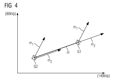
figure 4 shows an HU value diagram for two tissue types,
including the enhancement from an inventive
combination of two contrast agents;
figure 5 shows a schematic illustration of the 3 material
decomposition; and
figure 6 shows a schematic illustration of the enhancement
gradient plotted against atomic number.
In accordance with the invention, the concentration of a
contrast agent in body materials, in particular liver tissue,
is intended to be measured using computed tomography, with
another contrast agent being applied at the same time in which
only the CT value increase relative to the surroundings but not
the precise concentration is of interest.
Achieving the object requires two particular components: the X-
ray absorption must firstly be measured at least in respect of
two different energy spectra; this can preferably be effected
CA 02706188 2010-05-19
PCT/EP2008/056115 - 13a -
2006P25907WOUS
using a dual energy CT scanner which affords reconstruction of
two independent images for at least one axial slice through the
patient, which images were generated with different effective
X-ray
CA 02706188 2010-05-19
PCT/EP2008/056115 - 14 -
2006P25907WOUS
spectra. This can be implemented, for example, by
simultaneously scanning at two different tube voltages in a CT
scanner with two X-ray tubes (= dual source CT). Alternatively,
it is possible, for example, to use a simple CT scanner with an
energy selective detector. All that is important is that the
different absorption effects can be observed in two different
energy ranges. In the following text, the term dual energy CT
should represent an energy-specific CT in a generally
applicable fashion.
An example of such a CT system 1 is illustrated in figure 1.
This CT system 1 has a gantry housing 6 in which two X-ray
tubes 2 and 4 with opposing detector systems 3 and 5 are
arranged in an angularly offset fashion and rotate about a
system axis 9 in order to scan the patient 7 while the patient
7 is displaced through the measurement region of the CT system,
along the system axis 9 by means of the controllable patient
couch. In the example illustrated in this case, the two X-ray
tubes 2 and 4 are operated at different acceleration voltages
and so the two utilized X-ray spectra substantially differ from
one another and also supply different absorption values in the
associated detectors when the patient is irradiated.
A control and computational unit 10 is used for controlling,
reconstructing and executing the method according to the
invention; said unit comprises in its storage computer programs
Prgl-Prgn which execute the control and reconstruction during
operation. At least one program Prgx is also part of these
programs and it executes a method according to the invention.
The control and computational unit also controls a contrast
agent applicator 11, with the aid of which the contrast agents,
selected according to the invention, can be applied.
Specifically selected contrast agents are used to carry out the
method according to the invention, which contrast agents can be
distinguished using dual energy CT. A further requirement
CA 02706188 2010-05-19
PCT/EP2008/056115 - 15 -
2006P25907WOUS
is that at least the second contrast agent, in terms of its
absorption properties, has to be designed or selected
specifically for the utilized effective X-ray spectra and the
observed tissue surroundings.
A precondition for the algorithmic achievement of the object is
a known three material decomposition, as is described in the
patent application with the file reference
DE 10 2006 009 222.8. It can also be applied analogously for a
conventional 2 material decomposition.
In the following text, the method will be described
specifically for a liver examination, without this intending to
restrict the general applicability. Once a person skilled in
the art knows this specific solution, he or she is readily able
to extend the specific solution to different other tissue
combinations.
If the liver tissue in a patient is examined, it is ideally
composed of only glandular tissue, referred to as tissue in the
following text, and fat with in each case a constant density.
As a result of this assumption, the following linear dependence
of the HU value x as a function of the fat content f then
holds:
X = f X Xfat + (1-f) X Xtissue
Here, xfat and xtissue refer to the HU values depending on the
tube voltage of the pure materials mentioned in the indices.
If, as illustrated in figure 2, the determined HU value at
80 kVp acceleration voltage is plotted against the HU value at
140 kVp acceleration voltage in an HU value diagram, the HU
value pairs of HU values for all possible mixtures of fat and
tissue lie on a straight line with a gradient m which
corresponds to the base gradient within the scope of the
invention. In figure 2, the HU value diagram is illustrated for
CA 02706188 2010-05-19
PCT/EP2008/056115 - 15a -
2006P25907WOUS
the two radiation spectra with 80 kVp on the ordinate and
140 kVp on the abscissa, with the
CA 02706188 2010-05-19
PCT/EP2008/056115 - 16 -
2006P25907WOUS
points G1 and G2 representing the HU value pairs of tissue and
fat.
If a first contrast agent Kl, e.g. iodine, is now added, the HU
values in both spectra increase substantially. This is
illustrated in figure 2 by the arrows G1+K1 and G2+K1. The
gradients of these illustrated arrows correspond to the
enhancement of the contrast agent Ki in respect of the two
utilized energy spectra or the examined energy ranges. Since
the contrast agent cause significant absorption even in low
concentrations, it approximately holds true that the HU value
increases linearly with the concentration c of the admixed
contrast agent K1, with the absorption per molar concentration
of the contrast agent not depending on the organic material,
here the specific fat/tissue mixture. The resultant HU value in
respect of the 80 kVp or 140 kVp spectrum then results from:
X80/140 = f X Xfat801140 + (1-f) X Xtissue80/140 + CXcontrast agent80/140=
Using the three component decomposition mentioned initially, it
is thus possible to calculate the fat content f and the
contrast agent concentration c of the one contrast agent Kl by
measuring the energy-specific HU values for every point in the
HU value diagram.
It is obvious that this method only works if the direction of
the vectors plotted in figure 1, which illustrates the increase
in the CT value by the contrast agent, i.e. the enhancement of
the contrast agent, spans a sufficiently large angle with
respect to the fat/tissue connecting straight line because
otherwise the linear system of equations used to calculate the
component decomposition reacts very sensitively to noise. In
the extreme case in which the enhancement or the contrast agent
vector is parallel to the connecting straight line, the system
of equations cannot be solved at all.
CA 02706188 2010-05-19
PCT/EP2008/056115 - 16a -
2006P25907WOUS
In the case of existing contrast agents, which typically
comprise heavy atoms up to iodine, this is generally not
CA 02706188 2010-05-19
PCT/EP2008/056115 - 17 -
2006P25907WOUS
a problem. Iodine-containing contrast agents in particular are
even optimally suited to the method because they have a large
enhancement gradient.
In order to devise a suitable contrast agent K2 for the method
according to the invention, the following physical background
knowledge is required: The absorption spectrum of iodine is
dominated by the photoelectric effect with release of an
electron from the K shell of iodine. This absorption drops
sharply for photons which do not have sufficient energy for
this process (<33 keV) because then only L shell electrons
contribute to the absorption. For typical CT scanners, this "K
edge" lies below the utilized photon energy. A schematic
profile of the absorption is illustrated in figure 3.
The K edge of elements with a higher atomic number moves into
the energy range used by the dual energy CT scanner from below.
As a result, the ratio of the absorption at the lower tube
voltage relative to the absorption at the higher tube voltage
decreases at a constant concentration. For suitably high atomic
numbers, the reconstructed HU value at the lower tube voltage
can even become smaller than the HU value at the higher tube
voltage. For example, in the case of currently available
scanners from the applicants, this transition lies in the
region of the lanthanides.
Thus, in particular, it is possible to devise a contrast agent
K2 which, in the 80 kVp/140 kVp HU value diagram illustrated in
figure 2, has an increase vector which lies parallel to the
connecting line between fat and tissue. In particular, it is
possible to find a combination of two contrast agents Ki and
K2, wherein the first contrast agent K1 has an enhancement with
a gradient which unambiguously and significantly lies above the
base gradient between two tissue types Gl and G2, and wherein
the second contrast agent has an enhancement which
CA 02706188 2010-05-19
PCT/EP2008/056115 - 18 -
2006P25907WOUS
corresponds to the aforementioned base gradient, at least
within the error tolerance range of the CT system.
Such a combination is illustrated schematically in figure 4.
Here, the points Gl and G2 of the HU value pairs are plotted
for the two tissue types: liver tissue and fat tissue. The
connecting straight line G1-G2 between these points has the
gradient m, with vectors with the gradients ml and m2
originating from each point Gi and G2, which vectors correspond
to the enhancement of the contrast agents Kl and K2. The vector
with the gradient m2 at G2 is arranged slightly offset in
respect of the connecting line G1-G2 for improved clarity. In
this case, the contrast agent K2 was selected or "designed"
such that its enhancement gradient m2 corresponds to the base
gradient m.
If the described three material decomposition is now performed
in these conditions, the concentration of the contrast agent K1
(e.g. with iodine as a main component) is determined correctly,
while the contrast agent K2 (e.g. with erbium as a main
component) is interpreted as a mixture of fat and tissue, with
the factor f in the result also being able to be less than
zero.
The diagnostic use of the method is developed in particular if
a virtual native image is calculated. In this image, the
contrast agent K2 leads to a corresponding increase in HU
value. Thus what is obtained is, on the one hand, a pure
contrast agent image for the contrast agent Kl and a "virtual
native image" which looks like a traditional single energy CT
image using the contrast agent K2.
This method is advantageous in particular if the contrast
agents illustrate different functional aspects of the body
tissue, e.g. vascularization and cell activity. To this end, as
a carrier substance/carrier molecule of the two contrast
CA 02706188 2010-05-19
PCT/EP2008/056115 - 18a -
2006P25907WOUS
agents, a component mainly accumulating in the cardiovascular
system
CA 02706188 2010-05-19
PCT/EP2008/056115 - 19 -
2006P25907WOUS
can be selected on the one hand for the first contrast agent K1
with the higher enhancement gradient and a different component
mainly accumulating in pathological tissue cells, in particular
liver cells, can be selected as carrier substance/carrier
molecule for the second contrast agent. Compared to the method
of registration/subtraction, this has the advantage that there
are no registration artifacts. Moreover, only a single CT scan
is required and not two scans as is the case in the described
alternative method and so time and dose are saved.
The method according to the invention can be illustrated in an
abbreviated fashion in accordance with figure 5. Here, on the
left-hand side, the two image data records B1 and B2 are
recorded Al and A2, taking into account different energy
ranges, for example 50-80 keV and 70-140 keV. The contrast
agents selected according to the invention are used for this
purpose. Subsequently, a 3 component decomposition is performed
in method step 12, for example a 3 component decomposition in
accordance with the method described in the patent application
with the file reference DE 10 2006 009 222.8. This then results
in, on the one hand, an illustration BK1 of only the first
contrast agent Kl and, on the other hand, an illustration
BG1+G2+K2 of the two tissue components Gl and G2 with the
contrast agent K2. Thus, the first image or the first image
data record corresponds to a segmentation of the first contrast
agent Kl, while the second image or the second image data
record corresponds to a single image of a CT with an energy
range, using the second contrast agent K2.
Finally, figure 6 shows the profile 13 of the enhancement
gradient, that is to say the ratio of the HU value increases at
two X-ray spectra of 80 kVp X-ray radiation and 140 kVp X-ray
radiation used in an exemplary fashion for a CT system,
likewise used in an exemplary fashion, over the atomic number
of the contrast-generating element. The contrast-generating
CA 02706188 2010-05-19
PCT/EP2008/056115 - 19a -
2006P25907WOUS
element is, in an exemplary fashion, assumed to be dissolved in
water. The diagram shows four regions I to IV, with the region
CA 02706188 2010-05-19
PCT/EP2008/056115 - 20 -
2006P25907WOUS
I comprising the elements which are unsuitable as a contrast
agent due to a lack of absorption and a low enhancement
gradient. The region II contains the elements which are
suitable for the first contrast agent (up to iodine/early
lanthanides). The region III contains the elements which are
suitable for a second contrast agent, in particular the
lanthanides. At the same time, the elements located in region
IV are unsuitable for contrast agents since they are
predominately radioactive.
It is understood that the abovementioned features of the
invention can be used not only in the respectively specified
combination, but also in other combinations or on their own,
without departing from the scope of the invention.