Note: Descriptions are shown in the official language in which they were submitted.
CA 02706443 2010-05-20
WO 2009/070517 PCT/US2008/084470
TISSUE RETRACTORS
FIELD OF THE INVENTION
[0001] The present invention relates to methods and devices for manipulating
tissue.
BACKGROUND OF THE INVENTION
[0002] During certain surgical procedures, body tissue such as organs can
obstruct an area a
surgeon needs accessible for surgery. Relocating the tissue during all or part
of the procedure
can allow a surgeon to access an otherwise obstructed part of the body. The
tissue may also need
to be relocated to reduce chances of it being damaged as work is being done on
another, nearby
part of the body.
[0003] Visceral retractors have been developed that allow some movement of
tissue in a body
cavity during a surgical procedure. For example, a visceral retractor may be
inserted into the
body through an incision, and it can be used to push tissue aside to provide
access to an
underlying area. Current retractors include a rigid fan-type design, a spoon
or fork-like device,
or an inflatable bladder. While such visceral retractors can move tissue, they
typically move
small amounts of tissue and are difficult or impossible to keep in a fixed
position during use
without constant human interaction.
[0004] Accordingly, there remains a need for improved methods and devices for
manipulating
tissue.
SUMMARY OF THE INVENTION
[0005] The present invention generally provides tissue retractor devices as
well as methods for
performing various procedures using tissue retractors. In one embodiment, an
implantable tissue
retractor device is provided and includes a tissue retractor formed of a
flexible biocompatible
material defining an internal cavity having a plurality of granules, which can
be composed of a
biocompatible material. The tissue retractor has a first state in which it is
selectively
conformable to a target tissue in a body cavity in a desired configuration and
a second state in
which it is substantially rigid and in a substantially fixed conformation.
- 1 -
CA 02706443 2010-05-20
WO 2009/070517 PCT/US2008/084470
[0006] The tissue refractor can have a variety of configurations, but in one
embodiment, at least
one tab can be on the outside surface of the tissue retractor. In another
embodiment, a valve can
be located on an outer surface of the tissue retractor and can be in fluid
communication with the
internal cavity such that the valve can selectively allow passage of fluid
therethrough. The tissue
retractor can be configured from the first state to the second state by
removing fluid from within
the internal cavity. In some embodiments, the internal cavity can be formed
along a perimeter of
the tissue retractor with a mesh material disposed within a central opening
defined by the internal
cavity.
[0007] In other embodiments, the tissue retractor can also include at least
one conduit in fluid
communication with the internal cavity such that fluid can be removed from the
internal cavity
through the at least one conduit. The conduit can be detachable from the
tissue retractor.
Additionally, a valve in fluid communication with the internal cavity can be
coupled to the
conduit such that when the valve is coupled to the conduit and the valve is in
an open position,
the conduit is in fluid communication with the internal cavity.
[0008] In another embodiment, an implantable tissue retractor device includes
an implantable,
biocompatible retractor body having an internal cavity. The internal cavity
can, in some
embodiments, extend around at least a portion of a perimeter of the retractor
body, which may
include a flexible fabric disposed within the perimeter of the retractor body.
The tissue retractor
can have a default non-rigid state and can be disposed in a body cavity.
Constrictable material
can be disposed in the internal cavity, and constricting the material can
cause the retractor body
to have a rigid state in which the retractor body is effective to support
tissue in a body cavity in a
selected substantially fixed position. The material can include a viscous
fluid responsive to a
magnetic field, or, alternatively, biocompatible granules.
[0009] In other aspects, a surgical method is provided that in one embodiment
includes inserting
a conformable tissue retractor into a body cavity in a first orientation,
wherein the retractor has
an internal cavity comprising a plurality of granules. Tissue can be
positioned with respect to the
tissue retractor in a desired conformation that is different than the first
orientation such that the
tissue retractor supports a target tissue. The method can further include
evacuating a fluid from
within the internal cavity such that the granules compact together to maintain
the tissue retractor
- 2 -
CA 02706443 2010-05-20
WO 2009/070517 PCT/US2008/084470
in the desired conformation such that it is able to hold the target tissue in
a substantially fixed
position. In one embodiment, evacuating a fluid from within the internal
cavity can include
applying a vacuum force to withdraw fluid from within the internal cavity. The
method can also
include removing the vacuum force and allowing fluid to re-enter the internal
cavity to enable
the target tissue to be released from the substantially fixed position.
Removing the vacuum force
can include opening a valve on the tissue retractor that is in fluid
communication with the
internal cavity.
[0010] In another embodiment, a surgical method can include introducing a
pliable retractor into
a body cavity in a first conformable configuration. The retractor can be
configured in a rigid
state in a desired orientation with respect to a target tissue such that the
retractor is effective to
support tissue in the body cavity in a substantially fixed position. In some
embodiments,
configuring the retractor in the rigid state includes introducing a magnetic
field to the internal
cavity, while in other embodiments it includes introducing a vacuum to the
internal cavity. In
other embodiments, the method can include decompressing material disposed in
the internal
cavity such that the retractor can change from the rigid state to a non-rigid
state. Decompressing
the material can include removing a vacuum from the internal cavity. The
method can further
include positioning the retractor in the body cavity such that the retractor
supports tissue before
configuring the retractor in the rigid state.
BRIEF DESCRIPTION OF THE DRAWINGS
[0011] The invention will be more fully understood from the following detailed
description
taken in conjunction with the accompanying drawings, in which:
[0012] FIG. 1 is a schematic diagram of an embodiment of a retractor having an
internal cavity;
[0013] FIG. 2 is a cross-sectional view of the retractor of FIG. 1 in a
pliable state;
[0014] FIG. 3 is a cross-sectional view of the retractor of FIG. 1 in a
substantially rigid state;
[0015] FIG. 4 is a schematic diagram of an embodiment of a retractor having an
internal cavity
and tabs;
- 3 -
CA 02706443 2010-05-20
WO 2009/070517 PCT/US2008/084470
[0016] FIG. 5 is a schematic diagram of another embodiment of a retractor
having an internal
cavity and tabs;
[0017] FIG. 6 is a schematic diagram of yet another embodiment of a retractor
having an internal
cavity and tabs;
[0018] FIG. 7 is a schematic diagram of still another embodiment of a
retractor having an
internal cavity and tabs;
[0019] FIG. 8 is a schematic diagram of an embodiment of a retractor having an
internal cavity
around its perimeter;
[0020] FIG. 9 is a perspective view of the retractor of FIG. 1 shown disposed
in a body cavity;
[0021] FIG. 10 is a perspective view of the retractor of FIG. 9 showing tissue
positioned relative
to the retractor;
[0022] FIG. 11 is a perspective view of the retractor of FIG. 9 showing the
retractor manipulated
to move the tissue;
[0023] FIG. 12 is a perspective view of a retractor disposed in a body cavity
and being
manipulated to move tissue; and
[0024] FIG. 13 is a perspective view of the retractor of FIG. 8 disposed in a
body cavity and
coupled to trocars.
DETAILED DESCRIPTION OF THE INVENTION
[0025] Certain exemplary embodiments will now be described to provide an
overall
understanding of the principles of the structure, function, manufacture, and
use of the devices
and methods disclosed herein. One or more examples of these embodiments are
illustrated in the
accompanying drawings. Those skilled in the art will understand that the
devices and methods
specifically described herein and illustrated in the accompanying drawings are
non-limiting
exemplary embodiments and that the scope of the present invention is defined
solely by the
claims. The features illustrated or described in connection with one exemplary
embodiment may
- 4 -
CA 02706443 2010-05-20
WO 2009/070517 PCT/US2008/084470
be combined with the features of other embodiments. Such modifications and
variations are
intended to be included within the scope of the present invention.
[0026] The present invention generally provides methods and devices for
performing surgical
procedures using tissue retractors. In general, the methods and devices allow
a surgeon to use a
retractor to capture a large or small amount of tissue in the retractor, to
move the retractor to
relocate tissue to one or more convenient locations during a surgical
procedure, and to configure
the retractor from a pliable state to a substantially rigid state to hold the
retractor and the tissue in
a selected substantially fixed position during the procedure. The pliable
nature of the retractor
can allow the retractor to be moveable between an open position, in which the
retractor can
support tissue, and a closed position, in which the retractor can be rolled,
folded, or otherwise
compressed in size and fit through a relatively small port, e.g., a trocar or
an incision in a tissue
wall. Once the retractor is inside the body, the need to repeatedly position
tissue during a
procedure can be reduced because more than a small amount of tissue can be
held in the retractor
and moved at a time. The pliable nature of the retractor can allow more
freedom of movement in
positioning the retractor within the body and moving the tissue rather than a
retractor made of
non-pliable material, such as metal. Additionally, holding and moving tissue
in a retractor that
can be oriented in pliable and substantially rigid states can reduce the
chances of tissue slipping
or sliding away from the retractor, a common occurrence when using non-pliable
retractors. This
also reduces the need for tissue reengaging and repositioning. Furthermore,
the retractor can be
molded to the shape of tissue, thereby increasing the amount of tissue area
being supported by
the retractor and reducing the chances of the tissue from slipping or sliding
away from a desired
position. Another feature of the retractor is that it can be anchored and
maintain tissue in a
desired location without the need for a surgeon to constantly hold and
manipulate the retractor.
[0027] A person skilled in the art will appreciate that the devices disclosed
herein can be used in
numerous surgical procedures (including endoscopic, laparoscopic and hand-
assisted
laparoscopic surgery ("HALS") procedures), and in connection with numerous
body cavities and
body tissues. For example, the devices can be used in procedures that take
place in the
abdominal, thoracic, pelvic, and abdominopelvic cavities, and they can be used
to move any
tissue, including organs such as the bowel, small intestine, stomach, liver,
uterus, etc. The
- 5 -
CA 02706443 2010-05-20
WO 2009/070517 PCT/US2008/084470
devices can be introduced into the body in any way in any of the procedures,
such as through an
incision or percutaneously through an access device, such as a trocar or an
endoscopic device.
[0028] A person skilled in the art will also appreciate that the particular
configuration and
materials of the retractor can vary depending on factors such as the type of
procedure being
performed and the type of tissue to be moved or relocated. The retractor can
have any shape
with any number of sides, curves, and cut-out shapes, e.g., rectangular
(including square),
elliptical (including circular), triangular, hexagonal, trapezoidal, T-shaped,
U-shaped, etc. The
retractor can also be made from any flexible material appropriate for surgical
use and can include
zero, one, or more structural elements, e.g., tabs, compressible chambers,
grasping elements, etc.
Structural elements coupled to the retractor can be of any number and
configuration on the
fabric.
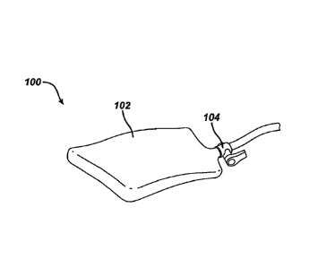
[0029] FIG. 1 illustrates one embodiment of a retractor 100 having a body 102
that can hold
tissue during a surgical procedure. The substantially rectangular shaped
retractor body 102 as
shown is formed of a flexible biocompatible material and is in a pliable
state. The retractor body
102 defines at least one internal cavity. At least one valve 104 located on an
outer surface of the
retractor body 102 can be in fluid communication with the internal cavity and
selectively allow
passage of fluid therethrough. Once inside the body and in the pliable state,
the retractor body
102 can be manipulated into a configuration to receive, hold, move, and
release tissue. From
such a configuration, the retractor body 102 can be conformed from the pliable
state to a
substantially rigid state by manipulating material disposed in the internal
cavity, as further
described below. The retractor body 102 can thereby support tissue in a
selected substantially
fixed position within the body. Additionally, while still inside the body, the
retractor body 102
can be released from the substantially fixed position by again manipulating
the material in the
internal cavity and changing the retractor body 102 from the substantially
rigid state to the
pliable state.
[0030] The retractor body 102 can have a variety of configurations that allow
the retractor body
102 to hold tissue and temporarily move tissue to another location during a
surgical procedure.
In the illustrated embodiment, the retractor body 102 has a substantially
rectangular shape,
although the retractor body 102 can have any shape as mentioned above. The
retractor body 102
- 6 -
CA 02706443 2010-05-20
WO 2009/070517 PCT/US2008/084470
can also have a two dimensional shape when in an open configuration as shown,
but in other
embodiments the retractor body 102 can have a third dimension. For example,
the retractor
body's 102 shape in an open position can be cone-shaped, domed, elliptical
(similar to a
parachute), or prism-shaped with one or more sides of the prism missing so as
to allow tissue to
be held in the retractor body 102.
[0031] The retractor body 102 can also have a variety of sizes, and different
sizes of the retractor
body 102 may be appropriate for relocation of different types of tissue, e.g.,
a larger body for
moving the liver than for moving the stomach. In one embodiment, the retractor
body 102 can
have dimensions that allow it to fit inside a commercially available cannula
so that the retractor
body 102 can be introduced into a body through the cannula.
[0032] The valve 104 attached to the retractor body 102 can also have any
structure. For
example, the valve 104 can include a stopcock (as illustrated in FIG. 1), a
connection port, a
check valve, and other similar structures. The valve 104 can have any shape,
such as elliptical
(including circular), and be of any size. The valve 104 can be configured to
have a shape and
size compatible to couple with commercially available fluid conduits such as
tubes, hoses, and
pumps, as further discussed below, thereby allowing fluid disposed in the
retractor body's
internal cavity to be introduced and/or evacuated through the valve 104. In
use, the valve 104 in
a closed position can maintain the retractor body 102 as a closed pouch able
to hold its shape and
internal pressure, e.g., by preventing fluid passage to and/or from the
retractor body 102. In
contrast, the valve 104 in an open position can allow the retractor body 102
to change shape
and/or internal pressure.
[0033] Any number of valves 104 (including zero, in some embodiments) can be
coupled to the
retractor body 102 in any configuration, and the valve 104 can be coupled to
the retractor body
102 at any point or points along the retractor body's perimeter or elsewhere
on its surface. If the
retractor 100 includes more than one internal cavity, each of the internal
cavities can have a
dedicated valve 104. Furthermore, the valve 104 can be used for both fluid
introduction and
evacuation, as in the embodiment shown in FIG. 1, or the retractor 100 can
include two valves
104, one for fluid introduction and one for fluid evacuation.
- 7 -
CA 02706443 2010-05-20
WO 2009/070517 PCT/US2008/084470
[0034] The valve 104 can be mated to the retractor body 102, or it can be
integrally formed with
the refractor body 102. For example, FIG. 1 illustrates the valve 104 mated to
the retractor body
102. The valve 104 is permanently coupled to the retractor body 102, but in
other embodiments,
the valve 104 can be removable.
[0035] The retractor body 102 and the valve 104 can each be made from any type
of and any
combination of biocompatible material appropriate for use in a body, such as
mesh (braided or
unbraided), fiber (natural or synthetic), gauze-like cloth, a polymer,
biocompatible metal, and
other similar types of material. The retractor body 102 can be made from two
or more layers of
material, e.g., a synthetic fiber outside surface that can come into contact
with tissue and a
polymerized inside surface defining the internal cavity. Moreover, the
retractor body 102 can be
fluid pervious or impervious, and the material can be treated to increase or
decrease its frictional
interaction with tissue. It is understood, of course, that portions of the
retractor body 102 that
define an internal cavity should be made of a fluid impervious material. The
retractor body 102
can also include structural elements such as grasping elements, described
further below. The
retractor body 102 is made from a flexible, elastic material, while the valve
104 can be made
from a flexible, elastic or non-flexible, non-elastic material.
[0036] As indicated above, the retractor body 102 defines an internal cavity
200, illustrated in
FIG. 2. In this embodiment, the internal cavity 200 is shown having a
substantially rectangular
shape, but the internal cavity 200 can have any shape as well as any size. If
the internal cavity
200 includes more than one chamber and/or one or more channels putting
multiple chambers in
fluid communication, each chamber and each channel can have any size,
different or the same
from any other chamber or channel included in the internal cavity 200.
[0037] The internal cavity 200 can have a variety of configurations. For
example, the internal
cavity 200 can be formed in the retractor body 102 as a defined space, e.g.,
two pieces of fabric
or other material mated together as discrete portions to create one or more
cavities therein. The
illustrated cavity 200 has one chamber, but the retractor body 102 can include
any number of
internal cavities including two or more cavities connected by any number of
channels (including
zero channels) through which material disposed in the internal cavity 200 can
flow. In use, fluid
can be introduced into and/or evacuated from the internal cavity 200 through
the valve 104, and
- 8 -
CA 02706443 2010-05-20
WO 2009/070517 PCT/US2008/084470
fluid can travel to and/or from one or more other internal cavities, if
present, via any number of
channels. Alternatively, the internal cavity 200 can include any number of
unconnected cavities,
and fluid can be separately introduced into each cavity to allow each cavity
to be manipulated in
a selected way.
[0038] Constrictable material, such as a plurality of granules 202 and/or a
fluid 204, can be
disposed in the internal cavity 200. The retractor 100 has a pliable state
(shown in FIG. 2) where
the granules 202 and/or the fluid 204 disposed in the internal cavity 200
allow the retractor body
102 to be selectively comformable to a target tissue in a body cavity, as
described further below.
The retractor 100 also has a substantially rigid state (shown in FIG. 3), also
described further
below, where the physical state of the granules 202 and/or the fluid 204
disposed in the internal
cavity 200 has been changed and the granules 202 have been constricted or
compacted, such as
by removing at least a portion of the fluid 204 from the internal cavity 200.
When in the
substantially rigid state, the retractor 100 in use can be held in a
substantially fixed conformation
such that it can support tissue in a selected substantially fixed position.
The retractor 100 can be
introduced to a body cavity in either the pliable state (typically the
retractor's default state) or the
substantially rigid state and can change between the states any number of
times. When the
retractor 100 is in the pliable state, it can maintain a substantially flat
yet pliable configuration
allowing the retractor 100 to be folded or otherwise compressed for easy
introduction into, or
removal out of, a body cavity.
[0039] The plurality of granules 202 are shown as substantially spherical
beads in this
embodiment, but the granules 202 can be of any type and have any shape. For
example, the
granules 202 can have a two-dimensional or three-dimensional ovular,
rectangular, cylindrical,
rod, or other similar shape. The granules 202 can also have any size, although
the granules 202
are typically of a size that prevents their passage through the valve 104. If
the internal cavity
200 includes two or more chambers, the granules 202 can be restricted from
passage between
chambers, such as by the absence of chamber-connecting channel(s) or by the
presence of
vent-like channel(s) that allow passage of the fluid 204 but not passage of
the granules 202
between the chambers. Such restricted passage between chambers can provide for
more even
distribution of the granules 202 throughout the internal cavity 200. While the
granules 202
disposed in the internal cavity 200 can have the same shape and size, any
number of the granules
- 9 -
CA 02706443 2010-05-20
WO 2009/070517 PCT/US2008/084470
202 can differ in shape and/or size from other granules 202 disposed in the
internal cavity 200.
Any number of the granules 202 can be disposed in the internal cavity 200. The
granules 202
can be made from any type of material, typically a biocompatible material
appropriate for use in
a body to minimize patient harm in the uncommon occurrence of retractor body
rupture. For
example, the granules 202 can be composed of medical grade polymers such as
polyethylene,
polypropylene, polyurethane foam or an organic compound, such as sugar. The
granules 202 can
be elastic or non-elastic.
[0040] The fluid 204 is shown as air in FIG. 2, but the fluid 204 can include
any type of gas or
liquid (e.g., saline, a viscous fluid, etc.). The type of fluid 204 disposed
in the internal cavity
200 is compatible with the valve 104 such that the fluid 204 can be introduced
and/or evacuated
through the valve 104. The fluid 204 is typically a biocompatible material
appropriate for use in
a body (although it typically does not come into contact with a body) and a
material compatible
with the granules' material. The amount of fluid 204 disposed in the internal
cavity 200 can
vary, but the amount of fluid 204 disposed in the internal cavity 200 when the
retractor 100 is in
the pliable state is more than when the refractor 100 in the substantially
rigid state. In some
embodiments, such as one further described below, the fluid 204 includes a
viscous fluid
responsive to a magnetic field and the granules 202 need not be present.
[0041] FIG. 4 illustrates another embodiment of a refractor 400 having a body
402 that can hold
tissue during a surgical procedure. The refractor 400 is similar to the
retractor 100 of FIG. 1 and
includes a retractor body 402 defining an internal cavity with fluid and/or
granules disposed
therein. The retractor 400 also includes a valve 404 mated to one corner of
the retractor body
402. The retractor body 402, the valve 404 (shown in this embodiment as a
connection port), the
internal cavity, the granules, and the fluid are similar to those described
with reference to
similarly named elements included in FIGS. 1-3, and the retractor 400 can
include variations as
described herein for various retractors.
[0042] The retractor body 402 has a central body 406 and two tabs 408a, 408b
extending from
the central body 406. The retractor body's internal cavity can extend between
the central body
406 and the tabs 408a, 408b as shown in FIG. 4, or the internal cavity can be
separated into one
or more chambers, e.g., a chamber for each of the central body 406 and the
tabs 408a, 408b. The
- 10 -
CA 02706443 2010-05-20
WO 2009/070517 PCT/US2008/084470
tabs 408a, 408b can have any shape (same or different from the central body
406) and in this
embodiment are substantially rectangular. The tabs 408a, 408b can also have
any size, although
the tabs 408a, 408b are typically each smaller in area than the central body
406. The tabs 408a,
408b can each be folded in one or more directions, such as backwards as shown
by the
directional arrow for the left tab 408a, to aid in conforming the retractor
400 to a target tissue
and/or to increase stability of the retractor 400 in its substantially rigid
state. The retractor body
402 can include scored, weakened, and/or thinned material at a junction
between the central body
406 and one or more of the tabs 408a, 408b to help facilitate tab folding.
Although not
illustrated, it is understood that one or more of the tabs 408a, 408b can have
one or more
apertures (e.g., grommets) to assist in securing the retractor 400 in place,
such as by way of
sutures.
[0043] The tabs 408a, 408b can have any configuration on the retractor 400. In
the illustrated
embodiment, the tabs 408a, 408b extend linearly from corners of the central
body 406, forming a
U-shaped retractor body 402. The tabs 408a, 408b, however, can be attached to
the retractor
body 402 in any configuration. For example, as shown in FIG. 5, tabs 500a,
500b, 500c, 500d
can extend diagonally from each corner of a retractor 502. The retractor 502
also includes a
valve 504 located in a non-corner position along its perimeter. Tabs 600a,
600b, 600c can also
extend from corners of a substantially triangular retractor 602 that has a
valve 604 located on its
top surface, as shown in FIG. 6. For another example, illustrated in FIG. 7,
tabs 700a, 700b,
700c, 700d can be located one each per side of central body 702 of a retractor
704 while a valve
706 is mated to one of the retractor's corners. Junctions between the tabs
700a, 700b, 700c,
700d and the central body 702 in this example include weakened regions 708a,
708b, 708c,
708d.
[0044] FIG. 8 illustrates another embodiment of a retractor 800 that includes
a retractor body
802 that can hold tissue during a surgical procedure. The retractor body 802
includes an internal
cavity extending around a perimeter 804 of the retractor body 802 that can be
of any size and
extend any distance from an edge of the retractor body 802 toward a center of
the retractor body
802. The retractor 800 also includes a valve 806 which, in an exemplary
embodiment is coupled
to one of four corners 808a, 808b, 808c, 808d of the retractor body 802,
although it can be at any
location on the retractor body 802. The retractor body 802, the internal
cavity, and the valve 806
- 11 -
CA 02706443 2010-05-20
WO 2009/070517 PCT/US2008/084470
are similar to those described with reference to similarly named elements
discussed above.
Although the retractor 800 is shown in FIG. 8 to be substantially rectangular,
it is understood that
it can be a variety of alternative shapes.
[0045] The retractor body 802 also includes an internal fabric 810 disposed
within a central
opening defined by the internal cavity around the perimeter 804. The internal
fabric 810 can be
made from any biocompatible material appropriate for use in a body (discussed
above), the same
or different material from the perimeter 804. The internal fabric 810 is
typically a more flexible
material (e.g., braided mesh fabric) than the rest of the retractor body 802
to provide increased
flexibility to the retractor 800. Braided mesh is a useful material for the
internal fabric 810
because tissue is generally less likely to stick or snag on braided mesh than
on other materials.
In one embodiment, the internal fabric 810 is fluid permeable.
[0046] The retractor body 802 also includes grasping elements 812a, 812b,
812c, 812d. The
grasping elements 812a, 812b, 812c, 812d, shown here as grommets, can be
coupled to each of
the retractor body's four corners 808a, 808b, 808c, 808d, although the
retractor 800 could
include any number of grasping elements at any location on the retractor body
802. Once inside
the body, the retractor 800 can be manipulated to receive, hold, move, and
release tissue by
grasping and pulling (including tightening and slackening) one or more
elements, such as the
grasping elements 812a, 812b, 812c, 812d. Additionally, the retractor 800, and
any tissue it
supports, can be held in a substantially fixed position within the body by
anchoring one or more
of the grasping elements 812a, 812b, 812c, 812d to a port, as further
described below.
[0047] The grasping elements 812a, 812b, 812c, 812d attached to the retractor
body 802 can also
have any structure. For example, the grasping elements 812a, 812b, 812c, 812d
can include any
combination of grommets, clips, wraparound ties/loops, hooks, magnetic clasps,
clamps, holes
formed in the retractor body 802, and other similar structures. The grasping
elements 812a,
812b, 812c, 812d can be formed of any biocompatible material appropriate for
use in a body
(discussed above). Each of the grasping elements 812a, 812b, 812c, 812d can be
made from the
same material, but one or more of the grasping elements 812a, 812b, 812c, 812d
can be made
from a material different from one or more of the other grasping elements
812a, 812b, 812c,
- 12 -
CA 02706443 2015-02-24
812d. The grasping elements 812a, 812b, 812c, 812d can be made from a non-
elastic material,
but they can be flexible or rigid.
[0048] The grasping elements 812a, 812b, 812c, 812d can have any shape, such
as elliptical
(including circular). The grasping elements 812a, 812b, 812c, 812d can also
have any length and
width. Preferably, the grasping elements 812a, 812b, 812c, 812d are of a shape
compatible to fit
around or otherwise couple to commercially available trocars, as further
discussed below,
thereby allowing the grasping elements 812a, 812b, 812c, 812d to be
manipulated around the
trocars when receiving, releasing, supporting, or moving tissue in the
retractor 800.
[0049] As indicated above, the grasping elements 812a, 812b, 812c, 812d can be
used to anchor
the retractor 800 in a substantially fixed position. The grasping elements
812a, 812b, 812c, 812d
can also be used for pulling the retractor 800 when introducing the retractor
800 into a body
cavity, when receiving tissue in or releasing tissue from the retractor 800,
and when moving
tissue held in the retractor 800. Any number of grasping elements 812a, 812b,
812c, 812d can be
coupled to the retractor body 802 in any configuration, and the grasping
elements 812a, 812b,
812c, 812d can be coupled to the retractor body 802 at any point or points
along the perimeter
804 and/or on the internal fabric 810. Preferably, there are at least two
grasping elements 812a,
812b, 812c, 812d coupled to the retractor 800 to provide adequate tension when
using the
grasping elements 812a, 812b, 812c, 812d in moving or securing the retractor
800. The grasping
elements 812a, 812b, 812c, 812d can be mated to the retractor body 802, or
they can be
integrally formed with the retractor body 802. For example, FIG. 8 illustrates
four individual
grasping elements 812a, 812b, 812c, 812d, each mated to the retractor body 802
in the perimeter
804 at the corners 808a, 808b, 808c, 808d with the retractor's internal cavity
surrounding the
grasping elements 812a, 812b, 812c, 812d. For another example, the grasping
elements 812a,
812b, 812c, 812d could include loops of fabric extending from one or more
places along the
retractor body's perimeter 804. The grasping elements 812a, 812b, 812c, 812d
are preferably
permanently coupled to the retractor body 802, but one or more of the grasping
elements 812a,
812b, 812c, 812d can be removable.
- 13 -
CA 02706443 2010-05-20
WO 2009/070517 PCT/US2008/084470
[0050] FIG. 9 illustrates the retractor 100 of FIG. 1 in use in a body cavity
900 (e.g., the
abdomen). With the retractor 100 disposed in the body cavity 900, the
retractor 100 can be
manipulated to position the retractor 100 where it can hold and/or move a
tissue 902. Although
the retractor 100 of FIG. 1 is shown, the illustrated methods can be performed
using any retractor
disclosed herein or known in the art.
[0051] The retractor 100 can be inserted into the body cavity 900 in a variety
of ways, such as
through a port, such as an incision (e.g., a HALS access port) made in a body
wall 904 (e.g., the
abdominal wall) or through an access device (e.g., a trocar 906, as shown, a
cannula, etc.)
extending from outside the body wall 904. Although the trocar 906 is shown in
a perpendicular
position relative to the body wall 904, the trocar 906 can be at any angle and
may move
horizontally and/or vertically during use. The retractor 100 can be introduced
into the body
cavity 900 in a closed position, in which the retractor 100 can be folded,
rolled, or otherwise
compressed in size and fit through a port, but once partially or fully
disposed in the body cavity
900, the refractor 100 can be moved to an open position, in which the
retractor body 102 can
support tissue. The retractor 100 is typically disposed in the body cavity 900
in a pliable state as
shown in FIG. 9, although it can be introduced in a rigid state.
[0052] A tube 908 capable of communicating fluid in and/or out of the
retractor's internal cavity
200 (see FIGS. 2 and 3) can be coupled to the retractor's valve 104, as
illustrated, either before
or after the retractor 100 has been disposed in the body cavity 900. The tube
908 is shown
extending through the trocar 906 used to introduce the retractor 100 into the
body cavity 900, but
the tube 908 can extend through any port.
[0053] Once the retractor 100 has been introduced into the body cavity 900, a
surgeon can
position the retractor 100 to hold the tissue 902. The retractor 100 can hold
any amount of the
tissue 902 and in any or all portions of the retractor 100. The tissue 902 can
include more than
one type of tissue, thereby allowing one retractor to simultaneously move
multiple types of
tissue. The tissue 902 can be held in more than one retractor, although only
one refractor 100 is
shown in the illustrated embodiment.
[0054] Referring to FIG. 10, the tissue 902 is shown positioned with respect
to the refractor 100
such that the retractor body 102 supports the tissue 902. The tissue 902 can
be positioned with
- 14 -
CA 02706443 2010-05-20
WO 2009/070517 PCT/US2008/084470
respect to the retractor 100 in a variety of ways that can be performed alone
or in any
combination. For example, positioning the tissue 902 with respect to the
retractor 100 can
include manipulating the retractor body 102 with at least one grasping device.
Examples of
grasping devices include fingers, hands, and any instrument safe for surgical
use and capable of
grasping the tissue 902 and/or the retractor 100 such as forceps, rods, a
spatula 1000 as shown,
and other similar instruments. The grasping device 1000 can grip, push, pull,
or otherwise move
the tissue 902 and/or the retractor 100 to position the tissue 902 with
respect to the retractor 100
or to position the retractor 100 in a location proximate to the tissue 712.
Gravity can move the
tissue 902 from the proximate location to a position such that the tissue 902
can be supported by
the retractor body 102.
[0055] In another example, positioning the tissue 902 can include manipulating
one or more
grasping elements coupled to the refractor 100 to move the retractor 100
around the tissue 902
(e.g., using a grasping device). One or more of the grasping elements can be
simultaneously or
sequentially pulled to position the tissue 902 with respect to the retractor
100 or to position the
retractor 100 in a location proximate to the tissue 902. As yet another
example, one or more tabs
coupled to the retractor body 102 can be simultaneously or sequentially folded
(e.g., using a
grasping device) to place the tissue 902 with respect to the retractor 100 or
to place the refractor
100 around the tissue 902.
[0056] Once the retractor 100 supports a desired amount of the tissue 902, the
retractor 100 can
be manipulated to move the tissue 902. As shown in FIG. 11, the retractor 100
has been
manipulated to move the tissue 902 supported by the retractor body 102. The
tissue 902 has
been moved from a first position 1100 (the tissue 902 shown with dotted lines)
to a second
position 1102 (the tissue 902 shown with solid lines). The two positions 1100,
1102 are
examples; the tissue 902 can be moved in any direction and between any number
of positions
during any one surgical procedure.
[0057] The tissue 902 can be moved while supported by the retractor 100 in a
variety of ways
that can be performed alone or in combination. For example, at least one
grasping element
and/or tab coupled to the retractor 100 can be manipulated. In another
example, a grasping
device can manipulate the retractor 100.
- 15 -
CA 02706443 2010-05-20
WO 2009/070517 PCT/US2008/084470
[0058] Once moved to a desired configuration such as the second position 1002,
the retractor
100 can be fixed to anchor the retractor 100 and thus the tissue 902 in the
second position 1002.
Fixing the retractor 100 can be accomplished by, for example, configuring the
retractor body 102
from a pliable state to a substantially rigid state, shown in FIG. 11. Fixed
in the second position
1002, the tissue 902 can be held in that particular position with minimal or
no human interaction
during a surgical procedure. The retractor 100 can still be easily adjusted,
e.g., by moving the
substantially rigid retractor body 102, manipulating grasping elements,
opening the valve 104 to
introduce and/or evacuate fluid from the internal cavity 200, etc.
[0059] Configuring the retractor body 102 in a substantially rigid state can
be accomplished in a
variety of ways. For example, a pump device 1104 (e.g., a surgical syringe)
coupled to the tube
908 (or, in some embodiments, coupled directly to the valve 104) can apply
suction to the
internal cavity 200 when the valve 104 is in an open position. The suction can
draw a vacuum
inside the internal cavity 200 by evacuating at least a portion of the fluid
204 such that the
granules 202 compact together in the positioned shape of the refractor body
102. In other words,
the ability of the granules 202 to move is constrained and a mass is created
within the internal
cavity 200 that becomes more rigid as more of the fluid 204 is evacuated.
Although the granules
202 are shown having the same spherical shapes in the pliable state of FIG. 2
and the
substantially rigid state of FIG. 3, one or more of the granules 202 may
themselves compress due
to the vacuum force, e.g., have a smaller diameter and/or a different shape.
Conversely,
introducing fluid into the internal cavity 200 through the tube 908 (using the
pump device 1104
or another fluid introduction device) can de-compact the granules 202 and make
the retractor
body 102 more pliable as more fluid is introduced to the internal cavity 200.
The retractor body
102 can be manipulated with a grasping device while the pump device 1104
evacuates the fluid
204 from and/or introduces fluid to the internal cavity 202, thereby allowing
the retractor 100,
and thus the tissue 902, to be continually positioned to reach a desired
conformation where the
retractor 100 can hold the tissue 902 in a substantially fixed position. When
a desired amount of
the fluid 204 has been evacuated from (and/or introduced to) the internal
cavity 200, the valve
104 can be closed, thereby putting the internal cavity 200 into an equilibrium
state. In the case
of fluid evacuation, the equilibrium state is a substantially rigid state
while in the case of fluid
introduction, it is a pliable state.
- 16 -
CA 02706443 2010-05-20
WO 2009/070517 PCT/US2008/084470
[0060] In other embodiments, illustrated in FIG. 12, configuring a retractor
1200 in a
substantially rigid state can be accomplished by applying a magnetic field
1202 to the retractor
1200 using a magnetic device 1204. The retractor 1200 includes an internal
cavity filled with
fluid, similar to the retractor 102, the internal cavity 200, and the fluid
204 of FIGS. 1-3.
However, the retractor 1200 typically does not include granules disposed in
its internal cavity
because the fluid disposed in the internal cavity can cause the retractor 1200
to have a
substantially rigid state without the presence of any granules. The fluid
within the retractor 1200
can include a viscous fluid (e.g., smart fluids and ferrofluids) responsive to
the magnetic field
1202. When introduced to the magnetic field 1202, the fluid can increase in
viscosity to the
point of forming a substantially rigid mass. With the fluid and hence the
retractor 1200 in such a
substantially rigid state, the retractor 1200 can support a target tissue 1206
in a desired
configuration. When the magnetic device 1204 and hence the magnetic field 1202
are removed,
the fluid can decrease in viscosity and approximately return to its original,
pliable state.
[0061] The retractor 1200 optionally includes a valve 1208 that can be coupled
to one of its
corners, although the valve 1208 can be located at any position on the
retractor 1200. The valve
1208 can be set to an open position at any time so fluid can be introduced
and/or evacuated from
the retractor's internal cavity as described above. However, the valve 1208
typically remains in
a closed position when the retractor 1200 is disposed in a body cavity 1210 as
shown. The valve
104 is more typically set to an open position when the retractor 1200 is
outside the body cavity
1210 so fluid in the internal cavity can be replaced because viscous fluid
responsive to the
magnetic field 1202 can decrease in effectiveness after repeated use.
[0062] In still other embodiments, illustrated in FIG. 13, one or more of the
refractor's grasping
elements 812a, 812b, 812c, 812d (see FIG. 8) can be manipulated to configure
the retractor 800
and a target tissue 1300 in a substantially fixed position. First and second
trocars 1302a, 1302b
are shown in use with the retractor 800 in a body cavity 1304. Two trocars
1302a, 1302b are
shown coupled to the retractor 800, but the retractor 800 can be coupled to
any number of
trocars. At least two trocars 1302a, 1302b are typically used to provide
adequate tension when
manipulating the retractor 800 to support tissue.
- 17 -
CA 02706443 2010-05-20
WO 2009/070517 PCT/US2008/084470
[0063] When the retractor 800 is in the body cavity 1304, one or more of the
grasping elements
812a, 812b, 812c, 812d can be used to couple to the trocars 1302a, 1302b,
typically using one
grasping element per trocar. Thus, one or more of the grasping elements 812a,
812b, 812c, 812d
can be manipulated to help position and/or secure the retractor 800 (and the
tissue 1300) from
one state to a desired state, e.g., a substantially fixed position.
[0064] The grasping elements 812a, 812b, 812c, 812d can couple to the trocars
1302a, 1302b in
a variety of ways. Generally, the grasping elements 812a, 812b, 812c, 812d can
each couple to
an outside surface of an access port (such as the trocars 1302a, 1302b)
inserted into the body
cavity 1304. When one or more of the grasping elements 812a, 812b, 812c, 812d
are coupled to
an outside surface of a trocar, an inside surface of the trocar remains
unobstructed to allow the
trocar to receive an instrument (e.g., a pump device or a magnetic device)
that can extend from
outside a body wall 1306 to inside the body cavity 1304. Although only two
grasping elements
812a, 812b are shown coupled to respective trocars 1302a, 1302b, the other
grasping elements
812c, 812d can be coupled to the same or other trocars in a similar manner.
[0065] The grasping elements 812a, 812b each have a shape that allows them to
be positioned
around the trocars 1302a, 1302b such that longitudinal axes Al of the grasping
elements 812a,
812b are initially substantially parallel to longitudinal axes A2 of trocars
1302a, 1302b. With the
axes Al, A2 so aligned, the grasping elements 812a, 812b can then be advanced
proximally up
their respective trocars 1302a, 1302b, e.g., in a direction from the body
cavity 1304 toward the
body wall 1306. A grasping device can be used to manipulate the grasping
elements 812a, 812b
on the trocars 1302a, 1302b.
[0066] Once the grasping elements 812a, 812b have been advanced on their
respective trocars
1302a, 1302b to desirable positions, the grasping elements 812a, 812b can be
simultaneously or
sequentially released from the grasping device. Releasing the grasping
elements 812a, 812b can
cause them to rotate on their respective trocars 1302a, 1302b due to gravity
and the weight of the
retractor body 802. The longitudinal axes Al of the grasping elements 812a,
812b can thereby
be oriented at non-parallel and non-perpendicular angles to the longitudinal
axes A2 of the
trocars 1302a, 1302b, as shown in FIG. 13. Alternatively or in addition to
relying on gravity, the
grasping elements 812a, 812b can be manipulated (e.g., with a grasping device)
to form the
- 18 -
CA 02706443 2010-05-20
WO 2009/070517 PCT/US2008/084470
non-parallel and non-perpendicular angles. The grasping elements 812a, 812b
can be at least in
part formed from a material (e.g., a high friction elastomeric material) to
increase their friction
holding capability with respect to the trocars 1302a, 1302b. Alternatively or
in addition, the
grasping elements 812a, 812b can engage locking elements 1308 (e.g., shown as
grooves formed
in the outside surface of the trocars 1302a, 1302b) located on one or both of
the trocars 1302a,
1302b to effectively lock the grasping elements 812a, 812b in position on the
trocars 1302a,
1302b. One or both of the grasping elements 812a, 812b can include one or more
locking
support structures such as protrusions from or notches in their surfaces to
help them engage the
locking elements 1308.
[0067] Although the locking elements 1308 are shown as grooves in this
illustrated embodiment,
the locking elements 1308 can have any structure. For example, the locking
elements 1308 can
including any combination of grooves, hooks, magnets, loops, ties,
protrusions, and other similar
structures. The locking elements' structure typically matches the structure of
the retractor's
grasping element(s), e.g., using magnets to engage magnetic grasping elements,
using
protrusions to engage clamps, or using hooks to engage grommets or loops. Any
number of
locking elements 1308 can be coupled to the trocars 1302a, 1302b in any
configuration, and the
locking elements 1308 can include elements of any size at one or more
locations along the
trocar's length. The locking elements 1308 can also have any depth, width, and
height.
Additionally, each of the trocars 1302a, 1302b used with the retractor 800 can
have any
combination of the same or varying locking elements 1308.
[0068] The locking elements 1308 can be coupled to the trocars 1302a, 1302b
using various
techniques. For example, as shown in FIG. 13, the locking elements 1308 are
formed in the
trocars 1302a, 1302b. As another example, the locking elements 1308 can
include a plurality
grooves cut circumferentially around the outside surface of the trocars 1302a,
1302b. In other
embodiments, the locking elements 1308 can be inlaid in or otherwise mated to
the outer surface
of the trocars 1302a, 1302b. The locking elements 1308 can be included as part
of a trocar's
manufacture or can be retrofitted to an existing trocar. The locking elements
1308 can be made
from any type of material appropriate for use in a body, such as the material
of the grasping
elements 812a, 812b and the material of the trocars 1302a, 1302b. The locking
elements 1308
are preferably made from a non-elastic material, but they can be flexible or
rigid.
- 19 -
CA 02706443 2010-05-20
WO 2009/070517 PCT/US2008/084470
[0069] With the grasping elements 812a, 812b anchored to the trocars 1302a,
1302b in the
locking elements 1308, the trocars 1302a, 1302b can still be otherwise used in
a surgical
procedure (as the trocars 1302a, 1302b also can before the grasping elements
812a, 812b couple
to them). For example, an instrument, e.g., an endoscope, can be inserted
through one or both of
the trocars 1302a, 1302b to extend from outside the body wall 1306 to inside
the body cavity
1304. For another example, another retractor could be inserted into the body
cavity 1304
through one or both of the trocars 1302a, 1302b.
[0070] Once the retractor 800 has been introduced into the body cavity 1304, a
surgeon can
position the retractor 800 to hold the tissue 1300 as described above. The
retractor 800 can be
positioned to hold the tissue 1300, and the tissue 1300 can be supported by
the retractor 800,
before and/or after any number of the grasping elements 812a, 812b, 812c, 812d
have been
coupled to the trocars 1302a, 1302b. In one embodiment, at least one of the
grasping elements
812a, 812b, 812c, 812d is coupled to at least one of the trocars 1302a, 1302b
before any tissue is
positioned in the retractor 800 to provide increased structural integrity to
the retractor 800 during
the refractor 800 and/or the tissue 1300 positioning. The retractor 800 can
hold any amount of
the tissue 1300 and in any or all portions of the retractor 800. The tissue
1300 can include more
than one type of tissue, thereby allowing one refractor to simultaneously move
multiple types of
tissue. The tissue 1300 can be held in more than one retractor (which may or
may not be joined
together) although only one retractor 800 is shown in the illustrated
embodiment.
[0071] The tissue 1300 is shown positioned in the retractor 800 such that the
retractor 800
supports the tissue 1300 in a pliable state and alternatively in a
substantially rigid state as
described above. The tissue 1300 can be positioned in the retractor 800 in a
variety of ways that
can be performed alone or in any combination. For example, positioning the
tissue 1300 in the
retractor 800 can include manipulating the internal cavity around the
retractor's perimeter 804
and/or the internal area 810 to move the retractor 800 around the tissue 1300.
As another
example, one or more of the grasping elements 812a, 812b can be adjusted
vertically between
any number of the locking elements 1308. One or more of the corners 808a,
808b, 808c, 808d
and/or other elements coupled to the refractor 800 can be simultaneously or
sequentially pulled
to position the tissue 1300 in the refractor 800 or to position the refractor
800 in a location
proximate to the tissue 1300. As illustrated, one of the corners 808c includes
a tab that has been
- 20 -
CA 02706443 2010-05-20
WO 2009/070517 PCT/US2008/084470
folded to support the tissue 1300. Gravity can move the tissue 1300 from the
proximate location
to a position such that the tissue 1300 can be supported by the retractor 800.
[0072] The devices disclosed herein can also be designed to be disposed of
after a single use, or
they can be designed to be used multiple times. In either case, however, the
device can be
reconditioned for reuse after at least one use. Reconditioning can include any
combination of the
steps of disassembly of the device, followed by cleaning or replacement of
particular pieces, and
subsequent reassembly. In particular, the device can be disassembled, and any
number of the
particular pieces or parts of the device can be selectively replaced or
removed in any
combination. Upon cleaning and/or replacement of particular parts, the device
can be
reassembled for subsequent use either at a reconditioning facility, or by a
surgical team
immediately prior to a surgical procedure. Those skilled in the art will
appreciate that
reconditioning of a device can utilize a variety of techniques for
disassembly,
cleaning/replacement, and reassembly. Use of such techniques, and the
resulting reconditioned
device, are all within the scope of the present application.
[0073] Preferably, the devices described herein will be processed before
surgery. First, a new
and/or used instrument(s) is obtained and if necessary cleaned. The instrument
can then be
sterilized. In one sterilization technique, the instrument is placed in a
closed and sealed
container, such as a plastic or TYVEK bag. The container and instrument are
then placed in a
field of radiation that can penetrate the container, such as gamma radiation,
x-rays, or
high-energy electrons. The radiation kills bacteria on the instrument and in
the container. The
sterilized instrument can then be stored in the sterile container. The sealed
container keeps the
instrument sterile until it is opened in the medical facility. It is preferred
that device is sterilized.
This can be done by any number of ways known to those skilled in the art
including beta or
gamma radiation, ethylene oxide, steam.
[0074] One skilled in the art will appreciate further features and advantages
of the invention
based on the above-described embodiments. Accordingly, the invention is not to
be limited by
what has been particularly shown and described, except as indicated by the
appended claims.
[0075] What is claimed is:
-21 -