Note: Descriptions are shown in the official language in which they were submitted.
CA 02706477 2015-08-06
1
Title: Improved FRET-probes and use thereof.
This invention relates to the detection of, among others,
tumor-specific fusion proteins and protein interactions. More specifically,
the invention relates to techniques that indicate the presence of a fusion
protein and/or interacting proteins at the single cell level.
The diagnosis and classification of malignancies is frequently based
on the detection specific protein molecules or sets of protein molecules, as
well as the detection of oncogenetic aberrations, mainly at the DNA level or
RNA level [1]. The current genomics and proteomics studies in normal and
malignant cells are drastically extending the information about gene
expression and genetic aberrancies. This leads to the discovery of multiple
new protein networks, which regulate cell-cell interactions, cell activation,
signaling pathways, proliferation, differentiation, apoptosis and many other
normal and abnormal cellular functions. Unravelling these protein
networks requires the specific detection of true co-localization of the
individual protein molecules.
Classification of malignancies is particularly based on cell lineage
and differentiation characteristics and the presence of specific chromosome
aberrations. Many of these chromosome aberrations result in fusion genes,
i.e. aberrantly coupled genes with the upstream part of one gene coupled to
the downstream part of the other gene and vice versa [2-5]. These fusion
genes are transcribed into fusion gene transcripts and translated into
fusion proteins (Figure 1), which are assumed to play an important role in
the oncogenic process. So far, more than hundred different types of fusion
genes have been described in leukemias, lymphomas, and solid tumors.
Exemplary fusion proteins are listed in Table 1.
CA 02706477 2010-05-20
WO 2009/067009 PCT/NL2008/050737
2
Consequently, the reliable detection of these tumor-specific fusion proteins
at
the single cell level would be a major step forward in the diagnosis and
classification of cancer patients as well as for monitoring of the
disappearance
of the malignant cells during treatment as measure of the effectiveness of the
applied therapy protocol.
TABLE 1. Examples of fusion proteins in malignancies3, which may be
detected via the FRET-technology using the nucleotide linker system for
close and stable juxtapositioning of differentially labeled antibodies
Malignancy Chromosome aberration Fusion protein
Chronic myeloid leukemia t(9;22)(q34;q11) BCR-ABL
Lymphoma t(2;5)(p23;q35) NPM-ALK
Prostate cancer t(21;21)(q22.3;q22.2) TMPRSS2-ERG
Ewing sarcoma t(11;22)(q24;q12) EWSR1-FLI1
Papillary renal cell t(X;1)(p 1 1;q23) PRCC-TFE3
carcinoma
Follicular thyroid t(2;3)(q13;p25) PAX8-PPARG
carcinoma
Fibromyxoid soft tissue t(7;16)(q33;p11) FUS-CREB3L2
sarcoma
Endometrial stromal t(7;17)(p15;q11) JAZF1-SUZ12
carcinoma
Soft tissue t(9;22)(q31;q12) EWSR1-NR4A3
chondrosarcoma
Desmoplastic small round t(11;22)(p13;q12) EWSR1-WT1
cell tumor
Poorly differentiated t(15;19)(q14;p13) BRD4-NUT
carcinoma affecting
midline structures
Initially, scientists tried to raise fusion-protein specific antibodies by
making antibodies against the fusion epitopes of the fusion proteins. This
approach has rarely been successful and if specific antibodies were obtained,
they generally were not applicable in fluorescence microscopy or flow
cytometry [6-8]. For example, the ER-FP1 antibody against the BCR-ABL p190
CA 02706477 2010-05-20
WO 2009/067009 PCT/NL2008/050737
3
fusion protein works nicely in Western blotting, but was not successful in
microscopic studies on human BCR-ABL positive leukemias [6,7].
Despite these initial problems, specific detection of fusion proteins has
become possible via the application of a catching antibody against one part of
the fusion protein and a labeled detection antibody against the other part of
the protein. In such systems, the catching antibody is bound to a solid layer,
such as a dipstick, an ELISA plate, or beads that can be analyzed by flow
cytometry [9]. Although elegant and easy to perform, these systems use cell
lysates and consequently do not allow detection of intracellular fusion
proteins
at the single cell level.
A close interaction between two different membrane-bound and/or
intracellular proteins, or the presence of fusion proteins can be investigated
by
use of antibodies that are conjugated with or linked to fluorochromes that are
suited for fluorescence resonance energy transfer (FRET). FRET technology is
based on the juxtapositioning of two different fluorochromes (with different
excitation wavelengths) of which one fluorochrome produces emission light
that excites the other fluorochrome (Figure 2). If the emission light of the
second fluorochrome can be detected by flow cytometry, this allows easy, high-
speed analysis of protein networks and detection of fusion proteins at the
single cell level. Using e.g. confocal laser scanning microscopy, it becomes
possible to evaluate the precise subcellular position of the interacting
protein
and fusion proteins.
Approximate colocalization of two FRET fluorochrome-conjugated
antibodies is not sufficient for the required light-energy transfer. True
colocalization of the detected proteins is needed so that close
juxtapositioning
of the fluorochromes linked to two different antibodies occurs (generally < 80
A, but preferably < 50 A, most preferably < 10 A), which is essential for
efficient light-energy transfer.
CA 02706477 2010-05-20
WO 2009/067009 PCT/NL2008/050737
4
We previously described that FRET technology can be used to detect the
presence of a fusion protein (W02004/042398) or to detect protein interactions
(W02004/042404) in a cell using a set of specifically designed probes.
According to W02004/042398 and W02004/042404, each probe is provided
with a dye wherein said dyes together allow FRET, and at least one probe is
provided with a reactive group. The addition of a "bridging" reagent capable
of
binding to the reactive groups allows juxtaposing the first and second probe
such that there is an increased likelihood of energy transfer between the FRET
dyes. W02004/042398 for example discloses a set of antibody probes A and B
directed against fragments A and B of an A-B fusion protein, wherein A and B
are labelled with different FRET dyes and wherein A and B were provided
with biotin reactive groups capable of binding a (strept)avidin bridging
substance. Upon addition of the bridging substance to the reactive groups, the
spatial organization of the antibody probes is modulated via the reactive
groups. This allows the individual FRET dyes that are attached to the probes
to come within a distance of each other that allows FRET to occur, i.e. within
about 80-100 Angstrom of each other. Whereas the probes and methods of
W02004/042398 and W02004/042404 can generally yield satisfactory results,
the present inventors sought to further improve the concept. In particular, it
is
a goal of the present invention to increase the sensitivity of FRET-based
methods for detecting fusion proteins and interacting proteins.
This goal was met by the realization that oligonucleotide moieties are
highly suitable to bring the FRET fluorochromes in close and stable
juxtaposition, such that FRET can occur with great efficiency. The small size
of
oligonucleotides allows for only a minimal spacing between the juxtaposed
dyes. Furthermore, complementary oligonucleotide sequences can be designed
to achieve highly specific and strong intermolecular interactions. Thus, the
inventors identified oligonucleotides (e.g. DNA or PNA or LNA molecules) as
CA 02706477 2010-05-20
WO 2009/067009
PCT/NL2008/050737
excellent reactive groups and bridging substance to mediate and/or enhance
close juxtapositioning of dye-labeled probes.
The invention therefore provides a set of at least a first and a second
molecular probe, each probe provided with a dye wherein said dyes together
5 allow energy transfer, each probe additionally provided with a reactive
group
allowing juxtaposing said at least first and second probe, wherein said
reactive
group is an oligonucleotide and wherein the oligonucleotide reactive group of
said first probe is not directly reactive with the oligonucleotide reactive
group
of said second probe. The latter is required to avoid false-positive signals
generated by unwanted self-association between the probes.
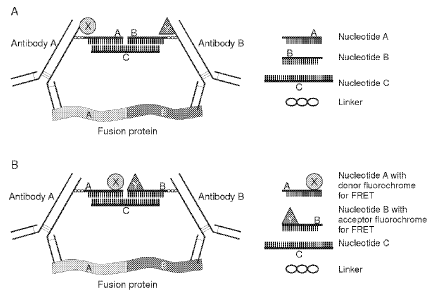
The principle underlying the present invention is schematically
illustrated in Figure 3. A first molecular probe (antibody A) is provided with
FRET dye X and with at least one reactive group, said reactive group
comprising or consisting of an oligonucleotide (Nucleotide A). The reactive
group is capable of binding specifically to a bridging substance (Nucleotide
C)
comprising or consisting of a nucleic acid sequence of which a part is
complementary to the sequence of at least a fragment of Nucleotide A..
Nucleotide C is also complementary to at least part of the sequence of the
reactive group (Nucleotide B) of a second molecular probe (Antibody B)
provided with FRET dye Y. The dyes X and Y together form a FRET pair. Only
if the first and second probe come into close proximity of each other (e.g.
because they are bound to adjacent epitopes on a fusion protein A-B as shown
in the figure, or to epitopes on interacting molecules), the oligonucleotide
reactive groups (Nucleotides A and B) are sufficiently close together for the
bridging substance (Nucleotide C) to bind to the reactive groups of both the
first and second probe. This interaction will reduce and stabilize the
distance
between the two probe-bound dyes such that a FRET signal can be detected.
FRET energy transfer efficiency is inversely proportional to the sixth
power of the distance between the donor dye and the acceptor dye. The very
small size of the oligonucleotide reactive groups and bridging substance of
the
CA 02706477 2010-05-20
WO 2009/067009
PCT/NL2008/050737
6
oligonucleotide linker system as disclosed herein allows a very close
proximity
of the dyes (e.g. within 10 Angstrom), resulting in a much stronger
fluorescence signal as compared to using the proteinaceous reactive groups
and bridging substance as disclosed in W02004/042398 or W02004/042404.
Furthermore, the base-pair recognition between the complementary sequences
of the reactive group and the bridging substance, yet not between the reactive
groups themselves, provides a high degree of specificity.
In this FRET approach with three oligonucleotide molecules as
stabilizing linker system, cells are typically first subjected to the
intracellular
labeling with the at least two probes each carrying an oligonucleotide
reactive
group, followed by (stringent) washing and subsequent incubation with the
specifically designed bridging oligonucleotide to obtain close and stable
linkage
of the two reactive antibodies. In a preferred embodiment, each of the probes
is
provided with a multiplicity of reactive groups, like 2-6 oligonucleotides
each
being reactive with a bridging oligonucleotide. Said reactive groups present
on
a single probe may be the same or different to each other.
As used herein, the expressions "reactive group oligonucleotide" and
"oligonucleotide reactive group" are used interchangeably, unless indicated
otherwise. Also, "bridging oligonucleotide" and "oligonucleotide bridging
substance" refer to the same entity.
The term "oligonucleotide" as used herein refers to a stretch of nucleic
acids or nucleic acid analogs joined in a long chain. The total length of the
oligonucleotide can vary, depending among others on the nature of the nucleic
acid or nucleic acid analog. In one embodiment, an oligonucleotide consists of
a
stretch of 5-50, preferably 10-30 nucleic acids or nucleic acid analogs joined
in
a long chain. A nucleic acid is for instance a nucleotide comprising a
nitrogenous base (A, G, T, or C in DNA; A, G, U, or C in RNA), a charged
phosphate moiety, and a sugar moiety (deoxyribose in DNA and ribose in
RNA).
CA 02706477 2010-05-20
WO 2009/067009
PCT/NL2008/050737
7
Suitable lengths for the probe-bound oligonucleotides (i.e. the reactive
groups) include those consisting of at least 8 nucleic acid residues,
preferably
10-18 nucleic acids or analogs. The lengths of the oligonucleotide reactive
groups on the respective probes may be different or the same. In one
embodiment, they are of the same length, such as 10-15, preferably 10-12
nucleic acids or analogs. The bridging oligonucleotide will generally be
longer
than the reactive group oligonucleotides. In one embodiment, the bridging or
linker oligonucleotide consists of 15-40 nucleic acids or analogs thereof, for
example 18-30, like from about 20 to about 25.
In a preferred embodiment, the oligonucleotide is a peptide nucleic acid
(PNA) oligomer. PNA is similar to DNA or RNA but differs in the composition
of its "backbone." DNA and RNA have a deoxyribose and ribose sugar
backbone, respectively, whereas PNA's backbone is composed of repeating N-
(2-aminoethyl)-glycine units linked by peptide bonds. The various purine and
pyrimidine bases are linked to the backbone by methylene carbonyl bonds.
PNAs are typically depicted like peptides, with the N-terminus at the first
(left) position and the C-terminus at the right.
The nucleic acid analog PNA is not known to occur naturally in existing
life on earth, but it can be artificially synthesized. It has been used in
certain
areas of biological research and medical treatments. Synthetic peptide nucleic
acid oligomers have been used in recent years in molecular biology procedures,
diagnostic assays and antisense therapies. Since the backbone of PNA contains
no charged phosphate groups, the binding between PNA/DNA strands is
stronger than between DNA/DNA strands due to the lack of electrostatic
repulsion. Early experiments with homopyrimidine strands (strands consisting
of only one repeated pyrimidine base) have shown that the Tm ("melting"
temperature) of a 6-base thymine PNA/adenine DNA double helix was 31 C in
comparison to an equivalent 6-base DNA/DNA duplex that denatures at a
CA 02706477 2010-05-20
WO 2009/067009 PCT/NL2008/050737
8
temperature less than 10 C. Mixed base PNA molecules are true mimics of
DNA molecules in terms of base-pair recognition.
PNA oligomers also show greater specificity in binding to
complementary DNAs, with a PNA/DNA base mismatch being more
destabilizing than a similar mismatch in a DNA/DNA duplex. This binding
strength and specificity also applies to PNA/RNA duplexes. PNAs are not
easily recognized by either nucleases or proteases, making them resistant to
enzyme degradation. PNAs are also stable over a wide pH range. Finally, their
uncharged nature makes crossing through cell membranes easier, which may
further improve their value for the present invention which involves detection
of a fusion protein in intact cells. In one aspect, a PNA oligonucleotide
consisting of about 10 to 16, like 12-15 PNA units is used as reactive group
oligonucleotide, optionally in combination with a bridging oligonucleotide
consisting of 20-30 PNA units. In a specific aspect, the probe-bound PNA
sequences each consist of 10-12 PNA units complementary to a bridging
oligonucleotide consisting of 20-25, like 21, PNA units.
In yet another embodiment, the oligonucleotide comprises Locked
Nucleic Acids (LNATm). LNA is a novel type of nucleic acid analog that
contains
a 2'-0, 4'-C methylene bridge. This bridge¨locked in 3'-endo conformation-
restricts the flexibility of the ribofuranose ring and locks the structure
into a
rigid bicyclic formation, conferring enhanced hybridization performance and
exceptional biological stability.
As will be understood by a person skilled in the art, for the reactive
groups and bridging substance various different combinations of types of
nucleic acid oligomers (e.g. DNA, RNA, LNA, PNA) can be used. The specific,
high affinity interaction between reactive group and bridging substance can be
effected through either homoduplex (e.g. DNA/DNA, PNA/PNA) or
heteroduplex (e.g. DNA/PNA) formation. In one embodiment, a probe set of the
invention comprises at least a first and a second probe, each probe provided
with a distinct oligonucleotide as reactive group, wherein said nucleic acid
CA 02706477 2010-05-20
WO 2009/067009 PCT/NL2008/050737
9
oligomer is a deoxyribonucleotide oligomer (DNA). These DNA reactive groups
can be clustered by different types of bridging substances, for instance a DNA
(homoduplex) or a PNA (heteroduplex) bridging substance. Alternatively, the
reactive groups comprise an oxyribonucleotide sequence (RNA) which can be
recognized and bound by an RNA (homoduplex) or PNA (heteroduplex)
bridging substance. It is also possible to use a homoduplex between a bridging
substance and the reactive group of an at least first probe and a heteroduplex
between the bridging substance and the at least second probe. For example,
probe A is provided with a PNA reactive group and probe B with a DNA or
RNA reactive group, both groups capable of being clustered by a PNA bridging
substance.
The extent or degree of complementarity between the bridging
oligonucleotide and either one of the oligonucleotide reactive groups can
vary,
as long as it allows for a specific and stable binding. In one embodiment,
there
is complementarity (i.e. base-pairing) between a reactive group and a bridging
substance over a stretch of at least 5, preferably at least 7 consecutive
nucleic
acids or analogs. As will be understood, the oligonucleotide reactive group of
the first probe is not directly reactive with the oligonucleotide reactive
group of
the second probe in order to avoid self association of the probes and
premature
energy transfer to occur between the attached dyes. This is important to
ensure that a FRET signal truly reflects juxtaposed probes.
In one embodiment, the bridging substance comprises a first sequence
that is complementary to at least part of a first oligonucleotide reactive
group
and a second sequence that is complementary to at least part of a second
oligonucleotide reactive group, wherein the first and second sequence are
separated by at least one nucleic acid or analog. For example, they are spaced
by a few e.g. 1-10 such as 2, 3, 4, or 5 nucleic acids. A spacing of 1-3 is
preferred. In another embodiment, the complementary sequences are spaced
by one or more amino acids residues, preferably 1-2 small amino residues like
glycine residues.
CA 02706477 2010-05-20
WO 2009/067009 PCT/NL2008/050737
However, it is also possible that the sequence complementary to at least
part of an oligonucleotide reactive group of a first probe is flanked
directly,
without spacing, by the sequence complementary to at least part of an
oligonucleotide reactive group of a second probe. It is preferred that both
5 termini of the bridging oligonucleotide are designed to participate in
the
binding to the oligonucleotide reactive groups, such that there are no single
stranded "free ends".
In addition, the oligonucleotide sequences should be selected such that
they are not cross-reactive with endogenous nucleotide sequences of the cell
in
10 which the fusion protein is to be detected. Thus, when designing any of
the
sequences used for practising the invention, complementarity with endogenous
(e.g. human) DNA and/or RNA sequences should be minimized or even
completely avoided in order to prevent unwanted blocking or scavenging of the
oligonucleotides.
In one embodiment, a first probe is provided, e.g. via a linker, with a
reactive group oligonucleotide consisting of the sequence 5'-CGA TTC TAT G-3'
and a second probe being provided, e.g. also via a linker, with a reactive
group
oligonucleotide comprising the sequence 5'-TGT ACC TTG A-3'. This set of
probes is advantageously used in combination with a bridging oligonucleotide
comprising or consisting of the sequence 5'-TCA DGG TAC A Gly Gly CAT
AGA ATC G-3'. The skilled person will however understand that the present
invention can be practiced using any set of sequences, be it DNA, RNA, PNA or
any combination thereof, that allows for sufficient binding strength and
binding specificity between the bridging substance and the respective probes.
A molecular probe is capable of specifically binding to a biological
molecule of interest via its so-called binding domain. Following binding of at
CA 02706477 2010-05-20
WO 2009/067009
PCT/NL2008/050737
11
least a first and a second probe to a molecule of interest via the binding
domain, a reactive group is used to modulate juxtapositioning. An
oligonucleotide reactive group remains available for modulating the spatial
organization of juxtaposed probes after the probe is bound to a molecule of
interest. In one embodiment, said molecule of interest is a protein,
preferably a
fusion protein, more preferably an oncogenic fusion protein. Particularly
preferred is a set of a first and a second molecular probe wherein each probe
is
capable of recognizing and binding to a binding site (epitope) positioned at
opposite sides of the fusion region of said fusion protein. Of course, when
using
a set of probes wherein each probe binds to a different epitope of a molecule
of
interest (e.g. epitopes at the C- and N-terminal side of the fusion region of
a
fusion protein), said different epitopes should not interact with each other
in
either an inter- or intramolecular fashion because this would obviously
interfere with probe binding. Different probes within a set of probes are
therefore capable of binding to different, essentially non-interacting
epitopes.
Provided that the probes recognize binding sites (epitopes) within a small
distance of each other, the mere binding of the probes to a fusion protein or
to
interacting molecules could, in theory, give rise to energy transfer between
the
dyes. However, by the "clustering" of juxtaposed reactive groups by a bridging
substance the spatial organization of the dyes can be modulated such that the
likelihood of energy transfer is dramatically enhanced.
The present invention also provides a diagnostic kit comprising a set of
probes according to the invention. In a preferred embodiment, the kit
additionally comprises an oligonucleotide bridging substance which has a
sequence that is complementary to at least part of the oligonucleotide
reactive
group of the first probe, and which is complementary to at least part of the
oligonucleotide of the second probe. For example, such a kit may be used for
monitoring and quantification of malignant cells, e.g. leukemic cells, via the
detection of tumor-specific fusion protein-positive cells. The diagnostic test
kit
CA 02706477 2010-05-20
WO 2009/067009
PCT/NL2008/050737
12
provided herein is useful at the time of diagnosis as well as during and after
treatment to evaluate the effectiveness of the applied cancer treatment
protocol.
A further aspect relates to a method using a set of probes for detecting
the presence of a fusion protein or interacting (proteinaceous) molecules in
the
diagnosis and / or classification of a disease as well as before, during and
after
treatment of a disease to evaluate the effectiveness of said treatment
Also provided is a method for producing a probe set according to the
invention comprising contacting each probe with an oligonucleotide reactive
group to form a conjugate between said probe and said reactive group and
purifying said conjugate. The reactive group oligonucleotide may be attached
to the probe directly or indirectly, for instance via spacer or linker moiety.
Also, the FRET dye can be attached to the probe directly or indirectly, e.g.
via
the reactive group. In a preferred embodiment, a probe comprises at least one
oligonucleotide reactive group, which reactive group is provided with a FRET
dye (see Fig. 3B). The oligonucleotide reactive group may be coupled directly
or
indirectly to the probe. The reactive group may be provided with a FRET dye
prior to or after its conjugation to a probe.
In a preferred embodiment of the invention, a probe set comprises a set
of at least two dye-oligonucleotide-conjugated antibodies, each antibody
capable of recognizing a binding site positioned at opposite sides of the
fusion
region of a fusion protein or at distinct interacting molecules, e.g. proteins
in a
protein complex. A suitable antibody comprises a conventional (poly- or
monoclonal) or a synthetic antibody or a binding fragment functionally
equivalent thereto, such as a Fab', Fab, a single chain Fy fragment, a diabody
(a single chain Fy dimer) and the like. For example, a chimeric fusion protein
A-B can be detected via FRET using a set of dye-conjugated probes, e.g. an
anti-A antibody and an anti-B antibody. In a preferred embodiment, a sample
is contacted with two antibodies, one against domain A and the other against
CA 02706477 2010-05-20
WO 2009/067009
PCT/NL2008/050737
13
domain B of a fusion protein to detect the presence of an A-B fusion protein
in
a cell sample. One antibody is labelled (preferably via its reactive group)
with
a FRET donor dye and an other with a FRET acceptor dye. Only when domain
A is in close proximity to domain B, e.g. when both are part of the same
protein
molecule, the two antibodies become sufficiently close together ('juxtaposed')
which allows the donor/acceptor pair to induce a detectable FRET fluorescence
signal.
In the present context, the term "reactive group" refers to a moiety
which allows modulating the spatial organization of FRET dyes such that
there is an increase in the probability of energy transfer to occur and / or
an
increase in energy transfer efficiency. The spatial organization refers to
both
the distance between the dyes as well as to their relative orientation.
Modulating the spatial organization includes adjusting and stabilizing the
spatial organization of dyes. One of the primary conditions for energy
transfer
to occur is that donor and acceptor molecules must be in close proximity,
typically 10-100 A. In a preferred embodiment, a reactive group allows
juxtaposing said dyes within a distance of 50 A of each other, more preferably
within 20 A of each other but most preferably within a distance of 10 A of
each
other.
In the present context, the term "dye" refers to a substituent which, in
concert with another dye, can be used for energy transfer analysis, such as
FRET analysis. As mentioned above, FRET is usually based on the interaction
between donor and acceptor dyes that are both fluorescent. In one
embodiment, the invention uses a set of probes wherein at least one of said
dyes is a fluorochrome. However, a nonfluorescent acceptor may also be used
and FRET is detected by quenching of donor fluorescence. As said, detecting
FRET by monitoring a decrease in donor fluorescence as a consequence of
juxtapositioned probes is often not as sensitive as detecting in increase in
acceptor fluorescence. Thus, in a preferred embodiment, at least two
fluorescently labeled probes are used to detect a fusion protein, as is
CA 02706477 2010-05-20
WO 2009/067009
PCT/NL2008/050737
14
exemplified in the detailed description. Examples of preferred fluorochromes
are those suitable for analysis by conventional flow cytometry and include
fluorescein labels, e.g. 5-(and 6)-carboxyfluorescein, 5- or 6-
carboxyfluorescein,
6-(fluorescein)-5-(and 6)-carboxamide hexanoic acid and fluorescein
isothiocyanate, AlexaFluorTM dyes such as AlexaFluor 488TM or AlexaFluor
594TM, cyanine dyes such as Cy2, Cy3, Cy5, Cy7, optionally substituted
coumarin, R-phycoerythrin, allophycoerythrin, Texas Red and Princeston Red
as well as conjugates of R-phycoerythrin and, e.g. Cy5 or Texas Red and
members of the phycobiliproteins. Other dyes of interest are quantum dot
dyes, which come in a nearly unlimited palette of colours. Extensive
information on donor/acceptor pairs suitable for energy transfer detection by
flow cytometry can be found in Szollosi et al.18 Preferred combinations of
fluorochromes comprise those dyes used in the classical tandem conjugates,
also referred to as duochromes 19. In a preferred embodiment, probes are
provided with a set of dyes that are used in LightCycler technology, such as
fluorescein in combination with LCRed64OTM or LCRed7OSTM.
Also provided herein is a method for detecting the presence of a fusion
protein
in a cell using a set of at least a first and a second molecular probe, each
probe
capable of recognizing a binding site (via its binding domain) positioned at
opposite sides of the fusion region of said fusion protein, each probe further
provided with a dye wherein said dyes together allow energy transfer, at least
one probe provided with an oligonucleotide reactive group allowing to
modulate juxtaposing said at least first and said second probe such that there
is an increased likelihood of energy transfer between said dyes, comprising
providing a set of probes, providing a sample comprising a cell, contacting
said
sample with said probes under conditions that allow juxtaposing said probes
on said fusion protein, removing any unbound and any non-specifically bound
probe and detecting juxtaposition of said probes via FRET to determine the
presence of said fusion protein.
CA 02706477 2010-05-20
WO 2009/067009
PCT/NL2008/050737
In one embodiment, a probe is provided with more than one
oligonucleotide reactive group, enabling said probe to interact with more than
one bridging substance. Providing a probe with more than one reactive group
will theoretically increase the likelihood of an interaction between said
probe
5 and a bridging substance.
Next, the invention provides a method for detecting a fusion protein at
the single cell level using of a set of probes according to the invention,
each
probe capable of binding to a binding site positioned at opposite sides of a
10 fusion region of said fusion protein via the binding domain of the probe
i.e. one
probe is directed against a protein fragment comprising the N-terminal
fragment of a fusion protein, and an other probe is directed against a protein
fragment comprising the C-terminal fragment of the same fusion protein. A
fusion protein comprises any kind of proteinaceous substance which is formed
15 after transcription and translation of a fusion gene. A fusion gene
comprises
one part of one or more genes combined with another gene or a part derived
thereof. A fusion protein may be the result of a chromosomal translocation,
inversion or deletion. In a preferred embodiment, a method provided is used to
detect a tumor-specific fusion protein. A fusion protein may be an
endogenously expressed protein or it may be the result of genetic engineering.
Fusion proteins in malignancies which can readily be detected using a method
according to the invention include but are not limited to those listed in
Table 1.
Many different applications could be envisaged using the proposed
oligonucleotide-based FRET method disclosed herein, for example:
- Detection of natural and oncogenic fusion proteins:
- oncogenic fusion proteins occurring in several leukemias and
solid tumors
- T-cell receptor or B-cell receptor proteins formed by fusions of
gene segments
CA 02706477 2010-05-20
WO 2009/067009 PCT/NL2008/050737
16
- Detection of association of specific T-cell receptor chains (e.g. a V62+
with a Vy9+ chain);
- Investigation of protein complexes: Are all components of a protein
complex present? How close are the components linked?
- Investigation of gene regulation by transcription complexes (e.g. the
AML1/CBFI3 core binding factor transcription complex, that is a critical
regulator of normal hematopoiesis);
- Investigation of activation of transcription by protein complexes (e.g.
binding of 13-catenin to Tcf-1 induces transcription of Wnt-target genes);
- Detection of protein-DNA or protein-RNA interactions, for
investigation of proteins involved in transcription or translation;
- Evaluation of cell-cell interactions via antibodies directed against
different cell types involved in the same interaction process.
The method disclosed herein has several advantages for application in tissue
sections and smears:
- application in parallel to other immunohistochemical stainings
- combination with split-signal FISH: detection of oncogenic events at
DNA level (fusion gene) and at the protein level (fusion protein).
It is of great relevance to note that the present method does not require
disruption of the cell integrity, e.g. the preparation of a cell lysate, to
detect
the presence of an intracellular fusion protein or molecular complex.
Preservation of the morphology integrity of a cell permits analysis at the
single
cell level, for example by flow cytometry or fluorescence microscopy.
Detection
of a FRET signal by flow cytometry offers the ability to perform rapid,
multiparametric analysis of specific individual cells in a heterogeneous
population. The main advantage of flow cytometry is that it directly gives
quantitative data and that it is very rapid (results can be obtained in a few
hours).
CA 02706477 2010-05-20
WO 2009/067009 PCT/NL2008/050737
17
The method provided in the present invention allows detection of a
fusion protein or interacting molecules at the single cell level. A sample
comprising a cell can be treated so as to obtain a permeabilisation of the
material and a preservation of the morphology. The preferred treatment is one
which fixes and preserves the morphological integrity of the cellular matrix
and of the proteins within the cell as well as enables the most efficient
degree
of probe, e.g. antibody, penetration.
Unlike for example a 'catching/detection' antibody method, which can
essentially only be applied to detect the presence of a fusion protein or
interacting proteins at the cell surface or in a cell lysate, the present
method
allows gating of subset of cells that are present in a mixture of cells via
immunophenotypic characteristics. Consequently, the method provided herein
permits detection in a rare population of malignant cells in a large
background
of normal cells. This is especially advantageous for detecting low frequencies
of
fusion-positive cells like in the case of detection of minimal residual
disease
(MRD) during or after treatment for evaluation of treatment effectiveness. In
preferred embodiment, the method provided includes multiparameter flow
cytometry to identify and / or isolate single cells to detect the presence of
a
fusion protein at the single cell level. All that is required for practicing
the
method provided is a flow cytometry facility. Importantly, the procedure can
be
performed in routine laboratories by personnel with ordinary skills.
More than a hundred different fusion genes and fusion proteins have
been described in various types of cancer. As said, the method provided allows
to discriminate between the presence of normal proteins and an aberrant
fusion protein at the single cell level. Theoretically, two antibodies
recognizing
two different domains of a fusion protein can cause a background staining by
binding to the domains on the normal proteins that are derived from the
normal genes instead of the fusion gene. However, in certain cases only one of
the two normal proteins reaches a detectable expression level in a target cell
population, as defined by cell surface and / or intracellular markers.
CA 02706477 2010-05-20
WO 2009/067009 PCT/NL2008/050737
18
Furthermore, the normal proteins and the fusion protein often differ in their
intracellular expression pattern, frequently resulting in a different
subcellular
localization. This implies that coincidental colocalisation of the two
different
normal proteins is unlikely to occur at a significant level. In particular,
coincidental juxtaposing probes sufficient for a FRET signal will be rare in
normal cells, if this occurs at all.
The method provided comprises providing a sample comprising a cell,
whereby said sample is optionally subject to fixation and permeabilization. A
sample may comprise a primary cell that is obtained from a biological sample.
A biological sample can be a body fluid sample including blood, serum, urine,
bone marrow, cerebrospinal fluid (CSF), saliva. It may also be a tissue
sample,
tissue homogenate. A sample comprises a cultured cell which may be a
cultured primary cell, for example tumor cells obtained from a lymph node
biopsy. Furthermore, a sample may comprise a cultured cell from an
established laboratory cell line, like a K562, KASUMI-1, REH or CEM cell
line, which can be obtained from a number of sources such as the American
Type Culture Collection (ATCC; www.atcc.org for an online catalog) or the
German Collection of Microorganisms and Cell Cultures (DSMZ; v7ww.dsmz.de
for an online catalog). The method provided is suitable to detect the presence
of
an endogenous fusion protein as well as a recombinant fusion protein in a
cell.
The method provided is also suitable to detect interactions between
recombinant proteins and/or endogenous molecules in a cell.
For analysing a sample comprising a suspension of cells, it is preferred
that the sample is treated so as to obtain a preservation of the morphology of
the material and permeabilisation in order to ensure sufficient accessibility
of
a molecule of interest to a probe. The type of treatment will depend on
several
factors, for instance on the fixative used, the extent of fixation and the
type
and properties of the molecule of interest. Fixation may be carried out with a
fixative such as formaldehyde.
CA 02706477 2010-05-20
WO 2009/067009
PCT/NL2008/050737
19
For the detection of in primary cells, it is especially advantageous to use
an additional marker to define a target cell population of interest. A number
of
important biological applications in infectious diseases, MRD detection and
monitoring, and gene therapy typically require the analysis and isolation of
rare cells (e.g. haemopoietic stem/progenitor cells) from a large "background"
of other cells. In one embodiment of the invention, the method includes
staining a sample for at least one cellular marker, like a cell surface marker
or
an intracellular marker, to define a target cell population within a mixture
of
cells comprising contacting said sample with a compound capable of selectively
binding to said marker. In a preferred embodiment, such a compound is
directly tagged with a fluorescent dye. A suitable compound comprises a
fluorescently labelled antibody or a binding fragment functionally equivalent
thereto. Also, a compound capable of selectively binding to a cellular marker
can be used which can be detected using a dye-conjugated secondary reagent
(e.g. a fluorescently labelled secondary antibody). A cellular marker
comprises
any kind of intracellular or membrane-bound marker which can be used to
distinguish a subpopulation of cells in a mixture of cells. A mixture of cells
comprises living cells. It also comprises permeabilized and / or fixed cells.
A
cellular marker can be a cluster of differentiation (CD) antigen. CD markers
are cell surface molecules of among others haemopoietic cells that are
distinguishable with monoclonal antibodies. Haemopoietic cells comprise
thymocytes, dendritic cells, Langerhans' cells, neutrophils, eosinophils,
germinal centre B cells, follicular dendritic cells, plasma cells and bone-
marrow cells. For example, suitable cellular markers comprise CD1, CD3,
CD4, CD8, CD10, CD19, CD20, CD33, CD34 and CD117. Monoclonal
antibodies directed against a large number of human CD markers can be
obtained from various suppliers, such as BD Biosciences or Ancell Immunology
Research Products, Bayport, USA. Often, antibodies are available that are
directly conjugated with a fluorochrome of choice e.g. CD1O-PE or CD19-FITC,
CA 02706477 2010-05-20
WO 2009/067009 PCT/NL2008/050737
which is obviously a preferred choice to practice a method according to the
invention.
In a preferred embodiment, a method is provided to identify and/ or
isolate rare single cells using multiparameter flow cytometry/cell sorting
5 techniques and to further characterize these cells by the presence or
absence of
a fusion protein of interest or by the identification of interacting
molecules.
Such a method is particularly suited for application to a number of important
problems in immune system development, infectious diseases, cancer and gene
therapy. Typically, prior to staining a cell sample with a probe set, cells
are
10 labeled with at least one relevant dye-conjugated antibody according to
standard procedures in order to define a target cell population. The choice of
dye should preferably, but not exclusively, aim at the usage of two or three
dyes for immunophenotyping in addition to the FRET dyes. For example, a
FRET probe set according to the invention can be combined with another dye
15 to mediate leukocyte subset gating via immunophenotypic characteristics,
e.g.
CD10, CD19 and CD20 to accurately define subsets of precursor-B-cells in
bone marrow, or CD1, CD4 and CD8 to define subsets of thymocytes, or CD34
and / or CD117 to identify stem/precursor cell populations. As shown herein in
the detailed description, the invention provides a method which allows the
20 detection of an intracellular fusion protein in a very small subset of
cells, i.e.
detection of MRD, which is essential for evaluating effectiveness of cancer
treatment.
Figure legends
Figure 1. Schematic diagram of a fusion gene consisting of the upstream (5')
part of gene A and the downstream (3') part of gene B. This A-B fusion gene is
transcribed into A-B mRNA and translated into an A-B fusion protein.
CA 02706477 2010-05-20
WO 2009/067009 PCT/NL2008/050737
21
Figure 2. Schematic diagram of the principle of fluorescence resonance energy
transfer (FRET) with fluorochrome X as donor dye and Y as acceptor dye.
A. The acceptor dye Y will not be excitated by the emission light of the donor
dye X, if the distance between X and Y is too large. B. If the distance
between
the donor and acceptor dye is sufficiently small (< 80 Angstrom but preferably
< 50 Angstrom), the emission light of the donor dye X will excitate the
acceptor
dye Y.
Figure 3. Schematic diagram depicting the use of oligonucleotides (e.g.
DNA/PNA) molecules to closely and stably link two antibodies. When both
antibodies come into close proximity of each other because they are bound to
both partners of a fusion protein A-B, bridging substance oligonucleotide C,
which is partly complementary to both oligonucleotide A and B, will reduce
and stabilize the distance between the two fluorochromes X and Y, and a
FRET signal can be detected. A. The donor and acceptor fluorochromes are
conjugated directly to the antibody probes. B. The donor and acceptor
fluorochromes are conjugated to the oligonucleotide reactive groups. The
oligonucleotide reactive groups are attached to the antibody probes via a
linker
moiety.
Figure 4. shows the results of the fluorescence detected in the case of either
reactive group oligonucleotide A, reactive group oligonucleotide B,
combination
of reactive group oligonucleotides A and B, or combination of A and B and
bridging oligonucleotide C. The arrow indicates the FRET signal induced by
the addition of complementary bridging oligonucleotide C. For details see the
Example below.
EXAMPLE
CA 02706477 2010-05-20
WO 2009/067009 PCT/NL2008/050737
22
The following oligonucleotides were synthesized according to standard
procedures:
Reactive group Oligonucleotide A: Linker CGA TTC TAT G Fluorescein
Reactive group Oligonucleotide B: Alexa-TGT ACC TTG A-linker
Bridging Oligonucleotide C: TCA DGG TAC A Gly Gly CAT AGA ATC G
10 pmol PNA of the different oligonucleotides were mixed in 200 4 phosphate
buffer pH 7,2, and their fluorescence was measured. The hybridisation was
complete within 5 minutes when PNA oligonucleotide C was added. For results
see Figure 4.
Instrument: Perkin Elmer LS 55
Excitation wavelength:fluorescein: 485 nm, Alexa 546: 546 nm.
excitation slit 5 nm emmision slit 10 nm.
References
1. Jaffe ES, Harris NL, Stein H, Vardiman JW (eds), World Health
Organization classification of tumours. Pathology and genetics of
tumours of haematopoietic and lymphoid tissues. Lyon: IARC Press,
2001.
2. Van Dongen JJM, Macintyre EA, Gabert JA, et al, Standardized RT-PCR
analysis of fusion gene transcripts from chromosome aberrations in acute
leukemia for detection of minimal residual disease. Report of the
BIOMED-1 Concerted Action: investigation of minimal residual disease
in acute leukemia. Leukemia 1999; 13: 1901-28.
3. Mitelman F, Johansson B, Mertens F, The impact of translocations and
gene fusions on cancer causation. Nat Rev Cancer 2007; 7: 233-45
CA 02706477 2010-05-20
WO 2009/067009
PCT/NL2008/050737
23
4. Look AT, Oncogenic transcription factors in the human acute leukemias.
Science 1997; 278: 1059-64.
5. Crans HN, Sakamoto KM, Transcription factors and translocations in
lymphoid and myeloid leukemia. Leukemia 2001; 15: 313-31.
6. Van Denderen J, Hermans A, Meeuwsen T, et al, Antibody recognition of
the tumor-specific bcr-abl joining region in chronic myeloid leukemia. J
Exp Med 1989; 169: 87-98.
7. Van Denderen J, ten Hacken P, Berendes P, et al, Antibody recognition
of the tumor-specific b3-a2 junction of bcr-abl chimeric proteins in
Philadelphia-chromosome-positive leukemias. Leukemia 1992; 6: 1107-
12.
8. Sang BC, Shi L, Dias P, et al, Monoclonal antibodies specific to the acute
lymphoblastic leukemia t(1;19)-associated E2A/PBX1 chimeric protein:
characterization and diagnostic utility. Blood 1997; 89: 2909-14.
9. Berendes P, Recognition of tumor-specific proteins in human cancer,
Ph.D. Thesis, Chapter 8. Rotterdam: Erasmus University Rotterdam,
1997: 111-27.