Note: Descriptions are shown in the official language in which they were submitted.
CA 02706890 2015-07-30
SPECIFICATION
OPHTHALMIC INTERFACE APPARATUS AND SYSTEM AND METHOD OF
INTERFACING A SURGICAL LASER WITH AN EYE
FIELD OF THE INVENTION
[0002] The field of the present invention is generally related to patient
interface
systems and, more particularly, to ophthalmic interface apparatus and system
and
method for interfacing surgical lasers with an eye.
BACKGROUND
[0003] Many advances have been made in the area of ophthalmic surgery in
recent years. In particular, lasers are being used more and more frequently in
certain
ophthalmic surgical procedures. For example, an ophthalmic surgical laser may
be
utilized to remove cataracts, re-shape the cornea, or the like. When providing
therapy
with the laser, one procedural aspect is to provide one or more reference
points for
aligning the laser. An accurate positioning of the eye in relationship to the
laser allows
the laser beam to be directed with a high degree of accuracy.
[0004] Devices have been constructed to stabilize the eye in relation to
the laser.
One example is a patient interface device. Patient interface devices typically
have a
corneal interface end and an attachment end for coupling to the laser. The
corneal
1
CA 02706890 2015-07-30
interface end is temporarily secured to a patient's cornea, and then the laser
is docked
to the attachment end to subsequently provide therapy. The corneal interface
end
typically uses a clear lens or interface glass to contact the cornea. This
patient interface
device is suited to stabilizing the eye and providing a fixed reference for
accurate
alignment of the laser with the patient's cornea. However, this and other
patient
interface devices lack mechanisms or tools that facilitate centering the
interface device
with respect to the patient's cornea. In some conventional patient interface
devices,
docking of the laser can be impeded by a limited field of view and/or the
accumulation of
moisture on the lens or interface contacting the cornea.
(0M) Accordingly, it is desirable to provide a system and method for
interfacing
a surgical laser with an eye and having a centering aid. It is also desirable
to provide a
system for interfacing a surgical laser with an eye that enhances viewing
through the
patient interface device. More particularly, it is desirable to provide a
system for
interfacing a surgical laser with an eye that assists in imaging,
illuminating, or both
imaging and illuminating a desired region of the eye. Additionally, it is
desirable to
provide a system for interfacing a surgical laser with an eye that increases
the field of
view and/or reduces moisture accumulation during patient interface.
Additionally, other
desirable features and characteristics of the present invention will become
apparent
from the subsequent detailed description taken in conjunction
with the accompanying drawings and the foregoing technical field and
background.
SUMMARY OF THE INVENTION
[0006] Apparatus, systems, and methods are provided for interfacing a
surgical
laser with an eye. In one embodiment, a patient interface device is provided
for
supporting therapy provided by a surgical laser to an eye. The patient
interface device
includes a frame having a first end and a second end, a lens disposed at the
first end, a
skirt affixed to the first end, and an ocular device including magnifying
optics. The
2
CA 02706890 2010-05-26
WO 2009/073502 PCT/US2008/084816
second end of the frame is adapted to couple with the surgical laser. The lens
has a
corneal side. The skirt is adapted to seal against an anterior surface of the
eye. The
ocular device is adapted to be removably coupled with the second end, and the
magnifying optics images a region on the corneal side of the lens when the
ocular
device is seated within the second end.
[0007] In another embodiment, a patient interface system is provided for
supporting therapy provided by a surgical laser to an eye. The patient
interface system
includes an interface device and an ocular device. The interface device
includes a
frame having a first end and a second end, a corneal interface lens disposed
at the first
end, and a skirt affixed to the first end. The second end of the frame is
adapted to
couple with the surgical laser. The skirt is adapted to seal against an
anterior surface of
the eye. The ocular device includes magnifying optics and is adapted to be
removably
seated within the second end. The magnifying optics image a region on a
posterior side
of the corneal interface lens when the ocular device is seated within the
second end.
[0008] In another embodiment, a patient interface system is provided for
supporting therapy provided by a surgical laser to an eye. The patient
interface system
includes an interface device and a centration device. The interface device
includes a
frame having a first end and a second end, a corneal interface lens disposed
at the first
end, and a skirt affixed to the first end. The second end is adapted to couple
with the
surgical laser, and the skirt is adapted to seal against an anterior surface
of an eye to
form an annular chamber. The centration device includes a first portion
adapted to be
removably coupled within the second end, and a second portion contiguous with
the first
portion. The first portion includes an indicator identifying an optical axis
of the interface
device when the centration device is coupled to the interface device. The
second
portion is adapted to secure the first portion to the second end and release
the first
portion from the second end.
3
CA 02706890 2010-05-26
WO 2009/073502 PCT/US2008/084816
[0009] In another embodiment, a system is provided for interfacing a
surgical
laser, having an output surface, with an eye. The system includes a first
piece having a
first end and a second end, and a second piece having a first end and a second
end.
The first end of the first piece is adapted to couple to the surgical laser,
and the second
end of the first piece includes an interface lens contiguous with the output
surface when
the surgical laser is coupled to the first end of the first piece. The first
end of the second
piece is adapted to couple to the second end of the first piece, and the
second end of
the second piece is adapted to couple to the eye.
[0010] In yet another embodiment, a method of interfacing an ophthalmic
surgical
laser with an eye is provided. The method includes positioning an interface
device
above a cornea of the eye, the interface device comprising a frame having a
corneal
interface end and an attachment end, a corneal interface lens disposed at the
corneal
interface end of the frame, and a skirt affixed to the corneal interface end
of the frame;
setting the skirt against the eye such that a posterior surface of the corneal
interface
lens contacts an anterior surface of the cornea; coupling an ocular device
within the
attachment end of the frame, the ocular device comprising an indicator
identifying an
optical axis of the interface device and magnifying optics; aligning, via the
ocular device,
the optical axis of the interface device with a pupil within the eye; securing
the skirt to
the eye; decoupling the centration device from the attachment end of the
frame; and
engaging a surgical tip of the ophthalmic surgical laser with the attachment
end of the
frame. The posterior surface of the corneal interface lens and the anterior
surface of
the cornea each have a different curvature.
BRIEF DESCRIPTION OF THE DRAWINGS
[0011] One or more exemplary embodiments of the present invention will
hereinafter be described in conjunction with the following drawings, wherein
like
reference numerals denote like components:
4
CA 02706890 2010-05-26
WO 2009/073502 PCT/US2008/084816
FIG. 1 is an exploded perspective view of a patient interface device in
accordance with one embodiment;
FIG. 2 is a sectional view of a patient interface system incorporating the
patient
interface device shown in FIG. 1 in accordance with one embodiment;
FIG. 3 is a top view of the ocular device shown in FIG. 2;
FIG. 4 is a patient interface device engaged with an ophthalmic surgical laser
in
accordance with another embodiment;
FIG. 5 is a perspective view of a centration device for use with the patient
interface device shown in FIG. 2 in accordance with another embodiment;
FIG. 6 is a perspective view of the patient interface device with the
centration
device shown in FIG. 5 coupled thereto;
FIG. 7 is a sectional view of the patient interface device and the centration
device
shown in FIG. 6;
FIG. 8 is a top view of the patient interface device and centration device
shown in
FIGS. 6 and 7;
FIG. 9 is a sectional view of a patient interface device in accordance with
another
embodiment; and
FIG. 10 is a sectional view of the patient interface device shown in FIG. 9
illustrating a laser head piece of the patient interface device docking with a
corneal
piece of the patient interface device.
DETAILED DESCRIPTION
(0012] Apparatus, system, and method for interfacing an ophthalmic
surgical
laser system with an eye are provided having a centration aid. Generally, the
centration
aid assists in the alignment of an ophthalmic patient interface device (e.g.,
a disposable
patient interface device or other patient interface device) with the cornea of
an eye (e.g.,
for ophthalmic procedures utilizing the ophthalmic surgical laser system or to
provide
CA 02706890 2015-07-30
alignment of the ophthalmic patient interface device for other procedures).
The
centration aid is removably coupled with the patient interface device to
facilitate
alignment and the subsequent therapy utilizing the patient interface device.
One
example of a patient interface device is described in U.S. Patent Application
No.
11/258,399, filed October 24, 2005, although other patient interface devices
may
be utilized with one or more of the exemplary embodiments.
[0013] Referring
to the drawings, a patient interface device ills shown in FIG. 1
in accordance with one embodiment. In use, the patient interface device Ills
adapted
to interface between an ophthalmic surgical laser system (not shown) and the
cornea of
an eye (not shown). The patient interface device 11 has a frame 13 with an
attachment
end 15 and a corneal interface end 17. In one embodiment, the frame 13 is
generally
conical shaped with the greater opening end forming the attachment end 15 and
the
constricted end forming the corneal interface end 17. The attachment end 15 of
the
frame 13 is preferably broad and open to accommodate an exit aperture of the
ophthalmic surgical laser system. The corneal interface end 17 of the frame 13
is
considerably narrower than the attachment end 15 to facilitate coupling with a
retainer
ring 19 and in turn, a patient's cornea. In this embodiment, the frame 13 also
has non-
perforated sidewalls 21. While the frame 13 is shown and described as having a
conical shape, the shape is generally a matter of design choice. The lack of
perforations in the sidewalls 21 reduces opportunities for cross-contamination
between
the eye and the ophthalmic surgical laser system during a surgical procedure.
In
another embodiment, the frame 13 may include perforations in the sidewalls 21,
although such perforations should be located to maintain a sterile barrier
between the
eye and the ophthalmic surgical laser system. Although the frame 13 and
retainer ring
6
CA 02706890 2010-05-26
WO 2009/073502 PCT/US2008/084816
19 are described as separate components, the frame 13 and retainer ring 19 may
be
constructed as a single, integrated piece.
[0014] The retainer ring 19 is constructed to be securely coupled with
the corneal
interface end 17. For example, the retainer ring 19 is constructed to have a
snap fit with
the corneal interface end 17 of the frame 13. A skirt 23 is affixed to the
retainer ring 19
to enable the patient interface device 11 to engage a patient's cornea.
Between the
retainer ring 19 and the frame 13 is seated a flexible support 25, to which a
corneal
interface lens 27 is affixed in snap fit engagement. In one embodiment, the
skirt 23 and
the flexible support 25 are both annular, although other shapes may also be
used. The
skirt 23 may be flared, straight, or otherwise shaped to couple with the
patient's cornea.
The snap fitting between the retainer ring 19 and the frame 13 secures the
flexible
support 25. Although the retainer ring 19 is snap fit with the frame 13 to
ease the
separate manufacture of such components, other means for securing the retainer
ring
19 with the frame 13 may be used,
[0016] FIG. 2 is a sectional view of an ophthalmic patient interface
system 12
incorporating the patient interface device 11 shown in FIG. 1 in accordance
with one
embodiment. In this embodiment, an ocular device 57 is shown coupled to (e.g.,
seated
on) the attachment end 15 of the frame 13. The flexible support 25 is seated
and
secured between the retainer ring 19 and the frame 13. The inner annular
surface of
the flexible support 25 is formed with a bead 29 about a circumference of the
inner
annular surface, which engages a groove 31 formed in the side of the corneal
interface
lens 27. This arrangement enables the corneal interface lens 27 to snap fit
into the
flexible support 25. Adhesives or other methods known to the skilled artisan
may also
be used to affix the corneal interface lens 27 to the flexible support 25. In
another
embodiment, the flexible support 25 and corneal interface lens 27 are
integrated
7
CA 02706890 2010-05-26
WO 2009/073502 PCT/US2008/084816
together in a single unit. This embodiment may further facilitate
disposability of one or
more components of the patient interface device 11.
[0016] The corneal interface lens 27 has an anterior surface 33 and a
posterior
surface 35, and may be planar, as shown, or one or both of the surfaces 33, 35
may be
curved. In operation, the posterior surface 35 of the corneal interface lens
27 contacts
the cornea during the surgical procedure and flattens, configures, or
otherwise shapes
the cornea for the surgical procedure as the posterior surface 35 is applied
to the
cornea. In one embodiment, the corneal interface lens 27 has a geometrical
configuration based upon the shape to which the cornea is to be conformed
during the
surgical procedure. The corneal interface lens 27 is preferably made of an
inexpensive
high strength transparent material, such as glass, plastic, or the like,
although other
transparent materials may be used.
[OM] The annular skirt 23 is preferably affixed to the retainer ring 19
using an
adhesive which is appropriate for the materials used. Such an adhesive should
be one
that will not quickly deteriorate when exposed to light from lasers generally
employed in
ophthalmic surgical laser systems. The annular skirt 23 includes two flexible,
annular
walls 36, 37 extending away from the retainer ring 19. These annular walls 36,
37 form
a channel which, when placed against the eye 39 as shown, form an annular
chamber
41 in combination with the anterior surface of the eye 39. The skirt 23 also
includes an
arm 43 housing a passageway 45, which fluidly connects the annular chamber 41
with a
vacuum pump 47 through a conduit 49 (e.g., a tube coupled to the arm 43).
Examples
of the vacuum pump 47 include a syringe or any other mechanical device capable
of
generating a negative pressure.
(0018] The patient interface device 11 is employed to substantially
immobilize the
eye during surgery. For this purpose, the skirt 23 is preferably constructed
of a soft,
pliable material. When the annular chamber 41 is formed by placing the skirt
23 against
8
CA 02706890 2010-05-26
WO 2009/073502 PCT/US2008/084816
the eye 39, the vacuum pump 47 may be used to create negative pressure within
the
chamber, thereby coupling the skirt 23, and thus the patient interface device
11, to the
eye 39.
[00191 During ophthalmic laser surgery, a secondary chamber 511s
typically
created when the patient interface device 11 is coupled to the eye 39. In one
embodiment, the secondary chamber 51 is formed by the posterior surface 35 of
the
corneal interface lens 27, the flexible support 25, the retainer ring 19, the
annular skirt
23, and the cornea of the eye 39. The volume of the secondary chamber 51
varies
based on the corneal interface lens 27 movement with respect to the flexible
support 25
(e.g., the lens movement affects the extent to which the cornea is flattened,
configured,
or otherwise shaped for the surgical procedure. As the volume of the secondary
chamber 51 varies, a localized variation of pressure typically occurs therein.
This
pressure variation can affect the desired cornea shaping (e.g., using the
corneal
interface lens 27).
[0020] Vent ports 53 are disposed within the retainer ring 19 and enhance
controlled movement of the corneal interface lens 27. For example, the vent
ports 53
equalize the relative pressure of air or fluids within the secondary chamber
51 with the
pressure of the environment (e.g., atmospheric pressure). The vent ports 53
are
preferably constructed so as not to compromise the sterile barrier between the
eye 39
and the ophthalmic surgical laser system as well as the established pressure
within the
annular chamber 41. The retainer ring 19 may include a single vent port or
multiple
vent ports. With multiple vent ports, the ports are preferably regularly
spaced in a ring
about an axis (not shown) perpendicular to the corneal interface lens 27. The
vent ports
53 help isolate the shaping of the cornea by the pressure applied to the
posterior
surface 35 of the corneal interface lens 27 (e.g., via the exit aperture of
the ophthalmic
surgical laser system). In one embodiment, the vent ports 53 are widened to
9
CA 02706890 2010-05-26
WO 2009/073502 PCT/US2008/084816
substantially encircle the retainer ring 19. Free or forced air flow below the
corneal
interface lens 27 assists in preventing condensation of moisture and fogging
on the
corneal interface lens 27.
[0021] As part of the process of coupling the patient interface device 11
with the
patient's eye 39, the corneal interface lens 27 is substantially centered with
the pupil
prior to reducing the pressure in the annular chamber 41. The ocular device 57
provides a centration aid and includes a sidewall 59 that is complimentary to
the frame
13. For example, the sidewall 59 is constructed such that the ocular device 57
may be
removably seated within the frame 13. The sidewall 59 internally supports
magnifying
optics, which are a series of lenses 61, 63, 65 in this embodiment. The
magnifying
optics are constructed and positioned to image and magnify a region near the
posterior
surface 35 of the lens 27 when the ocular device 57 is seated within the frame
13.
Preferably, the magnifying optics provide a wide angle view of the region.
Further,
when the ocular device 57 is seated within the frame 13, the optical axis of
the
magnifying optics is substantially aligned with the optical axis of the
corneal interface
lens 27 and thus, the magnifying optics share an optical axis with the corneal
interface
lens 27.
[0022] FIG. 3 is a top view of the ocular device 57 shown in FIG. 2
illustrating a
centration indicator 52. In this embodiment, the magnifying optics of the
ocular device
57 provide the centration indicator 52, which identifies the optical center of
the
magnifying optics. For example, referring to FIGS. 2 and 3, a first centration
mark may
be located on the anterior surface 67 of the first lens 65, and a second
centration mark
may be located on the posterior surface 69 of the second lens 63. Each of
these
centration marks identify the optical center of the magnifying optics.
Multiple centration
marks, such as when spaced-apart in relation to one another in this example,
provide
the advantage of reducing centration errors that may arise from parallax. In
another
CA 02706890 2010-05-26
WO 2009/073502 PCT/US2008/084816
embodiment, the ocular device 57 has a single centration mark. Other forms of
centration indicators known to those skilled in the art may also be used.
Thus, with the
ocular device 57 coupled to the frame, the centration marks identify the
optical axis of
the corneal interface lens 27 as well as the patient interface device 11.
[0023] To illuminate the corneal interface lens 27, the ocular device 53
utilizes a
light source that is coupled thereto. For example, LEDs 71 are included within
the
ocular device 53 and are positioned to illuminate the corneal interface lens
27. The
LEDs 71 are preferably powered by a battery and activated by a switch, neither
of which
are shown. Preferably, the LEDs 71 emit light in the visible spectrum. In
another
embodiment, the LEDs 71 emit light solely within a blue spectrum of visible
light.
[0024] During the process of coupling the patient interface device 11
with the
patient's eye 39, the patient interface device 11 is positioned above the
cornea and then
the skirt 23 is set against the eye. As part of this process, the posterior
surface 35 of
the corneal interface lens 27 comes into contact with the cornea and at least
part of the
anterior surface of the cornea contours with the posterior surface 35 of the
corneal
interface lens 27, thus forming a visible contour boundary. This contour
boundary is
substantially circular in shape, and the position of the patient interface
device 11 may be
adjusted to center the contour boundary with the patient's pupil. While this
centration
may be done with the naked eye, the ocular device 57 facilitates the process
by
illuminating the contour boundary, and the cornea, by providing magnification
of the
region and by providing one or more centration marks or indicators. For
example, the
patient's pupil may be centered or aligned with the optical axis of the
patient interface
device 11 using the centration marks. Once the contour boundary is centered on
the
pupil, the skirt 23 is secured to the eye. For example, in one embodiment, the
skirt 23
forms the annular chamber 41 in combination with the eye, and a negative
pressure is
generated in the annular chamber 41 to secure the position of the skirt 23 and
thus the
11
CA 02706890 2010-05-26
WO 2009/073502 PCT/US2008/084816
patient interface device 11. Other methods may also be used to secure the
skirt 23 to
the eye. The surgical tip of the ophthalmic surgical laser (e.g., associated
with the exit
aperture of the ophthalmic surgical laser system) is then engaged with the
patient
interface device 11, and the surgical procedure may proceed. Although not
preferred,
the patient interface device 11 may also be aligned with respect to other
reference
points of the eye using the centration indicator.
[0025] In one embodiment, the flexible support 25 and corneal interface
lens 27
are positioned within the retainer ring 19 at a pre-determined distance, and
this distance
is selected for pre-applanation of the patient's cornea. For example, although
not
shown, by coupling the ocular device 57 to the patient interface device 11
(e.gõ seating
the ocular device 57 within the frame 13), the flexible support 25 displaces
to a pre-
determined extent to contact and pre-applanate the patient's cornea. The
position of
the flexible support 25 and corneal interface lens 27 within the retainer ring
19 and the
extent of displacement for pre-applanation are preferably based at least on
the
dimensions of the ocular device 57, the position of the patient's cornea when
the patient
interface device 11 is coupled to the patient's eye 39, the flexibility of the
flexible
support 25, or any combination thereof. Additional factors may also be used to
pre-
determine the position of the flexible support 25 and the corresponding extent
of
displacement for pre-applanafion. By pre-applanating the patient's cornea with
the
ocular device 57, another methodology is provided for centering the patient
interface
device 11 with respect to the patient's cornea. For example, the degree of pre-
applanation can be selected to correspond with or mimic the degree of
applanation
desired during application of therapy by the ophthalmic surgical laser system.
[0026] FIG. 4 is a sectional view of a patient interface device 101
engaged with a
surgical tip 103 of the ophthalmic surgical laser in accordance with another
embodiment. This patient interface device 101 has an engagement end 109, a
corneal
12
CA 02706890 2010-05-26
WO 2009/073502 PCT/US2008/084816
interface end 111, and a frame 105 that includes a light guide 107 extending
from the
engagement end 109 to the corneal interface end 111. At the engagement end
109, the
light guide 107 includes a port 113 for receiving light from an external light
source 115
(e.g., an LED) and direct light toward the corneal interface end 111. For
example, the
port 113 has a bulbous lip that receives and gathers light from the external
light source
115. The corneal interface end 111 directs this gathered light out of the
light guide 107
toward the region near a posterior surface 117 of the corneal interface lens
119.
[0027] The patient interface device 101 also includes a flexible support
121 that
is formed of a material transparent to one or more spectra of visible light
emitted by the
light source 115, thus permitting light emerging from the light guide 107 to
pass through
the flexible support 121 and illuminate an indicated region of a cornea 123.
In this
embodiment, the light source 115 is separate from the patient interface device
101 to
further facilitate ease of manufacturing and disposability of the device 101.
Like the
LEDs described above for the ocular device 57 (FIG.2), the external light
source 115
emits light in a visible spectrum and may be configured to emit light solely
within a blue
spectrum of visible light. Additionally, in one embodiment with vent ports
(e.g., the vent
ports shown in FIG. 2), the vent ports are preferably configured to prevent
compromise
of the sterile barrier between the eye and the surgical tip 103 of the laser
or the ocular
device 57 (FIG. 2).
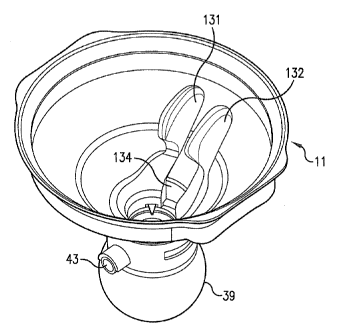
[0028] FIG. 5 is a perspective view of a centration device 130 for use
with the
patient interface device 11 shown in FIG. 2 in accordance with another
embodiment,
FIG. 6 is a perspective view of the patient interface device 11 with the
centration device
130 shown in FIG. 5 coupled thereto, FIG. 7 is a sectional view of the patient
interface
device 11 with the centration device 130 shown in FIG, 6 coupled thereto, and
FIG. 8 is
a top view of the patient interface device 11 and centration device 130 shown
in FIGS. 6
and 7. The centration device 130 may be removably coupled within the patient
interface
13
CA 02706890 2010-05-26
WO 2009/073502 PCT/US2008/084816
device 11 to provide yet another methodology for centering the patient
interface device
11 with respect to the patient's cornea. In this embodiment, the centration
device 130
has an attachment portion 137 for coupling to the frame 13 and has an
interface portion
135 to assist in removing and coupling the centration device 130 to the
patient interface
device 11. The attachment portion 137 includes a centration indicator 140,
141, 142,
143, which identifies the optical center of the corneal interface lens 27 as
well as the
patient interface device 11. In this embodiment, the vent ports 53 shown in
FIG. 2 are
preferably configured to prevent compromise of the sterile barrier between the
eye and
the centration device 130.
10029] The attachment portion 137 has an exterior surface 133 for
coupling to an
interior surface 134 of the frame 13. The interface portion 135 is angled from
the
attachment portion 137 (e.g., based on the shape of the frame 13) for ease of
inserting
and removing the centration device 130 with respect to the frame 13 while
minimizing
interference with centering the patient interface device 11 (e.g., maintaining
a
substantially unobstructed view of the centering aid). The interface portion
135 may be
angled away from the optical center of the corneal interface lens 27 to a
substantially
similar degree as the corresponding portion of the frame 13.
[0030] In this embodiment, the interface portion 135 has first and second
arms
131 and 132, respectively, that are coupled to the attachment portion for
releasing and
securing the attachment portion 137 with respect to the frame 13. For example,
the
centration device 130 may be fabricated from a semi-flexible or semi-rigid
material, such
as plastic, a plastic composite, or the like. When the attachment portion 137
is coupled
to the frame 13, the exterior surface 133 is radially biased against the
interior surface
134 of the frame 13 to secure the attachment portion 137 to the frame 13. By
squeezing or pinching the bias arms 131 and 132 together, the shape of the
exterior
surface 113 is temporarily modified to decrease this bias and thereby release
the
14
CA 02706890 2010-05-26
WO 2009/073502 PCT/US2008/084816
attachment portion 137 from the frame 13. Other mechanisms (e.g., a bead-and-
groove
mechanism or the like) may also be used to secure and release the attachment
portion
137 with respect to the frame 13. Additionally, the arms 131 and 132 are
angled away
from the optical axis of the patient interface device 11 and are preferably
angled
substantially parallel to the conical-shaped portion of the frame 13
substantially adjacent
to the interface portion 135 when the centration device 130 is coupled to the
patient
interface device 11.
[0031] Similar to the pre-applanation when the ocular device 57 (FIG. 2)
is
coupled with the patient interface device 11, the flexible support 25 and
corneal
interface lens 27 may also be positioned within the retainer ring 19 at a pre-
determined
distance selected for pre-applanation of the patient's cornea utilizing the
centration
device 130. For example, by coupling the centration device 130 to the patient
interface
device 11 (e.g., securing the centration device 130 to the frame 13), the
flexible support
25 displaces to a pre-determined extent to contact and pre-applanate the
patient's
cornea. The position of the flexible support 25 and corneal interface lens 27
within the
retainer ring 19 and the extent of displacement for pre-applanation are
preferably based
at least on the dimensions of the attachment portion 137, the position of the
patient's
cornea when the patient interface device 11 is coupled to the patient's eye
39, the
flexibility of the flexible support 25, or any combination thereof. Additional
factors may
also be used to pre-determine the position of the flexible support 25 and the
corresponding extent of displacement for pre-applanation.
[0032] FIG. 9 is a sectional view of a patient interface device 150 in
accordance
with another embodiment. FIG. 10 is a sectional view of the patient interface
device
150 shown in FIG. 9 illustrating a laser head piece 154 of the patient
interface device
docking with a corneal piece 152 of the patient interface device. In this
embodiment,
the patient interface device 150 has two separate components, the laser head
piece
CA 02706890 2010-05-26
WO 2009/073502 PCT/US2008/084816
154 and the corneal piece 152, which are not attached together or molded as a
single
unit, such as the patient interface device 11 shown in FIG. 2. The laser head
piece 154
has a first end 166 for receiving and coupling to a laser head 162, such as
may be used
with an ophthalmic surgical laser system (not shown), and second end 168 with
a
corneal interface lens 156 for contacting the patients eye 39. The corneal
piece 152
has a first end 170 for receiving the laser head piece 154 and the laser head
162 (e.g.,
during docking) and a second end 172 for coupling to the patient's eye 39.
[0033] In one embodiment, the laser head piece 154 is a disposable piece
having
a temporary biasing portion (e.g., a snap-on ring) to snap-fit the laser head
piece 154
onto the laser head 162. The laser head piece 154 is additionally rotation-
resistant
once fitted onto the laser head 162 to prevent twisting of the laser head
piece 154 in
relation to the laser head 162 and/or prevent abrasion of an output surface
174 of the
laser head 162. With the laser head piece 154 coupled to the laser head 162,
the
output surface 174 is contiguous with an interior surface 176 of the corneal
interface
lens 156. The corneal interface lens 156 may be attached to the second end 168
of the
laser head piece 174 via a flexible support 164 that is biased to abut the
output surface
174 with the interior surface 176 with the laser head piece 154 coupled to the
laser
head 162.
[0034] The first end 170 of the corneal piece 152 is shaped to receive
the laser
head piece 154, and may also be shaped to receive a portion of the laser head
162
(e.g., when the first end 170 is cone shaped). The second end 172 of the
corneal piece
152 includes a skirt 160 which, when placed against the eye 39 as shown, forms
a
chamber in combination with the anterior surface of the eye 39. The skirt 160
also
includes an arm 178 that provides a conduit 180 in fluid connection with the
chamber
such that a negative pressure may be generated in the chamber (e.g., via a
vacuum
16
CA 02706890 2010-05-26
WO 2009/073502 PCT/US2008/084816
pump, a syringe, or any other mechanical device capable of generating the
negative
pressure).
[0035] In operation, the laser head piece 154 is coupled to the laser
head 162,
and the precision of this placement may be determined for any errors. The
corneal
piece 152 is coupled to the eye 39 by setting the skirt 160 onto the eye 39
and applying
suction (e.g., generating a negative pressure within the chamber). Centration
of the
corneal piece 152 may be aligned using the ocular device 57 shown in FIG. 2 or
centration device 130 shown in FIGS. 5-8 and removing/reapplying suction. The
laser
head piece 154 and laser head 162 are then coupled to the corneal piece 152 by
guiding the second end 168 of the laser head piece 154 into the first end 170
of the
corneal piece 152. The eye 30 may be applanated by contacting the corneal
interface
lens 156 with the cornea and applanating to a desired degree (e.g., by
observing the
size of a meniscus formed by the cornea in contact with the corneal interface
lens 156).
The corneal piece 152 may be locked to the laser head 162 after applanation
and prior
to directing laser beams to the eye 39. For example, the laser head 162 may
have a
bladder (not shown) around a portion adjacent to the first end 170 of the
corneal piece
152. This bladder is inflated to bias the first end 170 of the corneal piece
152 against
the laser head 162. Other mechanisms may be used to temporarily secure the
laser
head 162 against the corneal piece 152.
[0036] Using a two-piece configuration, the laser head piece 154, when
coupled
with the laser head 162, has a substantially unimpeded field of view (e.g., as
viewed
through the corneal interface lens 156). For example, the corneal interface
lens 156 is
coterminous with the second end of the corneal piece 152 without additional
structure of
the corneal piece 152 that may limit the field of view through the corneal
interface lens
156. Additionally, the corneal interface lens 156 contacts the cornea after
docking the
laser head piece 154 and laser head 162 with the corneal piece 152, and thus
the
17
CA 02706890 2015-07-30
patient interlace device 150 avoids accumulation of moisture (e.g., from the
patient's
eye 39) on the corneal interface lens 156 prior to actual contact with the
cornea. The
sufficiency of contact between the corneal interface lens 156 and the output
surface 174
of the laser head 162 may be determined independent of and prior to docking
the
patient interface device 150 onto the patient's eye 39. Thus, placement errors
that
might be associated with coupling the corneal interface lens 156 with the
laser head 162
may be corrected prior to the ophthalmic procedure.
[00371 Thus, a centration device, an ophthalmic patient interface device,
an
ophthalmic patient interface system, and a method of interfacing an ophthalmic
surgical
laser system with an eye are disclosed. While one or more embodiments have
been
shown and described, it will be apparent to those skilled in the art that more
modifications are possible. The scope of the claims should not be limited by
the
preferred embodiments or the examples but should be given the broadest
interpretation
consistent with the description as a whole.
18