Note: Descriptions are shown in the official language in which they were submitted.
CA 02706954 2010-05-27
WO 2009/070686 PCT/US2008/084883
EXPANDABLE PLUGS AND RELATED DELIVERY APPARATUSES
AND METHODS
BACKGROUND
The present invention relates generally to medical devices and in particular
aspects to devices and methods for plugging fistulae and other passageways in
the
body.
As further background, there exist a variety of passages and other open
spaces in the body which can be plugged or otherwise filled to provide benefit
to
the patient. For example, it may be desirable to occlude a lumen or other open
space in the vasculature (e.g., a blood vessel such as a vein or artery). In
some
instances, a device is deployed within the venous system, e.g., within the
greater
and/or lesser saphenous vein, to treat complications, such as a varicose vein
conditions.
As well, it may be desirable to plug or otherwise fill a fistula. A variety of
fistulae can occur in humans. These fistulae can occur for a variety of
reasons,
such as but not limited to, as a congenital defect, as a result of
inflammatory bowel
disease, such as Chron's disease, irradiation, trauma, such as childbirth, or
as a side
effect from a surgical procedure. Further, several different types of fistulae
can
occur, for example, urethro-vaginal fistulae, vesico-vaginal fistulae, tracheo-
esophageal fistulae, gastro-cutaneous fistulae, and any number of anorectal
fistulae, such as recto-vaginal fistula, recto-vesical fistulae, recto-
urethral fistulae,
or recto-prostatic fistulae.
The path which fistulae take, and their complexity, can vary. A fistula may
take a take a "straight line" path from a primary opening to a secondary
opening,
known as a simple fistula. Alternatively, a fistula may comprise multiple
tracts
CA 02706954 2010-05-27
WO 2009/070686 PCT/US2008/084883
2
ramifying from a primary opening and have multiple secondary openings. This is
known as a complex fistula.
Anorectal fistulae can result from infection in the anal glands, which are
located around the circumference of the distal anal canal that forms the
anatomic
landmark known as the dentate line. Approximately 20-40 such glands are found
in humans. Infection in an anal gland can result in an abscess. This abscess
then
can track through soft tissues (e.g., through or around the sphincter muscles)
into
the perianal skin, where it drains either spontaneously or surgically. The
resulting
void through soft tissue is known as a fistula. The internal or inner opening
of the
fistula, usually located at or near the dentate line, is known as the primary
opening.
Any external or outer openings, which are usually located in the perianal
skin, are
known as secondary openings.
One technique for treating a perianal fistula is to make an incision adjacent
the anus until the incision contacts the fistula and then excise the fistula
from the
anal tissue. This surgical procedure tends to sever the fibers of the anal
sphincter,
and may cause incontinence. Other surgical treatment of fistulae involve
passing a
fistula probe through the tract of the fistula in a blind manner, using
primarily only
tactile sensation and experience to guide to probe. Having passed the probe
through the fistula tract, the overlying tissue is surgically divided. This is
known as
a fistulotomy. Since a variable amount of sphincter muscle is divided during
the
procedure, fistulotomy also may result in impaired sphincter control, and even
frank incontinence.
A gastrointestinal fistula is an abnormal passage that leaks contents of the
stomach or the intestine (small or large bowel) to other organs, usually other
parts
of the intestine or the skin. For example, gastrojejunocolic fistulae include
both
enterocutaneous fistulae (those occurring between the skin surface and the
intestine, namely the duodenum, the jejunum, and the ileum) and gastric
fistulae
CA 02706954 2010-05-27
WO 2009/070686 PCT/US2008/084883
3
(those occurring between the stomach and skin surface). Another type of
fistula
occurring in the gastrointestinal tract is an enteroenteral fistula, which
refers to a
fistula occurring between two parts of the intestine. Gastrointestinal
fistulae can
result in malnutrition and dehydration depending on their location in the
gastrointestinal tract. They can also be a source of skin problems and
infection.
The majority of these types of fistulae are the result of surgery (e.g., bowel
surgery), although sometimes they can develop spontaneously or from trauma,
especially penetrating traumas such as stab wounds or gunshot wounds.
Inflammatory processes, such as infection or inflammatory bowel disease
(Crohn's
disease), may also cause gastrointestinal fistulae. In fact, Crohn's disease
is the
most common primary bowel disease leading to enterocutaneous fistulae, and
surgical treatment may be difficult because additional enterocutaneous
fistulae
develop in many of these patients postoperatively.
Treatment options for gastrointestinal fistulae vary. Depending on the
clinical situation, patients may require IV nutrition and a period of time
without
food to allow the fistula time to close on its own. Indeed, nonsurgical
therapy may
allow spontaneous closure of the fistula, although this can be expected less
than
30% of the time according to one estimate. A variable amount of time to allow
spontaneous closure of fistulae has been recommended, ranging from 30 days to
6
to 8 weeks. During this preoperative preparation, external control of the
fistula
drainage prevents skin disruption and provides guidelines for fluid and
electrolyte
replacement. In some cases, surgery is necessary to remove the segment of
intestine involved in a non-healing fistula.
When surgery is deemed necessary, one operation for fistula closure is
resection of the fistula-bearing segment and primary end-to-end anastamosis.
The
anastomosis may be reinforced by greater omentum or a serosal patch from
adjacent small bowel. Still other methods for treating fistulae involve
injecting
sclerosant or sealant (e.g., collagen or fibrin glue) into the tract of the
fistula to
CA 02706954 2010-05-27
WO 2009/070686 PCT/US2008/084883
4
block the fistula. Closure of a fistula using a sealant is typically performed
as a
two-stage procedure, including a first-stage seton placement and injection of
the
fibrin glue several weeks later. This allows residual infection to resolve and
to
allow the fistula tract to "mature" prior to injecting a sealant. If sealant
or
sclerosant were injected as a one-stage procedure, into an "unprepared" or
infected
fistula, this may cause a flare-up of the infection and even further abscess
formation.
There remain needs for improved and/or alternative devices, systems and
methods for plugging passageways and other open spaces in the body. The
present
invention is addressed to those needs.
CA 02706954 2010-05-27
WO 2009/070686 PCT/US2008/084883
SUMMARY
The present invention provides, in certain aspects, unique devices for
insertion into passageways or other similar openings in the body. Such devices
in
5 some embodiments include a first element cooperable with a second element to
provide an implanted configuration to be received at the targeted insertion
site.
The cooperation between the two elements can be in a controlled fashion; e.g.
wherein portions of the first and second elements engage and potentially
translate
along one another in a fashion that is predictably controlled by engaged
surface
features of the first and second elements. Some of these devices have a
portion
that is outwardly displaced when contacted by another device component. In one
embodiment, a device comprises a first plug member and a removable second plug
member positioned in the first plug member. When so positioned, the second
plug
member is effective to radially expand at least a segment of the first plug
member.
This device can exhibit any suitable size, shape and configuration for
plugging a
passageway in the body, and be may be formed with one or more of a variety of
biocompatible materials including some that are naturally derived and some
that
are non-naturally derived. In a preferred embodiment, the first plug member
and/or the second plug member is comprised of a remodelable, angiogenic
material, for example, a remodelable extracellular matrix material such as
submucosa.
In another aspect, the invention provides an assembly for plugging a
passageway in the body that includes a first plug member and a second plug
member. The second plug member is positionable in the first plug member, and
in
the first plug member, is effective to outwardly displace at least part of the
first
plug member. Each of these plug members can exhibit a variety of shapes and
sizes, and the second plug member can be positioned at any suitable location
in the
first plug member for plugging the body passageway. Although not necessary to
broader aspects of the invention, in one form, the first plug member provides
a
CA 02706954 2010-05-27
WO 2009/070686 PCT/US2008/084883
6
designated opening (e.g., a lumen or other passage) into which the second plug
member can be positioned.
An additional embodiment of the invention provides a method for plugging
a passageway in the body, which utilizes a plugging assembly including a first
plug
member and a second plug member. In one step, the first plug member and the
second plug member are delivered to the body passageway. Thereafter, relative
movement between the first plug member and the second plug member is brought
about, wherein contact between the first plug member and the second plug
member
outwardly displaces at least part of the first plug member for plugging the
body
passageway. In some instances, such contact causes the circumference of the
first
plug member, or a portion thereof, to increase. Causing relative movement
between the first plug member and the second plug member can be achieved in a
variety of manners including some that involve pushing and/or pulling one or
both
plug members in the body passageway. In one aspect, a lumen extends through
the
first plug member, and a pulling device, which can be attached to or otherwise
associated with the second plug member, is passed through this lumen. This
pulling device (e.g., an attached suture or a grasping instrument) can then be
used
in positioning the second plug member in this first plug member lumen.
Another aspect of the invention provides an apparatus for plugging a
passageway in the body. This apparatus includes a delivery device that has a
lumen communicating with a distal end opening, and is configured for passage
through a body passageway. The apparatus also includes a first plug member and
a
second plug member, both received in the delivery device lumen. The second
plug
member is positionable in the first plug member, and in the first plug member,
is
effective to outwardly displace at least part of the first plug member for
plugging
the body passageway. This delivery device can exhibit any suitable size, shape
and
configuration for delivering the first plug member and second plug member into
the body passageway, and in some embodiments, is flexible to enhance its
travel
CA 02706954 2010-05-27
WO 2009/070686 PCT/US2008/084883
7
through particular body passageways. In one aspect, the apparatus includes a
pusher device, which is translatable through the delivery device lumen, and
can be
used in expelling the first plug member and/or the second plug member from the
distal end opening.
A further embodiment of the invention provides a method for plugging a
passageway in the body, which utilizes a plugging apparatus such as that
described
above. In one step, the delivery device is passed through at least a segment
of the
body passageway. In other steps, the first plug member and the second plug
member are removed from the delivery device lumen. The first plug member is
positioned at a location in the body passageway. Thereafter, relative movement
between the first plug member and the second plug member is brought about,
wherein contact between the first plug member and the second plug member
outwardly displaces at least part of the first plug member for plugging the
body
passageway.
Yet another aspect of the present invention provides a method for plugging
a passageway in the body. In this method, a plugging assembly comprising a
first
plug member and a second plug member is provided. The first plug member,
which has a lumen, is delivered to the body passageway. Thereafter, at least
part
of the first plug member lumen is filled with the second plug member.
In another embodiment, the invention provides an assembly for plugging a
passageway in the body. This assembly includes a first plug member and a
second
plug member. The first plug member has a cavity, and is positionable in a body
passageway. The second plug member is comprised of a porous, collagen-
containing matrix material, and includes a segment positionable in the cavity
of the
first plug member. Additionally, the second plug member has a first condition
suitable to deliver the segment to the first plug member cavity, and a second,
CA 02706954 2010-05-27
WO 2009/070686 PCT/US2008/084883
8
expanded condition providing a more snug fit of the segment in the first plug
member cavity relative to the first condition of the second plug member.
Other objects, embodiments, forms, features, advantages, aspects, and
benefits of the present invention shall become apparent from the detailed
description and drawings included herein.
CA 02706954 2010-05-27
WO 2009/070686 PCT/US2008/084883
9
BRIEF DESCRIPTION OF THE DRAWINGS
Figure 1 is a perspective view of an inventive assembly including a first
plug member and a second plug member.
Figure 2 shows the assembly of Figure 1 with the first plug member
positioned in the second plug member.
Figure 3 shows the assembly of Figure 1 received in a delivery device
lumen.
Figure 4 shows part of an inventive apparatus being used to deliver a
plugging assembly to a fistula tract.
Figure 5 shows the apparatus of Figure 4 at a different stage of delivery.
Figure 6 shows the apparatus of Figure 4 at a different stage of delivery.
Figure 7 shows the apparatus of Figure 4 at a different stage of delivery.
Figure 8 shows the apparatus of Figure 4 at a different stage of delivery.
Figure 9 shows the apparatus of Figure 4 at a different stage of delivery.
Figure 10 shows one step in delivering an inventive assembly to a body
passageway for plugging the passageway.
Figure 11 shows another step in delivering an inventive assembly to a body
passageway for plugging the passageway.
Figure 12 shows another step in delivering an inventive assembly to a body
passageway for plugging the passageway.
Figure 13 shows another step in delivering an inventive assembly to a body
passageway for plugging the passageway.
CA 02706954 2010-05-27
WO 2009/070686 PCT/US2008/084883
DETAILED DESCRIPTION
While the present invention may be embodied in many different forms, for
the purpose of promoting an understanding of the principles of the present
5 invention, reference will now be made to the embodiments illustrated in the
drawings, and specific language will be used to describe the same. It will
nevertheless be understood that no limitation of the scope of the invention is
thereby intended. Any alterations and further modifications in the described
embodiments and any further applications of the principles of the present
invention
10 as described herein are contemplated as would normally occur to one skilled
in the
art to which the invention relates.
As disclosed above, in certain aspects, the present invention provides
unique assemblies for plugging passageways in the body. One such assembly
comprises a first plug member and a second plug member, wherein the second
plug
member is positionable in the first plug member to expand at least a segment
of the
first plug member to plug a body passageway. In one embodiment, an assembly of
this sort is combined with a device that is suitable for delivering the
assembly into
a body passageway. An illustrative delivery device has a lumen communicating
with a distal end opening, wherein the first plug member and the second plug
member can be received in the delivery device lumen for removal from the
distal
end opening in the body. The present invention also provides methods for
plugging passageways in the body. One such method utilizes an assembly such as
that described above. In one step, a first plug member is positioned at least
partially in the body passageway, and in another step, a second plug member is
positioned in the first plug member, wherein at least a segment of the first
plug
member expands (e.g., radially expands) to plug the body passageway.
Assemblies and devices of the invention may be used to plug or otherwise
fill a variety of passages or other open spaces in the body. In some
instances, these
CA 02706954 2010-05-27
WO 2009/070686 PCT/US2008/084883
11
open spaces will occur naturally in the body, for example, as a native lumen
or
other open space in a bodily system, e.g., in an organ or other component of
the
circulatory, respiratory, digestive, urinary and reproductive, sensory, or
endocrine
systems. In certain aspects, a space to be filled is one that exists naturally
in the
body but relates to a disease, defect, deformation, etc. Alternatively, an
opening or
passage to be filled may be one resulting from an intentional or unintentional
trauma to the body including but not limited to some relating to vehicular
accidents, gunshots and other similar wounds, etc., as well as some formed by
passage of a medical instrument (e.g., a needle, trocar, etc.) through
cutaneous,
subcutaneous, and/or intracutaneous tissue.
Illustratively, inventive devices and assemblies, alone or in conjunction
with one or more other suitable objects, can be used to occlude, or at least
promote
and/or facilitate occlusion of, a lumen or other open space in the
vasculature, e.g., a
blood vessel such as a vein or artery, or a lumen or open space of a fallopian
tube,
e.g. in a procedure to provide sterility to a female patient. In certain
aspects, one
or more assemblies of the invention are deployed within the venous system
(e.g.,
within the greater and/or lesser saphenous vein) to treat complications, such
as a
varicose vein conditions. In other embodiments, inventive assemblies are used
as
contraceptive devices. In preferred embodiments, assemblies of the invention
can
be used to plug or otherwise fill fistulae such as but not limited to urethro-
vaginal
fistulae, vesico-vaginal fistulae, tracheo-esophageal fistulae, gastro-
cutaneous
fistulae, and any number of anorectal fistulae, such as recto-vaginal fistula,
recto-
vesical fistulae, recto-urethral fistulae, or recto-prostatic fistulae.
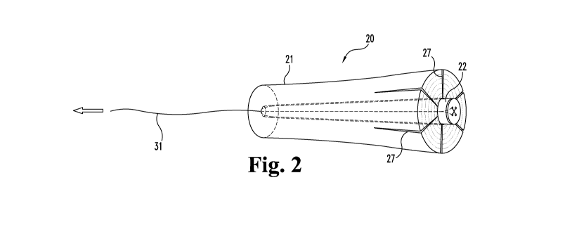
With reference now to Figure 1, shown is an assembly 20 which can be
used to plug a passageway or other open space in a patient's body. Assembly 20
includes a first plug member 21 and a second plug member 22. First plug member
21 is comprised of an elongate body 23 having a first end 24 and second end
25.
Body 23 is generally in the shape of a cylinder, although a variety of other
shapes
CA 02706954 2010-05-27
WO 2009/070686 PCT/US2008/084883
12
are contemplated as within the scope of the present invention. In general,
body 23
will be shaped and sized so that at least a portion of body 23, and in some
cases all
of body 23, can be positioned in a space to be plugged.
A lumen 26 extends through body 23 from its first end 24 to its second end
25. Although not necessary to broader aspects of the invention, such a plug
member lumen can provide a channel into which a second plug member such as
second plug member 22 can be received. When present, a plug member lumen can
exhibit a variety of shapes and sizes to suit a particular application, for
example,
having a constant or varying diameter along its length. In general, the
dimensions
of a plug member lumen, when used to receive one or more other plug members
therein, will be selected based on the characteristics of these one or more
other
plug members and/or other factors such as but not limited to conditions at the
treatment site and other characteristics of first plug member 21.
Continuing with Figure 1, second plug member 22 is comprised of an
elongate body 28 having a first end 29 and a second end 30. In general,
inventive
assemblies such as assembly 20 will include at least one plug member that can
be
caused or allowed to contact and move a portion of another plug member in
providing a plugging arrangement. Illustratively, second plug member 22 can be
positioned in first plug member 21, and when so positioned, is effective to
outwardly displace portions of first plug member 21. In this specific
illustrative
embodiment, such displacement provides radial expansion of at least part of
the
length of the first plug member, for example, a part that includes first end
24 as
generally shown in Figure 2. Body 28 is generally in the shape of a truncated
cone, although a variety of other shapes are contemplated as within the scope
of
the present invention. In this regard, it will be understood that a plug
member
(e.g., second plug member 22) can exhibit any suitable size and shape to
provide
the desired movement (e.g., outward displacement) of a portion of another plug
member (e.g., first plug member 21) in which it is positioned. Second end 30
of
CA 02706954 2010-05-27
WO 2009/070686 PCT/US2008/084883
13
second plug member 22 provides a leading or guiding portion for controlling
orientation of contact with first plug member 21.
Second plug member 22 has at least a segment that increases in
circumference moving from its second end 30 toward its first end 29, while
lumen
26 has a generally constant circumference along its length. Second end 30 has
roughly the same circumference as (or a slightly smaller circumference than)
lumen 26, and first end 29 has a somewhat larger circumference than lumen 26.
In
this regard, as the second end 30 of second plug member 22 is advanced through
lumen 26 from first end 24 toward second end 25, portions of second plug
member
having a larger circumference than lumen 26 (e.g., portions including first
end 29)
exert pressure on the wall defining lumen 26. This pressure causes portions of
first
plug member 21 to move in an outward direction, which in turn, increases the
circumference of at least a portion of first plug member 21. As described more
thoroughly below, one or more cuts or other adaptations for promoting and/or
facilitating such movement can be incorporated into elongate body 23. In an
alternative embodiment, the second plug member has a generally constant
diameter
along its length, and the first plug member lumen is tapered such that a
desired
displacement is achieved when the second plug member is advanced through the
lumen from the larger-diameter end toward the smaller-diameter end. In some
forms, a plug member having a tapered portion is positioned in a plug member
lumen having a tapered portion.
In addition what is shown in Figure 1, the invention provides a variety of
other plug body configurations such as that of second plug member 22, wherein
a
plug body occupies an increased volume moving along at least part of the plug
body, for example, having a gradually increasing volume moving from one end of
the plug body to the other. Illustratively, a plug member that is to be
positioned in
a lumen of another plug member can include a biocompatible sheet-form
material,
wherein one longitudinal portion of the sheet occupies an increased volume
(e.g., is
CA 02706954 2010-05-27
WO 2009/070686 PCT/US2008/084883
14
relatively wider and/or thicker) than another longitudinal portion of the
sheet (of a
similar length). In such forms, at least a portion of the sheet will be
deformable
upon impingement by the plug lumen wall, and will be sized and shaped so as to
be
deformable to a three-dimensional volumetric body filling at least a portion
of the
plug lumen. In this regard, as a portion of the sheet is drawn into the lumen,
it can
fold and/or roll over itself one or more times to conform to the lumen wall
and
gradually become "wedged" into the lumen when sufficiently pulled
therethrough.
In some instances, such positioning will exert pressure on the wall defining
the
lumen, causing at least a portion of that plug member to become outwardly
displaced. Also, such wedging or lodging may be sufficient to obviate the need
for
otherwise securing the sheet to the other plug member and/or soft tissues at
the
treatment site, although additional steps to secure the sheet in place (e.g.,
suturing
to the other plug member) may be taken.
In one aspect, a plug member to be positioned in another plug member
comprises a compliant, biocompatible sheet-form material, for example, one or
more layers of ECM material that can be pulled into a plug member lumen. Such
sheet-form plug members can be prepared, for example, as described in
International Patent Application Serial No. PCT/US2006/16233, filed April 29,
2006, and entitled "FISTULA GRAFT WITH DEFORMABLE SHEET-FORM
MATERIAL" (Cook Biotech Incorporated), which is hereby incorporated by
reference in its entirety.
A plugging assembly of the invention, or any component thereof, may be
formed with one or more of a variety of biocompatible materials including some
that are naturally derived and some that are non-naturally derived. In the
specific
illustrative embodiment depicted in Figure 1, body 23 is formed with layers of
sheet-form ECM material that are compressed and bonded together (e.g., around
a
mandrel) so as to form a substantially unitary, rolled construct. In other
embodiments, body 23 is formed with sheet-form material configured differently
CA 02706954 2010-05-27
WO 2009/070686 PCT/US2008/084883
than what is shown in Figure 1 (e.g., different number of layers, different
layer
thickness, differently rolled or otherwise assembled, etc.), or is formed with
a non-
sheet-form material as described elsewhere herein. Body 28 is similarly formed
with layers of sheet-form ECM material that are compressed and bonded together
5 so as to form a substantially unitary, rolled construct.
In certain aspects, a first plug member includes one or more adaptations for
enhancing expansion of external features of the first plug member when a
second
plug member is brought into contact with the first plug member. Such
adaptations
10 can include one or more perforations, cuts, channels, indentations, scores,
etc. in
the plug member. These and other adaptations for enhancing the expansive
ability
of the first plug member will be recognized by the skilled artisan and are
encompassed by the present invention. In the current embodiment, a plurality
of
cuts 27 is formed in elongate body 23. Each cut extends a distance from first
end
15 24 toward second end 25, as well as from an outer surface of body 23 to
lumen 26,
although additional cut configurations and placements in the plug member body
are contemplated as within the scope of the present invention. Illustratively,
a cut
or other adaptation can extend any suitable distance along a plug member
(e.g., can
run down the entire length of a plug member), and can extend through a plug
member any suitable distance and at any suitable angle.
In the current embodiment, positioning second plug member 22 in first plug
member 21 forces parts of first plug member 21 outward, causing the
circumference of at least a portion of first plug member 21 to increase. In
other
embodiments, positioning a second plug member in a first plug member also
forces
parts of the first plug member outward; however, the circumference of the
first
plug member increases very little or not at all. Rather, these parts of the
first plug
member are compressed as they are forced outward, which in turn, increases the
densities of these parts. Additionally or alternatively, positioning a second
plug
member in a first plug member can, in some aspects of the invention, compress
and
CA 02706954 2010-05-27
WO 2009/070686 PCT/US2008/084883
16
increase the density of a portion of the second plug member, for example, with
little or no outward movement of the first plug member. Such relatively higher
density plug portions may be beneficial in a variety of plugging operations.
For
example, having material with a relatively more dense structure at or near a
primary fistula opening can inhibit bacteria and other undesirable substances
from
passing from the alimentary canal and into the fistula.
Referring again to Figure 1, a pulling device in the form of a resorbable
suture 31 extends a distance from the second end 30 of second plug member 22.
This suture can extend any suitable distance from the second plug member, and
in
some cases, will extend from about 1 cm to about 100 cm, more typically from
about 20 cm to about 80 cm, and even more typically from about 40 cm to about
80 cm from its smaller end. As shown, suture 31 can extend through first plug
member lumen 26, and in this regard, is effective for pulling second plug
member
22 into first plug member 21. Second plug member 22 also includes an end cap
32
at its first end 29. End cap 32 may or may not be attached to first end 29. In
the
current illustrated embodiment, suture 31 passes through and around end cap
32,
and extends through second plug member 22 along its length. In certain
embodiments, suture 31 is attached to the material of second plug member 22,
e.g.
by being securely embedded therein or knotted thereto.
In accordance with the present invention, a plug member can be positioned
in contact with another plug member in any suitable manner including some that
involve directly or indirectly pushing and/or pulling one or both plug members
in
the body. As well, such positioning can be performed directly by hand in
situations where such access is possible, although in some embodiments,
positioning one plug member in another plug member will additionally or
alternatively involve the use of one or more instruments. In one aspect, a
lumen
extends through a first plug member, and a pulling device, which is attached
to or
otherwise associated with a second plug member, is passed through this lumen.
CA 02706954 2010-05-27
WO 2009/070686 PCT/US2008/084883
17
The pulling device can then be used in positioning the second plug member in
this
first plug member lumen.
For example and referring again to Figures 1 and 2, suture 31 or another
elongate flexible tether can be used to pull the second end 30 of second plug
member 22 into lumen 26 at the first end 24 of first plug member 21.
Thereafter,
second plug member 22 can be advanced through lumen 26 in the direction of the
arrow, i.e., toward the second end 25 of first plug member 21, until second
plug
member 22 is desirably seated in first plug member 21. Second plug member 22
may be positioned so that its first end 29 extends a distance from the first
end 24 of
first plug member 21 as shown in Figure 2, or alternatively, second plug
member
first end 29 may be pulled flush with first plug member first end 24 or a
distance
into lumen 26. In an alternative embodiment, a probe or other suitable
instrument
(e.g., a suitably configured pair of surgical hemostats) includes a portion
that is
passable through the lumen of a first plug member, and can be used to pull a
second plug member into this lumen, either by directly contacting the second
plug
member or by contacting an associated suture, etc. Such an instrument in
certain
forms can include a gripping portion for securing the second plug member or
suture. As will be understood by those skilled in the art, the second end 30
of
second plug member 22 could also be pushed into lumen 26 at the first end 24
of
first plug member 21 using an appropriate instrument or technique.
In certain embodiments, a plugging assembly includes a radiopaque
element. For example, an assembly component such as end cap 32 can be
comprised of a radiopaque substance or device such as but not limited to a
radiopaque coating, attached radiopaque object, or integrated radiopaque
substance
useful for determining the location of the component in the body. In certain
forms,
cap 32 can be formed of a polymeric material loaded with a particulate
radiopaque
material. In this regard, any suitable radiopaque substance, including but not
limited to, tantalum such as tantalum powder, can be incorporated into an
inventive
CA 02706954 2010-05-27
WO 2009/070686 PCT/US2008/084883
18
component. Other radiopaque markers may be comprised of gold, bismuth, iodine,
and barium, as well as other suitable radiopaque materials.
Turning now to a more detailed discussion of materials useful in forming
plug members of the invention, these materials should generally be
biocompatible,
and in advantageous embodiments of the assemblies, are comprised of a
remodelable material. Particular advantage can be provided by plug members
including a remodelable collagenous material. Such remodelable collagenous
materials, whether reconstituted or naturally-derived, can be provided, for
example, by collagenous materials isolated from a warm-blooded vertebrate, and
especially a mammal. Such isolated collagenous material can be processed so as
to
have remodelable, angiogenic properties and promote cellular invasion and
ingrowth. Remodelable materials may be used in this context to promote
cellular
growth on, around, and/or within tissue in which an plugging device of the
invention is implanted, e.g., around tissue defining a fistula tract, an
opening to a
fistula, or another space in the body.
Suitable remodelable materials can be provided by collagenous
extracellular matrix (ECM) materials possessing biotropic properties. For
example, suitable collagenous materials include ECM materials such as those
comprising submucosa, renal capsule membrane, dermal collagen, dura mater,
pericardium, fascia lata, serosa, peritoneum or basement membrane layers,
including liver basement membrane. Suitable submucosa materials for these
purposes include, for instance, intestinal submucosa including small
intestinal
submucosa, stomach submucosa, urinary bladder submucosa, and uterine
submucosa. Collagenous matrices comprising submucosa (potentially along with
other associated tissues) useful in the present invention can be obtained by
harvesting such tissue sources and delaminating the submucosa-containing
matrix
from smooth muscle layers, mucosal layers, and/or other layers occurring in
the
tissue source. For additional information as to some of the materials useful
in the
CA 02706954 2010-05-27
WO 2009/070686 PCT/US2008/084883
19
present invention, and their isolation and treatment, reference can be made,
for
example, to U.S. Patent Nos. 4,902,508, 5,554,389, 5,993,844, 6,206,931, and
6,099,567.
Submucosa-containing or other ECM tissue used in the invention is
preferably highly purified, for example, as described in U.S. Patent No.
6,206,931
to Cook et al. Thus, preferred ECM material will exhibit an endotoxin level of
less
than about 12 endotoxin units (EU) per gram, more preferably less than about 5
EU
per gram, and most preferably less than about 1 EU per gram. As additional
preferences, the submucosa or other ECM material may have a bioburden of less
than about 1 colony forming units (CFU) per gram, more preferably less than
about
0.5 CFU per gram. Fungus levels are desirably similarly low, for example less
than about 1 CFU per gram, more preferably less than about 0.5 CFU per gram.
Nucleic acid levels are preferably less than about 5 g/mg, more preferably
less
than about 2 g/mg, and virus levels are preferably less than about 50 plaque
forming units (PFU) per gram, more preferably less than about 5 PFU per gram.
These and additional properties of submucosa or other ECM tissue taught in
U.S.
Patent No. 6,206,931 may be characteristic of any ECM tissue used in the
present
invention.
A typical layer thickness for an as-isolated submucosa or other ECM tissue
layer used in the invention ranges from about 50 to about 250 microns when
fully
hydrated, more typically from about 50 to about 200 microns when fully
hydrated,
although isolated layers having other thicknesses may also be obtained and
used.
These layer thicknesses may vary with the type and age of the animal used as
the
tissue source. As well, these layer thicknesses may vary with the source of
the
tissue obtained from the animal source.
Suitable bioactive agents may include one or more bioactive agents native
to the source of the ECM tissue material. For example, a submucosa or other
CA 02706954 2010-05-27
WO 2009/070686 PCT/US2008/084883
remodelable ECM tissue material may retain one or more growth factors such as
but not limited to basic fibroblast growth factor (FGF-2), transforming growth
factor beta (TGF-beta), epidermal growth factor (EGF), cartilage derived
growth
factor (CDGF), and/or platelet derived growth factor (PDGF). As well,
submucosa
5 or other ECM materials when used in the invention may retain other native
bioactive agents such as but not limited to proteins, glycoproteins,
proteoglycans,
and glycosaminoglycans. For example, ECM materials may include heparin,
heparin sulfate, hyaluronic acid, fibronectin, cytokines, and the like. Thus,
generally speaking, a submucosa or other ECM material may retain one or more
10 bioactive components that induce, directly or indirectly, a cellular
response such as
a change in cell morphology, proliferation, growth, protein or gene
expression.
Submucosa or other ECM materials of the present invention can be derived
from any suitable organ or other tissue source, usually sources containing
15 connective tissues. The ECM materials processed for use in the invention
will
typically include abundant collagen, most commonly being constituted at least
about 80% by weight collagen on a dry weight basis. Such naturally-derived ECM
materials will for the most part include collagen fibers that are non-randomly
oriented, for instance occurring as generally uniaxial or multi-axial but
regularly
20 oriented fibers. When processed to retain native bioactive factors, the ECM
material can retain these factors interspersed as solids between, upon and/or
within
the collagen fibers. Particularly desirable naturally-derived ECM materials
for use
in the invention will include significant amounts of such interspersed, non-
collagenous solids that are readily ascertainable under light microscopic
examination with appropriate staining. Such non-collagenous solids can
constitute
a significant percentage of the dry weight of the ECM material in certain
inventive
embodiments, for example at least about 1%, at least about 3%, and at least
about
5% by weight in various embodiments of the invention.
CA 02706954 2010-05-27
WO 2009/070686 PCT/US2008/084883
21
The submucosa or other ECM material used in the present invention may
also exhibit an angiogenic character and thus be effective to induce
angiogenesis in
a host engrafted with the material. In this regard, angiogenesis is the
process
through which the body makes new blood vessels to generate increased blood
supply to tissues. Thus, angiogenic materials, when contacted with host
tissues,
promote or encourage the formation of new blood vessels into the materials.
Methods for measuring in vivo angiogenesis in response to biomaterial
implantation have recently been developed. For example, one such method uses a
subcutaneous implant model to determine the angiogenic character of a
material.
See, C. Heeschen et al., Nature Medicine 7 (2001), No. 7, 833-839. When
combined with a fluorescence microangiography technique, this model can
provide
both quantitative and qualitative measures of angiogenesis into biomaterials.
C.
Johnson et al., Circulation Research 94 (2004), No. 2, 262-268.
Further, in addition or as an alternative to the inclusion of such native
bioactive components, non-native bioactive components such as those
synthetically
produced by recombinant technology or other methods (e.g., genetic material
such
as DNA), may be incorporated into an ECM material. These non-native bioactive
components may be naturally-derived or recombinantly produced proteins that
correspond to those natively occurring in an ECM tissue, but perhaps of a
different
species. These non-native bioactive components may also be drug substances.
Illustrative drug substances that may be added to materials include, for
example,
anti-clotting agents, e.g. heparin, antibiotics, anti-inflammatory agents,
thrombus-
promoting substances such as blood clotting factors, e.g., thrombin,
fibrinogen, and
the like, and anti-proliferative agents, e.g. taxol derivatives such as
paclitaxel.
Such non-native bioactive components can be incorporated into and/or onto ECM
material in any suitable manner, for example, by surface treatment (e.g.,
spraying)
and/or impregnation (e.g., soaking), just to name a few. Also, these
substances
may be applied to the ECM material in a premanufacturing step, immediately
prior
to the procedure (e.g., by soaking the material in a solution containing a
suitable
CA 02706954 2010-05-27
WO 2009/070686 PCT/US2008/084883
22
antibiotic such as cefazolin), or during or after engraftment of the material
in the
patient.
Plug members of the invention can include xenograft material (i.e., cross-
species material, such as tissue material from a non-human donor to a human
recipient), allograft material (i.e., interspecies material, with tissue
material from a
donor of the same species as the recipient), and/or autograft material (i.e.,
where
the donor and the recipient are the same individual). Further, any exogenous
bioactive substances incorporated into an ECM material may be from the same
species of animal from which the ECM material was derived (e.g. autologous or
allogenic relative to the ECM material) or may be from a different species
from the
ECM material source (xenogenic relative to the ECM material). In certain
embodiments, ECM material will be xenogenic relative to the patient receiving
the
graft, and any added exogenous material(s) will be from the same species (e.g.
autologous or allogenic) as the patient receiving the graft. Illustratively,
human
patients may be treated with xenogenic ECM materials (e.g. porcine-, bovine-
or
ovine-derived) that have been modified with exogenous human material(s) as
described herein, those exogenous materials being naturally derived and/or
recombinantly produced.
ECM materials used in the invention may be essentially free of additional,
non-native crosslinking, or may contain additional crosslinking. Such
additional
crosslinking may be achieved by photo-crosslinking techniques, by chemical
crosslinkers, or by protein crosslinking induced by dehydration or other
means.
However, because certain crosslinking techniques, certain crosslinking agents,
and/or certain degrees of crosslinking can destroy the remodelable properties
of a
remodelable material, where preservation of remodelable properties is desired,
any
crosslinking of the remodelable ECM material can be performed to an extent or
in
a fashion that allows the material to retain at least a portion of its
remodelable
properties. Chemical crosslinkers that may be used include for example
aldehydes
CA 02706954 2010-05-27
WO 2009/070686 PCT/US2008/084883
23
such as glutaraldehydes, diimides such as carbodiimides, e.g., 1-ethyl-3-(3-
dimethylaminopropyl)carbodiimide hydrochloride, ribose or other sugars, acyl-
azide, sulfo-N-hydroxysuccinamide, or polyepoxide compounds, including for
example polyglycidyl ethers such as ethyleneglycol diglycidyl ether, available
under the trade name DENACOL EX810 from Nagese Chemical Co., Osaka,
Japan, and glycerol polyglycerol ether available under the trade name DENACOL
EX 313 also from Nagese Chemical Co. Typically, when used, polyglycerol ethers
or other polyepoxide compounds will have from 2 to about 10 epoxide groups per
molecule.
Turning now to a discussion of drying techniques that can be useful in
certain embodiments of the invention, drying by evaporation, or air drying,
generally comprises drying a partially or completely hydrated remodelable
material
by allowing the hydrant to evaporate from the material. Evaporative cooling
can
be enhanced in a number of ways, such as by placing the material in a vacuum,
by
blowing air over the material, by increasing the temperature of the material,
by
applying a blotting material during evaporation, or by any other suitable
means or
any suitable combination thereof. The amount of void space or open matrix
structure within an ECM material that has been dried by evaporation is
typically
more diminished than, for example, an ECM material dried by lyophilization as
described below.
A suitable lyophilization process can include providing an ECM material
that contains a sufficient amount of hydrant such that the voids in the
material
matrix are filled with the hydrant. The hydrant can comprise any suitable
hydrant
known in the art, such as purified water or sterile saline, or any suitable
combination thereof. Illustratively, the hydrated material can be placed in a
freezer
until the material and hydrant are substantially in a frozen or solid state.
Thereafter, the frozen material and hydrant can be placed in a vacuum chamber
and a vacuum initiated. Once at a sufficient vacuum, as is known in the art,
the
CA 02706954 2010-05-27
WO 2009/070686 PCT/US2008/084883
24
frozen hydrant will sublime from the material, thereby resulting in a dry
remodelable material.
In alternative embodiments, a hydrated ECM material can be lyophilized
without a separately performed pre-freezing step. In these embodiments, a
strong
vacuum can be applied to the hydrated material to result in rapid evaporative
cooling which freezes the hydrant within the ECM material. Thereafter, the
frozen
hydrant can sublime from the material thereby drying the ECM material.
Desirably, an ECM material that is dried via lyophilization maintains a
substantial
amount of the void space, or open matrix structure, that is characteristic of
the
harvested ECM material.
Drying by vacuum pressing generally comprises compressing a fully or
partially hydrated remodelable material while the material is subject to a
vacuum.
One suitable method of vacuum pressing comprises placing a remodelable
material
in a vacuum chamber having collapsible walls. As the vacuum is established,
the
walls collapse onto and compress the material until it is dry. Similar to
evaporative
drying, when a remodelable material is dried in a vacuum press, more of the
material's open matrix structure is diminished or reduced than if the material
was
dried by lyophilization.
In certain aspects, the invention provides plugging assemblies, devices, etc.
that include a multilaminate material. Such multilaminate materials can
include a
plurality of ECM material layers bonded together, a plurality of non-ECM
materials bonded together, or a combination of one or more ECM material layers
and one or more non-ECM material layers bonded together. To form a
multilaminate ECM material, for example, two or more ECM segments are
stacked, or one ECM segment is folded over itself at least one time, and then
the
layers are fused or bonded together using a bonding technique, such as
chemical
cross-linking or vacuum pressing during dehydrating conditions. An adhesive,
CA 02706954 2010-05-27
WO 2009/070686 PCT/US2008/084883
glue or other bonding agent may also be used in achieving a bond between
material
layers. Suitable bonding agents may include, for example, collagen gels or
pastes,
gelatin, or other agents including reactive monomers or polymers, for example
cyanoacrylate adhesives. As well, bonding can be achieved or facilitated
between
5 ECM material layers using chemical cross-linking agents such as those
described
above. A combination of one or more of these with dehydration-induced bonding
may also be used to bond ECM material layers to one another.
A variety of dehydration-induced bonding methods can be used to fuse
10 together portions of an ECM material. In one preferred embodiment, multiple
layers of ECM material are compressed under dehydrating conditions. In this
context, the term "dehydrating conditions" is defined to include any
mechanical or
environmental condition which promotes or induces the removal of water from
the
ECM material. To promote dehydration of the compressed ECM material, at least
15 one of the two surfaces compressing the matrix structure can be water
permeable.
Dehydration of the ECM material can optionally be further enhanced by applying
blotting material, heating the matrix structure or blowing air, or other inert
gas,
across the exterior of the compressed surfaces. One particularly useful method
of
dehydration bonding ECM materials is lyophilization.
Another method of dehydration bonding comprises pulling a vacuum on the
assembly while simultaneously employing the vacuum to press the assembly
together. Again, this method is known as vacuum pressing. During vacuum
pressing, dehydration of the ECM materials in forced contact with one another
effectively bonds the materials to one another, even in the absence of other
agents
for achieving a bond, although such agents can be used while also taking
advantage at least in part of the dehydration-induced bonding. With sufficient
compression and dehydration, the ECM materials can be caused to form a
generally unitary ECM structure.
CA 02706954 2010-05-27
WO 2009/070686 PCT/US2008/084883
26
It is advantageous in some aspects of the invention to perform drying and
other operations under relatively mild temperature exposure conditions that
minimize deleterious effects upon any ECM materials being used, for example
native collagen structures and potentially bioactive substances present. Thus,
drying operations conducted with no or substantially no duration of exposure
to
temperatures above human body temperature or slightly higher, say, no higher
than
about 38 C, will preferably be used in some forms of the present invention.
These
include, for example, vacuum pressing operations at less than about 38 C,
forced
air drying at less than about 38 C, or either of these processes with no
active
heating - at about room temperature (about 25 C) or with cooling. Relatively
low
temperature conditions also, of course, include lyophilization conditions.
As well, plugging assemblies of the invention may be comprised of
biocompatible materials derived from a number of biological polymers, which
can
be naturally occurring or the product of in vitro fermentation, recombinant
genetic
engineering, and the like. Purified biological polymers can be appropriately
formed into a substrate by techniques such as weaving, knitting, casting,
molding,
and extrusion. Suitable biological polymers include, without limitation,
collagen,
elastin, keratin, gelatin, polyamino acids, polysaccharides (e.g., cellulose
and
starch) and copolymers thereof.
Plugging assemblies of the invention can also include a variety of synthetic
polymeric materials including but not limited to bioresorbable and/or non-
bioresorbable plastics. Bioresorbable, or bioabsorbable polymers that may be
used
include, but are not limited to, poly(L-lactic acid), polycaprolactone,
poly(lactide-
co-glycolide), poly(hydroxybutyrate), poly(hydroxybutyrate-co-valerate),
polydioxanone, polyorthoester, polyanhydride, poly(glycolic acid), poly(D,L-
lactic
acid), poly(glycolic acid-co-trimethylene carbonate), polyhydroxyalkanaates,
polyphosphoester, polyphosphoester urethane, poly(amino acids),
cyanoacrylates,
poly(trimethylene carbonate), poly(iminocarbonate), copoly(ether-esters)
(e.g.,
CA 02706954 2010-05-27
WO 2009/070686 PCT/US2008/084883
27
PEO/PLA), polyalkylene oxalates, and polyphosphazenes. These or other
bioresorbable materials may be used, for example, where only a temporary
blocking or closure function is desired, and/or in combination with non-
bioresorbable materials where only a temporary participation by the
bioresorable
material is desired.
Non-bioresorbable, or biostable polymers that may be used include, but are
not limited to, polytetrafluoroethylene (PTFE) (including expanded PTFE),
polyethylene terephthalate (PET), polyurethanes, silicones, and polyesters and
other polymers such as, but not limited to, polyolefins, polyisobutylene and
ethylene-alphaolefin copolymers; acrylic polymers and copolymers, vinyl halide
polymers and copolymers, such as polyvinyl chloride; polyvinyl ethers, such as
polyvinyl methyl ether; polyvinylidene halides, such as polyvinylidene
fluoride
and polyvinylidene chloride; polyacrylonitrile, polyvinyl ketones; polyvinyl
aromatics, such as polystyrene, polyvinyl esters, such as polyvinyl acetate;
copolymers of vinyl monomers with each other and olefins, such as ethylene-
methyl methacrylate copolymers, acrylonitrile-styrene copolymers, ABS resins,
and ethylene-vinyl acetate copolymers; polyamides, such as Nylon 66 and
polycaprolactam; alkyd resins, polycarbonates; polyoxymethylenes; polyimides;
polyethers; epoxy resins, polyurethanes; rayon; and rayon-triacetate.
A plugging assembly and any of its components may be sized and
configured in a number of manners for use in accordance with the present
invention. In some forms, a plug member is comprised of an elongate plug body,
either having a constant or varying cross-sectional area along its length. For
example, elongate plug bodies useful in the invention may exhibit a generally
cylindrical shape, a conical shape, a shape having tapered and non-tapered
longitudinal portions, or other suitable shapes having rectilinear and/or
curvilinear
portions.
CA 02706954 2010-05-27
WO 2009/070686 PCT/US2008/084883
28
In embodiments where an inventive assembly is used to treat a fistula, such
an assembly will generally be configured to extend through a fistula tract (or
a
segment thereof), and in some cases, will be sufficient to plug or otherwise
fill at
least a segment of the tract. In certain embodiments, an assembly will have a
length of at least about 0.20 cm, and in many instances at least about 1 cm to
about
20 cm (approximately 1 to 8 inches) for plugging a fistula tract. In some
cases, an
assembly will have a length of from about 2 cm to about 5 cm, or
alternatively,
from about 2 inches to about 4 inches. Additionally, an assembly useful in the
invention, or any portion thereof, can have a diameter, which may or may not
be
constant along its length, from about 0.1 mm to about 25 mm, or more typically
from about 5 mm to about 15 mm. In certain forms, a generally conical assembly
is tapered along its length so that one end of the assembly has a diameter of
about 5
mm to about 15 mm, while the opposite end of the assembly has a diameter of
about 0.5 mm to about 5 mm. Such a taper may or may not be continuous along
the length of the assembly.
The plug members described herein can be formed in any suitable manner
including but not limited to by extrusion, using a mold or form, construction
around a mandrel, and/or combinations or variations thereof. In some
embodiments, a plug member is formed with a reconstituted or otherwise
reassembled ECM material. Plug members can also be formed by folding or
rolling, or otherwise overlaying one or more portions of a biocompatible
material,
such as a biocompatible sheet material. The overlaid biocompatible sheet
material
can be compressed and dried or otherwise bonded into a volumetric shape such
that
a substantially unitary construct is formed. In some forms, an inventive
assembly
component is constructed by randomly or regularly packing one or more pieces
of
single or multilayer ECM sheet material within a mold and thereafter
processing
the packed material. Plug member bodies useful in the invention can be
prepared,
for example, as described in International Patent Application Serial No.
PCT/US2006/16748, filed April 29,2006, and entitled "VOLUMETRIC GRAFTS
CA 02706954 2010-05-27
WO 2009/070686 PCT/US2008/084883
29
FOR TREATMENT OF FISTULAE AND RELATED METHODS AND
SYSTEMS" (Cook Biotech Incorporated), which is hereby incorporated by
reference in its entirety.
Methods for forming assembly components useful in the invention can
involve manipulating a material within a mold or form. It should be noted that
this
material may or may not be hydrated when placed in, on, around, etc. the mold
or
form. In some methods, a substantially dry ECM material (e.g., a powder or
sheet
material) can be placed in a mold and then suitably hydrated for further
processing.
In other methods, a hydrated starting material is placed in and/or on a mold
or
forming structure for further processing. For example, one or more hydrated
sheets of ECM material can be applied to a form, e.g., wrapped at least
partially
around a mandrel so that portions of the sheet(s) overlap. Then, the one or
more
sheets can be dried, and in some embodiments, dried while under compression,
to
form a unitary graft construct.
In some modes of operation, a hydrated graft material is provided within a
single- or multiple-part mold having a plurality of apertures or holes
extending
through a wall of the mold, thereby providing access to the mold interior from
an
external location. These apertures can serve to enhance drying of a hydrated
material during a processing step and in processes exerting vacuum pressure at
these apertures, can promote and/or facilitate formation of surface
protuberances
on the graft material as portions of the same are drawn toward the apertures
while
under vacuum. In one aspect, an amount of ECM material is retained in such a
mold, and needles or other material-displacing objects are inserted through
some or
all of the mold apertures and a distance into the ECM material, thereby
displacing
volumes of the ECM material. This can be performed when the graft material is
hydrated, partially hydrated or dehydrated. In some forms, with needles
inserted in
a hydrated ECM material and providing passages therein, the material is
subjected
to conditions (e.g., freezing and/or dehydrating conditions) which, alone or
in
CA 02706954 2010-05-27
WO 2009/070686 PCT/US2008/084883
combination with one or more other conditions, cause or allow the passages to
be
generally retained in the ECM material after the needles are removed.
In one embodiment, one or more sheets of hydrated ECM material are
5 suitably wrapped and/or randomly packed around a mandrel, and then a mold
having a plurality of holes extending through a wall of the mold is placed
around
the material-covered mandrel, for example, so that an amount of pressure is
placed
on the ECM material. The mandrel can then optionally be removed. Thereafter,
needles or other material-displacing objects are inserted through some or all
of the
10 holes and at least partially through the ECM material, thereby displacing
volumes
of the ECM material. The ECM material is then at least partially dried. In
some
aspects, a suitable lyophilization technique is employed, e.g., one with or
without a
pre-freezing step as described herein. In these or other drying methods in
which
needles or other penetrating elements are to be left within the mass during
drying,
15 these elements can optionally be provided with a plurality of apertures or
holes or
can otherwise be sufficiently porous to facilitate the drying operation by
allowing
the passage of hydrate from the wet mass. In one embodiment, a hydrated ECM
material with emplaced needles can be subjected to freezing conditions so that
the
material and any contained hydrate become substantially frozen. Thereafter,
the
20 needles can be removed from the ECM material, and the remaining construct
(with
the frozen material passages substantially retaining their shape) can be
placed
under a vacuum so that the frozen hydrant sublimes from the material, thereby
resulting in a dry graft construct with retained passages therein.
25 In other modes of operation, passage-forming stuctures can be incorporated
integrally into a mold so that passageways are formed upon introducing the
starting
material in and/or on the mold. In these aspects, the passage-forming
structures
can be part of the mold (e.g., extend from a surface of the mold), or they can
be
separate objects attached or otherwise coupled to the mold, to provide the
desired
30 passage or passages through the ultimately-formed graft body.
CA 02706954 2010-05-27
WO 2009/070686 PCT/US2008/084883
31
Although not necessary to broader aspects of the invention, in some
aspects, the formation of such a graft construct comprises wrapping one or
more
sheets of hydrated graft material around a mandrel a number of times. The
resulting roll of graft material is then introduced into a mold, and the
mandrel is
removed (optional), e.g., before or after applying the mold. Thereafter,
multiple
material-displacing objects such as but not limited to needles are forced
through
apertures in the mold and into the hydrated graft material, and the material
is
subjected to one or more drying techniques such as a lyophilization process.
In
other aspects, the formation of such a graft construct includes placing a
flowable
graft material into a mold and then subjecting the graft material to further
processing. For example, a flowable ECM material mass, such as a gel, paste or
putty, potentially incorporating a particulate ECM material, can be placed
into a
mold, and then with volumes of material displaced in the mass (e.g., by
penetrating
needles), the ECM material can be dried or otherwise caused to form an
integral
piece to provide a graft body having passages therein. Illustratively, each of
the
passages can be provided by forcing a single object through the material mass,
or
alternatively, where a mandrel is left in place to form a longitudinal lumen,
by
forcing two objects into the mass and toward one another from opposite
directions
until they abut the mandrel. The mass can then be processed to a solid graft
body
as discussed herein.
Now turning to a more detailed discussion of devices and methods useful in
delivering plugging assemblies of the invention into body passageways, in some
embodiments, an inventive assembly, or any component thereof, is delivered
into a
body passageway or other open space with the aid of a delivery device.
Illustratively, a plugging assembly can be deployed using a sheath or catheter
configured to enter the body passageway, and can optionally be located within
the
passageway over a guidewire or under endoscopic guidance. In these
CA 02706954 2010-05-27
WO 2009/070686 PCT/US2008/084883
32
embodiments, an assembly can be deployed in an over-the-wire configuration or
through an unobstructed delivery device lumen.
Delivery devices useful in certain aspects of the present invention have a
lumen communicating with a distal, open end. This "leading" distal end is
configured to pass into passageways and other open spaces in the body.
Although
not necessary to broader aspects of the invention, this distal end, or any
portion
thereof, may be particularly configured to enhance travel of the device
through
certain body passageways, for example, including a tapered portion and/or
having
a dome-shaped or otherwise rounded tip. Accordingly, such devices can exhibit
any suitable size, shape and configuration for performing the functions
described
herein, while avoiding substantially cutting or tearing surrounding soft
tissues.
In certain embodiments, a delivery device will be used to deliver an
assembly into a fistula tract. Such a device may have a length of about 2
inches to
about 12 inches, more typically about 3 inches to about 9 inches, and even
more
typically about 4 to about 8 inches. Also, these devices may have an outside
diameter of about 0.3 mm to about 3.2 mm, more typically about 0.5 to about
3.0
mm, and even more typically about 1.0 mm to about 2.5 mm.
In other embodiments, a delivery device is rigid or substantially rigid, and
is configured to be generally straight, for example, for use in treating
certain
simple or straight fistulae. Alternatively, delivery devices useful in the
invention
can be configured to include one or more portions that are curvilinear, bent,
or
otherwise suitably shaped. In certain aspects, the distal end of a delivery
device is
curved to a degree to allow for easier passage of the distal end through a
complex
fistula, e.g., a horseshoe fistula, and/or through the primary fistula opening
and into
the alimentary canal. In some forms, a delivery device is composed of a
malleable
material such as but not limited to a woven or spirally-configured metal or
alloy
material, or a plastic (hydrocarbon-based) material, which may be bent to the
CA 02706954 2010-05-27
WO 2009/070686 PCT/US2008/084883
33
necessary angle or curvature, for example, to allow passage through a fistula
tract.
The shape of such a delivery device may be adjusted at certain intervals of
the
procedure so as to allow the delivery device to pass further and further into
the
fistula tract, until the primary opening is identified. In some forms, the
delivery
device is generally straight in a relaxed condition but can flex to adapt to
contours
during passage.
In this regard, delivery devices, when used in the invention, can be formed
with one or more of a variety of materials. A particular material may be
selected to
take advantage of one or more of its properties such as but not limited to its
weight,
durability, flexibility, etc. For example, a device may comprise a material
having
properties that allow the device to traverse a body passageway without
buckling or
kinking or causing unacceptable damage to soft tissues defining the
passageway.
Illustratively, the device, or selected portions thereof (e.g., the distal
end), can
exhibit a degree of flexibility. In this regard, a delivery device, or any
portion
thereof, may be rigid, malleable, semi-flexible, or flexible. In certain
embodiments, an endoluminally advancable device is particularly adapted for
moving through and into body passages that angulate sharply or curve abruptly
such as when traversing the alimentary canal, passing through and into a
fistula
opening, traversing a fistula tract, etc. In some of these embodiments, the
device is
configured to be directable or steerable through the passageway, and
therefore,
exhibits desirable characteristics, e.g., sufficient stiffness, to allow an
operator to
apply an adequate degree of ante-grade force to the device to allow it to
traverse a
passageway in a desirable manner.
Suitable materials for forming delivery devices of the invention can include
but are not limited to metallic materials including stainless steel, titanium,
cobalt,
tantalum, gold, platinum, nickel, iron, copper and the like, as well as alloys
of
these metals (e.g., cobalt alloys, such as Elgiloy , a cobalt-chromium-nickel
alloy, MP35N, a nickel-cobalt-chromium-molybdenum alloy, and Nitinol , a
CA 02706954 2010-05-27
WO 2009/070686 PCT/US2008/084883
34
nickel-titanium alloy). Additionally or alternatively, the delivery device can
include material in the form of yarns, fibers, and/or resins, e.g.,
monofilament
yams, high tenacity polyester, and the like. A delivery device can also
include
other plastic, resin, polymer, woven, and fabric surgical materials, other
conventional synthetic surgical materials, such as a shape-memory plastic,
and/or
combinations of such materials. Further, appropriate ceramics can be used,
including, without limitation, hydroxyapatite, alumina and pyrolytic carbon.
Referring now to Figure 3, shown is an apparatus 40 that can be used to
plug a passageway or other open space in a patient's body. Apparatus 40
includes
a delivery device 41 having a distal end 42. Delivery device 41 also has a
lumen
43 communicating with a distal end opening 44. Distal end 42 is configured for
placement in a patient's body at or near a passageway or other opening to be
plugged, and in this regard, delivery device 41 (including its distal end 42)
may
exhibit any suitable size and shape for such placement. As well, delivery
device
41 may be formed with any suitable material for achieving desirable placement,
for
example, a material exhibiting a flexibility.
Assembly 20 may be positioned in delivery device lumen 43 as shown in
Figure 3, i.e., with second plug member 22 extending into lumen 26 but not far
enough to cause first plug member 21 to expand (or only causing minimal
expansion). Such a "pre-expanded" delivery configuration allows a smaller
diameter delivery device 40 to be used relative to what might be possible if
first
plug member was already fully or partially expanded. Apparatus 40 also
includes
an optional pusher device 45 having a lumen 46 to allow suture 31 to extend
therethrough. Pusher 45 is configured for translation through delivery device
lumen 43, and is effective to push assembly 20 out of distal end opening 44 or
hold
assembly 20 in position while the delivery device 41 is removed. Pusher 45 has
a
large enough diameter so that it does not enter first plug member lumen 26
when
pushing first plug member 21 through delivery device lumen 43.
CA 02706954 2010-05-27
WO 2009/070686 PCT/US2008/084883
In use, distal end 42 can be placed in a patient's body at or near a
passageway or other opening to be plugged. Thereafter, pusher 45 can be
manipulated to push first plug member 21 and second plug member 22 from
5 delivery device lumen 43 though distal end opening 44. At this point, first
plug
member 21 may need to be repositioned within the passageway as necessary, for
example, by pushing further with pusher 45 and/or indirectly pulling with
suture
31. First plug member 21 may or may not fit snugly within the passageway
before
second plug member 22 is positioned therein. Once first plug member 21 is in a
10 desirable position, suture 31 is used to pull second plug member 22 into
lumen 26,
while pusher 45 is placed in contact with the second end 25 of first plug
member
21, thus providing a counterforce against first plug member 21 and inhibiting
its
migration from a desirable position in the passageway. Thereafter, delivery
device
and pusher 45 are removed from the body passageway as necessary.
These and other apparatuses and methods of the invention are particularly
useful in treating gastro-cutaneous, entero-cutaneous, colo-cutaneous and
other
blind-ending fistulae, wherein a delivery device distal end can be advanced
through a fistula tract from a secondary fistula opening in the skin and
toward a
primary fistula opening at a subcutaneous location in the body. In these
instances,
it may be necessary or at least helpful to have some way to visualize the
delivery
device distal end and/or one or more parts of the plugging assembly during
delivery. Thus, such apparatus components can incorporate a radiopaque member
or other radiopaque element for this purpose, or equipment may otherwise be
provided for an apparatus component to be visualized. Suitable visualization
devices and techniques will be recognized by those skilled in the art, and
therefore,
are encompassed by the present invention.
In certain embodiments, a delivery apparatus includes a plugging assembly
such as assembly 20 in combination with a delivery catheter. In certain
beneficial
CA 02706954 2010-05-27
WO 2009/070686 PCT/US2008/084883
36
forms, the catheter is controllably separable longitudinally into two or more
pieces
for removal, for example, as occurs in Peel-Away catheters available from
Cook
Incorporated, Bloomington, Indiana, USA. Such an apparatus with a separable
catheter is particularly useful in treating fistulae that have a secondary
opening in
the outer skin surface and a primary opening that is relatively difficult to
access
other than through the fistula tract, e.g. as occurs in a large percentage of
enterocutaneous fistulae. In one form, such a catheter delivery system
comprises a
suitably sized and configured inner dilator, a Peel-Away sheath, and a
"pusher"
device that is translatable through the sheath, wherein all of these can be
received
over an emplaced guidewire.
Figures 4-9 depict an illustrative manner in which an apparatus of this sort
can be used to deliver a plugging assembly to a fistula tract. As shown in
Figure 4,
a delivery apparatus 60 includes a wire guide 61. The distal end 62 of the
wire
guide is passed into a fistula tract 64 (e.g., an enterocutaneous fistula
tract) through
a secondary fistula opening 65 and toward a primary fistula opening 66 under
fluoroscopic guidance. The wire is advanced until its distal end 62 enters the
alimentary canal 67 through the primary opening. Delivery apparatus further
includes a dilator 70 and a sheath 71. The over-the-wire dilator-sheath
combination, which is received over wire guide 61, can be advanced through the
tract in a similar manner until the distal ends of the two components are
positioned
at or the primary opening, for example, as depicted in Figure 5.
Dilator 70 is then removed, leaving sheath 71 (e.g., a check-flow sheath) in
the tract. The wire guide can be removed with the dilator, or alternatively,
can be
kept in place to assist in subsequent delivery steps. With reference now to
Figure
6, a suitably sized and shaped plugging assembly such as assembly 20 is loaded
into the proximal end of sheath 71 in such a way that end cap 32 enters the
sheath
first, and suture 31 extends from the secondary opening, i.e., remains outside
the
body. Again, assembly 20 is preferably positioned in the sheath lumen with
CA 02706954 2010-05-27
WO 2009/070686 PCT/US2008/084883
37
second plug member 22 extending into the first plug member lumen, but not far
enough to cause first plug member 21 to expand (or only causing minimal
expansion). An over-the-wire pusher 75 is positioned proximally of assembly
20,
and is introduced into the sheath second end (over suture 31). Pusher 75 is
advanced toward the sheath first end until at least a portion of assembly 20
is
desirably pushed from the sheath distal end, for example, so that first plug
member
first end 24 is positioned at or near the primary opening, and end cap 32
extends a
distance into the alimentary canal as shown in Figure 7. Again, positioning of
the
plug members can be aided by fluoroscopic imaging. Additionally, at any point
during a delivery procedure and whether or not wholly or partially inside or
outside
of the delivery sheath, the plug members can be pushed and/or pulled, or
otherwise
suitably manipulated until they are deemed to be in a desirable position for
placement.
Next, with pusher 75 held in contact with the first plug member second end
and providing back pressure, sheath 71 is wholly or partially withdrawn back
through the tract, thus maintaining desirable positioning of the plug members
in the
body. Sheath 71 can be removed entirely from the tract, or alternatively,
withdrawn a certain distance in the tract, for example, until it is covering
about half
20 of the first plug member as shown in Figure 8. Referring now to Figure 9,
suture
31 is pulled back through the tract a distance to draw second plug member 22
into
first plug member 21, and thereby expand first plug member 21 to plug at least
a
portion of the fistula tract. If still present, sheath 71 can be removed as
the second
plug member is being positioned in the first plug member, or alternatively, it
can
25 be removed after the second plug member is desirably positioned. Pusher 75
can
also be removed, and any desired plug adjustments, manipulations, fixation
steps,
etc. can be performed. Illustratively, suture 31 can be sutured to skin at or
near the
secondary opening, e.g., using a free needle, although in some cases, all or a
portion of the suture will be removed after the plugging assembly is desirably
seated. In placing the assembly, care should be taken to not block or
otherwise
CA 02706954 2010-05-27
WO 2009/070686 PCT/US2008/084883
38
close the secondary opening to facilitate drainage of the tract following the
implantation procedure, for example, during remodeling when a remodelable
material is utilized in the plugging assembly.
Suture 31 and other suitable pulling and/or coupling devices may be
comprised of one or more of a variety of suitable biocompatible materials
exhibiting a rate of degradation upon implantation in vivo, such as but not
limited
to a 2-0 vicryl suture material. Illustratively, suture 31 can be adapted to
desirably
hold end cap 32 in association with second plug member first end 29 during
product handling and implantation, and then upon implantation, to degrade at a
desirable rate. In some modes of operation, an end cap and plug member, at
least
due in part to degradation of suture 31, can uncouple or otherwise disengage
from
one another after a period of time following implantation, allowing end cap 32
to
be discarded, e.g., to pass through and out of the bowel with naturally
occurring
fecal mater. In some instances, such decoupling can be facilitated and/or
promoted
by naturally occurring forces generated during peristalsis.
Contacting and moving a portion of one plug member with another plug
member can be accomplished in a variety of manners. In general, such activity
will involve some way of bringing about relative motion between the two plug
members in the body. In providing a desirable plugging arrangement, one or
both
of these plug members may be caused or allowed to move. Illustratively, two
plug
members can each be equipped with one or more pull tethers enabling the plug
members to be simultaneously pulled in the body, for example, in generally
opposite directions in a bodily passageway. Additionally or alternatively,
providing a suitable plugging arrangement can involve directly or indirectly
pushing a plug member in the body.
In one embodiment, a first plug member has two sutures extending
therefrom, and a second plug member has a single suture extending therefrom.
CA 02706954 2010-05-27
WO 2009/070686 PCT/US2008/084883
39
The single suture of the second plug member can be passed through a lumen in
the
first plug member, wherein the suture(s) of the respective plug members can be
grasped and used in placing the second plug member at least partially into the
first
plug member lumen. In desirably seating the second plug member, the first plug
member sutures and/or the second plug member suture can be pulled. Thus, in
one
illustrative mode of placement, the second plug member suture is held steady
to
provide an effective counter force as the first plug member sutures are used
to pull
the first plug member over the second plug member.
Bringing about relative motion between two plug members in the body for
plugging purposes will, in some aspects of the invention, involve causing or
allowing at least one of the plug members to expand. In some preferred
embodiments, a portion of one plug member will be caused or allowed to expand
to contact and potentially move a portion of another plug member as part of
plugging a body passageway. In this regard, a plug portion having the capacity
to
expand can include those materials and/or objects that are considered self-
expanding, as well as those that require at least some manipulation in order
to
expand. Illustratively, an inventive plugging assembly can include a first
plug
member and a second plug member. The first plug member is positionable in a
body passageway, and has a cavity or other similar space occurring therein.
The
second plug member includes a segment positionable in the cavity of the first
plug
member. The second plug member has a first condition suitable to fit the
segment
in the first plug member cavity, and a second, expanded condition providing a
relatively more snug fit of the segment in the first plug member cavity. In
some
instances, the second plug member will be effective to contact and move a
portion
of the first plug member (e.g., in an outward direction) as it changes from
the first
condition to the second, expanded condition.
Three-dimensionally stable porous matrix materials, such as resilient foam
or sponge form materials, can be incorporated into plugging assemblies of the
CA 02706954 2010-05-27
WO 2009/070686 PCT/US2008/084883
invention. Illustrative sponge or foam matrices will generally comprise
porous,
three-dimensionally stable bodies formed from suitable biocompatible matrix
materials. For example, suitable biocompatible matrix materials include
naturally-
occurring polymers and/or synthetic polymers. More preferred sponge
5 compositions of the invention will comprise collagen as a matrix-forming
material,
either alone or in combination with one or more other matrix forming
materials. In
general, sponge matrices useful in certain embodiments of the present
invention
can be formed by providing a liquid solution or suspension of a matrix-forming
material, and causing the material to form a porous three-dimensionally stable
10 structure; however, a sponge or foam material can be formed using any
suitable
formation method, as is known in the art.
Illustratively, in the formation of a collageneous sponge or foam material, a
collagen solution or suspension can be prepared. The collagen may be derived
15 from mammalian or other animal sources, for example, bovine, porcine or
human
sources, and desirably is derived from remodelable ECM materials as discussed
herein. Synthetically-derived collagen may also be used. The determination of
suitable collagen concentrations in the solution will be within the purview of
those
skilled in the art, with concentration ranges of about 0.05 g/ml to about 0.2
g/ml
20 being typical.
Digestion of the collagen to form the collagen solution is usually carried
out under acidic conditions, starting with ground, minced or otherwise
comminuted
collagen-containing tissue. Optionally, enzymatic digestion may be utilized
using
25 known enzymes for this purpose such as pepsin, trypsin, and/or papain.
After
digestion, the enzymes can be removed by suitable, known techniques.
The collagenous solution and/or suspension can be employed as a moldable
or castable material in the formation of the foam or sponge. The cast material
can
30 be dried directly without chemical crosslinking or can be crosslinked with
a
CA 02706954 2010-05-27
WO 2009/070686 PCT/US2008/084883
41
suitable crosslinking agent and then dried. Illustrative crosslinking agents
for these
purposes include glutaraldehyde, formaldehyde, carbodiimides, UV irradiation,
or
other crosslinking agents. In preferred embodiments of the invention, the
crosslinking agent will contain polar groups that impart a hydrophilic
character to
the final sponge matrix material. Desirably, a polyepoxide crosslinker is
utilized
for this purpose, especially a polyglycidyl ether compound. Suitable such
compounds include ethylene glycol diglycidyl ether, available under the trade
name Denacol EX810 from Nagese Chemical Co., Osaka, Japan, and glycerol
polyglycidyl ether available under the trade name Denacol EX313 also from
Nagese Chemical Co. Typically, polyglycidyl ethers or other polyepoxide
compounds utilized in the invention will have from 2 to about 10 epoxide
groups
per molecule. The use of such epoxides and/or other crosslinking agents which
impart polar groups and a hydrophilic character to the resulting matrix will
provide
for good wettability and rapid hydration and expansion of certain plugging
devices
of the invention.
Preferred sources of collagen for forming sponge matrices useful in certain
embodiments of the invention include extracellular matrix materials such as
submucosa-containing collagenous tissue materials and other collagenous
materials
as described elsewhere herein. These include, for example, tissue materials
comprising small intestinal submucosa, stomach submucosa, urinary bladder
submucosa, liver basement membrane, and other basement membrane materials.
For additional information as to these collagenous matrix materials and their
preparation, reference can be made, for example, to U.S. Pat. Nos. 4,511,653,
4,902,508, 4,956,178, 5,554,389, and 6,099,567, and International Publication
Nos.
W09825637 and W09822158, each of which is hereby incorporated herein by
reference in its entirety. In forming sponge matrices, these materials are
preferably
processed and utilized under conditions which retain their favorable growth
properties. This may include, for example, processing under conditions in
which
native proteins and/or other materials, for instance biotropic agents, are
retained in
CA 02706954 2010-05-27
WO 2009/070686 PCT/US2008/084883
42
their bioactive form. For example, the collagen sources, and resulting sponge
matrices, may include active native substances such as one or more growth
factors,
e.g. basic fibroblast growth factor (FGF-2); transforming growth factor beta
(TGF-
beta); epidermal growth factor (EFG); platelet derived growth factor (PDGF);
and/or other substances such as glycosaminoglycans (GAGs); and/or fibronectin
(FN).
Sponge matrix materials that can be used to form illustrative devices of the
invention can be highly expandable when wetted, so as to achieve an expanded
configuration. Illustratively, expandable sponge materials can exhibit the
capacity
to expand at least 100% by volume, more preferably at least about 200% by
volume, and typically in the range of about 300% by volume to about 1000% by
volume, when wetted to saturation with deionized water. Sponge materials used
in
the invention can also exhibit advantageous rates of expansion, achieving
volume
expansions as noted above in less than about 10 seconds, more preferably less
than
about 5 seconds, when immersed in deionized water.
Highly compact, dense sponge matrices can be prepared by first hydrating
or otherwise wetting a porous sponge matrix, and then compressing and drying
the
element. Such preparative processes generally provide a more dense, rigid and
stably compressed sponge matrix than processes such as simple compaction of
the
dry sponge matrix. Drying can be conducted sufficiently to stabilize the
sponge
matrix. For example, preferred drying procedures will reduce the liquid (e.g.
water) content of the matrix to less than about 20% by weight, more preferably
less
than about 10% by weight. Compression forces can be applied so as to achieve a
final density and/or desirable configuration, and can be applied in one, two
or three
dimensions, including radially. The drying of the compacted element can
involve
lyophilization (or freeze drying) or vacuum drying at ambient or elevated
temperatures. When processed in this fashion, upon removal of the compaction
force, the sponge matrix is stabilized structurally and remains in its highly
dense
CA 02706954 2010-05-27
WO 2009/070686 PCT/US2008/084883
43
and compacted state until contacted with a liquid susceptible to absorption by
the
matrix, for example body fluids. The pores of the matrix are thereby stably
retained at a volume substantially reduced from their maximum volume, but
return
to a partially or fully expanded state when the matrix material is wetted.
Compressed sponge matrices used in plugging assemblies of the invention
can be highly dense, typically having densities of at least about 0.05 g/cm3,
preferably in the range of about 0.05 g/cm3 to about 0.2 g/cm3, and more
preferably about 0.075 g/cm3 to about 0.2 g/cm3. The compacted sponge matrix
can have sufficient rigidity to be deployed by passage through needles,
catheters or
sheaths, for example by utilizing a push rod or other pusher element to force
the
sponge matrix graft body through the needle and/or catheter cannula. Expanded
sponge densities (dry) will generally be less than the corresponding compacted
densities. Typical expanded densities (dry) will range from about 0.01 g/cm3
to
about 0.1 g/cm3, more preferably about 0.02 g/cm3 to about 0.07 g/cm3.
Compressed sponge materials may also contain agents which promote
further retention of the compressed, high density form of the matrices. These
may
include for example starch, cellulose, sugars such as dextrose, or glycerin.
Such
agents can optionally be included in the liquid (preferably aqueous) used to
hydrate
or otherwise wet the sponge prior to compaction and drying. For additional
information concerning foam or sponge form materials that can be useful in
certain
embodiments of the present invention, reference can be made, for example, to
U.S.
Pat. App. Pub. No. 2003/0013989.
In additional embodiments, plug members useful in the invention can
include ECM materials and other collagenous materials that have been subjected
to
processes that expand the materials. In certain forms, such expanded materials
can
be formed by the controlled contact of an ECM material with one or more
alkaline
substances until the material expands, and the isolation of the expanded
material.
CA 02706954 2010-05-27
WO 2009/070686 PCT/US2008/084883
44
Illustratively, the contacting can be sufficient to expand the ECM material to
at
least 120% of (i.e. 1.2 times) its original bulk volume, or in some forms to
at least
about two times its original volume. Thereafter, the expanded material can
optionally be isolated from the alkaline medium, e.g. by neutralization and/or
rinsing. The collected, expanded material can be used in any suitable manner
in
the preparation of a plugging device. Illustratively, the expanded material
can be
enriched with bioactive components, dried, and/or molded, etc., in the
formation of
a desirably shaped and configured plug construct. In certain embodiments, a
dried
plug member formed with the expanded ECM material can be highly compressible
(or expandable) such that the material can be compressed for delivery, such as
from within the lumen of a cannulated delivery device, and thereafter expand
upon
deployment from the device so as to become anchored within a particular space
(e.g., within a passageway or other similar space in the body, in a lumen or
cavity
of another plug, etc.) and/or cause closure of the space.
Expanded collagenous or ECM materials can be formed by the controlled
contact of a collagenous or ECM material with an aqueous solution or other
medium containing sodium hydroxide. Alkaline treatment of the material can
cause changes in the physical structure of the material that in turn cause it
to
expand. Such changes may include denaturation of the collagen in the material.
In
certain embodiments, it is preferred to expand the material to at least about
three,
at least about four, at least about 5, or at least about 6 or even more times
its
original bulk volume. The magnitude of the expansion is related to several
factors,
including for instance the concentration or pH of the alkaline medium,
exposure
time, and temperature used in the treatment of the material to be expanded.
ECM materials that can be processed to make expanded materials can
include any of those disclosed herein or other suitable ECM's. Typical such
ECM
materials will include a network of collagen fibrils having naturally-
occurring
intramolecular cross links and naturally-occurring intermolecular cross links.
CA 02706954 2010-05-27
WO 2009/070686 PCT/US2008/084883
Upon expansion processing as described herein, the naturally-occurring
intramolecular cross links and naturally-occurring intermolecular cross links
can be
retained in the processed collagenous matrix material sufficiently to maintain
the
collagenous matrix material as an intact collagenous sheet material; however,
5 collagen fibrils in the collagenous sheet material can be denatured, and the
collagenous sheet material can have an alkaline-processed thickness that is
greater
than the thickness of the starting material, for example at least 120% of the
original
thickness, or at least twice the original thickness.
10 Illustratively, the concentration of the alkaline substance for treatment
of
the remodelable material can be in the range of about 0.5 to about 2 M, with a
concentration of about 1 M being more preferable. Additionally, the pH of the
alkaline substance can in certain embodiments range from about 8 to about 14.
In
preferred aspects, the alkaline substance will have a pH of from about 10 to
about
15 14, and most preferably of from about 12 to about 14.
In addition to concentration and pH, other factors such as temperature and
exposure time will contribute to the extent of expansion, as discussed above.
In
this respect, in certain variants, the exposure of the collagenous material to
the
20 alkaline substance is performed at a temperature of about 4 to about 45 C.
In
preferred embodiments, the exposure is performed at a temperature of about 25
to
about 40 C, with 37 C being most preferred. Moreover, the exposure time can
range from at least about one minute up to about 5 hours or more. In some
embodiments, the exposure time is about 1 to about 2 hours. In a particularly
25 preferred embodiment, the collagenous material is exposed to a 1 M solution
of
NaOH having a pH of 14 at a temperature of about 37 C for about 1.5 to 2
hours.
Such treatment results in collagen denaturation and a substantial expansion of
the
remodelable material. Denaturation of the collagen matrix of the material can
be
observed as a change in the collagen packing characteristics of the material,
for
30 example a substantial disruption of a tightly bound collagenous network of
the
CA 02706954 2010-05-27
WO 2009/070686 PCT/US2008/084883
46
starting material. A non-expanded ECM or other collagenous material can have a
tightly bound collagenous network presenting a substantially uniform,
continuous
surface when viewed by the naked eye or under moderate magnification, e.g.
100x
magnification. Conversely, an expanded collagenous material can have a surface
that is quite different, in that the surface is not continuous but rather
presents
collagen strands or bundles in many regions that are separated by substantial
gaps
in material between the strands or bundles when viewed under the same
magnification, e.g. about 100x. Consequently, an expanded collagenous material
typically appears more porous than a corresponding non-expanded collagenous
material. Moreover, in many instances, the expanded collagenous material can
be
demonstrated as having increased porosity, e.g. by measuring for an increased
permeability to water or other fluid passage as compared to the non-treated
starting
material. The more foamy and porous structure of an expanded ECM or other
collagenous material can allow the material to be cast or otherwise prepared
into a
variety of sponge or foam shapes for use in the preparation of medical
materials
and devices. It can further allow for the preparation of constructs that are
highly
compressible and which expand after compression. Such properties can be
useful,
for example, when the prepared graft construct is to be compressed and loaded
into
a deployment device (e.g. a lumen thereof) for delivery into a patient, and
thereafter deployed to expand at the implant site.
After such alkaline treatments, the material can be isolated from the
alkaline medium and processed for further use. Illustratively, the collected
material can be neutralized and/or rinsed with water to remove the alkalinity
from
the material, prior to further processing of the material to form a plugging
assembly component.
A starting ECM material (i.e., prior to treatment with the alkaline
substance) can optionally include a variety of bioactive or other non-
collagenous
components including, for example, growth factors, glycoproteins,
CA 02706954 2010-05-27
WO 2009/070686 PCT/US2008/084883
47
glycosaminoglycans, proteoglycans, nucleic acids, and lipids. Treating the
material with an alkaline substance may reduce the quantity of one, some or
all of
suche non-collagenous components contained within the material. In certain
embodiments, controlled treatment of the remodelable material with an alkaline
substance will be sufficient to create a remodelable collagenous material
which is
substantially devoid of nucleic acids and lipids, and potentially also of
growth
factors, glycoproteins, glycosaminoglycans, and proteoglycans
In certain embodiments, one or more bioactive components, exogenous or
endogenous, for example, similar to those removed from an expanded material
during alkaline processing, can be returned to the material. For example, an
expanded material can include a collagenous material which has been depleted
of
nucleic acids and lipids, but which has been replenished with growth factors,
glycoproteins, glycosaminoglycans, and/or proteoglycans. These bioactive
components can be returned to the material by any suitable method. For
instance,
in certain forms a tissue extract, such as is discussed in U.S. Patent No.
6,375,989
which is hereby incorporated herein by reference in its entirety, containing
these
components can be prepared and applied to an expanded collagenous material. In
one embodiment, the expanded collagenous material can be incubated in a tissue
extract for a sufficient time to allow bioactive components contained therein
to
associate with the expanded collagenous material. The tissue extract may, for
example, be obtained from non-expanded collagenous tissue of the same type
used
to prepare the expanded material. Other means for returning or introducing
bioactive components to an expanded remodelable collagenous material include
spraying, impregnating, dipping, etc. as known in the art. By way of example,
an
expanded collagenous material may be modified by the addition of one or more
growth factors such as basic fibroblast growth factor (FGF-2), transforming
growth
factor beta (TGF beta), epidermal growth factor (EGF), platelet derived growth
factor (PDGF), and/or cartilage derived growth factor (CDGF). As well, other
biological components may be added to an expanded collagenous material, such
as
CA 02706954 2010-05-27
WO 2009/070686 PCT/US2008/084883
48
heparin, heparin sulfate, hyaluronic acid, fibronectin and the like. Thus,
generally
speaking, an expanded collagenous material may include a bioactive component
that induces, directly or indirectly, a cellular response such as a change in
cell
morphology, proliferation, growth, protein or gene expression.
Expanded collagenous materials can be used in preparing a wide variety of
plugging devices. Methods for preparing such plugging devices can include
contacting an ECM or other collagenous starting material with an alkaline
substance in an amount effective to expand the material, casting or otherwise
forming the expanded collagenous material into a plug member shape (e.g. one
of
those described herein), and lyophilizing the expanded material to form a
dried
plug member.
Compact, stabilized sponge materials and other expandable materials, when
used in the invention, can allow a plug member to attain a more low-profile
condition during a deployment step. For example, an illustrative plugging
assembly can include a first plug member and a second plug member. The first
plug member has a lumen or other similar open space therein. The second plug
member is comprised of an expandable material such that in a stabilized,
compressed first condition, the plug member can fit within an end of a
delivery
device (e.g., a probing device, catheter, delivery sheath, or other similar
instrument), which is sized and configured to at least partially enter the
lumen of
the first plug member. Thereafter, the second plug member can be pushed or
otherwise removed from the delivery device in a suitable manner to allow the
second plug member to attain an expanded second condition. In such an expanded
condition, the second plug member, which was previously able to easily pass
into
the first plug member lumen, is now somewhat lodged or at least relatively
more
lodged within the lumen.
CA 02706954 2010-05-27
WO 2009/070686 PCT/US2008/084883
49
Figures 10-13 show an illustrative manner in which another plugging
assembly of the invention can be delivered to a body passageway 80. A first
plug
member 82 having a central lumen 83 can be delivered to a location in the
passageway as shown in Figure 10. Such a plug member can exhibit a variety of
shapes and sizes, and in some cases, will by itself fit snugly in the body
passageway. Thereafter a delivery device 85 having a lumen communicating with
a distal end opening 86 is advanced a distance into the first plug member
lumen,
for example, as shown in Figure 11. Residing within the delivery device lumen
and positioned at or near its distal end opening 86 is an expandable, second
plug
member 87. This plug member can be formed with one or more of a variety of
materials. In a preferred embodiment, second plug member 87 includes a
collagen-containing material having the capacity to expand. A pusher 89 is
positioned proximal of the second plug member in the delivery device lumen,
and
can translate in the lumen. Thus, efforts to expel the second plug member from
the
lumen can involve holding the delivery device steady while advancing the
pusher,
holding the pusher steady while withdrawing the delivery device, or both.
Figure 12 shows second plug member 87 as it is being expelled from the
delivery device lumen and at least partially into the first plug member lumen.
In
this specific illustrative embodiment, such expulsion is accomplished by
holding
pusher 89 generally in place and in contact with the proximal end of the
second
plug member, while withdrawing the delivery device in the direction of the
arrow
shown. As second plug member 87 exits the delivery device lumen, it expands to
fill at least a portion of the first plug member lumen. Figure 13 shows the
plugging
assembly after second plug member 87 has been completely removed from the
delivery device lumen, and the delivery device and pusher have been withdrawn
from the body. While not necessary to broader aspects of the invention, in
some
embodiments, an expanded plug member such as that shown in Figure 13 will
exert a certain amount of force on walls of a plug member lumen in which it is
positioned. In this regard, a plug member such as plug member 87 can be
CA 02706954 2010-05-27
WO 2009/070686 PCT/US2008/084883
configured to exert varying amounts of force on a plug lumen wall as it
expands.
Also, when such a force is exerted, it may or may not be effective to
outwardly
displace a portion of the plug member to which it is applied.
5 Expansion of one plug member such as plug member 87 in a lumen or other
open space in another plug member may be sufficient to obviate the need for
otherwise securing the two plug members together, although additional steps to
secure the plug members (e.g., suturing, bonding, etc.) may be taken. In some
modes of operation, an adhesive will be placed on one or both of the plug
members
10 prior to expansion of the second plug member. Additionally, in certain
aspects, a
second plug member will include portions residing externally of the first plug
member lumen upon expansion of the second plug member in this lumen. In some
embodiments such as that shown in Figure 13, these portions, upon expanding,
will
have a diameter that is greater than that of the first plug member lumen. Such
15 external, expanded portions can provide enhanced closure of the first plug
member
lumen by the second plug member.
In certain aspects of the invention, treatment of a fistula includes an
endoscopic visualization (fistuloscopy) step that is performed prior to
implanting a
20 fistula plug. Such endoscopic visualization can be used, for example, to
determine
the shape and size of a fistula, which in turn can be used to select an
appropriately
sized and shaped fistula graft device for treating the fistula.
Illustratively, a very
thin flexible endoscope can be inserted into a secondary opening of the
fistula and
advanced under direct vision through the fistula tract and out through the
primary
25 opening. By performing fistuloscopy of the fistula, the primary opening can
be
accurately identified. Also, certain fistula treatment methods of the
invention
include a fistula cleaning step that is performed prior to implanting a
fistula graft.
For example, an irrigating fluid can be used to remove any inflammatory or
necrotic tissue located within the fistula prior to engrafting the graft
device. In
30 certain embodiments, one or more antibiotics are applied to the fistula
graft device
CA 02706954 2010-05-27
WO 2009/070686 PCT/US2008/084883
51
and/or the soft tissues surrounding the fistula as an extra precaution or
means of
treating any residual infection within the fistula.
Additionally, an inventive device, or any component thereof, can
incorporate an effective amount of one or more antimicrobial agents and/or
therapeutic agents otherwise useful to inhibit the population of the device
and
surrounding tissue with bacteria and/or other deleterious microorganisms.
Illustratively, a device can be coated with one or more antibiotics such as
penicillin, tetracycline, chloramphenicol, minocycline, doxycycline,
vancomycin,
bacitracin, kanamycin, neomycin, gentamycin, erythromycin and cephalosporins.
Examples of cephalosporins include cephalothin, cephapirin, cefazolin,
cephalexin,
cephradine, cefadroxil, cefamandole, cefoxitin, cefaclor, cefuroxime,
cefonicid,
ceforanide, cefotaxime, moxalactam, ceftizoxime, ceftriaxone, and
cefoperazone,
and antiseptics (substances that prevent or arrest the growth or action of
microorganisms, generally in a nonspecific fashion) such as silver
sulfadiazine,
chlorhexidine, glutaraldehyde, peracetic acid, sodium hypochlorite, phenols,
phenolic compounds, iodophor compounds, quaternary ammonium compounds,
and chlorine compounds. These or other therapeutic agents can be incorporated
directly on or in an inventive device, or they can be incorporated with a
suitable
binder or carrier material, including for instance hydrogel materials. The
carrier or
binder coating can be applied to the device by any suitable means including,
for
example, spraying, dipping, etc. as known in the art. The antimicrobial or
other
therapeutic agent can be added to the carrier/binder coating either prior to
or after
application of the coating to the device.
Further, the delivery systems and methods of the present invention can be
adapted for delivering plugging assemblies into one or multiple fistula tracts
in a
given medical procedure. In this context, the term "fistula tract" is meant to
include, but is not limited to, a void in soft tissues extending from a
primary fistula
opening, whether blind-ending or leading to one or more secondary fistula
CA 02706954 2010-05-27
WO 2009/070686 PCT/US2008/084883
52
openings, for example, to include what are generally described as simple and
complex fistulae. In cases of complex fistulae, for example a horse-shoe
fistula,
there may be one primary opening and two or more fistula tracts extending from
that opening. In such instances, a fistula graft may be delivered to any of
the
fistula tracts.
In some modes of operation, means for visualizing and/or irrigating a
fistula can be received within a delivery device lumen. Illustratively, such
means,
as well as other desirable instruments and/or materials, can be passed into
the
proximal end of a delivery device lumen (or alternatively, can be passed into
one
or more openings in a sidewall of the delivery device), and through at least a
portion of the delivery device lumen. For example, in certain aspects, a
delivery
device includes one or more ports in a sidewall thereof, wherein each port can
be
associated with a corresponding channel that extends from the port toward the
distal end of the delivery device. In some forms, one or more port and channel
combinations are each configured to receive one or more instruments and/or
materials therethrough. For example, a port can be configured to receive one
or
more optical fibers for visualization and/or illumination of the fistula and
surrounding soft tissues, for example, fiber-optic bundles including a
plurality of
glass fibers comprised of silicone, silicone dioxide, and/or a suitable
equivalent.
When used in the invention, these optical fibers are provided having suitable
characteristics for the particular application including but not limited to
suitable
lengths and diameters, as well as degrees of flexibility or malleability.
Suitable
delivery device ports can also be configured to receive fluids for the ante-
grade
irrigation of a fistula. Such fluids can be provided from an external bag of
fluid
that is connected to the port of the irrigation channel by means of flexible
tubing.
If necessary, the fluid can be infused under pressure using a pressure bag
applied to
the fluid source, to increase the pressure under which the fluid is infused.
Suitable
delivery device ports can further be configured to receive guide-wires,
drains,
solutions such as sealants or sclerosants, high intensity light sources, a
lever
CA 02706954 2010-05-27
WO 2009/070686 PCT/US2008/084883
53
system to steer the delivery device (e.g., wherein the delivery device and/or
its
distal tip is directable in one, two, or three planes), and/or any other
suitable
instruments and/or mateials. In some forms, a delivery device port is
configured to
receive an optical viewing and lens system that may be attached to a video
camera,
a video monitor, and a video recorder for viewing at the distal end of the
delivery
device.
All publications and patent applications cited in this specification are
herein
incorporated by reference as if each individual publication or patent
application
were specifically and individually indicated to be incorporated by reference.
Further, any theory, mechanism of operation, proof, or finding stated herein
is
meant to further enhance understanding of the present invention, and is not
intended to limit the present invention in any way to such theory, mechanism
of
operation, proof, or finding. While the invention has been illustrated and
described
in detail in the drawings and foregoing description, the same is to be
considered as
illustrative and not restrictive in character, it being understood that only
selected
embodiments have been shown and described and that all equivalents, changes,
and modifications that come within the spirit of the inventions as defined
herein or
by the following claims are desired to be protected.