Note: Descriptions are shown in the official language in which they were submitted.
CA 02707628 2014-11-12
=
WO 2009/088441 PCT/US2008/013967
SYSTEM FOR AND METHOD OF LOCATING AND CLOSING A TISSUE PUNCTURE
CROSS-REFERENCE TO RELATED PATENT APPLICATIONS
[0001] This application claims the benefit of U.S. patent application
11/967,979, filed
December 31, 2007,
10 The paragraphs shortly before the claims dictate the meaning to be given
to any term explicitly
recited herein subject to the disclaimer in the preceding sentence.
BACKGROUND
[0002] Various medical procedures, particularly cardiology related
procedures, involve
accessing a corporeal vessel or other bodily lumen through a percutaneous
sheath. Accessing the
vessel necessarily requires the formation of a hole or opening in the vessel
wall. The hole allows
medical equipment such as catheters to be inserted into the vessel so that the
physician can
perform the desired medical procedure. After the medical procedure has been
completed, the
sheath must eventually be removed from the vessel and the access hole in the
vessel wall must be
closed.
CA 02707628 2010-06-01
WO 2009/088441
PCT/US2008/013967
[0003] Historically, access holes to blood vessels were closed by applying
prolonged manual
pressure over the puncture site by a physician or other trained medical
professional. In most
situations, the time involved was extensive, especially if anticoagulants
and/or thrombolytic
agents were used during the procedure. Using manual pressure to close the
access holes resulted
in greater time demands on the medical professional and also increased the
patient's recovery
time. Consequently, the expense associated with the procedure also increased.
The discomfort
and delay in mobilization for patients resulting from this prolonged manual
pressure is
significant.
[0004] In response to these problems, a number of vascular closure
devices have been
developed to close an access hole in a vessel wall more efficiently. For
example, an access
opening in the vessel wall may be closed by positioning a resorbable sealing
plug adjacent to the
hole or sandwiching the hole between the sealing plug and an anchor. These
devices have been
found to be highly effective, but they may not be suitable for every
situation. Also, these devices
leave the anchor in the vessel, which may not be desirable in certain
situations. In an effort to
overcome some of these aspects of current vascular closure devices, closure
devices utilizing
balloons have been investigated. These closure devices may be used to close an
access hole to a
blood vessel by inserting the balloon through the opening in the vessel wall,
inflating the
balloon, pulling the balloon against the inner wall of the vessel, introducing
a sealing material to
the external side of the hole in the vessel wall, and withdrawing the balloon
catheter.
[0005] Unfortunately, there are a number of problems associated with using
balloon type
closure devices. As illustrated in FIGS. 1 and 2, one of the problems
associated with these
2
CA 02707628 2010-06-01
WO 2009/088441
PCT/US2008/013967
closure devices is that the balloon does not contact the vessel wall in a
uniform manner. FIGS. 1
and 2 show a vascular closure device 50 inserted into a blood vessel 52. The
vascular closure
device 50 includes a body 54 and a balloon 56 positioned perpendicular to the
body 54. As the
balloon 56 is pulled up against the wall 58, the uppermost tip contacts the
wall 58 first. As the
balloon 56 is pulled further, increasing amounts of the balloon 56 contact the
wall 58 until finally
the entire balloon 56 is in contact with the wall 58 as shown in FIG. 2.
Because the balloon 56
contacts the wall 58 in this way, the balloon 56 often deforms as shown in
FIG. 2 resulting in a
poor seal between the balloon 56 and the wall 58 of the blood vessel 52. The
poor seal may
allow the sealing material to pass through the hole in the blood vessel 52 and
into the
bloodstream. Also, it is difficult for the physician or other medical
professional to determine
when the balloon 56 is in position since the balloon 56 tends to provide a
similar amount of
tactile feedback from the time the balloon 56 first contacts the wall 58 and
the time the balloon
56 is fully in position as shown in FIG. 2. The lack of reliable tactile
feedback has caused
physicians, in some instances, to pull so hard on the balloon 56 that the
balloon 56 ruptures or
pulls through the hole in the blood vessel 52.
[0006] The problems with vascular closure devices that utilize balloons have
greatly hindered
commercial acceptance of these type of products. Accordingly, it would be
advantageous to
provide an improved vascular closure device that utilizes a balloon.
SUMMARY
[0007] A number of embodiments of vascular closure devices are described
herein. The
vascular closure devices may be used to close a hole in a blood vessel
following a medical
3
CA 02707628 2010-06-01
WO 2009/088441
PCT/US2008/013967
procedure or injury. For example, the vascular closure devices may be used to
close a hole used
to access the vascular system of a patient during a medical procedure such as
angioplasty,
electrophysiology study, and the like. It should be appreciated that the
vascular closure devices
may also be used to close any hole in a vessel regardless of whether the hole
was made
intentionally (e.g., vascular access hole used during a medical procedure) or
accidentally (e.g., an
accident that results in a punctured blood vessel).
[0008] A vascular closure device may include a main body and an
expandable portion
positioned at a distal end of the main body. The expandable portion of the
vascular closure
device may be configured to be inserted through the hole and into the vessel.
The expandable
portion may then be expanded and moved into contact with the inner wall of the
vessel to block
the hole. A sealing material may be applied to an area adjacent to the
exterior of the hole. In
one embodiment, the sealing material may flow over the hole as well as the
area adjacent to the
exterior of the hole. Once the sealing material is sufficiently in place, the
expandable portion
may be contracted and removed from the vessel.
[0009] The expandable portion may be oriented at an oblique angle relative to
the main body
when the expandable portion is in an expanded configuration. Since the main
body is often
inserted into the vessel at an oblique angle, this orientation results in the
expandable portion
being parallel to the inner wall of the vessel. As the expandable portion is
pulled into contact
with the inner wall, the expandable portion contacts the vessel wall
uniformly. The physician is
able to tactilely determine when the expandable portion is in contact with the
inner wall of the
4
CA 02707628 2010-06-01
WO 2009/088441
PCT/US2008/013967
vessel. Also, the expandable portion forms a uniform seal all the way around
the hole so that
sealing material and the like do not leak into vessel.
[0010] The vascular closure device may have any of a number of configurations.
For example,
in one embodiment, the expandable portion may be made from any suitable
elastomeric matei-ial.
In one embodiment, the expandable portion may be made, at least in part, from
a resilient
elastomeric material such as polyurethanes and/or silicone. The expandable
portion may be
coupled to a cylindrical tube (e.g., hypotube) that is used to direct fluid to
the expandable
portion. The fluid may be used to selectively expand and contract the
expandable portion. Any
suitable fluid may be used for this purpose such as saline, carbon dioxide
gas, etc. The
expandable portion may also be coupled to a tube such as a nitinol hypotube.
Also, a guidewire
may extend distally from the expandable portion to render the distal end of
the vascular closure
device atraumatic.
[0011]
The foregoing and other features, utilities, and advantages of the subject
matter
described herein will be apparent from the following more particular
description of certain
embodiments as illustrated in the accompanying drawings.
DRAWINGS
[0012]
The accompanying drawings illustrate various embodiments of the vascular
closure
devices and are a part of the specification. The illustrated embodiments are
intended to be
merely examples of certain embodiments of the vascular closure devices.
.r
5
CA 02707628 2010-06-01
WO 2009/088441
PCT/US2008/013967
[0013]
FIGS. 1 and 2 show a conventional vascular closure device that utilizes a
balloon to
close a hole in a blood vessel.
[0014] FIG. 3 is an exploded assembly view of one embodiment of an introducer
sheath and an
associated vascular closure device.
[0015] FIG. 4 is a perspective view of the vascular closure device inserted
into the introducer
sheath.
[0016]
FIG. 5 is a sectional side elevation view of one embodiment of a patient
with the
introducer sheath of FIG. 3 positioned within an arteriotomy and the
associated vascular closure
device extending through the introducer sheath and into a blood vessel.
[0017] FIG. 6 is a sectional side elevation view of the patient, introducer
sheath, and vascular
closure device of FIG. 5 where an expandable portion of the vascular closure
device is in an
expanded configuration and in contact with the inner wall of the arteriotomy.
[0018]
FIG. 7 is a sectional side elevation view of another embodiment of the
patient,
introducer sheath, and vascular closure device of FIG. 6 shown with the
introducer sheath
connected to a suction apparatus.
[0019]
FIG. 8 is a sectional side elevation view of another embodiment of the
patient,
introducer sheath, and the vascular closure device of FIG. 7 shown with the
introducer sheath
coupled to a sealant source.
6
CA 02707628 2010-06-01
WO 2009/088441
PCT/US2008/013967
[0020]
FIG. 9 is a sectional side elevation view of another embodiment of the
patient,
introducer sheath, and the vascular closure device of FIG. 8 with the
expandable portion being
contracted and being withdrawn through the sealant.
[0021]
FIG. 10 is a sectional side elevation view of the patient following
retraction of the
introducer sheath and vascular closure device from the situs of the hole in
the blood vessel.
[0022] FIG. 11 is a perspective view of one embodiment of a distal end of a
main body of the
vascular closure device.
[0023] FIG. 12 is a perspective view of another embodiment of a distal end of
a main body of
the vascular closure device where the expandable portion has a tail when the
expandable 'portion
is in an expanded configuration.
[0024] FIG. 13 shows a side view of another embodiment of a vascular closure
device.
[0025] FIG. 14 shows the vascular closure device from FIG. 13 inserted into a
blood vessel.
[0026] FIG. 15 shows the vascular closure device from FIG. 13 inserted into
the blood vessel
with a expandable portion in an expanded configuration and spaced apart from
the interior wall
of the blood vessel.
[0027] FIG. 16 shows the vascular closure device from FIG. 13 with the
expandable portion
positioned up against the interior wall of the blood vessel.
7
CA 02707628 2010-06-01
WO 2009/088441
PCT/US2008/013967
[0028] FIG. 17 shows the vascular closure device from FIG. 13 with the carrier
tube and
insertion sheath retracted to expose the sealing material to the tissue tract.
The sealing material
is beginning to change phase and fill in the tissue tract.
[0029] FIG. 18 shows the sealing material as it changes from a liquid/gel to a
cubic phase.
[0030] FIG. 19 shows the sealing material deployed adjacent to the hole in the
blood vessel after
the vascular closure device from FIG. 13 has been removed.
[0031] FIGS. 20-23 show another embodiment of the vascular closure device that
uses a
perforated tube to inject the sealing material into the tissue tract.
[0032] Throughout the drawings, identical reference numbers designate
similar, but not
necessarily identical, elements.
DETAILED DESCRIPTION
[0033] A number of embodiments of vascular closure devices are described
herein that may be
used to close a hole in a blood vessel. It should be appreciated that although
the vascular closure
devices may be used to close any hole in any animal, the following discussion
focuses:on
vascular closure devices that are used to close a hole in a blood vessel such
as an arteriotomy. It
should be appreciated, however, that the principles, concepts, and features
described herein may
apply to numerous other settings and may be used in connection with other uses
beyond closing
vascular holes (e.g., urinary tract, digestive tract, and the like). Also, it
should be appreciated,
that the features, advantages, characteristics, etc. of one embodiment may be
applied to any other
embodiment to form an additional embodiment unless noted otherwise.
8
CA 02707628 2010-06-01
WO 2009/088441
PCT/US2008/013967
[0034] As mentioned above, many medical procedures are performed that require
access to a
blood vessel through a hole, puncture, or opening in the vessel. The vascular
closure devices
described herein may be used to seal the hole in the blood vessel or
arteriotomy following
completion of the medical procedure. In most situations, the puncture extends
through the
patients skin and into the vessel at an oblique angle (e.g., approximately 20
to 45 ) relative to
the vessel. This makes it possible to insert a device such as a catheter into
the vessel without
bending the catheter significantly or damaging the blood vessel.
10035] In most situations, an introducer sheath extends through the
tissue tract and into the
blood vessel. The introducer sheath allows the medical personnel to quickly
and easily insert
different medical devices into the vessel without continually reinserting each
device through the
skin, underlying tissue, and the vessel. The introducer may be configured to
have a blood flow
indicator that provides a visual way for medical personnel to readily
determine when the
introducer has entered the blood vessel. It should be appreciated, however,
that a separate device
may be used to locate the blood vessel before the introducer sheath is put
into position.
[0036] Turning to the drawings, a variety of embodiments of vascular
closure devices are
shown. In some of the embodiments, the sealing material is injected or forced
into the tissue
tract area near the hole in the blood vessel. In other embodiments, the
sealing material may be
configured to melt and flow into the tissue tract. Numerous other embodiments
of vascular
closure devices can be used that have an expandable portion that can be used
to deploy sealing
material adjacent to the hole in the blood vessel.
9
CA 02707628 2010-06-01
WO 2009/088441
PCT/US2008/013967
[0037] Referring now to FIG. 3, one embodiment of a vascular closure
device 100
(alternatively referred to herein as a vascular closure device, vascular
puncture closure device,
tissue puncture closure device, or internal tissue puncture sealing apparatus)
is shown. The
vascular closure device 100 includes an elongated main body or conduit 102, a
fluid dispenser
116, and a valve assembly 118. The main body or balloon catheter 102 has a
distal or first,end
:f.
106 and a proximate or second end 108. The proximate end 108 is coupled to the
valve
assembly 118, which is in turn coupled to the fluid dispenser 116. A distal
tip 112 is provided on
the distal end 106 of the main body 102. An expandable portion 114
(alternatively referred to
herein as a balloon or inflatable portion) is also positioned at the distal
end 106 of the main body
102.
[0038] It should be noted that for purposes of this disclosure, the term
"coupled" means the
joining of two members directly or indirectly to one another. Such joining may
be stationary in
nature or movable in nature. Such joining may be achieved with the two members
or the ,two
members and any additional intermediate members being integrally formed as a
single unitary
body with one another or with the two members or the two members and any
additional
intermediate member being attached to one another. Such joining may be
permanent in nature or
alternatively may be removable or releasable in nature.
[0039] It should be appreciated that the configuration of the vascular closure
device 100 may
be altered in any of a number of ways such as by adding additional components,
removing
components, or rearranging components. For example, the valve assembly 118 may
be
integrated into the fluid dispenser 116 so that the resulting device appears
to be a single
CA 02707628 2010-06-01
WO 2009/088441
PCT/US2008/013967
component, but functions as both a fluid dispenser and a valve. In another
embodiment; the
valve assembly 118 may be eliminated entirely. Numerous other changes may also
be made to
the vascular closure device 100.
100401 The expandable portion 114 may be selectively expanded and/or
contracted using the
fluid from the fluid dispenser 116. The main body 102 may include a lumen or
passage 104 that
extends from the proximate end 108 of the main body 102 to the expandable
portion 114. The
lumen 104 is also in fluid communication with the valve assembly 118 and the
fluid dispenser
116. Thus, the main body 102 may form a conduit that is capable of delivering
a fluid from the
fluid dispenser 116 to the expandable portion 114. The fluid may be
selectively injected into or
suctioned out of the expandable portion 114 to move the expandable portion 114
between an
expanded configuration and a contracted configuration. In other words, the
expandable portion
114 may be selectively inflated and/or deflated with fluid from the fluid
dispenser 116. The
valve assembly 118 is positioned between the fluid dispenser 116 and the lumen
104 in the main
body 102. The valve assembly 118 can be used to selectively isolate the lumen
104 from the
fluid dispenser 116. Accordingly, when the valve assembly 118 is open, the
fluid dispenser 116
may be used to expand or inflate the expandable portion 114. Following
expansion, the valve
assembly 118 may be closed to prevent fluid from flowing back into the fluid
dispenser 116 and
thus maintain the expandable portion 114 in the expanded configuration. In
another
embodiment, the valve assembly 118 may be omitted and the fluid dispenser 116
may be
configured to provide the necessary force (e.g., friction of parts in the
fluid dispenser 116,
11
CA 02707628 2010-06-01
WO 2009/088441
PCT/US2008/013967
mechanical lock to hold fluid dispenser 116 in place, and so forth) to prevent
the fluid from
flowing back from the expandable portion 114 when the fluid dispenser 116 is
not being used.
100411 The fluid dispenser 116 may have any suitable configuration. For
example, as shown in
FIGS. 3 and 4, the fluid dispenser 116 may include a syringe that is capable
of injecting fluid
through the lumen 104 and to the expanding portion 114. Other suitable devices
or systems may
also be used as the fluid dispenser 116. Also, it should be appreciated that
the fluid dispenser
116 may be used to dispense any type of suitable fluid. In one embodiment, the
fluid provided
by the fluid dispenser 116 may include standard saline solution or any other
suitable liquid. In
another embodiment, the fluid may include a gas such as carbon dioxide or air.
Any fluid that is
suitable for medical applications may be used to expand and contract the
expandable portion 114
of the vascular closure device 110.
[0042] The main body 102 may have any suitable configuration and may be made
of any
suitable material. In one embodiment, the lumen 104 may be formed by a tube
such as hypotube.
In one embodiment, the hypotube may include one or more shape memory alloys
such as nickel-
titanium alloys and the like. In other embodiments, the hypotube may include
other materials
such as stainless steel and the like. The main body 102 may also include a
guidewire that
extends distally from the expandable portion 114. The main body 102 may also
include multiple
lumens to deliver a number of fluids to the distal end 106.
100431 The expandable portion 114 may be formed from any suitable expandable
material:, In
one embodiment, the expandable portion 114 includes a resilient expandable
portion. Suitable
examples of such materials include polyurethanes and/or silicones. In one
embodiment, the
12
CA 02707628 2010-06-01
WO 2009/088441
PCT/US2008/013967
expandable material used to provide the expandable portion 114 may be attached
to the main
body 102 using any suitable fastening technique or device. For example, the
expandable
material may be adhered or glued to the main body 102 so that the lumen 104 is
in fluid
communication with the interior of the expandable portion 114. Any suitable
adhesive may be
used for this purpose. Examples of particularly suitable adhesives include
cyanoacrylate
adhesives (cured with or without a light), acrylic adhesives (cured with or
without a light), epoxy
adhesives, and the like. In one embodiment, the expandable material may
include a urethane
balloon available from Advanced Polymers, Salem NH, as part number 050000030A
adhered to
nickel-titanium hypotube that is included as part of the main body 102 using
any of the following
adhesives available from Henkel Corp., Rocky Hill, Connecticut, as LOCTITE
brands 3911
(item number 36536), 3921 (item number 36484), 4011 (item number 18680), or
4061 (item
number 18686). In some embodiments, a primer may be applied before the
adhesive. A suitable
primer may also be obtained from Henkel Corp. as LOCTITE brand 770 (item
number 18396).
The surface of the hypotube may be etched or roughened in the areas where the
adhesive is
applied.
[0044] It should be appreciated that the main body 102 and the expandable
portion 114 may
have any of a number of suitable configurations. For example, instead of
attaching the
expandable portion 114 to the main body 102, the expandable portion 114 may be
integrally
formed as part of the main body 102. For example, the expandable portion 114
may be injection
molded with the remainder of the main body 102. Numerous other embodiments may
also be
used to provide the main body 102 and the expandable portion 114.
13
CA 02707628 2010-06-01
WO 2009/088441
PCT/US2008/013967
[0045] Turing to FIGS. 11 and 12, various embodiments of the main body 102 are
shown with
the expandable portion 114 in the expanded configuration. As shown in these
FIGS., the
expandable portion 114 is positioned at an oblique angle (e.g., about 20 to
40 ) relative to the
main body 102. The expandable portion 114 typically has a shape that is not
perfectly round.
Thus, the expandable portion 114 can have a shape that is spheroidal,
ellipsoidal, or the like. It
should be appreciated that referring to the expandable portion 114 as being
positioned at an
oblique angle relative to the main body 102, is meant to confer the general
idea that the side 115
of the expandable portion 114 that is intended to contact the inner surface of
the vessel is
positioned at an oblique angle. The angle between the expandable portion 114
and the main
body 102 is determined by measuring the angle between the general longitudinal
axis 107 of the
main body 102 and the side 115 of the expandable portion 114 that is intended
to contact the
inner surface of the vessel at the location where the axis 107 and the side
115 are the closest to
each other (FIG. 11). The expandable portion 114 may be oriented at an angle
of approximately
to 55 relative to the main body 102, approximately 20 to 45 relative to the
main body 102,
15 or, desirably, approximately 25 to 40 relative to the main body 102.
[0046] As shown in FIG. 11, the expandable portion 114 may be positioned so
that the distal
tip 112 of the main body 102 extends past the expandable portion 114. In this
embodiment, the
distal tip 112 may be atraumatic to facilitate insertion of the distal end 106
into a blood vessel
without damaging or harming the blood vessel. In one embodiment, the distal
tip 112 may be
formed by a guidewire that is coupled to the expandable portion 114 and/or the
main body 102.
The guidewire may be surrounded by a spring to make it atraumatic.
14
CA 02707628 2010-06-01
WO 2009/088441
PCT/US2008/013967
[0047] FIG. 12 shows another embodiment of the vascular closure device
100 where the
expandable portion 114 is positioned on the distal tip 112 of the main body
102. This
embodiment may be advantageous because only the deflated expandable portion
114 needs to be
withdrawn through the hole after the access hole is sealed.
[0048] Referring back to FIG. 3, the vascular closure device 100 also includes
an introducer
assembly or sheath 120. The introducer sheath 120 includes a valve 130
positioned at a proximal
end 126 and an elongated tube or conduit 122 that extends from the valve 130
to a distal end 124.
The tube 122 has a lumen 142 (FIG. 5) that is receptive of the main body 102.
The introducer
sheath 120 also includes at least one opening or side-port 128 positioned at
the proximal end 126
that is in fluid communication with the lumen 142. The valve 130 branches to a
suction port 132
and a sealing material port 134. It should be appreciated that in other
embodiments the suction
port 132 and sealing material port 134 may be one and the same so that the
valve 130 does not
branch.
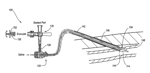
100491 As shown in FIG. 3, the suction port 132 is in fluid
communication with a suction
source 136 or other evacuator such as, for example, a syringe. Similarly, the
sealing material
port 134 is in fluid communication with a supply of sealing material, such as
a syringe 138 that
contains the sealing material. The valve 130 may comprise a translucent three-
way valve that
moves between a first or closed position where the suction port 132 and the
sealing material port
134 are both isolated from the lumen 142, a second position where the suction
port 132 is in fluid
communication with the lumen 142, and a third position where the sealing
material port 134 is in
CA 02707628 2010-06-01
WO 2009/088441
PCT/US2008/013967
fluid communication with the lumen 142. Details of the valve 130 and the
associated suction
port 132 and sealing material port 134 are shown in FIGS. 5-9.
[0050] Referring to FIG. 4, the main body 102 may be inserted into the
lumen 142 of the
introducer sheath 120 as shown. The main body 102 is sized so that it does not
fill the entire
lumen 142. Thus, the side-port 128 is in fluid communication with the portion
of the lumen 142
that is not filled.
[0051] A stopper sleeve or spacer 140 is shown disposed over the main body 102
to limit the
insertion distance of the main body 102 into the introducer sheath 120. The
length of the spacer
140 is chosen so that the distal end 106 of the main body 102 extends beyond
the distal end 124
of the introducer sheath 120 by a predetermined distance. According to one
embodiment, the
predetermined distance is approximately 2.5 cm to 4.0 cm. The distance is
chosen to allow the
expandable portion 114 of the main body 102 to pass through the introducer
sheath 120 and into
a blood vessel as discussed in more detail below. The spacer 140 may comprise
a split tube of
metal or plastic that can be easily removed as desired. =
[0052] Methods of closing a hole or puncture in a vessel such as an
arteriotomy 144 using the
vascular closure device 100 are discussed with reference to FIGS. 5-10.
Referring first to FIG. 5,
the vascular closure device 100 is shown with the introducer sheath 120
inserted through a hole
144 in a blood vessel 148. In one embodiment, the introducer sheath 120 may be
used for
introducing instruments during the medical procedure as well as to close the
hole 144. In other
embodiments, another introducer may be used during the procedure. When it is
time to close the
hole 144, the introducer may be swapped for the introducer sheath 120.
16
CA 02707628 2010-06-01
WO 2009/088441
PCT/US2008/013967
[0053] With the introducer sheath 120 in place, the main body 102 may be
inserted through the
lumen 142 until the expandable portion 114 extends beyond the tip of the
distal end 124 of the
introducer sheath 120 and into the blood vessel 148. The expandable portion
114 may be
expanded by opening the valve 118 and depressing the fluid dispenser 116. FIG.
5 shows the
expandable portion 114 after it has been expanded in the blood vessel 148.
Notably,: the
expandable portion 114 is positioned at an oblique angle relative to the main
body 102. The
valve 118 may be closed to maintain the expandable portion 114 in an expanded
position. The
main body 102 and the introducer sheath 120 are retracted until the expandable
portion 114
contacts an inner wall 150 of the blood vessel 148 and seals the internal side
of the hole 144 as
shown in FIG. 6. The expandable portion 114 is positioned so that it is
parallel to the inner wall
150 as it moves toward and contacts the inner wall 150. Thus, the expandable
portion 114 forms
a good seal over the hole 144 and provides sufficient tactile feedback to
allow the medical
professional to determine when the expandable portion 114 is in position. In
one embodiment,
the vascular closure device 100 may include a marking or some other indicia to
allow the
medical personnel to determine the rotational orientation of the main body
102. In this way, .the
medical personnel can reorient the main body 102 and the expandable portion
114 so that it is
parallel to the inner wall 150 before moving the expandable portion 114 into
contact with the
inner wall 150.
[0054] With the expandable portion 114 in place so that it internally blocks
or seals the hole
144, the side-port valve 130 is opened to allow fluid communication between
the unfilled space
1
of the lumen 142 and the suction source 136 as shown in FIG. 7. The pressure
is lowered in the
17
CA 02707628 2010-06-01
WO 2009/088441
PCT/US2008/013967
lumen 142 by withdrawing a stem 152 of the suction source 136 (in this
embodiment, a syringe)
or by some other suction device. As the pressure is lowered in the lumen 142
and communicated
to the tissue tract 146, a situs 154 of the hole 144 is aspirated, removing
fluids from the tissue
tract 146 via the lumen 142.
[0055] As the arteriotomy 144 is aspirated, a surgeon or other medical
professional may
visually inspect the fluid contents evacuated through the translucent valve
130 to assess blood
flow through the hole 144. This allows the medical professional to ensure that
the introducer
sheath 120 and/or the expandable portion 114 are properly positioned within
the blood vessel
148. A flow of blood may indicate that the expandable portion 114 is not
properly sealing the
hole 144.
[0056] When the surgeon is satisfied with the positioning of the introducer
sheath 120 and the
expandable portion 114, the valve 130 is toggled to create a fluid
communication path between
the lumen 142 and the sealing material contained in the syringe 138 or other
sealing material
supply as shown in FIG. 8. The syringe 138 holds a volume of sealing material
that is injected
into the introducer sheath 120 via the side-port 128 as a stem 156 is
depressed. The sealant
flows through the lumen 142 and into the tissue tract 146. Further, because
the tissue tract .146
has been evacuated and is in a vacuum condition, the sealing material is drawn
through the
annulus toward the hole 144. The vacuum condition of the situs 154 external to
the hole 144
causes the sealing material to quickly and efficiently fill all of the voids
around the hole 144 and
in the tissue tract 146. Preferably, the syringe 138 holds a volume of sealing
material sufficient
to fill the lumen 142 and therefore the tissue tract 146. As the sealing
material is injected (FIG.
18
.õ
CA 02707628 2010-06-01
WO 2009/088441
PCT/US2008/013967
8), the introducer sheath 120 is preferably withdrawn with respect to the
expandable portion 114
to allow the sealing material to fill the tissue tract 146. Therefore, in
order to facilitate retraction
of the introducer sheath 120, the spacer 140 (FIG. 4) is removed.
[0057] Following injection of the sealing material, it may be optionally
activated, cured, or set.
In many embodiments, the sealing material includes a liquid or gel sealant
that includes any of
the following thrombin, collagen, fibrin/fibrinogen, cyanoacrylate, polyvinyl
alcohol,
polyethylene glycol, chitosan, poly-n-acetyl glucosamine, and combinations
thereof (e.g.,
thrombin and collagen, fibrin/fibrinogen and collagen, cyanoacrylate and
collagen, or thrombin
and fibrin/fibrinogen). In other embodiments, the sealing material may include
implants
(implant is positioned adjacent to the exterior of the hole 146 using a
variety of different
techniques). Implants are typically provided as a solid, fiber, compressible
foam, or the like
while sealants are provided as a liquid, gel, or the like. The sealing
material may operate by
mechanically blocking the hole in the vessel, reacting with the blood or other
nearby tissue to
block the hole, or the like. In some embodiments, the sealing material may not
be dependent on
a biochemical reaction with blood or other bodily fluids to create a
hemostatic seal. However,
the gels or foams used according to some aspects of the present invention may
in some cases be
activated or cured by, for example, application of a second fluid, UV light,
or other activation
mechanisms.
[0058] Once the sealing material is in place adjacent the exterior of
the hole 144,. the
expandable portion 114 is contracted as shown in FIG. 9 by reopening the valve
118 (FIG. 4).
The stem 158 (FIG. 4) of the fluid dispenser 116 (FIG. 4) may be retracted to
ensure full
19
CA 02707628 2010-06-01
WO 2009/088441
PCT/US2008/013967
contraction of the expandable portion 114. The main body 102 and the
introduction sheath 120
are retracted, with the expandable portion 114 sliding through the sealing
material. According to
some embodiments, following removal of the main body 102 and the instruction
sheath 120,
manual pressure may be applied to the arteriotomy site to counteract any
sealing action
disruption caused by the act of pulling the expandable portion 114 through the
sealing material.
However, manual pressure is applied for only a fraction of the time allocated
to traditional
arteriotomy closures. For example, according to the principles described
herein, manual pressure
may be applied following retraction of the vascular closure device 100 for
only ten minutes or
less. The sealing material remains in the tissue tract 146 sealing the
arteriotomy 144 as shown in
FIG. 10.
[0059] Turning now to FIGS. 13-23, another embodiment of a vascular closure
device 250 is
shown. The vascular closure device 250 may be used to deploy sealing material
adjacent to and
outside of the hole in the blood vessel. The sealing material functions to
block the hole in the
blood vessel and/or the tissue tract to stop the bleeding. In one embodiment,
the sealing material
may be a lipid based sealing material. For example, the sealing material may
include
monoglycerides of saturated and unsaturated fatty acids. The sealing material
may include one
or more of such monoglycerides alone or in combination with other materials
such as therapeutic
agents, additives, and carrier materials. The therapeutic agents may include
drugs or other
substances that provide local or systemic therapeutic effect in the body.
Additives may be
included to alter the physical properties such as the melting point, strength,
resiliency, etc. of the
sealing material.
CA 02707628 2010-06-01
WO 2009/088441
PCT/US2008/013967
[0060] It should be appreciated that there are a wide number of substances,
mixtures,
molecules, etc. that may be used as the sealing material. In one embodiment,
the sealing Material
may comprise a monoglyceride including a fatty acid group having 12 to 22
carbon atoms: In
another embodiment, the sealing material may include glycerol monooleate,
glycerol
monostearate, glycerol monopalmitate, glycerol monolaurate, glycerol
monocaproate, glycerol
monocaprylate, glycerol monolinoleate, glycerol monolinolenate, glycerol
monomyristate,
and/or glycerol monoarachidonate. Those materials that may be preferable for
use as the sealing
material include glycerol, monooleate, glycerol monolinoleate, and/or glycerol
monolinolenate,
in any combination or amount.
[0061] The sealing material may melt, gel, or otherwise undergo a phase change
when
deployed adjacent to the hole in the blood vessel. In order for the sealing
material to melt, it is
desirable for the sealing material to have a melting point that is less than
bodily temperature but
high enough that the sealing material is a solid at room temperature. After
the sealing material
has been inserted into the tissue tract, it is heated by the patient's body
until it begins to melt or
gel. In one embodiment, the sealing material may have a melting point that is
no more than 37
C. In another embodiment, the sealing material may have a melting point that
is about 27 C to
37 C, about 30 C to 37 C, or about 34 C to 37 C.
[0062] Once the sealing material has melted, it may flow into the tissue tract
toward the hole in
the blood vessel. At the same time, the sealing material may begin to expand
and form a cubic
phase due to exposure to bodily fluids. In one embodiment, the sealing
material may expand up
to 46% of its original size. The sealing material may also exhibit adhesive
properties that help to
21
CA 02707628 2010-06-01
WO 2009/088441
PCT/US2008/013967
hold the sealing material in place in the tissue tract. The expansion of the
sealing material and
formation of the cubic phase (the sealing material becomes solid or non-
flowable in the cubic
phase) may act to hold the sealing material in place over the hole in the
blood vessel thereby
closing the hole in the blood vessel. It should be appreciated that any of the
foregoing sealing
materials may also be used with the vascular closure device 110.
=
[0063] The vascular closure devices 250 facilitates deployment of the sealing
material in the
tissue tract of the patient. The sealing material blocks the tissue tract and
stops the bleeding. In
one embodiment, the sealing material is bio-absorbable to allow it to be
removed by the body's
natural processes. In another embodiment, the sealing material may be deployed
with and
coupled to another bio-absorbable component such as a sealing plug (e.g.,
collagen plug) or
anchor both of which may also be bio-absorbable (e.g., PLA and PGA materials).
In one
embodiment, the vascular closure device 250 may be configured to not leave any
components
inside the blood vessel after the closure procedure is over (i.e., an extra-
vascular closure device).
In this embodiment, the sealing material and any other components left in the
patient are outside
of the blood vessel.
[0064] Referring to FIG. 13, one embodiment of the vascular closure device 250
is shown that
may be used to close and/or seal a hole or puncture in a blood vessel such as
an arteriotomy. The
vascular closure device 250 has a distal end 264 and a proximal end 265 and
includes a handle
251, a carrier tube or carrier member 252, sealing material 256, a stopper
254, and a vessel
locator assembly or vessel locator portion 260. The vessel locator assembly
260 includes a
central tube 259 that extends through the handle 251, the carrier tube 252,
the stopper 254, and
22
CA 02707628 2010-06-01
WO 2009/088441
PCT/US2008/013967
the sealing material 256. The vessel locator assembly 260 also includes a
expandable portion
266 positioned at the distal end 264 of the vascular closure device 250 and a
syringe 275
positioned at the proximal end 265 of the vascular closure device 250. The
syringe 275 is
coupled to and in fluid communication with the central tube 259.
[0065] The handle 251 is positioned at the proximal end 265 of the vascular
closure device 250
and allows the user to manipulate the various components of the device 250 to
facilitate closing
the hole in the blood vessel. In the embodiment shown in FIG. 13, the handle
251 includes a first
tube 261 having a distal end that is sized to slidably receive the carrier
tube 252 and a proximal
end that is sized to slidably receive a syringe 275. The first tube 261
includes a slot 267 that
receives an actuation member, protrusion, or pin 263 that extends outward from
the carrier tube
252. The user can reciprocally move the actuation member 263 proximally and
distally in .the
slot 267 to retract and extend, respectively, the carrier tube 252. Retracting
the carrier tube 252
when the vascular closure device 250 is deployed exposes the sealing material
256 to the tissue
tract.
[0066] Returning to the vessel locator assembly 260, the syringe 275 may be
used to
selectively expand and/or contract the expandable portion 266. Any suitable
fluid may be used
to expand the expandable portion 266. For example, fluids such as saline
solution, carbon
dioxide, or air may be suitable. Also a guide wire and a spring 268 may be
coupled proximally
to the expandable portion 266. The guide wire and the spring 268 are
configured to be
atraumatic to prevent the distal end 264 of the vascular closure device 250
from puncturing or
damaging the blood vessel.
23
CA 02707628 2010-06-01
WO 2009/088441
PCT/US2008/013967
[0067] The vascular closure device 250 is configured so that when it is
inserted into the tissue
tract, the expandable portion 266 is positioned inside the blood vessel. The
expandable portion
266 may be configured to move between the contracted configuration shown in
FIG. 13 and the
expanded configuration shown in FIG. 15. This allows the expandable portion
266 to be inserted
into the blood vessel, expanded, and then moved into contact with the interior
wall of the blood
vessel adjacent to the hole. The expandable portion 266 and the sealing
material 256 are spaced
apart a predetermined distance so that when the expandable portion 266 is
positioned against the
interior wall of the blood vessel, the sealing material 256 is positioned just
outside of the hole in
the blood vessel. In the embodiment shown in FIGS. 13-23, the expandable
portion 266 includes
a balloon that may use the same materials and/or otherwise be similar to the
balloon described in
connection with the expandable portion 114. For example, the expandable
portion 266 may be
positioned at an oblique angle like the expandable portion 114.
100681 It should be appreciated that the central tube 259 and any of the other
components of
the vessel locator assembly 260 may be made of any suitable material such as
metal, plastics, or
composites. Since the vascular closure device 250 is a medical device, the
materials used may
also be medical grade (medical grade metals, plastics, or composites). In one
embodiment, the
central tube 259 may be made of metals such as stainless steel or memory shape
metals such as
nitinol, and the like.
[0069] The stopper 254 is provided to prevent the sealing material 256
from Moving
proximally as the carrier tube 252 moves proximally. Accordingly, the stopper
254 is positioned
24
CA 02707628 2010-06-01
WO 2009/088441
PCT/US2008/013967
just proximal to the sealing material 256 inside the carrier tube 252 and the
stopper 254 is
coupled to the central tube 259 so that it is fixed in position.
[0070] Referring to FIG 13, the vascular closure device 250 may be
configured to indicate
when the expandable portion 266 is in contact with the interior wall of the
blood vessel. One
problem associated with locating the wall of the blood vessel is that the user
may be unable to
feel when the expandable portion 266 has contacted the wall of the blood
vessel. The user may
continue to pull on the vascular closure device 250 causing it to distort and
bend until it passes
through the hole in the blood vessel or the expanded expandable portion 266
may tear through
the hole in the wall of the blood vessel causing additional injury to the
patient.
[0071] The first tube 261 and the syringe 275 are coupled together in a manner
that signals to
the user when the expanded expandable portion 266 is positioned against the
interior wall of the
blood vessel. The syringe 275 is positioned to move lengthwise in the first
tube 261. The central
tube 259 is coupled to the syringe 275 so that when the expandable portion 266
contacts the
interior wall of the blood vessel, the tension on the central tube 259 pulls
the syringe 275 further
into the first tube 261. A spring 271 is positioned between the first tube 261
and the syringe 275
to bias the syringe 275 in the proximal direction and resistant the tension
exerted by the core
wire 270. The spring 271 is configured to provide just the right amount of
force so that the
spring 271 is only compressed, and consequently the syringe 275 moved, when
the expandable
portion 266 has contacted the interior wall of the blood vessel.
[0072] An indicator pin 298 extends outward from the syringe 275 and travels
in a slot 273 in
the first tube 261. As the spring 271 is compressed, the indicator pin 298
moves distally in the
CA 02707628 2010-06-01
WO 2009/088441
PCT/US2008/013967
slot 273. In operation, the user can pull back on the vascular closure device
250 while watching
the indicator pin 298. When the indicator pin 298 begins to move distally in
the slot 273, the
user knows that the expandable portion 266 is positioned against the interior
wall of the blood
vessel. The indicator pin 298 also prevents the spring 271 from biasing the
syringe 275 out the
proximal end of the first tube 261.
[0073] It should be appreciated that numerous other methods may be used to
signal the user
that the expandable portion 266 is positioned against the interior wall of the
blood vessel. The
signal may be visual, auditory, or any other suitable type of signal. In one
embodiment, the
vascular closure device 250 may be configured to emit a beep to alert the user
that the
expandable portion 266 is positioned against the interior wall of the blood
vessel.
;..
[0074] It should be appreciated that the design of the vascular closure
devices 250 may be
altered in any of a number of ways. For example, FIGS. 20-23 show another
embodiment of the
vascular closure device 250. In this embodiment, the vascular closure device
250 includes a
perforated tube 292 that is used to dispense the sealing material 256 into the
tissue tract. In one
embodiment, the vascular closure device 250 may be provided with another
syringe coupled to
the proximal end of the perforated tube 292. The syringe may be used to inject
the sealing
material 256 out through the holes 293 in the perforated tube 292. The distal
end. of the
perforated tube 292 may be blocked or closed so that the sealing material 256
is forced out the
sides of the perforated tube 292 against the walls of the tissue tract instead
of down against the
hole, which may result in sealing material entering the blood stream.
26
CA 02707628 2010-06-01
WO 2009/088441
PCT/US2008/013967
[0075] As shown in FIG. 21, the holes 293 in the perforated tube 292 may be
sized to regulate
the flow of the sealing material 256. For example, the holes 293 may get
larger moving in a
distal direction along the perforated tube 292 so that the largest holes 293
are positioned nearest
the distal end of the perforated tube 292. This configuration results in an
even amount of sealing
material 256 being dispensed along the perforated tube 292. It should be
appreciated that in
other embodiments, the holes 293 may be configured to be the same size or all
of the holes'293
may be unique sizes. Numerous configurations are possible.
[0076] A method of closing a hole 310 in a blood vessel 308 using the vascular
closure device
250 is described in connection with FIGS. 14-23. Once the procedure is over
and the user is
ready to close the hole in the blood vessel, the initial step may be to
exchange the procedural
access sheath for the introducer sheath 262. This is done by placing a
guidewire through the
procedural sheath and into the blood vessel 308. The procedural sheath is then
withdrawn from
the body while holding digital pressure on the blood vessel 308, upstream from
the sheath, and
while holding the guidewire in place. Next, a closure dilator is placed within
the introducer
sheath 262 and the distal tapered end of the closure dilator is back-loaded
onto the guidewire.
The closure dilator and the introducer sheath 262 are advanced together
distally over the
guidewire, through the tissue tract 312, and into the blood vessel 308.
[0077] In one embodiment, the introducer sheath 262 includes a distal side
hole (not shown)
near the distal end of the introducer sheath 262. The closure dilator also
includes a distal side
hole that is configured to align with the distal side hole in the introducer
sheath 262 when the
closure dilator is positioned in the introducer sheath 262. The closure
dilator also has a proximal
27
CA 02707628 2010-06-01
WO 2009/088441
PCT/US2008/013967
side hole at the proximal end of the closure dilator that is in fluid
communication with the distal
side hole of the closure dilator and the closure sheath. In one embodiment,
the distal and
proximal side holes may be fluidly connected by way of a dedicated lumen or
bore. In another
embodiment, the distal and proximal side holes may be fluidly connected by the
central lumen of
the closure dilator that the guidewire is positioned in.
[0078] The distal and proximal side holes in the introducer sheath 262 and the
closure dilator
are provided to allow blood to flash back when the introducer sheath 262 is
correctly positioned
in the blood vessel 308. Once blood flows out the proximal side hole of the
closure dilator, the
user pulls the introducer sheath 262 in a proximal direction until the blood
flow just stops. The
introducer sheath 262 is now placed in the correct position to continue the
procedure. The next
step is to withdraw the closure dilator and the guidewire while holding the
introducer sheath 262
in place.
[0079] The introducer sheath 262 is sized to slidably receive the vascular
closure device 250
therein. The distal ends of the introducer sheath 262 and the carrier tube 252
have a tapered
shape so that the tip will align with the lengthwise axis of the blood vessel
308 when the
introducer sheath 262 is inserted through the tissue tract 312 at an angle of
about 20-45 degrees
to the vessel axis.
[0080] After the introducer sheath 262 is in place, the vascular closure
device 250 is introduced
into the proximal end of the introducer sheath 262. The vascular closure
device 250 may be
configured to advance until it snaps, locks, or otherwise mates together with
the carrier tube 62.
In this position, the distal end 264 of the vascular closure device 250
extends out of the distal end
28
=
CA 02707628 2010-06-01
WO 2009/088441
PCT/US2008/013967
of the introducer sheath 262 and into the blood vessel 308. It should be noted
that the vascular
closure device 250 and the introducer sheath 262 may be configured so that
when they are
coupled together, the distal end 264 extends into the blood vessel 308 a
predetermined amount.
[0081] FIG. 14 shows the expandable portion 266 in position in the blood
vessel 308. The
expandable portion 266 is expanded using the syringe 275. FIG. 15 shows the
expandable
portion 266 in the expanded configuration. The introducer sheath 262 and the
vascular closure
device 250 are drawn away from the patient until the expandable portion 266
contacts the vessel
wall at the puncture site as shown in FIG. 16.
[0082] Now that the expandable portion 266 is in position, the introducer
sheath 262 and the
carrier tube 252 are withdrawn to expose the sealing material 256 to the
tissue tract 312. The
sealing material begins to melt as it is heated by the body and flows down
toward the hole 310 in
the blood vessel 308 as shown in FIG. 17. The expandable portion 266 blocks
the hole 310 so
that the sealing material 256 does not flow into the bloodstream. The sealing
material 256
;
begins to form a cubic phase upon exposure to bodily fluids such as blood and
the like. This
causes the sealing material 256 to expand and fill the tissue tract 312
adjacent to the hole 310 in
the blood vessel 308 as shown in FIG. 18. It should be appreciated that the
vascular closure
device 250 may be configured to use a second non-flowable sealing material or
anchor along
with the sealing material 256. For example, the vascular closure device 250
may be configured
to deposit a small collagen plug adjacent to the hole 310 to prevent the
sealing material 256 from
entering the blood vessel 308.
;..
29
CA 02707628 2010-06-01
WO 2009/088441
PCT/US2008/013967
[0083] Now that the sealing material 256 has been deployed and has
formed the solid or
somewhat firm cubic phase, the next step is to contract the expandable portion
266 and withdraw
the vessel locator assembly 260 and the remainder of the vascular closure
device 250 from the
tissue tract 312. As the vessel locator assembly 260 passes through the
sealing material 256, the
sealing material 256 swells or otherwise moves to fill the gap where the
vessel locator assembly
260 used to be. The hole in the blood vessel 308 is now sealed by clotting
action and the sealing
material 256 positioned in the tissue tract 312.
[0084] The method of using the vascular closure device 250 shown in FIGS. 20-
23 is similar to
the method of using the vascular closure device 250 shown in FIGS. 14-19.
However, instead of
passively allowing the sealing material 256 to melt and fill the tissue tract
312, the user can inject
any desired amount of sealing material 256 into the tissue tract 312 through
the perforated tube
292. This allows for additional sealing material 256 to be deployed. Also, the
user may inject
sealing material 256 through the perforated tube 292 as the perforated tube
292 is being
withdrawn so that the sealing material fills up the entire tissue tract 312.
[0085] It should be appreciated that the embodiments disclosed have many
components and the
methods described have many steps for operation and use. It is anticipated
that the number of
components and steps could be altered considerably without departing from the
broad scope of
what is described herein.
CA 02707628 2010-06-01
WO 2009/088441
PCT/US2008/013967
Illustrative Embodiments
[0086] Reference is made in the following to a number of illustrative
embodiments of the
subject matter described herein. The following embodiments illustrate only a
few selected
embodiments that may include the various features, characteristics, and
advantages of the subject
matter as presently described. Accordingly, the following embodiments should
not be considered
as being comprehensive of all of the possible embodiments. Also, features and
characteristics of
one embodiment may and should be interpreted to equally apply to other
embodiments or be
used in combination with any number of other features from the various
embodiments to provide
further additional embodiments, which may describe subject matter having a
scope that varies
(e.g., broader, etc.) from the particular embodiments explained below.
Accordingly, any
combination of any of the subject matter described herein is contemplated.
[0087] According to one embodiment, a method of closing a hole in a
vessel of a patient,
comprises: moving an expandable portion of a vascular closure device through
the hole and into
the vessel, the vascular closure device including a main body that extends
through the hole at an
oblique angle relative to the vessel; expanding the expandable portion of the
vascular closure
device; and moving the expandable portion into contact with an inner wall of
the vessel to block
the hole, the expandable portion being oriented at least substantially
parallel to the inner wall of
the vessel shortly before contacting the inner wall. The method may comprise
applying a sealing
material to the hole while the expandable portion is in contact with the inner
wall of the vessel.
The sealing material may include a sealant. The sealant may be applied using
suction. The
method may comprise applying a sealing material to the hole while the
expandable portion is in
31
CA 02707628 2010-06-01
WO 2009/088441
PCT/US2008/013967
=
contact with the inner wall of the vessel; contracting the expandable portion;
and removing the
expandable portion of the vascular closure device from the vessel. The method
may comprise
applying manual pressure to the hole after removing the expandable portion of
the vascular
closure device from the vessel. The vessel may include a blood vessel. The
expandable portion
may be oriented at an oblique angle relative to the main body shortly before
contacting the inner
wall. The expandable portion may be made, at least in part, of polyurethane.
The expandable
portion may include a tail. Moving the expandable portion of the vascular
closure device
through the hole and into the vessel may include moving the expandable portion
through an
introducer that extends into the vessel.
[0088] According to another embodiment, a method of closing a hole in a vessel
of a patient,
comprises: expanding an expandable portion of a vascular closure device inside
the vessel, the
expandable portion being oriented at an oblique angle relative to a main shaft
of the vascular
closure device; and moving the expandable portion into contact with an inner
wall of the vessel
to block the hole. The method may comprise applying a sealing material to the
hole while the
expandable portion is in contact with the inner wall of the vessel. The
sealing material may
include a sealant. The sealant may be applied using suction. The method may
comprise
applying a sealing material to the hole while the expandable portion is in
contact with the inner
wall of the vessel; contracting the expandable portion; and removing the
expandable portion of
the vascular closure device from the vessel. The method may comprise applying
manual
pressure to the hole after removing the expandable portion of the vascular
closure device from
the vessel. The vessel may include a blood vessel such as an artery. The
expandable portion
=
32
CA 02707628 2010-06-01
WO 2009/088441
PCT/US2008/013967
may be made, at least in part, of polyurethane. The expandable portion may
include a tail. The
method may comprise moving the expandable portion of the vascular closure
device through an
introducer that extends into the vessel.
[0089] According to another embodiment, a method of closing a hole in a blood
vessel of a
patient comprises: expanding an expandable portion of a vascular closure
device inside the blood
vessel, the expandable portion being oriented at an oblique angle relative to
a main shaft of the
vascular closure device; moving the expandable portion into contact with an
inner wall of the
vessel to block the hole; applying a sealing material to the hole while the
expandable portion is
in contact with the inner wall of the blood vessel; contracting the expandable
portion; and
removing the expandable portion of the vascular closure device from the blood
vessel. The
sealing material may include a sealant. The expandable portion may include a
tail.
[0090] According to another embodiment, a vascular closure device comprises: a
main body
having a distal end; and an expandable portion positioned at the distal end of
the main body, the
expandable portion being configured to be inserted into a hole in a vessel of
a patient; wherein
the expandable portion is oriented at an oblique angle relative to the main
body when the
expandable portion is in an expanded configuration. The main body may form a
conduit that is
in fluid communication with the expandable portion, the expandable portion
being selectively
expandable with fluid delivered by the conduit. The main body may include
hypotube that forms
the conduit. The main body may include a guidewire. The expandable portion may
be attached
to the guidewire. The guidewire may include hypotube to deliver fluid to the
expandable
portion. The vascular closure device may comprise a conduit to deliver sealant
to an area
33
=
CA 02707628 2010-06-01
WO 2009/088441
PCT/US2008/013967
adjacent to the expandable portion. The expandable portion may be positioned
adjacent to a
distal tip of the main body. The expandable portion may extend outward from a
distal tip of the
main body. The expandable portion may be oriented at an angle of approximately
200 to 70
relative to the main body when the expandable portion is in the expanded
configuration. :The
expandable portion may be made, at least in part, of polyurethane. The
expandable portion may
include a tail.
[0091] According to another embodiment, a vascular closure device comprises: a
guidewire;
and an expandable portion attached to the guidewire; wherein the expandable
portion is
configured to be inserted into a vessel of a patient through a hole in a wall
of the vessel, the
vascular closure device being configured to close the hole in the wall of the
vessel.
[0092] According to another embodiment, a vascular closure device comprises: a
cylindrical
tube; and an expandable portion attached to the cylindrical tube; wherein the
expandable portion
is configured to be inserted into a vessel of a patient through a hole in a
wall of the vessel, the
vascular closure device being configured to close the hole in the wall of the
vessel.
[0093] According to another embodiment, a vascular closure device comprises an
expandable
portion made at least in part of polyurethane, the expandable portion being
configured to be
inserted into a vessel of a patient through a hole in a wall of the vessel,
the vascular closure
device being configured to close the hole in the wall of the vessel.
[0094] According to another embodiment, an internal tissue puncture
sealing apparatus
comprises a first thin, elongated conduit having a first central lumen and
first and second ends.
34
CA 02707628 2010-06-01
WO 2009/088441
PCT/US2008/013967
The first end may be insertable through the internal tissue puncture and has
an inflation segment
in fluid communication with the central lumen. The first end may include an
expandable
member that is selectively inflatable with a fluid via the central lumen. The
apparatus may also
include a second thin, elongated conduit having a second central lumen
receptive of the first thin,
elongated conduit. The proximal end of the second conduit has at least one
valved side-port in
fluid communication with a space between the first and second conduits. The
valved side-port
may include a vacuum communication path and a sealant injection path, which
enable aspiration
of a tissue puncture site and sealing of the puncture.
[0095] According to another embodiment, a method of closing a hole in a vessel
wall may
include inserting an inflatable device through an introducer that is disposed
in the vessel,
inflating the inflatable device, sealing the inflatable device against an
inner wall of the vessel,
reducing the pressure inside of the introducer, injecting a sealant into the
introducer, deflating the
inflatable device, and removing the inflatable device through the sealant.
Following removal of
the inflatable device, manual pressure may be applied to the hole for a short
period of time to
'4
ensure continued hemostasis. A specially designed introducer may be swapped
with a standard
introducer used to facilitate insertion of vascular tools used to perform a
vascular procedure prior
to inserting the inflatable device.
[0096] The terms recited in the claims should be given their ordinary and
customary meaning
as determined by reference to relevant entries (e.g., definition of "plane" as
a carpenter's tool
would not be relevant to the use of the term "plane" when used to refer to an
airplane, etc.) in
dictionaries (e.g., widely used general reference dictionaries and/or relevant
technical
-1
CA 02707628 2010-06-01
WO 2009/088441
PCT/US2008/013967
dictionaries), commonly understood meanings by those in the art, etc., with
the understanding
that the broadest meaning imparted by any one or combination of these sources
should be given
to the claim terms (e.g., two or more relevant dictionary entries should be
combined to provide
the broadest meaning of the combination of entries, etc.) subject only to the
following
exceptions: (a) if a term is used herein in a manner more expansive than its
ordinary and
customary meaning, the term should be given its ordinary and customary meaning
plus the
additional expansive meaning, or (b) if a term has been explicitly defmed to
have a different
meaning by reciting the term followed by the phrase "as used herein shall
mean" or similar
language (e.g., "herein this term means," "as defined herein," "for the
purposes of this disclosure
[the term] shall mean," etc.). References to specific examples, use of "i.e.,"
use of the word
"invention," etc., are not meant to invoke exception (b) or otherwise restrict
the scope of the
recited claim terms. Other than situations where exception (b) applies,
nothing contained herein
should be considered a disclaimer or disavowal of claim scope. Accordingly,
the subject matter
recited in the claims is not coextensive with and should not be interpreted to
be coextensive with
any particular embodiment, feature, or combination of features shown herein.
This is true even if
only a single embodiment of the particular feature or combination of features
is illustrated and
described herein. Thus, the appended claims should be read to be given their
broadest
interpretation in view of the prior art and the ordinary meaning of the claim
terms.
[0097] As used herein, spatial or directional terms, such as "left," "right,"
"front," "back," and
the like, relate to the subject matter as it is shown in the drawing FIGS.
However, it is to be
understood that the subject matter described herein may assume various
alternative orientations
36
CA 02707628 2010-06-01
WO 2009/088441
PCT/US2008/013967
and, accordingly, such terms are not to be considered as limiting.
Furthermore, as used herein
(i.e., in the claims and the specification), articles such as "the," "a," and
"an" can connote the
singular or plural. Also, as used herein, the word "or" when used without a
preceding "either"
(or other similar language indicating that "or" is unequivocally meant to be
exclusive ¨ e.g., only
one of x or y, etc.) shall be interpreted to be inclusive (e.g., "x or y"
means one or both x or y).
Likewise, as used herein, the term "and/or" shall also be interpreted to be
inclusive (e.g., "x
and/or y" means one or both x or y). In situations where "and/or" or "or" are
used as a
conjunction for a group of three or more items, the group should be
interpreted to include one
item alone, all of the items together, or any combination or number of the
items. Moreover,
terms used in the specification and claims such as have, having, include, and
including should be
construed to be synonymous with the terms comprise and comprising.
[00981 Unless otherwise indicated, all numbers or expressions, such as
those expressing
dimensions, physical characteristics, etc. used in the specification (other
than the claims) are
understood as modified in all instances by the term "approximately." At the
very least, and not
as an attempt to limit the application of the doctrine of equivalents to the
claims, each numerical
parameter recited in the specification or claims which is modified by the term
"approximately"
should at least be construed in light of the number of recited significant
digits and by applying
ordinary rounding techniques. Moreover, all ranges disclosed herein are to be
understood to
encompass and provide support for claims that recite any and all subranges or
any and all
individual values subsumed therein. For example, a stated range of 1 to 10
should be considered
to include and provide support for claims that recite any and all subranges or
individual values
37
CA 02707628 2010-06-01
WO 2009/088441 PCT/US2008/013967
that are between and/or inclusive of the minimum value of 1 and the maximum
value of 10; that
is, all subranges beginning with a minimum value of 1 or more and ending with
a maximum
value of 10 or less (e.g., 5.5 to 10, 2.34 to 3.56, and so forth) or any
values from 1 to 10 (e.g., 3,
5.8, 9.9994, and so forth).
38