Note: Descriptions are shown in the official language in which they were submitted.
CA 02707898 2010-06-15
MRI BIOPSY CYLINDRACEOUS TARGETING GUIDE
BACKGROUND
[00011 Biopsy samples have been obtained in a variety of ways in various
medical
procedures using a variety of devices. Biopsy devices may be used under
stereotactic guidance, ultrasound guidance, MRI guidance, PEM guidance, BSGI
guidance, or otherwise. Merely exemplary biopsy devices are disclosed in U.S.
Pat. No. 6,273,862, entitled "Surgical Device for the Collection of Soft
Tissue,"
issued Aug. 14, 2001; U.S. Pat. No. 6,231,522, entitled "Biopsy Instrument
with
Breakable Sample Segments," issued May 15, 2001; U.S. Pat. No. 6,228,055,
entitled "Devices for Marking and Defining Particular Locations in Body
Tissue,"
issued May 8, 2001; U.S. Pat. No. 6,120,462, entitled "Control Method for an
Automated Surgical Biopsy Device," issued September 19, 2000; U.S. Pat. No.
6,086,544, entitled "Control Apparatus for an Automated Surgical Biopsy
Device," issued July 11, 2000; U.S. Pat. No. 6,077,230, entitled "Biopsy
Instrument with Removable Extractor," issued June 20, 2000; U.S. Pat. No.
6,017,316, entitled "Vacuum Control System and Method for Automated Biopsy
Device," issued Jan. 25, 2000; U.S. Pat. No. 6,007,497, entitled "Surgical
Biopsy
Device," issued Dec. 28, 1999; U.S. Pat. No. 5,980,469, entitled "Method and
Apparatus for Automated Biopsy and Collection of Soft Tissue," issued Nov. 9,
1999; U.S. Pat. No. 5,964,716, entitled "Method of Use for a Multi-Port Biopsy
Instrument," issued Oct. 12, 1999; U.S. Pat. No. 5,928,164, entitled
"Apparatus
for Automated Biopsy and Collection of Soft Tissue," issued July 27, 1999;
U.S.
Pat. No. 5,775,333, entitled "Apparatus for Automated Biopsy and Collection of
Soft Tissue," issued July 7, 1998; U.S. Pat. No. 5,769,086, entitled "Control
System and Method for Automated Biopsy Device," issued June 23, 1998; U.S.
CA 02707898 2010-06-15
-2-
Pat. No. 5,649,547, entitled "Methods and Devices for Automated Biopsy and
Collection of Soft Tissue," issued July 22, 1997; U.S. Pat. No. 5,526,822,
entitled
"Method and Apparatus for Automated Biopsy and Collection of Soft Tissue,"
issued June 18, 1996; U.S. Pub. No. 2008/0214955, entitled "Presentation of
Biopsy Sample by Biopsy Device," published September 4, 2008; U.S. Pub. No.
2007/0255168, entitled "Grid and Rotatable Cube Guide Localization Fixture for
Biopsy Device," published November 1, 2007; U.S. Pub. No. 2007/0118048,
entitled "Remote Thumbwheel for a Surgical Biopsy Device," published May 24,
2007; U.S. Pub. No. 2005/0283069, entitled "MRI Biopsy Device Localization
Fixture," published December 22, 2005; U.S. Pub. No. 2003/0199753, entitled
"MRI Compatible Biopsy Device with Detachable Probe," published Oct. 23,
2003; U.S. Pub. No. 2003/0109803, entitled "MRI Compatible Surgical Biopsy
Device," published June 12, 2003; U.S. Provisional Patent Application Serial
No.
60/874,792, entitled "Biopsy Sample Storage," filed December 13, 2006; and
U.S.
Provisional Patent Application Serial No. 60/869,736, entitled "Biopsy
System,"
filed December 13, 2006. The disclosure of each of the above-cited U.S.
Patents,
U.S. Patent Application Publications, and U.S. Provisional Patent Applications
is
incorporated by reference herein.
[00021 Some biopsy systems may provide an apparatus to guide a probe and/or
other
components of a biopsy device to a desired biopsy site. In some such biopsy
systems, a guide cube and positioning grid plate may be used. The guide cube
may be selectively located within an opening in the grid plate. The guide cube
may include guide holes to receive a portion of the probe and/or other
components, for example a needle, cannula, obturator, or combinations of these
or
other components. With the guide cube inserted in the grid plate, the probe or
other components can be guided through a selected guide hole of the guide cube
to arrive at a desired biopsy site. The desired biopsy site may or may not
have
been identified and/or targeted by one or more of the guidance approaches
mentioned above.
CA 02707898 2010-06-15
-3-
[0003] While several systems and methods have been made and used for obtaining
a
biopsy sample, it is believed that no one prior to the inventors has made or
used
the invention described in the appended claims.
BRIEF DESCRIPTION OF THE DRAWINGS
[0004] While the specification concludes with claims which particularly point
out and
distinctly claim the invention, it is believed the present invention will be
better
understood from the following description of certain examples taken in
conjunction with the accompanying drawings. In the drawings, like numerals
represent like elements throughout the several views.
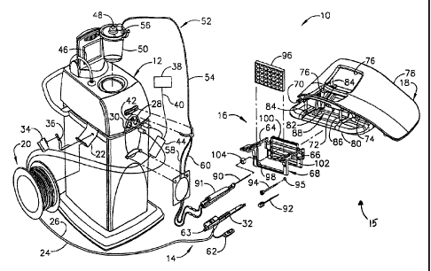
[0005] FIG. 1 is a perspective view of a biopsy system including a control
module
remotely coupled to a biopsy device, and including a localization assembly.
[0006] FIG. 2 is a perspective view of a breast coil of the localization
assembly of FIG.
1.
[0007] FIG. 3 is a perspective view of the biopsy device inserted through the
guide cube
of the localization assembly of FIG. 1.
[0008] FIG. 4 is a perspective view of the obturator and cannula of the biopsy
system of
FIG. 1.
[0009] FIG. 5 is an exploded perspective view of the obturator and cannula of
FIG. 4.
[0010] FIG. 6 is a perspective view of the guide cube inserted into the grid
plate of the
localization assembly of FIG. 1.
[0011] FIG. 7 is a perspective view of the obturator and cannula of FIG. 4
with a depth
stop device of FIG. 1 inserted through the guide cube and grid plate of FIG.
6.
[0012] FIG. 8 is a perspective view of an exemplary alternative grid plate.
[0013] FIG. 9 is a perspective view of the obturator and cannula of FIG. 4
with a depth
stop device of FIG. 1 inserted through the guide cube and grid plate of FIG.
8.
CA 02707898 2010-06-15
-4-
[0014] FIG. 10 is a front perspective view of an exemplary cylindraceous
targeting guide.
[0015] FIG. 11 is a front elevational view of the targeting guide of FIG. 10.
[0016] FIG. 12 is a rear perspective view of the targeting guide of FIG. 10.
[0017] FIG. 13 is a side perspective view of an exemplary alternative
cylindraceous
targeting guide.
[0018] FIG. 14 is a perspective view of the targeting guide of FIG. 10
inserted in the grid
plate of FIG. 6.
[0019] FIG. 15A is a front elevational view of the targeting guide and grid
plate of FIG.
14, with the targeting guide at a first rotational position.
[0020] FIG. 15B is a front elevational view of the targeting guide and grid
plate of FIG.
14, with the targeting guide rotated to a second rotational position.
DETAILED DESCRIPTION
[0021] The following description of certain examples should not be used to
limit the
scope of the present invention. Other features, aspects, and advantages of the
versions disclosed herein will become apparent to those skilled in the art
from the
following description, which is by way of illustration, one of the best modes
contemplated for carrying out the invention. As will be realized, the versions
described herein are capable of other different and obvious aspects, all
without
departing from the invention. Accordingly, the drawings and descriptions
should
be regarded as illustrative in nature and not restrictive.
[0022] As shown in the figures, an exemplary magnetic resonance imaging (MRI
or MR
imaging) compatible biopsy system may include a control module (12),
localization assembly (15), and biopsy device (14). In particular,
localization
assembly (15) is configured to localize a patient's breast and guide needle
(90) of
biopsy device (14) to a targeted area within the patient's breast; while
control
module (12) is operable to control biopsy device (14) after needle (90) has
been
CA 02707898 2010-06-15
-5-
introduced to the target site. These components and their sub-components will
be
discussed further below. In addition, targeting guides for use with various
localization assemblies will be discussed. While this disclosure may reference
the
biopsy system as compatible with MRI and MRI equipment and devices, it should
be appreciated that other imaging techniques and equipment and devices may be
used with the components described below, including but not limited to
stereotactic, ultrasound, PEM, BSGI, and/or other imaging techniques and
equipment.
[0023] I. Control Module
[0024] In FIGS. 1-3, MRI compatible biopsy system (10) has control module (12)
that
may be placed outside of a shielded room containing an MRI machine (not
shown) or at least spaced away to mitigate detrimental interaction with its
strong
magnetic field and/or sensitive radio frequency (RF) signal detection
antennas.
As described in U.S. Pat. No. 6,752,768, which is hereby incorporated by
reference in its entirety, a range of preprogrammed functionality may be
incorporated into control module (12) to assist in taking tissue samples.
Control
module (12) controls and powers biopsy device (14) that is used with
localization
assembly (15). Biopsy device (14) is positioned and guided by localization
fixture (16) attached to breast coil (18) that may be placed upon a gantry
(not
shown) of a MRI or other imaging machine.
[0025] In the present example, control module (12) is mechanically,
electrically, and
pneumatically coupled to biopsy device (14) so that components may be
segregated that need to be spaced away from the strong magnetic field and the
sensitive RF receiving components of a MRI machine. Cable management spool
(20) is placed upon cable management attachment saddle (22) that projects from
a
side of control module (12). Wound upon cable management spool (20) is paired
electrical cable (24) and mechanical cable (26) for communicating control
signals
and cutter rotation/advancement motions respectively. In particular,
electrical and
mechanical cables (24, 26) each have one end connected to respective
electrical
CA 02707898 2010-06-15
-6-
and mechanical ports (28, 30) in control module (12) and another end connected
to holster portion (32) of biopsy device (14). Docking cup (34), which may
hold
holster portion (32) when not in use, is hooked to control module (12) by
docking
station mounting bracket (36). It should be understood that such components
described above as being associated with control module (12) are merely
optional.
[00261 Interface lock box (38) mounted to a wall provides tether (40) to
lockout port (42)
on control module (12). Tether (40) is uniquely terminated and of short length
to
preclude inadvertent positioning of control module (12) too close to a MRI
machine or other machine. In-line enclosure (44) may register tether (40),
electrical cable (24) and mechanical cable (26) to their respective ports (42,
28,
30) on control module (12).
[00271 Vacuum assist is provided by first vacuum line (46) that connects
between control
module (12) and outlet port (48) of vacuum canister (50) that catches liquid
and
solid debris. Tubing kit (52) completes the pneumatic communication between
control module (12) and biopsy device (14). In particular, second vacuum line
(54) is connected to inlet port (56) of vacuum canister (50). Second vacuum
line
(54) divides into two vacuum lines (58, 60) that are attached to biopsy device
(14). With biopsy device (14) installed in holster portion (32), control
module
(12) performs a functional check. Saline may be manually injected into biopsy
device (14) or otherwise introduced to biopsy device (14), such as to serve as
a
lubricant and to assist in achieving a vacuum seal and/or for other purposes.
Control module (12) actuates a cutter mechanism (not shown) in biopsy device
(14), monitoring full travel of a cutter in biopsy device (14) in the present
example. Binding in mechanical cable (26) or within biopsy device (14) may be
optionally monitored with reference to motor force exerted to turn mechanical
cable (26) and/or an amount of twist in mechanical cable (26) may be sensed in
comparing rotary speed or position at each end of mechanical cable (26).
[00281 Remote keypad (62), which is detachable from holster portion (32),
communicates via electrical cable (24) to control panel (12) to enhance
clinician
CA 02707898 2010-06-15
-7-
control of biopsy device (14) in the present example, especially when controls
that would otherwise be on biopsy device (14) itself are not readily
accessible
after insertion into localization fixture (16) and/or placement of control
module
(12) is inconveniently remote (e.g., 30 feet away). However, as with other
components described herein, remote keypad (62) is merely optional, and may be
modified, substituted, supplemented, or omitted as desired. In the present
example, aft end thumbwheel (63) on holster portion (32) is also readily
accessible after insertion to rotate the side from which a tissue sample is to
be
taken.
[0029] Of course, the above-described control module (12) is merely one
example. Any
other suitable type of control module (12) and associated components may be
used. By way of example only, control module (12) may instead be configured
and operable in accordance with the teachings of U.S. Pub. No. 2008/0228103,
entitled "Vacuum Timing Algorithm for Biopsy Device," published September
18, 2008, the disclosure of which is incorporated by reference herein. As
another
merely illustrative example, control module (12) may instead be configured and
operable in accordance with the teachings of U.S. Patent Application Serial
No.
12/337,814, entitled "Control Module Interface for MRI Biopsy Device," filed
December 18, 2008, the disclosure of which is incorporated by reference
herein.
Alternatively, control module (12) may have any other suitable components,
features, configurations, functionalities, operability, etc. Other suitable
variations
of control module (12) and associated components will be apparent to those of
ordinary skill in the art in view of the teachings herein.
[0030] II. Localization Assembly
[0031] Localization assembly (15) of the present example comprises breast coil
(18) and
localization fixture (16). These components of localization assembly (15) are
described further below.
CA 02707898 2010-06-15
-8-
[0032] Left and right parallel upper guides (64, 66) of localization framework
(68) are
laterally adjustably received respectively within left and right parallel
upper tracks
(70, 72) attached to under side (74) and to each side of a selected breast
aperture
(76) formed in patient support platform (78) of breast coil (18). Base (80) of
breast coil (18) is connected by centerline pillars (82) that are attached to
patient
support platform (78) between breast apertures (76). Also, a pair of outer
vertical
support pillars (84, 86) on each side spaced about a respective breast
aperture (76)
respectively define lateral recess (88) within which localization fixture (16)
resides.
[0033] It should be appreciated that the patient's breasts hang pendulously
respectively
into breast apertures (76) within lateral recesses (88) in the present
example. For
convenience, herein a convention is used for locating a suspicious lesion by
Cartesian coordinates within breast tissue referenced to localization fixture
(16)
and to thereafter selectively position an instrument, such as needle (90) of
probe
(91) that is engaged to holster portion (32) to form biopsy device (14). Of
course,
any other type of coordinate system or targeting techniques may be used. To
enhance hands-off use of biopsy system (10), especially for repeated re-
imaging
within the narrow confines of a closed bore MRI machine, biopsy system (10)
may also guide obturator (92) encompassed by cannula (94). Depth of insertion
is
controlled by depth stop device (95) longitudinally positioned on either
needle
(90) or cannula (94). Alternatively, depth of insertion may be controlled in
any
other suitable fashion.
[0034] This guidance is specifically provided by a lateral fence in the
present example,
depicted as a grid plate (96), which is received within laterally adjustable
outer
three-sided plate bracket (98) attached below left and right parallel upper
guides
(64, 66). Similarly, a medial fence with respect to a medial plane of the
chest of
the patient, depicted as medial plate (100), is received within inner three-
sided
plate bracket (102) attached below left and right parallel upper guides (64,
66)
close to centerline pillars (82) when installed in breast coil (18). To
further refine
CA 02707898 2010-06-15
-9-
the insertion point of the instrument (e.g., needle (90) of probe (91),
obturator/cannula (92, 94), etc.), guide cube (104) may be inserted into grid
plate
(96).
[00351 In the present example, the selected breast is compressed along an
inner (medial)
side by medial plate (100) and on an outer (lateral) side of the breast by
grid plate
(96), the latter defining an X-Y plane. The X-axis is vertical (sagittal) with
respect to a standing patient and corresponds to a left-to-right axis as
viewed by a
clinician facing the externally exposed portion of localization fixture (16).
Perpendicular to this X-Y plane extending toward the medial side of the breast
is
the Z-axis, which typically corresponds to the orientation and depth of
insertion of
needle (90) or obturator/cannula (92, 94) of biopsy device (14). For clarity,
the
term Z-axis may be used interchangeably with "axis of penetration", although
the
latter may or may not be orthogonal to the spatial coordinates used to locate
an
insertion point on the patient. Versions of localization fixture (16)
described
herein allow a non-orthogonal axis of penetration to the X-Y axis to a lesion
at a
convenient or clinically beneficial angle.
[00361 It should be understood that the above-described localization assembly
(15) is
merely one example. Any other suitable type of localization assembly (15) may
be used, including but not limited to localization assemblies (15) that use a
breast
coil (18) and/or localization fixture (16) different from those described
above.
Other suitable components, features, configurations, functionalities,
operability,
etc. for a localization assembly (15) will be apparent to those of ordinary
skill in
the art in view of the teachings herein.
[00371 III. Biopsy Device
[00381 As shown in FIG. 1, one version of biopsy device (14) may comprise
holster
portion (32) and probe (91). Exemplary holster portion (32) was discussed
previously in the above section addressing control module (12). The following
CA 02707898 2010-06-15
-10-
paragraphs will discuss probe (91) and associated components and devices in
further detail.
[0039] In the present example, cannula (94) and obturator (92) are associated
with probe
(91). In particular, and as shown in FIGS. 4, 5, and 7, obturator (92) is slid
into
cannula (94) and the combination is guided through guide cube (104) to the
biopsy site within the breast tissue. Obturator (92) is then withdrawn from
cannula (94), then needle (90) of probe (91) is inserted in cannula (94), and
then
biopsy device (14) is operated to acquire one or more tissue samples from the
breast via needle (90).
[0040] Cannula (94) of the present example is proximally attached to
cylindrical hub
(198) and cannula (94) includes lumen (196) and lateral aperture (200)
proximate
to open distal end (202). Cylindrical hub (198) has exteriorly presented
thumbwheel (204) for rotating lateral aperture (200). Cylindrical hub (198)
has
interior recess (206) that encompasses duckbill seal (208), wiper seal (210)
and
seal retainer (212) to provide a fluid seal when lumen (196) is empty and for
sealing to inserted obturator (92). Longitudinally spaced measurement indicia
(213) along an outer surface of cannula (94) visually, and perhaps physically,
provide a means to locate depth stop device (95) of FIG. 1.
[0041] Obturator (92) of the present example incorporates a number of
components with
corresponding features. Hollow shaft (214) includes fluid lumen (216) that
communicates between imageable side notch (218) and proximal port (220).
Hollow shaft (214) is longitudinally sized to extend, when fully engaged with
cannula (94), piercing tip (222) out of distal end (202) of cannula (94).
Obturator
thumbwheel cap (224) encompasses proximal port (220) and includes locking
feature (226), which includes visible angle indicator (228), and which engages
cannula thumbwheel (204) to ensure that imageable side notch (218) is
registered
to lateral aperture (200) in cannula (94). Obturator seal cap (230) may be
engaged proximally into obturator thumbwheel cap (224) to close fluid lumen
(216). Obturator seal cap (230) of the present example includes locking or
CA 02707898 2010-06-15
-11-
locating feature (232) that includes visible angle indicator (233) that
corresponds
with visible angle indicator (228) on obturator thumbwheel cap (224), which
may
be fashioned from either a rigid, soft, or elastomeric material. In FIG. 7,
guide
cube (104) has guided obturator (92) and cannula (94) through grid plate (96).
[0042] While obturator (92) of the present example is hollow, it should be
understood
that obturator (92) may alternatively have a substantially solid interior,
such that
obturator (92) does not define an interior lumen. In addition, obturator (92)
may
lack side notch (218) in some versions. Other suitable components, features,
configurations, functionalities, operability, etc. for an obturator (92) will
be
apparent to those of ordinary skill in the art in view of the teachings
herein.
Likewise, cannula (94) may be varied in a number of ways. For instance, in
some
other versions, cannula (94) has a closed distal end (202). As another merely
illustrative example, cannula (94) may have a closed piercing tip (222)
instead of
obturator (92) having piercing tip (222). In some such versions, obturator
(92)
may simply have a blunt distal end; or the distal end of obturator (92) may
have
any other suitable structures, features, or configurations. Other suitable
components, features, configurations, functionalities, operability, etc. for a
cannula (94) will be apparent to those of ordinary skill in the art in view of
the
teachings herein. Furthermore, in some versions, one or both of obturator (92)
or
cannula (94) may be omitted altogether. For instance, needle (90) of probe
(91)
may be directly inserted into a guide cube (104), without being inserted into
guide
cube (104) via cannula (94). It should be noted that, while the term "cannula"
is
used to refer to cannula (94), which is configured to receive obturator (92),
needle
(90) of probe (91) may also be regarded as a "cannula," even though needle
(90)
of the present example does not receive obturator (92).
[0043] Another component that may be used with probe (91) (or needle (90)) is
depth
stop (95). Depth stop may be of any suitable configuration that is operable to
prevent cannula (94) and obturator (92) (or needle (90)) from being inserted
further than desired. For instance, depth stop (95) may be positioned on the
CA 02707898 2010-06-15
-12-
exterior of cannula (94) (or needle (90)), and may be configured to restrict
the
extent to which cannula (94) is inserted into a guide cube. It should be
understood that such restriction by depth stop (95) may further provide a
limit on
the depth to which the combination of cannula (94) and obturator (92) (or
needle
(90)) may be inserted into the patient's breast. Furthermore, it should be
understood that such restriction may establish the depth within the patient's
breast
at which biopsy device (14) acquires one or more tissue samples after
obturator
(92) has been withdrawn from cannula (94) and needle (90) has been inserted in
cannula (94). Exemplary depth stops (95) that may be used with biopsy system
(10) are described in U.S. Pub. No. 2007/0255168, entitled "Grid and Rotatable
Cube Guide Localization Fixture for Biopsy Device," published November 1,
2007, and incorporated by reference herein as mentioned previously.
[0044] In the present example, and as noted above, biopsy device (14) includes
a needle
(90) that may be inserted into cannula (94) after the combination of cannula
(94)
and obturator (92) has been inserted to a desired location within a patient's
breast
and after obturator (92) has been removed from cannula (94). Needle (90) of
the
present example comprises a lateral aperture (not shown) that is configured to
substantially align with lateral aperture (200) of cannula (94) when needle
(90) is
inserted into lumen (196) of cannula (94). Probe (91) of the present example
further comprises a rotating and translating cutter (not shown), which is
driven by
components in holster (32), and which is operable to sever tissue protruding
through lateral aperture (200) of cannula (94) and the lateral aperture of
needle
(90). Severed tissue samples may be retrieved from biopsy device (14) in any
suitable fashion.
[0045] By way of example only, biopsy device (14) may be configured and
operable in
accordance with the teachings of U.S. Pub. No. 2008/0228103, entitled "Vacuum
Timing Algorithm for Biopsy Device," published September 18, 2008, the
disclosure of which is incorporated by reference herein. As another merely
illustrative example, biopsy device (14) may be configured and operable in
CA 02707898 2010-06-15
- 13 -
accordance with the teachings of U.S. Patent Application Serial No.
12/337,874,
entitled "Mechanical Tissue Sample Holder Indexing Device," filed December
18, 2008, the disclosure of which is incorporated by reference herein. As
another
merely illustrative example, biopsy device (14) may be configured and operable
in accordance with the teachings of U.S. Patent Application Serial No.
12/337,674, entitled "Biopsy Device with Sliding Cutter Cover," filed December
18, 2008, the disclosure of which is incorporated by reference herein. By way
of
example only, cannula (94) may be replaced with any of the detachable needles
described in U.S. Patent Application Serial No. 12/337,674, entitled "Biopsy
Device with Sliding Cutter Cover." As another merely illustrative example,
biopsy device (14) may be configured and operable in accordance with the
teachings of U.S. Patent Application Serial No. 12/337,911, entitled "Biopsy
Device with Discrete Tissue Chambers," filed December 18, 2008, the disclosure
of which is incorporated by reference herein. As another merely illustrative
example, biopsy device (14) may be configured and operable in accordance with
the teachings of U.S. Patent Application Serial No. 12/337,942, entitled
"Biopsy
Device with Central Thumbwheel," filed December 18, 2008, the disclosure of
which is incorporated by reference herein. As yet another merely illustrative
example, biopsy device (14) may be configured and operable in accordance with
the teachings of any of the other U.S. Patents, U.S. Patent Application
Publications, and U.S. Provisional Patent Applications that are cited and
incorporated by reference herein. Alternatively, biopsy device (14) may have
any
other suitable components, features, configurations, functionalities,
operability,
etc. Other suitable variations of biopsy device (14) and associated components
will be apparent to those of ordinary skill in the art in view of the
teachings herein
[00461 IV. Alternative Localization Assembly
[00471 FIGS. 8-9 show an example of alternative grid plate (300) that may be
used with
localization assembly (15) in lieu of grid plate (96). In particular, grid
plate (300)
of this example may be received within laterally adjustable outer three-sided
plate
CA 02707898 2010-06-15
-14-
bracket (98) attached below left and right parallel upper guides (64, 66),
similar to
grid plate (300) as described above; and may be used to both compress a
patient's
breast and guide a portion of biopsy device (14) to reach a biopsy target site
in the
patient's breast. Grid plate (300) comprises a first face (302) and an
opposing
second face (304). A plurality of guide openings (306) provide passageways
from
first face (302) to second face (304). Guide openings (306) of the present
example are sized to receive a portion of a biopsy device (14). By way of
example only, and as shown in FIG. 9, a combination of obturator (92) and
cannula (94) may be inserted through a selected one of guide openings (306).
Grid plate (300) may thus be used to guide a combination of obturator (92) and
cannula (94) (or needle (90)) into a patient's breast without requiring a
guide cube
(104) or other similar component to be coupled with grid plate (300) at the
selected guide opening (306).
[0048] In some versions, a combined obturator (92) and cannula (94) are first
inserted
into a patient's breast via a selected one of guide openings (306). Obturator
(92)
is then removed from cannula (94), and needle (90) of probe (91) is then
inserted
into cannula (94) to obtain a tissue sample through side notch (218). In some
other versions, obturator (92) and cannula (94) are not used, and needle (90)
of
probe (91) is inserted directly into a patient's breast via a selected one of
guide
openings (306). In either case, grid plate (300) may substantially support
biopsy
device (14) when needle (90) is disposed directly or indirectly in a selected
guide
opening (306). Alternatively, some other component may directly support the
weight of at least a portion of biopsy device (14).
[0049] It should be understood that a depth stop (95) may be used as described
herein, to
restrict the depth to which the combination of obturator (92) and cannula (94)
(or
needle (90), etc.) may be inserted through a selected guide opening (306);
and,
hence, restrict the depth to which the combination of obturator (92) and
cannula
(94) (or needle (90), etc.) may be inserted into a patient's breast. Depth
stop (95)
may directly abut first face (302) of grid plate (300). Alternatively, a depth
stop
CA 02707898 2010-06-15
-15-
(95) may be used in any other suitable fashion; or may be modified,
substituted,
supplemented, or even omitted, as desired.
[0050] In the present example, guide openings (306) are spaced substantially
equidistantly from each other, and are arranged in a substantially symmetric
fashion. It should be understood, however, that guide openings (306) may
instead
be spaced and/or arranged in any other suitable fashion. For instance, guide
openings (306) in one or more sets of guide openings (306) may partially
overlap
each other, such as the overlapping guide holes shown in FIG. 18 of U.S. Pat.
No.
7,507,210, entitled "Biopsy Cannula Adjustable Depth Stop," issued March 24,
2009, the disclosure of which is incorporated by reference herein. Other
suitable
positions and arrangements of guide openings (306) will be apparent to those
of
ordinary skill in the art in view of the teachings herein.
[0051] It should also be understood that guide openings (306) may have any
suitable size.
For instance, as noted above, guide openings (306) of the present example are
sized to receive a cannula (94) or needle (90). In some settings, a user may
have
the option to use cannulas (94) and/or needles (90) of various gauges.
Accordingly, some versions of grid plate (300) may have guide openings (306)
of
various sizes that accept cannulas (94) and/or needles (90) of various gauges.
Alternatively, the size of guide openings (306) in a given grid plate (300)
may be
substantially consistent, with different grid plates (300) being available
that have
openings (306) of different sizes, such that the user may select a particular
grid
plate (300) based on the gauge of the cannula (94) and/or needle (90) to be
used.
As yet another merely illustrative example, and as will be described in
greater
detail below, guide openings (306) may be configured such that each guide
opening (306) may accept cannulas (94) and/or needles (90) of different sizes.
As
still another merely illustrative example, guide openings (306) may be larger,
and
may be sized to accept a cylindraceous targeting guide (400, 450) as described
in
greater detail below. Still other suitable sizes for guide openings (306) will
be
apparent to those of ordinary skill in the art in view of the teachings
herein.
CA 02707898 2010-06-15
-16-
[0052] Guide openings (306) of the present example are substantially round,
and are
configured to complement the cross-sectional shape of a cannula (94) or needle
that will be inserted into guide openings (306). By way of example only, guide
openings (306) may have a substantially circular cross-section, elliptical
cross-
section, oblong cross-section, "figure eight" type of cross-section, or any
other
suitable round cross-section. Alternatively, guide openings (306) may have a
non-round shape or may have any other suitable configuration.
[0053] In the present example, guide plate (300) is formed of a substantially
rigid
material, such as plastic. Of course, any other suitable material or
combination of
materials may be used. Also in the present example, the material defining the
interior (308) of guide openings (306) is substantially rigid. It should be
understood, however, that the material defining the interior (308) of guide
openings (306) may have any other suitable properties. For instance, an insert
(not shown) may be positioned in each guide opening (306), such that the
insert
defines the interior (308) of each guide opening (306). By way of example
only,
such an insert may have elastomeric properties. Suitable elastomeric materials
may include thermosetting plastics that may require vulcanization,
thermoplastic
elastomers (e.g., SantopreneTM among others), natural rubber, synthetic
rubbers
(e.g. ethylene propylene diene M-class-EPDM-among others), and/or any
other suitable materials, including combinations of materials. Providing
elastomeric properties or similar properties in guide openings (306) may
reduce
the likelihood that in instrument (e.g., needle (90) of biopsy device (14), a
combination of cannula (94) and obturator (92), etc.) that is inserted through
a
selected guide opening (306) will inadvertently slip along its longitudinal
axis,
inadvertently rotate about its longitudinal axis, etc. Providing elastomeric
properties or similar properties in guide openings (306) may also permit
openings
(306) to snugly accept insertion of cannulas (94) and/or needles (90) of
different
gauges. It should also be understood that when at least a portion of the
interior of
guide openings (306) is formed of an elastomeric material, guide openings
(306)
may be formed as longitudinally extending slits through such elastomeric
material
CA 02707898 2010-06-15
-17-
(e.g., instead of being formed as lumens having circular or otherwise round
cross-
sections along their length, etc.).
[0054] In addition to or in lieu of providing an elastomeric material within
guide
openings (306), a malleable material or other angulation feature may be
provided
in or by guide openings (306). For instance, in some settings, it may be
desirable
to insert a combination of obturator (92) and cannula (94) (or needle (90))
into a
patient's breast at a non-orthogonal angle (e.g., non-perpendicular to the
plane
defined by grid plate (300)). Having malleable material or some other type of
angulation feature may permit a user to adjust the angular orientation of a
selected
guide opening (306) to provide such a non-orthogonal axis of penetration. In
some such versions, the malleable material or angulation feature may permit
the
axis of penetration angle to be adjusted yet still maintain the adjusted axis
of
penetration angle after insertion of the combination of obturator (92) and
cannula
(94) (or needle (90)) without the user or another person or apparatus having
to
hold the combination of obturator (92) and cannula (94) (or needle (90)) at
the
desired orientation.
[0055] By way of example only, at least part of guide plate (300) near guide
openings
(306) may be formed of an elastomeric or otherwise flexible material, and one
or
more longitudinally extending wires (not shown) or other malleable members may
be provided within such elastomeric or otherwise flexible material. Such wires
may be made from a non-magnetic material such that no MRI artifact, or only a
minimal MRI artifact, will occur during an associated imaging procedure. Some
suitable materials for such wires may include, but are not limited to, cobalt
alloys
such as cobalt L605, aluminum alloys such as aluminum 6061, stainless steel
alloys such as 316L stainless steel, titanium alloys such as titanium 6,
nickel-
cobalt alloys such as MP35N, and other suitable alloys. Alternatively, such
wires
may be formed of any other suitable materials or combinations of materials. A
cannula (94), needle (90), or other instrument may be inserted into a selected
guide opening (306) (e.g., before grid plate (300) is positioned adjacent to
the
CA 02707898 2010-06-15
-18-
patient's breast), and may then be angled to a desired position (e.g.,
providing a
desired angular orientation for needle (90)). The action of such angling may
cause such wires to undergo a plastic deformation such that the wires are
malleable and hold their position once the cannula (94), needle (90), or other
instrument reaches a desired orientation. The elastomeric nature of the
portion of
guide plate (300) at the selected guide opening (306) may allow guide opening
(306) to conform to the angled orientation. Moreover, the construction of
guide
plate (300) may be such that the wires, once in their bent position, withstand
any
biasing forces by guide plate (300) that may attempt to return the guide
opening
(306) to its initial state; as well as any biasing forces that may be imposed
by the
weight of a coupled biopsy device (14). In such versions, the angled
orientation
of inserted biopsy device (14) may thus be maintained without the user or
another
person or apparatus holding biopsy device (14) at the desired position. Of
course,
in settings where an obturator (92) and cannula (94) are used, a user may
first
obtain a desired angular orientation with either cannula (94) or the
combination of
obturator (92) and cannula (94) before inserting needle (90) into cannula
(94).
100561 As one merely illustrative example of how another type of angulation
feature may
be provided, an adjustable bushing (not shown) may be positioned within a
guide
opening (306), and may include a rotating nut or other component that
selectively
provides locking and adjustability of the bushing. For instance, with the
rotating
nut or other component unlocking the bushing, the angular orientation of the
bushing (and, hence, the angular orientation of the guide opening (306)) may
be
adjusted. The rotating nut or other component may then be manipulated to lock
the adjusted angular orientation of the bushing (and, hence, the angular
orientation of the guide opening (306)). Still other suitable ways in which an
angulation feature may be provided will be apparent to those of ordinary skill
in
the art in view of the teachings herein.
[00571 While a several versions of grid plate (300) have been described above,
other
suitable features, configurations, components, functionalities, operability,
and
CA 02707898 2010-06-15
-19-
variations of grid plate (300) will be apparent to those of ordinary skill in
the art
in view of the teachings herein. It is therefore contemplated that grid plate
(300)
may take a variety of alternative forms and may be subject to a variety of
alternative uses.
[00581 V. Alternative Guides
[00591 FIGS. 10-15B show examples of targeting guides (400, 450) that may be
used in
lieu of guide cube (104) described above. In particular, targeting guide (400,
450)
may be inserted into a selected grid aperture (130) of guide plate (96) or a
selected guide opening (306) of grid plate (300). Targeting guides (400, 450)
may thus be used to guide insertion of one or more biopsy device (14)
components into a patient's breast as described herein.
[00601 As shown in FIGS. 10-12 and 14-15B, targeting guide (400) of the
present
example comprises a substantially cylindraceous body (402) having a disc-
shaped
flange (404) at one end. A plurality of passageways (406, 408, 410) are formed
through body (402) and flange (404). Flange (404) includes a knob (412), which
may be used to rotate targeting guide (400) about the axis defined by body
(402),
such as to selectively position passageways (406, 408, 410) as will be
described in
greater detail below. Passageways (406, 408, 410) are each configured to
insertingly receive cannula (94) or needle (90). For instance, in some
versions, a
combination of obturator (92) and cannula (90) are inserted into a patient's
breast
via a selected passageway (406, 408, 410) as described herein. Obturator (92)
is
then removed from cannula (94), and needle (90) is inserted into cannula (94)
to
obtain a tissue sample through side notch (218). Alternatively, cannula (94)
and
obturator (92) are not used in some other versions, such that needle (90) is
inserted directly through a selected passageway (406, 408, 410) and into the
patient's breast. In either case, targeting guide (400) may substantially
support
biopsy device (14) when needle (90) is disposed directly or indirectly in a
selected
passageway (406, 408, 410). Alternatively, some other component may directly
support the weight of at least a portion of biopsy device (14).
CA 02707898 2010-06-15
-20-
[00611 It should be understood that a depth stop (95) may be used as described
herein, to
restrict the depth to which the combination of obturator (92) and cannula (94)
(or
needle (90), etc.) may be inserted through a selected passageway (406, 408,
410);
and, hence, restrict the depth to which the combination of obturator (92) and
cannula (94) (or needle (90), etc.) may be inserted into a patient's breast.
Depth
stop (95) may directly abut flange (404) of targeting guide (400).
Alternatively, a
depth stop (95) may be used in any other suitable fashion; or may be modified,
substituted, supplemented, or even omitted, as desired.
[00621 In the present example, passageway (406) is positioned at the center of
flange
(404), such that passageway (406) is substantially coaxial with the
longitudinal
axis defined by body (402). Passageway (408) is positioned radially outwardly
from passageway (406), such that the outer diameter of passageway (408) is
tangent to the largest circle inscribed within grid aperture (130) when body
(402)
is inserted in grid plate (96) (FIG. 15A) as described in greater detail
below.
Passageway (410) is positioned at a diagonal of approximately 45 from
passageway (406), such that the outer diameter of passageway (410) is tangent
with the corner of grid aperture (130) when body (402) is inserted in grid
plate
(96) (FIG. 15A) as described in greater detail below. Of course, passageways
(406, 408, 410) may alternatively be provided in any other suitable
arrangement.
In addition, in some other versions, passageways (406, 408, 410) are arranged
such that at least one passageway (406, 408, 410) at least partially overlaps
another passageway (406, 408, 410), such as like the overlapping guide holes
shown in FIG. 18 of U.S. Pat. No. 7,507,210, entitled "Biopsy Cannula
Adjustable Depth Stop," issued March 24, 2009, the disclosure of which is
incorporated by reference herein. Furthermore, while targeting guide (400) of
the
present example has three passageways (406, 408, 410), it should be understood
that any other suitable number of passageways (406, 408, 410) may be provided
in targeting guide (400), such more or less than three.
CA 02707898 2010-06-15
-21-
[00631 It should also be understood that passageways (406, 408, 410) may have
any
suitable size. For instance, as noted above, passageways (406, 408, 410) of
the
present example are sized to receive a cannula (94) or needle (90). In some
settings, a user may have the option to use cannulas (94) and/or needles (90)
of
various gauges. Accordingly, some versions of targeting guide (400) may have
passageways (406, 408, 410) of various sizes that accept cannulas (94) and/or
needles (90) of various gauges. Alternatively, the size of passageways (406,
408,
410) in a given targeting guide (400) may be substantially consistent, with
different targeting guides (400) being available that have passageways (406,
408,
410) of different sizes, such that the user may select a particular targeting
guide
(400) based on the gauge of the cannula (94) and/or needle (90) to be used. As
yet another merely illustrative example, and as will be described in greater
detail
below, targeting guide (400) may be configured such that a given guide
passageway (406, 408, 410) may accept cannulas (94) and/or needles (90) of
different sizes. Still other suitable sizes for passageways (406, 408, 410)
will be
apparent to those of ordinary skill in the art in view of the teachings
herein.
[00641 Passageways (406, 408, 410) of the present example are substantially
round, and
are configured to complement the cross-sectional shape of a cannula (94) or
needle that will be inserted into passageways (406, 408, 410). By way of
example only, passageways (406, 408, 410) may have a substantially circular
cross-section, elliptical cross-section, oblong cross-section, "figure eight"
type of
cross-section, or any other suitable round cross-section. Alternatively,
passageways (406, 408, 410) may have a non-round shape or may have any other
suitable configuration. In addition, and as shown in FIGS. 10 and 12,
passageways (408, 410) are positioned such that they pass through at least a
portion of the longitudinal exterior surface of body (402). It should be
understood
that, in some such versions, a portion of a cannula (94) or needle (90) may
directly engage at least a portion of grid plate (96) when the cannula (94) or
needle (90) is disposed in such a passageway (408, 410). By way of example
only, and with reference to the orientation of targeting guide (400) as shown
in
CA 02707898 2010-06-15
-22-
FIG. 15A, a portion of a cannula (94) or needle (90) that is disposed in
passageway (410) may directly engage lip (140) and/or the upper left-hand
corner
of horizontal bar (132) and vertical bar (134). It should be understood,
however,
that passageways (406, 408, 410) in other versions of targeting guide (400)
may
not necessarily pass through at least a portion of the longitudinal exterior
surface
of body (402). A cannula (94) or needle (90) may thus be disposed in any
selected passageway (406, 408, 410) of some versions of targeting guide (400)
without such a cannula (94) or needle (90) necessarily contacting any portion
of
guide plate (96).
[0065] Targeting guide (400) of the present example is formed of a
substantially rigid
material, such as plastic. Of course, any other suitable material or
combination of
materials may be used. In addition, body (402) is dimensioned to fit snugly
within grid aperture (130) of guide plate (96). In some other versions,
targeting
guide (400) is formed of an elastomeric material. Suitable elastomeric
materials
may include thermosetting plastics that may require vulcanization,
thermoplastic
elastomers (e.g., SantopreneTm among others), natural rubber, synthetic
rubbers
(e.g. ethylene propylene diene M-class-EPDM-among others), and/or any
other suitable materials, including combinations of materials. For instance,
with
body (402) being formed of an elastomeric material, body (402) may conform
with variations in dimensions of grid aperture (130). As another merely
illustrative example, body (402) may be formed of a rigid plastic material
with an
elastomeric material overmolded about the exterior of body (402). Having an
elastomeric material at the exterior of body (402) may facilitate fitting of
body
(402) in differently sized grid apertures (130) (e.g., in different types of
grid
plates (96), etc.); and may also reduce the likelihood of targeting guide
(400)
inadvertently falling out of grid plate (96).
[0066] Regardless of whether body (402) is formed in whole or in part of an
elastomeric
material, body (402) may have a tapered configuration in some versions. For
instance, body (402) may be configured such that its outer diameter is greater
at
CA 02707898 2010-06-15
-23-
the rear face of flange (404) than the outer diameter at the free end of body
(402).
Such a tapered configuration may permit body (402) of targeting guide (400) to
securely interface with the grid aperture (130) in the grid plate (96) by
inserting
body (402) in the grid plate (96) up to the point where the tapered body (402)
contacts the interior walls of the grid aperture (130) in the grid plate (96).
In
some settings, such an interface may be securely provided regardless of
whether
the interior walls of the grid aperture (130) in the grid plate (96) are
substantially
horizontal and vertical along their length or at non-horizontal and/or non-
vertical
angles along their length. It should also be understood that such a tapered
configuration of body (402) may permit a given targeting guide (400) to be
used
with different grid plates (96) having grid apertures (130) of different sizes
(e.g.,
selectively use targeting guide (400) with a grid plate (96) having grid
apertures
(130) of one size or use the same targeting guide (400) with a grid plate (96)
having grid apertures (130) of a different size, etc.). Unless otherwise
explicitly
stated herein, the term "cylindraceous" shall be read to include bodies (402)
having a purely cylindrical shape as well as bodies (402) having a tapered
shape
(e.g., frusto-conical, etc.).
[00671 In addition to or in lieu of providing an elastomeric material at the
exterior of
body (402), an elastomeric material may be provided within the interior of
passageways (406, 408, 410), such as by forming interior regions of body (402)
out of elastomeric material, by providing elastomeric inserts within
passageways
(406, 408, 410), or otherwise. Providing elastomeric properties or similar
properties in passageways (406, 408, 410) may reduce the likelihood that in
instrument (e.g., needle (90) of biopsy device (14), a combination of cannula
(94)
and obturator (92), etc.) that is inserted through a selected passageway (406,
408,
410) will inadvertently slip along its longitudinal axis, inadvertently rotate
about
its longitudinal axis, etc. Providing elastomeric properties or similar
properties in
passageways (406, 408, 410) may also permit passageways (406, 408, 410) to
snugly accept insertion of cannulas (94) and/or needles (90) of different
gauges.
It should therefore be understood that the entirety of body (402) may be
formed of
CA 02707898 2010-06-15
-24-
an elastomeric material, that at least a portion of the exterior of body (402)
may
be formed of an elastomeric material, or that at least a portion of the
interior of
passageways (406, 408, 410) may be formed of an elastomeric material. It
should
also be understood that when at least a portion of the interior of passageways
(406, 408, 410) is formed of an elastomeric material, passageways (406, 408,
410)
may be formed as longitudinally extending slits through such elastomeric
material
(e.g., instead of being formed as lumens having circular or otherwise round
cross-
sections along their length, etc.).
[00681 In addition to or in lieu of providing an elastomeric material within
passageways
(406, 408, 410), a malleable material or other angulation feature may be
provided
in or by passageways (406, 408, 410). For instance, in some settings, it may
be
desirable to insert a combination of obturator (92) and cannula (94) (or
needle
(90)) into a patient's breast at a non-orthogonal angle (e.g., non-
perpendicular to
the plane defined by flange (404)). Having malleable material or some other
type
of angulation feature may permit a user to adjust the angular orientation of a
selected passageway (406, 408, 410) to provide such a non-orthogonal axis of
penetration. In some such versions, the malleable material or angulation
feature
may permit the axis of penetration angle to be adjusted yet still maintain the
adjusted axis of penetration angle after insertion of the combination of
obturator
(92) and cannula (94) (or needle (90)) without the user or another person or
apparatus having to hold the combination of obturator (92) and cannula (94)
(or
needle (90)) at the desired orientation.
[00691 By way of example only, at least part of body (402) near passageways
(406, 408,
410) may be formed of an elastomeric or otherwise flexible material, and one
or
more longitudinally extending wires (not shown) or other malleable members may
be provided within such elastomeric or otherwise flexible material. Such wires
may be made from a non-magnetic material such that no MRI artifact, or only a
minimal MRI artifact, will occur during an associated imaging procedure. Some
suitable materials for such wires may include, but are not limited to, cobalt
alloys
CA 02707898 2010-06-15
-25-
such as cobalt L605, aluminum alloys such as aluminum 6061, stainless steel
alloys such as 316L stainless steel, titanium alloys such as titanium 6,
nickel-
cobalt alloys such as MP35N, and other suitable alloys. Alternatively, such
wires
may be formed of any other suitable materials or combinations of materials. A
cannula (94), needle (90), or other instrument may be inserted into a selected
passageway (406, 408, 410) (e.g., before grid plate (96) is positioned
adjacent to
the patient's breast), and may then be angled to a desired position (e.g.,
providing
a desired angular orientation for needle (90)). The action of such angling may
cause such wires to undergo a plastic deformation such that the wires are
malleable and hold their position once the cannula (94), needle (90), or other
instrument reaches a desired orientation. The elastomeric nature of the
portion of
body (402) at the selected passageway (406, 408, 410) may allow body (402) to
conform to the angled orientation. Moreover, the construction of targeting
guide
(400) may be such that the wires, once in their bent position, withstand any
biasing forces by body (402) that may attempt to return the selected
passageway
(406, 408, 410) to its initial state; as well as any biasing forces that may
be
imposed by the weight of a coupled biopsy device (14). In such versions, the
angled orientation of inserted biopsy device (14) may thus be maintained
without
the user or another person or apparatus holding biopsy device (14) at the
desired
position. Of course, in settings where an obturator (92) and cannula (94) are
used,
a user may first obtain a desired angular orientation with either cannula (94)
or the
combination of obturator (92) and cannula (94) before inserting needle (90)
into
cannula (94).
[00701 As one merely illustrative example of how another type of angulation
feature may
be provided, an adjustable bushing (not shown) may be positioned within a
passageway (406, 408, 410), and may include a rotating nut or other component
that selectively provides locking and adjustability of the bushing. For
instance,
with the rotating nut or other component unlocking the bushing, the angular
orientation of the bushing (and, hence, the angular orientation of the
passageways
(406, 408, 410)) may be adjusted. The rotating nut or other component may then
CA 02707898 2010-06-15
-26-
be manipulated to lock the adjusted angular orientation of the bushing (and,
hence, the angular orientation of the passageways (406, 408, 410)). Still
other
suitable ways in which an angulation feature may be provided will be apparent
to
those of ordinary skill in the art in view of the teachings herein.
[00711 FIG. 13 shows a merely illustrative variation of targeting guide (400).
In
particular, FIG. 13 shows targeting guide (450), which includes the same
features
of and has the same operability of targeting guide (400) as described herein,
but
which also includes longitudinally extending ribs (452). Ribs (452) extend the
full length of body (402) in this example, and are spaced equidistantly about
the
exterior of body (402). Alternatively, ribs (452) may extend to any other
suitable
length; and may be at any other suitable spacing relative to each other. Ribs
(452)
may facilitate longitudinal retention of targeting guide (450) within grid
plate (96)
and/or reduce the risk of inadvertent rotation of targeting guide (450) about
the
longitudinal axis defined by targeting guide (450) when targeting guide (400)
is
inserted in grid plate (96). Ribs (452) may be rigid, elastomeric, or have any
other suitable properties. It should also be understood that a variety of
other
features may be provided on the exterior of body (402) in addition to or in
lieu of
ribs (452), including but not limited to flats (e.g., ten to twenty or more
flats),
circumferentially extending ribs, discrete bumps, other types of protrusions,
knurling, etc. Unless otherwise explicitly stated herein, the term
"cylindraceous"
shall be read to include bodies (402) having a purely cylindrical shape as
well as
bodies (402) having ribs (452) or other similar features.
[00721 As noted above, either type of targeting guide (400, 450) may be
inserted into a
selected grid aperture (106) of grid plate (96). As shown in FIG. 14,
targeting
guide (400, 450) may be inserted into a selected grid aperture (130) until
flange
(404) abuts bars (132, 134) of grid plate (96). Alternatively, as also noted
above,
either type of targeting guide (400, 450) may be inserted into a selected
guide
opening (306) of some versions of grid plate (300) (e.g., in versions of grid
plate
(300) where guide openings (306) are too large to accept just a cannula (94)
or
CA 02707898 2010-06-15
-27-
needle (90) but are large enough to accept a targeting guide (400, 450)).
Similar
to the engagement between targeting guide (400, 450) and grid plate (96), the
engagement between targeting guide (400, 450) and grid plate (300) may be such
that flange (404) abuts first face (302) of grid plate (300). Of course,
targeting
guide (400, 450) may alternatively be coupled with a variety of other types of
grid
plates having various configurations; or may be coupled with any other
suitable
structures.
[00731 As shown in FIGS. 15A-15B, targeting guide (400, 450) may be rotated
within
grid aperture (130) when body (402) is inserted in grid plate (96). In
particular,
FIG. 15A shows targeting guide (400, 450) in a first rotational position;
while
FIG. 15B shows targeting guide (400, 450) in a second rotational position. A
user
may rotate targeting guide (400, 450) by grasping knob (412) and using it to
rotate targeting guide (400, 450) about the longitudinal axis defined by
targeting
guide (400, 450). Such rotatability of targeting guide (400, 450) may present
the
user with a virtually infinite number of rotational positions and
corresponding
patterns of passageways (406, 408, 410), providing a significant degree of
access
to breast tissue. It should also be understood that some versions of targeting
guide (400, 450) may permit such rotatability while targeting guide (400, 450)
is
inserted in grid plate (96); while other versions of targeting guide (400,
450) may
require withdrawal from and re-insertion into grid plate (96) to effect
rotational
adjustments. While FIGS. 15A-15B depict targeting guide (400, 450) as being
rotatable within grid aperture (130) when targeting guide (400, 450) is
inserted
into grid plate (96), it should also be understood that targeting guide (400,
450)
may be rotatable within guide opening (306) when targeting guide (400, 450) is
inserted into grid plate (300).
[00741 While a several versions of targeting guide (400, 450) have been
described above,
other suitable features, configurations, components, functionalities,
operability,
and variations of targeting guide (400, 450) will be apparent to those of
ordinary
skill in the art in view of the teachings herein. It is therefore contemplated
that
CA 02707898 2010-06-15
-28-
targeting guide (400, 450) may take a variety of alternative forms and may be
subject to a variety of alternative uses.
[0075] Versions of the present invention may have application in conventional
endoscopic and open surgical instrumentation as well as application in robotic-
assisted surgery.
[0076] Versions of the devices disclosed herein can be designed to be disposed
of after a
single use, or they can be designed to be used multiple times. Versions may,
in
either or both cases, be reconditioned for reuse after at least one use.
Reconditioning may include any combination of the steps of disassembly of the
device, followed by cleaning or replacement of particular pieces, and
subsequent
reassembly. In particular, embodiments of the device may be disassembled, and
any number of the particular pieces or parts of the device may be selectively
replaced or removed in any combination. Upon cleaning and/or replacement of
particular parts, embodiments of the device may be reassembled for subsequent
use either at a reconditioning facility, or by a surgical team immediately
prior to a
surgical procedure. Those skilled in the art will appreciate that
reconditioning of
a device may utilize a variety of techniques for disassembly,
cleaning/replacement, and reassembly. Use of such techniques, and the
resulting
reconditioned device, are all within the scope of the present application.
[0077] By way of example only, versions described herein may be sterilized
before
and/or after a procedure. In one sterilization technique, the device is placed
in a
closed and sealed container, such as a plastic or TYVEK bag. The container and
device may then be placed in a field of radiation that can penetrate the
container,
such as gamma radiation, x-rays, or high-energy electrons. The radiation may
kill
bacteria on the device and in the container. The sterilized device may then be
stored in the sterile container for later use. A device may also be sterilized
using
any other technique known in the art, including but not limited to beta or
gamma
radiation, ethylene oxide, or steam.
CA 02707898 2010-06-15
-29-
[00781 Having shown and described various versions in the present disclosure,
further
adaptations of the methods and systems described herein may be accomplished by
appropriate modifications by one of ordinary skill in the art without
departing
from the scope of the present invention. Several of such potential
modifications
have been mentioned, and others will be apparent to those skilled in the art.
For
instance, the examples, versions, geometrics, materials, dimensions, ratios,
steps,
and the like discussed above are illustrative and are not required.
Accordingly,
the scope of the present invention should be considered in terms of the
following
claims and is understood not to be limited to the details of structure and
operation
shown and described in the specification and drawings.