Note: Descriptions are shown in the official language in which they were submitted.
CA 02709224 2010-06-14
WO 2009/048953
PCT/US2008/079205
TITLE OF THE INVENTION
Thrombopoietin Receptor Agonist (TpoRA) Kills Acute Human Myeloid Leukemia
Cells
BACKGROUND OF THE INVENTION
About 11,920 new cases of acute myelogenous leukemia (AML; also
known as acute myelocytic leukemia, acute myeloid leukemia, acute myeloblastic
leukemia, acute granulocytic leukemia or acute nonlymphocytic leukemia) were
diagnosed in the United States in 2005 (Surveillance, Epidemiology and End
Results
[SEER] Program, 2005). The most common acute leukemia affecting adults, AML
can
occur at any age, but adults age 65 and older are more likely to develop the
disease than
younger people. In addition, AML accounts for about 15 to 20 percent of
childhood
acute leukemia cases.
The malignant cell in AML is the myeloblast. In normal hematopoiesis,
the myeloblast is an immature precursor of myeloid white blood cells. However,
in AML,
a single myeloblast accumulates genetic changes which "freeze" the cell in its
immature
state and prevents differentiation. Such a mutation alone does not cause
leukemia;
however, when "differentiation arrest" is combined with other mutations which
disrupt
genes controlling proliferation, the result is the uncontrolled growth of an
immature clone
of cells (leukemic blasts) which fail to function as normal blood cells and
also block
production of normal marrow cells. This leads to a deficiency of red cells
(anemia),
platelets (thrombocytopenia), and normal white cells, especially neutrophils
(neutropenia)
in the blood, leading to the clinical presentation of AML.
Nearly all patients with AML require treatment as soon after diagnosis as
possible. In most patients, intensive chemotherapy (induction therapy), during
which at
least two different chemotherapeutic agents are administered, is required to
achieve
remission.
Remission is achieved when blood cell counts gradually approach normal
and leukemia cells cannot be identified in blood or marrow. However, in
remission,
residual leukemic cells are still present but inactive; they do not interfere
with normal
blood cell development but do have the potential to re-grow and cause a
relapse of the
CA 02709224 2010-06-14
WO 2009/048953
PCT/US2008/079205
leukemia. For this reason, additional chemotherapy with or without autologous
stem cell
infusion or allogeneic stem cell transplantation usually is advised.
Residual leukemic cells that cannot be detected in the blood or by marrow
examination remain in the body during remission. Optimal treatment of AML,
therefore,
usually requires additional intensive therapy after remission has been
achieved
(consolidation therapy). Even after the intensive chemotherapy of
consolidation therapy,
some patients have residual leukemic cells in their marrow (refractory
leukemia) and still
other patients suffer "relapse" after achieving remission.
One of the greatest difficulties to overcome when treating a patient with
AML is that the leukemia cells of some patients are insensitive to
chemotherapy drugs.
This can lead to a failure of treatment to induce or sustain remission.
There are three known mechanisms of drug resistance in the leukemia cell
that protect it from the effects of chemotherapy. First, specific genes encode
proteins that
evolved to protect the primitive cells from toxins (e.g. P-glycoprotein (multi-
drug
resistant protein), lung resistance protein, and breast cancer resistance
protein). These
proteins, and others, may decrease the effectiveness of chemotherapy in acute
leukemia
cells. Second, chemotherapy takes advantages of apoptosis gene pathways by
inducing
accentuated and accelerated programmed cell death. In some leukemias, however,
these
genes are either down-regulated or even blocked, literally blocking cell death
as a result
of chemotherapy. Third, specific gene families may be active in chemotherapy-
resistant
cells that result in relapse of the patient's leukemia. To date, no new
successful clinical
approaches have been found that block one or another of these pathways.
Although the proportion of patients with AML who enter remission, stay
in remission for years, or are actually cured, has increased over the past 30
years, AML
remains one of the most difficult blood cancers to cure. Because of this
difficulty, new
therapies for treating AML are essential. There is therefore, a longstanding,
urgent need
in the art for new methods of treating this devastating disease. The present
invention
fulfills this need.
SUMMARY OF THE INVENTION
2
CA 02709224 2010-06-14
WO 2009/048953
PCT/US2008/079205
One embodiment of the invention comprises a method of treating a human
diagnosed with acute myelogenous leukemia, the method comprising administering
a
composition comprising a thrombopoietic receptor agonist (TpoRA), a
derivative, or
variant thereof, to the human, further wherein the composition inhibits
leukemia cell
growth and proliferation in the human. In one aspect, the composition is
administered to
the human before, during or after the administration of a chemotherapeutic
agent. On
another aspect, a TpoRA, a derivative or variant thereof is administered to
the human as a
pharmaceutical composition comprising the TroRA, derivative or variant thereof
and a
pharmaceutical carrier. In another aspect, the pharmaceutical composition is
administered parentally to the human. In still another aspect, the
thrombopoietin receptor
agonist is Compound A.
Another embodiment of the invention comprises a method of treating a
human diagnosed with myelodysplastic syndrome, the method comprising
administering
a composition comprising a thrombopoietic receptor agonist (TpoRA), a
derivative, or
variant thereof, to the human, further wherein the composition inhibits
leukemia cell
growth and proliferation in the human. In one aspect, the composition is
administered to
the human before, during or after the administration of a chemotherapeutic
agent. In
another aspect, a TpoRA, a derivative or variant thereof is administered to
the human as a
pharmaceutical composition comprising the TroRA, derivative or variant thereof
and a
pharmaceutical carrier. In another aspect, the pharmaceutical composition is
administered parentally to said human. In still another aspect, the
thrombopoietin
receptor agonist is Compound A.
BRIEF DESCRIPTION OF THE DRAWINGS
For the purpose of illustrating the invention, there are depicted in the
drawings certain embodiments of the invention. However, the invention is not
limited to
the precise arrangements and instrumentalities of the embodiments depicted in
the
drawings.
Figure 1, comprising Figure lA through Figure 1C, is a series of charts
depicting the megakaryocyte colony forming assay. Figure lA is an image
depicting
human CD34+ cells grown in fibrin clots in presence of thrombopoietin (TPO).
Figure
3
CA 02709224 2010-06-14
WO 2009/048953
PCT/US2008/079205
1B is an image depicting human CD34+ cells grown in fibrin clots in presence
of
Compound A. Figure 1C is a graph depicting of the number of colonies formed
from
CD34+ cells after stimulation with TPO or Compound A.
Figure 2, comprising Figure 2A through Figure 2C, is a series of charts
depicting growth curves representing proliferation study of human primary
leukemia cells
exposed to TPO or Compound A. Figure 2A is a pair of charts depicting the
effect of
TPO, Compound A (SB), Control (CTRL) and DMSO on primary cells obtained from
two patients with acute myeloid leukemia (AML). The total number of cells is
shown on
the Y-axis and various time points (Day 0, 3, and 4) are shown on the X-axis.
Figure 2B
is a pair of charts depicting the effects of TPO, Compound A (SB), Control
(CTRL) and
DMSO on primary cells obtained from two patients with acute lymphoid leukemia
(ALL). The total number of cells is shown on the Y-axis and various time
points (Day 0,
1, 3, and 5) are shown on the X-axis. Figure 2C is a pair of charts depicting
the effects of
TPO, Compound A (SB), Control (CTRL) and DMSO on primary cells obtained from
two patients with chronic myeloid leukemia (CML). The total number of cells is
shown
on the Y-axis and various time points (Day 0, 3, and 6) are shown on the X-
axis.
Figure 3, comprising Figure 3A through Figure 3F, is a series of charts
depicting the effect of control, DMSO, 2.8 M TPO, 5 p.M Compound A (SB), 2.5
p.M
Compound A (SB), and 1 M Compound (SB) on growth of primary leukemia cells
obtained from patients with AML. Figure 3A is a chart depicting effects of 1,
2.5, and 5
1.tM of Compound A (SB) and 2.8 JIM TPO on cell growth of primary leukemia
cells
obtained from patient AML 857. Figure 3B is a chart depicting effects of 1,
2.5, and 5
1.tM of Compound A (SB) and 2.8 JIM TPO on cell growth of primary leukemia
cells
obtained from patient AML 794. Figure 3C is a chart depicting effects of 1,
2.5, and 5
1.tM of Compound A (SB) and 2.8 JIM TPO on cell growth of primary leukemia
cells
obtained from patient AML 342. Figure 3D is a chart depicting effects of 1,
2.5, and 5
1.tM of Compound A (SB) and 2.8 JIM TPO on cell growth of primary leukemia
cells
obtained from patient AML 332. Figure 3E is a chart depicting effects of 1,
2.5, and 5
i_tM of Compound A (SB) as well as 2.8 ilM TPO on cell growth of primary
leukemia
cells obtained from patient AML 774. Figure 3F is a chart depicting effects of
1, 2.5, and
5 1.tM of Compound A (SB) and 2.8 JIM TPO on cell growth of primary leukemia
cells
4
CA 02709224 2014-09-16
obtained from patient AML 759. Cells were counted on days 3, 5 and 8 in all
experiments.
Figure 4, comprising Figure 4A and Figure 4B, is a series of images
depicting Western blot analysis of phosphorylation of kinases involved in TPO
signaling.
Figure 4A is an image depicting a Western blot UT&-TPO cells. Figure 4B in an
image
depicting Western blots of human progenitor cells CD34+. Compound A is
designated as
SB.
Figure 5, comprising Figure 5A through Figure 5F, is a series of charts
depicting the results of proliferation assays. Figure 5A is a chart depicting
results of a
proliferation assay carried out on primary leukemia cells obtained from AML
patient 857.
Figure 5B is a chart depicting results of a proliferation assay carried out on
primary
leukemia cells obtained from AML patient 794. Figure 5C is a chart depicting
results of
a proliferation assay carried out on primary leukemia cells obtained from AML
patient
342. Figure 5D is a chart depicting results of a proliferation assay carried
out on primary
leukemia cells obtained from AML patient 332. Figure 5E is a chart depicting
results of
a proliferation assay carried out on primary leukemia cells obtained from AML
patient
774. Figure 5F is a chart depicting results of a proliferation assay carried
out on primary
leukemia cells obtained from AML patient 759. Compound A is designated as SB.
Figure 6 is an image depicting western blot analysis of ERK1/2, p70S6, S6
and STAT5 kinase phosphorylation in N2C-TPO cells exposed to both Compound A
and
rhTPO. Compound A is designated as SB.
Figure 7 is an image depicting a heat map illustrating the differences in
expression of all tested genes in N2C-TPO cells stimulated with TPO vs
Compound A at
different time points. Changes in gene expression in cells stimulated with
Compound A
indicated by a lighter color. Numbers 1 to 62 represent the following:
5
CA 02709224 2014-09-16
1-solute carrier family 1 (neutral amino acid 33-EF-hand calcium binding
protein 2
transporter 34-methylthioadenosine phosphorylase
2-E2F transcription factor 4, p107/p 1 30- 35.
binding 36-formin homology 2 domain containing 3
3-chromosome 4 open reading frame 9 37-NGFI-A binding protein 1 (EGR1
4-Fc Fragment of IgG, low affinity IIC binding protein 1)
5-forkhead box K2 38-SUMO l/sentrin specific peptidase 6
6-translocase of inner mitochondrial 39-adenosine dearninase, RNA_specific,
B1
membrane 44 40-SW1/SNF related matrix associated actin
7-engulfment and cell motility 2 depend.
8-PR domain containing 2, w/ ZNF domain 41-obscurin-like 1
9-Solute carrier family 2 (facil. Glucose 42-secretogranin V (7B2 protein)
transport.) 43-leucine-rich repeats and immunoglobulin
10-BMP2 inducible kinase like domain
11-chromosome 15 open reading frame 44 /// 44-chromosome 17 open reading
frame 86
12-zinc finger protein 410 45-amyloid beta (A4) precursor protein
13-EPH receptor A2 binding
14-chaperonin containing TCP1, subunit 6A 46-chromosome 4 open reading
frame 8
(zeta 1) 47-angiogenic factor with G patch and FHA
15-KIAA0586 domains 1
16-SPFH domain family member 2 48-carboxypeptidase A3 (mast cell)
I 7-Carboxypeptidase M 49-proteasome (prosome, macropain)
I 8-SH3 domain and and tetratricopeptide subunit. Beta
repeats 2 50-coatomer protein complex subunit alpha
19-neurotrophic tyrosine kinase receptor 51-transmembrane protein 118
type 3 52-allograft inflammatory factor 1
20-unknown protein L0051035 53-Jumanji AT rich interactive domain 1A
2I-Tyrosine 3-monooxygenase/tryptophan (RBBP2-like)
5-monox 54-c-myc binding protein
22-phospholipid scramblase 3 55-neutrophil cytosolic factor 4 40kDa
23-hypothetical protein DKFZp76112123 56-alpha thalassemia/mental
retardation
24-LDLR-FUT fusion protein syndrome X
25-villin 2 (ezin) 57-chromosome 17 open reading frame 75
26-KIAA0888 protein 58-natural killer tumor recognition
sequence
27-vacuolar protein sorting 53 (S. cerevisiae) 59-synaptonemal complex
protein I
28-bone morphogenetic protein recept, type 60-oxysterol binding protein-
like lA
IA 6I-Hypothetical protein LOC339524
29-synt aphilin 62-FK506 binding protein 8, 3 81(Da
30-mitogen activated protein kinase 13
31-chromosome 3 open reading frame 28
32-peroxinedoxin 2
Figure 8, comprising Figure 8A and Figure 8B, is a series of images
depicting heat maps depicting gene regulation in response to TPO vs. Compound
A
stimulation.
5-1
CA 02709224 2014-09-16
Figure 8A is an image depicting a heat map illustrating genes involved in
apoptotic pathway in N2C-TPO cells which are upregulated by stimulation with
TPO vs
Compound A. Numbers 1 to 56 represent the following:
1-13CI.2/adenovirus E1B 19kDa interacting 30-TRIAD3 protein
protein 31-SCAN domain containing 1
2-programmed cell death 10 32-tumor necrosis factor (ligand)
superfamily
3-Taxi (human T cell leukemia virus type 1 33-DNA fragmentation factor
40IcDa beta
binding polypeptide
4-SH3 domain GRB2 like enophilin B I 34-tumor necrosis factor (ligand)
superfamily
5-calreticulin 35-CD40 molecule, TNF receptor
superfamily
6-transglutaminase 2 (C polypeptide, protein 36-P21/Cdc42fRacl -activated
kinase 1 (STE20)
glutami...) 37-Islet amyloid polypeptide
7-CD27-binding Siva protein 38-Programmed cell death 6
8-Translocase of outer mitochondrail membrane 39-ATG12 autophagy related 12
homolog
40 40-death effector domain containing
9-baculoviral IAP repeat containing 5 (survivin) 41-tumor protein p53 (Li-
Fraumeni syndrome)
10-BCL2-associated X protein 42-zinc finger and BTB domain containing
16
11-testis enhanced gene transcript (BAX inhibitor 43-programmed cell death 4
(neoplastic
1) transformation)
12-EF hand domain (C terminal) containing 1 44-phosphoprotein enriched in
astrocytes 15
13-CD27 binding Siva protein 45-baculoviral IAP repeat containing 5
survivin
14-ATPase Ca++ transporting plasma membrane 46-Fas TNF receptor superfamily
member 6
15-CASP8 and FADD like apoptosis regulator 47-D site of albumin promoter
Albumin D box
16-BCL2/adenovirus El B 19kDa interacting 48-GULP engulfment adaptor PTB
domain
protein 1 49-CD40 molecule TNF receptor superfamily
I 7-non-metastatic cells 6, protein expressed in 50-NCK associated protein
I
(nucl...) 51-presenilin 1 (Alzheimer disease 3)
18-high mobility group box 1 52-Fragile X mental retardation autosomal
19-testis enhanced gene transcript (BAX inhibitor homolog 1
1) 53-death effector domain containing
20-SAP30 binding protein 54-ubiquitination factor E4B (UFD2
homolog
21-v-raf murine sarcoma viral oncogene homolog yeast)
B1 55-Translocase of outer mitochondria!
membrane
22-CASP8 associated protein 2 40
23-protein phosphatase 1 (formerly 2A) 56-CASP8 and FADD-like apoptosis
regulator
24-beclin 1 (coiled coil, myosin like BCL2
interacting...)
25-fission 1 (mitochondria] outer membrane)
homolog
26-nucleoporin 62kDa
27-rabaptin, RAB GTPase binding effector
protein 1
28-nucleophosmin (nulceolar phophoprotein B23)
29-voltage dependent anion channel 1
5-2
CA 02709224 2014-09-16
Figure 8B is an image depicting a heat map illustrating genes involved in
apoptotic pathway in N2C-TPO cells that are down-regulated by stimulation with
TPO vs
Compound A. Changes in gene expression in cells stimulated with Compound A
indicated by a lighter color. Numbers 1 to 38 represent the following:
1-suppressor of cytokine signaling 3)
2-protein phosphatase 1 regulatory inhibitor
3-Cell division cycle 2-like 2 (PITSLIZE proteins)
4-TNF receptor associated factor 4
5-myeloid cell leukemia sequence 1 BCL2-related
6-pim-1 oncogene /// pim-1 oncogene
7-myeloid cell leukemia sequence 1 BCL2-related
8-extra spindle poles like 1 (S. cerevisiae)
9-suppressor of cytokine signaling 2
10-caspase 9, apoptosis related cysteine peptidase
11-heat shock 70 kDa protein lA
12-promyelocytic leukemia
13-BCLA associated athanagene 3
14-1NFRSFIA associated via death domain
15-retinoic acid receptor alpha
16-cell division cycle 2-like 1 (PITSLRE protein)
17-engulfment and cell motility 2
18-ATP binding cassette sub family A (ABC1)
19-protein phosphatase 1 regulatory inhibitor subunit
20-caspase recruitment domain family, member 4
21-valosin containing protein
22-protein phosphatase IF (PP2C domain containing)
23-v-abl Abelson murine leukemia viral oncogene
24-promyelocytic leukemia
25-suppressor of cytokine signaling 2
26-cytokine induced apoptosis inhibitor 1
27-death associated protein 6
28-cell division cycle 2-like 1 (P1TSLRE proteins)
29-cathepsin B
30-transforming growth factor beta regulator 4
31-heat shock 70kDa protein 1B
32-TNF receptor associated factor 3
33-ras homolog gene family member 12
34-inhibitor of kappa light polypeptide gene enhancer
35-Iymphotoxin beta receptor (TNFR superfamily)
36-ras homolog gene family member 8
37-ma! T cell differentiation protein
38-protein phosphatase IF (PP2C domain containing)
5-3
CA 02709224 2014-09-16
Figure 9, comprising Figure 9A and Figure 9B, is a series of images
depicting heat maps showing the regulation of transcription factors in N2C-TPO
cells
stimulated with TPO vs. Compound A. Figure 9A is an image depicting a heat map
illustrating transcription factors that are upregulated in N2C-TPO cells
stimulated with
TPO vs Compound A. Numbers 1 to 18 represent the following:
1- zinc finger protein 345
2- transcription factor 25 (basic helix loop helix)
3- zinc finger protein 606
4- zinc finger & BTB domain containing 16
5- zinc finger protein 187
6- PR domain containing 2, with ZNF domain
7- zinc finger protein 307
8- protein rich nuclear receptor coactivator,
9- MADS box transcription enhancer factor 2
10- zinc finger protein 33B
11- SYR (sex determining region y) box 4
12- myeloid/lymphoid/mixed lineage leukemia
13- far upstream element binding protein 1
14- inhibitor of DNA binding 4, dom. neg.
15- empty spiracles homolog 2 (Drosophila)
16- protein kinesa C binding protein 1
17- nuclear factor I/X (CCAAT-binding transcription factor)
18- RAB11 B member ras oncogene family
5-4
CA 02709224 2014-09-16
Figure 9B is an image depicting a heat map illustrating
transcription factors that are downregulated in N2C-TPO cells stimulated with
TPO vs
Compound A. Changes in gene expression in cells stimulated with Compound A
indicated by a lighter color. Numbers 1 to 33 represent the following:
1-early growth response 3
2-inhibitor of DNA binding 1
3-early growth response 4
4-FOS like antigen 1
5-B cell CIA, lymphoma 3
6-early growth response 1
7-early growth response 2
8-v-fos FBJ murine osteosarcoma viral oncogene
9-1713J murine osteosarcoma viral oncogene
10-basic helix loop helix domain containing class B,2
1 1-v-mal musculoaponeurotic fibrosarcoma onc.
12-FOS-like antigen 2
13-SNF1-like k inase
14-jun B proto oncogene
15-early growth response 1
16-v-mal musculoaponeurotic fibrosarcoma onc.
17-nuclear receptor subfamily 2 group F memb.2
18-vitamin D (1,25 dihydroxyvitamin D3) receptor
19-nuclear receptor subfamily 2 group F memb.2
20-splicing factor 1
21-retinoic acid receptor alpha
22-zinc finger protein 324
23-distal-less homeobox 2
24-nuclear receptor subfamily 2 group F memb.2
25-SERTA domain containing 3
26-serum response factor (c-fos serum resp. elem.)
27-tribbles homolog 3 (Drosophila)
28-KIAA0 194
29-zinc finger protein 202
30-nuclear transcription factor Y, alpha
31-nuclear receptor subfamily 2 group F memb.2
32-thyroid hormone receptor alpha
33-ElA binding protein p400
5-5
CA 02709224 2014-09-16
Figure 10 is a series of images depicting the results of an apoptosis assay
performed on primary cells isolated from human patients diagnosed with either
AML or
ALL. The upper set of three panels depict data obtained from AML patient 774
where
isolated primary cells are exposed to control (left panel), rhTpo + DMSO
(middle panel)
or SB559457 (right panel) then assayed for apoptosis. The bottom set of three
panels
depict data obtained from ALL patient 710 where primary cells are exposed to
control
(left panel), rhTpo + DMSO (middle panel) or SB559457 (right panel) then
assayed for
apoptosis. Axes indicated the number of cells stained for either propidium
iodide (PI; y-
axis) or annexin V (x-axis).
Figure 11 is a series of graphs depicting a comparison of quantitative RT-
PCR analysis of GAPDH (top panel) and Reddl (bottom panel) mRNA level in
primary
AML cells stimulated with rhTpo (2.86 p.M) or SB559457 (5 for 6 hours.
Figure 12 is a series of images depicting the results of Western blot
analysis of p7OS and S6 kinase phosphorylation in three different samples of
primary
AML cells (AML 774, AML 794, and AML 971) after exposure to rhTpo or SB559457
for 1, 3, or 5 hours. Control = unstimulated cells; TPO = cells stimulated
with 2.86 61.1M
rhTpo + 0.05% DMSO; SB = cells stimulated with gM SB559457.
DETAILED DESCRIPTION OF THE INVENTION
The present invention provides methods of inhibiting human myeloid
leukemia cell growth and proliferation by administering a thrombopoietin
receptor
30
6
CA 02709224 2010-06-14
WO 2009/048953
PCT/US2008/079205
agonist (TpoRA), a derivative, or variant thereof, to an individual with AML.
In one
embodiment, a TpoRA, a derivative, or variant thereof is administered to an
individual
with AML. In another embodiment of the invention, TpoRA, a derivative, or
variant
thereof is administered to an individual with AML as part of a
chemotherapeutic regimen.
Definitions:
Unless defined otherwise, all technical and scientific terms used herein
have the same meaning as commonly understood by one of ordinary skill in the
art to
which the invention pertains. Although any methods and materials similar or
equivalent
to those described herein can be used in the practice for testing of the
present invention,
the preferred materials and methods are described herein. In describing and
claiming the
present invention, the following terminology will be used.
It is also to be understood that the terminology used herein is for the
purpose of describing particular embodiments only, and is not intended to be
limiting.
The articles "a" and "an" are used herein to refer to one or to more than
one (i.e. to at least one) of the grammatical object of the article. By way of
example, "an
element" means one element or more than one element.
An "amino acid" as used herein is meant to include both natural and
synthetic amino acids, and both D and L amino acids. "Standard amino acid"
means any
of the twenty L-amino acids commonly found in naturally occurring peptides.
"Nonstandard amino acid residues" means any amino acid, other than the
standard amino
acids, regardless of whether it is prepared synthetically or derived from a
natural source.
As used herein, "synthetic amino acid" also encompasses chemically modified
amino
acids, including but not limited to salts, amino acid derivatives (such as
amides), and
substitutions. Amino acids contained within the peptides, and particularly at
the carboxy-
or amino-terminus, can be modified by methylation, amidation, acetylation or
substitution with other chemical groups which can change a peptide's
circulating half life
without adversely affecting activity of the peptide. Additionally, a disulfide
linkage may
be present or absent in the peptides.
"About" as used herein when referring to a measurable value such as an
amount, a temporal duration, and the like, is meant to encompass variations of
20% or
7
CA 02709224 2010-06-14
WO 2009/048953
PCT/US2008/079205
10%, more preferably 5%, even more preferably 1%, and still more preferably
0.1%
from the specified value, as such variations are appropriate to perform the
disclosed
methods.
The terms "agonist" and "agonistic" when used herein refer to or describe
a molecule which is capable of, directly or indirectly, substantially
inducing, promoting
or enhancing biological activity or receptor activation.
The term "chemotherapy" as used herein, refers to course of treatment
wherein a chemotherapeutic agent is administered to an individual diagnosed
with a
cancer. A chemotherapeutic agent includes agents such as drugs which can
advantageously be administered to an individual with cancer, to treat said
cancer. The
chemotherapeutic agent often comprises an apoptosis inducing agent which
induces
apoptosis in cells, e.g., tumor cells. Cells, including cancer cells, can be
induced to
undergo programmed cell death, also known as apoptosis. Apoptosis is
characterized by
the selective programmed destruction of cells into relatively small fragments
with DNA
becoming highly fragmented (i.e. the resulting fragments typically have no
more than
about 200 bases). During apoptosis, cell shrinkage and internucleosomal DNA
cleavage
occurs, which results in the fragmentation of the DNA.
The term "derivative" is used to define a compound that has been derived
from another, specifically a compound comprising any modification of Compound
A of
the present invention that retains the bioactivity of Compound A as described
in the
present invention.
The term "DNA" as used herein is defined as deoxyribonucleic acid.
"Effective amount" or "therapeutically effective amount" are used
interchangeably herein, and refer to an amount of a compound, formulation,
material, or
composition, as described herein effective to achieve a particular biological
result. Such
results may include, but are not limited to, the inhibition of virus infection
as determined
by any means suitable in the art.
"Isolated" means altered or removed from the natural state. For example,
a nucleic acid or a peptide naturally present in a living animal is not
"isolated," but the
same nucleic acid or peptide partially or completely separated from the
coexisting
materials of its natural state is "isolated." An isolated nucleic acid or
protein can exist in
8
CA 02709224 2010-06-14
WO 2009/048953
PCT/US2008/079205
substantially purified form, or can exist in a non-native environment such as,
for
example, a host cell.
The phrase "leucopenia" as used herein, refers to a decrease below normal
in the concentration of blood leukocytes (white cells) in a mammal.
The phrase "mutation" as used herein, refers to an alteration in a gene that
results from a change to a part of the stretch of DNA that represents a gene.
A "germ cell mutation" is present in the egg or the sperm and can be
transmitted from parent(s) to offspring.
A "somatic cell mutation" occurs in a specific tissue cell and can result in
the growth of the specific tissue cell into a tumor. Most cancers start after
a somatic
mutation. In leukemia, lymphoma or myeloma, a primitive marrow or lymph node
cell
undergoes a somatic mutation(s) that leads to the formation of a tumor. In
these cases, the
tumors are usually widely distributed when detected; they involve the marrow
of many
bones or involve lymph nodes in several sites, usually.
The phrase "oncogene" as used herein, refers to a mutated gene that is the
cause of a cancer. Several subtypes of acute myelogenous leukemia, acute
lymphocytic
leukemia, lymphoma, and nearly all cases of chronic myelogenous leukemia have
a
consistent mutated gene (oncogene).
As used herein, the terms "peptide," "polypeptide," and "protein" are used
interchangeably, and refer to a compound comprised of amino acid residues
covalently
linked by peptide bonds. A protein or peptide must contain at least two amino
acids, and
no limitation is placed on the maximum number of amino acids that can comprise
a
protein's or peptide's sequence. Polypeptides include any peptide or protein
comprising
two or more amino acids joined to each other by peptide bonds. As used herein,
the term
refers to both short chains, which also commonly are referred to in the art as
peptides,
oligopeptides and oligomers, for example, and to longer chains, which
generally are
referred to in the art as proteins, of which there are many types.
"Polypeptides" include,
for example, biologically active fragments, substantially homologous
polypeptides,
oligopeptides, homodimers, heterodimers, variants of polypeptides, modified
polypeptides, derivatives, analogs, fusion proteins, among others. The
polypeptides
include natural peptides, recombinant peptides, synthetic peptides, or a
combination
9
CA 02709224 2010-06-14
WO 2009/048953
PCT/US2008/079205
thereof.
"Pharmaceutically acceptable" refers to those properties and/or substances
which are acceptable to the patient from a pharmacological/toxicological point
of view
and to the manufacturing pharmaceutical chemist from a physical/chemical point
of view
regarding composition, formulation, stability, patient acceptance and
bioavailability.
"Pharmaceutically acceptable carrier" refers to a medium that does not
interfere with the
effectiveness of the biological activity of the active ingredient(s) and is
not toxic to the
host to which it is administered.
The phrase "Polymerase Chain Reaction (PCR)" as used herein, refers to a
technique to expand trace amounts of DNA or RNA so that the specific type of
the DNA
or RNA can be studied or determined. This technique has become useful in
detecting a
very low concentration of residual leukemia or lymphoma cells, too few to be
seen using
a microscope. The technique can detect the presence of one leukemic cell among
five
hundred thousand to one million nonleukemic cells. PCR requires a specific DNA
(or
RNA) abnormality or marker, like an oncogene, in the leukemic or lymphomatous
cells
for its use to identify residual abnormal cells.
The term "polynucleotide" as used herein is defined as a chain of
nucleotides. Furthermore, nucleic acids are polymers of nucleotides. Thus,
nucleic acids
and polynucleotides as used herein are interchangeable. One skilled in the art
has the
general knowledge that nucleic acids are polynucleotides, which can be
hydrolyzed into
the monomeric "nucleotides." The monomeric nucleotides can be hydrolyzed into
nucleosides. As used herein polynucleotides include, but are not limited to,
all nucleic
acid sequences which are obtained by any means available in the art,
including, without
limitation, recombinant means.
The phrase "refractory (disease)" as used herein, refers to disease that does
not go into remission or improve substantially after initial treatment with
standard
therapy for the disease.
The phrase "relapse (recurrence)" as used herein, refers to a return of the
disease after it has been in remission following treatment.
The phrase "remission" as used herein, refers to a disappearance of
evidence of a disease, usually as a result of treatment. The terms "complete"
or "partial"
CA 02709224 2010-06-14
WO 2009/048953
PCT/US2008/079205
are used to modify the term "remission." Complete remission means all evidence
of the
disease is gone. Partial remission means the disease is markedly improved by
treatment,
but residual evidence of the disease is present. Long-term benefit usually
requires a
complete remission, especially in acute leukemia or progressive lymphomas.
The phrase "resistance to treatment" as used herein, refers to the ability of
cells to live and divide despite their exposure to a chemical that ordinarily
kills cells or
inhibits their growth. Refractory leukemia is the circumstance in which a
proportion of
malignant cells resist the damaging effects of a drug or drugs. Cells have
several ways to
develop drug resistance.
The term "therapeutic" as used herein means a treatment and/or
prophylaxis. A therapeutic effect is obtained by suppression, remission, or
eradication of
a disease state associated with liver disease.
The phrase "thrombocytopenia" as used herein, refers to a decrease below
normal in the concentration of the blood platelets in a mammal.
The term "treatment" as used within the context of the present invention is
meant to include therapeutic treatment as well as prophylactic, or suppressive
measures
for the disease or disorder. Thus, for example, the term treatment includes
the
administration of an agent prior to or following the onset of a disease or
disorder thereby
preventing or removing all signs of the disease or disorder. As another
example,
administration of the agent after clinical manifestation of the disease to
combat the
symptoms of the disease comprises "treatment" of the disease.
"Variant" as the term is used herein, is a nucleic acid sequence or a
peptide sequence that differs in sequence from a reference nucleic acid
sequence or
peptide sequence respectively, but retains essential properties of the
reference molecule.
Changes in the sequence of a nucleic acid variant may not alter the amino acid
sequence
of a peptide encoded by the reference nucleic acid, or may result in amino
acid
substitutions, additions, deletions, fusions and truncations. Changes in the
sequence of
peptide variants are typically limited or conservative, so that the sequences
of the
reference peptide and the variant are closely similar overall and, in many
regions,
identical. A variant and reference peptide can differ in amino acid sequence
by one or
more substitutions, additions, deletions in any combination. A variant of a
nucleic acid
11
CA 02709224 2014-05-16
or peptide can be a naturally occurring such as an allelic variant, or can be
a variant that
is not known to occur naturally. Non-naturally occurring variants of nucleic
acids and
peptides may be made by mutagenesis techniques or by direct synthesis. The
term
variant can also refers to a modification made to a molecule which does not
alter its
function.
Description:
The present invention provides methods using a thrombopoietin receptor
agonist to inhibit human myeloid leukemia cell growth and proliferation. The
method of
the present invention is useful in the treatment of cellular proliferative
and/or
differentiative disorders particularly those related to acute myeloid
leukemia.
Thrombopoietin Receptor Agonists in the Present Invention
The invention includes the use of a thrombopoietic receptor agonist
(TpoRA) to inhibit the growth and proliferation of AML cells. The terms
"thrombopoietin receptor agonist" or "TPO receptor agonist" (TpoRA) are used
interchangeably herein and include any pharmaceutical compound, small
molecule,
peptide or nucleic acid that possesses the property of binding to the
thrombopoietin
receptor, mpl, and having a biological property of a mpl agonist. In the
present
invention, the biological property of the TPO receptor agonist is the
inhibition of the
growth and proliferation of AML cells.
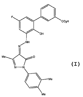
A preferred TpoRA useful in the method of the invention is 3% ( N'41-
(3,4-dimethyl pheny1)-3-methy1-5-oxo-1,5-dihydropyrazol-4-ylidene]hydrazine) -
5' -
fluoro-2' -hydroxybipheny1-3-carboxylic acid, hereafter known as Compound A.
Compound A is a compound which is disclosed (as Example 13) and claimed, along
with
pharmaceutically acceptable salts, hydrates, solvates and esters thereof, as
being useful as
an agonist of the TPO receptor, particularly in enhancing platelet production
and
particularly in the treatment of thrombocytopenia, in International
Application No.
PCT/US01/16863 (International Publication Number WO 01/89457; United States
Publication Number US2004/0019190 Al), and whose structure is as follows:
12
CA 02709224 2010-06-14
WO 2009/048953
PCT/US2008/079205
I.
F
101 CO,H
OH
N
I
Me 0
\
N--N
. Me
Me
Methods of Treatment
In one embodiment of the methods of the present invention, a TpoRA, a
derivative or a variant thereof, is administered to an individual diagnosed
with AML. In
one aspect of the invention, a TpoRA, derivative, or variant thereof is
administered to an
individual with AML as part of a chemotherapeutic regimen to augment the
efficacy of a
chemotherapeutic agent.
In another embodiment of the methods of the present invention, a TpoRA,
a derivative or a variant thereof, is administered to an individual diagnosed
with
myelodysplastic syndrome. In one aspect of the invention, a TpoRA, derivative,
or
variant thereof is administered to an individual with myelodysplastic syndrome
as part of
a chemotherapeutic regimen to augment the efficacy of a chemotherapeutic
agent.
By "augment the efficacy of a chemotherapeutic agent" it is meant that
administering a TpoRA, derivative, or variant thereof will bring about a
beneficial
clinical outcome including, but not limited to, increasing the survival of the
individual,
reducing clinical signs of AML or myelodysplastic syndrome in the individual,
or by
permitting a reduction in the dose of the chemotherapeutic agent or the
frequency of
administration of the chemotherapeutic agent, or both, thereby reducing the
undesirable
side effects associated with the toxicity of the chemotherapeutic agents and
making the
chemotherapeutic regimen more tolerable. A TpoRA, derivative, or variant
thereof may
be administered to an individual either before the administration of the
chemotherapeutic
agent, during the administration of the chemotherapeutic agent or after the
administration
of the chemotherapeutic agent, or some combination thereof, deemed to be
effective for
13
CA 02709224 2010-06-14
WO 2009/048953
PCT/US2008/079205
treatment of the individual. Establishing the optimal schedule for
administering a
TpoRA, a derivative, or variant thereof as part of a chemotherapeutic regimen
is well
within the skill of the art.
In another aspect of the invention, a TpoRA, a derivative, or variant
thereof is administered to the individual prior to the administration of
chemotherapy.
Without wishing to be bound by any theory, it will be appreciated by one
skilled in the art
that administering a TpoRA to the individual prior to the commencement of
chemotherapy would target leukemia cells resistant to chemotherapy and would
augment
the efficacy of a chemotherapeutic agent. In addition, all leukemia cells that
express a
TPO receptor are targets of a TpoRA, a derivative or variant thereof. It will
be apparent
to one skilled in the art that providing a TpoRA, derivative or variant
thereof to an
individual before commencing chemotherapy would make leukemia cells more
susceptible to chemotherapeutic agents, thereby making treatment more
efficacious.
In yet another aspect of the invention, a TpoRA, a derivative, or variant
thereof is administered to the individual after the individual has completed a
course of
chemotherapy. Without wishing to be bound by any theory, it will be
appreciated by one
skilled in the art that residual, undetected circulating leukemia cells
increase the risk of
relapse in AML patients after completion of chemotherapy. Administering a
TpoRA, a
derivative, or variant thereof to the individual who has completed a course of
chemotherapy targets residual leukemia cells while they are still inactive and
reduces the
risk of disease recurrence.
In still another aspect of the invention, a TpoRA, a derivative, or variant
thereof is administered to the individual in lieu of chemotherapy as the sole
method of
treating AML. It will be appreciated by one skilled in the art that in a
number of
individuals diagnosed with AML, the leukemia cells are refractory to
chemotherapy.
Treating these individuals with a TpoRA, a derivative, or variant thereof is
an alternative
therapy that bypasses the mechanisms that allow the leukemia cells to evade
chemotherapeutic agents.
In still another aspect of the invention, a TpoRA, a derivative, or variant
thereof is administered to the individual in lieu of chemotherapy as the sole
method of
treating AML.
14
CA 02709224 2010-06-14
WO 2009/048953
PCT/US2008/079205
The compositions and methods of the present invention can be used in
combination with other treatment regimens, including virostatic and virotoxic
agents,
antibiotic agents, antifungal agents, anti-inflammatory agents, pain-reduction
therapies,
as well as combination therapies, and the like.
The invention can also be used in combination with other treatment
modalities, such as chemotherapy, cryotherapy, hyperthermia, radiation
therapy, and the
like.
Therapies and Pharmaceutical Preparations
A TpoRA, a derivative, or a variant thereof can be administered to the
individual using any suitable route known in the art, including for example,
intravenous
treatment protocols. Administration can be either by rapid, direct injection
or over a
period of time as by slow infusion. Slow release formulation may also be used.
Furthermore, a TpoRA, a derivative, or variant thereof can be stably linked to
a polymer
such as polyethylene glycol to confer desirable properties such as solubility,
stability,
extended half-life and other pharmaceutically advantageous properties to the
TpoRA
(see, e.g. Burnham, 1994, AM. J. Hosp. Pharm. 51:210-8).
Phosphatase inhibitors and activators, and kinase inhibitors and activators,
can also be linked or conjugated to TpoRA to confer desirable properties such
as
solubility, stability, extended half-life and other pharmaceutically
advantageous
properties to the TpoRA.
The invention encompasses the use of pharmaceutical compositions to
practice the methods of the invention, the compositions comprising an
appropriate
therapeutic compound and a pharmaceutically-acceptable carrier.
As used herein, the term "pharmaceutically-acceptable carrier" means a
chemical composition with which a therapeutic compound may be combined and
which,
following the combination, can be used to administer the appropriate
therapeutic
compound to a mammal.
The pharmaceutical compositions useful for practicing the invention may
be administered to deliver a dose of between 1 ng/kg/day and 100 mg/kg/day.
The
precise dosage administered will vary depending upon any number of factors,
including
CA 02709224 2010-06-14
WO 2009/048953
PCT/US2008/079205
but not limited to, the type of animal and type of disease state being
treated, the age of the
animal and the route of administration. It is well within the skill of the art
to establish the
optimal dosage of a TpoRA, a derivative or variant thereof required for
maximal clinical
benefit.
Pharmaceutical compositions that are useful in the methods of the
invention may be administered systemically in intravenous formulations. In
addition to
the appropriate therapeutic compound, such pharmaceutical compositions may
contain
pharmaceutically-acceptable carriers and other ingredients known to enhance
and
facilitate drug administration.
The invention encompasses the preparation and use of pharmaceutical
compositions comprising a compound useful for treatment of AML disclosed
herein as an
active ingredient. Such a pharmaceutical composition may consist of the active
ingredient alone, in a form suitable for administration to a subject, or the
pharmaceutical
composition may comprise the active ingredient and one or more
pharmaceutically
acceptable carriers, one or more additional ingredients, or some combination
of these.
The active ingredient may be present in the pharmaceutical composition in the
form of a
physiologically acceptable ester or salt, such as in combination with a
physiologically
acceptable cation or anion, as is well known in the art.
As used herein, the term "physiologically acceptable" ester or salt means
an ester or salt form of the active ingredient which is compatible with any
other
ingredients of the pharmaceutical composition, which is not deleterious to the
subject to
which the composition is to be administered.
The formulations of the pharmaceutical compositions described herein
may be prepared by any method known or hereafter developed in the art of
pharmacology. In general, such preparatory methods include the step of
bringing the
active ingredient into association with a carrier or one or more other
accessory
ingredients, and then, if necessary or desirable, shaping or packaging the
product into a
desired single- or multi-dose unit.
Pharmaceutical compositions that are useful in the methods of the
invention may be prepared, packaged, or sold in formulations suitable for
parenteral,
intravenous or another route of administration.
16
CA 02709224 2010-06-14
WO 2009/048953
PCT/US2008/079205
A pharmaceutical composition of the invention may be prepared,
packaged, or sold in bulk, as a single unit dose, or as a plurality of single
unit doses. As
used herein, a "unit dose" is discrete amount of the pharmaceutical
composition
comprising a predetermined amount of the active ingredient. The amount of the
active
ingredient is generally equal to the dosage of the active ingredient which
would be
administered to a subject or a convenient fraction of such a dosage such as,
for example,
one-half or one-third of such a dosage.
The relative amounts of the active ingredient, the pharmaceutically
acceptable carrier, and any additional ingredients in a pharmaceutical
composition of the
invention will vary, depending upon the identity, size, and condition of the
subject treated
and further depending upon the route by which the composition is to be
administered. By
way of example, the composition may comprise between 0.1% and 100% (w/w)
active
ingredient.
In addition to the active ingredient, a pharmaceutical composition of the
invention may further comprise one or more additional pharmaceutically active
agents.
Controlled- or sustained-release formulations of a pharmaceutical
composition of the invention may be made using conventional technology.
As used herein, "parenteral administration" of a pharmaceutical
composition includes any route of administration characterized by physical
breaching of
a tissue of a subject and administration of the pharmaceutical composition
through the
breach in the tissue. Parenteral administration thus includes, but is not
limited to,
administration of a pharmaceutical composition by injection of the
composition, by
application of the composition through a surgical incision, by application of
the
composition through a tissue-penetrating non-surgical wound, and the like. In
particular,
parenteral administration is contemplated to include, but is not limited to,
intravenous,
subcutaneous, intraperitoneal, intramuscular, intrasternal injection, and
kidney dialytic
infusion techniques.
Formulations of a pharmaceutical composition suitable for parenteral
administration comprise the active ingredient combined with a pharmaceutically
acceptable carrier, such as sterile water or sterile isotonic saline. Such
formulations may
be prepared, packaged, or sold in a form suitable for bolus administration or
for
17
CA 02709224 2010-06-14
WO 2009/048953
PCT/US2008/079205
continuous administration. Injectable formulations may be prepared, packaged,
or sold in
unit dosage form, such as in ampules or in multi-dose containers containing a
preservative. Formulations for parenteral administration include, but are not
limited to,
suspensions, solutions, emulsions in oily or aqueous vehicles, pastes, and
implantable
sustained-release or biodegradable formulations. Such formulations may further
comprise one or more additional ingredients including, but not limited to,
suspending,
stabilizing, or dispersing agents. In one embodiment of a formulation for
parenteral
administration, the active ingredient is provided in dry (i.e. powder or
granular) form for
reconstitution with a suitable vehicle (e.g. sterile pyrogen-free water) prior
to parenteral
administration of the reconstituted composition.
The pharmaceutical compositions may be prepared, packaged, or sold in
the form of a sterile injectable aqueous or oily suspension or solution. This
suspension or
solution may be formulated according to the known art, and may comprise, in
addition to
the active ingredient, additional ingredients such as the dispersing agents,
wetting agents,
or suspending agents described herein. Such sterile injectable formulations
may be
prepared using a non-toxic parenterally-acceptable diluent or solvent, such as
water or
1,3-butane diol, for example. Other acceptable diluents and solvents include,
but are not
limited to, Ringer's solution, isotonic sodium chloride solution, and fixed
oils such as
synthetic mono- or di-glycerides. Other parentally-administrable formulations
which are
useful include those which comprise the active ingredient in microcrystalline
form, in a
liposomal preparation, or as a component of a biodegradable polymer systems.
Compositions for sustained release or implantation may comprise
pharmaceutically
acceptable polymeric or hydrophobic materials such as an emulsion, an ion
exchange
resin, a sparingly soluble polymer, or a sparingly soluble salt.
The compound may be administered to the individual as frequently as
several times daily, or it may be administered less frequently, such as once a
day, once a
week, once every two weeks, once a month, or even less frequently, such as
once every
several months or even once a year or less. The frequency of the dose will be
readily
apparent to the skilled artisan and will depend upon any number of factors,
such as, but
not limited to, the type and severity of the disease being treated, the type
and age of the
individual, etc.
18
CA 02709224 2010-06-14
WO 2009/048953
PCT/US2008/079205
EXPERIMENTAL EXAMPLES
The invention is further described in detail by reference to the following
experimental examples. These examples are provided for purposes of
illustration only,
and are not intended to be limiting unless otherwise specified. Thus, the
invention should
in no way be construed as being limited to the following examples, but rather,
should be
construed to encompass any and all variations which become evident as a result
of the
teaching provided herein.
The materials and methods employed in the experiments disclosed herein
are now described.
Compounds
Compound A was obtained from Glaxo SmithKline Pharmaceuticals
(Collegeville, PA). The compound was dissolved in 100% DMSO to prepare a 10mM
stock solution and then diluted in IMDM (Iscoves Modified Dulbecco's Medium,
Invitrogen; Carlsbad, CA) to obtain a 1 mM working solution. Full length
recombinant
human thrombopoietin (rhTP0) was obtained form R&D Systems (Minneapolis, MN)
and dissolved in IMDM medium to final concentration 5 ng/ml.
Cell culture
N2C-TPO cells were derived by culture of a megakaryblastic cell line in
rhTpo for 10 weeks and provided by Glaxo SmithKline Pharmaceuticals
(Collegeville,
PA). N2C-TPO cells were cultured in RPMI (Invitrogen, Carlsbad, CA) medium
supplemented with 10% Animal Serum Complex ¨ Fetalplex (Gemini Bio-Products,
West Sacramento, CA), 0.5% Penicillin/Streptomycin (Invitrogen; Carlsbad, CA)
and 20
ng/ml rhTPO.
Mo7e cells were cultured in DMEM (Dulbecco's Modified Eagle
Medium; Invitrogen, Carlsbad, CA) supplemented with 10% Fetalplex, 0.5%
Penicillin/Streptomycin, and GM-CSF lOng/m1 (R&D Systems, Minneapolis, MN).
Cells from both cell lines were starved for 24 hours before each experiment.
Human primary leukemia cells were obtained from Stem Cell and
Leukemia Core at the University of Pennsylvania. Cells were cultured in EGM-2
(Endothelial Cell Medium-2, Cambrex; East Rutherford, NJ). All cells were
cultured in
humidified incubator at 37 C and 5% CO2.
19
CA 02709224 2010-06-14
WO 2009/048953
PCT/US2008/079205
Megakaryocyte colony forming assay
Normal human progenitor cells were plated in fibrinogen clots at a density
of 5000 CD34+ cells per ml. Fibrinogen clots were prepared as follow: cells
were re-
suspended in IMDM medium supplemented with sodium bicarbonate (3 mg/ml); 3-
mercapto-1-2propanediol (0.002%); transferring (300 g/ml); CaC12(37 g/ml);
fatty
acid free, deionized BSA (10%); insulin (20 gimp; cholesterol (5.6 g/m1)
cytokines:
IL-3 (10 ng/ml), SCF (10 ng/ml) and rhTPO (100 ng/ml equal 2.8 M) or Compound
A
(5 M). Then clotting mix containing fibrinogen (0.2%) and thrombin (0.2
u/ml). Cells
were plated on 35mm culture dishes and cultured in humidified incubator at 37
C and 5%
CO2. After 10 days, colonies were fixed and stained for megakaryocyte marker
CD41a
with fluorescently labeled antibody. Megakaryocyte colonies were counted using
inverted fluorescent microscope.
Western blot analysis of kinases phosphorylation
Controls and cells stimulated with rhTPO or Compound A were washed
twice in PBS, then pelleted. The pellet was dissolved in triple-lysis buffer
comprised of
50 mM TRIS, 150 mM NaC1, 0.02% sodium azide, 0.1% sodium dodecyl sulphate
(SDS)
and 1% Igepal (Sigma, St. Louis, MO), and then incubated for 30 min on ice
with
vortexing every 10 min. The lysate was then spun at maximum speed in a
microcentrifuge at 4 C for 10 min. The extracted cell supernatant was used for
Western -
blot analysis.
Protein concentration was determined by a Bradford protein assay (Bio-
Rad, Hercules, CA). A total of 150 lig protein extract was resolved on a 10%
polyacrylamide gel (150V, 60 min.) then transferred to a poly vinylidene
fluoride
(PVDF) membrane (25V, 60 min). Condensed milk (5%) was used as a blocking
solution. The membrane was incubated overnight at 4 C with primary antibody at
a
1:1000 dilution. After incubation, the membrane was washed three times in TBS-
T
buffer and probed with secondary HRP-conjugated antibody (Amersham
Biosciences;
Piscataway, NJ) at dilution 1:1000 for 2 hour at room temperature. All
antibodies were
purchased from Cell Signaling Technology (Danvers, MA) .
CA 02709224 2010-06-14
WO 2009/048953
PCT/US2008/079205
Microarray analysis
N2C-TPO cells (1x10⁶ cells per 1 ml of RPMI medium supplemented with
10% FBS) were stimulated with 2.8 uM TPO or 5uM Compound A for 30 minutes, 1
and
3 hours. Then cells were washed twice in PBS and used to isolate RNA using
Qiagen's
RNaesy kit (Valencia, CA). Each condition was done in triplicate. RNA was
submitted to
Penn Microarray Facility (University of Pennsylvania; Philadelphia, PA).
Analysis was
performed using Affymetrix GeneChip U133A vs 2. Statistical data analysis was
performed in Penn Bioinformatics Core (University of Pennsylvania;
Philadelphia, PA)
using the GCRMA algorithm and Array Assistlite 3.4 program. Visualization of
gene
profile was done using Spotfire software.
Experimental Example #1: Cell culture evaluation of Compound A molecule in
comparison to recombinant human TPO
In order to determine if Compound A has the ability to function as a
thrombopoietin receptor (TpoR) agonist and stimulate proliferation and
differentiation of
human megakaryocytes, a number of cell culture experiments were performed
using the
Tpo dependent cell line (N2C-TPO) as well as normal human CD34+ cells. The
ability
of Compound A to stimulate proliferation of the TPO dependent cell line N2C-
TPO was
found to exhibit a dose dependent augmentation of cell proliferation between 1
and 10
uM, with maximal effects at 5-10 M. Consequently the 51LiM dose was used in
further
experiments where properties of rhTPO versus Compound A were directly
compared.
Normal human progenitor cells (CD34+) were used to evaluate capability
of Compound A to stimulate proliferation and differentiation of
megakaryocytes. CD34+
cells were plated in fibrin clots and cultured for 10 days, after which time
colonies were
fixed and stained with antibody specific for megakaryocytic marker CD41. The
number
of megakaryocyte colonies obtained from cells stimulated with Compound A was
slightly
lower when compare to the number of megakaryocyte colonies from cells
stimulated with
rhTPO, however there was no difference in size or shape of the colony (Figure
1). These
results were also confirmed by liquid culture of human progenitor cells. CD34+
cells
were incubated in cytokines with rhTPO or Compound A for periods of 7-10 days,
after
which time cells in culture were examined for degree of polyploidization, and
expression
21
CA 02709224 2010-06-14
WO 2009/048953
PCT/US2008/079205
of megakaryocyte lineage markers CD41 and CD61. 14% of cells grown in Compound
A showed greater than 4N DNA content compared to 8% in rhTPO. These same cells
expressed the megakaryocyte markers CD41 and CD6lon 35% of cells, comparable
to
the results with rhTPO.
Experimental Example #2: Differential effects of Compound A on primary
leukemia cells
Samples were obtained from 18 patients with acute myelogenous leukemia
(AML), 7 patients with acute lymphocytic leukemia (ALL) and 3 patients with
chronic
myelogenous leukemia (CML). In 17 out of 18 tested AML samples, Compound A
inhibited cell proliferation 70-90% when compared to untreated controls or
cells cultured
with rhTPO (Figure 2A). No significant effect of Compound A was observed in
primary
cells from ALL and CML patient samples (Figure 2B and C).
Further the effect of various doses of Compound A (1 M, 2.5 M and
5 M) on AML samples was examined. This experiment was performed on 6 different
samples. In 3 samples doses of 1 and 2.5 M Compound A had an effect on cell
proliferation. In samples 857 and 332 the inhibition of cell proliferation by
1 and 2.5 M
doses of Compound A was similar to the inhibition achieved with 5 M dose
(Figure 3).
In the remaining 3 samples, doses of 1 M and 2.5 M Compound A had no effect on
cell
proliferation and survival. In these cases, the only 5 M Compound A inhibited
leukemia
cell growth and proliferation. No significant effects on cell growth or
viability were
observed in the ALL or CML patient samples.
Experimental Example # 3: Comparison of signaling pathways stimulated by rhTPO
and
Compound A
To understand the mechanism by which Compound A triggers cell death
of AML cells, the intracellular signaling pathways stimulated by rhTPO and
Compound
A were compared. These studies were carried out on hematopoietic progenitor
cells
(CD34+) as well as a megakaryoblastic cell line, N2C-TPO, engineered to
express TpoR
and wherein cell proliferation is controlled by stimulation by Tpo.
The effect of Compound A on phosphorylation of kinases known to be
important in the Tpo signaling pathway was examined. Kinases evaluated include
STAT5, ERK, p7056, and ribosomal kinase S6. N2C-TPO cells stimulated with
rhTPO
for 10 or 30 minutes showed high phosphorylation level of all the kinases
listed above.
22
CA 02709224 2010-06-14
WO 2009/048953
PCT/US2008/079205
However, when cells were exposed to Compound A for 10 or 30 minutes, none of
those
kineses were activated (Figure 4A). The same experiment was performed on human
progenitor cells CD34+, which are stimulated by Compound A to differentiate
into the
megakaryocytes. In contrast to N2C-TPO cells, CD34+ cells showed activation of
ERK,
p70S6 and S6 ribosomal protein phosphorylation after being exposed to either
rhTPO
(2.8 M) or Compound A (5 M). However only cells exposed to rhTPO stimulated
phosphorylation of STAT5, a kinase, which is over-phosphorylated in AML cells
(Figure
4B).
Experimental Example #4: Can rhTPO stimulation block the effect of Compound A
on
AML cells
Proliferation assays were performed on cells obtained from 6 AML
patients. In some experiments, cells were first exposed to Compound A and then
stimulated with rhTPO five minutes later. To avoid the possibility of all
receptors being
occupied by Compound A and therefore preventing rhTPO from binding, other
experiments were also performed by first stimulating the cells with rhTPO
followed by
exposure to Compound A 5 minutes later. In 4 out of 6 samples (857, 774, 759,
342)
AML cell survival was not rescued either by adding rhTPO after stimulation
with
Compound A or by first stimulating cells with rhTPO prior to exposure to
Compound A.
However, in samples 794 and 332 adding rhTPO did attenuate the Compound A
effect
(Figure 5). These results suggest a number of possibilities: first, Compound A
may have
a much stronger affinity for the TpoR than rhTPO; second, that the KD rhTPO is
much
lower and consequently Compound A can displace rhTPO from the receptor; or,
third,
that Compound A stimulates other pathways that trigger cell death of AML
cells.
In an effort to address these possibilities, similar rescue experiments were
performed on N2C-TPO cells while evaluating phosphorylation of kinases
involved in
TPO pathway. In both sets of experiment (either adding rhTPO first and then
Compound
A, or adding Compound A first and then rhTPO), rescue of phosphorylation of
STAT5,
ERK p70S6 kinases and S6 ribosomal protein were observed, which were not
phosphorylated after stimulation with Compound A alone (Figure 6). These data
suggest
that Compound A activates another pathway which leads to death of AML cells.
Experimental Example #5: Microarray analysis of differences in gene expression
in cells
23
CA 02709224 2010-06-14
WO 2009/048953
PCT/US2008/079205
stimulated with TPO versus Compound A
To further understand the mechanism involved in Compound A signaling,
microarray analysis was performed on cells stimulated with Tpo or Compound A
using
Affymetrix GeneChips. In first set of experiments, the N2C-TPO cell line was
probed to
establish the optimal time point for further analysis using primary AML cells.
Cells were stimulated with either TPO (2.8 M) or Compound A (5 M)
for 30 minutes, 1 or 3 hours. RNA was then isolated from cells and used for
array
experiments. There were significant differences in gene expression after
stimulation with
Tpo vs Compound A. After 30 minute of stimulation, 200 genes were
differentially
expressed, after 1 hour of stimulation -400 genes were differentially
expressed and after
3 hours over 2000 genes were either up or down regulated in Compound A samples
as
copared to samples stimulated by TPO (Figure 7).
Among of these genes, differences in expression of number of genes
involved in apoptotic pathway were observed (Figure 8) as well as
transcription factors
(Figure 9). The most down regulated genes (50-200 times fold change) included
the
family of early growth factor response genes 1-4 (which encode proteins that
act as a
nuclear effectors of extracellular signals), a suppressor of cytokine
signaling 3, and
cytokine inducible SH2-containing protein involved in most cells' signaling
pathways.
Experimental Example 6: Apoptosis assay of isolated primary cells from human
cancer
patients
Annexin-V is a phospholipid binding protein with a high affinity for
phosphatidylserine (PS). Annexin V will not bind normal, intact cells, but
necrotic cells
are leaky enough to give Annexin V access to inner membrane PS. Propidium
iodide
stains DNA. The assay therefore identifies cells undergoing apoptosis.
Primary cells were isolated from either an AML (patient 774; top row) or
ALL (patient 710; bottom row) patient. The cells were then exposed to either
control
solution (left pair of panels), rhTpo + DMSO (middle pair of panels), or
5B559457 (right
pair of panels) for 72 hours. Figure 10 depicts an increase in Annexin V and
PI positive
cells in the AML samples exposed to 5B55945 for 72 hours (top, right panel) as
compared to control cells (top, left panel) and cells stimulated with Tpo +
DMSO (top,
middle panel). No significant increases in cellular apoptosis could be
observed in
24
CA 02709224 2014-05-16
primary cells isolated from the ALL patient. This suggests that SB559457
induces
apoptosis in AML cells.
Experimental Example 7: Molecular consequences of differential signaling in
AML cells
Affymetrix gene chip analysis was performed on 5 different primary AML
cell samples (AML patient 332, 342, 774, and 794). The primary cells were
stimulated
for 6 hours with either Tpo or SB559457. Statistically significant difference
in
expression was found in only 2 of 22,000 genes represented on the chips
(indicative of a
false discovery rate of less than 36%): glyceraldehyde-3-phosphate
dehydrogenase
(GAPDH) and DNA damage-inducible transcript 4 (also known as Redd 1). These
results
were confirmed using quantitative real time PCR (QRT-PCR). In primary AML
samples
the expression of GAPDH in cells treated with SB559457 was at least two times
higher
than in cells treated with rhTpo for the same time (6 hours). Similarly the
expression of
Reddl gene was ¨ 3 to 4 times higher in cells incubated with SB559457 than in
control
cells incubated with rhTpo (Figure 11).
In an effort to establish a correlation between cell signaling and array data,
phosphorylation of kinases involved in Tpo signaling pathway in primary
leukemia cells
was examined. Phosphorylation of ribosomal S6 and p70S6 kinases were compared
in
cells stimulated with SB559457 (5 M) and Tpo (2,8611M) for 1, 3 and 5 hours in
3
primary AML samples. In both AML samples, cells stimulated with SB559457 for 3
hours showed high phosphorylation of p70S6 kinase at Thr421/Ser424 and
ribosomal
kinase S6, while in unstimulated control cells and cells stimulated with Tpo,
none or very
little phosphorylation of those kinases was detected (Figure 12).
While this invention has been disclosed with reference to specific
embodiments, it is
apparent that other embodiments and variations of this invention may be
devised by
others skilled in the art The scope of the claims should not be limited by the
preferred
embodiments or the examples but should be given the broadest interpretation
consistent
with the description as a whole.
25