Note: Descriptions are shown in the official language in which they were submitted.
CA 02709430 2010-06-15
WO 2009/080253
PCT/EP2008/010704
Bivalent, bispecific antibodies
The present invention relates to novel bivalent, bispecific antibodies, their
manufacture and use.
Background of the Invention
Engineered proteins, such as bi- or multispecific antibodies capable of
binding two
or more antigens are known in the art. Such multispecific binding proteins can
be
generated using cell fusion, chemical conjugation, or recombinant DNA
techniques.
A wide variety of recombinant bispecific antibody formats have been developed
in
the recent past, e.g. tetravalent bispecific antibodies by fusion of, e.g. an
IgG
antibody format and single chain domains (see e.g. Coloma, M.J., et al, Nature
Biotech 15 (1997) 159-163; WO 2001077342; and Morrison, S.L. , Nature Biotech
25 (2007) 1233-1234).
Also several other new formats wherein the antibody core structure (IgA, IgD,
IgE,
IgG or IgM) is no longer retained such as dia-, tria- or tetrabodies,
minibodies,
several single chain formats (scFv, Bis-scFv), which are capable of binding
two or
more antigens, have been developed(Holliger, P., et al, Nature Biotech 23
(2005)
1126-1136; Fischer, N., Leger 0., Pathobiology 74 (2007) 3-14; Shen, J., et
al,
Journal of Immunological Methods 318 (2007) 65-74; Wu, C., et al, Nature
Biotech
(2007) 1290-1297).
20 All such formats use linkers either to fuse the antibody core (IgA, IgD,
IgE, IgG or
IgM) to a further binding protein (e.g.. scFv) or to fuse e.g. two Fab
fragments or
scFv.(Fischer N., Leger 0., Pathobiology 74 (2007) 3-14). While it is obvious
that
linkers have advantages for the engineering of bispecific antibodies, they may
also
cause problems in therapeutic settings. Indeed, these foreign peptides might
elicit
25 an immune response against the linker itself or the junction between the
protein
and the linker. Further more, the flexible nature of these peptides makes them
more
prone to proteolytic cleavage, potentially leading to poor antibody stability,
aggregation and increased immunogenicity. In addition one may want to retain
effector functions, such as e.g. complement-dependent cytotoxicity (CDC) or
antibody dependent cellular cytotoxicity (ADCC), which are mediated through
the
CA 02709430 2010-06-15
WO 2009/080253
PCT/EP2008/010704
- 2 -
Fc receptor binding , by maintaining a high degree of similarity to naturally
occurring.
Thus ideally, one should aim at developing bispecific antibodies that are very
similar in general structure to naturally occurring antibodies (like IgA, IgD,
IgE,
IgG or IgM) with minimal deviation from human sequences.
In one approach bispecific antibodies that are very similar to natural
antibodies
have been produced using the quadroma technology (see Milstein, C. and A.C.
Cuello, Nature, 305 (1983) 537-40) based on the somatic fusion of two
different
hybridoma cell lines expressing murine monoclonal antibodies with the desired
specificities of the bispecific antibody. Because of the random pairing of two
different antibody heavy and light chains within the resulting hybrid-
hybridoma
(or quadroma) cell line, up to ten different antibody species are generated of
which
only one is the desired, functional bispecific antibody. Due to the of
presence of
mispaired byproducts, and significantly reduced production yields, means
sophisticated purification procedures are required (see e.g. Morrison, S.L. ,
Nature
Biotech 25 (2007) 1233-1234). In general the same problem of mispaired
byproducts remains if recombinant expression techniques are used.
An approach to circumvent the problem of mispaired byproducts, which is known
as 'knobs-into-holes', aims at forcing the pairing of two different antibody
heavy
chains by introducing mutations into the CH3 domains to modify the contact
interface. On one chain bulky amino acids were replaced by amino acids with
short
side chains to create a 'hole'. Conversely, amino acids with large side chains
were
introduced into the other CH3 domain, to create a 'knob'. By coexpressing
these
two heavy chains (and two identical light chains, which have to be appropriate
for
both heavy chains), high yields of heterodimer formation ('knob-hole') versus
homodimer formation ('hole-hole' or `knob-knob') was observed (Ridgway, J.B.,
Presta LG, Carter P; and WO 1996027011). The percentage of heterodimer could
be
further increased by remodeling the interaction surfaces of the two CH3
domains
using a phage display approach and the introduction of a disulfide bridge to
stabilize the heterodimers (Merchant, A.M., et al, Nature Biotech 16 (1998)
677-
681; Atwell, S., Ridgway, J.B., Wells, J.A., Carter, P., J Mol Biol 270 (1997)
26-35).
New approaches for the knobs-into-holes technology are described in e.g. in
EP 1870459A1. Although this format appears very attractive, no data describing
progression towards the clinic are currently available. One important
constraint of
this strategy is that the light chains of the two parent antibodies have to be
identical
CA 02709430 2010-06-15
WO 2009/080253
PCT/EP2008/010704
- 3 -
to prevent mispairing and formation of inactive molecules. Thus this technique
is
not appropriate for easily developing recombinant, bivalent, bispecific
antibodies
against two antigens starting from two antibodies against the first and the
second
antigen, as either the heavy chains of these antibodies an/or the identical
light
chains have to be optimized.
Simon T. et al, EMBO Journal, 9 (1990) 1051 -1056 relates to domain mutants of
monospecific antibodies.
Summary of the Invention
The invention relates to a bivalent, bispecific antibody, comprising:
a) the light chain and heavy chain of an antibody specifically binding to a
first
antigen; and
b) the light chain and heavy chain of an antibody specifically binding to a
second antigen, wherein the constant domains CL and CH1 are replaced by
each other.
A further embodiment of the invention is a method for the preparation of an a
bivalent, bispecific antibody according to the invention
= comprising the steps of
a) transforming a host cell with
-vectors comprising nucleic acid molecules encoding the light chain and
heavy chain of an antibody specifically binding to a first antigen
-vectors comprising nucleic acid molecules encoding the light chain and
heavy chain of an antibody specifically binding to a second antigen,
wherein the constant domains CL and CH1 are replaced by each other;
b) culturing the host cell under conditions that allow synthesis of said
antibody
molecule; and
c) recovering said antibody molecule from said culture.
A further embodiment of the invention is a host cell comprising
CA 02709430 2010-06-15
WO 2009/080253
PCT/EP2008/010704
-4-
- vectors comprising nucleic acid molecules encoding the light chain and
heavy
chain of an antibody specifically binding to a first antigen
- vectors comprising nucleic acid molecules encoding the light chain and
heavy
chain of an antibody specifically binding to a second antigen, wherein the
constant domains CL and CH1 are replaced by each other.
A further embodiment of the invention is a composition, preferably a
pharmaceutical or a diagnostic composition of the antibody according to the
invention.
A further embodiment of the invention is a pharmaceutical composition
comprising an antibody according to the invention and at least one
pharmaceutically acceptable excipient.
A further embodiment of the invention is a method for the treatment of a
patient in
need of therapy, characterized by administering to the patient a
therapeutically
effective amount of an antibody according to the invention.
Detailed Description of the Invention
The invention relates to a bivalent, bispecific antibody, comprising:
a) the light chain and heavy chain of an antibody specifically binding to a
first
antigen; and
b) the light chain and heavy chain of an antibody specifically binding to a
second
antigen, wherein the constant domains CL and CH1 are replaced by each
other
Therefore said bivalent, bispecific antibody, comprises:
a) a first light chain and a first heavy chain of an antibody specifically
binding
to a first antigen; and
b) a second light chain and a second heavy chain of an antibody specifically
binding to a second antigen, wherein the constant domains CL and CH1 of
the second light chain and the second heavy chain are replaced by each
other.
CA 02709430 2010-06-15
WO 2009/080253 PCT/EP2008/010704
- 5 -
Thus for said antibody specifically binding to a second antigen the following
applies:
within the light chain
the constant light chain domain CL is replaced by the constant heavy chain
domain CH lof said antibody;
and within the heavy chain
the constant heavy chain domain CH1 is replaced by the constant light chain
domain CL of said antibody.
The term "antibody" as used herein refers to whole, monoclonal antibodies.
Such
whole antibodies consist of two pairs of a "light chain" (LC) and a "heavy
chain"
(HC) (such light chain (LC) /heavy chain pairs are abbreviated herein as
LC/HC).
The light chains and heavy chains of such antibodies are polypeptides
consisting of
several domains. In a whole antibody, each heavy chain comprises a heavy chain
variable region (abbreviated herein as HCVR or VH) and a heavy chain constant
region. The heavy chain constant region comprises the heavy chain constant
domains CH1, CH2 and CH3 (antibody classes IgA, IgD, and IgG) and optionally
the heavy chain constant domain CH4 (antibody classes IgE and IgM). Each light
chain comprises a light chain variable domain VL and a light chain constant
domain CL. The structure of one naturally occurring whole antibody, the IgG
antibody, is shown e.g. in Fig.'. The variable domains VI-I and VL can be
further
subdivided into regions of hypervariability, termed complementarity
determining
regions (CDR), interspersed with regions that are more conserved, termed
framework regions (FR). Each VH and VL is composed of three CDRs and four
FRs, arranged from amino-terminus to carboxy-terminus in the following order:
FR1, CDR1, FR2, CDR2, FR3, CDR3, FR4 ((Janeway, C.A., Jr, et al, (2001).
Immunobiology., 5th ed., Garland Publishing; and Woof, J., Burton, D., Nat Rev
Immunol 4 (2004) 89-99). The two pairs of heavy chain and light chain (HC/LC)
are capable of specifically binding to same antigen. Thus said whole antibody
is a
bivalent, monospecific antibody. Such "antibodies" include e.g. mouse
antibodies,
human antibodies, chimeric antibodies, humanized antibodies and genetically
engineered antibodies (variant or mutant antibodies) as long as their
characteristic
properties are retained. Especially preferred are human or humanized
antibodies,
especially as recombinant human or humanized antibodies.
There are five types of mammalian antibody heavy chains denoted by the Greek
letters: a, (5, E, y, and (Janeway, C.A., Jr, et al, (2001). Immunobiology.,
5th ed.,
CA 02709430 2010-06-15
WO 2009/080253
PCT/EP2008/010704
- 6 -
Garland Publishing). The type of heavy chain present defines the class of
antibody;
these chains are found in IgA, IgD, IgE, IgG, and IgM antibodies, respectively
(Rhoades RA, Pflanzer RG (2002). Human Physiology, 4th ed., Thomson
Learning). Distinct heavy chains differ in size and composition; a and 7
contain
approximately 450 amino acids, while A and 6 have approximately 550 amino
acids.
Each heavy chain has two regions, the constant region and the variable region.
The
constant region is identical in all antibodies of the same isotype, but
differs in
antibodies of different isotype. Heavy chains 7, a and (5 have a constant
region
composed of three constant domains CH1, CH2, and CH3 (in a line) , and a hinge
region for added flexibility (Woof, J., Burton, D., Nat Rev Immunol 4 (2004)
89-
99); heavy chains it and 6 have a constant region composed of four constant
domains CH1, CH2, CH3, and CH4 (Janeway, C.A., Jr. et al. (2001).
Immunobiology., 5th ed., Garland Publishing). The variable region of the heavy
chain differs in antibodies produced by different B cells, but is the same for
all
antibodies produced by a single B cell or B cell clone. The variable region of
each
heavy chain is approximately 110 amino acids long and is composed of a single
antibody domain.
In mammals there are only two types of light chain, which are called lambda
(k)
and kappa (x). A light chain has two successive domains: one constant domain
CL
and one variable domain VL. The approximate length of a light chain is 211 to
217
amino acids. Preferably the light chain is a kappa (lc) light chain, and the
constant
domain CL is preferably C kappa (x).
The terms "monoclonal antibody" or "monoclonal antibody composition" as used
herein refer to a preparation of antibody molecules of a single amino acid
composition.
The "antibodies" according to the invention can be of any class (e.g. IgA,
IgD, IgE,
IgG, and IgM, preferably IgG or IgE), or subclass (e.g., IgGl, IgG2, IgG3,
IgG4,
IgAl and IgA2, preferably IgG1), whereby both antibodies, from which the
bivalent
bispecific antibody according to the invention is derived, have an Fc part of
the
same subclass( e.g. IgGl, IgG4 and the like, preferably IgG1), preferably of
the same
allotype (e.g. Caucasian).
A "Fc part of an antibody" is a term well known to the skilled artisan and
defined
on the basis of papain cleavage of antibodies. The antibodies according to the
CA 02709430 2010-06-15
WO 2009/080253 PCT/EP2008/010704
- 7 -
invention contain as Fc part, preferably a Fc part derived from human origin
and
preferably all other parts of the human constant regions. The Fc part of an
antibody
is directly involved in complement activation, Clq binding, C3 activation and
Fc
receptor binding. While the influence of an antibody on the complement system
is
dependent on certain conditions, binding to Clq is caused by defined binding
sites
in the Fc part. Such binding sites are known in the state of the art and
described e.g.
by Lukas, T.J., et al., J. Immunol. 127 (1981) 2555-2560; Brunhouse, R., and
Cebra,
J.J., Mol. Immunol. 16 (1979) 907-917; Burton, D.R., et al., Nature 288 (1980)
338-
344; Thommesen, J.E., et al., Mol. Immunol. 37 (2000) 995-1004; Idusogie,
E.E., et
al., J. Immunol. 164 (2000) 4178-4184; Hezareh, M., et al., J. Virol. 75
(2001)
12161-12168; Morgan, A., et al., Immunology 86 (1995) 319-324; and EP 0 307
434.
Such binding sites are e.g. L234, L235, D270, N297, E318, K320, K322, P331 and
P329 (numbering according to EU index of Kabat, see below). Antibodies of
subclass IgG 1, IgG2 and IgG3 usually show complement activation, Clq binding
and C3 activation, whereas IgG4 do not activate the complement system, do not
bind Clq and do not activate C3. Preferably the Fc part is a human Fc part.
The term "chimeric antibody" refers to an antibody comprising a variable
region,
i.e., binding region, from one source or species and at least a portion of a
constant
region derived from a different source or species, usually prepared by
recombinant
DNA techniques. Chimeric antibodies comprising a murine variable region and a
human constant region are preferred. Other preferred forms of "chimeric
antibodies" encompassed by the present invention are those in which the
constant
region has been modified or changed from that of the original antibody to
generate
the properties according to the invention, especially in regard to Clq binding
and/or Fc receptor (FcR) binding. Such chimeric antibodies are also referred
to as
"class-switched antibodies.". Chimeric antibodies are the product of expressed
immunoglobulin genes comprising DNA segments encoding immunoglobulin
variable regions and DNA segments encoding immunoglobulin constant regions.
Methods for producing chimeric antibodies involve conventional recombinant
DNA and gene transfection techniques are well known in the art. See, e.g.,
Morrison, S.L., et al., Proc. Natl. Acad. Sci. USA 81(1984) 6851-6855; US
5,202,238
and US 5,204,244.
The term "humanized antibody" refers to antibodies in which the framework or
"complementarity determining regions" (CDR) have been modified to comprise the
CDR of an immunoglobulin of different specificity as compared to that of the
CA 02709430 2010-06-15
WO 2009/080253
PCT/EP2008/010704
- 8 -
parent immunoglobulin. In a preferred embodiment, a murine CDR is grafted into
the framework region of a human antibody to prepare the "humanized antibody."
See, e.g., Riechmann, L., et al., Nature 332 (1988) 323-327; and Neuberger,
M.S., et
al., Nature 314 (1985) 268-270. Particularly preferred CDRs correspond to
those
representing sequences recognizing the antigens noted above for chimeric
antibodies. Other forms of "humanized antibodies" encompassed by the present
invention are those in which the constant region has been additionally
modified or
changed from that of the original antibody to generate the properties
according to
the invention, especially in regard to Clq binding and/or Fc receptor (FcR)
binding.
The term "human antibody", as used herein, is intended to include antibodies
having variable and constant regions derived from human germ line
immunoglobulin sequences. Human antibodies are well-known in the state of the
art (van Dijk, M.A., and van de Winkel, J.G., Curr. Opin. Chem. Biol. 5 (2001)
368-
374). Human antibodies can also be produced in transgenic animals (e.g., mice)
that are capable, upon immunization, of producing a full repertoire or a
selection of
human antibodies in the absence of endogenous immunoglobulin production.
Transfer of the human germ-line immunoglobulin gene array in such germ-line
mutant mice will result in the production of human antibodies upon antigen
challenge (see, e.g., Jakobovits, A., et al., Proc. Natl. Acad. Sci. USA 90
(1993) 2551-
2555; Jakobovits, A., et al., Nature 362 (1993) 255-258; Bruggemann, M., et
al., Year
Immunol. 7 (1993) 33-40). Human antibodies can also be produced in phage
display libraries (Hoogenboom, H.R., and Winter, G., J. Mol. Biol. 227 (1992)
381-
388; Marks, J.D., et al., J. Mol. Biol. 222 (1991) 581-597). The techniques of
Cole et
al. and Boerner et al. are also available for the preparation of human
monoclonal
antibodies (Cole et al., Monoclonal Antibodies and Cancer Therapy, Alan R.
Liss, p.
77 (1985); and Boerner, P., et al., J. Immunol. 147 (1991) 86-95). As already
mentioned for chimeric and humanized antibodies according to the invention the
term "human antibody" as used herein also comprises such antibodies which are
modified in the constant region to generate the properties according to the
invention, especially in regard to Clq binding and/or FcR binding, e.g. by
"class
switching" i.e. change or mutation of Fc parts (e.g. from IgG1 to IgG4 and/or
IgGl/IgG4 mutation.)
The term "recombinant human antibody", as used herein, is intended to include
all
human antibodies that are prepared, expressed, created or isolated by
recombinant
CA 02709430 2010-06-15
WO 2009/080253
PCT/EP2008/010704
- 9 -
means, such as antibodies isolated from a host cell such as a NSO or CHO cell
or
from an animal (e.g. a mouse) that is transgenic for human immunoglobulin
genes
or antibodies expressed using a recombinant expression vector transfected into
a
host cell. Such recombinant human antibodies have variable and constant
regions
in a rearranged form. The recombinant human antibodies according to the
invention have been subjected to in vivo somatic hypermutation. Thus, the
amino
acid sequences of the VH and VL regions of the recombinant antibodies are
sequences that, while derived from and related to human germ line VH and VL
sequences, may not naturally exist within the human antibody germ line
repertoire
in vivo.
The "variable domain" (variable domain of a light chain (VL), variable region
of a
heavy chain (VH)) as used herein denotes each of the pair of light and heavy
chains
which is involved directly in binding the antibody to the antigen. The domains
of
variable human light and heavy chains have the same general structure and each
domain comprises four framework (FR) regions whose sequences are widely
conserved, connected by three "hypervariable regions" (or complementarity
determining regions, CDRs). The framework regions adopt a 13-sheet
conformation
and the CDRs may form loops connecting the 13-sheet structure. The CDRs in
each
chain are held in their three-dimensional structure by the framework regions
and
form together with the CDRs from the other chain the antigen binding site. The
antibody heavy and light chain CDR3 regions play a particularly important role
in
the binding specificity/affinity of the antibodies according to the invention
and
therefore provide a further object of the invention.
The terms "hypervariable region" or "antigen-binding portion of an antibody"
when
used herein refer to the amino acid residues of an antibody which are
responsible
for antigen-binding. The hypervariable region comprises amino acid residues
from
the "complementarity determining regions" or "CDRs". "Framework" or "FR"
regions are those variable domain regions other than the hypervariable region
residues as herein defined. Therefore, the light and heavy chains of an
antibody
comprise from N- to C-terminus the domains FR1, CDR1, FR2, CDR2, FR3, CDR3,
and FR4. CDRs on each chain are separated by such framework amino acids.
Especially, CDR3 of the heavy chain is the region which contributes most to
antigen
binding. CDR and FR regions are determined according to the standard
definition
of Kabat et al., Sequences of Proteins of Immunological Interest, 5th ed.,
Public
Health Service, National Institutes of Health, Bethesda, MD (1991).
CA 02709430 2010-06-15
WO 2009/080253
PCT/EP2008/010704
- 10 -
The "constant domains" of the heavy chain and of the light chain are not
involved
directly in binding of an antibody to an antigen, but exhibit various effector
functions. Depending on the amino acid sequence of the constant region of
their
heavy chains, antibodies or immunoglobulins are divided into the classes:
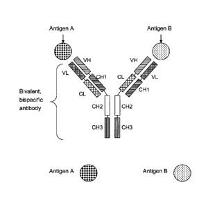
The term "bivalent, bispecific antibody" as used herein refers to an antibody
as
described above in which each of the two pairs of heavy chain and light chain
(HC/LC) is specifically binding to a different antigen, i.e. the first heavy
and the
first light chain (originating from an antibody against a first antigen) are
specifically
binding together to a first antigen, and , the second heavy and the second
light
chain (originating from an antibody against a second antigen ) are
specifically
binding together to a second antigen (as depicted in Fig. 2); such bivalent,
bispecific
antibodies are capable of specifically binding to two different antigens at
the same
time, and not to more than two antigens, in contrary to, on the one hand a
monospecific antibody capable of binding only to one antigen, and on the other
hand e.g. a tetravalent, tetraspecific antibody which can bind to four antigen
molecules at the same time.
According to the invention, the ratio of a desired bivalent, bispecific
antibody
compared to undesired side products can be improved by the replacement of
certain domains in only one pair of heavy chain and light chain (HC/LC). While
the
first of the two HC/LC pairs originates from an antibody specifically binding
to a
first antigen and is left essentially unchanged, the second of the two HC/LC
pairs
originates from an antibody specifically binding to a second antigen , and is
altered
by the following replacement:
- light chain: replacement of the constant light chain domain CL by the
constant heavy chain domain CH1 of said antibody specifically binding to a
second antigen , and
- heavy chain: replacement of the constant heavy chain domain CH1 by the
constant light chain domain CL of said antibody specifically binding to a
second antigen.
Thus the resulting bivalent, bispecific antibodies are artificial antibodies
which
comprise
a) the light chain and heavy chain of an antibody specifically binding to a
first
antigen; and
CA 02709430 2010-06-15
WO 2009/080253
PCT/EP2008/010704
- 11 -
b) the light chain and heavy chain of an antibody specifically binding to a
second
antigen;
wherein said light chain (of an antibody specifically binding to a second
antigen) contains a constant domain CH1 instead of CL
wherein said heavy chain(of an antibody specifically binding to a
second antigen) a constant domain CL instead of CH1.
In an additional aspect of the invention such improved ratio of a desired
bivalent,
bispecific antibody compared to undesired side products can be further
improved
by one of the following two alternatives:
A) First alternative (see Fig. 3):
The CH3 domains of said bivalent, bispecific antibody according to the
invention
can be altered by the "knob-into-holes" technology which described with in
detail
with several examples in e.g. WO 96/027011, Ridgway, J.B., et al, Protein Eng
9
(1996) 617-621; and Merchant, A.M., et al, Nat Biotechnol 16 (1998) 677-681.
In
this method the interaction surfaces of the two CH3 domains are altered to
increase
the heterodimerisation of both heavy chains containing these two CH3 domains.
Each of the two CH3 domains (of the two heavy chains) can be the "knob", while
the other is the "hole". The introduction of a disulfide bridge stabilizes the
heterodimers (Merchant, A..M., et al, Nature Biotech 16 (1998) 677-681;
Atwell, S.,
Ridgway, J.B., Wells, J.A., Carter, P., J Mol Biol 270 (1997) 26-35) and
increases the
yield.
Therefore in preferred embodiment the CH3 domains of a bivalent, bispecific
antibody wherein the first CH3 domain and second CH3 domain each meet at an
interface which comprises an original interface between the antibody CH3
domains
are altered by the "knob-into-holes" technology including further
stabilization by
introduction of a disulfide bridge in the CH3 domains (described in
WO 96/027011, Ridgway, J.B., et al, Protein Eng 9 (1996) 617-621; Merchant.
A.M,
et al., Nature Biotech 16 (1998) 677-681; and Atwell, S., Ridgway, J.B.,
Wells, J.A.,
Carter P., J Mol Biol 270 (1997) 26-35) to promote the formation of the
bivalent,
bispecific antibody.
Thus in one aspect of the invention said bivalent, bispecific antibody is
characterized in that
CA 02709430 2010-06-15
WO 2009/080253 PCT/EP2008/010704
- 12 -
the CH3 domain of one heavy chain and the CH3 domain of the other heavy chain
each meet at an interface which comprises an original interface between the
antibody CH3 domains;
wherein said interface is altered to promote the formation of the bivalent,
bispecific
antibody, wherein the alteration is characterized in that:
a) the CH3 domain of one heavy chain is altered,
so that within the original interface the CH3 domain of one heavy chain that
meets
the original interface of the CH3 domain of the other heavy chain within the
bivalent, bispecific antibody,
an amino acid residue is replaced with an amino acid residue having a larger
side
chain volume, thereby generating a protuberance within the interface of the
CH3
domain of one heavy chain which is positionable in a cavity within the
interface of
the CH3 domain of the other heavy chain
and
b) the CH3 domain of the other heavy chain is altered,
so that within the original interface of the second CH3 domain that meets the
original interface of the first CH3 domain within the bivalent, bispecific
antibody
an amino acid residue is replaced with an amino acid residue having a smaller
side
chain volume, thereby generating a cavity within the interface of the second
CH3
domain within which a protuberance within the interface of the first CH3
domain
is positionable.
Preferably said amino acid residue having a larger side chain volume is
selected
from the group consisting of arginine (R), phenylalanine (F), tyrosine (Y),
tryptophan (W).
Preferably said amino acid residue having a smaller side chain volume is
selected
from the group consisting of alanine (A), serine (S), threonine (T), valine
(V).
In one aspect of the invention both CH3 domains are further altered the
introduction of cysteine (C) as amino acid in the corresponding positions of
each
CH3 domain such that a disulfide bridge between both CH3 domains can be
formed.
CA 02709430 2010-06-15
WO 2009/080253 PCT/EP2008/010704
- 13 -
In another preferred embodiment of the invention both CH3 domains are altered
by the use of residues R409D; K370E (K409D) for knobs residues and D399K;
E357K for hole residues described eg. in EP 1870459A1.
or
B) Second alternative (see Figure 4):
by the replacement of one constant heavy chain domain CH3 by a constant heavy
chain domain CH1; and the other constant heavy chain domain CH3is replaced by
a constant light chain domain CL. The constant heavy chain domain CH1 by which
the heavy chain domain CH3 is replaced can be of any Ig class (e.g. IgA, IgD,
IgE,
IgG, and IgM), or subclass (e.g., IgG 1, IgG2, IgG3, IgG4, IgAl and IgA2).
The constant light chain domain CL by which the heavy chain domain CH3 is
replaced can be of the lambda (k) or kappa (lc) type, preferably the kappa
(lc) type.
Thus one preferred embodiment of the invention is a bivalent, bispecific
antibody,
comprising:
a) the light chain and heavy chain of an antibody specifically binding to a
first
antigen; and
b) the light chain and heavy chain of an antibody specifically binding to a
second
antigen, wherein the constant domains CL and CH1 are replaced by each
other,
and wherein optionally
c) the CH3 domain of one heavy chain and the CH3 domain of the other
heavy chain each meet at an interface which comprises an original
interface between the antibody CH3 domains;
wherein said interface is altered to promote the formation of the bivalent,
bispecific antibody, wherein the alteration is characterized in that:
ca) the CH3 domain of one heavy chain is altered,
CA 02709430 2010-06-15
WO 2009/080253
PCT/EP2008/010704
- 14 -
so that within the original interface the CH3 domain of one heavy chain
that meets the original interface of the CH3 domain of the other heavy
chain within the bivalent, bispecific antibody,
an amino acid residue is replaced with an amino acid residue having a
larger side chain volume, thereby generating a protuberance within the
interface of the CH3 domain of one heavy chain which is positionable in a
cavity within the interface of the CH3 domain of the other heavy chain
and
cb) the CH3 domain of the other heavy chain is altered,
so that within the original interface of the second CH3 domain that meets
the original interface of the first CH3 domain within the bivalent,
bispecific antibody
an amino acid residue is replaced with an amino acid residue having a
smaller side chain volume, thereby generating a cavity within the interface
of the second CH3 domain within which a protuberance within the
interface of the first CH3 domain is positionable;
or d)
one constant heavy chain domain CH3 is replaced by a constant heavy
chain domain CH1; and the other constant heavy chain domain CH3 is
replaced by a constant light chain domain CL
The terms "antigen" or "antigen molecule" as used herein are used
interchangeable
and refer to all molecules that can be specifically bound by an antibody. The
bivalent, bispecific antibody is specifically binding to a first antigen and a
second
distinct antigen. The term "antigens" as used herein include e.g. proteins,
different
epitopes on proteins (as different antigens within the meaning of the
invention),
and polysaccharides. This mainly includes parts (coats, capsules, cell walls,
flagella,
fimbrae, and toxins) of bacteria, viruses, and other microorganisms. Lipids
and
nucleic acids are antigenic only when combined with proteins and
polysaccharides.
Non-microbial exogenous (non-self) antigens can include pollen, egg white, and
proteins from transplanted tissues and organs or on the surface of transfused
blood
cells. Preferably the antigen is selected from the group consisting of
cytokines, cell
CA 02709430 2010-06-15
WO 2009/080253 PCT/EP2008/010704
- 15 -
surface proteins, enzymes and receptors cytokines, cell surface proteins,
enzymes
and receptors.
Tumor antigens are those antigens that are presented by MHC I or MHC II
molecules on the surface of tumor cells. These antigens can sometimes be
presented
by tumor cells and never by the normal ones. In this case, they are called
tumor-
specific antigens (TSAs) and typically result from a tumor specific mutation.
More
common are antigens that are presented by tumor cells and normal cells, and
they
are called tumor-associated antigens (TAAs). Cytotoxic T lymphocytes that
recognized these antigens may be able to destroy the tumor cells before they
proliferate or metastasize. Tumor antigens can also be on the surface of the
tumor
in the form of, for example, a mutated receptor, in which case they will be
recognized by B cells.
In one preferred embodiment at least one of the two different antigens (first
and
second antigen), to which the bivalent, bispecific antibody specifically binds
to, is a
tumor antigen.
In another preferred embodiment both of the two different antigens (first and
second antigen), to which the bivalent, bispecific antibody specifically binds
to, are
tumor antigens; in this case the first and second antigen can also be two
different
epitopes at the same tumor specific protein.
In another preferred embodiment one of the two different antigens (first and
second antigen), to which the bivalent, bispecific antibody specifically binds
to, is a
tumor antigen and the other is an effector cell antigen, as e.g. a T-Cell
receptor,
CD3, CD16 and the like.
In another preferred embodiment one of the two different antigens (first and
second antigen), to which the bivalent, bispecific antibody specifically binds
to, is a
tumor antigen and the other is an anti-cancer substance such as a toxin or a
kinase
inhibitor.
As used herein, "specifically binding" or "binds specifically to" refers to an
antibody
specifically binding an antigen. Preferably the binding affinity of the
antibody
specifically binding this antigen is of KD-value of 10-9 mo1/1 or lower (e.g.
100
mo1/1), preferably with a KD-value of 10-10 mo1/1 or lower (e.g. 10-12 mo1/1).
The
binding affinity is determined with a standard binding assay, such as surface
plasmon resonance technique (Biacore'').
CA 02709430 2010-06-15
WO 2009/080253
PCT/EP2008/010704
- 16 -
The term "epitope" includes any polypeptide determinant capable of specific
binding to an antibody. In certain embodiments, epitope determinant include
chemically active surface groupings of molecules such as amino acids, sugar
side
chains, phosphoryl, or sulfonyl, and, in certain embodiments, may have
specific
three dimensional structural characteristics, and or specific charge
characteristics.
An epitope is a region of an antigen that is bound by an antibody. In certain
embodiments, an antibody is said to specifically bind an antigen when it
preferentially recognizes its target antigen in a complex mixture of proteins
and/or
macromolecules.
An further embodiment of the invention is a method for the preparation of a
bivalent, bispecific antibody according to the invention
comprising
a) transforming a host cell with
-vectors comprising nucleic acid molecules encoding the light chain and
heavy chain of an antibody specifically binding to a first antigen
-vectors comprising nucleic acid molecules encoding the light chain and
heavy chain of an antibody specifically binding to a second antigen,
wherein the constant domains CL and CH1 are replaced by each other;
b) culturing the host cell under conditions that allow synthesis of said
antibody
molecule; and
c) recovering said antibody molecule from said culture.
In general there are two vectors encoding the light chain and heavy chain of
said
antibody specifically binding to a first antigen, and further two vectors
encoding the
light chain and heavy chain of said antibody specifically binding to a second
antigen. One of the two vectors is encoding the respective light chain and the
other
of the two vectors is encoding the respective heavy chain. However in an
alternative
method for the preparation of a bivalent, bispecific antibody according to the
invention, only one first vector encoding the light chain and heavy chain of
the
antibody specifically binding to a first antigen and only one second vector
encoding
the light chain and heavy chain of the antibody specifically binding to a
second
antigen can be used for transforming the host cell.
CA 02709430 2010-06-15
WO 2009/080253
PCT/EP2008/010704
- 17 -
The invention encompasses a method for the preparation of the antibodies
comprising culturing the corresponding host cells under conditions that allow
synthesis of said antibody molecules and recovering said antibodies from said
culture, e.g. by expressing
-a first nucleic acid sequence encoding the light chain of an antibody
specifically
binding to a first antigen;
-a second nucleic acid sequence encoding the heavy chain of said antibody
specifically binding to a first antigen;
-a third nucleic acid sequence encoding the light chain of an antibody
specifically
binding to a second antigen, wherein the constant light chain domain CL is
replaced by the constant heavy chain domain CH1; and
-a fourth nucleic acid sequence encoding the heavy chain of said antibody
specifically binding to a second antigen, wherein constant heavy chain domain
CH1
by the constant light chain domain CL.
A further embodiment of the invention is a host cell comprising
- vectors comprising nucleic acid molecules encoding the light chain and
heavy
chain of an antibody specifically binding to a first antigen
- vectors comprising nucleic acid molecules encoding the light chain and
heavy
chain of an antibody specifically binding to a second antigen, wherein the
constant
domains CL and CH1 are replaced by each other.
A further embodiment of the invention is a host cell comprising
a) a vector comprising a nucleic acid molecule encoding the light chain and a
vector
comprising a nucleic acid molecule encoding the heavy chain, of an antibody
specifically binding to a first antigen
b) a vector comprising a nucleic acid molecule encoding the light chain and a
vector comprising a nucleic acid molecule encoding the heavy chain, of an
antibody
specifically binding to a second antigen, wherein the constant domains CL and
CH1
are replaced by each other.
CA 02709430 2010-06-15
WO 2009/080253 PCT/EP2008/010704
- 18 -
A further embodiment of the invention is a composition, preferably a
pharmaceutical or a diagnostic composition of the bivalent, bispecific
antibody
according to the invention.
A further embodiment of the invention is a pharmaceutical composition
comprising a bivalent, bispecific antibody according to the invention and at
least
one pharmaceutically acceptable excipient.
A further embodiment of the invention is a method for the treatment of a
patient in
need of therapy, characterized by administering to the patient a
therapeutically
effective amount of a bivalent, bispecific antibody according to the
invention.
The term "nucleic acid or nucleic acid molecule", as used herein, is intended
to
include DNA molecules and RNA molecules. A nucleic acid molecule may be
single-stranded or double-stranded, but preferably is double-stranded DNA.
As used herein, the expressions "cell," "cell line," and "cell culture" are
used
interchangeably and all such designations include progeny. Thus, the words
"transformants" and "transformed cells" include the primary subject cell and
cultures derived therefrom without regard for the number of transfers. It is
also
understood that all progeny may not be precisely identical in DNA content, due
to
deliberate or inadvertent mutations. Variant progeny that have the same
function
or biological activity as screened for in the originally transformed cell are
included.
Where distinct designations are intended, it will be clear from the context.
The term "transformation" as used herein refers to process of transfer of a
vectors/nucleic acid into a host cell. If cells without formidable cell wall
barriers are
used as host cells, transfection is carried out e.g. by the calcium phosphate
precipitation method as described by Graham and Van der Eh, Virology 52 (1978)
546ff. However, other methods for introducing DNA into cells such as by
nuclear
injection or by protoplast fusion may also be used. If prokaryotic cells or
cells which
contain substantial cell wall constructions are used, e.g. one method of
transfection
is calcium treatment using calcium chloride as described by Cohen, F. N, et
al,
PNAS. 69 (1972) 7110ff.
Recombinant production of antibodies using transformation is well-known in the
state of the art and described, for example, in the review articles of
Makrides, S.C.,
Protein Expr. Purif. 17 (1999) 183-202; Geisse, S., et al., Protein Expr.
Purif. 8
(1996) 271-282; Kaufman, R.J., Mol. Biotechnol. 16 (2000) 151-161; Werner,
R.G.,
CA 02709430 2010-06-15
WO 2009/080253 PCT/EP2008/010704
- 19 -
et al., Arzneimittelforschung 48 (1998) 870-880 as well as in US 6,331,415 and
US 4,816,567.
As used herein, "expression" refers to the process by which a nucleic acid is
transcribed into mRNA and/or to the process by which the transcribed mRNA
(also
referred to as transcript) is subsequently being translated into peptides,
polypeptides, or proteins. The transcripts and the encoded polypeptides are
collectively referred to as gene product. If the polynucleotide is derived
from
genomic DNA, expression in a eukaryotic cell may include splicing of the mRNA.
A "vector" is a nucleic acid molecule, in particular self-replicating, which
transfers
an inserted nucleic acid molecule into and/or between host cells. The term
includes
vectors that function primarily for insertion of DNA or RNA into a cell (e.g.,
chromosomal integration), replication of vectors that function primarily for
the
replication of DNA or RNA, and expression vectors that function for
transcription
and/or translation of the DNA or RNA. Also included are vectors that provide
more
than one of the functions as described.
An "expression vector" is a polynucleotide which, when introduced into an
appropriate host cell, can be transcribed and translated into a polypeptide.
An
µ`expression system" usually refers to a suitable host cell comprised of an
expression
vector that can function to yield a desired expression product.
The bivalent, bispecific antibodies according to the invention are preferably
produced by recombinant means. Such methods are widely known in the state of
the art and comprise protein expression in prokaryotic and eukaryotic cells
with
subsequent isolation of the antibody polypeptide and usually purification to a
pharmaceutically acceptable purity. For the protein expression, nucleic acids
encoding light and heavy chains or fragments thereof are inserted into
expression
vectors by standard methods. Expression is performed in appropriate
prokaryotic
or eukaryotic host cells like CHO cells, NSO cells, SP2/0 cells, HEK293 cells,
COS
cells, yeast, or E.coli cells, and the antibody is recovered from the cells
(supernatant
or cells after lysis).The bivalent, bispecific antibodies may be present in
whole cells,
in a cell lysate, or in a partially purified or substantially pure form.
Purification is
performed in order to eliminate other cellular components or other
contaminants,
e.g. other cellular nucleic acids or proteins, by standard techniques,
including
alkaline/SDS treatment, column chromatography and others well known in the
art.
CA 02709430 2010-06-15
WO 2009/080253 PCT/EP2008/010704
- 20 -
See Ausubel, F., et al., ed., Current Protocols in Molecular Biology, Greene
Publishing and Wiley Interscience, New York (1987).
Expression in NSO cells is described by, e.g., Barnes, L.M., et al.,
Cytotechnology 32
(2000) 109-123; and Barnes, L.M., et al., Biotech. Bioeng. 73 (2001) 261-270.
Transient expression is described by, e.g., Durocher, Y., et al., Nucl. Acids.
Res. 30
(2002) E9. Cloning of variable domains is described by Orlandi, R., et al.,
Proc.
Natl. Acad. Sci. USA 86 (1989) 3833-3837; Carter, P., et al., Proc. Natl.
Acad. Sci.
USA 89 (1992) 4285-4289; and Norderhaug, L., et al., J. Immunol. Methods 204
(1997) 77-87. A preferred transient expression system (HEK 293) is described
by
Schlaeger, E.-J., and Christensen, K., in Cytotechnology 30 (1999) 71-83 and
by
Schlaeger, E.-J., in J. Immunol. Methods 194 (1996) 191-199.
The control sequences that are suitable for prokaryotes, for example, include
a
promoter, optionally an operator sequence, and a ribosome binding site.
Eukaryotic cells are known to utilize promoters, enhancers and polyadenylation
signals.
Nucleic acid is "operably linked" when it is placed into a functional
relationship
with another nucleic acid sequence. For example, DNA for a presequence or
secretory leader is operably linked to DNA for a polypeptide if it is
expressed as a
preprotein that participates in the secretion of the polypeptide; a promoter
or
enhancer is operably linked to a coding sequence if it affects the
transcription of the
sequence; or a ribosome binding site is operably linked to a coding sequence
if it is
positioned so as to facilitate translation. Generally, "operably linked" means
that the
DNA sequences being linked are contiguous, and, in the case of a secretory
leader,
contiguous and in reading frame. However, enhancers do not have to be
contiguous. Linking is accomplished by ligation at convenient restriction
sites. If
such sites do not exist, the synthetic oligonucleotide adaptors or linkers are
used in
accordance with conventional practice.
The bivalent, bispecific antibodies are suitably separated from the culture
medium
by conventional immunoglobulin purification procedures such as, for example,
protein A-Sepharose, hydroxylapatite chromatography, gel electrophoresis,
dialysis,
or affinity chromatography. DNA and RNA encoding the monoclonal antibodies is
readily isolated and sequenced using conventional procedures. The hybridoma
cells
can serve as a source of such DNA and RNA. Once isolated, the DNA may be
inserted into expression vectors, which are then transfected into host cells
such as
CA 02709430 2010-06-15
WO 2009/080253
PCT/EP2008/010704
- 21 -
HEK 293 cells, CHO cells, or myeloma cells that do not otherwise produce
immunoglobulin protein, to obtain the synthesis of recombinant monoclonal
antibodies in the host cells.
Amino acid sequence variants (or mutants) of the bivalent, bispecific antibody
are
prepared by introducing appropriate nucleotide changes into the antibody DNA,
or
by nucleotide synthesis. Such modifications can be performed, however, only in
a
very limited range, e.g. as described above. For example, the modifications do
not
alter the above mentioned antibody characteristics such as the IgG isotype and
antigen binding, but may improve the yield of the recombinant production,
protein
stability or facilitate the purification.
The following examples, sequence listing and figures are provided to aid the
understanding of the present invention, the true scope of which is set forth
in the
appended claims. It is understood that modifications can be made in the
procedures set forth without departing from the spirit of the invention.
Sequence Listing
SEQ ID NO: 1 amino acid sequence of wild type <IGF-1R> antibody
heavy
chain
SEQ ID NO: 2 amino acid sequence of wild type <IGF-1R> antibody
light
chain
SEQ ID NO: 3 amino acid sequence of the heavy chain** (HC**) of <IGF-
1R> CL-CH1 exchange antibody, wherein the heavy chain
domain CH1 is replaced by the light chain domain CL.
SEQ ID NO: 4 amino acid sequence of the light chain** (LC**) of
<IGF-
1R> CL-CH1 exchange antibody, wherein the light chain
domain CL is replaced by the heavy chain domain CH1.
SEQ ID NO: 5 amino acid sequence of IGF-1R ectodomain His-
Streptavidin
binding peptide-tag (IGF-1R-His-SBP ECD)
SEQ ID NO: 6 amino acid sequence of wild type ANGPT2 <ANGPT2>
antibody heavy chain
SEQ ID NO: 7 amino acid sequence of wild type ANGPT2 <ANGPT2>
antibody light chain
SEQ ID NO: 8 amino acid sequence of CH3 domain (Knobs) with a
T366W
exchange for use in the knobs-into-holes technology
CA 02709430 2010-06-15
WO 2009/080253 PCT/EP2008/010704
- 22 -
SEQ ID NO: 9 amino acid sequence CH3 domain (Hole) with a T366S,
L368A, Y407V exchange for use in the knobs-into-holes
technology
SEQ ID NO: 10 amino acid sequence of IGF-1R ectodomain His-
Streptavidin
binding peptide-tag (IGF-1R-His-SBP ECD)
Description of the Figures
Figure 1 Schematic figure of IgG, a naturally occurring whole
antibody
specific for one antigen with two pairs of heavy and light chain
which comprise variable and constant domains in a typical order.
Figure 2 Schematic figure of a bivalent, bispecific antibody,
comprising: a)
the light chain and heavy chain of an antibody specifically binding
to a first antigen; and b) the light chain and heavy chain of an
antibody specifically binding to a second antigen, wherein the
constant domains CL and CH1 are replaced by each other.
Figure 3 Schematic figure of a bivalent, bispecific antibody,
comprising: a)
the light chain and heavy chain of an antibody specifically binding
to a first antigen; and b) the light chain and heavy chain of an
antibody specifically binding to a second antigen, wherein the
constant domains CL and CH1 are replaced by each other, and
wherein the CH3 domains of both heavy chains are altered by the
knobs-into-holes technology.
Figure 4 Schematic figure of a bivalent, bispecific antibody,
comprising: a)
the light chain and heavy chain of an antibody specifically binding
to a first antigen; and b) the light chain and heavy chain of an
antibody specifically binding to a second antigen, wherein the
constant domains CL and CH1 are replaced by each other, and
wherein one of the constant heavy chain domains CH3 of both
heavy chains is replaced by a constant heavy chain domain. CH1,
and the other constant heavy chain domain CH3 is replaced by a
constant light chain domain CL.
Figure 5 Protein sequence scheme of the heavy chain** <IGF-1R>
HC** of
the <IGF-1R> CL-CH1 exchange antibody (with a kappa
constant light chain domain CL)
Figure 6 Protein sequence scheme of the light chain** <IGF-1R> LC** of
the <IGF-1R> CL-CH1 exchange antibody
CA 02709430 2010-06-15
WO 2009/080253 PCT/EP2008/010704
- 23 -
Figure 7 Plasmid map of heavy chain** <IGF-1R> HC** expression
vector
pUC-HC**-IGF-1R
Figure 8 Plasmid map of light chain** <IGF-1R> LC** expression
vector
pUC-LC**-IGF-1R
Figure 9 Plasmid map of the 4700-Hyg-OriP expression vector
Figure 10 Assay principle of cellular FAGS IGF-1R-ANGPT2 bridging
assay
on 124 IGF-1R expressing cells to detect the presence of functional
bispecific <ANGPT2-IGF-1R> CL-CH1 exchange antibody
Figure 11 Scheme IGF-1R ECD Biacore
Figure 12 SDS-PAGE and size exclusion chromatography of purified
monospecific, bivalent <IGF-1R> CL-CH1 exchange antibody
(IgG1**) with HC** and LC** isolated from cell culture
supernatants after transient transfection of HEK293-F cells.
Figure 13 Binding of monospecific <IGF-1R> CL-CH1 exchange
antibody
and wildtype <IGF-1R> antibody to the IGF-1R ECD in an
ELISA-based binding assay.
Figure 14 SDS-PAGE of and size exclusion chromatography <ANGPT2-
IGF-1R> CL-CH1 exchange antibody mix purified from cell
culture supernatants from transiently transfected HEK293-F cells.
Figure 15 Results for Samples A to F of cellular FAGS IGF-1R-ANGPT2
bridging assay on 124 IGF-1R expressing cells to detect the
presence of functional bispecific <ANGPT2-IGF-1R> CL-CH1
exchange antibody in purified antibody mix: Purified proteins
Samples A to F:
A = 124 untreated
B = 124 + 2 lig/mL hANGPT2 + hIgG Isotype
C = 124 + 2 vg/mL hANGPT2 + Mix from co-expression of
<IGF-1R> CL-CH1 exchange antibody and <ANGPT2> wildtype
antibody comprising bispecific <ANGPT2-IGF-1R> CL-CH1
exchange antibody
D: not present
E = 124 + 2 pg/mL hANGPT2 + <ANGPT2> wildtype antibody
F = 124 + 2 p.g/mL hANGPT2 + <IGF-1R> wildtype antibody
CA 02709430 2010-06-15
WO 2009/080253 PCT/EP2008/010704
- 24 -
Examples
Materials & general methods
General information regarding the nucleotide sequences of human
immunoglobulins light and heavy chains is given in: Kabat, E.A., et al.,
Sequences of
Proteins of Immunological Interest, 5th ed., Public Health Service, National
Institutes of Health, Bethesda, MD (1991). Amino acids of antibody chains are
numbered and referred to according to EU numbering (Edelman, G.M., et al.,
Proc.
Natl. Acad. Sci. USA 63 (1969) 78-85; Kabat, E.A., et al., Sequences of
Proteins of
Immunological Interest, 5th ed., Public Health Service, National Institutes of
Health, Bethesda, MD, (1991)).
Recombinant DNA techniques
Standard methods were used to manipulate DNA as described in Sambrook, J. et
al., Molecular cloning: A laboratory manual; Cold Spring Harbor Laboratory
Press,
Cold Spring Harbor, New York, 1989. The molecular biological reagents were
used
according to the manufacturer's instructions.
Gene synthesis
Desired gene segments were prepared from oligonucleotides made by chemical
synthesis. The 600 - 1800 bp long gene segments, which are flanked by singular
restriction endonuclease cleavage sites, were assembled by annealing and
ligation of
oligonucleotides including PCR amplification and subsequently cloned via the
indicated restriction sites e.g. KpnI/ Sad or AscI/PacI into a pPCRScript
(Stratagene) based pGA4 cloning vector. The DNA sequences of the subcloned
gene
fragments were confirmed by DNA sequencing. Gene synthesis fragments were
ordered according to given specifications at Geneart (Regensburg, Germany).
DNA sequence determination
DNA sequences were determined by double strand sequencing performed at
MediGenomix GmbH (Martinsried, Germany) or Sequiserve GmbH (Vaterstetten,
Germany).
CA 02709430 2010-06-15
WO 2009/080253 PCT/EP2008/010704
- 25 -
DNA and protein sequence analysis and sequence data management
The GCG's (Genetics Computer Group, Madison, Wisconsin) software package
version 10.2 and Infomax's Vector NT1 Advance suite version 8.0 was used for
sequence creation, mapping, analysis, annotation and illustration.
Expression vectors
For the expression of the described antibodies variants of expression plasmids
for
transient expression (e.g. in HEK293 EBNA or HEK293-F) cells based either on a
cDNA organization with a CMV-Intron A promoter or on a genomic organization
with a CMV promoter were applied.
Beside the antibody expression cassette the vectors contained:
- an origin of replication which allows replication of this plasmid in E.
coli, and
- a fi-lactamase gene which confers ampicillin resistance in E. coli.
The transcription unit of the antibody gene is composed of the following
elements:
- unique restriction site(s) at the 5' end
- the immediate early enhancer and promoter from the human cytomegalovirus,
- followed by the Intron A sequence in the case of the cDNA organization,
- a 5'-untranslated region of a human antibody gene,
- a immunoglobulin heavy chain signal sequence,
- the human antibody chain (wildtype or with domain echange) either as cDNA or
as genomic organization with an the immunoglobulin exon-intron organization
- a 3' untranslated region with a polyadenylation signal sequence, and
- unique restriction site(s) at the 3' end.
The fusion genes comprising the described antibody chains as decribed below
were
generated by PCR and/or gene synthesis and assembled with known recombinant
CA 02709430 2010-06-15
WO 2009/080253 PCT/EP2008/010704
- 26 -
methods and techniques by connection of the according nucleic acid segments
e.g.
using unique restriction sites in the respective vectors. The subcloned
nucleic acid
sequences were verified by DNA sequencing. For transient transfections larger
quantities of the plasmids were prepared by plasmid preparation from
transformed
E. coli cultures (Nucleobond AX, Macherey-Nagel).
Cell culture techniques
Standard cell culture techniques were used as described in Current Protocols
in Cell
Biology (2000), Bonifacino, J.S., Dasso, M., Harford, J.B., Lippincott-
Schwartz, J.
and Yamada, K.M. (eds.), John Wiley & Sons, Inc.
Bispecific antibodies were expressed by transient co-transfection of the
respective
expression plasmids in adherently growing HEK293-EBNA or in HEK29-F cells
growing in suspension as described below.
Transient transfections in HEK293-EBNA system
Bispecific antibodies were expressed by transient co-transfection of the
respective
expression plasmids (e.g. encoding the heavy and modified heavy chain, as well
as
the corresponding light and modified light chain) in adherently growing HEK293-
EBNA cells (human embryonic kidney cell line 293 expressing Epstein-Barr-Virus
nuclear antigen; American type culture collection deposit number ATCC # CRL-
10852, Lot. 959 218) cultivated in DMEM (Dulbecco's modified Eagle's medium,
Gibco) supplemented with 10% Ultra Low IgG FCS (fetal calf serum, Gibco), 2 mM
L-Glutamine (Gibco), and 250 itg/m1 Geneticin (Gibco). For transfection
FuGENETm 6 Transfection Reagent (Roche Molecular Biochemicals) was used in a
ratio of FuGENETm reagent ( 1) to DNA ( g) of 4:1 (ranging from 3:1 to 6:1).
Proteins were expressed from the respective plasmids using a molar ratio of
(modified and wildtype) light chain and heavy chain encoding plasmids of 1:1
(equimolar) ranging from 1:2 to 2:1, respectively. Cells were feeded at day 3
with L-
Glutamine ad 4 mM, Glucose [Sigma] and NAA [Gibco]. Bispecific antibody
containing cell culture supernatants were harvested from day 5 to 11 after
transfection by centrifugation and stored at -20 C. General information
regarding
the recombinant expression of human immunoglobulins in e.g. HEK293 cells is
given in: Meissner, P. et al., Biotechnol. Bioeng. 75 (2001) 197-203.
Transient transfections in HEK293-F system
CA 02709430 2010-06-15
WO 2009/080253 PCT/EP2008/010704
- 27 -
Bispecific antibodies were generated by transient transfection of the
respective
plasmids (e.g. encoding the heavy and modified heavy chain, as well as the
corresponding light and modified light chain) using the HEK293-F system
(Invitrogen) according to the manufacturer's instruction. Briefly, HEK293-F
cells
(Invitrogen) growing in suspension either in a shake flask or in a stirred
fermenter
in serumfree FreeStyle 293 expression medium (Invitrogen) were transfected
with a
mix of the four expression plasmids and 293fectin or fectin (Invitrogen). For
2 L
shake flask (Corning) HEK293-F cells were seeded at a density of 1.0E*6
cells/mL in
600 mL and incubated at 120 rpm, 8% CO2. The day after the cells were
transfected
at a cell density of ca. 1.5E*6 cells/mL with ca. 42 mL mix of A) 20 mL Opti-
MEM
(Invitrogen) with 600 p.g total plasmid DNA (1 pg/mL) encoding the heavy or
modified heavy chain, respectively and the corresponding light chain in an
equimolar ratio and B) 20 ml Opti-MEM + 1.2 mL 293 fectin or fectin (2 p1/mL).
According to the glucose consumption glucose solution was added during the
course of the fermentation. The supernatant containing the secreted antibody
was
harvested after 5-10 days and antibodies were either directly purified from
the
supernatant or the supernatant was frozen and stored.
Protein determination
The protein concentration of purified antibodies and derivatives was
determined by
determining the optical density (OD) at 280 nm, using the molar extinction
coefficient calculated on the basis of the amino acid sequence according to
Pace et
al., Protein Science, 1995,4, 2411-1423.
Antibody concentration determination in supernatants
The concentration of antibodies and derivatives in cell culture supernatants
was
estimated by immunoprecipitation with Protein A Agarose-beads (Roche). 60 [IL
Protein A Agarose beads are washed three times in TBS-NP40 (50 mM Tris, pH
7.5,
150 mM NaC1, 1% Nonidet-P40). Subsequently, 1 -15 mL cell culture supernatant
were applied to the Protein A Agarose beads pre-equilibrated in TBS-NP40.
After
incubation for at 1 h at room temperature the beads were washed on an
Ultrafree-
MC-filter column (Amicon] once with 0.5 mL TBS-NP40, twice with 0.5 mL 2x
phosphate buffered saline (2xPBS, Roche) and briefly four times with 0.5 mL
100
mM Na-citrate pH 5,0. Bound antibody was eluted by addition of 35 IA NuPAGE
LDS Sample Buffer (Invitrogen). Half of the sample was combined with NuPAGE
CA 02709430 2010-06-15
WO 2009/080253 PCT/EP2008/010704
- 28 -
Sample Reducing Agent or left unreduced, respectively, and heated for 10 min
at
70 C. Consequently, 5-30 I were applied to an 4-12% NuPAGE Bis-Tris SDS-
PAGE (Invitrogen) (with MOPS buffer for non-reduced SDS-PAGE and MES
buffer with NuPAGE Antioxidant running buffer additive (Invitrogen) for
reduced SDS-PAGE) and stained with Coomassie Blue.
The concentration of antibodies and derivatives in cell culture supernatants
was
quantitatively measured by affinity HPLC chromatography. Briefly, cell culture
supernatants containing antibodies and derivatives that bind to Protein A were
applied to an Applied Biosystems Poros A/20 column in 200 mM KH2PO4, 100
mM sodium citrate, pH 7.4 and eluted from the matrix with 200 mM NaC1, 100
mM citric acid, pH 2,5 on an Agilent HPLC 1100 system. The eluted protein was
quantified by UV absorbance and integration of peak areas. A purified standard
IgG1 antibody served as a standard.
Alternatively, the concentration of antibodies and derivatives in cell culture
supernatants was measured by Sandwich-IgG-ELISA. Briefly, StreptaWell High
Bind Strepatavidin A-96 well microtiter plates (Roche) were coated with 100
L/well biotinylated anti-human IgG capture molecule F(ab')2<h-Fcy> BI
(Dianova) at 0.1 pg/mL for 1 h at room temperature or alternatively over night
at
4 C and subsequently washed three times with 200 L/well PBS, 0.05% Tween
(PBST, Sigma). 100 L/well of a dilution series in PBS (Sigma) of the
respective
antibody containing cell culture supernatants was added to the wells and
incubated
for 1-2 h on a microtiterplate shaker at room temperature. The wells were
washed
three times with 200 L/well PBST and bound antibody was detected with 100 1
F(a13`)2<hFcy>POD (Dianova) at 0.1 pg/mL as detection antibody for 1-2 h on a
microtiterplate shaker at room temperature. Unbound detection antibody was
washed away three times with 200 L/well PBST and the bound detection antibody
was detected by addition of 100 1i1_, ABTS/well. Determination of absorbance
was
performed on a Tecan Fluor Spectrometer at a measurement wavelength of 405 nm
(reference wavelength 492 nm).
Protein purification
Proteins were purified from filtered cell culture supernatants referring to
standard
protocols. In brief, antibodies were applied to a Protein A Sepharose column
(GE
healthcare) and washed with PBS. Elution of antibodies was achieved at pH 2.8
followed by immediate neutralization of the sample. Aggregated protein was
CA 02709430 2010-06-15
WO 2009/080253 PCT/EP2008/010704
- 29 -
separated from monomeric antibodies by size exclusion chromatography (Superdex
200, GE Healthcare) in PBS or in 20 mM Histidine, 150 mM NaC1 pH 6Ø
Monomeric antibody fractions were pooled, concentrated if required using e.g.
a
MILLIPORE Amicon Ultra (30 MWCO) centrifugal concentrator, frozen and
stored at -20 C or -80 C. Part of the samples were provided for subsequent
protein
analytics and analytical characterization e.g. by SDS-PAGE, size exclusion
chromatography or mass spectrometry.
SDS-PAGE
The NuPAGE Pre-Cast gel system (Invitrogen) was used according to the
manufacturer's instruction. In particular, 10% or 4-12% NuPAGE Novex Bis-
TRIS Pre-Cast gels (pH 6.4) and a NuPAGE MES (reduced gels, with NuPAGE
Antioxidant running buffer additive) or MOPS (non-reduced gels) running buffer
was used.
Analytical size exclusion chromatography
Size exclusion chromatography for the determination of the aggregation and
oligomeric state of antibodies was performed by HPLC chromatography. Briefly,
Protein A purified antibodies were applied to a Tosoh TSKgel G3000SW column in
300 mM NaC1, 50 mM KH2PO4/K2HPO4, pH 7.5 on an Agilent HPLC 1100
system or to a Superdex 200 column (GE Healthcare) in 2 x PBS on a Dionex
HPLC-System. The eluted protein was quantified by UV absorbance and
integration of peak areas. BioRad Gel Filtration Standard 151-1901 served as a
standard.
Mass spectrometry
The total deglycosylated mass of crossover antibodies was determined and
confirmed via electrospray ionization mass spectrometry (ESI-MS). Briefly, 100
lig
purified antibodies were deglycosylated with 50 mU N-Glycosidase F (PNGaseF,
ProZyme) in 100 mM KH2PO4/K2HPO4, pH 7 at 37 C for 12-24 h at a protein
concentration of up to 2 mg/ml and subsequently desalted via HPLC on a
Sephadex
G25 column (GE Healthcare). The mass of the respective heavy and light chains
was
determined by ESI-MS after deglycosylation and reduction. In brief, 50 p.g
antibody
in 115 1 were incubated with 60 ill 1M TCEP and 50 Ill 8 M Guanidine-
hydrochloride subsequently desalted. The total mass and the mass of the
reduced
CA 02709430 2010-06-15
WO 2009/080253 PCT/EP2008/010704
- 30 -
heavy and light chains was determined via ESI-MS on a Q-Star Elite MS system
equipped with a NanoMate source.
IGF-1R ECD binding ELISA
The binding properties of the generated antibodies were evaluated in an ELISA
assay with the IGF-1R extracellular domain (ECD). For this sake the
extracellular
domain of IGF-1R (residues 1-462) comprising the natural leader sequence and
the
LI-cysteine rich-12 domains of the human IGF-IR ectodomain of the alpha chain
(according to the McKern et al., 1997; Ward et al., 2001) fused to an N-
terminal
His-Streptavidin binding peptide-tag (His-SBP) was cloned into a pcDNA3 vector
derivative and transiently expressed in HEK293F cells. The protein sequence of
the
IGF-1R-His-SBP ECD is given in SEQ ID NO: 10. StreptaWell High Bind
Strepatavidin A-96 well microtiter plates (Roche) were coated with 100 4/well
cell
culture supernatant containing soluble IGF-1R-ECD-SBP fusion protein over
night
at 4 C and washed three times with 200 L/well PBS, 0.05% Tween (PBST, Sigma).
Subsequently, 100 L/well of a dilution series of the respective antibody and
as a
reference wildtype <IGF-1R> antibody in PBS (Sigma) including 1% BSA (fraction
V, Roche) was added to the wells and incubated for 1-2 h on a microtiterplate
shaker at room temperature. For the dilution series the same amount of
purified
antibody were applied to the wells. The wells were washed three times with 200
4/we1l PBST and bound antibody was detected with 100 4/well
F(abc)2<hFcy>POD (Dianova) at 0.1 vg/mL (1:8000) as detection antibody for 1-2
h on a microtiterplate shaker at room temperature. Unbound detection antibody
was washed away three times with 200 pL/well PBST and the bound detection
antibody was detected by addition of 100 111, ABTS/well. Determination of
absorbance was performed on a Tecan Fluor Spectrometer at a measurement
wavelength of 405 nm (reference wavelength 492 nm).
IGF-1R ECD Biacore
Binding of the generated antibodies to human IGF-1R ECD was also investigated
by
surface plasmon resonance using a BIACORE T100 instrument (GE Healthcare
Biosciences AB, Uppsala, Sweden). Briefly, for affinity measurements Goat-Anti-
Human IgG, JIR 109-005-098 antibodies were immobilized on a CM5 chip via
amine coupling for presentation of the antibodies against human IGF-1R ECD-Fc
tagged. Binding was measured in HBS buffer (HBS-P (10 mM HEPES, 150 mM
NaC1, 0.005% Tween 20, ph 7.4), 25 C. IGF-1R ECD (R&D Systems or in house
CA 02709430 2010-06-15
WO 2009/080253
PCT/EP2008/010704
- 31 -
purified) was added in various concentrations in solution. Association was
measured by an IGF-1R ECD injection of 80 seconds to 3 minutes; dissociation
was
measured by washing the chip surface with HBS buffer for 3 - 10 minutes and a
KD
value was estimated using a 1:1 Langmuir binding model. Due to low loading
density and capturing level of <IGF-1R> antibodies monovalent IGF-1R ECD
binding was obtained. Negative control data (e.g. buffer curves) were
subtracted
from sample curves for correction of system intrinsic baseline drift and for
noise
signal reduction. Biacore T100 Evaluation Software version 1.1.1 was used for
analysis of sensorgrams and for calculation of affinity data. Figure 11 shows
a
scheme of the Biacore assay.
Examples 1
Production, expression, purification and characterization of monospecific,
bivalent
<IGF-1R> antibody, wherein the variable domains CL and CH1 are replaced by
each other (abbreviated herein as <IGF-1R> CL-CH1 exchange antibody).
Example 1A
Making of the expression plasmids for the monospecific, bivalent <IGF-1R> CL-
CH1 exchange antibody
The sequences for the heavy and light chain variable domains of the
monospecific,
bivalent <IGF-1R> CL-CH1 exchange antibody including the respective leader
sequences described in this example are derived from a human <IGF-1R> antibody
heavy chain (SEQ ID NO: 1, plasmid 4843-pUC-HC-IGF-1R) and a light chain
(SEQ ID NO: 2, plasmid 4842-pUC-LC-IGF-1R) described in WO 2005/005635,
and the heavy and light chain constant domains are derived from a human
antibody (C-kappa and IgG1).
The gene segments encoding the <IGF-1R> antibody leader sequence, heavy chain
variable domain (VH) and the human kappa-light chain domain (CL) were joined
and fused to the 5'-end of the Fc domains of the human yl-heavy chain constant
domains (Hinge-CH2-CH3). The DNA coding for the respective fusion protein
resulting from the exchange of the CH1 domain by the CL domain (CH1-CL
exchange) was generated by gene synthesis and is denoted <IGF-1R> HC** (SEQ
ID NO: 3) in the following.
CA 02709430 2010-06-15
WO 2009/080253 PCT/EP2008/010704
- 32 -
The gene segments for the <IGF-1R> antibody leader sequence, light chain
variable
domain (VL) and the human y 1 -heavy chain constant domain (CH1) were joined
as independent chain. The DNA coding for the respective fusion protein
resulting
from the exchange of the CL domain by the CH1 domain (CL-CH1 exchange) was
generated by gene synthesis and is denoted <IGF-1R> LC** (SEQ ID NO: 4) in the
following.
Figure 5 and Figure 6 show a schematic view of the protein sequence of the
modified <IGF-1R> HC** heavy chain and the modified <IGF-1R> LC** light
chain.
In the following the respective expression vectors are briefly described:
Vector pUC-HC**-IGF-1R
Vector pUC-HC**-IGF-1R is an expression plasmid e.g. for transient expression
of
a CL-CH1 exchange <IGF-1R> heavy chain HC** (cDNA organized expression
cassette; with CMV-Intron A) in HEK293 (EBNA) cells or for stable expression
in
CHO cells.
Beside the <IGF-1R> HC** expression cassette this vector contains:
- an origin of replication from the vector pUC18 which allows replication
of this
plasmid in E. coli, and
- a 8-lactamase gene which confers ampicillin resistance in E. coli.
The transcription unit of the <IGF-1R> HC** gene is composed of the following
elements:
- the AscI restriction site at the 5'-end
- the immediate early enhancer and promoter from the human cytomegalovirus,
- followed by the Intron A sequence,
- a 5'-untranslated region of a human antibody gene,
- a immunoglobulin light chain signal sequence,
CA 02709430 2010-06-15
WO 2009/080253 PCT/EP2008/010704
- 33 -
- the human <IGF-1R> mature HC** chain encoding a fusion of the human heavy
chain variable domain (VH) and the human kappa-light chain constant domain
(CL) fused to the 5'-end of the Fc domains of the human 71-heavy chain
constant
domains (Hinge-CH2-CH3).
- a 3' untranslated region with a polyadenylation signal sequence, and
- the restriction site SgrAI at the 3'-end.
The plasmid map of the heavy chain** CL-CH1 exchange <IGF-1R> HC**
expression vector pUC-HC**-IGF-1R is shown in Figure 7. The amino acid
sequence of the <IGF-1R> HC** (including signal sequence) is given in SEQ ID
NO: 3.
Vector pUC-LC**-IGF- 1R
Vector pUC-LC**-IGF-1R is an expression plasmid e.g. for transient expression
of
a CL-CH1 exchange <IGF-1R> light chain LC** (cDNA organized expression
cassette; with CMV-Intron A) in HEK293 (EBNA) cells or for stable expression
in
CHO cells.
Beside the <IGF-1R> LC** expression cassette this vector contains:
- an origin of replication from the vector pUC18 which allows replication
of this
plasmid in E. coli, and
,
- a 8-lactamase gene which confers ampicillin resistance in E. coli.
The transcription unit of the <IGF-1R> LC** gene is composed of the following
elements:
- the restriction site Sse8387I at the 5' end
- the immediate early enhancer and promoter from the human cytomegalovirus,
- followed by the Intron A sequence,
- a 5'-untranslated region of a human antibody gene,
- a immunoglobulin heavy chain signal sequence,
CA 02709430 2010-06-15
WO 2009/080253 PCT/EP2008/010704
- 34 -
- the human <IGF-1R> antibody mature LC** chain encoding a fusion of the
human light chain variable domain (VL) and the human )4-heavy chain constant
domains (CH1).
- a 3' untranslated region with a polyadenylation signal sequence, and
- the restriction sites Sall and FseI at the 3'-end.
The plasmid map of the light chain** CL-CH1 exchange <IGF-1R> LC**
expression vector pUC-LC**-IGF-1R is shown in Figure 8. The amino acid
sequence of the <IGF-1R> LC** (including signal sequence) is given in SEQ ID
NO: 4.
Plasmids pUC-HC**-IGF-1R and pUC-LC**-IGF-1R can be used for transient or
stable co-transfections e.g. into HEK293, HEK293 EBNA or CHO cells (2-vector
system). For comparative reasons the wildtype <IGF-1R> antibody was
transiently
expressed from plasmids 4842-pUC-LC-IGF-1R (SEQ ID NO: 2) and 4843-pUC-
HC-IGF-1R (SEQ ID NO: 1) analogous to the ones described in this example.
In order to achieve higher expression levels in transient expressions in
HEK293
EBNA cells the <IGF-1R> HC** expression cassette can be sub-cloned via AscI,
SgrAl sites and the <IGF-1R> LC** expression cassette can be sub-cloned via
5se8387I and FseI sites into the 4700 pUC-Hyg_OriP expression vector
containing
- an OriP element, and
- a hygromycine resistance gene as a selectable marker.
Heavy and light chain transcription units can either be sub-cloned into two
independent 4700-pUC-Hyg-OriP vectors for co-transfection (2-vector system) or
they can be cloned into one common 4700-pUC-Hyg-OriP vector (1-vector
system) for subsequent transient or stable transfections with the resulting
vectors.
Figure 9 shows a plasmid map of the basic vector 4700-pUC-OriP.
CA 02709430 2010-06-15
WO 2009/080253
PCT/EP2008/010704
- 35 -
Example 1B
Making of the monospecific, bivalent <IGF-1R> CL-CHI exchange antibody
expression plasmids
The <IGF-1R> fusion genes (HC** and LC** fusion genes) comprising the
exchanged Fab sequences of the wildtype <IGF-1R> antibody were assembled with
known recombinant methods and techniques by connection of the according
nucleic acid segments.
The nucleic acid sequences encoding the IGF-1R HC** and LC** were each
synthesized by chemical synthesis and subsequently cloned into a pPCRScript
(Stratagene) based pGA4 cloning vector at Geneart (Regensburg, Germany). The
expression cassette encoding the IGF-1R HC** was ligated into the respective
E. coli
plasmid via PvuII and BmgBI restriction sites resulting in the final vector
pUC-
HC**-IGF-1R; the expression cassette encoding the respective IGF-1R LC** was
ligated into the respective E. coli plasmid via PvuII and Sall restriction
sites
resulting in the final vector pUC-LC**-IGF- IR. The subcloned nucleic acid
sequences were verified by DNA sequencing. For transient and stable
transfections
larger quantities of the plasmids were prepared by plasmid preparation from
transformed E. coli cultures (Nucleobond AX, Macherey-Nagel)
Example 1C
Transient expression of monospecific, bivalent IGF-1R> CL-CHI exchange
antibody, purification and confirmation of identity by mass spectrometry
Recombinant <IGF-1R> CL-CHI exchange antibody was expressed by transient
co-transfection of plasmids pUC-HC**-IGF-1R and pUC-LC**-IGF-1R in
HEK293-F suspension cells as described above.
The expressed and secreted monospecific, bivalent <IGF-1R> CL-CH1 exchange
antibody was purified from filtered cell culture supernatants by Protein A
affinity
chromatography according as described above. In brief, the <IGF-1R> CL-CH1
exchange antibody containing cell culture supernatants from transient
transfections
were clarified by centrifugation and filtration and applied to a Protein A
HiTrap
MabSelect Xtra column (GE Healthcare) equilibrated with PBS buffer (10 mM
Na2HPO4, 1 mM KH2PO4, 137 mM NaCl and 2.7 mM KC1, pH 7.4). Unbound
proteins were washed out with PBS equilibration buffer followed by 0.1 M
sodium
CA 02709430 2010-06-15
WO 2009/080253 PCT/EP2008/010704
- 36 -
citrate buffer, pH 5.5 and washed with PBS. Elution of antibody was achieved
with
100 mM sodium citrate, pH 2,8 followed by immediate neutralization of the
sample
with 300111 2 M Tris pH 9.0 per 2 ml fraction. Aggregated protein was
separated
from monomeric antibodies by size exclusion chromatography on a HiLoad 26/60
Superdex 200 prep grade column (GE Healthcare) in 20 mM Histidine, 150 mM
NaC1 pH 6.0 and monomeric antibody fractions were subsequently concentrated
using a MILLIPORE Amicon Ultra-15 centrifugal concentrator. <IGF-1R> CL-
CH1 exchange antibody was frozen and stored at -20 C or -80 C. The integrity
of
the <IGF-1R> CL-CH1 exchange antibody was analyzed by SDS-PAGE in the
presence and absence of a reducing agent and subsequent staining with
Coomassie
brilliant blue as described above. Monomeric state of the <IGF-1R> CL-CH1
exchange antibody was confirmed by analytical size exclusion chromatography.
(Figure 12) Characterized samples were provided for subsequent protein
analytics
and functional characterization. ESI mass spectrometry confirmed the
theoretical
molecular mass of the completely deglycosylated <IGF-1R> CL-CH1 exchange
antibody.
Example 1D
Analysis of the IGF-1R binding properties of monospecific, bivalent IGF-1R> CL-
CH1 exchange antibody in an IGF-1R ECD binding ELISA and by Biacore
The binding properties of monospecific, bivalent <IGF-1R> CL-CH1 exchange
antibody were evaluated in an ELISA assay with the IGF-1R extracellular domain
(ECD) as descried above. For this sake the extracellular domain of IGF-1R
(residues
1-462) comprising the natural leader sequence and the LI-cysteine rich-12
domains
of the human IGF-IR ectodomain of the alpha chain (according to the McKern et
al., 1997; Ward et al., 2001) fused to an N-terminal His-Streptavidin binding
peptide-tag (His-SBP) was cloned into a pcDNA3 vector derivative and
transiently
expressed in HEK293F cells. The protein sequence of the IGF-1R-His-SBP ECD is
given in see above. The obtained titration curve showed that <IGF-1R> CL-CH1
exchange antibody was functional and showed comparable binding characteristics
and kinetics as the wildtype <IGF-1R> antibody within the error of the method
and
thus appeared fully functional (Figure 13).
These findings were corroborated by Biacore data with the respective purified
antibodies that showed that the monospecific, bivalent <IGF-1R> CL-CH1
exchange antibody with a KD value of 3.7 pM had a comparable affinity and
CA 02709430 2010-06-15
WO 2009/080253 PCT/EP2008/010704
- 37 -
binding kinetics for IGF-1R ECD as the original wildtype <IGF-1R> antibody
with
a KD value of 3.2 nM:
Example 1G
Analysis of the IGF-1R binding properties of monospecific, bivalent IGF-1R> CL-
CH1 exchange antibody by FAGS with IGF-1R over-expressing 124 cells
In order to confirm the binding activity of <IGF-1R> CL-CH1 exchange antibody
to the IGF-1R over-expressed on the surface of 124 cells (NIH3T3 cells
expressing
recombinant human IGF-1R, Roche) is studied by FAGS. Briefly, 5x10E5 I24cells
per FAGS tube are incubated with a dilution of purified <IGF-1R> CL-CH1
exchange antibody and wildtype <IGF-1R> antibody as a reference and incubated
on ice for 1 h. Unbound antibody is washed away with 4 ml ice cold PBS (Gibco)
+
2% FCS (Gibco). Subsequently, cells are centrifuged (5 min at 400 g) and bound
antibody is detected with F(a13`)2 <hFcy>PE conjugate (Dianova) on ice for 1 h
protected from light. Unbound detection antibody is washed away with 4 ml ice
cold PBS + 2% FCS. Subsequently, cells are centrifuged (5 min 400 g),
resuspended
in 300-500 pt PBS and bound detection antibody is quantified on a FACSCalibur
or FAGS Canto (BD (FL2 channel, 10.000 cells per acquisition). During the
experiment the respective isotype controls are included to exclude any
unspecific
binding events. Binding of <IGF-1R> CL-CH1 exchange antibody and wildtype
<IGF-1R> reference antibody to IGF-1R on 124 cells result in a comparable,
concentration dependent shift of mean fluorescence intensity.
Examples 2:
Description of a monospecific, bivalent <ANGPT2> wildtype antibody
Example 2A
Making of the expression plasmids for the monospecific, bivalent <ANGPT2>
wildtype antibody
The sequences for the heavy and light chain variable domains of a
monospecific,
bivalent ANGPT2 <ANGPT2> wildtype antibody including the respective leader
sequences described in this example are derived from a human <ANGPT2>
antibody heavy chain (SEQ ID NO: 6) and a light chain (SEQ ID NO: 7) described
CA 02709430 2010-06-15
WO 2009/080253 PCT/EP2008/010704
- 38 -
in WO 2006/045049 and the heavy and light chain constant domains are derived
from a human antibody (C-kappa and IgG1).
The wildtype <ANGPT2> antibody was cloned into plasmids SB04-pUC-HC-
ANGPT2 (SEQ ID NO: 6) and SB06-pUC-LC-ANGPT2 (SEQ ID NO: 7) that are
analogous to the vectors described in the previous example 1A.
For comparative reasons and for co-expression experiments (see example 3) the
wildtype <ANGPT2> antibody was transiently (co-)expressed from plasmids SB04-
pUC-HC-ANGPT2 and SB06-pUC-LC-ANGPT2.
Example 2B
Making of the monospecific, bivalent <ANGPT2> wildtype antibody expression
plasmids
The nucleic acid sequences encoding the ANGPT2> HC and LC were each
synthesized by chemical synthesis and subsequently cloned into a pPCRScript
(Stratagene) based pGA4 cloning vector at Geneart (Regensburg, Germany). The
expression cassette encoding the <ANGPT2> HC was cloned into the respective E.
coli plasmid resulting in the final vector SB04-pUC-HC-ANGPT2; the expression
cassette encoding the respective <ANGPT2> LC was cloned into the respective E.
coli plasmid resulting in the final vector SB06-pUC-LC-ANGPT2. The subcloned
nucleic acid sequences were verified by DNA sequencing. For transient and
stable
transfections larger quantities of the plasmids were prepared by plasmid
preparation from transformed E. coli cultures (Nucleobond AX, Macherey-Nagel).
CA 02709430 2010-06-15
WO 2009/080253 PCT/EP2008/010704
- 39 -
Examples 3
Expression of bispecific, bivalent <ANGPT2-IGF-1R> antibody, wherein in the
heavy and light chain specifically binding to IGF-1R, the constant domains CL
and
CH1 are replaced by each other (abbreviated herein as <ANGPT2-IGF-1R> CL-
CH1 exchange antibody)
Example 3A
Transient co-expression and purification of <IGF-1R> CL-CH1 exchange antibody
and <ANGPT2> wildtype antibody in HEK293 EBNA cells to yield bispecific
<ANGPT2-IGF-1R> CL-CH1 exchange antibody
In order to generate a functional bispecific antibody recognizing IGF-1R via
the
<IGF-1R> CL-CH1 exchange antibody Fab on one side and <ANGPT2> via the
<ANGPT2> wildtype Fab region on the other side the two expression plasmids
coding for the <IGF-1R> CL-CH1 exchange antibody (example 1A) were co-
expressed with two expression plasmids coding for the <ANGPT2> wildtype
antibody. (example 2A). Assuming a statistical association of wildtype heavy
chains
HC and CL-CH1 exchange heavy chains HC** this results in the generation of
bispecific and bivalent <IGF-1R-ANGPT2> CL-CH1 exchange antibody. Under
the assumption that both antibodies are equally well expressed and without
taking
side products into account this should result in a 1:2:1 ratio of the three
main
products A) <IGF-1R> CL-CH1 exchange antibody, B) bispecific <IGF-1R-
ANGPT2> CL-CH1 exchange antibody, and C) <ANGPT2> wildtype antibody.
Several side products can be expected. However, due to the exchange of only
the
CL-CH1 domains the frequency of side products should be reduced compared to
the complete Fab crossover. Please note as the <ANGPT2> wildtype antibody
showed higher expression transient expression yields than the <IGF-1R>
wildtype
and <IGF-1R> CL-CH1 exchange antibodies the ratio of <ANGPT2> wildtype
antibody plasmids and <IGF-1R> CL-CH1 exchange antibody plasmids was shifted
in favour of the expression of <ANGPT2> wildtype antibody.
To generate the mix of the main products A) <IGF-1R> CL-CH1 exchange
antibody, B) bispecific <ANGPT2-IGF-1R> CL-CH1 exchange antibody, and C)
<ANGPT2> wildtype antibody the four plasmids pUC-HC**-IGF-1R and pUC-
LC**-IGF-1R and plasmids SB04-pUC-HC-ANGPT2 and SB06-pUC-LC-ANGPT2
were transiently co-transfected in suspension HEK293-F cells as described
above
The harvested supernatant contained a mix of the main products A) <IGF-1R> CL-
CA 02709430 2010-06-15
WO 2009/080253 PCT/EP2008/010704
- 40 -
CH1 exchange antibody, B) bispecific <ANGPT2-IGF-1R> CL-CH1 exchange
antibody, and C) <ANGPT2> wildtype antibody and is denoted as "Bispecific CL-
CH1 exchange mix". Bispecific CL-CH1 exchange mix containing cell culture
supernatants, were harvested by centrifugation and subsequently purified as
decribed above. Figure 14
The integrity of the antibody mix was analyzed by SDS-PAGE in the presence and
absence of a reducing agent and subsequent staining with Coomassie brilliant
blue
as described. The SDS-PAGE showed that there were 2 different heavy and light
chain presents in the preparation as expected (reduced gel). The monomeric
state
of the antibody mix was confirmed by analytical size exclusion chromatography
and showed that the purified antibody species were in a monomeric state.
Characterized samples were provided for subsequent protein analytics and
functional characterization.
Example 3B
Detection of functional bispecific <ANGPT2-IGF-1R> CL-CH1 exchange antibody
in a cellular FACS bridging assay on 124 IGF-1R expressing cells
In order to confirm the presence of functional bispecific <ANGPT2-IGF-1R> CL-
CH1 exchange antibody in the purified bispecific CL-CH1 exchange mix of the
main products A) <IGF-1R> CL-CH1 exchange antibody, B) bispecific <ANGPT2-
IGF-1R> CL-CH1 exchange antibody, and C) <ANGPT2> wildtype antibody from
the transient co-expression described in example 3A, a cellular FAGS IGF-1R-
ANGPT2 bridging assay on 124 cells (NIH3T3 cells expressing recombinant human
IGF-1R, Roche) was performed. The assay principle is depicted in Figure 10. A
bispecific <ANGPT2-IGF-1R> CL-CH1 exchange antibody that is present in the
purified antibody mix is capable of binding to IGF-1R in 124 cells and to
ANGPT2
simultaneously; and thus will bridge its two target antigens with the two
opposed
Fab regions.
Briefly, 5x10E5 I24cells per FACS tube were incubated with total purified
antibody
mix and incubated on ice for 1 h (titration 160 pg/m1 mix). The respective
purified
antibodies wildtype <IGF-1R> and <ANGPT2> were applied to the 124 cells as
controls. Unbound antibody was washed away with 4 ml ice cold PBS (Gibco) + 2%
FCS (Gibco), cells were centrifuged (5 min at 400 g) and bound bispecific
antibody
was detected with 50 Ill 2 pg/mL human ANGPT2 (R&D Systems) for 1 h on ice.
CA 02709430 2010-06-15
WO 2009/080253 PCT/EP2008/010704
- 41 -
Subsequently, unbound ANGPT2 was washed away once or twice with 4 ml ice cold
PBS (Gibco) + 2% FCS (Gibco), cells were centrifuged (5 min at 400 g) and
bound
ANGPT2 was detected with 50 1 5 g/mL <ANGPT2>mIgG1 -Biotin antibody
(BAM0981, R&D Systems) for 45 min on ice; alternatively, cells were incubated
with 50 p.1 5 pg/mL mIgGl-Biotin-Isotype control (R&D Systems). Unbound
detection antibody was washed away with 4 ml ice cold PBS (Gibco) + 2% FCS
(Gibco), cells were centrifuged (5 min at 400 g) and bound detection antibody
was
detected with 50 ill 1:400 Streptavidin-PE conjugate (Invitrogen/Zymed) for 45
min
on ice protected from light. Unbound Streptavidin-PE conjugate was washed away
with 4 ml ice cold PBS + 2% FCS. Subsequently, cells were centrifuged (5 min
400
g), resuspended in 300-500 1., PBS and bound Streptavidin-PE conjugate was
quantified on a FACSCalibur (BD (FL2 channel, 10.000 cells per acquisition).
During the experiment the respective isotype controls were included to exclude
any
unspecific binding events. In addition, purified monospecific, bivalent IgG1
antibodies <IGF-1R> and <ANGPT2> were included as controls.
The results in Fig. 15 show that the incubation with purified antibody
crossover
mix (<ANGPT2-IGF-1R> CL-CH1 exchange antibody) from the co-expression of a
crossover antibody (<IGF-1R> CL-CH1 exchange antibody) with a wildtype
antibody (<ANGPT2> wildtype antibody) resulted in a significant shift in
fluorescence indicating the presence of a functional bispecific <ANGPT2-IGF-
1R>
CL-CH1 exchange antibody that was capable of binding to IGF-1R in 124 cells
and
to ANGPT2 simultaneously; and thus bridges its two target antigens with the
two
opposed Fab regions. In contrast to this the respective <IGF-1R> and <Ang-2>
control antibodies did not result in shift in fluorescence in the FAGS
bridging assay
Taken together these data show that by co-expressing the respective wildtype
and
crossover plasmids functional bispecific antibodies can be generated. The
yields of
correct bispecific antibody can be increased by forcing the correct
heterodimerization of wildtypoe and modified crossover heavy chains e.g. using
the
knobs-into-holes technology as well as disulfide stabilization See examples 4)
CA 02709430 2010-06-15
WO 2009/080253 PCT/EP2008/010704
- 42 -
Example 4
Expression of bivalent, bispecific <ANGPT2-IGF-1R> CL-CH1 exchange antibody
with modified CH3 domains (knobs-into-holes)
To further improve the yield of the bispecific <ANGPT2-IGF-1R> CL-CH1
exchange antibody the knobs-into-holes technology is applied to the co-
expression
of < IGF-1R > CL-CH1 exchange and wildtype <ANGPT2> antibodies to obtain a
homogenous and functional bispecific antibody preparation. For this purpose,
the
CH3 domain in the heavy chain* HC* of the <IGF-1R> CL-CH1 exchange
antibody is replaced by the CH3 domain (Knobs) of the SEQ ID NO: 8 with a
T366W exchange and the CH3 domain in the heavy chain of the wildtype
<ANGPT2> antibody is replaced by the CH3 domain (Hole) of the SEQ ID NO: 9
with a T366S, L368A, Y407V exchange or vice versa. In addition, a disulfide
can be
included to increase the stability and yields as well as additional residues
forming
ionic bridges and increasing the heterodimerization yields (EP 1870459A1).
The transient co-expression, and the purification of the resulting bivalent,
bispecific
<ANGPT2-IGF-1R> CL-CH1 exchange antibody with modified CH3 domains
(knobs-into-holes) is performed as described in Example 3.
It should be noted that an optimization of heterodimerization can be achieved
e.g.
by using different knobs-in-holes technologies such as the introduction of an
additional disulfide bridge into the CH3 domain e.g. Y349C into the "knobs
chain"
and D356C into the "hole chain" and/or combined with the use of residues
R409D; K370E (K409D) for knobs residues and D399K; E357K for hole residues
described by EP 1870459A1.
Analogously to example 4 further bivalent, bispecific CH1-CL exchange
antibodies
with modified CH3 domains (knobs-into-holes) directed against ANGPT2 and
another target antigen (using the above described ANGPT2 heavy and light chain
and the CH1-CL exchange heavy and light chain** HC** and LC** of an antibody
directed against said other target, whereby both heavy chains are modified by
"knobs-in-holes"), or directed against IGF-1R and another target (using the
heavy
and light chain of an antibody directed against said other target and the
above
described IGF-1R CH1-CL exchange heavy and light chain** HC** and LC**,
whereby both heavy chains are modified by "knobs-in-holes") can be prepared.