Note: Descriptions are shown in the official language in which they were submitted.
CA 02709619 2010-07-12
SURGICAL DEVICE HAVING INDICIA FOR CUTTING TO SIZE
BACKGROUND
1. Technical Field
The present disclosure relates to a seal for use in a surgical procedure. More
particularly,
the present disclosure relates to a seal anchor member adapted for insertion
into an incision in
tissue, and, for the sealed reception of one or more surgical objects such
that a substantially
fluid-tight seal is formed with both the tissue and the surgical object, or
objects.
2. Background of the Related Ai -t
Today, many surgical procedures are perfoned through small incisions in the
skin, as
compared to the larger incisions typically required in traditional procedures,
in an effort to
reduce both trauma to the patient and recovery time. Generally, such
procedures are referred to
as "endoscopic", unless performed on the patient's abdomen, in which case the
procedure is
referred to as "laparoscopic". Throughout the present disclosure, the term
"minimally invasive"
should be understood to encompass, e.g., endoscopic, laparoscopic,
arthroscopic, thoracic
procedures.
During a typical minimally invasive procedure, surgical objects, such as
surgical access
devices, e.g., trocar and cannula assemblies, or endoscopes, are inserted into
the patient's body
through the incision in tissue. In general, prior to the introduction of the
surgical object into the
patient's body, insufflation gases are used to enlarge the area surrounding
the target surgical site to
-9-
CA 02709619 2010-07-12
create a larger, more accessible work area. Accordingly, the maintenance of a
substantially
fluid-tight seal is desirable so as to inhibit the escape of the insufflation
gases and the deflation
or collapse of the enlarged surgical site.
To this end, various valves and seals are used during the course of minimally
invasive
procedures and are widely known in the art. However, a continuing need exists
for a seal anchor
member that can be inserted directly into an incision in tissue in a narrow
area, such as a cavity
between two ribs, and that can accommodate a variety of surgical objects while
maintaining the
integrity of an insufflated workspace.
SUMMARY
According to an embodiment of the present invention, there is provided a
surgical
apparatus for positioning within a tissue tract accessing an underlying body
cavity includes a seal
anchor member comprising a compressible material and being adapted to
transition between a first
expanded condition and a second compressed condition. The first expanded
condition facilitates a
securing of the seal anchor member within the tissue tract and in substantial
scaled relation with
tissue surfaces defining the tissue tract, and the second compressed condition
facilitates an at least
partial insertion of the seal anchor member within the tissue tract. The seal
anchor member may
be formed of a foam material, which may be at least partially constituted of a
material selected
from the group consisting of polyisoprene, urethane, and silicone.
Alternatively, the seal anchor
member may be formed of a gel material.
The seal anchor member includes proximal and distal ends that define
elongated, e.g., oval
or oblong, perimeters to facilitate the positioning of the seal anchor member
within a tissue tract
accessing an underlying body cavity. At least one of the proximal and distal
ends of the seal anchor
member may exhibit an arcuate configuration, which may be either concave or
convex. The seal
anchor member may be rolled, twisted, or otherwise deformed to fit nonlinearly
into the tissue
tract. The seal anchor member may also be cut to better suit a surgical
procedure.
-2-
CA 02709619 2010-07-12
At least one port extends between the proximal and distal ends and is adapted
for the
reception of an object whereby compressible material defining the at least one
poet is adapted to
deform to establish a substantial sealed relation with the object. The at
least one port may contain
at least an undercut to protect against fluid leaks. The seal anchor member
may include a
plurality of ports that may be configured linearly with respect to the major
diameter of the
perimeter of at least one of the distal and proximal ends. Each port may be
spaced equally from
its neighboring ports.
According to an embodiment of the present invention, there is provided a
surgical
apparatus for positioning within a tissue tract accessing an underlying body
cavity, which
comprises: a seal anchor member comprising a compressible material. The seal
anchor member
may be adapted to transition between a first condition for insertion of at
least a portion of the seal
anchor member within a tissue tract and a second condition to facilitate a
securing of the seal
anchor member within a tissue tract and in substantial sealed relation with
tissue surfaces defining
a tissue tract. The seal anchor member may have proximal and distal ends and
define at least one
port extending between the proximal and distal ends, the at least one port
being adapted for the
reception of an object whereby compressible material defining the at least one
port is adapted to
deform to establish a substantial sealed relation with the object. The seal
anchor member may
have a non-circular cross-section.
The seal anchor member may be formed of a foam material. The foam material may
be at
least partially constituted of a material selected from the group consisting
of polyisoprene,
urethane, and silicone. The seal anchor member may also be formed of a gel
material. The at
least one port may include at least one undercut to reduce the likelihood of
leaks therethrough.
Also, the surgical apparatus may include indicia that indicates to a user a
location at which the
apparatus may be cut. The seal anchor member may include a plurality of ports,
and the plurality
of ports may be configured linearly with respect to each other. Each port of
the plurality of ports
-3-
CA 02709619 2010-07-12
may be spaced equally from its neighboring ports. In use, the seal anchor
member may have an
initial expanded condition, and may be adapted to be compressed by an external
compressing force
from the initial expanded condition to the first condition to facilitate
insertion of at least a portion
of the seal anchor member within a tissue tract, the anchor seal member being
further adapted upon
removal of the compressing force to expand towards its initial expanded
condition and to its
second condition to facilitate a securing of the seal anchor member within a
tissue tract and in
substantial sealed relation with tissue surfaces defining a tissue tract.
According to another embodiment of the present invention, there is provided a
surgical
apparatus for positioning within a tissue tract accessing an underlying body
cavity, which
comprises: a seal anchor member comprising a compressible material; the seal
anchor member
being adapted to transition between a first condition for insertion of at
least a portion of the seal
anchor member within a tissue tract and a second condition to facilitate a
securing of the seal
anchor member within a tissue tract and in substantial sealed relation with
tissue surfaces defining
a tissue tract, the seal anchor member having proximal and distal ends and
defining a plurality of
ports extending between the proximal and distal ends, at least one of the
plurality of ports being
adapted for the reception of an object whereby compressible material defining
the at least one port
is adapted to deform to establish a substantial sealed relation with the
object, wherein the plurality
of ports are arranged linearly relative to each other.
The seal anchor member may be formed of a foam material. The foam material may
be at
least partially constituted of a material selected from the group consisting
of polyisoprene,
urethane, and silicone. The seal anchor member may also be formed of a gel
material. The port
may include at least one undercut to reduce the likelihood of leaks
therethrough. The surgical
apparatus may include indicia that indicates to a user a location at which the
apparatus maybe cut.
The seal anchor member may have a non-circular cross-section. Each port may be
spaced equally
from its adjacent ports. In use, the seal anchor member may have an initial
expanded condition,
-4-
CA 02709619 2010-07-12
and may be adapted to be compressed by an external compressing force from the
initial expanded
condition to the first condition to facilitate insertion of at least a portion
of the seal anchor member
within a tissue tract, the anchor seal member being further adapted upon
removal of the
compressing force to expand towards its initial expanded condition and to its
second condition to
facilitate a securing of the seal anchor member within a tissue tract and in
substantial scaled
relation with tissue surfaces defining a tissue tract.
According to still another embodiment of the present invention, there is
provided a surgical
apparatus for positioning within a tissue tract accessing an underlying body
cavity, which
comprises: a seal anchor member comprising a compressible material; the seal
anchor member
being adapted to transition between a first condition for insertion of at
least a portion of the seal
anchor member within a tissue tract and a second condition to facilitate a
securing of the seal
anchor member within a tissue tract and in substantial sealed relation with
tissue surfaces defining
a tissue tract, the seal anchor member having proximal and distal ends and
defining at least one
port extending between the proximal and distal ends, the at least one port
being adapted for the
reception of an object whereby compressible material defining the at least one
port is adapted to
deform to establish a substantial sealed relation with the object, and wherein
the at least one port
includes an undercut to reduce the likelihood of leaks therethrough.
According to still another embodiment of the present invention, there is
provided a seal
anchor member being configured and dimensioned to be compressed for insertion
into an
incision and, once inserted, to expand so as to be secured within and seal
against the incision, the
seal anchor member defining at least one port extending generally
longitudinally and being
adapted for sealed reception of a surgical object, the seal anchor member
having indicia and being
formed from a material suitable to be cut along the indicia by, e.g., a
surgeon's scalpel. The
indicia may be located at positions such that, when the seal anchor member is
separated, e.g., cut,
along the indicia, the seal anchor member has a cross-sectional shape that is
different from the
-5-
CA 02709619 2010-07-12
original cross-sectional shape of the anchor seal member. The indicia may be
one of lines or
markings on a surface of the seal anchor member. Coincident with the indicia,
the seal anchor
member may also include an area of weakening to ease the cutting of the seal
anchor member.
The area of weakening may include a perforation and/or a slit.
According to still another embodiment of the present invention, there is
provided a seal
anchor member being configured and dimensioned to be compressed for insertion
into an incision
and, once inserted, to expand so as to be secured within and seal against the
incision, the seal anchor
member defining at least one port extending generally longitudinally and being
adapted for sealed
reception of a surgical object, the seal anchor member having indicia that
indicate to a user a
position on the seal anchor member of an area of weakening, the area of
weakening enabling the
seal anchor member to be reduced in size. The area of weakening may be one of
a perforation or
a slit. The seal anchor member may include a plurality of ports, the indicia
indicating a position at
which, when the scat anchor member is separated at the indicia, the number of
ports of the seal
anchor member is reduced. Alternatively, the indicia may indicate a position
at which, when the
seal anchor member is separated at the indicia, the seal anchor member is
reduced in size so as to
be accommodated in a relatively smaller incision without reducing the number
of ports.
BRIEF DESCRIPTION OF THE DRAWINGS
Various embodiments of the present disclosure are described hereinbelow with
references
to the drawings, wherein:
FIG. 1 is a top, perspective view of a surgical apparatus in accordance with
the principles
of the present disclosure shown in an expanded condition illustrating a seal
anchor member
positioned relative to the tissue;
FIG. 2 is a side, schematic view of the seal anchor member of FIG. 1;
FIG. 3 is a cross-sectional view of the seal anchor member of FIG.1 taken
along section
-6-
CA 02709619 2010-07-12
line 3-3 of FIG. I illustrating a plurality of ports defining undercuts;
FIG. 4 is a side, schematic view of a port of the seal anchor member of FIG. 2
with a
surgical object inserted therethrough;
FIG. 5 is a perspective, schematic view of the seal anchor member of FIG.1
shown in a
compressed condition prior to the insertion thereof into an incision in
tissue;
FIG. 6 is a perspective, schematic view of the seal anchor member of FIG.1
shown in the
expanded condition and subsequent to its insertion into the incision;
FIG. 7 is a top, plan view of the seal anchor member of FIG. 1 in a rolled
state; and
FIGS. 8A-8D are perspective views of a surgical apparatus in accordance with
another
embodiment of the present disclosure illustrating a seal anchor member cut to
varying lengths.
DETAILED DESCRIPTION OF THE EMBODIMENTS
In the drawings and in the description which follows, in which like references
numerals
identify similar or identical elements, the term "proximal" will refer to the
end of the apparatus
which is closest to the clinician during use, while the tens "distal" will
refer to the end which is
furthest from the clinician, as is traditional and known in the art.
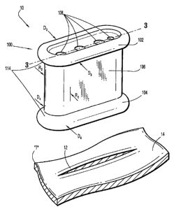
With reference to FIGS. 1-4, a surgical apparatus 10 for use in a surgical
procedure, e.g., a
minimally invasive procedure is illustrated. Surgical apparatus 10 includes
seal anchor member 100
having proximal end 102 and distal end 104. Seal anchor member 100 includes
one or more ports
108, i.e., lumen, that extend through seal anchor member 100 between proximal
end 102 and
distal end 104.
Seal anchor member 100 is formed from a suitable foam material having
sufficient
compliance to form a seal about one or more surgical objects, shown generally
as surgical object
"I" (FIG. 4), and also establish a sealing relation with tissue "T". The foam
is sufficiently
compliant to accommodate motion of the surgical object "I". In one embodiment,
the foam
-7-
CA 02709619 2010-07-12
includes a polyisoprene material. An example of an anchor member formed of,
e.g., foam, is
disclosed in applicant's co-pending U.S. Patent Application Serial Number
12/244,024, filed
October 2,2008, the entire contents of which are hereby incorporated by
reference herein.
Proximal end 102 of seal anchor member 100 defines a first major diameter Dr
and distal
end 104 defines a second major diameter D2. In an embodiment of seal anchor
member 100, the
respective first and second major diameters D1, D2 of the proximal and distal
ends 102, 104 are
substantially equivalent, as seen in FIG. 2, although an embodiment of seal
anchor member 100 in
which diameters Di, D2 are different is also within the scope of the present
disclosure. Also,
proximal end 102 of seal anchor member 100 defines a first minor diameter D3
distal end 104
defines a second minor diameter D4. In an embodiment of seal anchor member
100, the
respective first and second minor diameters D3, D4 of the proximal and distal
ends 102, 104 are
substantially equivalent, as seen in FIG. 2, although an embodiment of seal
anchor member 100 in
which diameters D3, D4 are different is also within the scope of the present
disclosure.
Advantageously, first and second major diameters Dr, D2 of the proximal and
distal ends 102, 104
are greater than first and second minor diameters D3,D4 of the proximal and
distal ends 102, 104,
such that the seal anchor member 100 has, in cross-section, a non-circular,
e.g., oblong, oval, race-
track, etc., shape.
As depicted in FIG. 1, positioning members 114 of proximal and distal ends
102, 104
may define arcuate surfaces to assist in the insertion of seal anchor member
100 within a tissue
tract 12 defined by tissue surfaces 14 and formed in tissue "T", e.g., an
incision, as discussed in
further detail below. Alternatively, proximal and distal ends 102,104 may
define substantially
planar surfaces or substantially arcuate surfaces. Embodiments are
contemplated herein in which
either or both of proximal and distal ends 102, 104 define surfaces that are
either or both arcuate or
planar. The arcuate surfaces may be either or both concave or convex.
Intermediate portion 106 extends between proximal and distal ends 102, 104 to
define a
-8-
CA 02709619 2010-07-12
dimension, or length, "L" therealong. Intermediate portion 106 further defines
an intermediate
major diameter "R" substantially parallel to major diameters D1, D2. The
dimension "R" of
intermediate portion 106 may remain substantially uniform along the dimension
"L" thereof.
Alternatively, the dimension "R" of intermediate portion 106 may vary along
the dimension, or
length, "L" thereof, thereby defining a cross-sectional dimension that varies
along its length "L",
which facilitates the anchoring of seal anchor member 100 witNn tissue "T". In
addition,
intermediate portion 106 may further define an intermediate minor diameter
"R2" substantially
perpendicular to major diameter R. Advantageously, the intermediate minor
diameter "R2" being
smaller than the major diameter R, such that the seal anchor member 100 has,
in cross-section, a
non-circular, e.g., oblong, oval, race-track, etc., shape.
The dimension "R" of intermediate portion 106 may be appreciably less than the
respective major axes Dr, D2 of proximal and distal ends 102, 104 to assist in
anchoring seal
anchor member 100 within tissue "T", as discussed in further detail below.
However, in an
alternate embodiment, the dimension "R" of intermediate portion 106 maybe
substantially
equivalent to the respective major axes Dr, D2 of proximal and distal ends
102, 104. In cross
section, intermediate portion 106 may exhibit any suitable elongated
configuration, e.g.,
substantially oval or oblong, for insertion into a narrow incision.
Each port 108 is configured to removably receive the surgical object "I".
Prior to the
insertion of surgical object "I", port 108 is in a first state in which port
108 defines a first or
initial dimension DNr. Port 108 may define an opening within seal anchor
member 100 having an
initial open state. Alternatively, DPr maybe about 0mm such that the escape of
insufflation gas
(not shown) through port 108 of seal anchor member 100 in the absence of
surgical object "I" is
substantially inhibited. For example, port 108 may be a slit extending the
length "L" of seal
anchor member 100 through proximal and distal ends 102, 104.
-9-
CA 02709619 2010-07-12
Upon the introduction of surgical object "I", port 108 transitions to a second
state in
which port 108 defines a second, larger dimension Dp2 that substantially
approximates the diameter
D1 of surgical object "I" such that a substantially fluid-light seal is formed
therewith, thereby
substantially inhibiting the escape of insufflation gas (not shown) through
port 108 of seal anchor
member 100 in the presence of surgical object "I". D1, and thus DP2, will
generally lie within the
range of about 51yun to about 12mm, as these dimensions are typical of the
surgical objects used
during the course of minimally invasive procedures. However, a seal anchor
member 100
including a port 108 that is capable of exhibiting substantially larger, or
smaller dimensions in the
second slate thereof is not beyond the scope of the present disclosure. Seal
anchor member 100
may include a plurality of generally tubular port segments (not shown)
defining ports 108. In
addition, seal anchor 100 may be devoid of ports 108. With this arrangement,
ports 108 are
created within seal anchor member 100 during the insertion of the surgical
object "I". In
accordance with this embodiment, seal anchor member 100 is formed of a
flowable or sufficiently
compliant material such as a foam material, e.g., an open-cell polyurethane
foam, or a gel.
Ports 108 may include ports 108a, which contain at least one undercut 118 that
collects
insufflation gas that leaks through the substantially fluid-tight seal between
a surgical instrument
"I" and a port 108a. Each undercut 118 defines a diameter DP3 greater than DP2
and a length
along a port 108a less than "L". hisufflation gas that leaks through a
substantially fluid-tight seal
between an instrument "I" and a port 108a may collect in an undercut 118 to
inhibit further
leakage of the gas through the substantially fluid-tight seal. Furthermore,
the undercuts 118
provide edges (where the respective diameters Dpl of the lumen 108 transition
to the diameters
DP3 of the undercut 118) that engage the outer surfaces of the instruments "I"
inserted
therethrough to further reduce leakage. Ports 108 may also include ports 108b,
which do not
contain undercuts 118, or any combination of ports 108a and ports 108b.
Generally, ports 108 are arranged linearly with respect to major diameter D1.
Ports 108
-10-
CA 02709619 2010-07-12
may alternatively be arranged linearly with respect to major diameter D2 or
dimension "R".
However, embodiments in which ports 108 are arranged nonlinearly, e.g., an
oval or zigzag
pattern, are also within the scope of this disclosure. Each port 108 may be
spaced equally from its
neighboring ports. However, embodiments in which ports 108 are spaced
unequally are also within
the scope of this disclosure.
Referring now to FIGS.1 and 5, seal anchor member 100 is adapted to transition
from an
expanded condition (FIG. 1) to a compressed condition (FIG. 5) so as to
facilitate the insertion
and securement thereof within tissue tract 12 in tissue "T". In the expanded
condition, seal
anchor member 100 is at rest and the respective major axes D1, D2 of the
proximal and distal ends
102, 104 of seal anchor member 100, as well as the dimension "R" of the
intermediate portion
106 are such that the seal anchor member 100 cannot be inserted within tissue
tract 12. However,
as seen in FIG. 5, in the compressed condition, proximal and distal ends 102,
104 of seal anchor
member 100 as well as intermediate portion 106 are dimensioned for insertion
into tissue tract 12.
Seal anchor member 100 is formed of a biocompatible compressible material that
facilitates the resilient, reciprocal transitioning of seal anchor member 100
between the expanded
and compressed conditions thereof. In one embodiment, the compressible
material is a "memory"
foam. An external force "F" is applied to seal anchor member 100 to cause the
seal anchor
member 100 to assume the compressed condition. External force "F" is directed
inwardly and
when seal anchor member 100 is subjected thereto, e.g., when seal anchor
member 100 is squeezed,
seal anchor member 100 undergoes an appreciable measure of deformation,
thereby transitioning
into the compressed condition.
As depicted in FIG. 5, as seal anchor member 100 is compressed under the
influence of
external force "F", an internal biasing force "FBI" is created within seal
anchor member 100 that
is directed outwardly, opposing force "F". Internal biasing force "FBI"
endeavors to expand seal
anchor member 100 and thereby return seal anchor member 100 towards the
expanded condition
-11 -
CA 02709619 2010-07-12
thereof. Accordingly, as long as seal anchor member 100 is subject to external
force "F" greater
than biasing force "FBI", seal anchor member 100 is compressed, and, once
compressed, as
long as external force "F" at least equals biasing force "FBI", seal anchor
member 100 remains
in the compressed condition. Upon the removal of external force "F" biasing
force "FBI"
acts to return seal anchor member 100 towards the expanded condition.
The compressible material comprising seal anchor member 100 also facilitates
the
resilient transitioning of port 108 between its first stale (FIGS. 1-3) and
its second state (FIG.
5). As previously discussed, prior to the insertion of surgical object "I",
port 108 is in its first
stale in which port 108 defines a first or initial dimension DPI. Port 108 may
incorporate a slit
extending the length "L" of seal anchor member 100. In this first state, port
108 is at rest and
is not subject to any external forces. However, upon the introduction of
surgical object "I"
through port 108 as depicted in FIG. 4, the surgical object "I" exerts a force
"FI" upon port 108
that is directed radially outward. Force "FI" acts to enlarge the dimensions
of port 108 and
thereby transition port 108 into the second state thereof in which port 108
defines a second,
larger dimension DP2 that substantially approximates the diameter Dr of
surgical object "I".
Consequently, an internal biasing force "FB2" is created that is directed
radially inward, in
opposition to force "FI". Internal biasing force "F112" endeavors to retuni
port 108 to reduce the
internal dimension of port 108 and thereby return port 108 to the first state
thereof. Internal
biasing force "FD2" is exerted upon surgical object "I" and acts to create a
substantially fluid-
tight seal therewith. The significance of forces "Far" and "F112" will be
discussed in further
detail below.
Referring again to FIG. 1, one or more positioning members 114 maybe
associated
with either or both of proximal end 102 and distal end 104 of seal anchor
member 100.
Positioning members 114 may be composed of any suitable biocompatible material
that is at least
semi-resilient such that positioning members 114 maybe resiliently deformed
and may exhibit
-12-
CA 02709619 2010-07-12
any suitable elongated configuration, e.g., substantially oblong or oval.
Prior to the insertion of
seal anchor member 100, positioning members 114 are deformed in conjunction
with the
respective proximal and distal ends 102, 104 of seal anchor member 100 to
facilitate the
advancement thereof through tissue tract 12 (FIG. 6). Subsequent to the
insertion of seal anchor
member 100 within tissue tract 12, the resilient nature of positioning members
114 allows
positioning members to return towards their normal, e.g., substantially oblong
or oval,
configuration, thereby aiding in the expansion of either or both of the
respective proximal and
distal ends 102, 104 and facilitating the transition of seal anchor member 100
from its compressed
condition to its expanded condition. Positioning members 114 also may engage
the walls
defining the body cavity to further facilitate securement of seal anchor
member 100 within the
body tissue. For example, positioning member 114 at leading end 104 may engage
the internal
peritoneal wall and positioning member 114 adjacent trailing end 102 may
engage the outer
epidermal tissue adjacent the incision 12 within tissue "T". In another
embodiment of seal
anchor member 100, one or more additional positioning members 114 may be
associated with
intermediate portion 106.
The use of seal anchor member 100 will be discussed during the course of a
typical
minimally invasive procedure. Initially, the peritoneal cavity (not shown) is
insufflated with a
suitable biocompatible gas, such as CO2 gas, such that the cavity wall is
raised and lifted away
fi-om the internal organs and tissue housed therein, providing greater access
thereto. The
insufflation maybe performed with an insufflation needle or similar device, as
is conventional in
the art. Either prior or subsequent to insufflation, a tissue tract 12 is
created in tissue "T", the
dimensions of which may be varied dependent upon the nature of the procedure.
Prior to the insertion of seal anchor member 100 within tissue tract 12, seal
anchor member
100 is in its expanded condition in which the dimensions thereof prohibit the
insertion of seal
anchor member 100 into tissue tract 12. To facilitate insertion, the clinician
transitions seal
-13-
CA 02709619 2010-07-12
anchor member 100 into the compressed condition by applying a force "F"
thereto, e.g., by
squeezing seal anchor member 100. Force "F" acts to reduce the dimensions DI
and D2 of the
proximal and distal ends 102, 104, respectively, to Dr' and D2' (FIG. 5)
including positioning
members 114 (if provided) and to reduce the dimension "R" of intermediate
portion 106 to "R"'
such that seal anchor member 100 may be inserted into tissue tract 12. As best
depicted in FIG. 6,
subsequent to its insertion, distal end 104, positioning member 114 (if
provided), and at least a
section 112 of intermediate portion 106 are disposed beneath the tissue "T".
Seal anchor
member 100 is caused to transition from the compressed condition to the
expanded condition by
removing force "F" therefrom.
During the transition from the compressed condition to the expanded condition,
the
dimensions of seal anchor member 100, i.e., the respective dimensions Dr', D2'
(FIG. 5) of the
proximal and distal ends 102, 104 are increased towards Dr and D2 (FIG. 6) and
the dimension
"R"' is increased towards "R". The expansion of distal end 104 is relatively
uninhibited given
the disposition thereof beneath tissue "T", and accordingly, distal end 104 is
permitted to expand
substantially, if not completely. However, as seen in FIG. 5, the expansion of
the section 112 of
the intermediate portion 106 is limited by the tissue surfaces 14 (FIG. 1)
defining tissue tract 12,
thereby subjecting intermediate portion 106 to an external force "F" that is
directed inwardly. As
discussed above, this creates an internal biasing force "FBI" that is directed
outwardly and
exerted upon tissue surfaces 14, thereby creating a substantially fluid-tight
seal between the seal
anchor member 100 and tissue surfaces 14 and substantially inhibiting the
escape of insufflation
gas around seal anchor member 100 and through tissue tract 12.
In the expanded condition, the respective dimensions DI, D2 of the proximal
and distal
ends 102, 104 are larger than the dimension "R" of the intermediate portion
106. Subsequent to
insertion, the dimension D2 of distal end 104 and positioning member 114 is
also substantially
larger than the dimensions of the tissue tract 12. Consequently, seal anchor
member 100 may not
-14-
CA 02709619 2010-07-12
be removed from tissue tract 12 in the expanded condition and thus, seal
anchor member 100 will
remain anchored within the tissue "T" until it is returned to its compressed
condition.
After successfully anchoring seal anchor member 100 within the patient's
tissue "T", one
or more surgical objects "I" maybe inserted through ports 108. FIG. 6
illustrates a surgical
object "I" introduced through one of ports 108. As previously discussed, prior
to the insertion of
surgical object "I", port 108 is in its first state in which port 108 defines
an initial dimension DPr
which may be negligible in that port 108, in one embodiment, is a slit.
Accordingly, prior to the
escape of insufflation gas through port 108, in the absence of surgical object
"I" is minimal,
thereby preserving the integrity of the insufflated workspace.
Surgical object "I" maybe any suitable surgical instrument and, accordingly,
may vary in
size. Suitable surgical objects to be introduced within one or more of the
poets 108 include
minimally invasive grasper instruments, forceps, clip-appliers, staplers,
cannula assemblies, etc.
Upon the introduction of surgical object "I", port 108 is enlarged, thereby
transitioning into its
second state in which port 108 defines a second dimension DP2 (FIG. 4) that
substantially
approximates the diameter Dr of surgical object "I", thereby creating a
substantially fluid-tight
seal with surgical object "I" and substantially inhibiting the escape of
insulation gas (not
shown) through port 108 of seal anchor member 100 in the presence of a
surgical object "I", as
previously discussed.
Turning now to FIGS. 8A-8D, a surgical apparatus, in accordance with an
alternate
embodiment of the present disclosure, is generally designated as 20. Surgical
apparatus 20 is
substantially identical to surgical apparatus 10 and thus will only be
discussed in detail herein to
the extent necessary to identify differences in construction and operation
thereof.
As seen in FIG. 8A, surgical apparatus 20 comprises a seal anchor member 200
defining a
plurality of ports 208, wherein the seal anchor member 200 has features that
facilitate a reduction
in size of the seal anchor member 200, such as by separating, e.g., cutting.
Advantageously, the
-15-
CA 02709619 2010-07-12
seal anchor member 200 is formed from a material, e.g., foam, that may be cut
by a surgeon's
scalpel. If seal anchor member 200 defines more ports 208 than are required
for a particular
surgical procedure, seal anchor member 200 maybe cut to have a fewer number of
ports 208.
Similarly, if a surgeon desires to have a relatively smaller incision than
would typically be used
for the seal anchor member 200, seal anchor member 200 may be cut to reduce
its, e.g., major
diameter, thereby reducing the size of the incision needed to achieve optimal
sealing when
positioned therewithin and providing a biasing force/sealing force that is
more appropriate for the
smaller incision. Depending on the position of the cut to be made, cutting the
seal anchor member
may reduce the number of ports remaining, or the number of ports may stay the
same. FIGS. 8B-
8D illustrate resulting seal anchor members 210, 220, and 230 when seal anchor
member 200 is
cut along segment lines 8B-8B, 8C-8C, and 8D-8D respectively. Seal anchor
member 200 may
include indicia, e.g., lilies or markings along segment lines 8B-8B, 8C-8C,
and 8D-8D, etc., that
indicate to a user a location at which to make such a cut if desired.
Additionally or alternatively,
the seal anchor member 200 may include, at a position that is coincident with
the indicia, a
weakened region, e.g., perforations, slits, etc., at such locations that
facilitate the making of such a
cut, e.g., by helping insure that a cut made at the indicia is passes through
the entire seal anchor
member without straying, and/or ease the making of such a cut, e.g., by
reducing the effort that a
surgeon might need to exert in making the cut. Seal anchor member 200 and
resulting seal anchor
members 210, 220, and 230, may be used in a surgical procedure in a
substantially similar manner
to seal anchor member 100 as discussed hereinbefore.
As set forth above, the prevent invention, according to various embodiments
thereof, may
provide particular advantages for. e.g., thoracic procedures (for example,
thymectomies,
lobectomies, pneumonectomy, esophagectomy, mediastinal tumor resection,
sympathectomy, etc.)
and/or single incision laparoscopic procedures in which it may be desirable to
access an
abdominal cavity off-midline. For example, during thoracic procedures, access
is typically
-16-
CA 02709619 2010-07-12
attained by placing cannulas or instruments between a patient's ribs. The
elongated shape, when
viewed in cross-section, of the seal anchor member, along with the linear
arrangement of the ports
therethrough, allows the seal anchor member to be inserted between a patient's
ribs and to move
with the natural curvature of the ribcage. In single incision laparoscopic
procedures, the shape of
the seal anchor member may enable it to be positioned between muscle groups,
e.g., parallel to and
on the lateral edge of the rectus abdorninus muscles. Advantageous positioning
of the seal anchor
member, as described hereinabove, may provide additional benefits of reducing
stretching, trauma
and post-operative pain.
In some instances, thoracic procedures may not require insufflation. For other
types of
surgical procedures, e.g., laparoscopic procedures, insufflation may be used -
for these types of
procedures, the seal anchor member may be provided with insufflation tubing
(not shown) or one
of the ports may be specifically employed for insufflation purposes.
Although the illustrative embodiments of the present disclosure have been
described
herein with reference to the accompanying drawings, the above description,
disclosure, and
figures should not be construed as limiting, but merely as exemplifications of
particular
embodiments. It is to be understood, therefore, that the disclosure is not
limited to those precise
embodiments, and that various other changes and modifications may be effected
therein by one
skilled in the art without departing from the scope or spirit of the
disclosure.
-17-