Note: Descriptions are shown in the official language in which they were submitted.
CA 02710191 2010-06-18
WO 2009/085214 PCT/US2008/013911
MAGNETIC RESONANCE SYSTEM WITH IMPLANTABLE COMPONENTS AND
METHODS OF USE THEREOF
RELATED APPLICATIONS
[001] This application claims priority to and the benefit of U.S. Provisional
Application No.
61/008,646, filed December 21, 2007; and this application claims priority to
and the benefit
of U.S. Provisional Application No. 61/008,669, filed December 21, 2007; and
this
application claims priority to and the benefit of U.S. Provisional Application
No. 61/127,514,
filed May 14, 2008. The entire contents of the prior applications are
incorporated herein by
reference in their entirety.
BACKGROUND OF THE INVENTION
[002] Current diagnostic systems involve, for example, microarray technology,
polymerase
chain reaction (PCR), in situ hybridization, antibody-based immunoassays (e.g.
enzyme-
linked immunosorbant assays), chemiluminescence, nephelometry, and/or
photometry.
Generally, these systems cannot perform the diversity of assays at high
sensitivity that is
possible with an NMR-based nanosensor system.
[003] Nuclear magnetic resonance (NMR) systems make use of nuclear magnetic
resonance
of atomic nuclei contained in a sample and are known to be able to provide a
large variety of
information characterizing a sample and corresponding sample components.
Systems
include, for example, magnetic resonance imaging (MRI) devices, magnet
resonance
spectrometers and magnetic resonance relaxometers. The nature of nuclear
magnetic
resonance phenomenon requires the presence of a magnetic field upon excitation
with a
radiofrequency electromagnetic wave. Thus, generally, NMR systems include a
magnet and
a radiofrequency coil, either as separate system components or combined in a
probehead.
[004] Magnets that are typically preferred in magnetic resonance systems
provide magnetic
fields with high magnetic field strength and high homogeneity. Magnets known
to satisfy
such requirements are usually large and/or expensive. They are therefore not
suitable for
portable devices and/or implantation devices, and/or not suitable as part of
disposable
CA 02710191 2010-06-18
WO 2009/085214 PCT/US2008/013911
-2-
probeheads. Thus, a need exists for small, inexpensive probeheads for use in
magnetic
resonance systems, allowing portability, implantation and/or one-time use
applications.
[005) Magnetic nanosensors are derivatized superparamagnetic nanoparticles
that form
clusters (aggregates) or nanoassemblies as a function of the presence or
concentration of their
intended molecular target. It is thought that when superparamagnetic
nanoparticles assemble
into clusters and the effective cross sectional area becomes larger, a
nanoassembly becomes
more efficient at dephasing the spins of surrounding water (or other solvent)
protons, leading
to a measurable change of the relaxation rates (1/T2). Examples of magnetic
nanosensors
are described, for example, in Perez et al., "Use of Magnetic Nanoparticles as
Nanosensors to
Probe for Molecular Interactions," Chem Bio Chem, 2004, 5, 261-264, and in
U.S. Patent
Application Publication No. US2003/0092029 (Josephson et al.).
[0061 Provided magnetic resonance systems and methods of the present invention
address
some of the limitations in the art. Provided implantable systems and methods
are particularly
suitable but not limited to magnetic relaxation measurements, because
relaxation
measurements require less homogenous fields.
SUMMARY OF THE INVENTION
10071 One embodiment of the present invention is a nuclear magnetic resonance
system for
measuring magnetic resonance signals from a sample contained in a sample
volume in-vivo.
The system comprises a magnet or magnetic field generator positioned to
provide a magnetic
field in a sample volume, the magnetic field being suitable to allow measuring
magnetic
resonance signal; a probehead suitable for partial or complete implantation in
a subject, the
probehead comprising a radiofrequency circuit that includes a radiofrequency
coil wound to
form a space capable of accommodating a sample volume and a port, the port
allowing a
sample to enter the sample volume, wherein the sample volume contains magnetic
particles
and the port is adapted to allow an analyte to enter the sample volume and to
prevent, partly
or completely, the magnetic particles from leaving the sample volume; an
external coil for
disposition outside the subject's body, wherein the external coil is suitable
for inductive
coupling to the radiofrequency circuit to form a radiofrequency resonant
circuit; and a control
unit for disposition outside the subject's body, wherein the control unit is
connected to the
external coil and comprises logic circuitry to control the radiofrequency
resonant circuit and
CA 02710191 2010-06-18
WO 2009/085214 PCT/US2008/013911
-3-
allows acquisition and processing of magnetic resonance signals received by
the
radiofrequency resonant circuit.
[008] Another embodiment of the present invention is nuclear magnetic
resonance system
for measuring magnetic resonance relaxation signals from a sample contained in
a sample
volume in-vivo. The system comprises: a single-sided permanent magnet or
magnetic field
generator for disposition outside a subject's body and near a sample volume to
provide a
magnetic field in the sample volume, the magnetic field being suitable to
allow measuring
magnetic resonance relaxation signals; a probehead suitable for partial or
complete
implantation in a subject, the probehead comprising a radiofrequency circuit
that includes a
radiofrequency coil wound to form a space capable of accommodating a sample
volume and a
port, the port allowing a sample to enter the sample volume; an external coil
for disposition
outside the subject's body, wherein the external coil is suitable for
inductive coupling to the
radiofrequency circuit to form a radiofrequency resonant circuit; and a
control unit for
disposition outside the subject's body, wherein the control unit is connected
to the external
coil and comprises logic circuitry to control the radiofrequency resonant
circuit and allows
acquisition and processing of magnetic resonance relaxation signals received
by the
radiofrequency resonant circuit.
[009] Yet another embodiment of the invention provides a nuclear magnetic
resonance
system for measuring magnetic resonance relaxation signals from a sample
contained in a
sample volume in-vivo. The system comprises: a probehead suitable for partial
or complete
implantation in a subject, the probehead comprising a radiofrequency circuit
that includes a
capacitor and a radiofrequency coil wound to form a space capable of
accommodating a
sample volume and a port, the port allowing a sample to enter the sample
volume; and a
permanent magnet positioned near or around the radiofrequency coil to provide
a magnetic
field in the sample volume, the magnetic field being suitable to allow
measuring magnetic
resonance relaxation signal; an external coil for disposition outside the
subject's body,
wherein the external coil is suitable for inductive coupling to the
radiofrequency circuit to
form a radiofrequency resonant circuit; and a control unit for disposition
outside the
subject's body, wherein the control unit is connected to the external coil and
comprises logic
circuitry to control the radiofrequency resonant circuit and allows
acquisition and processing
of magnetic resonance relaxation signals received by the radiofrequency
resonant circuit.
CA 02710191 2010-06-18
WO 2009/085214 PCT/US2008/013911
-4-
[0101 Still another embodiment of the present invention is nuclear magnetic
resonance
system for measuring magnetic resonance relaxation signals from a sample
contained in a
sample volume in-vivo. The system comprises: a single-sided permanent magnet
or magnetic
field generator for disposition outside a subject's body and near a sample
volume to provide a
magnetic field in the sample volume, the magnetic field being suitable to
allow measuring
magnetic resonance relaxation signals; a probehead suitable for partial or
complete
implantation in a subject, the probehead comprising a radiofrequency circuit
that includes a
radiofrequency coil wound to form a space capable of accommodating a sample
volume and a
port, the port allowing a sample to enter the sample volume, wherein the
sample volume
contains at least one sensor particle and the port is adapted to allow an
analyte to enter the
sample volume and to prevent, partly or completely, the sensor particles from
leaving the
sample volume; an external coil for disposition outside the subject's body,
wherein the
external coil is suitable for inductive coupling to the radiofrequency circuit
to form a
radiofrequency resonant circuit; and a control unit for disposition outside
the subject's body,
wherein the control unit is connected to the external coil and comprises logic
circuitry to
control the radiofrequency resonant circuit and allows acquisition and
processing of magnetic
resonance relaxation signals received by the radiofrequency resonant circuit.
[0111 Yet another embodiment of the invention provides a nuclear magnetic
resonance
system for measuring magnetic resonance relaxation signals from a sample
contained in a
sample volume in-vivo. The system comprises: a probehead suitable for partial
or complete
implantation in a subject, the probehead comprising a radiofrequency circuit
that includes a
capacitor and a radiofrequency coil wound to form a space capable of
accommodating a
sample volume and a port, the port allowing a sample to enter the sample
volume, wherein
the sample volume contains at least one sensor particle and the port is
adapted to allow an
analyte to enter the sample volume and to prevent, partly or completely, the
sensor particles
from leaving the sample volume; and a permanent magnet positioned near or
around the
radiofrequency coil to provide a magnetic field in the sample volume, the
magnetic field
being suitable to allow measuring magnetic resonance relaxation signal; an
external coil for
disposition outside the subject's body, wherein the external coil is suitable
for inductive
coupling to the radiofrequency circuit to form a radiofrequency resonant
circuit; and a
control unit for disposition outside the subject's body, wherein the control
unit is connected
to the external coil and comprises logic circuitry to control the
radiofrequency resonant
CA 02710191 2010-06-18
WO 2009/085214 PCT/US2008/013911
-5-
circuit and allows acquisition and processing of magnetic resonance relaxation
signals
received by the radiofrequency resonant circuit.
[012] Still other embodiments include methods for determining a magnetic
resonance
relaxation parameter associated with a sample contained in a sample volume in-
vivo in a
subject using a nuclear magnetic resonance system. In certain embodiments, the
method
comprises: positioning a magnet or magnetic field generator of the nuclear
magnetic
resonance system near or around the sample to provide a magnetic field in the
sample
suitable to allow measuring magnetic resonance signals; implanting partially
or completely a
probehead of the nuclear magnetic resonance system within a subject's body,
the probehead
comprising: a radiofrequency circuit that includes a radiofrequency coil wound
to form a
space capable of accommodating a sample volume and a port, the port allowing a
sample to
enter the sample volume, wherein the sample volume contains at least one
sensor particle
and the port is adapted to allow an analyte to enter the sample volume and to
prevent, partly
or completely, the one or more sensor particles from leaving the sample
volume, and at least
part the sensor particles aggregate in the presence of analyte to change a
magnetic resonance
signal of a sample; positioning an external coil outside the subject's body,
wherein the
external coil is inductively coupled to the radiofrequency circuit to form a
radiofrequency
resonant circuit; controlling with a control unit positioned outside the
subject's body the
radiofrequency circuit to apply the radiofrequency pulse or pulse sequence to
the sample
volume in the presence of the magnetic field; and acquiring and processing
part or effectively
all of the magnetic resonance signals from the sample in the sample volume
sensed by the
radiofrequency resonant circuit to determine a magnetic resonance relaxation
parameter.
[013] Additional embodiments provide methods of monitoring analytes in a body
of a
patient. In certain embodiments the method comprises: implanting an
implantable diagnostic
device at an exposed treatment site; and detecting or measuring the presence
and/or
concentration of one or more analytes using magnetic resonance measurement.
[014] In another embodiment, the present invention provides a method of
determining organ
transplant rejection or acceptance in a patient. The method comprises:
implanting an
implantable magnetic resonance diagnostic device in a patient having undergone
or
undergoing an organ transplant surgery, the implanted diagnostic device
detecting or
measuring the presence and/or concentration of one or more analytes; and
monitoring the one
or more analytes in response to the organ transplant using the output of the
implanted
CA 02710191 2010-06-18
WO 2009/085214 PCT/US2008/013911
-6-
diagnostic device; wherein the output of the implanted device conveys
information indicating
whether an organ transplant is being rejected or accepted in the patient.
[015] The foregoing will be apparent from the following more particular
description of
example embodiments of the invention, as illustrated in the accompanying
drawings in which
like reference characters refer to the same parts throughout the different
views. The drawings
are not necessarily to scale, emphasis instead being placed upon illustrating
embodiments of
the present invention.
BRIEF DESCRIPTION OF THE DRAWINGS
[016] FIG. 1 provides a schematic representation of two nuclear magnetic
resonance
systems with probeheads implanted in mammalian body, one (system A) employing
a
probehead with a double sided magnet and the other (system B) a probehead
without a
magnet but an external one-sided magnet.
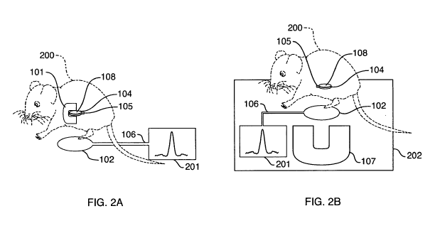
[017] FIG. 2 provides a schematic representation of two nuclear magnetic
resonance (NMR)
systems with probeheads implanted in mouse, one (system A) employing a
probehead with a
double sided magnet and the other (system B) a probehead without a magnet but
an external
one-sided magnet.
[018] FIG. 3 presents two schematic views (a top view and a side view) of a
probehead that
is suitable for the systems of the present invention, the probehead including
a radiofrequency
coil surrounding by a double-sided magnet.
[019] FIG. 4 presents two schematic views (a top view and a side view) of a
probehead that
is suitable for the systems of the present invention, the probehead including
a planar
radiofrequency coil adjacent to a magnet.
[020] FIG. 5 shows a probehead in a corresponding pickup assembly for
connection to a
spectrometer.
[021] FIG.6 provides a magnitude spectrum obtained with the device of FIG. 5.
[022] FIG. 7 provides a T2 relaxation curve obtained with the device of FIG.
5.
DETAILED DESCRIPTION OF CERTAIN EMBODIMENTS OF THE INVENTION
10231 One aspect of the present invention is an implantable device for in vivo
Magnetic
Resonance (MR) sensing. It allows measuring MR signals from a selected volume
inside the
body of an animal. Sensing is performed by using an apparatus configuration
where
components of a device include a resonant radio-frequency (RF) circuit (see
Figure 1-A) or a
CA 02710191 2010-06-18
WO 2009/085214 PCT/US2008/013911
-7-
resonant RF circuit and a small magnet (see Figure 1-B) implanted in the body
of a subject
(e.g., an animal), wherein an RF spectrometer is not included in the
implantable components.
A static magnetic field needed for an MR sensing method is generated by either
a small
implanted permanent magnet (e.g., a magnet implanted in the proximity of or
around an
implanted RF coil) (Fig. A), or by an external magnet (Fig. B). An external
magnet, e.g., a
portable open permanent magnet, can be placed at an external location of a
subject close to
the location of an implanted device for generation of a magnetic field. An
implanted RF coil,
when part of an RF resonant circuit, is electromagnetically (inductively, non
contact) coupled
to an external coil. MR measurements are controlled with an external
spectrometer,
connected to the external coil. Provided configurations allow measuring
signals from a
specific location inside the body and may also allow monitoring properties of
body fluids
and/or tissues by their interaction with a small biological or chemical sensor
particle (e.g., a
magnetic particle) implanted together with an MR device.
[024] Another aspect of the present invention is a means for obtaining
magnetic resonance
relaxation measurements. from an implanted radiofrequency (RF) detection coil,
wherein the
means includes an external RF coil and instrument that is not physically
attached to the
detection coil (e.g., inductive coupling of an RF coil and external pickup
coil). No power or
additional electronics are required to be on board the implanted coil. This
invention is
particularly useful for detecting the relaxation signals from implanted
devices such as MRSw
relaxation switches.
[025] Implantation of an implantable diagnostic device allows for real-time,
intermittent, or
continuous monitoring of one or more analytes of interest. Implantable
diagnostic devices
suitable for the use with the methods of the present invention are described
herein. One
advantage of the methods of implantation is in conjunction with a surgical
procedure,
wherein a treatment site of a subject is exposed during a surgical procedure.
Particular
embodiments thus provide methods for implanting an implantable diagnostic
device before,
after, or during any type of surgery (e.g. endoscopic, laparoscopic, or open
surgery). In some
embodiments a surgery is conducted for a purpose other than implantation of an
implantable
diagnostic device.
CA 02710191 2010-06-18
WO 2009/085214 PCT/US2008/013911
-8-
[026] As used herein, the terms "subject" and "patient", which in some
embodiments can be
a mammal, are used interchangeably. The terms "subject" and "patient" refer to
an animal
(e.g., a bird such as a chicken, quail or turkey, or a mammal). In some
embodiments a
subject is a mammal, including a non-primate (e.g., a cow, pig, horse, sheep,
rabbit, guinea
pig, rat, cat, dog, and mouse) and a primate (e.g., a monkey, chimpanzee and a
human). In
one embodiment, a subject is a non-human animal such as a farm animal (e.g., a
horse, cow,
pig, sheep or fish), or a companion animal (e.g., a dog, cat, guinea pig or
rabbit). In a
particular embodiment, the subject is a human.
[027] It is known in the art that magnetic resonance encoding and detection
can occur
within a detection coil, which consists of an inductor and a capacitor, by
means of a remote
RF coil connected to a transceiver (transmitter and detector). Inductive
coupling requires a
detection coil, which is usually made of an inductor (the "coil") and a
capacitor which
comprises an RF circuit, and a pickup coil that consists of an inductor
connected to an RF
transmitter and receiver (also known as transceiver) and a magnetic field.
When a detection
coil is in the presence of a magnetic field, the proton within a detection
volume of the
detection coil can be detected by means of a coupled pickup coil. This is
possible because
the pickup coil and the. detection coil are electromagnetically coupled,
comprising a resonant
circuit.
[028] The present invention also provides for affordable means for implantable
MR-
relaxometry based biosensors. The nuclear magnetic resonance systems of the
present
invention do not require the highly homogeneous magnetic fields necessary in
prior
implantable MR devices to obtain magnetic resonance information, and thus
allow greater
flexibility in terms of the requisite instrumentation and hardware. For
example, a magnetic
field conveyed upon an implanted coil can be generated by an implanted
miniature magnet,
an external permanent magnet such as a single sided magnet, or by a Halbach
array, u-
magnet, torroidal magnet, or even a superconducting magnet. A dedicated magnet
array can
be designed to generate desired levels of magnetic field strength at the
position of the
implanted coil. The magnetic field strength may be in the range of 0.1 Tesla
to 2 Tesla, most
commonly in the range of 0.2 to 0.7 Tesla. If the field is generated by an
external magnet, the
configuration can be enclosed or open. Enclosed configurations typically allow
for more
homogenous magneto field distributions and therefore are less sensitive to the
relative
position of the implanted coil and the magnet. Open configurations generate
less homogenous
CA 02710191 2010-06-18
WO 2009/085214 PCT/US2008/013911
-9-
fields, but have been demonstrated to be effective for magnetic resonance and
magnetic
resonance measurements (P.J. Prado. Single-Sided Imaging Sensor. Magn. Reson.
Imaging,
2003, 21, 397-400 - U.S. Patent 6,977,503. System and Method for Single-Sided
Magnetic
Resonance Imaging - Bluemich B, Bluemler P, Eidmann G, Guthausen A, Haken R,
Schmitz
U, Saito K, Zimmer G, The NMR-mouse: Construction, excitation, and
applications. Magn
Reson Imaging 1998; 16:479). Because the region of interest in the current
implantable
application is significantly smaller than that of conventional MRI
diagnostics, compact
magnets may be utilized to generate the magnetic field. Open magnets can be
fabricated in
robust, low cost, compact forms. As an example a dipole type magnet (U.S.
Patent 6,977,503)
generates flat surfaces of constant magnetic field outside the magnet. If the
implanted coil is
positioned at a know distance from the permanent magnet array, the frequency
of resonance
is known and magnetic resonance signals can be generated and picked up by the
inductive
coupling device.
[029] In one embodiment, the invention consists of an implantable device that
contains a
chamber (e.g. a sample volume) containing MRSw nanoparticles. The chamber is
permeable
to analyte and solution from the surrounding matrix but does not allow the
nanoparticles to
diffuse out. The chamber is within the detection volume of a radiofrequency
coil (e.g.,
solenoid, Helmholtz). In certain embodiments a RF coil is a solenoid. An-RF
coil is usually
an independent circuit. In some embodiments a chamber is located within the
magnetic field
of one or more implantable magnets. In other embodiments a-chamber is not
permanently
located within a magnetic field; rather a magnetic field is introduced by an
external magnet
positioned outside the subject. Signal from an RF coil enclosing a chamber is
detected by
means of an external radiofrequency coil (e.g., a pickup coil) appropriately
tuned to couple
(inductively coupled) with the implanted coil as known in the art. This tuning
and matching
characteristic of the circuit is a function of the distance between the two
coils. Proper signal
acquisition is also a function of the magnetic field. External magnets for use
in the provided
systems and methods for tuning and matching a circuit appropriately are known
in the art and
can be adapted for the current systems. Rollwitz WL. Agricultural Engineering
1985; 66:12;
P.J. Prado. Single-Sided Imaging Sensor. Magn. Reson. Imaging, 2003, 21, 397-
400; P.J.
Prado. NMR Hand-Held Moisture Sensor. Magn. Reson. Imaging, 2001, 19, 506-5
08; P.J.
Prado, B. Blumich, and U. Schmitz. One-dimensional Imaging with a Palm-Size
Probe. J.
Magn. Reson. 2000, 144, 200-206; U.S. Patent 6,977,503. System and Method for
Single-
CA 02710191 2010-06-18
WO 2009/085214 PCT/US2008/013911
- 10-
Sided Magnetic Resonance Imaging; U.S. Patent 6,489,767: Apparatus for and
Method of
Single-sided Magnetic Resonance Imaging with a Palm-size Probe; Bluemich B,
Bluemler P,
Eidmann G, Guthausen A, Haken R, Schmitz U, Saito K, Zimmer G, The NMR-mouse:
Construction, excitation, and applications. Magn Reson Imaging 1998; 16:479;
MacDonald
PJ. Stray field magnetic resonance imaging. Progress in NMR 1997; 30:69 -99.
[030] As used herein, the term "nanoparticle" refers to MRSw nanoparticles,
e.g.
"magnetic particles," as described below.
[031] Provided devices and methods include an implanted MR biosensor that by
means of
reversible magnetic resonance switch (MRSw) assays allows for the real-time
sensing of
analyte levels. MRSw, which undergo a switch between clustered and dispersed
states due to
the presence of molecular, cellular, viral analytes, can be read by a change
in a MR relaxation
rate. MRSw assays and sensors (e.g., provided devices) can be configured to
function in a
reversible fashion.
[032] Rather than necessitating implantation of a complete RF probe or
spectrometer as
used in prior devices and methods, only a chamber including an RF coil is
required to be
implanted. Further, prior devices and methods including implanted chambers use
a high-field
MRI scanner; however this is costly and inconvenient or requires implantation
of some
combination of MR spectrometers. Using an inductively coupled coil as provided
herein
allows for a portable reader to be used and allows for the implantable device
to be cheap and
disposable. The portable reader contains a customized compact magnet array
with significant
lower complexity and cost than conventional superconducting counterparts. If
the magnetic
field is generated by an implanted permanent magnet by the implanted RF coil,
the array can
be even significantly smaller.
1033] Provided devices and methods can be used for a variety of applications.
Relevant
applications include but are not limited to the following. A permeable sample
volume
(chamber) can be configured to contain the sensing volume of an implantable
device (e.g.,
biosensor) within the sensitive region of the implanted RF coil. Measurements
of relaxation
rates (e.g., T2, TI, etc) with a variety of pulse sequences can be used to
deduce the state of
the biosensor. A biosensor can be configured to link presence and/or amount of
analyte (e.g.,
target levels, physiological status, or biomolecular levels) to an MR
parameter using one of
the following methods:
CA 02710191 2010-06-18
WO 2009/085214 PCT/US2008/013911
-11-
[034] Encapsulation of a smart polymer, or responsive polymers. Such polymers
have been
shown to be sensitive to pH, ionic strength, and specific molecular and
biomolecular analytes.
In these cases the hydration level, cross-link density, or other
characteristic of the polymer
would change in response to a change in the animal. This change in polymer
state would be
detected by an implanted RF coil.
[035] The real-time sensing capabilities of the devices and methods provided
herein can be
used, for example, in small animals or humans to monitor response to drug or
other therapies
over a course of time by monitoring appropriate biomarker levels without
requiring removal
of body fluids, obtaining one or more specimens or sacrificing the animal.
Examples of this
use include, for example, monitoring drug delivery and dosing efficiencies for
development
or treatments such as patient dosing for chemotherapy, drug development
studies, monitoring
circulating active agent (e.g., insulin) levels or other biomarker levels in a
non-invasive
manner, real-time monitoring of foreign substances in a subject (a
pharmaceutically active
agent, a toxin, a poison). Also, relaxation measurements of tissue, organs,
blood vessels, or
other body parts of a subject can be performed.
[036] A "magnet" as used herein can be any material or combination of
materials that
provides a magnetic field in at least some volume around the material.
Typically, the magnet
is a permanent magnet. Suitable materials include but are not limited to
NdFeB, SmCo, and
the like. Magnets can be configured to form new magnets, that is, magnet
arrays, for
example, a permanent magnet with a c-shaped yoke, a Halbach magnet (cylinder
and other
configurations), u-magnet, torroidal magnet and the like.
[037] The magnets, magnet configurations and magnetic field generators of the
present
systems can be weak and/or provide magnetic fields that are inhomogeneous.
Typically,
maximum magnetic field strength values provided by the magnets and/or magnet
configurations of the present invention are between about 0.2 Tesla and about
2 Tesla. More
typically, they are between about 0.3 and about 1.5 Tesla. Even more
typically, they are
between about 0.4 and about 1.1 Tesla. Most typically, they are between about
0.45 and 0.8
Tesla. In some embodiments the magnetic field strength is less than about 2
Tesla. In certain
embodiments the magnetic field strength is less than about 1.1 Tesla. In
certain embodiments
the magnetic field strength is less than about 0.8 Tesla.
[038] The term "inhomogeneous" refers to magnetic fields that are lower in
uniformity than
those required for spectroscopy. Homogeneity is dependent on the space in
which the
CA 02710191 2010-06-18
WO 2009/085214 PCT/US2008/013911
-12-
measurement is defined. Homogeneities of the magnetic fields can range between
about
10000 ppm and about 10 ppm. In some embodiments homogeneities can range
between
about 50 ppm and 5000 ppm. In particular embodiments, homogeneities can range
between
about 100 ppm and about 1000 ppm.
[039] Also, typically, the magnetic fields employed in the present systems are
effectively
static, that is, they do not change substantially over time. Changes in
magnetic field such as
due to temperature fluctuations are considered to be not substantial,
corrected by an
additional controlled magnetic field, a shift in the resonance frequency, or
can be taken into
consideration during the magnetic resonance measurement.
[040] The magnets in the present systems can be positioned within a subject's
body and/or
outside of the subject's body. In contrast to magnets for disposition outside
a subject's body,
magnets for complete implantation within a subject's body are preferred to be
small to lessen
the invasiveness of the implantation. Typically, magnets for implantation are
smaller than
about 2 inches in any dimension. More typically, magnets for implantation are
smaller than
about 1 inch in any dimension. Most typically, magnets for implantation are
smaller than
about 0.5 inches in any dimension. Each dimension may be independently
determined.
[041] A "magnetic field generator" as used herein, is a device that provides a
magnetic field
in at least some volume around the device. Typically, a magnetic field
generator requires a
power supply and provides the targeted magnetic field only when powered.
Examples of
magnetic field generators include but are not limited electromagnets with and
without a metal
pole (see Cardot et al. Sensors and Actuators 1994). In certain embodiments, a
magnetic field
generator used in the present system can be positioned within a subject's
body. In other
embodiments, a magnetic field generator used in the present system can be
positioned outside
of a subject's body. Because magnetic field generators tend to be large and
complex (e.g.,
requiring, for example, power supply), in typical configurations a magnetic
field generator
will be used for disposition outside a subject's body.
[042] One or more magnet(s) and magnetic field generator(s) for use in the
present systems
are selected such that the combination and position of selected components
will provide a
magnetic field of sufficient strength in the sample volume to allow measuring
a magnetic
resonance signal (e.g., a relaxometry measurement).
[043] The magnetic field strength of a given magnet or magnetic field
generator in a given
volume, for example, a sample volume can be calculated and/or approximated
using methods
CA 02710191 2010-06-18
WO 2009/085214 PCT/US2008/013911
- 13-
known in the art. Typically, the magnetic field strength depends on the nature
of the magnet
or magnetic field generator and the position of the magnet or magnetic field
generator relative
to the sample volume. Also, magnetic field strength of a given magnet or
magnetic field
generator in a sample volume can be measured using methods and devices known
in the art,
for example, gaussmeters, teslameters, hall effect probes, and the like. In
some
embodiments, magnetic field strengths within a sample volume of between about
0.2 and
about 2 Tesla are sufficient to allow measuring magnetic resonance signals. In
certain
embodiments, magnetic field strengths within a sample volume of between about
0.3 and
about 1.5 Tesla are sufficient to allow measuring magnetic resonance signals.
In other
embodiments, magnetic field strengths within the sample volume of between
about 0.45 and
about 1.5 Tesla are sufficient to allow measuring magnetic resonance signals.
In still other
embodiments, magnetic field strengths within the sample volume of between
about 0.45 and
about 1.1 Tesla are sufficient to allow measuring magnetic resonance signals.
[044] A "probehead" as used herein is the part of the nuclear magnetic
resonance system
that is to be implanted, partially or completely, in a subject's body.
Minimally, a probehead
refers to a radiofrequency circuit that includes a radiofrequency coil wound
to form a space
capable of accommodating a sample volume and allowing sample to enter. In one
embodiment, a probehead comprises a space capable of accommodating a sample
volume
and/or a port. In certain embodiments, a space capable of accommodating a
sample volume
and port can be, for example, a radiofrequency coil (as part of a
radiofrequency circuit)
wound to enclose a sample volume while providing an opening (i.e., space
capable of
accommodating a sample volume) to allow a sample volume to be placed within
the opening.
In other embodiments, a space capable of accommodating a sample volume and/or
port is
distinct from the opening of a radiofrequency coil but adapted to a given
radiofrequency coil,
for example, formed to enclose part or all of a detection volume of the
radiofrequency coil.
For example, a capillary for containment of a sample volume, positioned within
a
radiofrequency coil.
[045] In some embodiments a radiofrequency coil is wound to enclose a volume
of about 1
1 to about lOml. In some embodiments a volume is about lml. In some
embodiments a
volume is about 500 l. In certain embodiment a radiofrequency coil is wound
to enclose a
volume of less than about 100 l. In still other embodiments a radiofrequency
coil is wound
to enclose a volume of less than about 10 l are used. In still further
embodiment a
CA 02710191 2010-06-18
WO 2009/085214 PCT/US2008/013911
14-
radiofrequency coil is wound to enclose a volume of less than about 5 l. In
particular
embodiments a radiofrequency coil is wound to enclose a volume of less than
about 1.6 l.
In still further particular embodiments a radiofrequency coil is wound to
enclose a volume of
less than about I l.
[046] A material used to forma sample volume, and, in particular, any material
that may be
in contact with a biological sample or tissue is typically biocompatible, and
constructed of
materials that allow for proper function of both the device and a host
animal's biological
functions. Surfaces of components of a magnetic resonance system of the
present invention
that will be in direct contact with tissue when implanted can be made of or
coated with
biocompatible material to prevent, partly or completely, biofouling and/or
immune responses.
Also, parts of the present systems for implantation can partially or entirely
composed of
biodegradable material(s). In certain embodiments a device may be coated, in
whole or in
one or more parts with a physiologically acceptable coating as known in the
art to render an
implantable device bioinert, biomimetic, or bioactive, as desired. Suitable
materials include
titanium, inert silicone elastomers, ceramics, glass, polymeric materials,
poly-[i-
hydroxybutyrate (PHB) and the like. One or more sample volumes and
corresponding ports
can be fabricated using methods known in the art. Suitable methods include
form or injection
molding methods, and microfabrication methods for sample containers smaller
than a few
millimeters, for example, two-photon three-dimensional lithography.
[047] A probehead may include a "housing" that encloses components of a
probehead such
as, for example, a radiofrequency coil and magnet. In certain embodiments at
least one
component of a probehead (e.g., a magnet, a magnetic field generator, a
radiofrequency coil)
is attached to the housing.
[048] The probeheads of the present invention typically are for implantation
within a patient
(e.g., implantation in tissue, organ and/or bone of a mammal). Methods for
implanting an
object, whether permanently or temporarily in a mammalian body are well known
in the art
and such methods are adaptable for implantation of device (e.g., a probehead,
a magnetic
resonance system) of the present invention. Suitable probeheads are disclosed
but not limited
to the ones described in International Patent Application No. PCT/US08/12592,
filed
November 6, 2008, entitled "Small Magnet and RF Coil for Magnetic Resonance
Relaxometry," (Attorney's Docket No. 1014-002), the entire teachings of which
are hereby
incorporated by reference.
CA 02710191 2010-06-18
WO 2009/085214 PCT/US2008/013911
-15-
[049] A "port" as used herein, refers in the simplest case.to an opening as
provided above
but can also be a structure or device that allows one or more analytes to
enter and/or exit the
sample volume, and may prevent other sample components to enter the sample
volume. A
port can be, for example, a feature or structural part of a probehead or a
component thereof
(e.g. radiofrequency coil) or a separate structure attached to or contained
within a probehead.
Furthermore, a port can be, for example, a structure with one or more
openings, a semi-
permeable membrane, or the like. In some embodiments, a port allows analytes
that lead to
aggregation of magnetic particles in a sample volume to enter the sample
volume and
prevents sample components that would hinder the aggregation process from
entering the
sample volume. A port also prevents assay components, for example, magnetic
particles,
from leaving the device.
[050] In certain embodiments, a probehead includes one or more separate sample
volumes.
In some embodiments a probehead includes between about 1 and about 100 sample
volumes.
In some embodiments a probehead includes between about 1 and about 10 sample
volumes.
In some embodiments a probehead includes two sample volumes. In certain
embodiments a
probehead includes one sample volume.
[051] A probehead containing more than one sample volume may comprise a
radiofrequency coil with an associated detection volume encompassing at least
part of each
sample volume. Alternatively, a probehead may have more than one
radiofrequency coil
and/or radiofrequency circuit, one for each sample volume or a subgroup of the
sample
volumes. In certain embodiments, a probehead comprises at least two
radiofrequency coils.
[052] For probeheads that include a plurality of separate sample volumes but
only one
radiofrequency coil that is employed to probe the plurality of sample volumes
simultaneously, multiplexing methods may be used to distinguish the magnetic
resonance
signal or information from the separate sample chambers. For example, one
multiplexing
method that may be used is based on extracting decay constant values, for
example, values of
spin-spin relaxation constant T2 from multi-exponential relaxation curves (see
T.J. Lowery et
al., Anal. Chem. (2008), 80, 1118-1123.). Relaxation data obtained using a
probehead of the
present invention may be fit to a decaying exponential curve defined by the
following
equation:
f(t)=~A,expl ,(o)
CA 02710191 2010-06-18
WO 2009/085214 PCT/US2008/013911
- 16-
where f(t) is the signal intensity as a function of time, t, A; is the
amplitude coefficient for the
ith component, and (T); the decay constant (such as T2) for the ith component.
For relaxation
phenomenon discussed here the detected signal is the sum of a discrete number
of
components (i=1,2,3,4...n). Such functions are called mono-, bi-, tri-, tetra-
or multi-
exponential, respectively. Due to the widespread need for analyzing multi-
exponential
processes in science and engineering, there are several established
mathematical methods for
rapidly obtaining estimates of A; and (T); for each coefficient (Istratov, A.
A. & Vyvenko, 0.
F. 1999. Exponential analysis in physical phenomena. Rev. Sci. Inst. 70 (2):
1233-1257).
[053] Further, for probeheads that include a magnet, the magnet can be single-
sided,
double-sided or multi-sided. In some embodiments, the magnet is double-sided.
For systems
with a magnet for disposition outside the subject's body, typically, the
magnet is one-sided.
[054] Also, probeheads that include a plurality of separate sample volumes
with associated
radiofrequency coils for each sample volume may have each radiofrequency coil
connected to
a switch and associated circuitry that allows to wirelessly turn on or off
each of the
radiofrequency coils. This allows selectively measuring the magnetic resonance
signal of a
sample in a sample volume with switched on radiofrequency coil.
[055] Alternatively, magnets or magnetic field generators providing part or
all of the
magnetic field in a sample volume of a probehead can be implanted separately
from the
probehead.
[056] In embodiments containing multiple sample volumes, separate sample
volumes can
each independently have the same dimensions, different dimensions but equal
volumes, or
different dimensions and different volumes. Typically, each separate sample
volume is
between about 1 fL and about 10 mL. More typically, each separate sample
volume is
between about 1 pL and about 1 mL. Most typically, each separate sample volume
is
between about 1 L and about 200 L.
[057] Implantable devices of the present invention may be used to sense and/or
measure
magnetic resonance signals as part of a magnetic resonance system, wherein one
or more
sensing reagent(s) are included within an implantable device. In particular,
one or more
sample volume(s) of the present invention may include one or more sensing
agent. Typically,
each sample volume contains one sensing agent.
[058] A "sensing agent" as used herein is an agent that senses, responds to,
or is influenced
by a sample characteristic to correlate the presence and/or extent of the
sample characteristic
CA 02710191 2010-06-18
WO 2009/085214 PCT/US2008/013911
- 17
with the presence, change or magnitude of the magnetic resonance signals
associated with a
sample. The term "sample characteristic" as used herein refers to any chemical
and/or
physical property of a given sample. Suitable sample characteristics can be,
but are not
limited to concentration of an analyte (that is, a molecule, ion, or radical
of interest in the
sample), pH-value, ionic strength, hydration state (e.g., of tissue, that is,
concentration of
water in tissue, temperature, and the like.
[059] Suitable sensing agents can be, but are not limited to dry reagent
compositions,
magnetic particles, responsive polymers, magnetic resonance contrast agents,
and the like.
[060] Dried reagent compositions that are suitable include, for example, dried
biotinylated
coated nanoparticles (see T.J. Lowery et al., Anal. Chem. (2008), 80, 1118-
1123), for
example, based on the following formulation (216 L, 0.083 mM Fe, 10 mM PBS,
20 mg/ml
dextran, pH 7.4). Dried reagent compositions can be prepared by placing a
magnetic particle
solution, for example, biotinylated coated nanoparticle solution into a
container, for example,
a container such as a glass tube, and freezing the container in a freeze dryer
(e.g., VirTis
freeze dryer (Gardiner, NY)), for example, at -80 C for 24h. Each of the one
or more
separate volumes of the sample containers may be filled by transfer of the
dried reagent
composition from the container that was used during freeze drying.
[061] "Magnetic particles" as used herein, are particles that respond to or
are influenced by
a sample characteristic to correlate the presence and/or extent of the sample
characteristic
with the presence, change or magnitude of the magnetic resonance signals
associated with the
sample. Typically, the magnetic particles respond by aggregating or
dispersing.
Accordingly, "forward" (clustering or aggregation) or "reverse" (declustering
or dispersion)
types of assays can be employed by the methods of the present invention.
Additionally,
nanoassembly formation can be designed to be reversible (e.g., by temperature
shift, chemical
cleavage, pH shift, etc.) so that "forward" or "reverse" assays can be
developed for detection
of specific analytes. Forward (clustering) and reverse (declustering) types of
assays can be
used to detect a wide variety of biologically relevant materials. Furthermore,
the spin-lattice
relaxation time (Ti) is considered independent of nanoparticle assembly
formation and can
be used to measure concentration in both nano-assembled and dispersed states
within the
same solution.
[062] Also, typically, magnetic particles have an average particle size of
between about 1
nm and 54m. Magnetic particles may be paramagnetic or superparamagnetic. They
can have
CA 02710191 2010-06-18
WO 2009/085214 PCT/US2008/013911
- 18-
binding moieties on their surface. The binding moieties are preferably
operative to alter the
aggregation of the magnetic particles as a function of the presence or
concentration of the
analyte. The magnetic particles may include an oxide and/or a hydroxide of Fe,
Si, Sri, An,
Ti, Bi, Zr, and/or Zn. In some embodiments the magnetic particles are
superparamagnetic
and have crystallite size from about 1 nm to about 100 rim. In some
embodiments the
magnetic nanoparticles have a metal oxide core of about 1 to about 25 nm, from
about 3 to
about 10 nm, or about 5 nm in diameter. The binding moieties may include one
or more
species of one or more of the following: an amino acid, a nucleic acid, an
oligonucleotide, a
therapeutic agent, a metabolite of a therapeutic agent, a peptide, a
polypeptide, a protein, a
carbohydrate, a polysaccharide, a virus, and/or bacteria. For example, in one
embodiment,
the binding moieties may include one, two, or more types of oligonucleotides
and/or one,
two, or more types of proteins. The binding moieties may be a polymer, or may
be part of a
polymer that is linked to, or otherwise associated with one or more of the
magnetic particles.
In some embodiments the binding moieties include functional groups, for
example, the
binding moieties may include one or more species of one or more of the
following: an amino
group, a carboxyl group, a sulfhydryl group, an amine group, an imine group,
an epoxy
group, a hydroxyl group, a thiol group, an acrylate group, and/or an isocyano
group. The
analyte may include one or more species of one or more of the following: a
protein, a
peptide, a polypeptide, an amino acid, a nucleic acid, an oligonucleotide, a
therapeutic agent,
a metabolite of a therapeutic agent, RNA, DNA, an antibody, an organism, a
virus, bacteria, a
carbohydrate, a polysaccharide, and glucose. The analyte may also include, for
example, a
lipid, a gas (e.g., oxygen, carbon dioxide), an electrolyte (e.g., sodium,
potassium, chloride,
bicarbonate, BUN, creatinine, glucose, magnesium, phosphate, calcium, ammonia,
lactate), a
lipoprotein, cholesterol, a fatty acid, a glycoprotein, a proteoglycan, and/or
a
lipopolysaccharide.
[0631 For example, magnetic particles can be adapted to respond to glycated
hemoglobin.
For example, amino-CLIO nanoparticles, that is, iron oxide nanoparticles
coated with amino-
functionalized cross-linked dextran, may be decorated with boronate compounds
by standard
solution-phase chemistries. The boronate compounds such as boronic acid,
phenylboronic,
boric acid and boronate, etc. have an affinity for HbAlc, a specific type of
glycated
hemoglobin designated based on its separation from other species of glycated
hemoglobin.
Hemoglobin is composed of four subunits, two a chains and two (3 chains
therefore HbA I c is
CA 02710191 2010-06-18
WO 2009/085214 PCT/US2008/013911
-19-
divalent. The divalency allows HbAIc to facilitate the boronic acid
functionalized
superparamagnetic iron oxide particle agglomeration. Boronate reacts with
HbA1c in a
sample through the cis-diol moiety of glucose bound to hemoglobin, forming a
five-
membered ring structure. A boronate group can be attached to a solid phase
covalently or
electrostatically by a variety of chemistries. Solid phases such as amino-CLIO
nanoparticles
can be decorated with boronate compounds by standard solution-phase
chemistries. Amino-
CLIO are iron oxide nanoparticles coated with amino-functionalized cross-
linked dextran.
The dextran polymer coating endows these nanoparticles with solubility and
enabled
solution-phase chemistries. Suitable boronate compounds include but are not
limited to 4-
carboxyphenylboronic acid, 3-nitro-5-carboxyphenylboronic acid, and m-
aminophenylboronic acid (APBA).
1064] "Nanosensors" are paramagnetic or superparamagnetic magnetic particles,
typically
of nanometer scale, that comprise a polymer matrix layer about a magnetic core
and/or are
derivatized/functionalized with binding moieties or affinity groups for a
target compound or
analyte. Suitable nanosensors include responsive polymer-coated magnetic
nanoparticles.
These nanosensors can exploit the ability of magnetic nanoparticles to dephase
nuclear spins
detectable by nuclear magnetic resonance (NMR), hereinafter generally
exemplified as the
protons of water molecules, for detection without aggregation of
nanoparticles. Each
nanoparticle has a polymer matrix layer which expands or contracts when
exposed to an
analyte and/or condition to be detected. The resulting change in nanoparticle
size affects the
dephasing of freely-diffusing water molecules in the vicinity of the
nanoparticles, which
affects one or more NMR-detectable properties. By calibrating the NMR-detected
properties
with known reference samples, the existence of the condition and/or analyte of
interest may
be detected in test samples via NMR techniques using the probeheads of the
present
invention.
1065] In the case where the detected nuclei are water protons, the polymer
matrix preferably
takes the form of a stimuli or molecule sensitive hydrogel comprising a
polymer "mesh" that
is cross-linked by binding moieties that affects the volume, permeability and
the proton
content of the matrix as a function of a physical or chemical stimulus or a
physical parameter
of the analyte under study. This is accomplished by design of the matrix as a
hydrophilic
polymer network comprising (as pendent groups or as part of the polymer
backbone) binding
moieties that influence water permeability (and/or permeability of other
molecules in the
CA 02710191 2010-06-18
WO 2009/085214 PCT/US2008/013911
-20-
environment) through formation of one or more covalent or hydrogen bonds, van
der Waals
interactions, or physical entanglement with a component of the analyte. The
presence of
analyte induces a change in the crosslink density of the polymer, which leads
to a change in
the volume fraction of the solution occupied by the polymer. The change in
cross link
density also leads to a change in the diameter of the nanoparticles, which
leads to a change in
their diffusion time. Both diffusion time and specific volume are proportional
to the T2
relaxivity observed for a solution, as shown in the proportionality:
1/T2 a (VP)(R2/D)
where VP is the specific volume fraction of the particles in solution, R the
radius of the
particles, and D the diffusion constant of water. The term R2/D is equal to
the diffusion time,
Td. This is the time necessary for a water molecule to diffuse past a
particle, and is
proportional to the extent of T2 relaxation that occurs.
[066] The binding moiety may be a chemical binder, an electroactive mediator,
an electron-
pair donor, and/or an electron-pair acceptor. It may contain an amino,
carboxyl, sulfhydryl,.
amine, imine, epoxy, hydroxyl, thiol, acrylate, or isocyano group, or a
mixture thereof. For
example, the binding moiety may be an acetic acid moiety such as in
poly(acrylic acid) for
sensing pH, or phenylboronic acid for sensing the presence of diols, such as
glucose
Alternatively, the binding moieties are binding pairs, or binding pendants,
such as antibodies
that serve as cross-linkers in the presence of their cognate antigen, or
antigens that serve as
cross-linkers in the presence of their cognate antibodies, and which mediate
the water proton
flux in and out of the matrix and change in specific volume by competitive
affinity reactions.
This typically is accomplished as the extent of cross-linking of matrix
polymer is mediated as
a function of the physical parameter under study so as to control the
permeability of water,
including its amount and rate of translational diffusion in an out of the
matrix and within the
matrix volume in proximity to the magnetic particle(s). For example, the
binding pairs may
be a ligand binding protein such as concanavalin A bound to a low-affinity
ligand such as a
carbohydrate. Addition of glucose to this system would displace the low
affinity ligand and
change the crosslinking of the matrix. Another example is a matrix-immobilized
antibody,
antibody fragment, or peptide that crosslinks the matrix by binding to its
matrix-immobilized
antigen or target. The presence of a higher affinity analyte would lead to
disruption of the
cross-linked matrix and a swelling of the matrix.
CA 02710191 2010-06-18
WO 2009/085214 PCT/US2008/013911
-21-
[067] The responsive matrix may comprise a matrix of material which includes
one or more
monomers and/or polymers. The one or more monomers and/or polymers contain
functional
groups that enable the binding moiety to be attached to or otherwise in stable
association with
the nanoparticle to form the conjugate. The polymer can be a natural polymer,
a synthetic
polymer, a combination of natural and synthetic polymers, shape memory
polymers, block
co-polymers (PEO, PPO), or derivatives of each type. For example, the matrix
polymer may
be poly (N-isopropylacrylamide). The matrix polymer may also be (or include),
for example,
Poly(N-isopropylacrylamide) (PNIAAm), Poly(N,N-diethyacrylamide) (PDEAAm),
P(NIAAm-co-BMA), PEO-PPO-PEO (e.g., Pluronic ), N,N-diethylaminoethyl
methacrylate
(DEA), 2-hydroxypropyl methacrylate (HPMA), Poly-(methacrylic acid-g-ethylen
glycol),
Poly(2-glucosyloxyethyl methacrylate), Poly(N-vinyl-2pyrrolidone - co - 3-
(acrylamido)phenylboronic acid), and/or N-(S)-sec-butylacrylamide. The
functional groups
can be any appropriate chemical functional group, e.g. carboxy, amino, or
sulfhydryl groups.
A specific moiety or moieties may be attached to the nanoparticle via
conjugation to these
groups, or by physical adsorption and/or through hydrogen bonds or van der
Waals
interactions. A responsive polymer matrix, through physical and/or chemical
stimuli,
mediates the specific volume of the polymer layer, leading to a detectable
change in NMR-
measurable properties such as T2 relaxivity.
[068] "Responsive polymers" (also referred to herein as "smart polymers") are
polymers
that are, for example, sensitive to pH, ionic strength, and/or specific
molecular and/or
biomolecular analytes. When utilized, the hydration level, cross-link density,
or other
relevant characteristic of the polymer changes in response to a change in a
sample, for
example, a biofluid. This change in polymer state/characteristic leads to
changes in a
magnetic resonance signal that can be detected by an implanted radiofrequency
coil. Suitable
smart polymers are known in the art, and described, for example, in
Gemeinhart, RA, Chen,
J, Park, H, Park, K. 2000. pH-sensitivity of fast responsive superporous
hydrogels. J.
Biomater. Sci. Polym. Ed. 11: 1371-1380; Murakami, Y, Maeda, M. 2005. DNA-
responsive
hydrogels that can shrink or swell. Biomacromolecules, 6: 2927-2929; Miyata,
T, Uragami,
T, Nakamae, K. 2002. Biomolecule-sensitive hydrogels. Adv Drug Deliv Rev, 54:
79-98; and
Zhang, R, Bowyer, A, Eisenthal, R, Hubble, J. 2006. A smart membrane based on
an antigen-
responsive hydrogel. Biotechnol Bioeng.
CA 02710191 2010-06-18
WO 2009/085214 PCT/US2008/013911
-22-
[069] A system of the present invention can comprise a plurality of
probeheads, which can
be identical or different. For example, two or more probeheads can each
independently differ
structurally and/or functionally. However, each probehead is adapted to form a
radiofrequency resonant circuit with an external coil (also referred to herein
as a "pickup
coil"), and can be controlled with a control unit. A plurality of probeheads
tuned at one or
more than one frequency can be implanted in a given subject and/or in a number
of subjects.
A probehead can be implanted chronically, that is, for an extended period of
time, for
example, years, months or weeks, or temporarily, that is, for a short period
of time, for,
example, days, hours or minutes.
[070] A "radiofrequency circuit" as used herein is a circuit that includes a
radiofrequency
coil and may also include one or more capacitors for tuning.
[071] Many different radiofrequency coils are known in the art. Suitable
radiofrequency
coils include planar coils and "whole volume" coils such as might be
constructed of opposed
saddle coils, solenoids, Helmholtz coils and the like. In particular
embodiments,
radiofrequency coils employed in the systems of the present invention include
solenoids.
Radiofrequency coils can be used to sense and/or detect magnetic resonance
signals in an
associated detection volume. Optionally, a radiofrequency coil also applies
and/or emits
radiofrequency signals (e.g., pulses with an associated pulse length(s)) to a
sample under
investigation in a corresponding detection and excitable volume that is
associated with a
given radiofrequency coil design as part of a magnetic resonance system. In
certain
embodiments a detection volume and an excitable volume are identical.
[072] "Detection volume" as used herein refers to a volume associated with a
given
radiofrequency coil from which magnetic resonance signals, in principle, are
detectable with
the given radiofrequency coil as part of a given magnetic resonance system.
"Detectable" as
used herein refers to distinguishable from background noise level, that is, a
magnetic
resonance signal is detectable if a signal can be distinguished from
background noise level
with a given radiofrequency coil as part of a given magnetic resonance system.
The detection
volume for a given radiofrequency coil-magnetic resonance system combination
can be
calculated, approximated and/or measured using methods known in the art.
Typically,
however, it is sufficient to approximate a detection volume. For example, for
a solenoid coil,
typically, the detection volume is effectively, the volume enclosed within a
coil, which,
typically, is of about cylindrical shape. In certain embodiments a
radiofrequency coil is a
CA 02710191 2010-06-18
WO 2009/085214 PCT/US2008/013911
-23-
cylinder shape. Thus, for a solenoid a good approximation of the detection
volume is the
volume of the enclosed cylinder, which can be calculated very easily. Similar
approximations are known in the art for other types of radiofrequency coils
(see, e.g.,
Mispelter, J., Lupu, M., Briquet, A. "NMR Probeheads for biophysical and
biomedical
experiments" 2006 Imperial College Press, London.) In certain embodiments a
radiofrequency coil is wound to enclose a coil volume having a shape of about
cylindrical
shape and the associated detection volume is effectively the volume of the
cylindrical shape.
In some embodiments a radiofrequency coil is positioned to have the coil
volume include
between about 80 percent and about 100% of the excitable volume. In still
other
embodiments a radiofrequency coil is positioned to have the coil volume
include effectively
all of the excitable volume.
[073] An "excitable volume" as used herein is a volume of hydrogen nuclei
within a sample
volume that are transitioned to a higher energy state by a radiofrequency
pulse of a given
pulse length in the presence of a magnetic field provided by a magnet and/or
magnet field
generator. For example, hydrogen nuclei of a sample that are outside of the
excitable volume
cannot directly be excited.
[074] An "external coil" as used herein is a coil that is suitable to couple
electromagnetically (i.e., inductively) with a radiofrequency coil of a given
radiofrequency
circuit to form a radiofrequency resonant circuit. Typically, the external
coil is connected to
a control unit as part of a receiver circuit that is part of a spectrometer.
[075] Suitable combinations of radiofrequency coil and external coil, that is,
coil assemblies
include but are not limited to solenoid and single-turn, solenoid and
solenoid, surface coil and
surface coil, surface coil and single-turn, helmholtz and solenoid, and the
like. In particular
embodiments, the coils are two solenoid coils.
[076] Coupling of an external coil with an implanted radiofrequency coil
allows control of
an implanted radiofrequency coil without direct connection between the
external coil and an
implanted radiofrequency coil. It also allows inductively powering the
implanted
radiofrequency circuit from an external source, without the need for an
implanted power
supply in a subject. In certain embodiments, however, an implanted power
supply, for
example, included in the probehead may be used. For example, an autonomous
device may
be implanted which has its own internal power, and may be active while signal
is detected by
a passive external receiver.
CA 02710191 2010-06-18
WO 2009/085214 PCT/US2008/013911
-24-
[077] In certain embodiments systems of the present invention include
components, for
example, one or more capacitors in a radiofrequency circuit and/or one or more
capacitors in
a receiving circuit to allow tuning and matching of the coil assembly to form
a
radiofrequency resonant circuit according to methods known in the art. In
other
embodiments a coil assembly may be pre-tuned.
[078] During operation of the systems of the present invention, an external
coil is
inductively coupled to an implanted radiofrequency coil. Accordingly, magnetic
resonance
signals sensed by the implanted radiofrequency coil are effectively provided
to the external
coil. Likewise, control of a coupled external coil with a control unit allows
inductive control
of an implanted radiofrequency coil by applying a radiofrequency pulse or
pulse sequence to
the excitable volume of the radiofrequency coil. Typically, the excitable
volume
encompasses at least part of a sample volume.
[079] A "radiofrequency pulse sequence" as used herein is a sequence of
radiofrequency
pulses, the pulse characteristics including a frequency of the radiofrequency
pulses selected
such that application of the radiofrequency pulse sequence to part or all of a
sample volume
leads to magnetic resonance signal that can be acquired and is associated with
at least one
sample contained in a sample volume. Once acquired, the raw data allows for,
e.g.,
processing to obtain one or more nuclear magnetic resonance parameters
associated with the
sample. Data or NMR signal acquisition can start one or more times before,
during and/or
after a radiofrequency pulse sequence is applied. Typically, data or NMR
signal acquisition
starts between pulses of the radiofrequency pulse sequence. Standard
radiofrequency pulse
sequences that are suitable are known in the art, for example, a Carr-Purcell-
Meiboom-Gill
(CPMG) can be used if a relaxation constant T2 is to be determined.
Optimization of
radiofrequency pulse sequences, including selection of the frequency of
radiofrequency
pulses in a sequence, depends on the system under investigation and is
performed using
procedures known in the art. Radiofrequency pulse sequences of the present
invention may,
for example, combine pulse sequences known in the art with one or more filter
radiofrequency pulse sequence to allow determination of nuclear magnetic
resonance
parameters of a sample in the presence of one or more additional samples.
[080] "Filter-based methods" as used herein, are methods that include the step
of applying a
filter radiofrequency pulse sequence to the samples. This is typically done as
part of the
pulse program, that is, at the "sample encoding" stage prior to data
acquisition. Sample
CA 02710191 2010-06-18
WO 2009/085214 PCT/US2008/013911
-25-
encoding is an inherent step in magnetic resonance based analytics and
consists of inputting
information into the sample in the form of radiofrequency pulses and delays.
The concept of
tailoring sample encoding to extract desired information from the sample is
analogous to
what is commonly referred to as a pulse sequence in the fields of magnetic
resonance
imaging and spectroscopy.
[0811 For example, one way of distinguishing multiple samples within a single
coil is to
apply a filter that suppresses the signal from one sample while allowing the
detection of
signal from another. A Ti-based filter represents one possibility for particle-
based
diagnostics. Other possible filters include those based on T2, T1-rho, and
pulsed field
gradients. It is believed that a T1-filter based approach is particularly
amenable to magnetic
relaxation switch assays because the 1/T1 of a superparamagnetic nanoparticle
solution, for
example, comprising monocrystalline iron oxide depends linearly on the iron
content of a
solution and does not significantly change for analyte-free versus analyte-
bound
nanoparticles, as does T2. Therefore, samples with high iron concentrations
that have short
T1 values can be discriminated from those with low iron concentrations and
long T1 values
prior to signal acquisition. This can be achieved, for example, by using a
pulse sequence that
combines a filter radiofrequency (RF) pulse sequence with the Carr-Purcell-
Meiboom-Gill
(CPMG) sequence:
180 - T1 - 90 - icp - [180 -- 2Tcp(acq)],,p - Trd (1)
where 1800 and 90 represent RF-pulses with 180 and 90 tip angles, T1 is an
inversion
recovery delay, Tcp the CPMG inter-echo delay, (acq) refers to signal
acquisition, and Trd is
the recycle delay between scans. Optimization of Tcp, Trd, and pulse lengths
is achieved as for
a standard CPMG sequence, but T1 is tuned such that the longitudinal
magnetization of one or
more samples is at a null value at the time of the 90 pulse that begins the
CPMG readout. If
a CPMG sequence is used at this point to collect signal from within an RF
coil, only signal
from the second sample will be present in the echo train. Likewise, if a T2
measurement
begins at 228 ms then only the signal from the first sample will be collected.
The concept is
that the signals in both chambers are manipulated and based on their
properties, which can be
inherent or engineered; the signal of one sample is effectively zero at the
point of detection of
the second sample. A single or a combination of filters can be used in
conjunction with the
devices and methods of the present invention. The radiofrequency pulse
sequences thus
CA 02710191 2010-06-18
WO 2009/085214 PCT/US2008/013911
-26-
allow for detection of discrete NMR relaxation information by means of a
tailored nature of
the particle assay and a one dimensional pulse sequence.
[082] Both filter-based methods as well as signal processing-based methods
allow the
assignment of which signals originated from which sample chambers. In some
cases such an
assignment is not necessary, but in many cases assignment will enhance the
quality of the end
result delivered to a user. One means relies on a correlation between the
volume of the
sample and the amplitude coefficient in the fitting algorithm. If each sample
has different
relative volumes then the corresponding amplitude and T2 coefficient can be
appropriately
assigned.
[083] Yet another means for assigning a corrected relaxation rate to an
appropriate sample
is by tailoring a T1 relaxation rate of each sample. The T1 relaxation rate of
a magnetic
particle solution is inversely proportional to the concentration of magnetic
particles.
Therefore, two different magnetic particle solutions can be detected and
assigned by means of
the T1-filter approach described above, based on the respective delays used to
null signal
from each chamber. An added advantage of this method is that different dynamic
ranges can
be sampled using different concentrations of magnetic particle assays due to
the centers of an
assay's dynamic range being roughly proportional to the magnetic particle
concentration.
[084] Filter-based methods and signal processing based methods can be combined
to detect
signal from more than two sample chambers, for example, four chambers. One
possible
application for the combination of the T1-filter and signal-processing methods
would be to
measure a sample and reference assay for two different magnetic particle
concentrations.
[085] For a given magnet or magnet array, measuring magnetic field strengths
as provided
above allows to determine a one, two or three dimensional magnetic field map,
that is, the
functional dependence of the magnetic field strength in the selected one, two
or three
dimensions of space. As known in the art, the magnetic field map allows
analyzing volumes
inside or surrounding a magnet or magnet array in terms of magnetic field
strengths and the
homogeneity of the magnetic field. Typically, given suitable field strengths
as provided
above, at least limited homogeneity of the magnetic field, e.g., at least
within a region
corresponding to a suitable or desirable sample volume, is a desirable
feature. Thus, an
excitation frequency and bandwidth appropriate for a probehead configuration
(e.g., magnet,
coil configuration (e.g., a single external magnet configuration, and
implantable magnet
CA 02710191 2010-06-18
WO 2009/085214 PCT/US2008/013911
-27-
configuration)) guides the magnet and coil design, as well as the probehead
configuration
design.
[0861 In some embodiments probeheads include one or more magnets, wherein the
magnets
are in a fixed relative position and orientation to a radiofrequency coil of
the radiofrequency
circuit such that the sensitive volume of the radiofrequency coil encloses a
volume of the
magnetic field with suitable field strengths and better homogeneity (relative
to other volumes
that could be selected). "Sensitive volume" as used herein refers to the
overlap volume
between the excitable volume and the detection volume, and is the volume from
which
magnetic resonance signals can be detected with a radiofrequency coil. A
sensitive volume is
determined by a fill factor (i.e., a fraction of the detection volume of an RF
coil which is
filled with a sample volume). An advantage of systems including probeheads
with implanted
magnet(s) is that an optimized relative position and orientation between
magnet(s) and a
radiofrequency coil can be pre-determined and fixed and, thus, sample volume
can be
determined and fixed prior to implantation of the probehead in a subject's
body.
[0871 In some embodiments systems have one or more magnets for disposition
outside a
subject's body to provide a magnetic field in a sample volume, wherein the
magnet(s) is/are
positioned and oriented relative to an implanted radiofrequency coil of the
radiofrequency
circuit such that the sensitive volume of the radiofrequency coil encloses a
volume of the
magnetic field with suitable field strengths and better homogeneity (relative
to other volumes
that could be selected). Positioning of the magnet in such systems necessarily
occurs after
implantation of a given probehead. Thus, in certain embodiments provided
systems include
means for determining a position and/or orientation of a probehead within a
subject's body to
optimize the position of a magnet(s) relative to the probehead. Alternatively,
for example,
the position and/or orientation of the magnet(s) and/or the subject body can
be incrementally
adjusted until a magnetic resonance signal is detected and/or maximized in
strength. In still
additional or alternative embodiments, a probehead may include a magnetic
field detector for
detecting the strength of a magnetic field within or adjacent to a sample
volume, and a
transmitter for transmitting a signal indicative of the strength of the
magnetic field to a
control unit. The control unit in such a situation includes a radio-frequency
receiver for
receiving a signal indicative of the strength of the magnetic field, and the
position and/or
orientation of a magnet can then be adjusted to increase the magnetic field
strength within the
sample volume.
CA 02710191 2010-06-18
WO 2009/085214 PCT/US2008/013911
-28-
[0881 Other embodiments include devices in the systems of the present
invention to
determine a position of an implanted probehead in a subject's body, and
include, for example,
devices that are based on x-ray telemetric location determination, near IR
telemetric location
determination, ultrasonic telemetric location determination, magnetic
resonance telemetric
location determination and the like.
[089] A "control unit" as used herein is any device that is suitable to be
connected to an
external coil and comprises logic circuitry to control a radiofrequency
resonant circuit to
allow acquisition and processing of magnetic resonance signals sensed by a
radiofrequency
circuit and received by the external coil as part of the radiofrequency
resonant circuit. A
control unit may be custom made or a control unit of an existing magnetic
resonance device,
for example, a spectrometer such as a KEA spectrometer from Magritek with
associated
software, and then used and/or adapted for use in the magnetic resonance
systems of the
present invention.
[090] In certain embodiments magnetic particles for use in the systems and
methods
provided herein are paramagnetic. In particular embodiments the magnetic
particles are
superparamagnetic. In certain embodiments magnetic particles are coated with a
polymer
matrix coating, for example, but not limited to, a dextran coating. In yet
other embodiments
magnetic particles are functionalized with one or more binding moieties that
bind to one or
more target analytes. Suitable binding moieties include at least one of an
amino group, a
carboxyl group, a sulfhydryl group, an amine group, an imine group, an epoxy
group, a
hydroxyl group, a thiol group, an acrylate group, or an isocyano group.
Additional suitable
binding moieties include at least one of an amino acid, a nucleic acid, an
oligonucleotide, a
therapeutic agent, a metabolite of a therapeutic agent, a peptide, a
polypeptide, a protein, a
carbohydrate, a polysaccharide, an antibody, a virus, or a bacterium.
[091] The systems of the present invention measure magnetic resonance signals.
The logic
circuitry of the control unit can be adapted using known methods known in the
art to process
the sensed and acquired magnetic resonance signals, and to determine magnetic
resonance
relaxation parameters such as T1, T2, Tirho and the like, and other parameters
such as signal
intensity, signal lifetime, signal linewidth, signal integral and the like.
Typically, the logic
circuitry allows determination of magnetic resonance relaxation parameters.
Most typically,
the logic circuitry allows determination of T2.
CA 02710191 2010-06-18
WO 2009/085214 PCT/US2008/013911
-29-
[092] The systems of the present invention may further include a drug delivery
unit, the
drug delivery unit comprising one or more pharmaceutically active agents
suitable for
administration to a subject, and the control unit further comprising logic
circuitry for
controlling the drug delivery unit, the method further comprising the step of
controlling the
drug delivery unit with the control unit to deliver a pharmaceutically active
agent from the
drug delivery unit to the subject's body in response to the sensed magnetic
resonance signals,
and in particular, determined magnetic relaxation parameter(s).
[093] Part or all of the components of the present systems can be part of
wearing apparel.
This design is particularly suitable for systems that employ probeheads with
included magnet.
For example, for a probehead implanted in a particular implantation side in
the chest of a
human, the wearing apparel, for example, a jacket would be designed to
position the external
coil near, and in certain embodiments, on top of the implantation side.
[094] One embodiment of the present invention is a nuclear magnetic resonance
system for
measuring magnetic resonance signals from a sample contained in a sample
volume in-vivo.
The system comprises (a) a permanent magnet for disposition outside a
subject's body and
near the sample volume to provide a static magnetic field in the sample
volume, (b) a
radiofrequency circuit suitable for partial or complete implantation within a
subject's body,
the radiofrequency circuit comprising a radiofrequency coil to allow applying
a
radiofrequency pulse or pulse sequence to the sample volume in the presence of
a magnetic
field provided by the permanent magnet and to sense magnetic resonance signals
from the
sample in the sample volume, (c) an external coil for disposition outside the
subject's body,
the external coil being suitable to couple electromagnetically (without
contact) to the
radiofrequency circuit to form a radiofrequency resonant circuit, and (d) a
control unit for
disposition outside the subject's body, the control unit being connected to
the external coil
and comprising logic circuitry to control the radiofrequency resonant circuit
and to allow
acquisition and processing of magnetic resonance signals sensed by the
radiofrequency
circuit and received by the external coil as part of the radiofrequency
resonant circuit.
[095] A related embodiment of the present invention is a nuclear magnetic
resonance
system for measuring magnetic resonance signals from a sample contained in a
sample
volume in-vivo. The system comprises (a) a permanent magnet for disposition
outside a
subject's body and near a sample volume to provide a static magnetic field in
the sample
volume, (b) a radiofrequency circuit suitable for partial or complete
implantation within a
CA 02710191 2010-06-18
WO 2009/085214 PCT/US2008/013911
30-
subject's body, the radiofrequency circuit comprising a radiofrequency coil to
allow
applying a radiofrequency pulse or pulse sequence to the sample volume in the
presence of a
magnetic field provided by the permanent magnet and to sense magnetic
resonance signals
from the sample in the sample volume, (c) an external coil for disposition
outside the
subject's body, the external coil being suitable to couple electromagnetically
(without
contact) to the radiofrequency circuit to form a radiofrequency resonant
circuit, and (d) a
control unit for disposition outside the subject's body, the control unit
being connected to the
external coil and comprising logic circuitry to control the radiofrequency
resonant circuit and
to allow acquisition and processing of magnetic resonance signals sensed by
the
radiofrequency circuit and received by the external coil as part of the
radiofrequency resonant
circuit to determine magnetic resonance relaxation parameters.
[096] A further related embodiment of the present invention is a nuclear
magnetic resonance
system for measuring magnetic resonance signals from a sample contained in a
sample
volume in-vivo. The system comprises (a) a permanent magnet for disposition
outside a
subject's body and near the sample volume to provide a static magnetic field
in the sample
volume, (b) a radiofrequency circuit suitable for partial or complete
implantation within a,
subject's body, the radiofrequency circuit comprising a radiofrequency coil to
allow
applying a radiofrequency pulse or pulse sequence to the sample volume in the
presence of a
magnetic field provided by the permanent magnet and to sense magnetic
resonance signals
from the sample in the sample volume, (c) an external coil for disposition
outside the
subject's body, the external coil being suitable to couple electromagnetically
(without
contact) to the radiofrequency circuit to form a radiofrequency resonant
circuit, and (d) a
control unit for disposition outside the subject's body, the control unit
being connected to the
external coil and comprising logic circuitry to control the radiofrequency
resonant circuit and
to allow acquisition and processing of magnetic resonance signals sensed by
the
radiofrequency circuit and received by the external coil as part of the
radiofrequency resonant
circuit to determine T2.
[097] Another embodiment of the present invention is a nuclear magnetic
resonance system
for measuring magnetic resonance signals from a sample contained in a sample
volume in-
vivo. The system comprises (a) a permanent magnet for disposition outside a
subject's body
and near the sample volume to provide a static magnetic field in the sample
volume, (bl) a
probehead suitable for partial or complete implantation within a subject's
body, the
CA 02710191 2010-06-18
WO 2009/085214 PCT/US2008/013911
-31-
probehead comprising a radiofrequency circuit comprising a radiofrequency coil
wound to
form a space capable of accommodating a sample volume and a port, and allowing
applying a
radiofrequency pulse or pulse sequence to the sample volume in the presence of
a magnetic
field provided by the permanent magnet and to sense magnetic resonance signals
from the
sample in the sample volume; wherein the sample volume contains
superparamagnetic
particles and the port is adapted to allow an analyte to enter the sample
volume and to
prevent, partly or completely, the magnetic particles from leaving the sample
volume, part or
effectively all of the magnetic particles aggregating in the presence of the
analyte to change
the magnetic resonance signal sensed by the radiofrequency circuit, (c) an
external coil for
disposition outside the subject's body, the external coil being suitable to
couple
electromagnetically (without contact) to the radiofrequency circuit to form a
radiofrequency
resonant circuit, and (d) a control unit for disposition outside the subject's
body, the control
unit being connected to the external coil and comprising logic circuitry to
control the
radiofrequency resonant circuit and to allow acquisition and processing of
magnetic
resonance signals sensed by the radiofrequency circuit and received by the
external coil as
part of the radiofrequency resonant circuit to determine T2.
[098] Another embodiment of the present invention is a nuclear magnetic
resonance system
for measuring magnetic resonance signals from a sample contained in a sample
volume in-
vivo. The system comprises (a) a permanent magnet for partial or complete
implantation
within a subject's body and near the sample volume to provide a static
magnetic field in the
sample volume, (b) a radiofrequency circuit suitable for partial or complete
implantation
within a subject's body, the radiofrequency circuit comprising a
radiofrequency coil to allow
applying a radiofrequency pulse or pulse sequence to the sample volume in the
presence of a
magnetic field provided by the permanent magnet and to sense magnetic
resonance signals
from the sample in the sample volume, (c) an external coil for disposition
outside the
subject's body, the external coil being suitable to couple electromagnetically
(without
contact) to the radiofrequency circuit to form a radiofrequency resonant
circuit, and (d) a
control unit for disposition outside the subject's body, the control unit
being connected to the
external coil and comprising logic circuitry to control the radiofrequency
resonant circuit and
to allow acquisition and processing of magnetic resonance signals sensed by
the
radiofrequency circuit and received by the external coil as part of the
radiofrequency resonant
circuit.
CA 02710191 2010-06-18
WO 2009/085214 PCT/US2008/013911
-32-
[099] A related embodiment of the present invention is a nuclear magnetic
resonance
system for measuring magnetic resonance signals from a sample contained in a
sample
volume in-vivo. The system comprises (a) a permanent magnet for partial or
complete
implantation within a subject's body and near the sample volume to provide a
static magnetic
field in the sample volume, (b) a radiofrequency circuit suitable for partial
or complete
implantation within a subject's body, the radiofrequency circuit comprising a
radiofrequency
coil to allow applying a radiofrequency pulse or pulse sequence to the sample
volume in the
presence of a magnetic field provided by the permanent magnet and to sense
magnetic
resonance signals from the sample in the sample volume, (c) an external coil
for disposition
outside the subject's body, the external coil being suitable to couple
electromagnetically
(without contact) to the radiofrequency circuit to form a radiofrequency
resonant circuit, and
(d) a control unit for disposition outside the subject's body, the control
unit being connected
to the external coil and comprising logic circuitry to control the
radiofrequency resonant
circuit and to allow acquisition and processing of magnetic resonance signals
sensed by the
radiofrequency circuit and received by the external coil as part of the
radiofrequency resonant
circuit to determine magnetic resonance relaxation parameters.
[0100] A further related embodiment of the present invention is a nuclear
magnetic resonance
system for measuring magnetic resonance signals from a sample contained in a
sample
volume in-vivo. The system comprises (a) a permanent magnet for partial or
complete
implantation within a subject's body and near the sample volume to provide a
static magnetic
field in the sample volume, (b) a radiofrequency circuit suitable for partial
or complete
implantation within a subject's body, the radiofrequency circuit comprising a
radiofrequency
coil to allow applying a radiofrequency pulse or pulse sequence to the sample
volume in the
presence of a magnetic field provided by the permanent magnet and to sense
magnetic
resonance signals from the sample in the sample volume, (c) an external coil
for disposition
outside the subject's body, the external coil being suitable to couple
electromagnetically
(without contact) to the radiofrequency circuit to form a radiofrequency
resonant circuit, and
(d) a control unit for disposition outside the subject's body, the control
unit being connected
to the external coil and comprising logic circuitry to control the
radiofrequency resonant
circuit and to allow acquisition and processing of magnetic resonance signals
sensed by the
radiofrequency circuit and received by the external coil as part of the
radiofrequency resonant
circuit to determine T2.
CA 02710191 2010-06-18
WO 2009/085214 PCT/US2008/013911
- 33 -
[0101] Another embodiment of the present invention is a nuclear magnetic
resonance system
for measuring magnetic resonance signals from a sample contained in a sample
volume in-
vivo. The system comprises (a) a permanent magnet for partial or complete
implantation
within a subject's body and near the sample volume to provide a static
magnetic field in the
sample volume, (b 1) a probehead suitable for partial or complete implantation
within a
subject's body, the probehead comprising a radiofrequency circuit suitable for
partial or
complete implantation within a subject's body to allow applying a
radiofrequency pulse or
pulse sequence to the sample volume in the presence of a magnetic field
provided by the
permanent magnet and to sense magnetic resonance signals from the sample in
the sample
volume, the radiofrequency circuit comprising a radiofrequency coil wound to
form a space
capable of accommodating a sample volume and a port; wherein the sample volume
contains
superparamagnetic particles and the port is adapted to allow an analyte to
enter the sample
volume and to prevent, partly or completely, the magnetic particles from
leaving the sample
volume, part or effectively all of the magnetic particles aggregating in the
presence of the
analyte to change the magnetic resonance signal sensed by the radiofrequency
circuit, (c) an
external coil for disposition outside the subject's body, the external coil
being suitable to
couple electromagnetically (without contact) to the radiofrequency circuit to
form a
radiofrequency resonant circuit, and (d) a control unit for disposition
outside the subject's
body, the control unit being connected to the external coil and comprising
logic circuitry to
control the radiofrequency resonant circuit and to allow acquisition and
processing of
magnetic resonance signals sensed by the radiofrequency circuit and received
by the external
coil as part of the radiofrequency resonant circuit to determine T2-
[01021 Another embodiment of the present invention is nuclear magnetic
resonance system
for measuring magnetic resonance relaxation signals from a sample contained in
a sample
volume in-vivo. The system comprises: (a) a probehead suitable for partial or
complete
implantation within a subject's body, the probehead comprising: a permanent
magnet; a
radiofrequency circuit to allow applying a radiofrequency pulse or pulse
sequence to a
sample volume in the presence of a magnetic field provided by the permanent
magnet and to
sense magnetic resonance signals from a sample in the sample volume, wherein
the
radiofrequency circuit comprises a radiofrequency coil wound to form a space
capable of
accommodating a sample volume and a port, the port allowing the sample to
enter the sample
volume; and a capacitor, wherein the permanent magnet is positioned near or
around the
CA 02710191 2010-06-18
WO 2009/085214 PCT/US2008/013911
-34-
radiofrequency coil to provide a magnetic field in the sample volume, the
magnetic field
being suitable to allow measuring magnetic resonance relaxation signals, (b)
an external coil
for disposition outside the subject's body, the external coil being suitable
to couple
electromagnetically (without contact) to the radiofrequency circuit to form a
radiofrequency
resonant circuit, and (d) a control unit for disposition outside the subject's
body, the control
unit being connected to the external coil and comprising logic circuitry to
control the
radiofrequency resonant circuit and to allow acquisition and processing of
magnetic
resonance relaxation signals sensed by the radiofrequency circuit and received
by the external
coil as part of the radiofrequency resonant circuit, wherein the sample volume
contains
magnetic particles and the port is adapted to allow an analyte to enter the
sample volume and
to prevent, partly or completely, the magnetic particles from leaving the
sample volume, part
or effectively all of the magnetic particles aggregating in the presence of
the analyte to
change the magnetic resonance signal sensed by the radiofrequency circuit.
[0103] Other embodiments of the present invention are the systems described in
the
preceding paragraphs, wherein the sample volume and the detection volume
overlap in a
sample-detection volume, the magnetic field within the sample-detection volume
has a
minimum magnetic field strength value, and the magnetic field varies by more
than about
0.5%, more than about 0.75%, more than about 1%, more than about 1.25%, more
than about
1.5%, more than about 1.75%, more than about 2%, more than about 2.25%, more
than about
2.5 %, more than about 2.75 %, or more than about 3% within the sample-
detection volume
with respect to the minimum magnetic field strength value.
[0104] Additionally, other embodiments of the invention are the systems
described herein,
wherein the sample volume and the detection volume overlap in a sample-
detection volume,
the magnetic field within the sample-detection volume has a minimum magnetic
field
strength value of between about 0.1 Tesla and between about 0.8 Tesla and a
maximum
magnetic field strength value of between about 0.5 Tesla and between about 1.1
Tesla, and
the magnetic field varies by more than about 0.5%, more than about 0.75%, more
than about
1%, more than about 1.25%, more than about 1.5%, more than about 1.75%, more
than about
2%, more than about 2.25%, more than about 2.5 %, more than about 2.75 %, or
more than
about 3% within the sample-detection volume with respect to the minimum
magnetic field
strength value.
CA 02710191 2010-06-18
WO 2009/085214 PCT/US2008/013911
-35-
[01051 Even further embodiments of the present invention are directed to
methods of using
any of the above described magnetic resonance systems with one or more
probeheads
implanted in one or more subjects. Typically, these methods are used to obtain
in-vivo
medical diagnostic information. More typically, these methods are used to
obtain magnetic
resonance relaxation information such as the above described nuclear resonance
relaxation
parameters.
[0106] Use of magnetic resonance systems of the present invention employing a
probehead
with a magnet or magnetic field generator contained within the probehead,
position the
magnet or magnetic field generator concurrently with implantation of the RF
coil and sample
volume because the probeheads are designed with a magnet or magnetic field
generator
positioned near or around the sample volume to provide a magnetic field in the
sample
volume suitable to allow measuring magnetic resonance signals. Accordingly,
implanting a
probehead including a magnet or magnetic field generator in a subject body
automatically
positions the magnet or magnetic field generator near or around the sample.
[0107] Use of magnetic resonance systems of the present invention employing a
probehead
that does not include a magnet or magnetic field generator requires separate
positioning of the
magnet or magnetic field generator. These systems may, for example, use an
external magnet
or an internal magnet that is not included in the probehead or separate from a
radiofrequency
coil. Typically, methods of using these systems require external positioning
of a magnet or
magnetic field generator after the radiofrequency coil or probehead has been
implanted.
[0108] Positioning of an external coil may include adjusting the position of
the external coil
only, adjusting the position(s) of the subject body and, thus, the implanted
probehead, and/or
a combination of both.
[0109] One aspect of the invention provides a method for determining a
magnetic resonance
relaxation parameter associated with a sample contained in a sample volume in-
vivo in a
subject using a nuclear magnetic resonance system. In one embodiment, the
method
comprising the steps of. positioning a magnet or magnetic field generator of
the nuclear
magnetic resonance system near or around the sample to provide a magnetic
field in the
sample suitable to allow measuring magnetic resonance signals; implanting
partially or
completely a probehead of the nuclear magnetic resonance system within a
subject's body,
the probehead comprising: a radiofrequency circuit that includes a
radiofrequency coil wound
to form a space capable of accommodating a sample volume and a port, the port
allowing a
CA 02710191 2010-06-18
WO 2009/085214 PCT/US2008/013911
-36-
sample to enter the sample volume, wherein the sample volume contains magnetic
particles
and the port is adapted to allow an analyte to enter the sample volume and to
prevent, partly
or completely, the magnetic particles from leaving the sample volume, and at
least part the
magnetic particles aggregate in the presence of analyte to change a magnetic
resonance signal
of a sample; positioning an external coil outside the subject's body,. wherein
the external coil
is inductively coupled to the radiofrequency circuit to form a radiofrequency
resonant circuit;
controlling with a control unit positioned outside the subject's body the
radiofrequency
circuit to apply the radiofrequency pulse or pulse sequence to the sample
volume in the
presence of the magnetic field; and acquiring and processing part or
effectively all of the
magnetic resonance signals from the sample in the sample volume sensed by the
radiofrequency resonant circuit to determine a magnetic resonance relaxation
parameter.
[0110] In some embodiments a control unit comprises logic circuitry for
processing the
magnetic resonance signals is adapted to determine one or a combination of
signal intensity,
signal lifetime, signal linewidth or signal integral.
[0111] In some embodiments the method comprises detecting a magnetic resonance
relaxation parameter T1, T2, and/or Tirho. In certain embodiments the detected
magnetic
resonance relaxation parameter is T2.
[0112] In certain embodiments the method comprises utilizing a CPMG
radiofrequency
pulse.
[0113] In certain embodiments, a method comprises use of a device comprising
magnetic
particles wherein the magnetic particles are paramagnetic. In certain
embodiments the the
magnetic particles are superparamagnetic. In some embodiments at least one of
the magnetic
particles comprises a polymer matrix coating. In some embodiments the magnetic
particles
have an average particle size of between about 1 nm and 5 m.
[0114] In certain embodiments the magnetic particles are functionalized with
one or more
binding moieties that bind to one or more analytes. In some embodiments the
one or more
binding moieties comprises at least one of an amino group, a carboxyl group, a
sulfhydryl
group, an amine group, an imine group, an epoxy group, a hydroxyl group, a
thiol group, an
acrylate group, or an isocyano group. In particular embodiments the one or
more binding
moieties comprises at least one of an amino acid, a nucleic acid, an
oligonucleotide, a
therapeutic agent, a metabolite of a therapeutic agent, a peptide, a
polypeptide, a protein, a
carbohydrate, a polysaccharide, a virus, or a bacteria.
CA 02710191 2010-06-18
WO 2009/085214 PCT/US2008/013911
-37-
[0115] In particular embodiments a sensing agent is a responsive polymer and
the sample
characteristic is one of pH value, ionic strength, or the concentration or
presence of an
analyte, wherein a characteristic of the responsive polymer changes in
response to changes in
the sample characteristic to change the magnetic resonance signal sensed by
the
radiofrequency circuit.
[0116] In one embodiment a sample contains water in a hydration state and a
sample
characteristic detected is the hydration state of a sample.
[0117] In certain methods, one or more provided method steps are repeated one
or more
times to determine one or more magnetic resonance relaxation parameters. In
particular
embodiments a given magnetic resonance parameter is determined two or more
times to
monitor a sample characteristic over time.
[0118] In particular methods, the steps further comprise administering a
pharmaceutically
active agent to a subject, the pharmaceutically active agent or therapeutic
response changing
the sample characteristic.
[0119] In particular embodiments a subject's body contains a foreign
substance, the foreign
substance being one or a combination of a pharmaceutically active agent, a
toxin, or a poison,
and the foreign substance or subject's body response to the foreign substance
changing the
sample characteristic.
[0120] In still other embodiments a nuclear magnetic resonance system further
comprises a
drug delivery unit, the drug delivery unit comprising a pharmaceutically
active agent suitable
for administration to the subject, and the control unit further comprising
logic circuitry for
controlling the drug delivery unit, the method further comprising the step of
controlling the
drug delivery unit with the control unit to deliver a pharmaceutically active
agent from the
drug delivery unit to the subject's body in response to the determined
magnetic resonance
relaxation parameter.
[0121] In certain embodiments a probehead is coated with a coating suitable to
prevent,
partly or completely, biofouling or immune responses. In certain embodiments a
method
comprises using a probehead is partially or completely composed of
biodegradable materials.
[0122] In particular embodiments a magnet or magnetic field generator is
outside the
subject's body, the nuclear magnetic resonance system further comprises one or
more
probeheads, the probeheads being implanted in different locations in the
subject's body. In
CA 02710191 2010-06-18
WO 2009/085214 PCT/US2008/013911
-38-
certain embodiments methods of using a device comprise steps are performed one
or more
times for each of the probeheads.
[0123] In still other embodiments a magnet or magnetic field generator, an
external coil
and/or a control unit are independently or collectively part of wearing
apparel for wear by the
subject and/or attached to the subject's body.
[0124] In certain embodiment a radiofrequency coil has an associated detection
volume and
the sample volume overlaps completely with the detection volume. In some
embodiments a
radiofrequency coil forms all of the space accommodating the sample volume and
port.
[0125] In some embodiments the magnet or magnetic field generator is single-
sided. In other
embodiments a magnet or magnetic field generator is two-sided.
[0126] In some embodiments the magnet or magnetic field generator is
positioned outside the
subject's body.
[0127] In some embodiments the probehead comprises a radiofrequency coil and a
parallel
capacitor and the magnet, the magnet being a permanent magnet with a position
fixed relative
to the position of the sample chamber. In some embodiments the probehead
comprises a
radiofrequency coil and a parallel capacitor, and a permanent magnet near or
around the
radiofrequency coil. In certain embodiments the probehead further comprises
means for
allowing removing and/or loading the sample volume with sensing agent, and the
port is
adapted to prevent, partly or completely, the sensing agent from leaving the
sample volume.
In particular embodiments a method utilizing a device further comprises a step
of loading the
sample volume with the sensing agent.
[0128] In certain embodiments the probehead further comprises a magnetic field
detector for
detecting the strengths of a magnetic field within or adjacent to the sample
volume, and a
transmitter for transmitting a signal indicative of the strength of the
magnetic field; and the
control unit further comprises a receiver for receiving the signal indicative
of the strength of
the magnetic field.
[01291 In still other embodiments a nuclear magnetic resonance system further
comprises
means for determining the position of the probehead within the subject's body.
In certain
embodiments the means for determining the position of the probehead is based
on x-ray
telemetric location determination, near IR telemetric location determination,
ultrasonic
telemetric location determination, or magnetic resonance telemetric location
determination.
In particular embodiments the probehead further comprises a radiofrequency
identification
CA 02710191 2010-06-18
WO 2009/085214 PCT/US2008/013911
-39-
(RFID) emitter for emitting radiofrequency signals, and the means for
determining the
position of the probehead comprising (i) a radiofrequency receiver for
receiving the emitted
radiofrequency signals, and (ii) logic circuitry for determining the position
of the sample
volume from the received radiofrequency signals.
[0130] In certain embodiments a method of using a device comprises positioning
an external
coil by varying a position of the external coil until the presence of the
resonant
radiofrequency circuit is detected. In particular embodiments the external
coil is part of a
wand.
[0131] In some embodiments a probehead comprises a plurality of separate
sample volumes
and a radiofrequency coil with an associated excitable volume and detection
volume, each of
the sample volumes at least partly overlapping with the excitable volume and
detection
volume.
[0132] In certain embodiments a probehead comprises two sample volumes, and a
method of
using the device further comprises: controlling with a control unit positioned
outside the
subject's body the radiofrequency circuit to apply a readout sequence
simultaneously to two
samples in the respective two sample volumes in the presence of the magnetic
field, wherein
applying the readout sequence comprises acquiring radiofrequency signal to
obtain raw data;
and
processing with the control unit part or effectively all of the magnetic
resonance signals from
the samples in the sample volumes sensed by the radiofrequency circuit to
determine the
magnetic resonance relaxation parameter for at least one of the two samples.
[0133] In particular embodiments, a method further comprises prior to applying
the readout
sequence: selecting one of the two samples as target sample and the other as
filter sample;
and controlling with the control unit positioned outside the subject's body
the radiofrequency
circuit to apply a filter radiofrequency pulse sequence simultaneously to the
two samples in
the respective sample volumes in the presence of a magnetic field, wherein the
filter
radiofrequency pulse sequence is designed to lead to a partial or complete
reduction of
radiofrequency signal associated with the filter sample during the step of
acquiring signal.
[0134] In certain embodiments a nuclear magnetic resonance parameter is a spin-
spin
relaxation constant T2. In particular embodiments a readout sequence is a Carr-
Purcell-
Meiboom-Gill (CPMG) sequence. In more particular embodiments a filter
radiofrequency
pulse sequence comprises a radiofrequency pulse with a tip angle of about 180
degrees. In
CA 02710191 2010-06-18
WO 2009/085214 PCT/US2008/013911
-40-
certain embodiments a filter radiofrequency pulse sequence further comprises
an inversion
recovery delay that follows the radiofrequency pulse, the inversion recovery
delay being
selected such that the longitudinal magnetization of the at least two samples
but the target
sample is at about a null value when the CPMG sequence is applied.
[0135] In still additional embodiments a probehead comprises a plurality of
separate sample
volumes and a radiofrequency coil for each sample volume, each radiofrequency
coil being
connected to a switch and associated circuitry that allows to wirelessly turn
on or off each of
the radiofrequency coils, and each radiofrequency coil having an associated
excitable volume
and detection volume that at least partly overlaps with each corresponding
sample volume.
[0136] In other embodiments a radiofrequency circuit comprises a
radiofrequency coil and a
reporter coil, any coupling of the external coil to the radiofrequency circuit
being inductive
and substantially via the reporter coil.
[0137] Many surgical procedures require significant follow up testing. For
example, after
organ transplantation, patients must be closely monitored to assess organ
acceptance or
rejection. Several biomarkers as well as therapeutic agents can be monitored.
Levels of
biomarker(s) and therapeutic agent(s) are typically measurable in interstitial
fluid, blood,
tissue and/or at a site of surgery. Implanting one or more diagnostic
monitoring devices that
can report biomarker or therapeutic agent levels in a real-time manner would
allow non-
invasive monitoring after surgery.
[0138] In some instances, a patient may need to wear a device that can
communicate with an
implanted sensor. This device may be used for acquiring data as well as data
logging and
transmitting of data to an appropriate health professional or medical center.
[0139] A device may have a limited or prolonged functional lifetime. In some
cases a device
may be completely biodegradable and in others a device may become simply
biologically
inert after a period of time. In limited cases, it may need to be replaced or
inactivated.
[0140] A device can be coupled to a drug delivery system to afford real-time
adjustments in
therapeutic treatment. It can also be coupled to an early-response system or
other types of
data management to improve patient care.
[0141] As mentioned above, provided methods include a method of in vivo
monitoring of
one or more analytes in a body of a subject. In one embodiment a method
comprises (a) in a
surgery, surgically operating on a patient to expose a treatment site in said
patient; (b)
therapeutically or surgically treating an exposed treatment site; and (c)
implanting an
CA 02710191 2010-06-18
WO 2009/085214 PCT/US2008/013911
-41-
implantable diagnostic device at the exposed treatment site, said implanted
diagnostic device
detecting or measuring the presence and/or concentration of one or more
analytes. One or
more analytes can be monitored following the surgery to assess outcome of the
surgery. One
or more analytes can be, independently, biomarkers or therapeutic drugs. One
or more
analytes can be molecules present in interstitial fluid, blood, and/or tissue
and/or organ(s) at
or around a site of surgery. In certain embodiments a surgical operation
performed can be an
endoscopic surgery, a keyhole surgery, a laparoscopic surgery, or an open
surgery. In certain
embodiments a surgery is an organ transplant surgery (e.g., a kidney
transplant, a liver
transplant, a skin graft, etc).
[0142] As used herein, the term "endoscopic surgery" refers to a type of a
minimally invasive
diagnostic medical procedure that is used to assess the interior surfaces of
an organ by
inserting a tube into the body. The instrument may have a rigid or flexible
tube and not only
provide an image for visual inspection and photography, but also enable taking
biopsies and
retrieval of foreign objects.
[0143] As used herein, the term "keyhole surgery" refers to a surgery which
involves very
small incisions and less pain and trauma for the patient than in conventional
surgery. The
surgeon can see the area to be operated on by looking through a fine tube with
a light on the
end (known as a fiber optic light source) and carries out the operation by
using special
instruments inserted through the tube. Removal of gall bladder or gallstones
and some
operations on the prostate gland or on joints may be suitable for keyhole
surgery. The
operations are carried out under anesthetic.
[0144] As used herein, the term "laparoscopic surgery" refers to a surgical
procedure similar
to keyhole surgery but refers especially to operations performed inside the
abdomen and in
the peritoneum (the lining of the abdomen).
[0145] As used herein, the term "open surgery" refers to a surgical procedure
which involves
cutting skin and tissues so the surgeon has a direct access to the structures
or organs involved.
Examples of open surgery include the removal of organs, such as the
gallbladder or kidney.
[0146] In another embodiment, an operation can be "minimally invasive", which
refers to
any surgical technique that does not require a large incision. This allows the
patient to
recuperate faster and with less pain.
[0147] In various embodiments, a surgery can be a diagnostic surgery, a
preventive surgery, a
curative surgery, or a palliative surgery.
CA 02710191 2010-06-18
WO 2009/085214 PCT/US2008/013911
-42-
[0148] As used herein, the term "diagnostic surgery" refers to a surgery
performed for the
purpose of establishing a diagnosis. Examples of diagnostic surgeries include
biopsies.
[0149] As used herein, the term "preventive surgery" refers to removal by a
surgeon of a
tissue that does not yet contain diseased tissue (e.g., cancer cells), but has
the probability of
becoming diseased (e.g., cancerous) in the future.
[0150] As used herein, the term "curative surgery" refers a surgical procedure
that involves a
therapeutic step. Examples include appendectomy, mastectomy, and the like,
implantation of
artificial limbs, heart valves and the like, repairs to blood vessels, torn
ligaments and the like.
[0151] As used herein, the term "palliative surgery" refers to surgeries that
result in
temporary improvements in patient's quality of life but do not result in
elimination of the
cause of disease or disorder. For example, the procedure may involve the
removal of a
painful primary or metastatic tumor mass such as a solitary spinal metastasis.
[0152] In one embodiment, the surgery can be a "microsurgery", which is a term
referring to
a surgery performed using a magnifying device to enable the surgeon to operate
on tiny
structures such as small arteries, nerves, the bones of the middle ear or
inside the eye.
[0153] Certain embodiments include a method of determining organ transplant
rejection or
acceptance in a patient. The method comprises implanting an implantable
diagnostic device
in a subject, wherein the implanted diagnostic device is capable of detecting
or measuring the
presence and/or concentration of one or more analytes. A device may be
implanted at a site
of organ transplant before, during, or after the actual organ transplantation.
Following
implantation, the method includes monitoring one or more analytes using the
output of the
implanted diagnostic device.
[0154] In certain embodiments, one or more analytes being detected are
selected from
inflammation markers (e.g., cytokines (e.g., interleukins, TNF-alpha), COX-2,
CRP);
diabetes markers (e.g., insulin, glucose, glycosolated hemoglobin, Foxp3, KIM-
1, NGAL);
metabolic markers (e.g., cholesterol, triglycerides, TSG), electrolytes (e.g.,
potassium,
calcium); cardiac markers (e.g., troponin, BNP, CK-Mb, D-dimer); viral markers
(e.g., CMV,
EBV, HBV, HCV, VZV, HHV-6, HHV-8); pathogens (e.g., Streptococcus pneumonia,
mycobacteria), therapeutic agents (e.g., rapamycin, mycofenitol, cyclosporine,
Tacrolimus,
Sirolimus, coumadin, warfarin); and hormones (e.g, estradiol, FSH, LH, TSH,
T3, T4). In
some embodiments, the one or more analytes being detected are selected from
cytokines
(e.g., IL1, IL2, 116, IL8, TNF), calcineurin, creatinine, and/or
immunosuppressive
CA 02710191 2010-06-18
WO 2009/085214 PCT/US2008/013911
-43-
therapeutic agents (e.g., cyclosporine A, Tacrolimus, Sirolimus). In one
embodiment, the one
or more analytes being monitored is an immunosuppressive therapeutic agent. In
a particular
embodiment, the one or more analytes being detected is cyclosporine A and/or
Tacrolimus
(FK506). In another embodiment, the one or more analytes being monitored is a
cytokine
(e.g., IL1, IL2, IL6, IL8, TNF).
[0155] Most transplant recipients will remain on immunosuppressive agents for
the
remainder of their lives in order to prevent rejection episodes. Controlled
doses of
immunosuppressive therapeutic agents are required to prevent over-medication,
which may
lead to patient susceptibility to opportunistic infection and drug toxicity
effects; or under-
medication, which may lead to shortened graft survival because of rejection
episodes.
[0156] Cyclosporine A (CsA) and Tacrolimus (FK506) are two of the most popular
immunosuppressant agents given to transplant patients today. These compounds
have
transformed clinical transplantation, both in terms of success and of quality-
of-life of the
patient. The mechanism of action of these two compounds has been elucidated
and
extensively reviewed. See Scott et al. "Tacrolimus." Drugs 2003; 63(12):1247-
97, G.
Wiederrecht, E. Lam, S. Hung, M. Martin and N. Sigal; "The mechanism of action
of FK-506
and Cyclosporin A." Annals of the New York Academy of Sciences, Vol 696, Issue
1 9-19,
1993; Clipstone NA, Crabtree GR. " Identification of calcineurin as a key
signaling enzyme
in T-lymphocyte activation." Nature 1992; 357:695-697, Schreiber SL, Crabtree
GR. "The
mechanism of action of cyclosporin A and FK506." Immunol Today 1992; 13:136-
142.
[0157] It is well known that these immunosuppressive agents act by inhibiting
calcineurin
activity. Calcineurin is a phosphatase that activates many transcription
factors involved in
cytokine transcription; including the up regulation of mRNA for IL-2. See
Wiederrecht G,
Lam E, Hung S, Martin M, Sigal N. "The mechanism of action of FK-506 and
cyclosporin
A." Ann NY Acad Sci USA1993; 696:9-19. By inhibiting calcineurin, these
immunosuppressive agents, prevent IL-2 production, the expression of IL-2
receptors and
consequently inhibit T-cell activation. See Brabletz T, Pfeuffer I, Schorr E,
Siebelt F, Wirth
T, Serfling E. "Transforming growth factor beta and cyclosporin A inhibit the
inducible
activity of the interleukin-2 gene in T cells through a noncanonical octamer-
binding site."
Mol Cell Biol. 1993; 13:1155-1162. The result of this inhibition is that the
humoral and
cellular immune responses are abolished, resulting in successful transplant
acceptance.
CA 02710191 2010-06-18
WO 2009/085214 PCT/US2008/013911
-44-
[0158] Cytokine monitoring is also a potentially useful tool in the management
of transplant
patients. The utility of cytokine monitoring to predict the onset of
significant rejection has
been reported in the literature. Blood levels of cytokines involved in
inflammation and
immune activation (IL-1, IL-2, IL-6, IL-8, and TNF) have been shown to
correlate with
clinical outcome. It has been reported that following the levels of selected
cytokines may in
fact aid in the control of host versus graft rejection and may be a mechanism
for the early
detection of graft rejection. See Visentainer et al. "Serum cytokine levels
and acute graft-
versus-host disease after HLA-identical hematopoietic stem cell
transplantation."
Experimental Hematology, Volume 31, Issue 11, Pages 1044 - 1050 J.; Soichi et
al.
"Detection of IL-2 Receptor Gene Expression in Peripheral Blood from Renal
Transplant
Patients." Surgery Today 2001 Volume 31, Number 12, p. 1058-1064; and P.A.
Corris and J.
A. Kirby "A role for cytokine measurement of therapeutic monitoring of
immunosuppressive
drugs following lung transplantation." Clinical and Experimental Immunology
2005, 139:
176-178.
[0159] Immunosuppressive agents can be monitored, for example, by a classic
immunoassay
system in which an antibody: antigen interaction is detected. In the case of
cyclosporine A
(CsA) and tacrolimus (FK506), compounds would be coupled to a nanoparticle.
Detection
and quantitation of therapeutic agent levels would be done by a competitive
assay format in
which binding of anti-CsA or anti-Tacrolimus antibodies would compete for
binding to an
agent on a nanoparticle versus binding to agent present in a patient sample as
it passes
through the in vivo device. A binding moiety would provide a competitive
interaction
between bound compound and antibody and free compound in a subject sample and
antibody.
[0160] Cytokines levels can also be monitored by an immunoassay. In this case
an antibody
specific to a cytokine of interest would be coupled to the nanoparticle.
Binding of cytokine
from a subject sample to the antibody-coupled nanoparticle would be detected
as specimen
passes through the in vivo device. A binding moiety would be an antibody
specific to
cytokine of interest. A binding interaction detected would be an analyte,
(i.e. cytokine of
interest) to its specific antibody coupled to a nanoparticle.
[0161] Any of a variety of antibodies raised against one or more analytes to
be monitored can
be employed in the methods of the present invention. For example, an anti-
FK506 antibody
available from United States Biological Inc., catalog no. F4150-15, can be
used. In another
embodiment, an anti-CsA antibody, available from Abcam, Cambridge, UK, catalog
no.
CA 02710191 2010-06-18
WO 2009/085214 PCT/US2008/013911
- 45 -
ab8312, can be used. Methods of conjugating antibodies to the magnetic
particles are well
known in the art. For example see Sun, E. et al.., "Development of
Nanoparticle Libraries for
Biosensing", Bioconjugate Chem. 2006, 17, 109-113; and Sun, E. et al.,
"Clickable"
Nanoparticles for Targeted Imaging, Molecular Imaging 2006, 5, 122-128. See
also
Hermanson, G. T., Bioconjugate Techniques, San Diego: Academic Press, 1996;
and Wong,
S.S., Chemistry of Protein Conjugation and Cross-Linking, CRC Press, 1991.
Other
reagents, including anti-cytokine antibodies and other agent binding moieties
are available
and can be purchased and/or generated using standard procedures known in the
art.
[0162] Yet another aspect of the invention includes methods of monitoring
analytes in a body
of a patient. In some embodiments the method comprises: implanting an
implantable
magnetic resonance diagnostic device at an exposed treatment site; and
detecting or
measuring the presence and/or concentration of one or more analytes using
magnetic
resonance measurement.
[0163] In some embodiments the method comprises detecting one or more analytes
to assess
outcome of a surgery. In some embodiments, one or more analytes are being
monitored any
of before, during, or following a surgery. In certain embodiments the one or
more analytes
being monitored are, independently, a biomarker or a therapeutic agent. In
some
embodiments one or more analytes are molecules present in the interstitial
fluid, the blood, or
the tissue at the site of surgery.
[0164] In some embodiments a method of monitoring analytes in a body of a
patient
comprises implantation in conjunction with a surgery selected from an
endoscopic surgery,
keyhole surgery, laparoscopic surgery, or open surgery. In particular
embodiments the
surgery is minimally invasive.
[0165] In certain embodiments a surgery is selected from the group consisting
of diagnostic
surgery, preventive surgery, curative surgery, and palliative surgery. In
particular
embodiments the surgery is an organ transplant surgery.
[0166] In certain embodiments a method comprises using a implantable
diagnostic device
comprising: a sensor suitable for partial or complete implantation within the
patient's body,
the sensor comprising a probehead suitable for partial or complete
implantation in a subject,
the probehead comprising a radiofrequency circuit that includes a
radiofrequency coil for
applying a radiofrequency pulse sequence to the sample volume in the presence
of a magnetic
field provided by the magnet or magnetic field generator, wherein the coil is
wound to form a
CA 02710191 2010-06-18
WO 2009/085214 PCT/US2008/013911
-46-
space capable of accommodating a sample volume and a port, the sample volume
containing
magnetic particles, and the port allowing an analyte to enter the sample
volume and
preventing, partly or completely, the magnetic particles from leaving the
sample volume,
wherein an extent of aggregation of the magnetic particles is indicative of
the presence or
concentration of the analyte in the sample volume; a reader for disposition
outside the
patient's body, the reader providing results based on sensor indication of
presence or
concentration of the analyte in the sample volume; a magnet or magnetic field
generator; and
means for determining the position of the sensor within the subject's body.
[01671 In certain embodiments the method comprises using an implantable
diagnostic device
comprising: a conduit having an inlet for receiving the body fluid; sensor
comprising a
probehead suitable for partial or complete implantation in a subject, the
probehead
comprising a radiofrequency circuit that includes a radiofrequency coil for
applying a
radiofrequency pulse sequence at or near the Larmor frequency of water within
the sample
volume to the sample volume in the presence of a magnetic field provided to
induce emission
of echo radiofrequency signals from the water within the sample volume,
wherein the coil is
wound to form a space capable of accommodating a sample volume and a port, the
sample
volume containing magnetic particles, and the port allowing an analyte to
enter the sample
volume and preventing, partly or completely, the magnetic particles from
leaving the sample
volume, wherein an extent of aggregation of the magnetic particles is
indicative of the
presence or concentration of the analyte in the sample volume; a magnet or
magnetic field
generator for applying a magnetic field to the sample volume; a radio
frequency coil for
receiving the echo radiofrequency signals; and logic circuitry for calculation
of a nuclear
magnetic resonance parameter influenced by the presence or concentration of
the analyte
within the sample volume.
[0168] In some embodiments a method comprises using an implantable diagnostic
device
comprising a probehead that includes: a radiofrequency circuit comprising a
radiofrequency
coil wound to form a space capable of accommodating a sample volume and a
port, wherein
the sample volume contains a sensing agent and the port is adapted to allow
the sample to
enter and exit the sample volume and prevent, partly or completely, the
sensing agent from
exiting the sample volume, the sensing agent responding to a sample
characteristic to
correlate the extent of the sample characteristic with a change of the
magnetic resonance
relaxation parameter, and wherein the radiofrequency coil allows applying a
radiofrequency
CA 02710191 2010-06-18
WO 2009/085214 PCT/US2008/013911
-47-
pulse or pulse sequence to the sample volume in the presence of a magnetic
field provided by
a magnet or magnetic field generator and to sense magnetic resonance signals
from the
sample in the sample volume, and wherein the radiofrequency coil has an
associated excitable
volume and detection volume that partly or completely overlaps with the sample
volume.
[0169] In particular embodiments a method further comprises positioning a
magnet or
magnetic field generator of the nuclear magnetic resonance device near or
around the sample
to provide a magnetic field in a sample suitable to allow measuring magnetic
resonance
signals; positioning an external coil outside the patient's body to
inductively couple to the
radiofrequency circuit to form a radiofrequency resonant circuit; controlling
with a control
unit positioned outside the patient's body the radiofrequency circuit to apply
the
radiofrequency pulse or pulse sequence to the sample volume in the presence of
the magnetic
field; and processing with the control unit part or effectively all of the
magnetic resonance
signals from the sample in the sample volume sensed by the radiofrequency
circuit to
determine the magnetic resonance relaxation parameter,wherein the control unit
is connected
to the external coil and comprises logic circuitry to control the
radiofrequency resonant
circuit and to allow acquisition and processing of magnetic resonance signals
sensed by the
radiofrequency circuit and received by the external coil as part of the
radiofrequency resonant
circuit.
[0170] In still additional embodiments a method of determining organ
transplant rejection or
acceptance in a patient is provided, the method comprising: implanting an
implantable
magnetic resonance diagnostic device in a patient, wherein the patient has
undergone is
undergoing, or will undergo an organ transplant surgery, the implanted
diagnostic device
detecting or measuring the presence and/or concentration of one or more
analytes; and
monitoring the one or more analytes using the output of the implanted
diagnostic device in
response to the organ transplant; wherein the output of the implanted device
conveys
information indicating whether an organ transplant is being rejected or
accepted in the
patient.
[0171] In certain embodiments the method comprises using an implantable
diagnostic device
comprising a sensor suitable for partial or complete implantation within the
patient's body,
the sensor comprising a radiofrequency coil for applying a radiofrequency
pulse sequence to
the sample volume in the presence of a magnetic field provided by the magnet
or magnetic
field generator, the radiofrequency coil wound to form a space capable of
accommodating a
CA 02710191 2010-06-18
WO 2009/085214 PCT/US2008/013911
-48-
sample volume and a port, the sample volume containing magnetic particles, and
the port
allowing the analyte to enter the sample volume and preventing, partly or
completely, the
magnetic particles from leaving the sample volume, the extent of aggregation
of the magnetic
particles being indicative of the presence or concentration of the analyte in
the sample
volume; a reader for disposition outside the patient's body, the reader
providing results based
on sensor indication of presence or concentration of the analyte in the sample
volume; a
magnet or magnetic field generator; and means for determining the position of
the sensor
within the patient's body.
[0172] In particular embodiments a diagnostic device for use in the method
comprises: a
conduit having an inlet for receiving the body fluid;a sensor comprising a
radio frequency
coil for transmitting a probe radiofrequency pulse sequence at or near the
Larmor frequency
of water within the sample volume to the sample volume in the presence of the
magnetic field
to induce emission of echo radiofrequency signals from the water within the
sample volume;
the radiofrequency coil wound to form a space capable of accommodating a
sample volume
and a port; the sample volume containing magnetic particles, and the port
allowing the
analyte to enter the sample volume and preventing, partly or completely, the
magnetic
particles from leaving the sample volume, the extent of aggregation of the
magnetic particles
being indicative of the presence or concentration of the analyte in the sample
volume; a
magnet or magnetic field generator for applying a magnetic field to the sample
volume; a
radio frequency coil for receiving the echo radiofrequency signals; and logic
circuitry for
calculation of a nuclear magnetic resonance parameter influenced by the
presence or
concentration of the analyte within the sample volume.
[0173] In some embodiments the method comprises using an implantable
diagnostic device
comprising a probehead that includes: a radiofrequency circuit comprising a
radiofrequency
coil wound to form a space capable of accommodating a sample volume and a
port, wherein
the sample volume contains a sensing agent and the port is adapted to allow
the sample to
enter and exit the sample volume and prevent, partly or completely, the
sensing agent from
exiting the sample volume, the sensing agent responding to a sample
characteristic to
correlate the extent of the sample characteristic with a change of the
magnetic resonance
relaxation parameter, and wherein the radiofrequency coil allows applying a
radiofrequency
pulse or pulse sequence to the sample volume in the presence of a magnetic
field provided by
a magnet or magnetic field generator and to sense magnetic resonance signals
from the
CA 02710191 2010-06-18
WO 2009/085214 PCT/US2008/013911
- 49 -
sample in the sample volume, and wherein the radiofrequency coil has an
associated excitable
volume and detection volume that partly or completely overlaps with the sample
volume.
[0174] In still additional embodiments a method of determining organ
transplant rejection or
acceptance in a patient further comprises the steps of: positioning a magnet
or magnetic field
generator of the nuclear magnetic resonance device near or around the sample
to provide a
magnetic field in the sample suitable to allow measuring magnetic resonance
signals;
positioning an external coil outside the subject's body to inductively couple
to the
radiofrequency circuit to form a radiofrequency resonant circuit; controlling
with a control
unit positioned outside the patient's body the radiofrequency circuit to apply
the
radiofrequency pulse or pulse sequence to the sample volume in the presence of
the magnetic
field; and processing with the control unit part or effectively all of the
magnetic resonance
signals from the sample in the sample volume sensed by the radiofrequency
circuit to
determine the magnetic resonance relaxation parameter, wherein the control
unit is connected
to the external coil and comprises logic circuitry to control the
radiofrequency resonant
circuit and to allow acquisition and processing of magnetic resonance signals
sensed by the
radiofrequency circuit and received by the external coil as part of the
radiofrequency resonant
circuit.
EXEMPLIFICATION
[0175] Two nuclear magnetic resonance systems with probeheads implanted in a
mammalian
body 100 are schematically presented in Figure 1. The systems differ with
respect to the
employed magnet and, accordingly, the probehead. System A employs an internal
double-
sided magnet 101 whereas System B employs a single-sided external magnet 107.
Further,
the probehead of System A consists of a radiofrequency circuit 105 and the
magnet 101, and
System B employs the radiofrequency circuit 105 itself as the probehead. In
both systems,
the radiofrequency circuit 105 includes a radiofrequency coil (also referred
herein as "internal
coil") 104 and a capacitor 103. Also, both systems include a receiver circuit
106 that
includes an external coil 102, the internal coil 104 defines a sample volume
and port 108, and
the external coil 102 is connected to (item 109) a control unit (not shown).
[0176] Two nuclear magnetic resonance systems with probeheads implanted in a
mouse 200
are schematically presented in Figure 2. System A employs an internal double-
sided magnet
101 and a radiofrequency circuit 105 as components of the probehead. System B
employs a
CA 02710191 2010-06-18
WO 2009/085214 PCT/US2008/013911
-50-
single-sided external magnet 107, and an implanted radiofrequency circuit 105
as
components of the probehead. In both systems, the radiofrequency circuit 105
includes a
radiofrequency coil (also referred herein as "internal coil") 104 and a
capacitor (not shown).
Also, both systems include a receiver circuit 106 that includes an external
coil 102.
Furthermore for both systems, the internal coil 104 defines a sample volume
and port 108,
and the external coil 102 is connected to a control unit 201 via the receiver
circuit 106. In
contrast to System A, System B encloses the control unit and single-sided
external magnet in
a separate housing 202. An advantage of System A is that due to the absence of
an external
magnet, the external components of the magnetic resonance system are smaller.
This allows
for a mobile "wand" design for rapid in-vivo sample measurements.
Additionally, the fixed
relative position of internal magnet and radiofrequency coil makes tuning and
matching of
the system easier. An advantage of System B is that the probehead can be
smaller due to the
absence of an internal magnet.
[0177] Figure 3 presents two schematic views (a top view and a side view) of a
probehead
that is suitable for the systems of the present invention. The probehead
includes a double
sided magnet 101 surrounding a radiofrequency coil 104 and a sample volume
with opening
(i.e. port) 108, the radiofrequency coil 104 being part of a radiofrequency
circuit 105 that also
includes a capacitor 103.
[0178] Figure 4 presents two schematic views (a top view and a side view) of a
probehead
that is suitable for the systems of the present invention. The probehead
includes a double
sided magnet 101 adjacent to a planar radiofrequency coil 400 and a sample
volume with
opening (i.e. port) 108, the radiofrequency coil 104 being part of a
radiofrequency circuit 105
that also includes a capacitor 103.
[0179] The probehead design of Figure 3 provides a larger sensitive volume
than the
probehead design of Figure 4. On the other hand, the probehead design of
Figure 4 allows
easier coupling to an external coupling coil of the magnetic resonance system
and provides a
better fill factor.
[0180] Custom parts were machined to position the pickup coil (i.e., external
coil 102)
relative to the single-sided magnet 107 such that the sensitive volume of the
pickup coil
contained effectively the most homogeneous region of the one-sided magnet. Two
parts were
machined, a surface plate 502 from cast acrylic and a coil and connector
scaffold 503 from
CA 02710191 2010-06-18
WO 2009/085214 PCT/US2008/013911
-51-
Delrin acetal resin (DuPont). These two parts were fastened to each other
with brass screws
and the resulting assembly was fastened to the single-sided magnet with
aluminum screws.
[0181] A single turn pickup coil 505 was fabricated from 28 gauge enamel-
coated copper
wire and wound using a cylindrical support as a guide. It was fastened to the
Delrin acetal
resin (DuPont) scaffold using transparent adhesive tape. The ends of the
enamel coated
copper wire were sanded to remove the enamel coating and were soldered to a
SMA-
bulkhead connector that was attached to a SMA BNC cable 504 connected to a
spectrometer
(not shown).
[0182] A four-turn sample coil 506 was fabricated from 28 gauge enamel-coated
copper wire
wound using a -10 mm outer diameter support as a guide. Spacing between the
turns was
dictated by co-winding with 24 gauge enamel-coated copper wire. Adhesive tape
was used to
maintain the coil architecture. The ends of the coil wire were sanded to
remove the enamel
coated prior to attaching two non-magnetic 10-120 pF variable capacitors 507
with solder. An
additional fixed capacitor (390 pF) 508 was attached to the sample coil used
for sample
detection 8mm from the pickup coil (see bottom part of Figure 5).
[0183] A KEA spectrometer (not shown) and Prospa software (Magritek,
Wellington New
Zeeland) were used for signal acquisition and processing. The sample and
pickup coil
combination was tuned by using the "wobb" macro and by changing the
capacitance of the
variable capacitors in the sample coil resonant circuit. Signal was acquired
using the
"cpmgadd" and "cpmgint" macros as provided by the Prospa software. Pulses were
calibrated and echo times set using the standard approach.
[0184] Sample was presented to the sample coil inside of a plastic sample well
obtained by
modifying a 96-well sample plate. Signal was acquired from the sample coil
when the center
of it was positioned 3mm from the pickup coil (see top part of Figure 5) and
when it was
8mm from the pickup coil (see bottom part of Figure 5) to demonstrate the
capability of
obtaining signal from the sample coil when it is not adjacent to the pickup
coil.
[0185] Figure 6 shows a magnitude spectrum obtained with the above described
setup,
wherein measurements were obtained with the pickup coil positioned 6mm from
the sample
coil. Figure 7 shows a T2 relaxation curve obtained with the device of FIG. 5,
wherein the
pickup coil was positioned 6mm from the sample coil.
[0186] An additional multi-turn coil was prepared, with a sample well machined
from
Delrin acetal resin (DuPont), and T2 values measured from samples in an hCG
assay that
CA 02710191 2010-06-18
WO 2009/085214 PCT/US2008/013911
-52-
consisted of antibody-functionalized CLIO nanoparticles clustering in the
presence of the
hCG target (reference Kim, G. Y., Josephson, L., Langer, R., and Cima, M. J.,
Magnetic
Relaxation Switch Detection of Human Chorionic Gonadotrophin, Bioconj. Chem.
2007, 18,
2024-2028.), designed to detect the presence of hCG in a device configuration
similar to those
described above (reference Daniel, K. D., Kim, G. Y., Vassiliou, C. C., Jalili-
Yazdi, F., Langer,
R. and Cima, M. J., Multi-reservoir device for detecting a soluble cancer
biomarker, Lab on a
Chip 2007, 7, 1288-1293). Measurements were obtained when the center of the
sample coil
was about 6 mm from the pickup coil. T2 Measurements were effectively obtained
from the
prepared Delrin acetal resin (DuPont) sample well. (data not shown.) Further,
it was found
that the obtained T2 resolution was significantly enhanced by entrapping the
same solutions
inside of a 0.5% agarose matrix (data not shown). The increase in resolution
was
demonstrated in hCG assays wherein no significant change in T2 values was
observed in
sample solutions in buffer alone, however, when embedded in 0.5% agarose a 15%
(fifteen
percent) decrease in T2 was observed. It is believed that the diffusion effect
of water,
nanoparticles, and/or nanoparticle clusters is attenuated by placing sample in
a polymer
matrix, enabling discrimination of clustering states of nanoparticles; and
further, that
additional polymer matrices would similarly improve signal resolution.
[0187] While this invention has been particularly shown and described with
references to
example embodiments thereof, it will be understood by those skilled in the art
that various
changes in form and details may be made therein without departing from the
scope of the
invention encompassed by the appended claims.