Note: Descriptions are shown in the official language in which they were submitted.
CA 02710312 2010-06-21
WO 2009/082699 PCT/US2008/087730
COMPOSITIONS FOR FORMING IMMOBILIZED
BIOLOGICAL LAYERS FOR SENSING
Cross Reference to Related Applications
The present invention claims priority to co-pending U.S. Application No.
11/961,498, filed
December 20, 2007, the entire contents and disclosure of which is hereby
incorporated by
reference.
Field of the Invention
A composition for forming immobilized biological layers is disclosed. More
particularly, the invention is directed to curable compositions of matter for
the formation of
immobilized biological layers.
Background of the Invention
The development of miniaturized sensors for the measurement of biologically
significant analyte species in biological fluids is becoming increasingly
important,
particularly because of the need for increasingly smaller devices that permit
the measurement
of such analyte species in the field or in the home. Notwithstanding advances
in the field of
sensor fabrication, there still exist major challenges in the miniaturization
and fabrication of
such sensors. One such challenge is the degree of complexity involved with the
mass
production of commercially viable sensors that comprise biological active
molecules. Of
major concern is the compatibility of the inherently harsh physical and
chemical processes
associated with existing semiconductor manufacturing methods, with sensitive
organic
compounds and labile biologically active molecules, both of which comprise
parts of a
functioning biological sensor. Another major challenge surrounding the
miniaturization and
fabrication of such sensors is the production of sensors that are sensitive
and that can be
made in mass quantities with a high degree of reproducibility. There is
therefore a need for
processes for forming sensors that take into account the sensitivity of the
biologically active
molecules used in the sensors, as well as the need for a highly uniform sensor
when the
sensor is produced in large quantities.
CA 02710312 2010-06-21
WO 2009/082699 PCT/US2008/087730
Brief Description of the Drawings
The present invention will be better understood in view of the following non-
limiting
figures, wherein:
FIG. 1 shows a topological elevation cross-sectional view of a blood urea
nitrogen
(BUN) sensor fabricated on a silicon wafer in combination with a reference
electrode;
FIGS. 2(a)-(c) show plan views of a BUN sensor on a silicon chip (2 mm x 3 mm)
at
different steps of manufacture, as follows: FIG. 2(a) the bare silver-silver
chloride electrode
353; FIG. 2(b) as for (a) with a microdispensed ammonium ion-selective
membrane 355;
FIG. 2(c) as for (b) with an ultraviolet spot-cured acrylamide urease enzyme
layer 356.
FIGS. 3(a)-(c) show electron micrograph views of finished BUN sensors, as
follows:
FIG. 3(a) shows an immobilized enzyme layer formed by the conventional ELVACE
process
(a vinyl acetate ethylene copolymer composed of hydrophilic and hydrophobic
domains)
process; FIG. 3(b) a UV spot-cured acrylamide urease enzyme layer with the
desired uniform
domed shape; and FIG. 3(c) a UV flood cured acrylamide urease enzyme layer;
FIG. 4 shows a sensor output data (chronopotentiometric graph) for an
acrylamide
BUN sensor using the composition described in FIG. 14(a), in going from
calibrant fluid to
blood;
FIG. 5 shows a sensor correlation data for acrylamide BUN sensors (EIL) of the
type
shown in FIG. 4, in whole blood (WB) for both a heated and un-heated chip
compared to the
ELVACE based (wood glue) enzyme immobilization membrane;
FIG. 6 shows a view of dispensing apparatus and UV spot-curing subsystem;
FIG. 7 shows details of the spot-curing subsystem and the UV light box;
FIGS. 8 (a)-(b) shows details of (a) the dispensing and (b) the spot-curing
subsystems;
FIG. 9 shows process algorithm steps and timing for dispensing and spot-
curing;
FIG. 10 is an isometric top view of a sensor cartridge cover;
FIG. 11 is an isometric bottom view of a sensor cartridge cover;
FIG. 12 is a top view of the layout of a tape gasket for a sensor cartridge;
FIG. 13 is an isometric top view of a sensor cartridge base;
FIG. 14 (a)-(b) show a table of reagents for matrix with preferred actual
mixture
compositions where FIG. 14(a) shows the components for the urease containing
enzyme
2
CA 02710312 2010-06-21
WO 2009/082699 PCT/US2008/087730
immobilization layer with urease as the only enzyme, and FIG. 14(b) is similar
to 14(a) with
the addition of carbonic anhydrase;
FIG. 15 (a)-(b) shows chronopotentiometric data of signals generated from
enzyme
immobilization layers generated using (a) acrylamide, methyacrylamide,
poly(ethylene
glycol) acrylate (PEGA), and N-[3-(dimethylamino)propyl]-methacrylamide
(DMAPMA) as
monomers and 1,4-bis(acryloyl)piperazine as dimer, (b) acrylamide as monomer
and 1,4-
bis(acryloyl)piperazine, polyethylene glycol diacrylate poly(ethylene glycol)
diacrylate
(PEGDA), N,N'-(1,2-dihydroxyethylene)bis-acrylamide (DHEBA) and
trimethylolpropane
ethoxylate triacrylate (TMPETA) as dimers;
FIG. 16 shows chronopotentiometric data of signals generated from an
acrylamide/1,4- bis(acryloyl)piperazine based enzyme immobilization layer
after different
intensity and times of exposure to UV light at 310 nm, demonstrating no
significant impact
of a range of time and intensity of exposures used in this experiment to UV
light;
FIG. 17 shows data on the impact of the humectant at different concentrations
with
different shelf-life and storage conditions; and
FIG. 18 demonstrates the relatively minor effect of print thickness of the EIL
membrane on sensor performance using five different aqueous control fluids.
Summary of the Invention
The present invention provides compositions for forming immobilized biological
layers for use in sensors for the measurement of biologically significant
analyte species in
biological fluids. The compositions are used in methods for forming devices,
e.g., sensors,
having more uniform performance characteristics across a large manufacturing
lot. The
compositions may be used to form sensors that include biological layers that
substantially
resist swelling when contacted with a liquid, such as, e.g., calibrant fluid,
control fluid and
blood. While not bound by any particular theory, it is believed that the
biological layers
produced using the compositions resist swelling because there is a significant
level of
crosslinking in the layers. It has been found that a membrane that resists
swelling in this way
is desirable for the operation of the sensor, as biological layers that
exhibit significant
swelling can give inconsistent signals and even delaminate from the surface.
3
CA 02710312 2010-06-21
WO 2009/082699 PCT/US2008/087730
In one embodiment, the invention is to an enzyme immobilization composition,
comprising one or more enzymes, a humectant, an acrylic-based monomer, a water-
soluble
organic photo-initiator and a water-soluble acrylic-based cross-linker in a
substantially
homogeneous aqueous mixture.
The one or more enzymes may, for example, be selected from the group
consisting of
glucose oxidase, lactate oxidase, creatinase, creatininase, sarcosine oxidase,
catalase,
carbonic anhydrase,. NAD(P)H oxidase, cholesterol oxidase, alcohol oxidase,
choline
oxidase, glycerol-3-phosphate oxidase, thiamine oxidase, pyruvate oxidase,
pyridoxal
oxidase, D-amino acid oxidase, L-amino acid oxidase, urease, alkaline
phosphatase and
horseradish peroxidase. The monomer optionally is selected from the group
consisting of
acrylamide, methacrylamide, N-[3-(dimethylamino)propyl]methacrylamide,
hydroxyethylmethacrylate and combinations thereof. The organic photo-initiator
optionally
is selected from the group consisting of 2-hydroxy-1-[4-(2-
hydroxyethoxy)phenyl]-2-methyl-
1-propanone, 2-hydroxy-3-(3,4-dimethyl-9-oxo-9H-thioxanthen-2-yloxy)-N,N,N-
trimethy-l-
propanaminium chloride and combinations thereof. The cross-linker may be
selected from
the group consisting of 1,4- bisacryloyl piperazine, N,N'-(1,2-
dihydroxyethylene)bis-
acrylamide, N,N'-bis(acryloyl)cystamine, N,N'-methylenebisacrylamide, ethylene
glycol
diacrylate, (+)-N, N'-diallyltartramide and combinations thereof.
In one aspect, the composition further comprises one or more stabilizing
components
selected from the group consisting of a pH buffer, an acrylic-based
crosslinking molecule, a
disulfide bond reducing agent, a divalent ion chelating agent, a protease
inhibitor, a bovine
serum albumin, a salt, a sugar and a biocide. In another aspect, the
composition further
comprises one or more stabilizing components selected from the group
consisting of TRIS
buffer, dithiothreitol, ethylene diamine tetraacetate, sucrose, aprotinin,
bovine serum
albumin, sodium chloride, potassium chloride, sodium azide and glycerol.
The composition optionally includes a pH buffer selected from the group
consisting
of sodium barbital, HEPES, PIPES, MES, MOPS, Tricine, BIS-TRIS, phosphate,
phosphate-
saline, SSC, SSPE, TAPS, TAE, TBE and combinations thereof.
In one aspect, the composition further comprises a disulfide bond reducing
agent
selected from the group consisting of 2-mercaptoethanol, tris(2-carboxyethyl)
phosphine
HCI, dithiothreitol and combinations thereof.
4
CA 02710312 2010-06-21
WO 2009/082699 PCT/US2008/087730
Optionally, the composition further comprises a divalent ion chelating agent
selected
from the group consisting of ethylene diamine tetraacetic acid, sodium
citrate, ethylene
glycol tetraacetic acid, diethylene triamine pentaacetic acid, ethylenediamine
and
combinations thereof.
In another embodiment, the composition further comrpises a protease inhibitor
selected from the group consisting of aprotinin, chicken egg white cystatin,
antipain,
cystarnine dihydrochloride, chymostatin, 3,4-dichloroisocoumarin, E-64,
ebselen, Gly-Gly-
Tyr-Arg synthetic peptide, leupeptin, alpha2-macroglobulin, N-alpha-tosyl-L-
lysine
chloromethyl ketone hydrochloride, N-alpha para tosyl-L-phenylalanine
chloromethyl ketone
hydrochloride, pepstatin A, pesinostreptin, epsilon-amino-n-caproic acid, 4-(2-
aminoethyl)
benzenesulfonyl fluoride hydrochloride, antithrombin III, bdellin, complement
C 1 esterase
inhibitor, 3,4-dichloroisocoumarin, diisopropyl fluorophosphage, elastatinal,
gabexate
mesylate, leupeptin, alpha2-macroglobulin, N-acetyl-glu-ser-met-asp-al, N-
acetyl-ile-gly-thr-
asp-al, diisopropyl fluorophosphates, Na-T-Boc-deacetylleupeptin, acetyl-
pepstatin, histatin
5, Cbz-Leu-Leu-Phe-al, Cbz-Leu-Leu-Leu-B(OH)2, lactacystin, clasto-lactacystin
beta-
lactone, diisopropyl fluorophosphates, phenylmethylsulfonyl fluoride,
pepstatin A, D-His-
Pro-Phe-His-Leu-psi-(CH2NH)-Leu-Val-Tyr, diethylenetriaminepentaacetic acid,
1,10-
phenanthroline monohydrase, phosphoramidon, diisopropyl fluorophosphates, N-
acetyl-
eglin C, gabexate mesylate, hirudin, N alpha-(2-naphthalenesulfonylglycyl)-4-
amidinoDL-
pheylalaninepiperidide, D-Val-Leu-Lys-chloromethyl ketone,
paraaminobenzamidine
dihydrochloride, ecotin, trypsin inhibitor, trypsin-chymotrypsin inhibitor,
Glu-Gly-Arg-
chloromethyl ketone and combinations thereof.
The composition may include a biocide selected from the group consisting of
sodium
azide, 2-methyl-4-isothiazolin-3 -one, 5-chloro-2-methyl-4-isothiazolin-3 -
one, thimerosal,
hypochlorite and combinations thereof.
The humectant optionally is selected from the group consisting of glycerol,
propylene
glycol, glyceryl triacetate, sorbitol, xylitol, maltitol, polydextrose,
quillaia, lactic acid,
lithium chloride, 1,2-propanediol and combinations thereof.
Optionally, the composition further comprises a salt selected from the group
consisting of sodium chloride, potassium chloride, sodium phosphate, potassium
phosphate
and combinations thereof.
5
CA 02710312 2010-06-21
WO 2009/082699 PCT/US2008/087730
The composition preferably is stable, e.g., stable in frozen form.
Preferably, the composition is capable of being microdispensed onto a
substantially
planar surface and exposed to UV radiation to cause cross-linking to form an
adhered
immobilized enzyme layer on said surface, e.g., an adhered non-swelling
immobilized
enzyme layer on said surface, which preferably comprises an electrode, e.g.,
an ion-selective
electrode, an amperometric electrode, a conductimetric electrode or sensor.
The sensor, for
example, optionally is selected from the group consisting of optical sensor,
fiber optic sensor,
surface acoustic wave sensor, evanescent sensor, surface plasmon resonance
sensor and
optical wave guide sensor.
The composition preferably is capable of being applied to a surface by means
selected
from the group consisting of spin-coating, dip-coating, spray coating, screen
printing, ink jet
printing, laser printing, painting and contact printing; and exposed to UV
radiation to cause
crosslinking to form an adhered immobilized enzyme layer on said surface.
Detailed Description Of The Invention
In one aspect, the invention relates to an enzyme immobilization composition
comprising one or more enzymes, a humectant, an acrylic-based monomer, a water-
soluble
organic photo-initiator and a water-soluble acrylic-based cross-linker in a
substantially
homogeneous aqueous mixture.
Enzymes
The one or more enzymes included in the composition of the present invention
may
vary widely. In some embodiments, the one or more enzymes comprises urease.
The
enzyme urease is particularly well suited for incorporation into biosensors
that quantify the
blood urea nitrogen (BUN) content in an assay. BUN assays are useful in
measuring the
levels of urea nitrogen, a waste product of protein metabolism that is cleared
by the kidneys,
in the blood. BUN assays therefore assess renal function. Clinically useful
BUN values are
2-140 mg/100 mL (dL). The condition known as azotemia, i.e., increased BUN
levels, can
indicate impaired renal function, congestive heart failure, dehydration,
shock, hemorrhage
into the gastrointestinal tract, stress, acute myocardial infarction or
excessive protein intake.
Alternatively, decreased BUN values may indicate liver failure, malnutrition,
anabolic
steroid use, pregnancy and siliac disease.
6
CA 02710312 2010-06-21
WO 2009/082699 PCT/US2008/087730
Urea in whole blood is detected in a two-step process. First, the urea is
enzymatically
converted to the products NH4+ and HC03_ in "the urease reaction" via a
mechanism that is
not well understood. The second step in the detection of urea is the
potentiometric
determination of ammonium ion activity by the NH4+ ion-selective electrode
(ISE). See, D.
Freifelder, Physical Biochemistry: Applications to Biochemistry and Molecular
Biology
Chapter 4 (2d ed. 1982). The BUN sensor response, i.e., change in potential
due to changes
in the concentration of NH4, is calibrated at known levels of urea in blood. A
plot of a
sensor response curve, (chronopotentiometric graph in millivolts as a function
of time), thus
can be used to indicate the concentration of ammonium ion within the sensor
membrane,
which provides an estimate indirectly of the urea concentration in the blood.
Since the enzymatic breakdown of urea by urease produces the species H+ and
CO2 as
byproducts from the decomposition of HCO3, the BUN content can be determined
using
sensors that detect changes in the H+ or CO2. Detection of ammonium ion is
preferred
because of the relatively low background concentration of ammonium ions in the
blood. In
contrast, blood has a significant background of H+, CO2 and HC03 The
production of ions
during the urease reaction also increases the conductivity of the sample,
which can be
detected with a conductivity sensor.
An ideal property of urease is that it has a low residual level of associated
product
(ammonium ions <0.00001 pmol/enzyme unit) and other nitrogenous compounds. The
urease that is used in the enzyme immobilization compositions of the present
invention
should ideally be free of contaminating proteases and should have specific
activities greater
than 500 U/mg protein at 25 C. In addition, the enzyme should also be of high
purity. In
some embodiments, the urease should have a Km in the range of from about 1 to
about 100
mM, e.g., from about 1 to about 50 mM or from about 25 to about 75 mM, and
preferably
about 50 mM. In addition, the urease should have a Vmax greater than 16,000
(micromol/ml/min). Finally, the urease should have a Kcat of about 5 x 10⁵ min-
' or greater.
In a preferred embodiment, the urease is Jack Bean urease (E.C. 3.5.1.5)
(Biozyme
Laboratories, San Diego, CA). Other sources of Jack Bean Urease (E.C. 3.5.1.5)
include; (i)
Sigma-Aldrich Canada Ltd. (Oakville, Ontario, Canada); (ii) Toyobo (Tokyo,
Japan); (iii)
Worthington Biochemical Corporation (Lakewood, NJ), (iv) Genzyme Diagnostics
(Cambridge, MA).
7
CA 02710312 2010-06-21
WO 2009/082699 PCT/US2008/087730
In some embodiments the enzyme immobilization compositions of the present
invention comprise carbonic anhydrase and urease. Carbonic anhydrase converts
bicarbonate
formed by the urease reaction to carbon dioxide, thereby increasing the rate
of ammonium
ion production as described in U.S. Patent Application Serial No. 11/216,041,
the entirety of
which is incorporated herein by reference. The carbonic anhydrase that is used
in these
compositions should ideally have low residual levels of nitrogenous compounds,
be free of
contaminating proteases and should otherwise be of high purity. In addition,
the carbonic
anhydrase should have a specific activity greater than 2500 Wilbur-Anderson
units/mg
protein at 0 C (Wilber, K.M. and N.G. Anderson, Journal of Biological
Chemistry 176: 147-
154 (1948)). In some embodiments, the carbonic anhydrase should have a Km
value between
1 to 50 mM , where the preferred Km is 1 to 5. In addition, the carbonic
anhydrase should
have a Vma,, greater than 50 (microl/ml/min), preferably above 10,000
(microl/ml/min).
Finally, the carbonic anhydrase should have a Keat value greater than 75 min-
I, preferably
greater than 5 x 10⁵ min-'.
In a preferred embodiment, the carbonic anhydrase is bovine carbonic anhydrase
(E.C. 4.2.1.1) (Sigma-Aldrich Canada Ltd., Oakville, Ontario, Canada; Km: 1.31
mM; Vmax:
64.4 micromol/ml/min; Keat: 76.24 min-'). Another source of Bovine carbonic
anhydrase
(E.C. 4.2.1.1) is Worthington Biochemical Corporation (Lakewood, NJ).
Although in some preferred embodiments, the enzyme used in the enzyme
immobilization compositions of the present invention is urease, any other
enzyme that is
compatible with the immobilization compositions can be used individually or in
combination
with another enzyme (e.g., urease and carbonic anhydrase). In some
embodiments, the
enzyme can be selected from the group consisting of glucose oxidase, lactate
oxidase,
creatinase, creatininase, sarcosine oxidase, catalase. NAD(P)H oxidase,
cholesterol oxidase,
alcohol oxidase, choline oxidase, glycerol-3-phosphate oxidase, thiamine
oxidase, pyruvate
oxidase, pyridoxal oxidase, D-amino acid oxidase, L-amino acid oxidase,
urease, alkaline
phosphatase, horseradish peroxidase and combinations thereof. It can be
appreciated that the
enzyme used determines the analyte that is being sensed. Thus, for example,
glucose oxidase
can be used in a sensor to detect glucose; lactate oxidase can be used to
detect lactate; and a
combination of urease and carbonic anhydrase can be used for the simultaneous
detection of
BUN content.
8
CA 02710312 2010-06-21
WO 2009/082699 PCT/US2008/087730
Monomers, photo-initiators and cross-linkers
As discussed above, the enzyme immobilization compositions of the present
invention comprise an acrylic-based monomer, a water-soluble organic photo-
initiator and a
water-soluble acrylic-based cross-linker. In some embodiments, the acrylic-
based monomer
comprises an acrylamide. In other embodiments, the monomer comprises a
methacrylamide,
poly(ethylene glycol) acrylate, N-[3-(dimethylamino)propyl]
methacrylamide, hydroxyethylmethacrylate, or mixtures thereof.
The organic photo-initiator can be any photo-initiator that is capable of
polymerizing
a monomer. In some embodiments, the photo-initiator is selected from the group
consisting
of 2,6-bis(4-azidobenzylidene)cyclohexanone; 2,6-bis(4-azidobenzylidene)-4-
methylcyclohexanone; 4,4-diazidostilbene-2,2'-disulfonic acid disodium salt;
ammonium
dichromate; 1 -hydroxy-cyclohexyl-pentyl-keton (Irgacure 907); 2-methyl-1 [4-
(methylthio)phenyl]-2-morpholinopropane-l-one (Irgacure 184C); 2-hydroxy-2-
methyl-l-
phenyl-propane-l-one(Darocur 1173); a mixed photo-initiator (Irgacure 500) of
50 wt % of
Irgacure 184C and 50 wt % of benzophenone; a mixed initiator (Irgacure 1000)
of 20 wt %
of Irgacure 184C and 80 wt % of Darocur 1173; 2-hydroxy-l-[4-(2-
hydroxyethoxy)phenyl]-
2-methyl-l-propanone (Irgacure 2959); methylbenzoylformate (Darocur MBF);
alpha, alpha-
dimethoxy-alpha-phenylacetophenone (Irgacure 651); 2-benzyl-2-(dimethylamino)-
1-[4-(4-
morpholinyl)phenyl]-1-butanone (Irgacure 369); a mixed initiator (Irgacure
1300) of 30 wt %
of Irgacure 369 and 70 wt % of Irgacure 651; diphenyl (2,4,6-trimethylbenzoyl)-
phosphine
oxide (Darocur TPO); a mixed initiator (Darocur 4265) of 50 wt % of Darocur
TPO and 50
wt % of Darocur 1173; a phosphine oxide; phenyl bis(2,4,6-trimethyl benzoyl)
(Irgacure
819); a mixed initiator (Irgacure 2005) of 5 wt % of Irgacure 819 and 95 wt %
of Darocur
1173; a mixed initiator (Irgacure 2010) of 10 wt % of Irgacure 819 and 90 wt %
of Darocur
1173; a mixed initiator (Irgacure 2020) of 20 wt % of Irgacure 819 and 80 wt %
of Darcocur
1173; bis (etha 5-2,4-cyclopentadiene-l-yl) bis [2,6-difluoro-3 -(1H-pyrrole-l-
yl)phenyl]titanium (Irgacure 784); a mixed initiator containing
benzophenone(HSP 188); and
derivatives thereof. In a preferred embodiment, the photo-initiator comprises
2-hydroxy-l-
[4-(2-hydroxyethoxy)phenyl]-2-methyl-l-propanone. In other embodiments, the
photo-
initiator comprises 2-hydroxy-3-(3,4-dimethyl-9-oxo-9H-thioxanthen-2-yloxy)-
N,N,N-
9
CA 02710312 2010-06-21
WO 2009/082699 PCT/US2008/087730
trimethy-l-propanaminium chloride. Additionally these photo-initiators may be
used in
combination.
In a preferred embodiment the cross-linker is any chemical entity that is able
to
promote the cross-linking of the polymer formed from the monomer. In one
embodiment,
the cross-linker comprises 1,4-bisacryloyl piperazine (BAP). In other
embodiments, the
cross-linker comprises N,N'-(1,2-dihydroxyethylene)bis-acrylamide, N,N'-
bis(acryloyl)cystamine, NN'-methylenebisacrylamide, poly (ethylene glycol)
diacrylate,
trimethylolpropane ethoxylate triacrylate, (+)-N, N'-diallyltartramide, or
mixtures thereof.
Humectants, buffers and other components
In addition to the acrylic-based monomer, water-soluble organic photo-
initiator and
water-soluble acrylic-based cross-linker, the enzyme immobilization matrix can
optionally
further comprise other stabilizing components that include, e.g., a pH buffer,
a disulfide bond
reducing agent, a divalent ion chelating agent, a protease inhibitor, an
albumin, a salt, a
sugar, a biocide, a humectant and a plasticizer. In a preferred embodiment the
enzyme
immobilization matrix comprises TRIS buffer, bisacrylamide, dithiothreitol,
ethylene
diamine tetraacetate, sucrose, aprotinin, bovine serum albumin, sodium
chloride, potassium
chloride, sodium azide and glycerol. See FIG. 14.
In some embodiments, the compositions of the present invention comprise a
humectant. When a humectant is added, it is preferably selected from glycerol,
propylene
glycol, glyceryl triacetate, sorbitol, xylitol, maltitol, polydextrose,
quillaia, lactic acid,
lithium chloride and 1,2-propanediol. In one embodiment, the concentration of
humectant in
the composition is on the order of 2-20%, e.g., about 2-15%, about 2-10% or
about 2-8%
(v/v). In some cases, it has been found that at too high of a concentration,
the humectant can
reduce product shelf-life. Humectants, e.g., glycerol, are added to prevent
the matrix from
drying during the microdispensing process and prior to the curing step. If the
microdispensed drop dries too soon, the components of the formulation can
precipitate out of
solution and this can adversely affects cross-linking and curing. As a result
of the small size
of the drops dispensed onto a substrate, microdispensed in a low humidity
manufacturing
environment, is easily prone to rapid drying. Accordingly, it is important to
control the
ambient temperature and humidity. Preferable ranges for the processes
described here are 4
to 25 C and 5 to 30% relative humidity.
CA 02710312 2010-06-21
WO 2009/082699 PCT/US2008/087730
The formulations of the present invention preferably comprise biochemical
buffer
components useful for maintaining and optimizing the enzymatic activity.
Exemplary
buffers include tris(hydroxymethyl)aminomethane (TRIS), sodium barbital, 4-(2-
hydroxyethyl)- 1-piperazineethanesulfonic acid (HEPES), piperazine
diethanesulfonic acid
(PIPES), 2-(N-morpholino)ethanesulfonic acid (MES), 3-(N-
morpholino)propanesulfonic
acid (MOPS), Tricine, BIS-TRIS, phosphate, phosphate-saline, saline sodium
citrate (SSC),
saline sodium phosphate ethylene diamine tetraacetic acid (SSPE), N-
tris(hydroxymethyl)
methyl-3-aminopropanesulfonic acid (TAPS), tris acetate ethylene diamine
tetraacetic acid
(TAE), tris borate ethylene diamine tetraacetic acid (TBE), and mixtures
thereof. In a
preferred embodiment, the buffer is TRIS.
In some embodiments, the compositions of the present invention optionally
comprise
a reducing agent. Exemplary reducing agents include dithiothreitol (DTT), 2-
mercaptoethanol, tris(2-carboxyethyl)phosphine HC1, dithioerythritol,
glutathione and
mixtures thereof. In a preferred embodiment, the reducing agent is DTT. It may
be
advantageous to add a reducing agent to the compositions of the presenting
invention to
prevent enzymes comprised in the compositions from forming inactive multimers.
In some embodiments, the compositions of the present invention optionally
comprise
a cation binder, preferably, a divalent cation binder. Exemplary divalent
cation binders
include ethylene diamine tetraacetic acid (EDTA), sodium citrate, ethylene
glycol tetraacetic
acid, diethylene triamine pentaacetic acid, ethylenediamine, and mixtures
thereof. Such
cation binders are added as metal chelators to prevent metal ion inactivation,
as well as to
prevent the activation of proteases.
In some embodiments, the compositions of the present invention comprise a
protease
inhibitor. Exemplary protease inhibitors include aprotinin, chicken egg white
cystatin,
antipain, cystamine dihydrochloride, chymostatin, 3,4 - dichloroisocoumarin, E-
64, ebselen,
Gly-Gly-Tyr-Arg synthetic peptide, leupeptin, alpha2-macroglobulin, N-alpha-
tosyl-L-lysine
chloromethyl ketone hydrochloride, N-alpha para tosyl-L-phenylalanine
chloromethyl ketone
hydrochloride, pepstatin A, pesinostreptin, epsilon-amino-n-caproic acid, 4-(2-
aminoethyl)
benzenesulfonyl fluoride hydrochloride, antithrombin III, bdellin, complement
Cl esterase
inhibitor, 3,4-dichloroisocoumarin, diisopropyl fluorophosphage, elastatinal,
gabexate
mesylate, leupeptin, alpha2-macroglobulin, N-acetyl-glu-ser-met-asp-al, N-
acetyl-ile-gly-thr-
11
CA 02710312 2010-06-21
WO 2009/082699 PCT/US2008/087730
asp-al, diisopropyl fluorophosphates, Na-T-Boc-deacetylleupeptin, acetyl-
pepstatin, histatin
5, Cbz-Leu-Leu-Phe-al, Cbz-Leu-Leu-Leu-B(OH)2, lactacystin, clasto-lactacystin
beta-
lactone, diisopropyl fluorophosphates, phenylmethylsulfonyl fluoride,
pepstatin A, D-His-
Pro-Phe-His-Leu-psi-(CH2NH)-Leu-Val-Tyr, diethylenetriaminepentaacetic acid,
1, 10-
phenanthroline monohydrase, phosphoramidon, diisopropyl fluorophosphates, N-
acetyl-eglin
C, gabexate mesylate, hirudin, N alpha-(2-naphthalenesulfonylglycyl)-4-amidino-
DL-
pheylalaninepiperidide, D-Val-Leu-Lys-chloromethyl ketone, para-
anlinobenzamidine
dihydrochloride, ecotin, trypsin inhibitor, trypsin-chyrnotrypsin inhibitor
and Glu-Gly-Arg-
chloromethyl ketone. In a preferred embodiment, aprotinin is added as a
protease inhibitor
for any protease that may contact the membrane at the time of blood sample
analysis and also
may be present in the matrix formulation which would affect product shelf-
life.
In some embodiments, the compositions of the present invention comprise a
biocide.
Exemplary biocides include sodium azide, 2-methyl-4-isothiazolin-3 -one, 5-
chloro-2-methyl-
4- isothiazolin-3-one, thimerosal, hypochlorite, and mixtures thereof. In a
preferred
embodiment, sodium azide is added as a biocide for prophylactic protection of
the
formulation from microorganisms either before or after spot curing.
In some embodiments, the compositions of the present invention comprise a
plasticizer. Exemplary plasticizers include glycerol, propylene glycol,
polyethylene glycol,
and mixtures thereof. In a preferred embodiment, the plasticizer is glycerol.
It should be
appreciated that some plasticizers, e.g., glycerol, can act both as
plasticizers and as
humectants, thus making the compositions of the present invention more
flexible. This
prevents the cured acrylic resin from cracking and delaminating during
temperature changes
and other conditions that might place the material under stress, known as
environmental
stress cracking (ESC).
In some embodiments, the compositions of the present invention comprise a
salt.
Exemplary salts include sodium chloride, potassium chloride, sodium phosphate,
potassium
phosphate, and mixtures thereof. The addition of a low concentration of salts,
preferably
sodium chloride and potassium chloride, was found to reduce the rate of
delamination, i.e.,
loss of membrane adhesion when subsequently contacting a calibrant fluid or
blood sample.
While not being bound by any theory, this improvement is believed to be due to
the reduction
in the difference in osmotic concentrations between the sample and the
membrane with the
12
CA 02710312 2010-06-21
WO 2009/082699 PCT/US2008/087730
addition of endogenous salt. As the ion selective electrode (ISE) is sensitive
to sodium and
potassium ions, the concentration was optimized to reduce this background
impact on the
ISE. In some embodiments, the salt concentration is from about 0.1 mM to about
140 mM,
e.g., from about 30 mM to about 140 mM, from about 0.1 mM to about 1 mM, from
about 20
mM to about 50 mM, or from about 20 mM to about 40 mM. In a preferred
embodiment, the
compositions of the present invention comprise 0.9 mM KC1 and 35 mM NaCl is
used.
In some embodiments, the compositions of the present invention comprise an
anhydrobiotic protectant. Exemplary anhydrobiotic protectants include sucrose,
trehalose,
mannitol and mixtures thereof. In a preferred embodiment, the anhydrobiotic
protectant is
sucrose. Sucrose is preferably added as an anhydrobiotic protectant, to
enhance membrane
stability so that the test cartridge in which the sensor is packaged exhibits
an extended shelf-
life, e.g., 6-12 months or longer. Bovine serum albumin (BSA) can also be
added as it was
observed to increase cartridge shelf-life and ensure good membrane adhesion.
Preparation of compositions
In some embodiments, the compositions of the present invention are mixed,
aliquoted
and then stored frozen, until an aliquot is thawed and used for
microdispensing. In a
preferred embodiment, the monomer (e.g., acrylamide) and the cross-linker
(e.g., BAP) are
mixed together in solution. To the monomer/cross-linker solution is added the
photoinitiator
(e.g., Irgacure 2959). In some embodiments, it is desirable to add the photo-
initiator last, as
it is the most reactive and this precaution reduces its exposure to light. In
a preferred
embodiment samples were frozen at -60 C and found to be stable for at least 4
months.
Sample freezing can range from -20 to -120 C where colder temperatures are
preferable. At
these cryogenic temperatures, the formulation can remain stable for several
years.
While the compositions of the present invention are preferably used to
immobilize
enzymes, those skilled in the art will recognize that they can also be used to
immobilize other
biologically active materials, instead of, or as well as enzymes, e.g.,
antibodies, antibody
fragments, RNA, single stranded DNA and double stranded DNA. See, e.g., Rehman
et al.,
1999, "Immobilization of acrylamide-modified oligonucleotides by co-
polymerization,"
Nucleic Acids Research, 27: 649-655 (1999), which is incorporated herein by
reference.
When formulating the enzyme immobilization compositions of the present
invention,
it is necessary to consider both solubility and buffering of the composition.
Enzymes
13
CA 02710312 2010-06-21
WO 2009/082699 PCT/US2008/087730
generally require an aqueous buffered solution near pH 7, but there are
exceptions, e.g.,
alkaline phosphatase. For example, the optimum pH for urease is reported 8.0
(Wall &
Laidler, The Molecular Kinetics of the Urea-Urease System: IV The Reaction in
an Inert
Buffer, Archives of Biochemistry and Biophysics 43: 307-311 (1953)). It has
been found,
however, that in order to obtain an optimal enzyme activity in the
compositions of the present
invention, it is advantageous to use a pH less than pH 8Ø In some
embodiments the pH of
the compositions of the present invention is from about 6.5 to about 7.4. The
pH of the
compositions may be maintained in that range by using well known buffers. An
exemplary
buffer includes, but is not limited to 100 mM TRIS, at pH 7.6. TRIS buffers
ranging from 10
to 200 mM can also be used in the pH range from about pH 6.5 to about 8Ø
Other buffers
useful in the present invention include sodium phosphate, potassium phosphate,
TRIS
(trishydroxymethylaminomethane), e.g., TRIS-H2SO4, HEPES, TRIS-HC1 buffer and
barbitone.
Most photo-initiators also have limited solubility in aqueous based solvents.
Additionally, acrylic resin cross-linkers are also only slightly soluble in
aqueous solutions.
For example, the photo-initiator 2-hydroxy-l-[4-(2-hydroxyethoxy)phenyl]-2-
methyl-l-
propanone, when dissolved in an acrylic resin solution comprising a monomer
and a cross-
linker, was found to be slightly more soluble and could be dissolved into the
aqueous
solution. Higher concentrations of photo-initiator are preferred. It is
important, however,
that the photo-initiator does not precipitate out of solution. Accordingly,
identifying an
appropriate concentration range is important. In some embodiments, the
concentration range
of photo-initiator is from about 0.5 to about 10%, e.g., from about 0.5 to
about 5%, or from
about 0.5 to about 4.0%.
A microdispensed layer of the preferred matrix comprising urease, 2-hydroxy-1-
[4-
(2-hydroxyethoxy)phenyl]-2-methyl-l-propanone, bisacryloyl piperazine and
acrylamide
monomer is shown in FIGS. 2(c) and 3(b). The matrix is dispensed in a
controlled volume
onto an ammonium ion-selective membrane (see FIG. 2(b)) sufficient to cover
the
membrane, and then exposed to sufficient UV radiation to form an immobilized
urease layer
adhered to the membrane. The nominal volume of the microdispensed matrix is
preferably
about 50 nL, but a wide range of volumes can be used. For sensors with the
dimensions
shown in FIG. 2, the range is preferably 10-200 nL.
14
CA 02710312 2010-06-21
WO 2009/082699 PCT/US2008/087730
FIG. 3 shows scanning electron micrographs (SEMs) that illustrate typical
prints
created using different protocols. With spot-curing (see FIG. 3(b)), each
microdispensed
drop is exposed to UV at a predetermined time interval after the
microdispensing event.
Control of the time domain for each individual printed membrane was as follows
for FIG.
3(b): to dispense membrane, tl apply UV, t2 stop UV, where to to tl is 0.5
second and tl to t2 is
0.6 second. For the immobilized enzyme layer shown in FIG. 3(b), the UV
radiation
wavelength was 310 nm, and the UV intensity was 2 W/cm2. Preferably, the
dispense
membrane matrix step to to tl ranges from about 0.05 to about 2.0 seconds,
e.g., from about
0.1 to about 1.0 seconds. The UV radiation step preferably ranges from about
0.05 to 120
seconds, e.g., from about 0.1 to about 60 seconds. The UV radiation wavelength
can vary,
for example, from about 260 to about 360 nm, and preferably is specific to the
photoinitiator.
Preferably, with 2-hydroxy- l - [4-(2-hydroxyethoxy)phenyl]-2-methyl- 1 -
propanone
photoinitiator, the UV wavelength is 310 rim. The UV radiation intensity can
vary from
about 0.005 to about 50 W/cm2, e.g., from about 0.01 to about 10 W/cm2. The UV
radiation
intensity and time are related characteristics of the process, wherein a
reduction in one
typically necessitates an increase in the other parameter. Further, shorter
wavelengths of UV
radiation can have a negative impact on sensitive biological materials.
Accordingly,
wavelengths above about 300 nm are preferred.
By way of comparison, FIG. 3(c) depicts a curing step using a flood UV system.
The
UV flood cure system requires that all the drops of matrix are microdispensed
onto a
substrate, e.g., a wafer, before the flood curing step is executed. This means
that the earlier
drops dry (or set-up) for longer than the later drops. This delay can lead to
time-dependent
variations in the cured structure. Given the small dimensions of the printed
drops, they can
dry quickly with the components becoming insoluble, and thus are less amenable
to being
UV cured. For comparison, FIG. 3(a) shows an enzyme layer formed by the
conventional
ELVACE process. "ELVACE" is a vinyl acetate ethylene copolymer composed of
hydrophilic and hydrophobic domains. The ELVACE process does not involve a UV
curing
step. It is noted that the variable surface using the ELVACE structure may
undesirably
contribute to performance variability, as can the structure shown in FIG.
3(c).
It has been found that the UV spot cure process provides the most consistent
domed
structures, as shown in FIG. 3(b), and surprisingly and unexpectedly yields
superior sensor
CA 02710312 2010-06-21
WO 2009/082699 PCT/US2008/087730
performance characteristics. FIG. 4 shows typical sensor output data for a
spot-cured
acrylamide BUN sensor, in going from calibrant fluid to blood, and FIG. 5
shows typical
sensor correlation data for the spot-cured acrylamide BUN sensor in blood.
Note that these
structures are also more robust mechanically having superior adhesion compared
to the other
structures.
In FIG. 4, the chronopotentiometric graph shows the potential difference when
the
calibrant solution is measured at time point 200, and then a test sample or in
this case two
different blood samples with a low and high urea concentration, are added to
the sensor at
time point 201. After a short time for the sensor output to stabilize, the
potential difference is
measured at time point 202. The difference between the potential at time
points 202 and 201
can be used to determine the urea concentration in the sample. This is based
on the Nernst
equation where the slope and intercept are empirically determined. The change
in voltage at
time points 202 and 201 can be semi-log plotted against the logarithm of
analyte
concentration giving a graph with a linear response to voltage based on the
analyte
concentration.
In FIG. 5 the new UV cured enzyme immobilization layer (EIL) is compared to
the
prior art enzyme formulated in ELVACE (also termed a film-forming latex).
Experiments
used two different biosensor chips, one heated (BCL4-5) and the other un-
heated (BCL3-5).
Two different blood donor samples, 169M (male) and 658F (female) were used.
Some
samples were tested without spiking with additional urea, and others were
amended by
adding urea, identified as low spike and high spike. Five cartridges were
built for each test
condition and the raw potential difference in mV was recorded. The standard
deviation (SD)
of the 5 samples was calculated for each sample tested. These data showed
comparable
standard deviation to the prior art ELVACE-based process.
Importantly, with the EIL process, it was found that the impact of having
consistently
reproduced sensors is an increase in precision, reproducibility of print
thickness, improved
ease of manufacture and improved product yield. For example, FIG. 18
demonstrates that for
a range of print thicknesses, the potential difference measured was not
significantly impacted
by print thickness, attesting to the robust nature of this EIL process. CV 1,
CV2, CV3, CV4
and CV5 are standard test fluids containing concentrations of urea at 152.5,
57.8, 10.7, 5.9
and 3.4 mg/dL BUN, respectively. In addition to urea, these solutions also
contain other salts
16
CA 02710312 2010-06-21
WO 2009/082699 PCT/US2008/087730
and buffering components. In FIG. 18, prints of thickness of 57.3, 71.4, 97.3
m were tested
and graphed.
When formulating an enzyme immobilization composition comprising one or more
enzymes, an acrylic-based monomer, a water-soluble organic photo-initiator and
a water-
soluble acrylic-based cross-linker in a substantially homogeneous aqueous
mixture, it is
typically necessary to consider both solubility and buffering. Enzymes
generally require an
aqueous buffered solution near pH 7, but there are exceptions, e.g., alkaline
phosphatase.
Most photo-initiators also have limited solubility in aqueous based solvents.
Additionally,
acrylic resin cross-linkers are also only slightly soluble in aqueous
solutions. The preferred
1- [4-(2-Hydroxyethoxy)-phenyl]-2-hydroxy-2-methyl-l-propane-l-one
(manufactured by
Ciba-Geigy, Irgacure 2959) when dissolved in the acrylic resin solution
(monomer and cross-
linker) was found to be slightly more soluble and could be dissolved into the
aqueous
solution. Higher concentrations of photo-initiator are preferred. It is
important, however,
that the photo-initiator does not precipitate out of solution and identifying
an appropriate
concentration range is important. Concentrations ranging from about 0.5 to
about 4.0% (w/v)
are preferred. As this photo-initiator is sensitive to UV light at 310 nm, it
was found to be
generally insensitive to indoor light, and was thus found to be useful for a
production
process without the need for red room or yellow room manufacturing conditions.
FIG. 14 (a) and (b) show a table of reagents for the spot curing matrix with
preferred
actual mixture compositions. The first example is for a spot cured sensor with
the only
enzyme being urease (FIG. 14(a)), and the second example is one for a urease
and carbonic
anhydrase combination (FIG. 14(b)). In the preferred urease mixture, the order
of mixing is
as follows:
Acrylic resins that are typically used for electrophoresis gels are at high
monomer to
cross-linker concentration compared to the acrylic resin formulations
preferably employed in
the present application. Electrophoresis type resins (e.g., acrylamide and bis-
acrylamide) are
typically at monomer and cross-linker concentrations of 0.2 and 0.007 g/ml,
respectively,
whereas the preferred matrix for the present invention has an acrylic resin
formulation
containing monomer and cross-linker concentrations of 0.05 and 0.02 g/ml,
respectively.
This higher cross-linker to monomer ratio is believed to be advantageous in
reducing the
physical change in the microdispensed print during curing and drying. In
various
17
CA 02710312 2010-06-21
WO 2009/082699 PCT/US2008/087730
embodiments, the composition includes a cross-linker to monomer weight ratio
greater than
about 0.04:1, e.g., greater than about 0.1:1 or greater than about 0.3:1.
These ratios are also
advantageous to sensor hydration with a biological sample (e.g., blood). To
accomplish the
desired monomer: cross-linker ratios and prevent precipitation out of
solution, the typical gel
electrophoresis cross-linker bis-acrylamide was replaced in the preferred
embodiment with
1,4-bis(acryloyl)piperazine (BAP), which exhibits higher aqueous solubility.
Wafer fabrication and biosensors
Silicon wafers are preferably used as the solid substrate on which biosensor
chips are
created. Other materials e.g., plastics alumina and glass, can be substituted
for silicon,
however the former is a convenient material for manufacture of planar
structures at high
volume, e.g., many millions of devices per year.
Onto the silicon, layers of materials are added at specific locations to
create a set of
individual chips. These processes are well known to those skilled in the art.
For the present
invention, a thin layer of silicon dioxide is preferably formed over the
silicon by pyrolysis.
Then titanium or a titanium-tungsten alloy is sputtered down and photoformed
on top of the
silicon dioxide layer. This is used as a layer for silver and other metals to
adhere to. In this
example silver and silver oxide are formed on the chip, as depicted in FIG.
2(a) using well
known processes. In FIG. 2(a), the contact pads 350 for connecting to the
analyzer device
are connected to the various sensors and electrical connectors. Specifically,
the underlying
layer of the BUN sensor 353 contains silver and silver chloride. The chloride
sensor 352 also
has a silver/silver chloride layer. The ground electrode 351 is a photoformed
silver chloride
electrode with multiple contact points with the sample. It forms the ground
potential for
electrical measurements of other sensors on the chip. The reference electrode
structure is
depicted by 354 and described in detail in U.S. Patent No. 4,933,048 and
Published U.S.
Appl. No. 20070015977, the entireties of which are incorporated herein by
reference. These
layers preferably use several photo-definable masks in their process to permit
accurate
deposition of materials at specific locations.
FIG. 2(b) depicts a next step in the process wherein the ammonium ionophore
layer
355 is deposited on top of the BUN silver/silver chloride deposition shown in
FIG. 2(a). The
ammonium ionophore layer and methods for its preparation are described in U.S.
Patent No.
5,200,051, the entirety of which is incorporated herein by reference. This is
followed by
18
CA 02710312 2010-06-21
WO 2009/082699 PCT/US2008/087730
microdispensing the chloride (CL) sensor 352, shown in FIG. 2(c), along with
the UV based
BUN EIL membrane 356 of the composition described in FIG. 14.
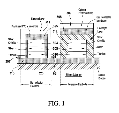
FIG. 1 depicts in a topological manner the cross sectional layers of a BUN
sensor
according to one non-limiting embodiment of the present invention. The BUN
sensor shown
includes reference sensors described in the process reflected by FIGS. 2(a)-
(c). The silicon
wafer 320 is covered with a silicon dioxide layer 315, along with titanium or
titanium-
tungsten alloy 310, followed by silver 305 and silver chloride 304. A PVC
ionophore
composition 325 is printed above the silver/silver chloride layer, followed by
the EIL enzyme
layer 311 containing the urease enzyme.
In FIG. 1, the reference electrode also contains all the above described
layers found
up to the silver/silver chloride layer 304, but also contains an electrolyte
layer 312, a gas
permeable membrane 308 and optionally is processed with a photoresist cap 309.
Automated integrated microdispensing and spot curing system
In a preferred aspect of the invention, the microfabricating process of the
invention is
an automated system, which is able to microdispense precise and programmable
amounts of
the materials in select regions of a planar surface material used in the
sensors of interest and
additionally provide for integrated curing, preferably spot-curing, by means
of UV radiation.
The key concept in this aspect is controlling the time domain with regard to
the
microdispensing step and the timing and duration of the subsequent UV exposure
step. Here,
control of the time domain is typically in fractions of seconds. This is
consistent with high
volume manufacturing processes familiar for microfabricated devices.
In one embodiment, the dispensing head comprises a syringe needle with a
reservoir
for the matrix, and a displacement means for controlling the dispensed volume
from the
syringe and onto the selected surface. The apparatus also includes a step and
repeat
mechanism for moving the surface, e.g., silicon wafer, with respect to both
the dispensing
head and said UV radiation source, thus enabling the formation of an array of
immobilized
enzyme layers at a set of pre-selected locations.
Preferably the controlled volume that is dispensed is in the range of from
about 1 nL
to about 10 L, e.g., from about 5 nL to about 1 gL or from about 50 nL to
about 0.1 L, and
the dispensed volume will cover an area in the range of about 10 square
microns to about 75
19
CA 02710312 2010-06-21
WO 2009/082699 PCT/US2008/087730
square millimeters. Preferably, this area is substantially circular with
radial dimensions in
the range of from about 5 m to about 5 mm.
In a preferred embodiment, the system provides a method of forming an
organized
array of immobilized layers on a substantially planar surface by dispensing a
sequence of
controlled volumes of a photoformable matrix, e.g., the above-described enzyme
immobilization compositions of the present invention, at a pre-selected set of
locations on a
surface. This is followed by applying UV radiation beam onto an area
substantially covering
each of the pre-selected locations. Importantly, this occurs in sequence,
starting at a
predetermined time after each controlled volume has been dispensed and
applying said
radiation at a predetermined intensity for a predetermined duration, to form
said immobilized
array of immobilized layers. Preferably, the predetermined time is in the
range of from about
0.1 to about 10 seconds, and the predetermined duration is in the range of
from about 0.1 to
about 10 seconds. Preferably, the method uses UV radiation in the wavelength
range of from
about 185 to about 400 run and having an intensity in the range of from about
100 mW/cm2
to about 10 W/cm2. As is known by experts in the field, curing can be effected
by generating
a specific dosage of UV radiation at a selected site. It is well known that
the parameters of
time, intensity and distance all impact the UV radiation dose and can be
adjusted to generate
a specific UV dose. Typically, a high intensity can generate more heat and can
have other
effects on the process. The preferred method also uses a planar surface that
is a silicon wafer
and the pre-selected set of locations is an array of sensors on said wafer,
typically based on
unit cells in an X-Y array. In terms of the sequencing of the UV exposure
after the printing
step, the method preferably operates in a manner where the UV radiation beam
is applied to
the Nth minus X pre-selected location while dispensing occurs at the Nth pre-
selected
location. Typically, X is equal to an integer from 1 to 10. In the preferred
embodiment for
the urease membrane (311 and 356), the parameters UV exposure parameters
include a 310
nm wavelength, 0.56 seconds of exposure to 4.2 W/cm2 of radiation.
FIG. 6 illustrates a microdispensing system according to one embodiment of the
present invention. As shown, the microdispensing system comprises a vacuum
chuck 106
and a syringe 102 and 105, each of which are attached to separate means for
altering one or
more of the vertical, horizontal, lateral, or rotational displacement of these
elements. For the
sake of economy, it is sufficient to have means for changing the vertical
displacement of the
CA 02710312 2010-06-21
WO 2009/082699 PCT/US2008/087730
syringe so long as one can change the position of the vacuum chuck multi-
directionally. The
movements of both elements may be controlled via a personal computer. In one
aspect, the
position of the vacuum chuck may be reproducible within 51 microns or better
in either or
both the x and/or y directions and the flatness of the chuck is within 1
micron.
The matrix formulations of the preferred embodiments of the present invention
can be
loaded into a microsyringe assembly 102 for the purpose of establishing layers
in a
controllable manner. The microsyringe assembly is preferably equipped with 25
to 30 gauge
needles 105 having an internal diameter of 150 gm and an external diameter of
300 gm.
Typically, the microsyringe needle 105, which includes an elongated member and
a needle
tip, is made of a metallic material, such as, for example, stainless steel.
Additional layers may
be coated onto the needle to change its surface properties. Furthermore, other
materials such
as synthetic polymers may also be employed in manufacturing the main body of
the needle,
itself. Depending on the pretreatment of the electrode surface and the volume
amount of fluid
applied, membrane layers of a thickness ranging from about 1 to about 200 gm
can be
obtained consistently.
The UV cure microdispensing subsystem (FIG. 6) optionally comprises a valve
101
connected to tubing which connects to the needle holder and barrel 102, as
well as the needle
105. The microdispensed drops can be optically monitored using a microscope
100. The
microdispensed drops are cured using the UV light from the focusing lens
assembly 104
using radiation from optional light-guide fiber 103.
Non-limiting FIG. 7 shows the radiation focused in focusing lens assembly 104
using
radiation from the light-guide fiber 103, which radiation is generated by a UV
bulb 151 in the
UV light box 150. As shown, the amount of light radiation and the time of
exposure is
controlled using a shutter/aperture 152 in the light box 150. The latter is
optionally
performed with an algorithm in a computer.
One non-limiting embodiment of the microdispense system is further illustrated
in
FIG. 8(a) where the pneumatic valve 101 generates pressure for the system
along tubing 132
into the needle holder and barrel 102. As shown, needle holding and barrel 102
is held by
syringe housing 131, which holds syringe 130. Syringe 130 allows the delivery
of
microdispensed drops through needle 105.
21
CA 02710312 2010-06-21
WO 2009/082699 PCT/US2008/087730
An optional embodiment of the UV focusing lens assembly 104 is further
illustrated
in FIG. 8(b) wherein the UV light is provided to the concentrator housing 401
via light-guide
fiber 103 through light-guide 405. The concentrator housing 401 is attached to
light-guide
fiber 103 using a mounting bracket 400. The light is focused in this example
with two lenses
403 held in place with a lense spacer 404 in the lense bracket 402. One lense
might be
sufficient, but additional optional lenses are optional, and understood by
those skilled in the
art.
The optionally computer controlled process for the microdispensing and UV
curing
may be run by an algorithm depicted in FIG. 9 wherein the needle is moved up
and away
from the X-Y tray which in turn is placed into the first print position. The
needle is moved
down into close proximity to the printing location, followed by print pressure
being
generated for a brief time period. The needle is then raised after the print
pressure has
finished dispensing a droplet. Concurrently with the microdispensing process,
the trailing
UV cure process is started once the X-Y tray has positioned itself to a new
print location and
finishes prior to it moving again. This process is repeated for each print
location until all
positions are printed and cured.
The drop sizes that can be dispensed reproducibly extend over a wide range.
For
volume sizes between about 5 to about 500 nanoliters (nL), the drops can be
applied
preferably with a precision of about 5%. A solenoid having a 0.1% precision
rating is
sufficient for this purpose. The height of the tip of the syringe needle above
the sensor
preferably is between about 0.1 to about 1 mm, depending on the volume to be
dispensed.
Generally, the smaller the volume of the drop, the lower the elevation of the
needle from the
sensor. The precise alignment of the syringe needle with the preselected area
of the sensor
can be achieved optically by means of a camera and a reticle. Such an
operation can be
performed manually by an operator or automatically by means of a visual
recognition system.
The latter is preferred.
It is useful to consider the dynamics involved when a single drop of fluid is
formed
and expelled from a needle. As more fluid is expelled from the needle tip, the
drop will grow
in size until the gravitational force acting on the mass of the drop exceeds
the opposing
forces maintaining contact with the needle tip. These opposing forces include
the adhesive
forces between the needle tip and the fluid or liquid, and surface tension of
the liquid itself. It
22
CA 02710312 2010-06-21
WO 2009/082699 PCT/US2008/087730
is well established that at low liquid flow rates where discrete drop
formation is complete, the
drop volume is fixed. However, the volume may be changed by varying any of the
fluid
related parameters discussed above, or by changing the diameter of the needle
tip thus
changing the available surface area for fluid adhesion. For example, a
hydrophobic
polytetrafluoroethylene (PTFE) coating applied to the needle tip reduces the
natural drop size
of an aqueous based matrix material by reducing the adhesive forces between
the drop and
the needle tip. In circumstances where a controlled volume must be
microdispensed onto a
surface, it is possible to have the microsyringe tip positioned above the
planar surface at a
height which does not allow the drop to form completely (and then fall to the
surface under
the influence of gravity), but the partially formed drop actually contacts the
surface and the
new adhesive forces between the liquid and the surface begin to spread the
drop. If the needle
tip is now retracted in the Z-direction a sufficient distance away from the
surface, then the
cohesive forces of the liquid is overcome and a volume of liquid less than the
fixed drop size
will remain in contact with the surface. This technique can be used to
dispense reproducibly
any volume of liquid from about one-one thousandth of the fixed drop size and
greater.
The surface tension between a pure liquid and its vapor phase can be changed
by
adding reagents. For example, a fatty acid added to water reduces the surface
tension,
whereas added salts can increase surface tension. The microdispensable fluid
compositions of
the present invention preferably are prepared to have a controlled optimized
surface tension.
Suitable additives may be used when necessary. The hydrophobicity or
hydrophilicity of the
fluid is controlled in the same manner. Where a cured membrane is required as
the end
product, the solids content and volatile solvents content preferably are
carefully adjusted.
Moreover, the ratio of these components is also used to control the viscosity.
The preferred microdispensable compositions for the ammonium ion sensor
comprises PVC polymer, plasticizers, ionophores and solvents with viscosities
generally
higher than those used for planar casting (e.g., spin-coating) of membranes.
These higher
viscosity compositions cure or dry without deformation of the membrane layer.
Related
problems, e.g., that of ensuring the homogeneity of the matrix at high
viscosity and thus
preventing phase separation of materials after time (i.e., considerations
related to shelf-life)
are also alleviated by these compositions. Other additives are also used to
prevent long-term
degradation of the membranes.
23
CA 02710312 2010-06-21
WO 2009/082699 PCT/US2008/087730
In addition to the factors described above relating to controlled volumetric
dispensing
of fluids having an optimized surface tension associated with a prescribed
composition,
tailoring the surface free energy of the substrate, or surface onto which the
fluid is dispensed,
provides control over the final dimensions, especially the thickness, of the
resulting layer.
The resulting process is highly versatile, allowing the deposition of arrays
of layers of varied
composition and utility. For establishing thick membranes, (e.g., 40-60 m
thick), the surface
is preferably tailored so that the contact angle which the microdispensed
fluid makes with the
surface is large. For example, before an aqueous based enzyme matrix is
microdispensed, the
surface may be first plasma treated to give a controlled contact angle. For
the preferred
urease matrix, a carbon tetrafluoride plasma step yields a contact angle in
the range 50 -70 .
An improved aspect of the microdispensing system, described here, is the
integration
of an automatic spot curing component. An EXFO Omnicure UV system is preferred
for
integration due to its ability to continually monitor and adjust the light
aperture to assure that
the radiation intensity remained consistent throughout the process. This
ameliorates the issue
of a typical UV bulb intensity decreasing over its lifetime (2000 working
hours) by using
50% intensity as the set point. As there is a relationship between cure time
and bulb
intensity, a reasonably high setting is required to reduce the product
processing time.
Another aspect of the UV cure process is the desire to focus the beam on the
specific
sensor to avoid UV exposure to other sensors. Focusing the beam needs to be
appropriate to
avoid being too limiting. This is because the visualization system used to
align each sensor
that is being processed needs enough flexibility to assure a robust process in
the event that
they are not accurately aligned. Intensity is related to the distance of the
UV beam to the cure
site, therefore, by being closer the intensity is increased and the product
processing time is
decreased.
While the invention is described primarily in terms of a silicon wafer with
microfabricated ion-selective electrodes, other types of sensors can be
fabricated to
incorporate a surface onto which the disclosed composition can be dispensed or
coated.
These include optical sensors, fiber optic sensors, surface acoustic wave
sensors, evanescent
sensors, surface plasmon resonance sensors and optical wave guide sensors. It
also includes
various base sensors, e.g., electrodes, ion-selective electrodes,
potentiometric electrodes,
amperometric electrodes, conductimetric electrodes, enzyme electrodes,
biosensors, optical
24
CA 02710312 2010-06-21
WO 2009/082699 PCT/US2008/087730
sensors, fiber optic sensors, surface acoustic wave sensors, evanescent
sensors, surface
plasmon resonance sensors and optical wave guide sensors. Substantially planar
surfaces for
sensor fabrication can include silicon wafers, alumina wafers, liquid crystal
substrates, glass
substrates and plastic substrates and flexible plastic substrates. In
preferred embodiments,
membrane-forming compositions are exposed to sufficient UV radiation to cause
significant
cross-linking, thus forming an adhered non-swelling immobilized enzyme layer
on the
surface.
The integrated microdispense and UV cure device is preferably automatically
programmed in order to optimize the manufacturing process time and to effect
UV curing.
The microdispensing and UV curing steps preferably are run in tandem. In a
preferred
aspect, the UV curing step takes approximately 0.5 seconds, whereas the
microdispense step
takes about 0.3 to 0.4 seconds. Therefore, the microdispense step is typically
rate limited by
the indexing time of approximately 0.1 seconds between print sites. The
microdispense and
the UV cure subsystems preferably operate at two different, but adjacent
physical locations
during the same time period, wherein the microdispense step occurs before and
ahead of the
UV cure operation. FIG. 9 provides a preferred algorithm for operation.
The dispensing apparatus with the integrated UV radiation source preferably
has a
registration and alignment means capable of focusing a beam of radiation onto
an area
substantially covering the location at which a drop of matrix has been
dispensed. A
computer means is able to switch the UV radiation on and off, and this occurs
at a
predetermined time and for a predetermined duration (and also at a
predetermined intensity),
after the matrix has been dispensed. The registration and alignment means
permits a beam to
be focused on a selected area of said surface and illuminate an area in the
range of about 10
square microns to about 75 square millimeters.
Each wafer preferably is manufactured with a plurality of chips (typically
about one
thousand on a 5 inch wafer), each containing one or more sensors and in this
case each
containing the BUN sensor. These sensors are desirably arranged in a uniform X-
Y
arrangement on the wafer. For processing of the wafers in the preferred
embodiment, the
chips are preferably generated by first placing an adhesive tape on the back
of the wafer
followed by cutting the wafer into individual chips using, for example, a
diamond dicing
saw. This process causes a slight displacement and uneven arrangement compared
to the
CA 02710312 2010-06-21
WO 2009/082699 PCT/US2008/087730
original location of the chip on the wafer. To compensate for this, a
microscope 100, as
depicted in FIG. 6, is used along with visual recognition software to realign
the chip for the
microdispense process. For the initial alignment and registration of the UV
cure system, a
UV sensitive paper is used to determine the proper alignment. Alternatively, a
visible light
source can be inserted in place of the UV light source 151 above the light
guide 103 in order
to align and focus the UV cure site. Yet another alternative approach would be
use a UV
monitoring device (radiometer) which has a focusing point and the intensity
and activity is
used to align and focus the UV cure site. Note that alternatively the dicing
step can be
performed after dispensing and spot-curing, however care is required to ensure
that the water
coolant used for the dicing blade does not result in dicing dust damaging the
cured
membranes. Where the substrate is plastic rather than a silicon wafer, dicing
is by a simpler
cutting process where dust damage is not an issue. Here dicing after
dispensing and spot-
curing is preferred.
Cartridge Construction for the use of improved sensors
The diced silicon chips described above are then preferably used as
subcomponents
for disposable plastic cartridges. Each cartridge typically contains several
features allowing
it to process a patient sample with an analyzer device and determine the
presence or amount
of an analyte, e.g., urea, in the sample.
Referring to the figures, the cartridge for accepting chips of the present
invention
comprises a cover (two views), FIGS. 10, 11, a base, FIG. 13, and a thin-film
adhesive
gasket, FIG. 12, disposed between the base and the cover and securing them
together.
Specifically, the backside of the cover shown in FIG. 10 mates with the
exposed face of the
gasket of FIG. 12, and the backside of the gasket mates with the exposed face
of the base of
FIG. 13. Referring now to FIG. 10, the cover 1 is made of a rigid material,
preferably plastic,
and capable of repetitive deformation at flexible hinge regions 5, 9, 10
without cracking. The
cover comprises a lid 2, attached to the main body of the cover by a flexible
hinge 9. In
operation, after introduction of a sample into the sample holding chamber 34,
the lid can be
secured over the entrance to the sample entry port 4, preventing sample
leakage by means of
deformable seal 11, and the lid is held in place by hook 3. The cover further
comprises two
paddles 6, 7, that are moveable relative to the body of the cover, and which
are attached to it
by flexible hinge regions 5, 10. In operation, when operated upon by a pump
means, paddle 6
26
CA 02710312 2010-06-21
WO 2009/082699 PCT/US2008/087730
exerts a force upon an air bladder comprised of cavity 43, which is covered by
thin-film
gasket 21, to displace fluids within conduits of the cartridge. When operated
by a second
pump means, paddle 7 exerts a force upon the gasket 21, which can deform. The
cartridge is
adapted for insertion into a reading apparatus, and therefore has a plurality
of mechanical and
electrical connections for this purpose. It should also be apparent that
manual operation of
the cartridge is possible. Thus, after insertion of the cartridge into a
reading apparatus, the
reading apparatus transmits pressure onto a fluid-containing foil pack filled
with
approximately 130 L of calibrant fluid located in cavity 42, rupturing the
package upon
spike 38, and expelling fluid into conduit 39, which is connected via a short
transecting
conduit in the base to the sensor conduit, 16. When the calibrant fluid
contacts the sensors,
they wet-up and establish a signal associated with the amount of calibrating
ion or molecule
in the fluid.
Referring to FIG. 12, thin-film gasket 21 comprises various holes and slits to
facilitate transfer of fluid between conduits within the base and the cover,
and to allow the
gasket to deform under pressure where necessary. Holes 30 and 33 permit one or
more urea
sensors and one or more reference electrode that are housed within either
cutaway 35 or 37,
to contact fluid within conduit 16.
Referring to FIG. 13, conduit 34 is the sample holding chamber that connects
the
sample entry port 4 to first conduit 16 in the assembled cartridge. Cutaways
35 and 37 are
locations in the housing for accepting the chips of the present invention.
Optionally they also
house a conductimetric sensor for determining the position of air-liquid
boundaries. Recess
42 houses a fluid-containing package, e.g., a rupturable pouch, in the
assembled cartridge
that is pierced by spike 38 because of pressure exerted upon paddle 7 upon
insertion into a
reading apparatus. Fluid from the pierced package flows into the second
conduit at 39 and
then into conduit 16. An air bladder is comprised of recess 43 which is sealed
on its upper
surface by gasket 21. The air bladder is one embodiment of a pump means, and
is actuated by
pressure applied to paddle 6 which displaces air in conduit 40 and thereby
displaces the
sample from sample chamber 34 into conduit 16.
Improved method of forming and curing membranes arrays
In one embodiment, the method of manufacture of a BUN sensor requires two
separate printing events. The first step involves an NH4+ ion-selective
electrode (ISE) print
27
CA 02710312 2010-06-21
WO 2009/082699 PCT/US2008/087730
step followed by a urease layer print step. As described above, the printing
process
preferably uses a pneumatic pump and valve with a fine gauge needle on top of
an X-Y table
to accurately microdispense a drop onto a specific location on a wafer.
Several controllable
factors contribute to overall sensor performance. These include: (i) accurate
registration of
the printed drop over the desired location, (ii) the appropriate viscosity and
surface tension of
the drop, (iii) the associated hydrophobicity of the surface, (iv) the height
and width (or
volume) of the drop, (v) the height of the drop after drying on the surface,
and (vi) good
adhesion. Furthermore, the formation of crystals (partitioning) or cracking of
the dried
droplet can adversely affect sensor performance.
The prior art enzyme membrane (see, e.g., U.S. Patent No. 5,200,051) is
composed of
a film-forming latex, preferably ELVACE (Forbo Adhesives Synthetic Polymers,
Morris,
Illinois). However, this heterogeneous material can be susceptible to drying
and blocking the
microdispensing needle tip which can have an adverse affect on manufacturing.
The surface
tension of the ELVACE can also create irregular shaped structures which may
impact the
performance of the sensor. See FIG. 3(a). Additionally, ELVACE, which is a
vinyl acetate
ethylene (VAE) copolymer composed of hydrophilic and hydrophobic domains, can
degrade
over time, most likely generating acetate which makes the material more
acidic. This time
dependent process reduces the usable lifetime of the material. As a result, it
is desirable to
replace this heterogeneous matrix with a substantially more stable
composition, preferably a
homogeneous aqueous matrix which can be photoformed and provides a stable
immobilization environment for enzymes, e.g., urease.
The present invention solves several lifetime issues including: the lifetime
of the raw
materials, the lifetime of the aqueous matrix prior to print, and the lifetime
of the printed and
cured matrix in the completed sensor. It also survives contacting a blood
sample in the final
product without dissolving away. This provides evidence of good adhesion
characteristics
that are highly desirable for reliable sensor performance.
Most importantly for a reliable manufacturing method, the present invention
provides
a microdispensable matrix that remains in solution at storage temperatures in
the range of
about 4 C to about 35 C without precipitation of the sub-components. It can
also be stored
frozen and melted for use without deleterious effect. Advantageously, the
matrix
28
CA 02710312 2010-06-21
WO 2009/082699 PCT/US2008/087730
composition also has adequate UV transmission, printed at thicknesses in a
range extending
to about at least 200 m, to have a high degree of polymer conversion
throughout the matrix.
The present invention is advantageous as the matrix exists in a state of low
viscosity
during the printing process, and then is controllably converted by UV
radiation into a gelled
or fixed state. This controlled approach using a UV cure process requires the
incorporation
of a UV photo-initiator in the formulation. This also requires a curing system
that can
generate controlled UV radiation directed to the part being cured. This
specification of this
UV curing system needs to provide for (i) simple automation, (ii) operation on
a short cycle
time compatible with the printing system, (iii) avoiding racking diced wafers
during the
curing process, (iv) avoiding the need for an oven or heat curing step, and
(v) avoiding the
need for continuous matrix mixing. In addition, it is desirable to avoid
unnecessary material
wastage. As previously mentioned, from a manufacturing perspective it is also
useful if a
large batch of material is made, pre-aliquoted and stored in frozen form.
The present invention permits a robust manufacturing process. The aspects of a
robust
manufacturing process permit some range of precision at each step in the
process while
generating a consistent product result. For one step in the process, the print
thickness can
vary slightly. From data (see FIG. 18), the mean potentiometric signal was
advantageously
found to be independent of the thickness of the print. Further, the standard
deviation of these
values across a lot of wafers is fairly independent of print thickness. This
allows a wide
range of enzyme layer thicknesses without impacting product performance.
Various UV systems may be used including a light wand, UV flood cure with or
without an oven step, and a light-guide system. A UV laser can also be used
for this process
to deliver a focused beam of radiation to the printed sensor site. For the
process of curing
printed membranes on a silicon wafer or similar substrate, e.g., glass and
plastic, a light wand
is required to be positioned over every print location. This can increase the
cure step time by
n x t, where n is the number of chips/print locations and t is the time in
seconds of each UV
cure step. In one embodiment, a UV cure step is provided by a system
accurately moving a
light wand to each successive print location. The light wand movement is
integrated with the
print event at each print location.
For a system where the UV source moves independent of the print head, the
matrix is
first printed at a specific location by the print needle. The print needle
moves away from the
29
CA 02710312 2010-06-21
WO 2009/082699 PCT/US2008/087730
surface, preferably in the Z direction. The UV source then moves into
location, followed by
a UV cure exposure at the print location.
In a system where the UV source trails by a fixed off-set from the print site,
the
print needle prints in the fashion of a typewriter style print process (e.g.,
from left to right,
followed by a return to the left side and an index to the next line of print
sites, which are
again processed from left to right). A UV source is located one or more print
sites away, n,
from the print needle location. The print needle prints the first print site,
and then another
print site until it reaches n print sites. Trailing along behind the print
needle is the UV
source. Once the UV source is located at the first print site on the left, n
sites away from the
print needle, the UV source exposes and cures the print site. As the print
needle moves to the
next print site, the UV source moves to the next site and while the print
needle dispenses the
matrix, the UV source exposes the print site. This print and trailing UV cure
process
continues until the print needle reaches the right side of the planar surface.
In order to finish
the UV cure process, the needle continues to move to the right with the
associated movement
of the UV source which continues to cure the remaining n sites. The print
needle may be
programmed to stop printing for the remaining print sites. The print needle
and UV source
are indexed to the next row to be printed and the setup begins at the left
side of the planar
surface. It should be understood by those skilled in the art that the print
needle and UV
source could be fixed in their position, and the table holding the planar
surface moved to
effect the movement of the print needle and UV source to each individual print
site. Those
skilled in the art will recognize that other engineered registration means can
be used to
accomplish the objective of ensuring consistent control of the time domain,
such that each
dispensed layer is cured at a fixed time and in a fixed manner after it is
printed.
In the preferred embodiment a light-guide is used. The light-guide is
effectively a
conduit for light where the light is to be focused onto a small region of a
wafer and where it
is not delivered in a straight line from the UV source. The UV radiation is
directed along the
light-guide by a light fiber connection. This has the advantage of a light
wand system where
the lamp and filters are contained in the power box and the "flashlight" uses
a very small
footprint at the location of the cure site. Additionally, the UV radiation
contains less
attendant heat from infrared radiation and therefore keeps the cured part
cool.
CA 02710312 2010-06-21
WO 2009/082699 PCT/US2008/087730
Where the particular photoinitiator present in the matrix requires specific
radiation wavelengths, this is achieved by selection of specific UV bulbs
appropriate for that
specific photoinitiator. Most standard UV bulbs generate multiple wavelengths,
some
desired and some not necessarily desired. In applications with biological
materials, it is
preferred to have a specific radiation wavelength with no additional
extraneous radiation at
other wavelengths. Note that it is well known in the scientific literature
that UVC radiation
is less damaging biologically than UVA or UVB. Therefore, avoiding or limiting
these
wavelengths is preferable for biological samples. In the preferred matrix
formulation which
uses Irgacure 2959, the preferred radiation wavelength is about 310 nm. Common
UV bulbs
use mercury (type "H") and metal halide ("D"), which can both be used to cure
as they
generate UV A and UV B radiation required for the Irgacure 2959 photoinitiator
containing
matrix. However the "H" bulb has less extraneous radiation wavelengths and is
preferred.
It is beneficial for the UV system to have an integrated optical filter
allowing the
passage of specific wavelengths of non-ionizing radiation, while preventing
the transmittance
of undesired wavelengths. In the preferred embodiment, radiation near 310 nm
is required
for the photoinitiator. As a means to limit the exposure of the sensor to
deleterious
wavelengths of radiation, a narrowband filter such as the Gilway and
International Light
Technologies, Inc. (Peabody, MA) Narrowband Filter NS313 which efficiently
only permits
wavelengths from about 300 to 340 nm is useful for preferred embodiments.
Other filters
used singly or in combination will be apparent to those skilled in the art to
affect appropriate
radiation wavelengths specific for certain other photoinitiators.
In a preferred embodiment, the light-guide system (FIGS. 6, 7 and 8b) has the
advantages of spot curing with the capability to direct light to a specific
location. In addition
it can easily be filtered and does not generate the heat found in flood
exposure based systems
filtering Infrared (IR) radiation. It also does not require a separate shutter
system, as this
feature is already integrated into the device. This approach is also desirable
for integration
with the microdispensing system, as the power supply can be positioned outside
of the
microdispense housing with only the light-guide and concentrator housing with
its associated
lenses inserted inside the microdispensing unit minimizing footprint.
It is desirable that the UV-curable enzyme matrix has the following
characteristics: (i) compatibility with enzymes and particularly for the
preferred embodiment
31
CA 02710312 2010-06-21
WO 2009/082699 PCT/US2008/087730
the enzyme urease enzyme, (ii) exhibit the appropriate flow and viscosity for
good printing
characteristics, (iii) compatibility with a reliable chemical formulation
which can support a
high-yield manufacturing process, e.g., incorporate an antimicrobial agent for
improving
matrix shelf-life and reagents for stabilizing an enzyme, (iv) be a water
based technology to
support biological reagents, (v) achieve reliable adhesion to a surface after
UV curing step,
(vi) provide for rapid wet-up when used in conjunction with a sensor, (vii)
exhibit the
appropriate enzyme substrate and water permeability when formed as a layer to
support and
sustain the enzyme reaction (viii) exhibit good electrical characteristics
when used with an
electrochemical sensor, and (ix) show an extended post-processing lifetime of
greater than
about 6 months at room temperature or under refrigeration, e.g., be compatible
with genuine
commercial product requirements. The compositions described in FIG. 14 have
these desired
characteristics.
The UV curing systems described herein can be used with each UV curable
formulation to characterize many operating parameters including the precision
and accuracy
of the dimensions of cured membranes and adhesion of the membrane to the
surface. The
sensor can then be tested in cartridges to determine sensor performance with a
given matrix
and curing combination. This can include intra-wafer and inter-wafer
variations, where each
wafer may contain as many as a thousand sensors.
In FIG. 16 the effect of radiation on the sensor performance based on the
matrix
of FIG. 14(a). Batches of sensors were prepared using a flood lamp process and
processes
with 0.5s, Is, 2s and 3s spot-cure UV radiation with radiation doses of 190,
380, 760 and
1140 mJ, respectively. These data show that the processes are robust with
various conditions
of radiation giving acceptable test results.
FIG. 15 demonstrates that compositions containing alternative monomers and
crosslinkers generated similar sensor signals with test solutions. The
monomers tested
included acrylamide, methacrylamide, poly(ethylene glycol) acrylate (PEGA),
and N-[3-
(Dimethylamino)propyl]-methacrylamide (DMAPMA). Additionally, the crosslinkers
tested
included 1,4-bis(acryloyl)piperazine, polyethylene glycol diacrylate
poly(ethylene glycol)
diacrylate (PEGDA), N,N'-(1,2-dihydroxyethylene)bis-acrylamide (DHEBA) and
trimethylolpropane ethoxylate triacrylate (TMPETA).
32
CA 02710312 2010-06-21
WO 2009/082699 PCT/US2008/087730
FIG. 17 shows an experiment where the absence of the humectant glycerol under
accelerated lifetime storage conditions (e.g., 30 C or 40 C) had an affect on
performance. By
adding glycerol to the present matrix (see compositions in FIG. 14), a process
with good
performance and shelf-life is achieved.
Improved blood urea nitrogen sensor manufacture
In one aspect, the intent of the present invention is to improve on the
manufacture
of BUN (blood urea nitrogen) sensors. The preferred embodiment of the new BUN
sensor is
manufactured using a combination of thin-film microfabrication processes and
microdispensing techniques. It comprises a thin film silver-silver chloride
indicator electrode
operating in combination with a thin-film silver-silver chloride reference
electrode of the
type described in U.S. Patent No. 4,933,048, incorporated by reference herein.
A more
preferable reference electrode is described in Published U.S. Appl. No.
20070015977,
incorporated by reference herein.
In the initial step, a substrate wafer of silicon is overlaid with an
insulating layer
of silicon dioxide, by thermal oxidation. Metal layers of a titanium and
tungsten alloy (TiW)
and then silver are subsequently deposited onto the silicon dioxide base wafer
and then
patterned using photolithographic techniques. An electrically insulating layer
such as
polyimide polymer or additional silicon dioxide is then photo-patterned to
isolate adjacent
sensor circuitry. The silver-silver chloride indicator electrode (diameter -
200 microns) is
prepared from the patterned silver using standard techniques, e.g.,
electrochemical, chlorine
gas plasma and oxidation of Ago by an inorganic oxidant such as Cr2072- or Fe
3+ in the
presence of chloride ion.
The remaining layers of the BUN electrode include two thick-film structures:
(i) a
semi-permeable membrane film, comprising an organic polymer layer (e.g.,
poly(vinyl
chloride) - PVC), and an ammonium ion ionophore; and (ii) the outermost
biolayer,
comprising in this particular sensor, a spot photo-cured acrylamide urease
layer that
optionally includes carbonic anhydrase. These layers are deposited by a
microdispensing
technique as described in U.S. Patent No. 5,554,339, incorporated by reference
herein. In the
present invention, however, the microdispensing assembly described in the `339
Patent has
been substantially improved to include an integrated ultra-violet spot-curing
component
33
CA 02710312 2010-06-21
WO 2009/082699 PCT/US2008/087730
system enabling automated printing and curing in a controlled time domain, as
described
above.
The thick-film ammonium ion-sensitive structure comprises a poly(vinyl
chloride)
(PVC) binder, tris(2-ethylhexyl)phosphate as a plasticizer, and nonactin as
the ionophore.
The indicator electrode can be made selective for different ions by using the
same (or
similar) binder and plasticizer composition but with different ionophores. For
example,
valinomycin, monensin and (methyl)monensin, and tridodecylammonium chloride
have been
used to make potassium, sodium, or chloride-ion selective electrodes,
respectively. Other
ionophores may include, but are not limited to crown ethers, trialkylamines,
or phosphate
esters, and the like. Alternatively, other polymeric binder materials may be
used besides
PVC. These polymers may include, for example, silicon rubber,
polytetrafluoroethylene
plastics, or derivatives of PVC containing ionizable functional groups (e.g.,
carboxylates).
Other plasticizers suitable for use in the present invention may include, but
are not limited to
tris(2-ethylhexyl)phosphate, nitrocymene, 2-nitrophenyloctyl ether, dibutyl
sebacate, diethyl
adipate, phthalates, propylene carbonate, 5-phenylpentanol, or mixtures
thereof. Still other
binders and ionophore combinations may occur to those skilled in the art,
which are within
the scope of the present invention. The resulting semi-permeable ion-selective
film may have
a thickness in the range of about 2 microns to about 200 microns, preferably
about 10 to
about 30 microns. In the preferred embodiment, the ammonium ion-selective
membrane
solvent system is selected to provide the appropriate surface tension and
stability. The solids
content (wt %) of plasticizer, PVC polymer, and ionophore are preferably 60-
80%, 15-40%
and 0.5-3%, respectively.
Various methods can be used to define a layer on a planar substrate. If a
thick
layer (about 5 to about 200 microns) is required, microdispensing of a viscous
matrix, e.g.,
the photo-curable urease matrix described above, is generally preferred. Other
methods for
defining a layer on a planar substrate include, without limitation, spin-
coating, dip-coating,
spray coating, screen printing, ink jet printing, laser printing, painting and
contact printing
are alternative methods and may be better suited to a different applications.
For example, the
preferred urease photoformable matrix may be screen printed onto a wafer in a
single pass at
a specific time (t=0). The screen optionally has an opening of 300 m
diameter, with each
opening registered for alignment with an array of ammonium ion-selective
membranes on the
34
CA 02710312 2010-06-21
WO 2009/082699 PCT/US2008/087730
wafer. After the printing step a flood UV exposure of the wafer is performed
at t=1.
Similarly to the spot-curing method described above, this method also gives
control of the
time domain from printing to the UV step for each individual matrix. In this
embodiment t=1
is preferably automatically set at 2-20 seconds after t=0. Automated equipment
for moving
wafers from a printing station to an exposure station is well known in the
microfabrication
art. The preferred urease matrix can also be spin-coated and photo-patterned
with a mask, in
the manner widely used in the microfabrication art.
Referring now to the topological illustration in FIG. 1, the substrate wafer,
320, is
silicon, with an overlaid insulating layer of silicon dioxide, 315. In
addition there is a
polyimide layer 301 with two circumferential print wells (302, 303) which are
used to
confine the printed layers. The first metal layer, 310, is TiW and serves the
functions of a
conductor and an adhesion layer to the wafer. Succeeding layers 305 and 304,
are the silver
and silver chloride layers. On the left side of FIG. 1, the remaining layers
of the indicator
electrode include (i) a semi-permeable membrane film, 325, comprising an
organic polymer
layer (e.g., polyvinyl chloride (PVC)) and an ammonium ion ionophore; and (ii)
the
outermost biolayer, 311, comprising in this particular embodiment, an
acrylamide photo-
cured urease layer of the preferred composition described above.
The reference electrode portion of the unit cell may be comprised of overlaid
structures as shown in FIG. 1. In this particular embodiment, the metal and
chloridized
layers of the reference electrode are covered by an electrolyte layer, which
may comprise any
material which is able to hold a high concentration of salt but which is,
preferably,
photoformable. In this respect, a polyvinyl alcohol (PVA) formulation is the
preferred
material and may first be photo-patterned and forms a water-permeable matrix
that can
subsequently be saturated with a salt, such as potassium chloride. A separate
gas permeable
membrane, may also be present which serves to diminish the loss of electrolyte
or salt to the
bulk analytical sample but allows the rapid wet-up (i.e., passage of water or
other small
gaseous molecules) of the reference electrode prior to commencing the sample
analysis. The
patterning process can be either of those described in U.S. Patent No.
4,933,048 and
Published U.S. Appl. No. 20070015977 both incorporated herein by reference.
Alternatively, a reference electrode structure can be used in which the
distance between the
liquid junction and the surface of the silver/silver chloride is sufficiently
large, such that the
CA 02710312 2010-06-21
WO 2009/082699 PCT/US2008/087730
concentration of electrolyte in the immediate vicinity of the Ag/AgC1
structure is
substantially constant for a period of time sufficient to perform a
measurement of the
potential difference between the indicator electrode and the reference
electrode.
Referring now to FIG. 2, indicator electrode and the adjacent reference
electrode
are each connected by an overpassivated signal line to a contact pad. The
overpassivation
(polyimide layer) includes print wells 302 and 303 formed as concentric
circles. The unit cell
is confined within a rectangular area, which is repeated in an array several
hundred times on
a single silicon wafer. In particular embodiments of the instant invention,
other indicator
electrodes may be present in the unit cell for the simultaneous measurement of
other species
(e.g., Na+, K+, Cl') in addition to ammonium ion.
To manufacture the BUN base sensor, a silicon wafer with a topical layer of
silicon dioxide, which had previously been cleaned, scrupulously with a
mixture of
concentrated sulfuric acid and hydrogen peroxide is placed into a plasma
deposition system
and layers of TiW (0.1 m) and silver (0.5 gm) are sputtered consecutively
onto the wafer
surface. The silver-titanium bilayer is then processed to localize it to a
region, which in the
final device acts as the ammonium ion sensor. This process is achieved by a
standard
lithographic technique in which the wafer is spin-coated with positive resist
(Shipley AZ
1370 SF). After UV exposure of the photoresist through a mask and development
(Shipley
AZ 351), the exposed silver is removed by an aqueous solution of ferric
nitrate (0.9 mM) as
the etchant. The underlying titanium layer is then processed by means of the
same
photolithographic steps, but using an aqueous mixture of nitric acid (3.9M)
and hydrofluoric
acid (0.78 M) as the etchant. N-methylpyrrolidone solvent is then used to
remove the
remaining photoresist to expose the required silver structures (diameter about
150 m).
To passivate the signal lines a photo-definable polyimide (DuPont 2703) is
spin-
coated onto the wafer. Once the wafer is UV exposed and developed with a
solvent the
polymer is baked in an oven at 350 C for 30 minutes under an inert atmosphere
and left to
cool to 150 C before removal. While the mask used for patterning defines the
perimeter of
the layer, it also defines print wells 302 and 303. These are subsequently
used to control the
dimensions of the two respective microdispensed membranes.
The silver is preferably then chloridized by dipping the entire wafer into an
aqueous solution of potassium dichromate (12 mM) and hydrochloric acid (60
mM). The
36
CA 02710312 2010-06-21
WO 2009/082699 PCT/US2008/087730
wafer is then washed and partially diced. Over these patterned silver chloride
electrodes is
placed an ammonium ion sensitive membrane. The membrane material is made by
dissolving
low molecular weight PVC (Sigma) and high molecular weight carboxylated PVC
(Type
Geon, Goodrich) (1:1 w/w) in a solvent system of cyclohexanone, propiophenone,
and N-
methylpyrrolidone (1:1:1 v/v/v) to a total solids content of 10 g/dL of
solution. Dissolution is
accomplished by heating the mixture at 70 C for 30 minutes. To this mixture
the plasticizer
tris(2-ethylhexyl)phosphate (Fluka) is added, to provide a total solids
content of 35 g/dL. The
resulting mixture is then allowed to cool to 45 C and nonactin (Fluka) is
added in the amount
equivalent to 2 percent of the total solids in the mixture. At room
temperature, 10-100 nL of
this final material is microdispensed onto each of the silver chloride
indicator electrodes on
the wafer, overlapping on all sides by at least about 30 m. Print well 302 is
preferably used
to define the perimeter. Curing is accomplished by placing the wafer on a 60 C
hot-plate for
30 minutes. This process yields a stable, rugged structure having a thickness
of about 15 m.
In the final step the preferred urease matrix, described above, is
microdispensed
onto individual membranes and UV spot cured using the apparatus. Print well
303 is
preferably used to define the perimeter. As mentioned above, in the preferred
formulation
the components are mixed together and frozen in a cryofreezer prior to use.
This allows
consistent production of product day-to-day and a long storage lifetime prior
to
microdispensing. This formulation can also be quality control (QC) tested
prior to a
microdispense event as the mixture can be assayed for enzymatic activity using
a standard
reference method using a spectrophotometer. This reduces the cost and waste in
making
product in the manufacturing process.
Regarding the preferred standard assay method, the urease enzyme activity from
an aliquot of the thawed frozen formulation is assessed using a modification
of the method of
Kaltwasser & Schlegel, "NADH-dependent coupled enzyme assay for urease and
other
ammonia-producing systems,"Analytical Biochemistry 16: 132-138 (1966). The
method
measures the spectrophotometric change of NADH to NAD+ at 340 nm in a
glutamate
dehydrogenase assay coupled to urease.
37
CA 02710312 2010-06-21
WO 2009/082699 PCT/US2008/087730
Cartridge analyses using the improved BUN sensor
In the preferred embodiment, the finished chips containing the sensors are
then
assembled into test cartridges and used to make BUN measurements in blood. One
embodiment of a cartridge of the present invention is shown in FIGS. 10-13.
The cartridge is preferably adapted for insertion into a reading apparatus,
and
therefore has a plurality of mechanical and electrical connections for this
purpose. It should
also be apparent that manual operation of the cartridge is possible. Thus,
after insertion of
the cartridge into a reading apparatus, the reading apparatus transmits
pressure onto a fluid-
containing foil pack filled with approximately 130 L of calibrant fluid
located in cavity 42,
rupturing the package upon spike 38, and expelling fluid into conduit 39,
which is connected
via a short transecting conduit in the base to the sensor conduit, 16. When
the calibrant fluid
contacts the sensors, they wet-up and establish an electrical signal
associated with the
amount of calibrating ion or molecule in the fluid.
Referring to FIG. 12, thin-film gasket 21 comprises various holes and slits to
facilitate transfer of fluid between conduits within the base and the cover,
and to allow the
gasket to deform under pressure where necessary. Holes 30 and 33 permit one or
more urea
sensors and one or more reference electrode that are housed within either
cutaway 35 or 37,
to contact fluid within conduit 16. Referring to FIG. 13, conduit 34 is the
sample holding
chamber that connects the sample entry port 4 to first conduit 34 in the
assembled cartridge.
Cutaways 35 and 37 optionally houses a conductimetric sensor for determining
the position
of air-liquid boundaries. Recess 42 houses a fluid-containing package, e.g., a
rupturable
pouch, in the assembled cartridge that is pierced by spike 38 because of
pressure exerted
upon paddle 7 upon insertion into a reading apparatus. Fluid from the pierced
package flows
into the second conduit at 39 and then into conduit 16. An air bladder is
comprised of recess
43 which is sealed on its upper surface by gasket 21. The air bladder is one
embodiment of a
pump means, and is actuated by pressure applied to paddle 6 which displaces
air in conduit
40 and thereby displaces the sample from sample chamber 34 into conduit 16.
In the preferred embodiment, the BUN sensor is packaged into a cartridge of
the
type disclosed in U.S. Patent No. 5,096,669, which also contains a calibrant
solution. It is
contained in a calibrant package (cal-pack), which is ruptured during the
blood sample
analysis. The typical sequence of events includes the cal-pack being ruptured
and then the
38
CA 02710312 2010-06-21
WO 2009/082699 PCT/US2008/087730
calibration solution passing over the sensor and wetting up the sensor.
Typically, the cal-
pack of prior art cartridges contained the following ions, sodium, potassium,
calcium,
chloride, bicarbonate and also HEPES buffer, glucose, lactate, urea, creatine
and creatinine.
As a diagnostic test for research purposes, urea was replaced with ammonia.
This permits
studies of the ammonium ionophore performance independent of the urease
containing
enzyme immobilization layer. The preferred constitution of the cal-pack for
the new BUN
sensor preferably include sodium, potassium, calcium, chloride, urea, HEPES
buffer,
glucose, lactate, creatine and creatinine.
A potentiometric chemical sensor for urea can be viewed as a system, which is
constructed from functionally dissimilar components. In one embodiment of the
blood urea
nitrogen (BUN) sensor, the outermost layer, the one in contact with the
analyte solution,
permits the transport of urea while also serving to immobilize the active
enzyme molecules.
These enzymes catalyze the hydrolysis of urea to ammonia as described above.
At neutral pH
values, the ammonia thus produced exists predominantly as ammonium ions. By
interposing
a separate layered structure, which contains an ionophore with high
sensitivity and selectivity
for ammonium ions between the enzyme containing layer and a silver-silver
chloride
indicator electrode, the ammonium ion concentration at the electrode interface
can be
measured. In this type of measurement, the potential difference between the
indicator
electrode and a reference electrode is recorded. This is done with a
potentiometric circuit in
an instrument (or analyzer) which makes connection with the two electrodes, as
is well
known in the electrochemical measurement art. The analytical value of the
measurement is
derived from the fact that the magnitude of the potential difference is
related by the Nicolsky
equation to the concentration of the analyte, in this case, urea:
E = E. + RT/nF log [A + E (a,b) k(a,b)B]
where E is the measured electromotive force (signal), R is the gas law
constant, T is the
absolute temperature, n is the absolute value of the charge on analyte species
a (e.g., n = 1 for
the ammonium ion), F is the Faraday constant, A is the activity of the analyte
species a, B is
the activity of an interfering chemical species b, k(a,b) is the interference
coefficient
associated with the effect of the presence of chemical species b on the
electrochemical
39
CA 02710312 2010-06-21
WO 2009/082699 PCT/US2008/087730
potentiometric determination of the activity of the analyte species a, and E.
is a constant
independent of T, A, or B. See Amman, D., Ion-Selective Microelectrodes,
Springer, Berlin
(1986) p. 68, and references cited therein, which is incorporated by reference
herein.
Data presented in FIGS. 4, 5 and 15-18 were recorded and/or analyzed in using
these principles. These data are presented using a commercial BUN testing
system for
comparison. The performance of the new sensor is superior to the established
technology.
Examples
Example 1
A solution of aprotinin is prepared by dissolving 0.01 g of aprotinin in 50 mL
of
deionized water, generating a 0.02% concentration stock solution. An enzyme
buffer
solution is prepared by the addition of 4.32 g of glycerol, 130 g of deionized
water, 130 g of
1M TRIS at pH 7.6, 2.6 g of 0.5M EDTA at pH 8.0, 13 g of the above 0.02%
aprotinin stock
solution, 0.2 g of 1,4-dithioerythritol, 2.7 g of sodium chloride, 0.097 g of
potassium
chloride, 0.05 g of sodium azide, 92.8 g of sucrose and 29 g of BSA. The
mixture is
vortexed until fully dissolved. A solution containing the acrylic resin
components is then
prepared, including 72.2 g of acrylamide, 150 g of deionized water, 29.4 g of
1,4-
bis(acryloyl)piperazine and 2.5 g of activated carbon. This acrylic resin
solution is mixed
until in solution and filtered using a 0.2 um filter into a clean container.
To this solution 17.8
g of 1-[4-(2-hydroxyethoxy)-phenyl]-2-hydroxy-2-methyl-l-propane-l-one is
added, mixed
and the solution is kept from excessive exposure to light in a container
wrapped in aluminum
foil. Finally 40 g of urease is added to about 333 g of the above filtered
solution and mixed.
About 44 g of deionized water is added, and finally 184 g of the acrylic resin
solution
(prepared as above) is added. The material is kept covered in aluminum foil
and mixed until
in solution. For long term storage, this solution is aliquoted in 1 ml
portions and frozen at -
60 C.
Example 2
In another embodiment of the urease enzyme immobilization layer included the
enzyme carbonic anhydrase. The order of mixing is as follows: A solution of
aprotinin is
prepared by dissolving 0.01 g of aprotinin in 50 mL of deionized water,
generating a 0.02%
concentration stock solution. An enzyme buffer solution is prepared by the
addition of 4.32
CA 02710312 2010-06-21
WO 2009/082699 PCT/US2008/087730
g of glycerol, 130 g of deionized water, 130 g of 1M Tris at pH 7.6, 2.6 g of
0.5M EDTA at
pH 8.0, 13 g of the above 0.02% aprotinin stock solution, 0.2 g of 1,4-
dithioerythritol, 2.7 g
of sodium chloride, 0.097 g of potassium chloride, 0.05 g of sodium azide,
92.8 g of sucrose,
29 g of BSA and vortexed until the contents are fully dissolved. A solution
containing the
acrylic resin components is then prepared, including 72.2 g of acrylamide, 150
g of deionized
water, 29.4 g of 1,4-bis(acryloyl)piperazine, and 2.5 g of activated carbon.
This acrylic resin
solution is mixed until in solution and filtered using a 0.2 um filter into a
clean container. To
this solution 17.8 g of 1-[4-(2- hydroxyethoxy)-phenyl]-2-hydroxy-2-methyl-l-
propane-1 -
one is added, mixed and the solution is kept from excessive exposure to light
using aluminum
foil. Finally 40 g of urease and 14 mg of carbonic anhydrase is added to about
333 g of the
above filtered enzyme solution and mixed. About 44 g of deionized water is
added, and
finally 184 g of the acrylic resin solution prepared above is added. The
material is kept
covered in aluminum foil and mixed until in solution. This solution is
aliquoted in 1 ml
portions and frozen at -60 C.
Any feature described or claimed with respect to any disclosed implementation
may be combined in any combination with any one or more other feature(s)
described or
claimed with respect to any other disclosed implementation or implementations,
to the extent
that the features are not necessarily technically incompatible, and all such
combinations are
within the scope of the present invention. Furthermore, the claims appended
below set forth
some non-limiting combinations of features within the scope of the invention,
but also
contemplated as being within the scope of the invention are all possible
combinations of the
subject matter of any two or more of the claims, in any possible combination,
provided that
the combination is not necessarily technically incompatible.
41