Note: Descriptions are shown in the official language in which they were submitted.
CA 02710366 2015-09-10
-1-
CAPPING BIOPROSTHETIC TISSUE TO REDUCE CALCIFICATION
Field of the Invention
=
[0002] The present invention relates generally to methods for treating
bioprosthetic tissue,
materials, and devices to reduce post-implantation calcification, and reducing
post-implantation
calcification, in particular for bioprosthetic tissue used in heart valves.
Background of the Invention
[0003] Heart valve replacement may be indicated when there is a narrowing of
the native heart
valve, commonly referred to as stenosis, or when the native valve leaks or
regurgitates, such as when the
leaflets are calcified. In one therapeutic solution, the native valve may be
excised and replaced with either
a biologic or a mechanical valve. Certain medical conditions may require
grafting or suturing a tissue
patch to repair physiological abnormalities. These include, but are not
limited to hernia repair, vascular
wounds, congenital heart defect repair and reconstruction, and bladder wall
repair.
100041 Tissue-type or "bioprosthetic" valves have flexible tissue leaflets
supported by a base
structure that project into the flow stream and ftmction much like those of a
natural human heart valve by
coapting against each other to ensure one-way blood flow. In tissue-type
valves, a whole xenograft valve
(e.g., porcine) or a plurality of xenograft leaflets (e.g., bovine or equine
pericardium) typically provide
fluid occluding surfaces. Synthetic tissue leaflets have also been proposed.
One or more flexible leaflets
mount within a peripheral support structure, for example as seen in the
CARPENTIER- EDWARDS
Porcine Heart Valve and PERIMOUNTTm Pericardial Heart Valve available from
Edwards Lifesciences
of Irvine, California.
CA 02710366 2010-06-21
WO 2009/085199
PCT/US2008/013879
- 2 -
[0005] Implantable biological tissues can be formed of human tissues
preserved by freezing (i.e., cryopreservation) the homograft tissues, or of
animal
tissues preserved by chemically fixing (i.e., tanning) the xenograft tissues.
The
type of biological tissues used as bioprostheses include cardiac valves, blood
vessels, skin, dura mater, pericardium, small intestinal submucosa ("SIS
tissue"), ligaments and tendons. These biological tissues typically contain
connective tissue proteins (i.e., collagen and elastin) that act as the
supportive
framework of the tissue. The pliability or rigidity of each biological tissue
is
largely determined by the relative amounts of collagen and elastin present
within the tissue and/or by the physical structure and configuration of its
connective tissue framework. Collagen is the most abundant connective tissue
protein present in most tissues. Each collagen molecule is made up of three
(3)
polypeptide chains intertwined in a coiled helical configuration.
[0006] The techniques used for chemical fixation of biological tissues
typically involve exposing the biological tissue to one or more chemical
fixatives (i.e., tanning agents) which form cross-links between the
polypeptide
chains within a given collagen molecule (i.e., intramolecular cross-linkages),
or
between adjacent collagen molecules (i.e., intermolecular cross-linkages).
Examples of chemical fixative agents that have been used to cross-link
collagenous tissues include: formaldehyde, glutaraldehyde, dialdehyde starch,
hexamethylene diisocyanate and certain polyepoxy compounds.
[0007] One problem associated with the implantation of many
bioprosthetic materials is that the connective tissue proteins (i.e., collagen
and
elastin) within them can become calcified following implantation in the body.
Such calcification can result in undesirable stiffening or degradation of the
bioprosthesis. This damage to the collagenous tissue leads to valve failure.
[0008] Of the various chemical fixatives available, glutaraldehyde (also
referred to as simply "glut") has been the most widely used since the
discovery
of its anti-immunological and anti-degenerative effects by Dr. Alain
Carpentier
in 1968. See Carpentier, A., J. Thorac. Cardiovascular Surgery, 58: 467-69
(1969). In addition, glutaraldehyde is one of the most common sterilization
CA 02710366 2010-06-21
WO 2009/085199
PCT/US2008/013879
- 3 -
agents. Glutaraldehyde is therefore used as the preferred fixative and
sterilant
for many commercially available bioprosthetic products, such as in the
bioprosthetic heart valves available from Edwards Lifesciences of Irvine,
California. Glutaraldehyde creates potential calcium binding sites within the
tissue that can lead to calcification in vivo, such as residual aldehydes,
acids,
Schiff bases, etc. These groups can contribute to calcification unless
mitigated
via capping. Mitigating such calcification is particularly important during
storage, especially when the tissue is not being stored in aqueous solution.
100091 Various techniques have been proposed for mitigating the in vivo
calcification of glutaraldehyde-fixed bioprostheses or for otherwise improving
the glutaraldehyde fixation process. Among these are methods described in U.S.
4,729,139 (Nashef); U.S. 4,885,005 (Nashef et al.); U.S. 4,648,881 (Carpentier
et al.); U.S. 5,002,566 (Carpentier); EP 103947 (Pollock et al.), U.S.
5,476,516
(Seifter et al.), and U.S. 5,215,541 (Nashef et al.). The techniques in U.S.
5,862,806 (Cheung) include dehydration using an organic solution (i.e.
ethanol,
but no glycerol) of glutaraldehyde-treated tissues, prior to the application
of a
chemical reducing agent such as sodium cyanoborohydride or sodium
borohydride in an organic solvent. This process involves only the addition of
a
reducing agent without any capping agents, such as proteins, amines or amino
acids. The calcification mitigation techniques disclosed in U.S. 6,471,723 and
U.S. 4,786,287 involve the addition of a variety of amines to detoxify the
aldehyde groups in glutaraldehyde-fixed tissue. These chemicals are not
permanently attached to the tissue (e.g., by addition of a reducing agent),
and so
diffuse out of the tissue over time, which dramatically lowers the calcium
mitigation efficacy of these treatments. The techniques in U.S. 5,476,516
involve the addition of polyols (e.g., glycerol) and alcohols to bioprosthetic
tissues as a calcification mitigation treatment alone, but do not address any
oxidation mitigation (i.e., capping) strategies. U.S. 6,509,145 and U.S.
7,078,163 address oxidation of bioprosthetic tissue for the purpose of
calcification mitigation. U.S. 6,630,001 and U.S. 6,277,555 discuss the use of
glycerol preservation and lyophilization of tissue, but do not discuss
chemical
CA 02710366 2010-06-21
WO 2009/085199
PCT/US2008/013879
- 4 -
methods to prevent oxidation. U.S. 6,352,708 includes glycerol preservation of
fresh, "non-fixed" tissue, and treatments with glycerol and heparin, but does
not
include combinations of chemical treatments to prevent oxidation or reduce
calcification with a glycerol drying step.
[0010] Recently a new technique of calcium mitigation by elevated-
temperature fixation of the tissue in glutaraldehyde was described in U.S.
6,561,970 (Carpentier et al.) and in combination with relative tissue/fluid
movement in U.S. 5,931,969 (Carpentier et al.). Another technique, involving
adjusting the pH of a glutaraldehyde fixation solution, is disclosed in U.S.
6,878,168 (Carpentier et al.) The Edwards Lifesciences XenoLogiXTM Tissue
Treatment eliminates up to 98% of phospholipids in an attempt to reduce
calcium binding sites. In the Carpentier-Edwards ThermaFixTm Advanced
Heart Valve Tissue Process, also from Edwards Lifesciences, both thermal and
chemical treatments are used to remove unstable glutaraldehyde molecules and
thus reduce calcium binding sites, resulting in a marked reduction in calcium
uptake versus glutaraldehyde-only controls.
[0011] Bioprosthetic valves are generally stored in glutaraldehyde or
formaldehyde solution, and must be rinsed prior to implantation.
Glutaraldehyde is widely used as a storage solution due to its sterilant
properties
but is known to contribute to calcification. Strategies to minimize
glutaraldehyde content in the final product have been demonstrated to mitigate
in vivo calcification. Studies have shown that storage solutions without
gluaraldehyde reduce in vivo calcification compared to those with
glutaraldehyde. (Mirzaie, et al. Ann Thorac Cardiovasc Surg 2007 13:102).
[0012] One such strategy to avoid glutaraldehyde as a storage solution is
to dehydrate the bioprosthetic tissue in a glycerol/ethanol mixture, sterilize
with
ethylene oxide, and package the final product "dry". This process circumvents
the potential toxicity and calcification effects of glutaraldehyde as a
sterilant
and storage solution. There have been several methods proposed to use
glycerine, alcohols, and combinations thereof as post-glut processing methods
so that the resulting tissue is in a "dry" state rather than a wet state with
excess
CA 02710366 2010-06-21
WO 2009/085199
PCT/US2008/013879
- 5 -
glut. These approaches avoid the use of aqueous liquid aldehyde, or liquid
sterilant as storage solutions for tissue and devices. Glycerol-based methods
can
be used for such storage as described in the following examples. The storage
of
heart valve tissue in glycerol was described by Parker et al. (Thorax 1978
33:638), but does not include any calcification mitigation techniques and does
not describe any advantages. Also, U.S. 6,534,004 (Chen et al.) describes the
storage of bioprosthetic tissue in polyhydric alcohols such as glycerol.
However, neither of these addresses mitigating potential oxidation of the
tissue.
[0013] In processes where the tissue is dehydrated in an ethanol/glycerol
solution, the tissue may be sterilized by ethylene oxide, gamma irradiation,
or
electron beam irradiation. Ethylene oxide sterilization requires exposing the
tissue to increased temperatures and water vapor which will generate oxidative
damage in the tissue (Olde Damink, LH. et al. J Biomed Mater Res 1995
29:149). Gamma irradiation is known to generate significant reactive oxygen
species in collagenous substrates which causes backbone scission and breakage
of collagen fibrils (Ohan, MP et.al. J Biomed Mater Res A 2003 67:1188). This
damage will lead to decreased mechanical and biochemical functionality in the
tissue. Electron beam irradiation will also cleave the collagen backbone and
lead to deterioration of the tissue structure and reactivity (Grant, RA et al.
J Cell
Sci 1970 7:387). Damage from oxidation during sterilization and/or storage
will
contribute to valve deterioration and structural failure. U.S. 6,605,667
discusses
the addition of various antioxidant stabilizers to polymerizable adhesives,
but
does not address damage mitigation to bioprosthetic tissue by ionizing
radiation
or oxidation during storage.
[0014] Although these glycerol-based methods are useful as alternatives
to storage in aqueous, liquid-type solutions, they do not address the fact
that the
post-process functional groups (i.e. aldehydes) can oxidize over time and thus
increase the potential for calcification. The present invention describes a
capping method such that oxidation and other changes are dramatically reduced
with storage time. The prior art does not address the changes within
dehydrated
bioprosthetic tissue during storage that occur as a result of in vitro
oxidation by
CA 02710366 2010-06-21
WO 2009/085199
PCT/US2008/013879
- 6 -
air or in vivo oxidation. The high aldehyde content in glutaraldehyde-fixed
tissue is highly susceptible to oxidation, which results in calcification and
tissue
failure. Thus, the present invention teaches an improved tissue treatment
method for implantable tissue devices.
[0015] The present invention addresses certain detrimental changes
within dehydrated tissue that can occur as a result of oxidation either from
sterilization, atmospheric exposure during storage and handling, or from in
vivo
oxidation. Storage of bioprosthetic tissue in glutaraldehyde provides some
antioxidant effect and helps to prevent oxidation of the aldehyde functions in
the tissue that are likely to contribute to increased calcification. In
processes
where the tissue is dehydrated and stored in air, the tissue is not protected
from
oxidation and will lead to biochemical damage from reactive oxygen species.
The resulting oxidative biomarkers, such as carboxylic acids, are likely to
promote calcium binding and proceed to failure of the bioprosthesis due to
calcification. The permanent capping of the aldehyde groups in the tissue
(reductive amination) will prevent significant oxidation of the tissue and
lead to
longer service lifetimes of the material. The present invention involves the
chemical capping of aldehydes (and other species) or otherwise neutralizing of
the tissue to prevent oxidation in dehydrated tissue.
[0016] The invention also describes the addition of chemicals (e.g.
antioxidants) to the dehydration solution (ethanol/glycerol) to prevent
oxidation
of the tissue during sterilization (ethylene oxide, gamma irradiation,
electron
beam irradiation, etc.) and storage. Dehydrated bioprosthetic tissue is
particularly susceptible to oxidation during sterilization and storage. The
prior
art does not discuss the chemical prevention of this damage for this type of
bioprosthetic material.
Summary of the Invention
[0017] One object of the invention is to provide a method of mitigating
calcification in bioprosthetic implant tissue, comprising: a) treating
bioprosthetic implant tissue with a capping agent that reacts with functional
CA 02710366 2010-06-21
WO 2009/085199
PCT/US2008/013879
- 7 -
groups on said tissue, and b) dehydrating the capped tissue with a non-aqueous
solution.
100181 Another object is to provide calcification-resistant tissue,
comprising: a) bioprosthetic implant tissue that has been treated with a
capping
agent which reacts with functional groups on said tissue, and b) dehydrated
with
a non-aqueous solution.
[0019] A further understanding of the nature and advantages of the
present invention are set forth in the following description and claims,
particularly when considered in conjunction with the accompanying drawings in
which like parts bear like reference numerals.
Brief Description of the Drawings
[0020] Figure 1 is a graph showing the aldehyde and acid content in
bovine pericardial tissue after several different chemical treatments;
[0021] Figure 2 is a graph correlating the calcium content of in vivo
tissue with the corresponding acid and aldehyde content for tissue treated
three
different ways;
[0022] Figure 3 is a graph illustrating the acid and aldehyde content of
various tissue treatments;
[0023] Figure 4 is a chart showing the decrease in calcification by
capping, reduction and drying (GLX process);
[0024] Figure 5 is a chart that indicates the reduction in calcification
variability by capping, reduction and drying (GLX process);
[0025] Figure 6 is a chart showing the decrease in calcification by
capping, reduction and drying (GLX process) after 80 days of real time shelf
life;
[0026] Figure 7 is a box and whisker plot of 35 Day Rabbit
Intramuscular Study.
[0027] Figure 8 is a box and whisker plot of 35 Day Rabbit
Intramuscular Study, Short Term Shelf Life and Calcification
CA 02710366 2015-09-10
-8-
Description of the Preferred Embodiments
[0028] The present invention provides an improved bioprosthetic tissue
treatment process that
greatly reduces the potential for calcification after implantation by blocking
free aldehyde groups prior to
a dehydration step and/or the addition of chemical agents to prevent oxidative
damage during
sterilization. "Bioprosthetic tissue" includes, without limitation, bovine
pericardium and porcine tissue
which are commonly used in bioprosthetic heart valves, and blood vessels,
skin, dura mater, pericardium,
small intestinal submucosa ("SIS tissue"), tissue heart valves, ligaments and
tendons. "Implants" in the
present application refers not only to heart valves, including transcatheter
heart valves, but also to
vascular prostheses and grafts, tissue grafts, bone grafts, and orbital
implant wraps, among others.
[0029] A "bioprosthetic heart valve" refers to a fully assembled prosthetic
valve made at least
partly from bioprosthetic tissue. Some whole porcine valves are used in so-
called "stentless" bioprosthetic
valves in which there is very little if any synthetic material added for
support or anchoring purposes. A
"stented" bioprosthetic valve typically has some kind of synthetic (e.g.,
polymer or metallic) support for
the leaflets, which may be the leaflets of a whole porcine xenograft or
separate bovine pericardial leaflets.
Heart valves contemplated herein include surgical heart valves, transapical
heart valves, transfemoral
heart valves and other types of heart valves.
[0030] Prior art tissue treatments address crosslinking, microbes, and other
aspects of the tissue in
a "static" setting, and typically involve immersion of the tissue in
glutaraldehyde, TweenTm
(polyoxyethylene 20 sorbitan monooleate), ethanol, formaldehyde, and other
agents to mitigate post-
implant calcification. Some prior art processes include the addition of
various chemicals to reduce the
toxicity of the crosslinked tissue or mitigate calcification via the use of
metal ions (i.e., A13+ or Fe3+ - see
U.S. 5,746,775, Levy) or bulk blocking agents (i.e., 2-amino oleic acid - see
U.S. 4.976.733, Giradot).
But each of these methods is only applied to initially processed tissue, not
to dehydrated tissue or tissue
devices to prevent oxidative damage. The prior art processes are limited
CA 02710366 2010-06-21
WO 2009/085199
PCT/US2008/013879
- 9 -
to the addition of chemical or biological agents to crosslinked tissue that
are
temporarily attached to the tissue, or they are limited to reduction or
oxidation
of the tissue alone without any addition of "capping agents" (e.g., U.S.
5,862,806, Cheung).
[0031] Unlike the various prior art tissue processes, where the goal is to
fix (i.e. crosslink, etc.) the tissue, this invention describes an additional
process
whereby acids and other potential binding sites formed from the prior art
fixation processes are "capped." It also involves "capping" the binding sites
and
potential binding sites that are generated from oxidation of fixed tissue.
Tissue
treatment with glutaraldehyde, Tween (polyoxyethylene 20 sorbitan
monooleate), ethanol, formaldehyde, and other agents can provide useful
fixation of the tissue. However, it will also generate binding sites capable
of
interacting with or attracting calcium, phosphate, immunogenic factors, or
other
precursors to calcification. For example, there are many negatively charged
carboxylic acid groups formed after glutaraldehyde fixation of the tissue, and
these groups are capable of attracting calcium ions (due to their negative
charge
and electrostatic interactions with positively charged ions) leading to
calcification of the tissue or adverse cellular interactions. Carboxylic acid
groups like those in glutamic acid or gamma carboxy glutamic acid are known
to bind calcium atoms (Hauschka et al. PNAS 1975 72:3925). Calcium binding
proteins such as bone sialoprotein contain carboxylic acid-rich domains
designed to attract and bind calcium, leading to hydroxyapatite formation
(calcification). The overall level and location of acid groups in these
proteins
determines the ability of the protein to efficiently bind calcium and form
hydroxyapatite. The term "acid potential" of the tissue refers to the level of
these chemical functional groups within the fixed tissue which may eventually
form acid groups or "binding sites" by oxidation, dehydration, hydration, or
similar processes.
[0032] The inventors have discovered that such binding, causes
significant post-implant damage in bioprosthetic materials, especially tissues
used for heart valve leaflets. For example, the oxidative damage that occurs
CA 02710366 2010-06-21
WO 2009/085199
PCT/US2008/013879
- 10 -
during storage and handling of dehydrated or "dry" tissue can create
carboxylic
acid groups that will bind calcium and lead to tissue failure. This
progressive
leaflet damage process can create new binding sites or potential binding sites
that are precursors to calcification and immunogenic related pathways. The
present invention describes a method to cap these newly formed binding sites
prior to implantation of the tissue for tissue-based bioprosthetic into the
body.
The inventors have also discovered that bioprosthetic tissue exposed to
oxidation from the atmosphere when not submersed in a glutaraldehyde solution
or during sterilization is likely to contain more acid groups that contribute
to
calcification and inflammation. In dry storage, the dehydrated tissue is
sterilized
and stored "dry" without the protective effect of the glutaraldehyde solution.
The ease of handling and storage of this new product is greatly facilitated
due to
the absence of the glutaraldehyde storage solution. This technology can be
improved by treating such bioprosthetic tissue with a capping agent and/or
adding a chemical protectant during the dehydration phase.
[0033] One chemical target within the invention is the permanent
"capping" of the acid groups which dramatically reduces their ability to
attract
calcium, phosphate, immunogenic factors, or other groups. The term "capping"
refers to the blocking, removal, or alteration of a functional group that
would
have an adverse effect on the bioprosthesis properties. For example, the
addition
of 1-ethyl-343-dimethylaminopropyl]carbodiimide hydrochloride (EDC), N-
hydroxysulfosuccinim ide (sulfo-NHS), and ethanolamine will effectively cap
the acid groups with a non-reactive alcohol group.
[0034] In addition to acid binding sites, tissues treated with
glutaraldehyde or other aldehyde-containing agents also yield tissue with many
free aldehyde groups that cause increased toxicity, higher calcification, and
involvement in immunogenic responses. These aldehyde groups can easily
oxidize into carboxylic acid groups via air oxidation, in vivo blood
oxidation,
macrophage oxidation, and other similar oxidation pathways. Thus, an
additional target of the invention includes the permanent capping of aldehyde
groups, which are potential binding sites, in a way that would prevent or
CA 02710366 2010-06-21
WO 2009/085199
PCT/US2008/013879
- I I -
mitigate their ability to transform into acids or other groups and thus
further
mitigate the potential for calcification in the body (in vivo). In addition to
acids
and aldehydes there are other possible binding sites such as immunogenic and
antigenic factors, capping which is included within the scope of this
invention.
[0035] The present capping process includes chemical reduction of the
tissue, which, when applied to the tissue in the presence of a capping agent,
will
permanently connect the capping agent to the target group. For example, the
addition of ethanolamine to the tissue will cap the aldehyde groups, while the
reducing agent (e.g., sodium borohydride) reduces any Schiff base created by
reaction of the aldehyde with the amine group of ethanolamine. Thus an
aldehyde is ultimately replaced by a hydroxyl group, which may be beneficial
for tissue hydration, flexibility, and cell interactions. Of course, other
capping
agents can be used instead of ethanolamine and other reducing agents other
than
sodium borohydride and are known by those skilled in the art and which are
included in the scope of this patent. Another preferred strategy is to oxidize
the
tissue aldehydes to acids, and then cap the acid groups. This may involve the
addition of 1-ethyl-3 -dimethylam inopropyl]carbod i im ide hydrochloride
(EDC), N-hydroxysulfosuccinimide (sulfo-NHS), and ethanolamine. These new
"capped" groups will reduce the attraction of calcium, phosphate, immunogenic
factors, or other similar agents.
[0036] In one specific embodiment, the invention provides a method of
treating bioprosthetic implant tissue to reduce in vivo calcification of
comprising at least partially cross-linking bioprosthetic implant tissue, then
treating the cross-linked tissue with an aldehyde (or acid) capping solution
to
mitigate calcification, and dehydrating the tissue in an ethanol/glycerol
solution. The glycerol solution may include an antioxidant treatment and may
contain a water-soluble wax. The tissue is then allowed to dry and then
subjected to final sterilization (e.g., ethylene oxide, gamma irradiation, or
electron beam irradiation). The following steps describe an implementation of
this process in the manufacture of prosthetic heart valves.
CA 02710366 2015-09-10
-12-
[00371 Aldehyde Capping. After the valve leak and flow inspection, the valves
are briefly rinsed
in 20% ethane to remove any excess glutaraldehyde adhering to the tissue. This
is thought to enhance the
capping process by ensuring that the capping solution can attach to aldehydes
on the tissue rather than
free glutaraldehyde in solution. The valves are then exposed to a capping
solution of ethanolamine and
sodium borohydride, at room temperature under agitation for 4 hours. Valves
are removed from the
capping solution, and rinsed for a few minutes at room temperature with the
same solution used in the
final bioburden reduction process to remove excess capping solution.
(0038] Glycerol Treatment. After the valves have been processed through a
standard final
bioburden reduction step, they undergo the glycerol treatment in a solution of
75 wt% glycerol and 25
wt% ethanol. Valves are soaked in this solution for one hour at room
temperature. During this time, most
of the water molecules present in the pericardial tissue are replaced with
glycerol. Valves are removed
from the solution and placed in a clean hood to allow any excess solution to
evaporate or drip off the
valves.
100391 EO Sterilization. The dehydrated valves are then ready for packaging.
They are packaged
in double sterile barrier packaging consisting of a rigid tray (PETG) with a
TyyekTm lid. The package is
sealed in a cleanroom, and sterilized in 100% ethylene oxide.
[0040] The calcification mitigant preferably contains a capping agent selected
from:
an amine,
an amino acid,
an amino sulfonate,
a hydrophilic multirunctional polymer,
a hydrophobic multifilnctional polymer,
a-dicarbonyl,
a hydrazides,
a N,N-disuccinimidyl carbonate,
a carbodi im ide,
CA 02710366 2010-06-21
WO 2009/085199
PCT/US2008/013879
- 13 -2-chloro-l-methylpyridinium iodide (CMPI),
an antibiotic,
a cell recruiting agent,
a hemocompatibility agent,
an antiinflamatory agent,
an antiproliferative agent,
an immunogenic suppressing agent,
a reducing agent, and
a mono-, di- or polyepoxy alkane.
[0041] The chemical anti-oxidant is desirably selected from :
a water soluble antioxidant such as
ascorbic acid,
a fat soluble antioxidant such as
tocopherols,
a carbohydrate such as
fructose,
sucrose,
or mannitol
a hindered phenol such as
butylated hydroxytoluene (BHT),
a hindered amine light stabilizer (HALS) such as
p-phenylamine diamine,
trimethyl dihydrodquinoline,
or alkylated diphenyl amines
a phosphite/phosphonite such as
triphenyl phosphine,
and a thioester such as
a thiocinnamate
[0042] The calcification mitigant (capping agent) and/or the chemical
oxidation protectant is desirably delivered in one or a combination of the
following selected solutions:
CA 02710366 2010-06-21
WO 2009/085199
PCT/US2008/013879
- 14 -
an aqueous solution such as an aqueous buffered solution, water,
short chain alcohols, glycerol, or plasticizers,
an organic solvent, and
an organic buffered solution.
[0043] The tissue is preferably fully cross-linked prior to the capping
process. In one embodiment, the tissue comprises precut heart valve leaflets
mounted and treated in a suitable apparatus. Alternatively, the tissue may be
bulk sheets of tissue treated in a suitable apparatus.
[0044] Examples of capping agents, provided in species and subspecies,
are:
amines,
alkyl amines,
amino alcohols,
ethanolamine,
amino acids,
lysine,
hydroxylysine,
amino sulfonates,
taurine,
amino sulfates,
dextran sulfate,
chondroitin sulfate,
hydrophilic multifunctional polymer,
polyvinyl alcohol,
polyethyleneimine,
hydrophobic multifunctional polymer,
a-dicarbonyls
methylglyoxal
3-deoxyglucosone
glyoxal
hydrazides
CA 02710366 2010-06-21
WO 2009/085199
PCT/US2008/013879
- 15 -
adipic hydrazide
N,N-disuccinimidyl carbonate
carbodiim ides
1-ethy1-343-dimethylaminopropyllcarbodiimide
hydrochloride (EDC)
N-cyclohexyl-N'-(2-morpholinoethyl)carbodiimide
(CMC)
1,3-dicyclohexyl carbodiimide (DCC)
2-chloro-1-methylpyridinium iodide (CMPI)
an antibiotic,
a cell recruiting agent,
a hemocompatibility agent,
heparin,
an anti-inflammatory agent,
an antiproliferative agent,
an immunogenic suppressing agent,
a reducing agent,
sodium cyanoborohydride,
sodium borohydride,
sodium bisulfite + acetylacetone,
formic acid + formaldehyde, and
mono-, di- or polyepoxy alkanes.
[0045] The effect of preferred capping agents is to not only block
functional groups that will attract calcium, phosphate, immunogenic factors,
or
other similar agents, but to replace those groups with a superior biological
functionality. The term "biological functionality" is defined as the effect of
tissue components on the local biology of the implanted material. Improved
biological functionality of a tissue treatment may include reduction in
overall
charge of the tissue, better hemocompatibility, increased tissue hydration,
better
cell interactions, better flexibility, etc. For example, capping aldehyde
functions
with ethanolamine blocks the aldehyde group from oxidizing into an acid and
CA 02710366 2010-06-21
WO 2009/085199
PCT/US2008/013879
- 16 -
replaces it with a hydroxyl group, which may be beneficial for tissue
hydration,
flexibility, and cell interactions. The desired biological functionality of
the
capped tissue will determine the type of capping compounds employed.
[0046] The capping strategy is also designed to block the biological
functionality of components of the tissue that may contribute to adverse
cellular
reactions. Some of these targets include, but are not limited to a-gal, MHC-1
associated proteins, HLA antigens and the like. The invention addresses the
capping or blocking of proteins, carbohydrates, lipids, and other components
of
the tissue that may contribute to cellular reactions. For example, the a-gal
carbohydrate may be blocked by treatment with 2-chloro- 1 -methylpyridinium
iodide (CMPI) and other agents that neutralize the hydroxyl groups which are
known by those skilled in the art. Another example includes MHC-1 associated
proteins that may be capped or effectively neutralized by treatment with 1-
ethyl-3 -dimethylam inopropyl] carbodi imide hydrochloride (EDC) and
ethanolamine. Also included in the invention's capping process is the
targeting
of proteins, carbohydrates or lipids associated with cell and vessel remnants.
For example, fibronectin may be capped or blocked by the addition of
methylglyoxal to the tissue. This dicarbonyl is known to bind critical
arginine
functions within proteins and impairs the biological functionality of these
proteins.
[0047] Another aspect of the invention includes the activation of
capping technology upon sterilization. For example, the treatment of tissue
with
specific capping agents (e.g. glucose and ethanolamine or taurine) prior to
gamma irradiation sterilization would produce activation of the capping agents
upon irradiation. The capping agents added to the tissue would effectively cap
targets within the tissue immediately, but sterilization (i.e. ethylene oxide,
electron beam irradiation, or gamma irradiation) would generate new binding
sites that would then be capped by the residual capping agents within the
tissue.
Gamma irradiation of collagen is known to cleave peptide bonds within the
backbone and produce new functional groups that may have adverse effects on
the tissue. These new functional groups are included in the targets or binding
CA 02710366 2010-06-21
WO 2009/085199
PCT/US2008/013879
- 17 -
sites described herein and may be capped or blocked by the capping agents
listed herein.
[0048] Immunogenic factors are defined as anything causing or involved
in stimulating an immunogenic response. These include any chemical or
biological agent capable of inducing an immune type response. For example,
' vessel and cell
membrane components left in the tissue may cause some type of
immunogenic or antigenic response from the body's natural immune system.
The invention includes capping agents capable of masking, replacing, or
blocking these immunogenic elements in the tissue either statically or
dynamically. For example, a whole valve could be fixed, then capped with a
non-immunogenic or more hemocompatible capping agent such as heparin prior
to dehydration and sterilization. This is different from prior art processes
that
add heparin to fixed tissue without any dehydration of the valve or any
consideration of the post-process oxidation conditions. The invention process
can be supplemented with a decellularization process to reduce immunologic or
antigenic binding sites and potential binding sites and is also within the
scope of
this invention.
[0049] To better understand the principles underlying the treatment
techniques of the present invention, a number of graphs in (see the Figures)
are
presented based on actual testing. As mentioned above, the invention generally
comprises treating tissue so that calcium or phosphate binding sites, or other
such sites which could trigger calcification, are capped. The correlation
between acid binding sites and tissue calcification can be seen in (Figure 2
see
also Hauschka et al. PNAS 1975 72:3925.) and it appears that acid templating
directs mineralization in a variety of species. Thus, the amount of free acids
and/or aldehydes in the tissue at the time of implantation correlates with the
number of such binding sites and, therefore, increases the potential for
calcification. The amount of free acids and aldehydes present in tissue can be
measured by known methods, for example, a standard spectrophotometric assay.
[0050] Figure 1 is a graph showing both the free aldehyde and free acid
content in bovine pericardial tissue as measured by the aforementioned
CA 02710366 2015-09-10
-18-
technique. It should be understood that all of the tests referred to herein
involve glutaraldehyde-fixed
bovine pericardial tissue. The tissues studied have all been chemically
treated, one with glutaraldehyde
only, and two others with tissue treatments that have been used by Edwards
Lifesciences to prepare
commercial bioprosthetic tissue for implant. However, other cross-linking and
tissue processing methods
can be used and are within the scope of this invention.
100511 The aldehyde and acid content of the tissues is measured subsequent to
the chemical
treatments, and without any damage or stress imparted to the tissue. On the
right side of the graph of
Figure 1, the tissue samples have been treated in glutaraldehyde only, in
particular in a solution of 0,625%
glutaraldehyde for a period of 14 days. A total of 10 samples were treated and
subsequently tested. The
measurements showed an average level of about 40 nanomoles of aldehydes and
about 17 nanomoles of
acids per milligram of dry weight of the tissue.
10052] The middle of the graph of Figure 1 shows the results from testing a
total of 10 tissue
samples subjected to Treatment A, which is commercially known as the
XenoLogiXTM tissue treatment
process from Edwards Lifesciences of Irvine, CA. Treatment A involves first
fixing with glutaraldehyde
then treating twice with a sterilant including a cross-linking agent such as
formaldehyde, a surfactant such
as Tween-80Tm (Polyoxyethylene sorbitan monooleate), and a denaturant such as
ethyl alcohol. Both the
aldehyde and acid content of the tissue subjected to Treatment A were less
than that of tissue treated with
glutaraldehyde alone, with the aldehyde content decreasing by about 25% and
the acid content by about
50%. This reduction has been attributed to the further reduction of
phospholipids which are sources of
acid binding sites as well as hemiacetal formation from alcohol and aldehyde
groups.
100531 The left side of Figure 1 shows the results from testing a total of 10
samples subjected to
Treatment B, which is commercially known as the Carpentier-Edwards ThermaFixTm
tissue treatment
process from Edwards Lifesciences. Treatment B is essentially the same as
Treatment A, with the
CA 02710366 2010-06-21
WO 2009/085199
PCT/US2008/013879
- 19 -
addition of a heat treating step after fixing and prior to sterilizing. Both
the
aldehyde and acid content of the tissue subjected to Treatment B were less
than
that of tissue treated glutaraldehyde alone, with the aldehyde content
decreasing
by about 33% and the acid content by more than 50%. In addition, Treatment B
reduces both the aldehyde and acid content relative to Treatment A by between
10-20%.
[0054] Figure 2 is a graph that repeats the results of aldehyde/acid
content measurements for the three tissue treatments shown in Figure 1, and
also superimposes measurements of calcium uptake from like tissue samples
implanted subcutaneously in rabbits from a separate study. These acid levels
are measured in the tissue prior to implant and are likely to increase in
vivo.
Figure 2 reveals that the amount of calcium found in the implanted tissues
correlates with the levels of aldehydes/acids from the three tissue
treatments.
That is, as the level of free aldehydes and free acids in the various tissue
samples decreases, the amount of calcium absorbed upon implant also
decreases. Again, it is understood that a number of factors contribute to
calcium uptake, but the availability of certain calcium and phosphate binding
sites, among others, is a prime indicator of future calcification. The graph
of
Figure 2 therefore suggests that decreasing the levels of aldehydes/acids in
the
tissue will reduce the propensity for the tissue to calcify.
[0055] As mentioned above, it is now understood that oxidation of the
aldehyde groups in tissue to carboxylic acid groups produces an increase in
calcification. Evidence of this phenomenon is provided in the graph of Figure
3. Specifically, as explained above, the level of acids in the tissue
correlates
directly with the propensity to calcify after implant. Figure 3 indicates the
acid
levels in various tissue samples. The types of tissue treatments are fresh
untreated tissue, glutaraldehyde-fixed tissue, XenoLogiX (XLX), ThermaFix
(TFX), ThermaFix tissue treated with glycerol and ethanol only, and the GLX
process, which includes treatment with glutaraldehyde, capped with
ethanolamine while being reduced with sodium borohydride, and dehydrated
with glycerol and ethanol.
CA 02710366 2010-06-21
WO 2009/085199
PCT/US2008/013879
- 20 -
[0056] Figures 4 and 5 are the results of a rabbit intramuscular implant
study indicating that the GLX process (described in this invention)
significantly
reduces calcification over current technology (TFX) and over simple glycerol
treatment (GLY-treated). These data also indicate that the GLX process reduces
variability in calcification. All calcification measurements were measured by
atomic absorption spectroscopy and normalized to dry weight of lyophilized
tissue.
[0057] Figures 6 and 7 illustrate that after 80 days of real time shelf life,
the GLX treated tissue shows significantly less calcification than TFX or the
glycerol treatment alone. The GLX process also reduces variability after 80
days of shelf life. All calcification measurements were measured by atomic
absorption spectroscopy and normalized to dry weight of lyophilized tissue.
[0058] Based on the foregoing empirical results, the inventors believe
that the oxidative damage of dehydrated tissue imparted on bioprosthetic
tissue
greatly contributes to the propensity for calcification of tissue. In
particular,
heart valve leaflets not stored in glutaraldehyde are subjected to significant
oxidative damage. This deleterious tissue damage process can create new
binding sites not previously detected or recognized, as potential attachment
sites
of calcium and phosphate ions, thereby initiating calcification.
[0059] To help prevent this post-implant damage-calcification process,
the present invention involves mitigating oxidation by capping the numerous
aldehydes that are susceptible to oxidation and increased calcification
initiation.
[0060] The preferred embodiments include, but are not limited to:
1. The fixed tissue valve or tissue sheet is treated in a solution
containing an aldehyde capping agent.
2. Embodiment 1, but where a sterilization step is added during
or after the capping process.
3. Embodiments 1 and 2, but where the processing is agitated.
4. Embodiments 1, 2, and 3, but where the capping agent is for
the other binding sites such as acids or biological-immune
related sites.
CA 02710366 2010-06-21
WO 2009/085199
PCT/US2008/013879
-21-
5. The aldehyde capping solution may contain an amine (10
mM ethanolamine) and a reducing agent (132 mM sodium
borohydride) in 50 mM phosphate buffer at pH 7.4.
6. The tissue or valve is then dehydrated in a glycerol solution
7. The tissue or valve is then sterilized with ethylene oxide
[0061] Example 1 - Aldehyde capping using ethanolamine and sodium
borohydride of glutaraldehyde-fixed tissue. Bioprosthetic tissue was removed
from 0.625% glutaraldehyde just after heat treatment step and rinsed in
ethanol:
saline (20% / 80%) for 2 minutes. One liter of capping solution was prepared
containing 10mM ethanolamine (0.06%), and 110mM sodium borohydride
(0.42%) in 50mM phosphate buffer (pH 7.3 - 7.8)
[0062] The capping solution was placed on an orbital shaker, then
tissues (leaflets or valves) were placed in the solution so that they were
completely submerged. The ratio of tissue to solution was 3 leaflets per 100m1
or one valve per 100 ml. The container was partly covered but not completely
sealed because hydrogen gas liberated by the chemical reaction with water
could cause the container to explode. The orbital shaker was operated at
between 80-100 rpm for 4 hours at room temperature. The tissue was removed
and rinsed in FET solution (formaldehyde, ethanol, tween-80) for three
minutes.
[0063] Example 2 - Glycerol dehydration process for pericardial valve
bioprosthesis. Pericardial valves were dehydrated by holding each valve with
forceps on the sewing ring of the valve and placing the valve in a
glycerol/ethanol (75%/25%) mixture. Beakers containing the valves were
placed on an orbital shaker operating between 50-60 RPM for at least one (1)
hour but not more than four (4) hours then immediately treated to remove
excess glycerol. This was done by holding them with forceps on the sewing ring
of the valve, taking the valve out of the glycerol/ethanol mixture and then
placing it on an absorbent towel in a wide mouth jar. After being allowed to
dry
for at least 5 minutes at room temperature the jar was attached to a
lyophilizer
CA 02710366 2010-06-21
WO 2009/085199
PCT/US2008/013879
- 22 -
and dried for 2 hours. Valves were then transferred to ethylene oxide gas
permeable packages and sterilized with ethylene oxide.
[0064] Example 3 - Calcification Mitigation ¨ Small Animal Model. In
order to evaluate the calcification mitigation properties of GLX treated and
EO
sterilized pericardial tissue, two small animal feasibility studies were
conducted.
These studies demonstrate that, 1) GLX is superior to TFX in mitigating the
occurrence of calcification in tissue, and 2) real time aged GLX tissue is
also
superior to TFX in mitigating calcification. In both studies, GLX valves
demonstrated reduced variability in calcification data when compared to TFX
valves. Test methods and results of each are summarized below.
[0065] Example 3A - Rabbit Study #1. Calcification Potential of
Ethylene Oxide Sterilization on GLX Processed Pericardial Tissue in a 35 Day
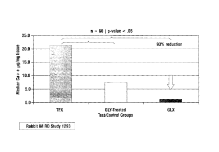
Rabbit Intramuscular Study. A study utilizing sixty (60) rabbits was conducted
to look at the effects on calcification of GLX treated pericardium sterilized
using 100% ethylene oxide. The control group was ThermaFix (TFX) processed
bovine pericardium and the test group was GLX processed pericardium. Using a
sign-rank test, GLX tissue was found to be significantly different (p=0.0004)
when compared to TFX, and demonstrated a 93% calcification reduction over
TFX. GLX data also showed reduced outliers and reduced variability. Box and
Whisker plots show an appreciable reduction in variability with the GLX
process. Data are presented in Figure 7, with the y-axis measuring pg calcium
/
mg dry weight tissue.
[0066] Example 3B - Rabbit Study #2. Effects of a Short Term Shelf
Life on Calcification of GLX Processed Pericardial Tissue in a 35 Day Rabbit
Intramuscular Study. A second study utilizing sixty (60) additional rabbits
was
conducted to look at the effects on calcification of short term shelf life of
GLX
processed pericardium. The control group was ThermaFix (TFX) processed
bovine pericardium and the test group was GLX processed pericardium. The
CA 02710366 2010-06-21
WO 2009/085199
PCT/US2008/013879
- 23 -
GLX tissue samples were packaged in Tyvek pouches and sterilized via 100%
ethylene oxide. GLX samples were stored in a controlled steady state chamber
at 25 oC and 60% humidity for a period of 83 days. The TFX samples had not
been aged. In this study the GLX processed tissue demonstrated significantly
reduced levels of calcification, 73% compared (p=0.009) to TFX as well as
reduced outliers and reduced variability in the data. Data are presented in
Figure
8, with the y-axis measuring jig calcium / mg dry weight tissue.
[0067] While the invention has been described in its preferred
embodiments, it is to be understood that the words which have been used are
words of description and not of limitation. Therefore, changes may be made
within the appended claims without departing from the true scope of the
invention.