Note: Descriptions are shown in the official language in which they were submitted.
CA 02710408 2016-12-13
VARIABLE CURRENT DENSITY SINGLE NEEDLE ELECTROPORATION
SYSTEM AND METHOD
TECHNICAL FIELD
10011 The present invention relates generally to the use of electric pulses to
increase the
permeability of cells, and more specifically to methods and devices for
applying controlled
electroporative electric fields to in vivo tissues of humans and animals for
the delivery of
pharmaceutical compounds and nucleic acids into cells thereof. Further, this
invention
relates to an improved and novel electrode design for carrying out
electroporation that
provides for focused current density near the tissue treatment site undergoing
electroporation and a simultaneous nonelectroporative electric field of
decreased current
density away from said tissue treatment site, which design provides for both
focusing of
electroporative electric pulses in a predetermined and measurable tissue
volume, such as in
skeletal muscle and/or dermal and subdermal tissues while providing
additionally for a
substantial reduction in electric current to nerve sensory cell-containing
mammalian
surface tissues.
BACKGROUND
[0021 The following description includes information that may be useful in
understanding the present invention. It is not an admission that any such
information is
prior art, or relevant, to the presently claimed inventions, or that any
publication
specifically or implicitly referenced is prior art.
[0031 The classical mode of administering vaccines and other pharmaceutical
agents into
the body tissues is by direct injection into muscle or skin tissues using a
syringe and
needle. As has been well disclosed in the art, incorporating electroporative
pulses of
electric energy with direct injection provides for delivery of such vaccines
or agents
directly into the cells within the tissue. Such direct delivery to cells using
electroporative
electric pulses can have a profound clinical effect on the quality of the
response of the
body's metabolic and/or immune systems over that of simple syringe and needle
injection.
Moreover, the capability of direct delivery of substances into the cell via
electroporation
has enabled the effective delivery of expressible naked DNA encoding a
polypeptide,
having any number of functions, including antigenic for eliciting of immune
responses, or
alternatively, metabolic for affecting various biologic pathways that result
in a clinical
effect.
1
CA 02710408 2013-10-24
[004] Although electroporation technology allows for a more advanced delivery
of
substances to the cellular compartments in the body, the electroporative
process, as
presently commonly performed using tissue penetrating electrode arrays such as
disclosed
in U.S. patents 6,041,252, 6,278,895, and 7,245963, has at least two distinct
drawbacks for
practical clinical use. These include first, the need to penetrate the skin
barrier with
multiple trauma inducing needles and second, no ability to easily determine
the tissue
volume undergoing electroporation. Classical electroporation technique, using
arrays of
spaced tissue-piercing needle electrodes provides for a relatively spread out
area of tissue
being electroporated. Typically, the tissue volume undergoing electroporation
when using
an array of spaced electrodes is larger than the volume bounded by the
electrodes of the
array. This is because of the natural flow of electric lines of force through
the in vivo
tissue between the positive and negative electrodes. How far around the
outside of the
array the elecroporative forces are capable of traveling is not easily
quantifiable. This
makes a quantifiable measure of the amount of drug being taken up by the cells
very
difficult. Thus, regarding control of therapeutic dose delivery, there remains
a need to
quantify the amount of tissue undergoing electroporation and consequently the
dosage of
drug being delivered into the cells of said tissue using electroporation.
[005] With regard to tissue penetration, the typical spaced needle array
design also
causes substantial sensation of not only penetration of a multiplicity of
needles into the
flesh, but because of the exposed electrically conductive lengths of the
penetrating
electrodes the recipient of the electroporative pulse will experience a
noticeable electric
shock even if the upper portion of the inserted needle has a nonconductive
coating. By
upper portion here is meant that length of the needle that is in contact with
surface and
dermal tissues. Commonly, the electric pulse in the electroporation process is
noticeable
due to the fact that the pulse being sent between two exposed elongate
electrodes sets up
an electric field and an electric current through the entire depth of flesh
penetrated by said
electrodes. Since the skin tissues possess substantial nervous sensory cells,
it is currently
understood that the sensation of electric shock in the outer tissue regions is
substantial.
This typically unpleasant sensation is a drawback to the widespread acceptance
and use of
electroporation in such applications as vaccination. Further, assuming any
sensation of
= electric shock is directly related to the tissue area or volume subject
to the electric current
of a certain strength, then it would reasonably appear, given that effective
electroporation
in a mammal is possible using only a single needle.
2
The use of spaced needle electrode arrays cause a far greater area of tissue
to be subject
to the electric pulse and consequent excitation of sensory nerve cells than is
necessary.
Thus, there is a need in the arts to find design configurations for delivering
electroporative pulses while reducing the excitement of tissue surface and
skin-based
nerve cells.
[006] Concerning the noticeable sensation of the electroporative pulse of
electric energy,
the level of sensation is also due in part to the design and typically bare
metal nature of
the electrodes used. For example, electrodes are typically constructed in
various
configurations such as, for example, calipers, meander electrodes, and
noninvasive
needle arrays for delivering an electric pulse to the surface of the skin, and
underlying
tissues close to the skin, and elongate and penetrating needle arrays for
delivering
electric pulses to deep tissue. Placement of electrodes directly onto the skin
or piercing
through it sets the electrode in areas of tissue where sensitivity to pain via
nerve
stimulation is very pronounced. Therefore, without a mechanism for lessening
the
current and current density in the areas of tissue having a high concentration
of sensory
nerve endings, the sensation of shock will likely remain.
[007] Thus, there still exists a need in the art for electroporative methods,
electrodes and
systems that can provide for the ability to quantifiably measure the volume of
tissue
actually undergoing electroporation as well as provide for a substantial
reduction in the
electric energy directed in nerve sensory cell-containing tissues so as to
provide for the
possibility of reducing sensory cell excitement during the electroporation-
assisted
delivery of a therapeutic substance.
SUMMARY
[007a] Certain exemplary embodiments provide an electroporation system for
delivering a
molecule into cells of a focused area of tissue by providing a differential
current density to a
mammal, the electroporation system comprising: a) a geometric planar ring
electrode with a
multiplicity of electrically conductive projections thereon; b) a tissue-
piercing elongate
electrode having a proximal end and a distal end, said elongate electrode
having a conductive
portion and a nonconductive portion, said nonconductive portion lying between
said proximal
end and between about 2.5 cm and at least 0.1 cm from the distal end, said
nonconductive
portion comprising either an insulator coating on said elongate electrode or a
nonconductive
3
Date Re9ue/Date Received 2020-09-24
material; c) a housing associated with said ring and elongate electrodes; d) a
charging unit for
charging a capacitor; e) a computer in electrical communication with said
charging unit, said
computer comprising software capable of performing programming functions for
said system;
0 wherein the elongate electrode defines a plurality of apertures, such that:
1) the apertures
are positioned on the conductive portion such that there are between
approximately 10 and
approximately 100 apertures per centimeter of length of the conductive
portion; 2) the
apertures are spaced around the entire circumference of the conductive
portion; 3) the
apertures have a diameter in a range of 30 microns to 80 microns; and 4) the
aperture
positioning, spacing, and diameters are configured to provide even
distribution of an injection
fluid over an entire length of the conductive portion.
[007b] Other exemplary embodiments provide a variable current density
electrode system
for in vivo electroporation comprising: a) a geometric planar ring electrode
with a
multiplicity of electrically conductive projections thereon; b) a partially
insulated elongate
needle electrode having a proximal end, a distal end, and an electrically
conductive portion;
and c) a surface area ratio between said ring electrode and needle electrode
selected from the
group consisting of a range between 1000:1 and 5: 1, wherein when said
electrode system is
activated by providing an electric pulse in a body tissue, electric current
density in said
tissue at or near said needle electrode is higher than current density in said
tissue at or near
said ring electrode; d) wherein the needle electrode defines a plurality of
apertures, such that:
1) the apertures are positioned on the conductive portion such that there are
between
approximately 10 and approximately 100 apertures per centimeter of length of
the
conductive portion; 2) the apertures are spaced around the entire
circumference of the
electrically conductive portion of the needle electrode; 3) the apertures have
a diameter in a
range of 30 microns to 80 microns; and 4) the aperture positioning, spacing,
and diameters
are configured to provide even distribution of an injection fluid over an
entire length of the
conductive portion.
[0007c] Yet other exemplary embodiments provide an electroporation system for
delivering
a molecule into cells of a focused area of tissue by providing a differential
current density to
a mammal, the electroporation system comprising: a) a geometric planar ring
electrode with
a multiplicity of electrically conductive projections thereon; b) a tissue-
piercing elongate
electrode having a proximal end and a distal end, said elongate electrode
having a
conductive portion and a nonconductive portion, said nonconductive portion
lying between
3a
Date Re9ue/Date Received 2020-09-24
said proximal end and between about 2.5 cm and at least 0.1 cm from the distal
end, said
nonconductive portion comprising either an insulator coating on said elongate
electrode or a
nonconductive material; c) a housing associated with said ring and elongate
electrodes; d) a
charging unit for charging a capacitor; e) a computer in electrical
communication with said
charging unit, said computer comprising software capable of performing
programming
functions for said system; and 0 an actuator configured to mechanically drive
the elongate
electrode in a linear motion along a travel length from 0.5 cm to 4 cm, the
actuator further
configured to drive a fluid medium from a reservoir through a lumen in said
elongate
electrode to and out a plurality of apertures at a distal portion of said
elongate electrode
during at least a portion of the linear motion, wherein said driving of the
actuator creates a
pressure that allows said fluid medium to pass through each aperture at
equivalent flow
dynamics such that a uniform volume of fluid is ejected about the elongate
electrode.
[007d] Still yet other exemplary embodiments provide an electroporation system
for
delivering a molecule into cells of a focused area of tissue by providing a
differential current
density to a mammal, the electroporation system comprising: a) a geometric
planar ring
electrode with a multiplicity of electrically conductive projections thereon,
wherein said ring
electrode is electrically isolatable into two electrically conductive halves;
b) an elongate
electrode having a proximal end and a distal end, the distal end defining a
tip for piercing the
tissue, said elongate electrode having a nonconductive portion and a
conductive portion, said
nonconductive portion lying between said proximal end and between about 2.5 cm
and at
least 0.1 cm from the distal end, said nonconductive portion comprising either
an insulator
coating on said elongate electrode or a nonconductive material; c) a housing
associated with
said geometric planar ring electrode and said elongate electrode; d) a
charging unit for
charging a capacitor; and e) a computer in electrical communication with said
charging unit,
said computer comprising software capable of performing programming functions
for said
system; f) wherein the elongate electrode defines a plurality of apertures
that are spaced
around an entire circumference of the conductive portion of the tissue
piercing elongate
electrode such that there are between approximately 10 and approximately 100
apertures per
centimeter of length of the conductive portion, the apertures have a diameter
in a range of 30
microns to 80 microns, and the tip of the elongate electrode does not include
an aperture,
such that all of the plurality of apertures are located around the
circumference of the
3b
Date Re9ue/Date Received 2020-09-24
conductive portion, wherein the aperture spacing and diameters are configured
to provide
even distribution of an injection fluid over an entire length of the
conductive portion.
[007e] Still yet further exemplary embodiments provide an electrode system for
in vivo
electroporation, comprising: an elongate needle electrode that is tubular and
capable of
channeling an injection fluid from a reservoir, the elongate needle electrode
having a
proximal end and a distal end, wherein the elongate needle electrode includes:
an electrically
non-conductive portion located along a proximal region of the elongate needle
electrode and
being electrically non-conductive, and a distal electrically conductive
portion; electronic
circuitry connected to the elongate electrode needle for delivering an
electroporative pulse to
cells of a body tissue, wherein the elongate needle electrode defines
apertures, such that:
1) the apertures are positioned on the conductive portion such that there are
between
approximately 10 and approximately 100 apertures per centimeter of length of
the
conductive portion; 2) the apertures are spaced around the entire
circumference of the
electrically conductive portion of the needle electrode; 3) the apertures have
a diameter in a
range of 30 microns to 80 microns; and 4) the aperture positioning, spacing,
and diameters
are configured to provide even distribution of an injection fluid over an
entire length of the
conductive portion.
[008] Turning now to the advantages of selected embodiments, disclosed is an
apparatus
for conducting the electroporation-assisted delivery to in vivo tissues of a
mammal of
therapeutic substances including expressible nucleic acid sequences encoding
therapeutic
polypeptides, or therapeutic forms of nucleic acids, or derivatives thereof.
In a preferred
embodiment the apparatus can be used to deliver directly to cells DNA
sequences linked
to a promoter and capable of expressing the polypeptide encoded thereby. In
other
alternate preferred embodiments, the apparatus can be used to deliver
therapeutic
substances comprising any of RNA, RNAi, siRNA, micro RNA, and shRNA.
Therapeutic
substances can further include expressible nucleic acid sequences encoding
cytokines,
3c
Date Re9ue/Date Received 2020-09-24
CA 02710408 2010-06-21
WO 2009/091578 PCT/US2009/000273
hormones and other functional molecules useful in therapeutic treatment of
disorders and
diseases.
[009] The present invention also comprises an in vivo method, using pulsed
electric
fields to deliver therapeutic agents into cells of the skin, including dermal
and underlying
muscle compartments of the skin for local and systemic treatments. In a
particularly
preferred embodiment of the present invention, there is provided an in vivo
method for
introducing a therapeutic agent into body tissues and cells, such as cells
within the dermis
and muscle cells, particularly muscle cells in the dermis and skeletal muscle
cells located
in deeper tissue. Therapeutic agents contemplated for use with the invention
method
include naked or formulated nucleic acid, including RNAi, siRNA, microRNA, and
shRNA, polypeptides and chemotherapeutic agents, and other therapeutic agents
that can
be employed directly as palliative agents (i.e., those which directly exert a
therapeutic
effect), or as agents with a less direct effect (e.g., genes encoding
polypeptides that elicit
an immune response).
[010] In another embodiment, the apparatus of the invention provides for the
capability
of delivering to in vivo tissue an electroporative pulse of electric energy
comprising a high
current density at and near the tissue treatment site undergoing
electroporation and,
simultaneously, a nonelectroporative electric field having a correspondingly
substantially
lowered or diffused current density away from said tissue treatment site.
Specifically, as
disclosed herein, the invention apparatus comprises a single tissue
penetrating needle
electrode and a corresponding ring counter electrode, described further below,
comprising
a planar and generally circular or ovoid structure spatially situated with
respect to the
elongate electrode such that the elongate electrode is preferentially central
and
perpendicular to the planar ring electrode surface as shown in Figure 2. The
actual shape
of the "ring" electrode can comprise variable geometries such as for example
round,
ovoid, triangular, square, rectangular, pentagonal, hexagonal, etc.
[011] In another embodiment the single central elongate electrode has a tissue
piercing
distal end and proximate end mounted to a substrate. The elongate electrode
can be solid
or tubular, in which latter case said electrode is capable of delivering a
fluid substance
therethrough. In alternative embodiments the tubular configuration can
comprise a
fenestrated hypodermic needle (i.e., ports for expelling fluid substance are
along the sides
of the needle) or, in an alternate particularly preferred embodiment, the
tubular electrode
can comprise a fenestrated needle wherein there is no aperture at the tip of
the tubular
needle. In such an arrangement fluid media expressed through the tube will not
expel out
4
CA 02710408 2010-06-21
WO 2009/091578 PCT/1JS2009/000273
the tip of the needle but instead exclusively through the side ports. In a
further
embodiment the side ports are positioned on the elongate electrode along at
least the
electrically conducting distal 0.1 to 1.5 cm portion of the tube. In a related
embodiment,
the apertures forming the multiplicity of side ports provides the surprising
capability of
uniform distribution of the injected substance into the tissues intended to
undergo
electroporation where the diameter of said apertures are smaller than about
120 microns
and present in number generally about between 10 and 100, preferably between
20 and 60
and even more preferably between 20 and 40 apertures per 1 cm length of
electrode. This
arrangement provides for the ability to easily apply a constant force/
pressure on the fluid,
such as animatedly by applying thumb pressure on a plunger on a syringe in
fluid
communication with the needle, and maintain approximately even distribution
into the
tissue along the entire length of the fenestrated part of the needle.
10121 In still further related embodiments, the needle electrode is not placed
in a static or
fixed position with respect to the ring electrode. Rather, the elongate needle
electrode can
be attached to a reservoir such as a hypodermic syringe or the like, via the
substrate at the
electrode upper end, wherein the reservoir and needle electrode are movable in
a plane
perpendicular to the plane of the ring electrode surface such that the
reservoir and
electrode can be moved by an actuator mechanism so as to move the
needle/reservoir from
a first position to a second position relative to the ring electrode. The
first position
comprises a resting position wherein the electrode needle tip lies no further
towards the
plane of the ring electrode surface (i.e., the surface intended to contact the
tissue) than the
plane of the ring electrode. In such position, the needle does not contact the
tissue. The
second position comprises an extended position wherein the tip of the needle
lies between
0.5 and 4.0 cm away from the plane of the ring electrode in the direction of
the tissue
which would therefore place the needle tip in a position between 0.5 and 4.0
cm into the
tissue when the ring electrode is in contact with the tissue surface.
10131 With respect to tubular electrode embodiments of the elongate needle
electrode,
the electrode is capable of passing flowable medium, such as injection
substance, from a
reservoir through the ports of the electrode (i.e., ports at the tip of the
needle or
alternatively fenestrated ports. The connection can be made by any number of
methods
such as for example where the substrate at the end of the electrode comprises
a plastic hub
and locking mechanism of a typical hypodermic needle. In a further embodiment,
said
elongate electrode has an electrically non-conducting surface along said
electrode
extending from the needle substrate mount to within between 2.5 and 0.1 cm
from the
CA 02710408 2010-06-21
WO 2009/091578 PCT/US2009/000273
needle distal end. In a further preferred embodiment, when said electrode is
in contact
with body tissues, electric current will not transmit from the electrode into
the tissues
along the section of the electrode having a non-conductive surface. In a
further related
embodiment the non-conductive surface can comprise any type of electrically
inert
substance. In a particularly preferred embodiment the material comprising the
non-
conductive surface can, as one of skill in the art will comprehend, be
selected from any
material that is biocompatible as well as nonconductive such as for example
paralene,
epoxy, rubber, plastic, TeflonTM, and the like.
[014] In accordance with the preferred embodiments of the present invention,
the "ring"
electrode comprises several useful attributes. In a first embodiment the
electrode is
generally of a ring- or ovoid shape, and an electrode surface area having a
relatively
uniform symmetry placement with respect to the central needle electrode. In a
preferred
embodiment, the ring electrode intended to be brought into contact with the
skin has a
surface area of at least about 2.5 cm2 or more. In a further related
embodiment, the
surface area of the ring electrode is proportioned to the surface area of the
electrically
conductive portion of the elongate electrode so as to provide for substantial
differences in
current densities between said electrodes when an electric pulse is sent
between the ring
and elongate electrodes. Specifically, the current density at the elongate
needle electrode
surface (JE) is related to the current density at the ring electrode surface
IR described by
the formula:
[015] IE 11R = (AR /AE)
[016] Where IE is the current density (Amps/cm2) at the elongate electrode
which is
expressed as a ratio of surface Area of the ring electrode (AR) over surface
Area of the
elongate electrode (AE) and IR is the current density in Amps/cm2 at the ring
electrode.
Thus, for example, if the current is 0.5 Amps, and surface area of the
elongate electrode is
0.20 cm2 and the surface area of the ring electrode is 20 cm2, then the
average current
density at the surface of the ring electrode is 0.0125 Amps/cm2 and the
average current
density at the needle electrode is 1.25 Amps/cm2 during the duration of the
electroporation
pulse. The exposed surface area of the elongated electrode above is calculated
for a 23
gauge needle with a nominal 0.64 mm diameter and a 1.0 cm un-insulated length
as
follows:
6
CA 02710408 2010-06-21
WO 2009/091578 PCT/US2009/000273
Surface Area = (Length)x(Circumference) = (lcm)x(2nR) = (1
cm)x(2)x(3.14159)x(0.032
cm) = 0.20 cm2
1017] In a particularly preferred embodiment, the presence of said non-
conductive
surface on said elongate electrode provides for targeting or focusing of
electric current of a
density sufficient to cause electroporation of the cells in the vicinity of
the distal portion of
said elongate electrode. In such embodiment, electroporation of cells
preferably takes
place in areas surrounding said conductive portion of said electrode and
extending into
said tissue towards the ring electrode to a distance where a lowered current
density is
incapable of supporting sufficient electric energy to cause cell poration. In
other words,
the area of tissue undergoing electroporation is that area immediately
surrounding the
electrically conductive area on the elongate needle and into the tissue
laterally and
upward therefrom (i.e., towards the tissue surface) toward the ring electrode
for a distance
of at least between 0.0 and 0.5 cm depending upon the strength of the
electroporative
energy pulse. As the distance from said elongate electrode increases towards
the ring
electrode, the electrical field strength and the current density becomes too
low to cause
electroporation. In a particularly preferred embodiment, the sensation of
electricity, which
sensation thereof is related to the density of electric current, is likely
greatly diminished at
the tissue or skin surface due to the reduced current density. Further, given
that cellular
tissues such as skin and muscle tissues (i.e., epidermis, dermis, subdermis,
muscle)
possess an average conductivity, one can now determine experimentally the
volume of the
tissue subjected to an electric pulse having a sufficient field strength and
current density to
electroporate cells outward from the needle electrode into the tissue to a
given distance.
This advance allows for aligning drug volume/dose to be dispensed into a
predetermined
definable tissue volume with desired treatment outcome.
10181 In another related embodiment, the ring electrode is designed as a
"split" ring
electrode that provides for the capability to monitor the proper placement of
the electrode
onto the skin surface prior to sending an electroporative pulse. Specifically,
the ring is
electrically isolatable in two or more parts, preferably in two electrically
equal halves.
This arrangement allows the electrode to be placed against the tissue surface
and a sensory
electric signal generated to sense the resistance between the surface of the
electrode and
the tissue surface. Once the sensor determines that the electrode is properly
in contact
with the tissue surface, as calculated by the relative resistances between
each half of the
electrode and tissue surface, the two halves of the ring electrode are brought
into electrical
7
CA 02710408 2010-06-21
WO 2009/091578 PCT/1JS2009/000273
communication with one another, the elongate needle electrode deployed into
the tissue,
and an electroporative pulse delivered to the in vivo tissue. This embodiment
provides for
ensuring that the effect of electric impedance of the tissue is uniform with
respect to the
ring electrode prior to delivering an electroporative pulse. In an alternate
and/or
simultaneous application with a split ring impedance sensor, the invention
device can
further include a pressure sensor associated with the ring electrode. In this
embodiment
the pressure sensor provides for determining a predetermined level of pressure
the user
must place with respect to the contact of the device onto the tissue surface
of a subject
before the apparatus will be pulsed. Sensing the pressure allows for the user
of the device
to tell when the device has been placed properly with respect to the tissue
surface in order
to maintain good electrical contact for an electroporative energy pulse.
[019] In another embodiment, the invention apparatus can provide for
manipulating the
tissue surface to be drawn against the apparatus for making consistent contact
with the
tissue surface. In this embodiment, the apparatus can be equipped with a
suction cup
arrangement formed as a pliable diaphragm comprising the central section of
the ring
electrode. In this embodiment, the diaphragm is shaped as a suction cup as in
a toy dart
gun, the outer circumference in sealable connection with the inner
circumference of the
ring. Additional related embodiments provide for assisting in the generation
of active
suction of the cup which can include a spring activated pulling of the cup
slightly outward
from the plane of the ring electrode such that when the ring is pressed
against a surface
tissue, the tissue is urged upward into the cup recess. Following placement of
the tissue in
the cup recess, the elongate needle electrode can be driven through the
suction cup
diaphragm and into the tissue to the desired depth.
[020] In yet another embodiment the invention apparatus provides for the
sensing of the
tissue type into which the elongate electrode is placed. In a preferred
embodiment the
invention device through its elongate electrode is equipped with a sensor
capable of
measuring the impedance of the tissue as the needle is inserted into said
tissue. Thus, for
example, as the electrode passes from one tissue type into another, such as
for example
adipose tissue to deep muscle tissue, the impedance sensed by the electrode
changes
thereby providing a direct indication that the electrode has passed from one
type of tissue,
e.g., adipose tissue, to another type, namely muscle.
[021] In a particularly preferred related embodiment, the invention device is
programmable for setting delivery of a fluid therapeutic substance through the
side ports in
the electrode at a predetermined position within a tissue type. Thus, for
example, the
8
CA 02710408 2010-06-21
WO 2009/091578 PCT/US2009/000273
device can deliver substances after the tip of the electrode has passed
between 0.5 and 1.5
cm beyond, or deeper, than a tissue type interface, i.e., once the needle has
passed beyond
the adipose/muscle tissue interface, for example, the substance to be
electroporated can be
expelled into the muscle tissue. In a particularly preferred embodiment where
the injectate
is intended to be delivered to muscle tissue, fluid is not expelled until the
tip of the
elongate needle electrode has passed the adipose/muscle tissue interface and
into the
muscle tissue by between 0.5 and 1.5 cm. Alternatively, any depth of
penetration can be
programmed so that the specific delivery location in the tissue of substances
can be
predetermined. For example, it may be desirable, depending upon the
indication, to
deliver to dermal tissue, to adipose tissue, or to muscle tissue. Thus, it is
another
embodiment that the sensor can be used to indicate location of the penetrating
needle for
delivery of substances and electroporative pulses to any depth of tissue.
10221 In still further embodiments, the invention device has a novel
arrangement of
electrical components such that the device is portable and can be used without
attachment
to a fixed source of electrical energy such as a wall outlet. In a preferred
embodiment, the
invention device possesses at least one capacitor having a nominal capacitance
potential of
2000 uF (microFarads). In a further preferred embodiment the capacitor can be
charged to
a value of up to 200 Volts before sending energy discharges from said
capacitor to the
electrodes. In a particularly preferred embodiment, the circuitry is designed
so as to
relatively over-charge the capacitor and then, upon discharge of the
capacitor, use a
regulated voltage circuitry which allows for a constant voltage pulse or a
relatively clean
square wave pulse over the length of the pulse period to the patient even
though the
capacitor voltage drops due to dissipation of charge from the capacitor
through the
electrodes and treated tissue. Consequently, such arrangement allows for
simulating a
constant current pulse even though it is the voltage discharge that is
regulated. By
"regulated voltage" is meant a down-regulated voltage output during the pulse
from the
capacitor that is below the voltage at which the capacitor is charged, as
shown in Figure 6.
Using such lower voltage allows for the pulse voltage to remain at a constant
output
during discharge of the capacitor. The voltage drop during the pulse (delta V)
is
approximated by the formula below where "i" is the current into the tissue
being treated,
"Q" is the charge on the capacitor having a maximum capacitance of "C", and t
is the
pulse length.
9
CA 02710408 2010-06-21
WO 2009/091578 PCT/1JS2009/000273
i = dQ/dt :24 C AV/tp
V ------------------------ AV
tp
[023] Thus, the regulated voltage output pulse is set below the maximum
voltage V
minus the expected drop AV across the pulse (or pulse train) so that each
pulse is a
relatively clean square wave thereby delivering a substantially constant
voltage to the
tissue. Since it has been determined that the tissue impedance seen between
the elongate
electrode and the ring electrode is fairly constant throughout the delivered
pulse
(particularly as for pulses intended for use in the electroporative delivery
of therapeutic
agents into cells), the substantially constant voltage will result in a
substantially constant
current into the tissue throughout the pulse length.
[024] In yet further embodiments, the electrical circuitry allows for the
capacitor to be
charged either via a fixed electrical energy source such as an alternating
current source
directly or by induction, or by a battery-charging type unit.
[025] In still another embodiment, the electric charge placed on the elongate
electrode is
the negative charged pole while the ring electrode is the positively charged
pole. By
negatively charged is meant electrons emit therefrom while by positive charged
is meant
that electrons are attracted thereto. This aspect provides the novel feature
of providing for
minimizing positive metal ion contamination into the body tissues from metal
ions being
generated from the positive electrode. As disclosed herein it has been found
that ion
shedding takes place almost exclusively at the positive pole. Specifically,
metal is shed
essentially only from the positive electrode during electroporation. Thus, the
instant
invention provides the capability of providing an electroporative electric
pulse of energy
into the body tissues while minimizing the contamination of the biologic
environment with
potentially toxic metallic ions. Although ions are capable of being shed from
the ring
electrode proportional to the strength of the current and length of the pulse,
the metal ions
shed at the skin surface should stay outside the skin barrier and the body's
biologic
environment.
CA 02710408 2010-06-21
WO 2009/091578 PCT/US2009/000273
[026] In accordance with another embodiment of the present invention, there is
provided
a method for inducing an immune response in a subject, comprising applying a
pulsed
electric field to cells within body tissues, particularly dermal and/or muscle
cells of the
subject, substantially contemporaneously with the application of an immune
response-
inducing agent to said body tissues, such that the immune response-inducing
agent is
introduced into said cells thereby inducing in the subject an immune response.
[027] In accordance with still another embodiment of the present invention,
there is
provided a method for the therapeutic application of electroporation to cells
within certain
tissues including such as muscle cells within the dermis and underlying
skeletal muscle
cells of a subject for introducing a metabolic or otherwise systemic effect to
the recipient.
For example, the methods contemplated include gene therapy treatments wherein
a gene
encoding an expressible cytokine or chemokine or hormone or other polypeptide
that has a
direct therapeutic effect is administered to a mammal.
[028] Still another embodiment contemplates an electrode kit for use in
conjunction with
electroporation therapy, said kit having a ring electrode assembly, said
assembly
comprising a ring electrode and elongate central electrode, said assembly
designed for
connecting to a device for handling said ring and elongate electrode assembly
and using it
with a source of fluid injectate and au electric energy source.
[029] In Still further embodiments, the device design of the current invention
can be
tailored for use in vaccinating or otherwise treating domestic herd/food
source animals
such as cattle, sheep, goats and horses. In this embodiment, the ring
electrode is designed
with a multiplicity of short electrically conductive projections thereon. Such
projections
provide both the required total surface area ratio with the needle electrode
and allows for
proper contact with the skin surface tissue, the projections allowing for the
ring/surface
electrode to penetrate the animal's fur, pelt, hair, or wool coat.
BRIEF DESCRIPTION OF THE FIGURES
[030] This specification contains at least one figure executed in color.
Copies hereof with
color drawing(s) will be provided upon request and payment of the necessary
fee.
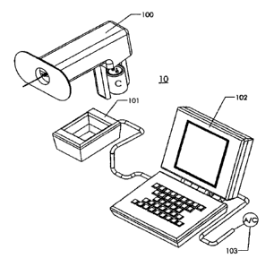
10311 Figure 1 shows a schematic drawing of an electroporation device system
10
comprising a ring-shaped electrode 99 (here the ring is depicted as ovoid in
shape), a
hand-held portable device 100 supporting the ring electrode and its assembly,
a charging
unit 101 for charging a capacitor (C) which is in electrical communication
with said ring
electrode assembly, a computer 102 operated software for setting pulse
parameters and
11
CA 02710408 2010-06-21
WO 2009/091578 PCT/US2009/000273
monitoring and recording pulsing conditions, and quality of charge imparted to
said
capacitor, which computer is powered by an external alternating current power
source 103
or alternatively, a DC battery (not shown).
[032] Figure 2 is a perspective drawing of one example of the relative spatial
arrangement between the elongate 120 and ring 200 electrodes. Specifically,
the elongate
electrode 120 is placed in a plane perpendicular with respect to the ring
electrode 200 such
that the elongate electrode 120 lies along an axis central to the ring
electrode 200 and is in
fluid communication with a reservoir 140. The elongate electrode 120 further
comprises a
section 130 that is nonconductive to electric current. The figure further
depicts the
substrate 121 comprising the proximal end of the elongate electrode as well as
a support
substrate 201 for supporting the ring electrode.
[033] Figures 3A, B and C are related drawings showing in Fig. 3A a cross
sectional
representation of the ring and elongate electrode in tissue. Specifically,
ring electrode 200
is shown engaged with tissue surface 15, with elongate electrode 120 having
insulated
section 130 in said tissue. Depicted are theoretical lines of force 310 due to
a typical
electroporative energy pulse that are concentrated to a higher current density
at the
elongate electrode 120 and less concentrated at the surface of the ring
electrode 200.
Figure 3B depicts a close-up view of the elongate needle 120 with a
multiplicity of bores
150 in the distal region of said needle 120. The figure further depicts
ejectate 140 from
said bores 150 and theoretical lines of electric force 310. Figure 3C is a top
view
depicting theoretical lines of electric force 310 radiating from central high
current density
needle electrode 120 to the low current density ring electrode 200.
[034] Figures 4A and B are perspective drawings showing split ring electrode
embodiments wherein a ring electrode is either physically split into two
halves with a
small air gap 152 therebetween (Fig. 4A) or that is physically split but
connected by a non-
conducting substrate 160. In these embodiments, each half of the ring is
electronically
isolatable from one another.
[035] Figures 5A, B, C, and D are planar drawings depicting the electrode side
155 of split
ring electrodes mounted in support substrate 156 for various useful shapes of
a split ring
electrode.
[036] Figure 6 is a graph depicting a regulated voltage potential generated
from charging
a capacitor to a higher voltage potential than actually employed during the
pulse for the
purposes of obtaining a relatively flat or constant voltage discharge.
12
CA 02710408 2010-06-21
WO 2009/091578 PCT/US2009/000273
[037] Figure 7 is a schematic drawing of a likely shape of an electroporated
bolus of
delivered substance using the ring electrode system of the current invention.
[038] Figure 8 is a cross-sectional drawing of the ring electrode support 201
covering the
ring electrode 200 with centrally located pliable suction cup 210. As
depicted, the suction
cup 210 in this embodiment is connected to spring 215 assisted riser substrate
220 in
slidable relation to a portion of either the assembly housing 205.
[039] Figures 9A, B, C, D, E and F depict color green fluorescent protein
(GFP) staining
photographs superimposed against a representation of the placement of a 25 cc
surface
area ring, and a needle electrode showing that the actual focus of
electroporative energy in
the the tissue as exhibited by the GFP staining corresponds to the
theoretically expected
result disclosed in Figure 7 depending upon the field strength and
corresponding current
density of the pulse. In Figs. 9A (GFP staining) and 9B (GFP and visible light
field) are
shown results in rabbit muscle tissue using a nominal 289 mAmp / 64 volt
pulse. Fig. 9C
shows a nominal 384 mAmp / 81 volt pulse wherein a greater tissue volume than
in Fig.
9A has undergone an electroporation pulse. In Fig. 9D is shown GFP staining
following a
nominal 579 mAmp 1103 volt pulse exhibiting an even greater tissue volume
undergoing
electroporation, and in Figs. 9E (GFP staining) and 9F (GFP and visible light
field) are
shown GFP staining following a nominal 758 mAmp/ 138 volt pulse of still
greater tissue
volume electroporation.
[040] .Figures 10A and B are color photographs showing GFP (Fig. 10A) and
combined
GFP and visible light (Fig.10B) where tissue was subjected to a 189 mAmp/ 58
volt pulse
using a ring electrode having a 2.5 cc surface area. As depicted the tissue
volume is
confined to tissue closely surrounding the elongate needle.
[041] Figures 11A and B show schematic drawings depicting positioning of
apertures
along the electrically conductive portion of the elongate tubular electrode
shaft. The
apertures can be between 20 and 120 microns in diameter. As depicted, the
apertures are
spaced along the needle length (Fig. 11A) and at 90 degrees with respect to
one another
around the circumference of the needle shaft (Fig. 11B).
[042] Figure 12 is a color photograph showing a mixed fluorescent and visible
light GFP
staining in muscle tissue following dispersion of injection of the GFP through
60 micron
apertures. As observed the distribution of injection substance is evenly
dispersed about
the needle track in the muscle tissue.
[043] Figures 13A and B are perspective drawings of alternative ring
embodiments
designed for use in delivering electroporative pulses to hair, fur, or wool
covered domestic
13
CA 02710408 2010-06-21
WO 2009/091578 PCT/US2009/000273
herd animals. In this embodiment, the ring 301, whether round, ovoid, split or
not, is
equipped with a plurality of electrically conductive projections 300 that can
penetrate a
hair, fur, or wool coat to contact the animal's skin surface.
[044] Figures 14A,B, and C are graphs depicting the sensing of divergent
tissue type
interfaces, specifically adipose or fat tissue and muscle tissue. In Fig. 14A,
In Fig. 14B, In
Fig 14C.
[045] Figures 15 A, B, C and D are pictorial diagrams showing the process of
sensing
tissue types followed by simultaneous driving of the needle into tissue while
injecting the
injection substance. Figure 15A shows that the electrodes used for sensing in
this
configuration are on the injection needle. Specifically, electrode 400 is
electrically
isolated from the tip of the needle which acts as the return electrode 401.
[046] Figures 16A-E are GFP staining photographs showing the successful
electroporation of animal muscle tissue using the ring electrode haying a
plurality of
projections. Each successive photo is that of adjacent tissue slices.
[047] Figures 17A, B, C and D are diagrams showing construction examples for
needles
useful for sensing tissue resistance for determining tissue interfaces. In
Figure 17A, the
tip of a typical syringe needle can be constructed wherein the tip section
acts as one
electrically conductive pole while a second electrically conductive pole is
cased about the
tip electrode with an insulative material therebetween. In Figure 17B, a dual
needle
arrangement is depicted wherein the central needle of the current system is
actually two
closely spaced delivery tubes and each can act as individual electrodes. In
Figures 17C
and D, two additional design formats are depicted for a double central
electrode system.
In Figure 17C the depicted electrodes can have insulation so as to focus
sensing to only
the region the tip of the electrodes are driven past, while in Figure 17D, the
electrodes not
only possess insulated portion but also fenestrated ports per the presently
disclosed
invention elements.
[048] Figures 18A and B are graphs showing in Figure 18A results of expression
of
plasmid gWiz-SEAP, a plasmid encoding secreded alkaline phosphatase, following
electroporation in rabbit tissues using the methods and devices of the
invention, namely
use of a planar ring electrode and a ring electrode with projections (comb
electrode). Also
depicted are control negative and control positive using an elgen device. In
Figure 18B
induction of anti-HBsAg antibodies in rabbits is shown for the same electrodes
using same
delivery conditions.
14
CA 02710408 2010-06-21
WO 2009/091578 PCT/1JS2009/000273
DETAILED DESCRIPTION OF THE INVENTION
[049] Turning now to further embodiments of the current invention, as used
herein,
"biocompatible" or "biocompatible material" means a material that is suitable
for
introduction into the human body for therapeutic purposes. For example, with
respect to
electrodes and materials such as insulation used for covering the electric
conducting
surfaces, such covering comprises materials that are inert and do not elicit
irritation or
allergy in the tissue of a mammal.
[050] As used herein, "injection substance" means any injectable composition
of a
therapeutic agent to be delivered to a target tissue. As described herein,
therapeutic agents
comprising injection substances contemplated for use in the practice of the
present
invention include nucleic acids, polypeptides, chemotherapeutic agents and the
like
without limitation such as any nucleic acid disclosed throughout this letters
patent as well
as nucleic acid sequences encoding polypeptides such as for example, encoding
IL-2, IL-
12, ICAM-1, ICAM-2, ICAM-3, PSA, PSMA, PAP, MUC-1, Her-2, NS 3 and 4 etc., and
nucleic acids comprising RNA, DNA, RNAi, siRNA, micro RNA, and shRNA. For
purposes of this letters patent, injection substance can also include DNA
coding for green
fluorescing protein (GFP) and other substances used in visualizing location of
materials
injected into the tissue. Injection substances can further include
pharmaceutical
formulations comprising salts, excepients, and other materials for acceptable
buffering as
is will understood by those of skill in the pharmaceutical arts.
[051] As used herein with respect to the application of an electroporative
pulse of electric
energy in tissue concomitant with an injection substance, the term
"substantially
contemporaneously" means that the electric pulse and the injection substance
are delivered
to the tissue reasonably close together in time. Preferably, the injection
substance is
administered prior to or concurrently with an electroporative pulse of
electric energy.
When applying multiple electrical pulses, the injection substance can be
administered
before or after each of the pulses, or at any time between the electrical
pulses.
10521 As used herein, the terms "impulse," "pulse," "electrical impulse,"
"electrical
pulse," "electric pulse," "electropulse" and grammatical variations thereof
are
interchangeable and all refer to an electrical stimulus. Although the various
terms are
frequently used herein in the singular, the singular forms of the terms
include multiple
pulses. Preferred electrical impulses are pulsed electric fields applied for
the purposes of
reversible poration of cellular membranes. The pulse can be unipolar, bipolar,
exponential
or of a square wave or other form. Electric pulses contemplated for use in the
practice of
CA 02710408 2010-06-21
WO 2009/091578 PCT/US2009/000273
the present invention include those pulses of sufficient voltage, current,
current density,
and duration and frequency to cause electroporation in specified locations
within a body
tissue.
The Ring Electrode System
[053] In a first embodiment, the invention device comprises an electrode
system for
performing electroporation of cells in vivo. In a preferred embodiment, the
system
comprises (1) a generally ring- or ovoid-shaped positive electrode, (2) an
elongate tissue
piercing single needle negative electrode comprising both a conductive and a
non-
conductive portion thereof, said single needle electrode positioned so as to
lie along an
axis central to said ring electrode and perpendicular to the plane of the ring
electrode such
that electric current can be directed to a limited electrode surface area on a
distal portion
of said needle electrode, (3) a mechanism for driving the single needle
electrode into the
tissue, (4) a mechanism for injecting a fluid containing an effective amount
of a
therapeutic agent through said elongate needle electrode, and (5) a source of
electrical
energy for charging a capacitor the discharge of which comprises at least one
electric
pulse, preferably a square wave regulated voltage pulse, for delivering an
electroporative
pulse of electric energy to the electrodes.
[054] An example of the general embodiments of the current invention is shown
in
Figure 1. Specifically, the system 10 includes a portable hand manipulateable
housing
100 which is associated with the ring/needle electrode, a charging unit 101, a
computer
software system 102, and a source of electric power 103. This power source 103
can be
A/C or D/C.
10551 The housing element 100, further comprises drivers, which can be
mechanically
operated using cams, gears, and or levers, or by inanimate means such as an
electric
motor, for driving the single elongate electrode from a starting position to a
terminal
position, which movement of the electrode can be between a displacement of
about
between 0.5 and 4 cm. In a preferred embodiment, the single elongate electrode
120
(Figure 2) acts also as an injection needle. Hence, at least a part of the
injection needle
120 must be formed of a material possessing qualities of an electrical
conductor. In a
preferred embodiment, the driver manipulates the elongate needle by driving
the reservoir
itself which is attached to the needle. In a particularly preferred
embodiment, the driver
manipulates the body of a syringe such that the syringe is carried with the
combination
electrode/ injection needle from the starting to the terminal positions.
16
CA 02710408 2010-06-21
WO 2009/091578 PCT/1JS2009/000273
10561 In particularly preferred embodiments, the needle electrode can be
modified to
provide for radial delivery of an injection substance, for example, by
providing one or
more apertures disposed along its length and proximal to the needle tip (i.e.,
a fenestrated
needle), wherein said apertures are in fluid communication with the hollow
interior of the
injection needle. The needle 120 can be formed of a biocompatible metal such
as stainless
steel, gold, silver, etc. In a further preferred embodiment, the elongate
electrode 120 can
be designed so as to not have a port at the tip of the needle in contrast to
typical syringe
needles. In such embodiment, the ports (apertures) are located only around the
electrically
conductive portion of the electrode shaft so that fluid expelled therefrom is
directed to
tissue directly in the area intended for delivering an electroporative pulse
of electric
energy as depicted in Figure 3B. In contrast to typical fenestrated needles
(wherein
ejected fluid flows predominantly out of the port located at the tip of the
needle, if present,
or alternatively through the upper or first side ports contacted by the
expressed fluid along
the needle path due to fluid dynamics as is well understood by those of skill
in the
hydrologic arts), the fenestrated needles of the current invention, due to the
size range of
the apertures and the elimination of a tip aperture, provide for even
distribution of fluid
through said apertures along the entire length of the needle having said ports
using only
nominal pressures for injecting the fluid injection substances. By nominal
pressures is
meant that the pressure required to expel fluid from electrodes having micron
sized
aperatures is only the pressure typically required during the injection of a
substance
through a standard hypodermic needle. This surprising finding is brought about
by sizing
the apertures along the needle shaft to a diameter in the micron range as well
as including
a multiplicity of such apertures ranging from between 10 and 100, more
preferably
between 20 and 60 and even more preferably between 20 and 40 such apertures
per 1 cm
length of electrically conductive needle/electrode. Preferably, the aperture
diameter for
obtaining even expression of fluid through each aperature is between 20 and
120 microns,
more preferably between 30 and 100 microns, and even more preferably between
30 and
80 microns. Specific diameters include 20, 25, 30, 40, 50, 60, 70, 80, 90, and
100 microns
and any incremental diameters therebetween. Numbers of apertures preferably
arc at least
20/cm, more preferably at least 30/cm and even more preferably at least 40/cm
of
electrically conductive electrode length. As depicted in Figures 11A and B, a
multiplicity
of micron sized apertures can be spaced along the needle shaft and for
cylindrical
distribution therefrom, as depicted in Fig. 11B, the apertures can be spaced
around the
needle circumference at 90 degree angles such that 4 apertures oppose one
another as
17
CA 02710408 2010-06-21
WO 2009/091578 PCT/US2009/000273
shown or can be spaced around the needle circumference at 60 degrees to obtain
more
apertures per needle length. Further still, the apertures can be formed in the
needle shaft
in a spiral configuration such that when, for example, a 60 degree cross
sectional
placement of the apertures is employed, the apertures are not in the same
cross section of
the needle but are staggered off of a cross sectional plane so that the
formation of the
apertures along the needle shaft are in a spiral format. As further disclosed
in Figure 12,
results from a GFP experiment show consistent distribution about the needle
track of the
expressed material as indicated by the cylindrical localization of the GFP. In
this
experiment the transfected volume was measured at 1.2 cm3, the tissue having
been
subjected to two successive 6o millisec 98 V, 768 mAmp pulses delivered 200
millisec
apart.
10571 In still other embodiments, the single needle electrode has an insulated
portion 130
that is not electrically conducting. In a particularly preferred embodiment,
the non-
conductive portion can be provided either by an insulation coating such as a
biocompatible
plastic, paralene, Teflon TM, epoxy or other material that will not allow
current to pass.
Still further, the non-conducting portion of said elongate electrode is
located on said
electrode along its proximal region. Specifically, the elongate electrode will
have a
nonconductive surface between the proximal end, (which is in fluid
communication with a
reservoir containing an injection substance for delivery to body tissues) and
terminating
between 0.1 cm and 2.5 cm from the distal end of the electrode.
[058] In a further embodiment, the ring-shaped electrode 200 may be formed in
any
planar shape having symetry, including but not limited to, a circular ring, a
donut circle, an
oval donut, a rectangular ring, isosceles triangle donut, an equilateral
triangle donut, a
square ring donut, a rectangular donut, a pentagonal ring donut, and hexagonal
ring donut
or the like as shown, for example, in Figs. 5A-D as long as the electrode is
formed in a
plane, is generally symmetrical (i.e., has a form that can be recognized as
having a shape
that is evenly divisable into two relatively equal electrically conductive
portions), and is
conductive at least on one side of the plane (i.e., the side facing the body
surface against
which the electrode is placed), the electrode further having an area central
with respect to
the ring structure that is empty of electrically conductive material such as a
void or hole or
alternatively a nonconductive material such as, for example, a pliable
material such as
rubber or silicon. Where such pliable material is present, such material can
further be
designed to act as a suction cup for urging surface tissue to be pulled
outward from the
18
CA 02710408 2010-06-21
WO 2009/091578 PCT/US2009/000273
tissue surface. In further embodiments the empty or nonconductive area forming
the
center of the ring allows for the elongate electrode to pass therethrough,
whether through a
void and directly into contact with a surface tissue or alternatively, through
said resilient
suction cup material and then into contact with said tissue.
[059] In another attribute, the ring electrode is designed so that it can be
electrically
isolateable into two halves. Specifically, the ring can be manufactured either
as two
separate halves or can comprise two halves connected together by a non-
conducting
substrate (See Figures 4A and B). In this aspect, the ring electrode halves
can be
electrically isolated from one another which electrical arrangement provides
for the
capability of using the electrode to monitor quality of electrical contact of
the ring
electrode with the tissue surface. Specific electrical arrangements for
carrying out sensing
contact is easily understandable by one of ordinary skill in the electrical
arts. In operation,
for example, the ring electrode of the invention apparatus is pressed against
the skin. The
ring electrode system circuitry includes electric leads to each half of the
split ring and an
impedance check can be made for each half regarding the detected current or
alternatively,
resistance, measured between the electrode and surface tissue upon sending a
nominal
electric signal through each electrode half. If the ring electrode is properly
placed against
the tissue surface the resistance or current measured in each half will be
essentially the
same indicating that the user has applied the ring electrode and device to the
tissue surface
evenly so that when the single elongate electrode is driven into the tissue
and the electric
pulse sent between the elongate and ring electrodes, the current flow between
the elongate
and two halves of the split ring with be equivalent. Numerous split ring
electrode shapes
can be used including, for example, shapes depicted in Figures 5A-D.
[060] With respect to embodiments wherein the void area in the center of the
ring
electrode comprises a rubber or other pliable nonconductive material for
acting as a
suction cup, the tissue surface can be urged into the cup by a suction
mechanism causing
the tissue to be drawn up against the resilient material thereby providing for
additional
close and consistent contact of the tissue with the electrode to support
consistent electrical
conductivity between the tissue surfaces and the ring electrode. The drawing
up of the
tissue against the suction cup further provides for maintaining a consistency
in the depth
of delivery of injection substances as between different treated subjects.
When the tissue
is drawn up against the suction, the elongate needle can be driven through the
suction cup
rubber and into the tissue to a consistent predetermined depth.
19
CA 02710408 2010-06-21
WO 2009/091578 PCT/1JS2009/000273
[061] As shown in Figure 8, for example, dmechanism can be incorporated into
the ring
electrode assembly comprising the resilient suction cup 210 in sealable
connection with
the ring electrode substrate 201. Specifically, for example, a spring 215
loaded riser
substrate 220 in slideable relation to the assembly housing substrate 205 can
be connected
to the ring electrode for aiding the outward (from the tissue surface) pull of
the suction cup
which in use will provide for urging the surface tissue outward.
[062] In a preferred embodiment the elongate electrode is in fluid
communication at its
proximate end to a reservoir containing an injection substance. In a further
preferred
embodiment, the invention apparatus includes a driver mechanism for driving
the elongate
electrode and optionally said reservoir attached thereto from a starting
position to a
terminal position in relation to said housing and ring electrode. Preferably,
the length of
travel of the actuated elongate electrode can be anywhere between 0.5 and 4
cm. For
embodiments comprising a suction cup, the needle is directed to pierce through
the suction
cup and into the tissue.
[063] In still another concomitant and/or alternate embodiment, the ring
electrode system
can comprise a pressure sensor associated with the ring electrode as one of
skill in the
electrical and mechanical arts will understand how to make. In this embodiment
a
pressure sensor is arranged such that when the ring electrode is pressed
against the tissue
surface, the sensor will measure the physical pressure applied to the device
against said
tissue. If the value of pressure is sufficient, the device will be capable of
activating the
sending of an electroporative pulse of electric energy to the electrodes. In
preferred
embodiments the requisite pressure for activation of the device can be between
0.5 and 1
lbs/sq inch. As one of skill in the respective arts will understand, the
invention apparatus
includes software for measuring said pressure for determining the amount of
physical
force placed on the ring electrode against the tissue surface. It should be
understood that
the application of such physical force is intended to assist good electrical
contact between
the ring electrode and tissue surface. For embodiments comprising a suction
cup, such
pressure also assists the function of the suction cup so that when the suction
is activated,
the tissue will be easily drawn against said suction cup.
[064] In still further embodiments, the current invention provides for a
heretofore not
discovered capability of avoiding the shedding of potentially toxic metal ions
into the
tissues of a mammal when using standard stainless steel tissue penetrating
electrodes.
Whereas it is known that use of gold-coated stainless steel electrodes will
provide non-
shedding of toxic heavy metals present in stainless steel into the tissues as
disclosed in US
CA 02710408 2010-06-21
WO 2009/091578 PCT/US2009/000273
patent application no.10/516,757, use of gold is not as desired as stainless
steel because of
the added cost of the gold as well as the application of gold to an electrode.
Surprisingly,
as here in disclosed, stainless steel can be used in tissue penetrating
electrodes where, as in
the instant case, there is but a single tissue penetrating electrode and a
corresponding
counter electrode that is non-tissue penetrating. In this instance the
stainless steel
penetrating electrode will shed minimal amounts of metallic ions into the
tissue if such
electrode acts as the negatively charged electrode while the non-tissue
penetrating
electrode, such as the herein disclosed ring electrode, acts as the positive
electrode.
[065] In experiments by the current inventors, standard stainless steel
hypodermic
injection needles, set as either the negative or positive electrode, were
tested against a gold
electrode, also set as either the negative or positive electrode, in
physiologic saline.
Where the gold electrode was set as the negative and the stainless steel
electrode set as the
positive, metallic ions found in the solution following two 60 millisecond
clectroporation
pulses of a 400 mAmp current and 40 Volts were as follows: Manganese 0.035 ppm
(parts
per million), Nickel 0.200 ppm, Molybdenum less than 0.003 ppm, Chromium 0.413
ppm,
and Iron 0.977 ppm. In contrast, when the gold electrode was set as the
positive and the
stainless steel electrode set as the negative under the same conditions there
was virtually
no detectable metallic ions shed into solution, i.e., in each case, whether
Manganese,
Nickel, Molybdenum, Chromium, or Iron, less than 0.003 ppm were observed.
[066] In a particularly preferred embodiment, the surface area of the ring
electrode is
proportional to the surface area of the elongate electrode. Generally, the
ratio of the ring
electrode surface area to the surface area of the needle electrode is at least
5:1,
respectively. Preferably, the ratio of the surface areas of the ring to the
elongate electrode
is between 10:1 to 1000:1. Ratios of 10:1 are preferable for use in human
subjects while a
ratio between 5:1 and 10:1 is acceptable for use in herd animals. In related
embodiments
the ring electrode can have a surface area of between 1 cm2 and 100 cm2 and
the
electrically conductive portion of the elongate electrode can have a length of
between 0.01
cm and 3.0 cm. For elongate electrodes having such a linear dimension range
the surface
areas corresponding to such lengths depend upon the gage of the electrodes
(i.e., their
respective outer diameters). As shown in the following Schedule A elongate
electrode
surface areas are as delineated using the formula: Area = CL = 7c1)1_, to
calculate where D
is diameter and L is exposed length:
21
CA 02710408 2010-06-21
WO 2009/091578 PCT/US2009/000273
Schedule A
Needle Gauge Dia (mm) Conductive Length (cm)
0.5 1.0 1.5 2.0 2.5 3.0
20 0.91 0.14 0.29 0.43 0.57
0.71 0.86
21 0.82 0.13 0.26 0.39 0.52
0.64 0.77
22 0.72 0.11 0.23 0.34 0.45
0.57 0.68
23 0.64 0.10 0.20 0.30 0.40
0.50 0.60
24 0.57 0.09 0.18 0.27 0.36
0.45 0.54
25 0.51 0.08 0.16 0.24 0.32
0.40 0.48
26 0.46 0.07 0.14 0.22 0.29
0.36 0.43
27 0.41 0.06 0.13 0.19 0.26
0.32 0.39
28 0.36 0.06 0.11 0.17 0.23
0.28 0.34
Bold numbers in table are area in cm2
10671 With respect to the above list of useful elongate electrode surface
areas, in a
particularly preferred embodiment the gauge of the electrode needle can be
between 22
and 24 gauge ranging from about 0.1 to 0.6 cm2 for up to an insertion depth of
up to 4.0
cm.
10681 With regard to the above ring and elongate electrode surface area
ratios, such ratios
correlate to current density wherein the relationship between the ring and
elongate
electrodes is described by the following formula as previously noted:
AR/AE -IE/1R
where AR is the surface area of the ring electrode, AE is the surface area of
the elongate
electrode, IE is the average current density at the elongate electrode, and IR
is the average
current density at the ring electrode. Thus, for any given surface areas of
the elongate and
ring electrodes, the ratios are directly proportional to the current density
observable at the
elongate and ring electrode surfaces. In a particularly preferred embodiment,
the ratio of
average current density of the ring electrode to the exposed elongated
electrode is intended
to have a value of anywhere from 1000:1 to 50:1, more preferably between 200:1
to 100:1.
Further, such value ratios are directly associated with obtaining
electroporative electric
energies near the elongate electrode while obtaining nonelecroporative
electric energies
closer to the ring electrode. Such ratios further provide for a lessened
current available to
excit sensory nerve cells and thus provide for potential lessening of
sensation of electric
shock in the surface tissues near the ring electrode. This is particularly the
case when
delivering pulses of electric energy having a nominal current of between 0.01
Amp and
1.0 Amp (of constant current discharge pulse).
22
CA 02710408 2010-06-21
WO 2009/091578 PCT/1JS2009/000273
[069] As is understandable to one of skill in the art, the difference in
surface area of the
elongate and ring electrodes provides for a condition while pulsing where
electric current
density is non-uniform throughout the volume of tissue lying between the
electrodes.
Specifically, current density is very high at the non-insulated portion of the
elongate
electrode (at least high enough to provide for electroporation of cells
adjacent or near the
electrode) and substantially lower at the ring-electrode surface as depicted
in Figures 3A,
B and C. In a particularly preferred embodiment, where the ring electrode
assembly is
designed with its symmetry imbued ring electrode at one end and the conductive
portion
of the elongate electrode at the other end, the current established in the
tissue at any given
distance between the ring and elongate electrode in a given plane
perpendicular to the
elongate electrode will, largely due to the symmetrical shape of the ring
electrode, have
the same current density. Further, the current density decreases relatively
evenly in the
tissue at each incremental measure towards the ring electrode. Thus,
essentially all of the
tissue in a given plane in the tissue around and extending into the tissue
from the elongate
electrode will experience the same lower current density than the density at
the elongate
electrode. Further still, the area of tissue intended to undergo
electroporation is that tissue
along the needle track in the conductive region of the elongate electrode and
extending
into the tissue to a distance therefrom sufficient to become electroporated
(i.e., at least
some of the cells lying between at least 0 and 0.5 cm from the needle track
along that
portion of electrode that is not insulated are subject to electroporation
depending on the
local field strength). The distance into the tissue from the needle track
where
electroporation will occur is dependent upon the pulse energy used as well as
other factors.
The higher the field strength of the pulse the farther into the tissue towards
the ring
electrode will be the threshold point for electroporation to occur.
[070] Regarding the phenomenon of field strength as it relates to
electroporation of intact
tissues in the present invention, a way to visualize "Current Density" as it
relates to "Field
Strength" is as follows. For the theoretical condition of two parallel plates
of area 1 cm2
and a 100 volt potential across them, and further separated by a length L = 1
cm, as shown
below;
23
CA 02710408 2010-06-21
WO 2009/091578 PCT/US2009/000273
A = 1 cm2
electrical field lines and the direction of electrical current are illustrated
by the arrows.
For parallel plate electrodes, the average Field Strength between the plates
is V/L = 100
Volts / 1 cm = 100 V/cm. The current between the plates depends on the
impedance of the
tissue in-between them. If this impedance is, say, 100 ohms, then the current
between the
plates is I = V/R = 100/100 = 1.0 Amp. This would result in a Current Density
of I/A =
1000 mAmps / 1 cm2. For different electrode configurations and different
tissue
impedances, the relationship between Field Strength and Current Density will
differ;
however, they will approximately vary in proportion with each other. In the
configuration
of an elongate electrode and a ring electrode, the measure of Current Density
can more
readily be used to determine effective electroporation. This threshold has
been
experimentally measured in rabbit muscle to be near 300 mAmps/cm2. Tissues
which
experience current densities above the threshold will be electroporated, and
tissues below
the threshold will not. By determining the boundary within a three-dimensional
region of
tissue that represents this threshold, the volume and shape within this
boundary can be
predicted to electroporate.
10711 As shown in Figures 9A to F, the volume of tissue exposed to an
electroporative
pulse of energy can be essentially dialed-in without spreading of the
electroporative
energy beyond a measurable distance/volume of tissue. In Fig. 9A the prototype
invention
device was tested in New Zealand white rabbit leg quadriceps muscle pulsed at
a 289
mAmps actual measurement correlating to a 64 V pulse, in Fig. 9C a larger
tissue volume
was electroporated using a 384 mAmps actual measurement correlating to an 81V
pulse.
In Fig. 9D a still larger tissue volume was electroporated using 579 mAmps
correlating to
a 103 V pulse, and in Fig. 9E a still larger tissue volume was electroporated
at 758
mAmps measured correlating to 138 V. In Figs. 9B and F both GFP fluorescence
alone
and GFP and visible light photos are disclosed showing the electoporative
spread behavior
of the treatment zone into the muscle tissue. In each of these experiments
(Figs. 9A, B, C,
E and F) 280u1 (microliters) volume of GFP encoding plasmid DNA solution was
injected
into the rabbit muscle followed immediately by the requisite pulse. In Fig. 9D
only 70u1
GFP DNA was used to show that electroporation will encompass all of the tissue
volume
containing GFP DNA (i.e., saturation of injectate not reached). Thus, in the
Fig. 9D,
24
CA 02710408 2010-06-21
WO 2009/091578 PCT/US2009/000273
actual electroporated volume measured is smaller (See Table II) than in Figs.
9B and E
even though in Fig. 9B the volume injected was about 2/3 more and electrical
energy was
less while in Fig. 9E the volume injected was about 2/3 more and electrical
energy was
more. The ring electrode conductive surface area for this experiment was
approximately
25 cm' and the elongate electrode conductive surface area was approximately
0.22 cm2.
[072] The ring electrode surface area is correlated with the current density
as shown in
Table I below. Table I shows figures for a system equipped with an ovoid ring
electrode
having a surface area of 25 cm' and various applied field strengths.
Table I
Elongate Ring Surface Charge Electrode Current Current Calculated
Calculated
electrode electrode area applied Current density at
density at EE Field RE Field
(EE) (RE) ratio (Volts) (mA) EE RE strength strength
surface surface (mA/cm2) (mA/cm2) (V/cm) at (V/cm) at
area (cm2) area 11.4 ratio
11.4 ratio
(cm2)
0.22 25 >100 25 86 391 3.4 34 0.29
0.22 25 >100 50 171 777 6.8 68 0.60
0.22 25 >100 100 342 1554 13.7 136 1.20
0.22 25 >100 150 514 2336 20.6 205 1.81
0.22 25 >100 200 689 3132 27.6 275 2.42
0.44 (Elgen) na 50 500 1136 na 100 na
Note: Electrode Current is based on nominal tissue impedance per formula I=V/R
of
292S2 (calculated as an average of impedance measurements obtained for tissue
between
2310 - 3801))
[073] Table I shows calculations of current density at the ring (RE) and
elongate (EE)
electrodes. In these calculations, for the elongate electrode, the average of
surface areas of
hypodermic needles between a 22 gauge needle (0.7 mm OD A 0.4 mm ID) and a 23
gauge needle (0.64mm. OD A 0.1 mm ID) were used. Specifically, the various
gauge
needles have the following dimensions:
22ga, .028"OD x 25.4 = 0.71 mm
23ga, .025"OD x 25.4 = 0.64 mm
where Area = CL = TcDL = 3.14159 x 0.07 cm x 1 cm = 0.22 cm2 (22 gauge needle,
of the
total length of the needle, the distal 1 cm was used in the calculations since
it is that
portion of the electrode that was non-insulated). The area of a 23 gauge
needle is, for
example, 0.20 cm2.
[074] Other gauge needles can be used such as, for example 24ga, 0.022"OD x
25.4 =
0.56 mm, 25ga, 0.020"OD x 25.4 = 0.51 mm, and 26ga, 0.018"OD x 25.4 = 0.46 mm.
For
each, the same type of ratios for current density can be generated, yet are
not shown here.
[075] Table I also shows current densities for an electroporation device (the
Elgen
electroporation device, Inovio Biomedical Corp., San Diego, as disclosed in
U.S. Patent
Application Serial No. 10/612,304, filed July 3, 2003) that uses two 2 cm
length needle
electrodes of 22 gauge having a surface area, of 0.44cm2, (all 2 cm non-
insulated),
thereby having a higher surface area than in the elongate electrode of the
current
invention. With respect to the Elgen device calculation, it is clear that the
nominal field
strength between two non-insulated parallel elongate electrodes at a distance
of 0.5 cm
remains at a high value ( about 100 V/cm) capable of generating substantial
nerve
stimulation while the field strength at the ring electrode of the instant
invention is on the
order of 1/50th that value (between 0.29 and 2.42 V/cm).
[076] The field strength applied (V/cm) can be interpreted as Volts between
needle and
ring electrodes at a closest distance (arbitrarily calculated at 1 cm) between
them. For
example, the distance between electrodes of the invention device can range
between at
least 1 and 4 cm, with 4 cm measured between the needle tip and the farthest
outer edge
on the ring electrode (here calculated for a 25 cm2 ring electrode disclosed
above). The
exact "field strength" in V/cm between the elongate and ring electrodes cannot
easily be
calculated because the field strength is not constant between the electrodes
but is
diminishing from the elongate electrode towards the ring electrode surface
which has a
broad lateral profile. However, such calculation can be made for a system that
employs
parallel electrodes, such as in the Elgen device. In such a device current
density
(mAmp/cm2) and field strength can be determined because the electrodes
represent well
defined parallel sources of electric current, and because of the uniform
nature of the field
(simulating two parallel electrode plates). This ratio or relationship can be
used to
estimate an equivalent field strength for the ring electrode arrangement, at
the strongest
point next to the needle. This ratio is calculated for example as follows. A
50 V discharge
across the Elgen device electrodes that are 0.5 cm apart results in a field
strength of 100
V/cm. Using an impedance value for tissue of 100Q between the two electrodes
of the
Elgen device results via the formula I=V/R= 50/100 = 500 mA. Use of two 22
gauge
needles of 2 cm length, and surface area of approximately 0.44cm2 results in a
current
26
Date Recue/Date Received 2021-08-09
density of I/A= 500 mA/0.44 = 1136 mA/cm2. Therefore the ratio between current
density
(1136 mA/cm2) and field strength (100 V/cm) is 11.4. This value is reflected
in Table I.
[077] The data in Table I shows therefore that with a ring electrode system of
the present
invention, the current density and field strength in V/cm can be reduced to a
marginal
value in areas of the body tissue, namely the skin tissues containing sensory
nerve cells. At
an applied voltage of 50 Volts, the V/cm experienced at the ring electrode is
only 0.61
while an applied voltage of 100 V results in a V/cm at the ring electrode of
only 1.2 V/cm.
Even where the applied voltage is as high as 200, the V/cm at the ring
electrode is only 2.4
volts. In contrast, applying only 50 V across the prior art Elgen device
experiences a V/cm
of 100 at both electrodes. Merely reducing the area of the electrically
conductive portion of
the elongate needles in an Elgen or other similar electrode arrangement, such
as disclosed
in any of U.S. patents 6,041,252, 6,278,895, and 7,245,963, such as by adding
insulation to
a portion of the electrodes will not lower the V/cm but in fact may well
increase it. Thus,
the use of insulation on the central electrode of the present invention is
substantially
different than as applied in prior tissue penetrating electrodes.
[078] Additional examples of the instant invention wherein smaller ring
electrode
dimensions are used, such as where the surface area of the ring electrode is
2.5 cm, are
shown to provide for the same control over the tissue volume intended for
electroporation.
With ring electrode embodiments wherein the ring has a relatively small
diameter, current
is directed less laterally through the tissue and more along the vicinity of
the elongate
electrode, similar to a non-ring single needle system, such as disclosed in
pending U.S.
patent application serial number 11/804,703. As disclosed in Figures 10A and
B, an
experiment in NZ White rabbit quadriceps muscle using a 200 mA setting
(measured 189
mA and 58V) results in a narrow tissue volume being electroporated. Here the
average
current density at the elongate electrode was calculated at 189 mA/0.22 cm2 =
859
mA/cm2 and the average current density at the ring electrode was calculated at
189 mA/25
cm2 = 7.6 mA/cm2. Thus, whether the ring electrode is small or of larger
dimension, the
tissue volume undergoing electroporation can be experimentally predetermined,
such as
by measuring GIP expression in the tissue, and correlating the tissue volume
electroporated to the injection volume/concentration of therapeutic substance.
[079] Additional embodiments of the invention device include the capability of
measuring the volume of tissue undergoing electroporation. This aspect
provides a
27
Date Recue/Date Received 2021-08-09
CA 02710408 2010-06-21
WO 2009/091578 PCT/US2009/000273
substantial advantage over prior electroporation systems in that it is now
possible to
predetermine the volume of tissue that will be exposed to an electroporative
pulse of
electric energy. Thus, the amount of substance delivered can be dosed to a
predetermined
tissue volume undergoing electroporation. As discussed herein, the ring
electrode system
provides for a variable current between the elongate and ring electrodes. This
arrangement allows electroporation energy to propagate into the tissue away
from the
elongate electrode to a predetermined average distance and consequently
measurable
tissue volume (the length of the electrically conductive portion of the
elongate electrode is
known, the distance away from the electrode that electroporation has occurred
as
determinable by calculation and by prior empirical experimentation.) Both
should
correlate as shown herein. Since the lines of force from any electric pulse is
directed
generally outward from a central or core position and upward from the
electrically
conducting portion of the elongate electrode to the laterally positioned ring
electrode, the
actual electroporated volume of tissue will be generally cylindrical or even
conical, cup or
bowl shaped as depicted in Figure 7. Further, the distance into the tissue the
eleetroporative energy is propagated, is dependent upon the strength of the
electric pulse
and the natural resistance of the biological tissue. In a preferred
embodiment, any level of
electric energy pulse can be used having a value of between 1 and 200 V or
alternatively,
calculated for constant current, an amperage at the elongate electrode of
between 0.01 and
1.0 amps.
[080] Calculating the distance into tissue that an electrical field strength
will be sufficient
to porate cells, and consequently the tissue volume subjected to such field
strength, can be
accomplished empirically by measuring the amount of tissue subject to GFP
expression.
As shown in Table II, the volume of tissue affected does not correlate to the
classic
formula for calculating a cylinder or a cone volume. Rather, the volume
affected is
dependent upon the resistance of the biologic tissue and other physical
parameters.
Moreover, the volume undergoing electroporation is highly sensitive to the
volume of GFP
encoding plasmid injected into the tissue. As shown also in Table II, where
one fourth the
volume (70u1 vs 280u1) is injected, the tissue volume undergoing
electroporation is clearly
greater than the region infused with a sufficient concentration of GFP
plasmid. This can
be calculated given that the greater current used (579mA vs 384mA) causes only
about
half the tissue volume expressing GFP, whereas, if an amount of GFP enough to
flood the
tissue were used (i.e., 280u1), the tissue volume undergoing GFP expression
would be
28
CA 02710408 2010-06-21
WO 2009/091578 PCT/US2009/000273
between 1.4 and 1.9 cm3. Thus, empirical calculations for volume of
electroporated tissue
must be conducted using a volume of GFP plasmid, or other like indicator,
sufficient to
completely flood the area of tissue being tested.
[081] Table!!
GFP Injected Electric Electric Elongate
Electroportion Tissue Tissue Tissue
bolus volume Current voltage electrode Maximum Radius
volume volume volume
of equvalent (mA) (Volts) Length empirically actual
cylinder# cone $
concentration (cm) determined (cm) (cm3)%
280 ul 189 58 2.0 0.1 (small ring)* 0.1 0.1 0.03
280u1 289 64 2.0 0.4 (big ring)** 0.4 1.0 0.3
280u1 384 81 2.0 0.5 (big ring)** 1.4 1.6 0.5
70u1 579 103 2.0 0.5 (big ring)** 0.9 1.6 0.5
280u1 758 138 2.0 1.0 (big ring)** 1.9 6.3 2.1
Figures rounded to nearest 1/10. *small ring = 2.5 em2, ** big ring = 25.0
cm2, #
Theoretical based on classical formula V=bh=(pi x r2)h; $ Theoretical volume
based on
formula for cone volume V=1/3pixr2h; A- actual volume calculated using all
slices of
tissue having GFP staining.
[082] With the ability to determine the likely volume of tissue undergoing
electroporation, one can now align the volume/concentration of substance to be
delivered
with the volume of cells in the tissue that are available to take up said
substance directly
by the temporarily porated cells. This advantage allows for proper dosing and
avoidance
of over- or under-dosing and prevention of wasting of therapeutic materials.
In other
words, the current invention provides for the ability to deliver a calculated
"effective
dose" of, for example, an expressible nucleic acid based on knowledge of the
volume of
cells exposed to an electroporative pulse of any given current and nominal
field strength,
and knowing the volume of delivered material that is capable of being
completely taken up
by said cell volume.
[083] The instant invention apparatus further comprises a mechanism for
providing a
charge of electric energy sufficient to cause electroporation in the tissue.
In a particularly
preferred embodiment, said mechanism comprises a capacitor situated in said
apparatus
having a capacitance of between 1000 and 2,200 uF (micro Farads). The
capacitor can be
placed in the device housing, such as for example in the portion comprising a
hand
manipulateable housing, and in electrical communication with the electrodes
and the
source of electric energy. In another preferred embodiment, the source of
electric energy
can be an external source such as stationary alternating current (wall
socket), or a battery
bank. Further, the capacitor can be energized in advance to a predetermined
capacitance
as desired for the specific treatment parameter to be used in any particular
treatment
29
CA 02710408 2010-06-21
WO 2009/091578 PCT/1JS2009/000273
regimen by charging it with a charging unit that is itself energizeable via
the external
source of energy. In a particularly preferred embodiment the capacitor is
charged up to
200 Volts.
Circuitry Components
[084] An electrical control circuit (not shown) is connected to both the
elongate electrode
needle 120 and the ring-shaped electrode 200 to produce desired electrical
pulses sent
from the capacitor, causing electroporation and electroporation-augmented
delivery of the
injected substance to cells in the target tissue.
[085] The electronic circuit can comprise a battery, typically between 1.5 and
9 Volts, a
capacitor that can store sufficient energy and voltage such that it can supply
the desired
electroporation output voltage for the desired output duration, and a control
circuit that
charges the capacitor to the correct voltage, and controls the output pulse
voltage and
duration.
[086] In an exemplary embodiment, the circuitry arrangement provides for the
electronic
potential charging of a capacitor to a voltage that, when discharged for a set
time period
provides electronic energy pulses sufficient to electroporate cells in situ.
The discharge of
the capacitor can be regulated so as to provide for either bipolar discharge
or monopolar
discharge. For example, the discharge in a monopolar arrangement can be
regulated to
provide for maintenance of a set voltage pulse over the pulse time period.
Specifically, in
one embodiment wherein the device is set to generate 100 Volts for 100
milliseconds, for
example, the pulse will have a wave form as depicted in Figure 6. In such
arrangement,
the device is equipped with a capacitor capable of supplying, for example, 1
Amp for 100
milliseconds, and, at the end of this pulse, must remain above 100 Volts so
that a voltage
regulator using the capacitor's voltage to make the 100 Volt output does not
drop out of
regulation. By way of example, in one embodiment the invention device can
comprises a
1000 microFarad capacitor. As is well understandable by one of skill in the
electric arts,
equation VxC=Q describes the energy needed to obtain a 100 Volt output where V
is
Volts, C is micro Farads, and Q is Coulombs. Specifically, in order to obtain
a discharge
with a regulated constant 100 V potential over the 100 millisecond pulse, the
voltage
charge to the capacitor must be 200 Volts, i.e., V= 200, C= 2200, Q= 0.44
based on the
formula Q=CV. In a variation of this embodiment, there could be a succession
of two or
more pulses, at the end of which the capacitor's voltage is still above the
100 Volts so that
the output does not drop out of regulation for any of the pulses within the
succession.
CA 02710408 2010-06-21
WO 2009/091578 PCT/US2009/000273
[087] Virtually any capacitor value can be used for a circuit employed in the
current
invention device. For example a 100 uF (microFarad) capacitor used to
discharge 0.1
coulomb of electric charge requires the capacitor to be charged to 1000 Volts
above the
regulated target voltage to the patient. Alternatively, the same discharge can
be
accomplished with a 10,000 uF capacitor charged to only 10 Volts above the
regulated
target voltage. In the case of the current invention, in one embodiment a
capacitor having
a capacitance of about between 2000 to 4000 uF can be used which will in such
case
provide for a 100 millisecond pulse of 100 Volts. The voltage required to
supply the
aforementioned pulse is approximately 150 Volts depending on the tissue
impedance
between electrodes.
[088] In one embodiment of the invention device, the circuit comprises 1) a
microprocessor to control the charging and discharging process, and to manage
a control
and safety circuit; 2) a charging circuit that is controlled by the
microprocessor and that
brings the capacitor to the calculated correct voltage for the desired
electroporation pulse
time period wherein when the capacitor reaches the desired voltage potential
the
microprocessor turns off the charging circuit; 3) the charge capacitor; 4) a
linear regulator
controlled by the microprocessor that can be turned on and off quickly so that
it can
supply pulses of voltage and duration programmed into the microprocessor; 5)
safety and
monitoring circuits that protect the circuit from abuse and also check and
guarantee that
the voltage only appears on the output when it is requested, for safety.
[089] In further related embodiments, the charging circuit can comprise a
current limited
fly-back regulator. In such a circuit, the regulation voltage is set to a
value higher than
required for any voltage level used to electroporate tissue. The current limit
allows
optimization of the lifetime of a battery. If the charge is taken from a
battery too quickly
the battery life is significantly shortened. To maximize battery life, the
current limit on
the regulator is set for short charging times. In the case of a 9 Volt
battery, for example,
the optimum current is approximately 200 mA, and for a 100 Volt, 120
millisecond
electroporation output. In this case the charge times are around 15 seconds.
[090] In further embodiments, the capacitor is charged to an optimum voltage
required
by the linear regulator such as a charge to volt ratio (Vout) as in the
following formula,
(1+4.5 t) +10 Volts, where Vout is the electroporation output voltage and t is
the total
electroporation pulse duration (i.e., the sum of the times for all the
electroporation output
31
CA 02710408 2010-06-21
WO 2009/091578 PCT/US2009/000273
pulses). By charging the capacitor to the voltage described by this equation,
the circuit has
a minimal amount of energy lost that is not delivered to the electroporation
process.
[091] In another embodiment, a wideband linear regulator can be used wherein
the
voltage reference used to determine the output voltage is supplied by a pulse
width
modulation circuit within a microprocessor. When the circuit is off, the
microprocessor
derived pulse width modulation duty cycle is zero. For the electroporation
pulse duration,
the pulse width modulation output from the microprocessor is set to a value
corresponding
to the desired regulated value, that is, the average value times the gain of
the regulator
which equals the pulse output value. At the end of the desired electroporation
pulse, the
pulse width modulation output is again set to zero.
[092] In further embodiments, the invention device circuitry comprises safety
and
monitor circuits. A safety switch in this circuit can turn off the output in
the event of any
detected failure. In this embodiment the microprocessor measures the output
voltage
before the pulse is to be applied to the electrodes. The processor then
measures the output
pulse voltage magnitude during the pulse. In a preferred embodiment the
voltage
measurement must be within 10% of the intended voltage output. The
microprocessor
further measures the current of the pulse and also measures the voltage at the
end of the
pulse to verify that the pulse terminated properly. With regard to each of the
tested
conditions noted above, if any such conditions are not within specified
parameters, the
device's ability to pulse is terminated and the user is advised of a system
error. Types of
failure can be: output short circuit, output voltage incorrect, output period
too long, and
the like.
[093] In still further embodiments, the control microprocessor contains
software
programming capability, including parameters for the analog-to-digital inputs,
and the
control lines to charge and discharge the storage capacitor to manage the
circuit as
described. The device circuitry further includes EEPROM (Electrically Erasable
Programmable Read Only Memory) to allow the user, by using a computer
interface, to
change the pulse timing and settings recorded in the software, thereby
changing the output
pulse durations and levels. In further embodiments, output values for pulsing
can be saved
even if power to the board is removed. The values are check summed to not
allow
erroneous values to control the outputs.
[094] The waveform of the electrical signal provided by the electrical power
source can
be an exponentially decaying pulse, a square pulse, a unipolar pulse or pulse
train, a
bipolar oscillating pulse, or a combination of any of these pulse forms. The
nominal
32
CA 02710408 2010-06-21
WO 2009/091578 PCT/US2009/000273
electric field strength can be from about 10 V/cm to about 200V/cm
corresponding to
current of approximately 0.05 Amps and 1.0 Amps, respectively. Many different
specific
pulse energies can be employed such as for example, 10V/cm, 15V/cm, 20V/cm,
30, 40,
50, 60, 70, 80, 90, 100, 110, 120, 130, 140, 150, 160, 170, 180, 190, and
200V/cm. Each
of such pulse voltages and field strengths have a corresponding current
density at each of
the elongate and ring electrodes and corresponding volumes of tissue that will
be subject
to pulses of electricity sufficient to electroporate cells.
[095] Pulse length can be about 10 microseconds to about 100 milliseconds. In
particular, specific ranges and times can be used such as, for example, 10
milliseconds,
20ms, 30ms, 40ms, 50ms, 60ms, 70ms, 80ms, and 90ms. There can also be any
desired
number of pulses, typically one to 100 pulses, more typically 2 to 6 pulses,
even more
usually 2 to 4 pulses. The time interval between pulses can be any desired
time, such as
one second or less, more typically 10 milliseconds or less, even more usually
5
milliseconds or less. The waveform, electric field strength and pulse duration
may also
depend upon the type of cells and the type of molecules that are to enter the
cells via
elcctroporation. Each pulse wave form has particular advantages; square wave
form pulses
provide increased efficiencies in transporting compounds into mammalian cells
in
comparison to exponential decay wave form pulses. Preferably, the waveform
used is an
exponential or a square wave monopolar pulse.
[096] In addition to the ring electrode assembly and attendant circuitry
components as
disclosed above, the invention apparatus can have a variety of further
functionalities. For
example, the apparatus can have a data display for indicating apparatus
function and status
settings, the various pulse parameter settings including, for example,
voltage, capacitance,
pulse duration, time delay between pulses, pulse wave type, number of pulse(s)
applied,
and parameters of the applied pulse(s) (e.g., voltage, capacitance, pulse
duration, pulse
wave type, number of pulses), or combinations thereof. Such display can be
visual,
audible, or combinations thereof. For example, a single audible "beep" can
indicate that
the "apparatus is ready," two audible "beeps" can indicate that a pulse has
been correctly
applied and three audible "beeps" can indicate a malfunction or that the pulse
was not or
was improperly applied. Visual displays include analog or digital alpha-
numeric displays
(e.g., LCD, LED and the like), as in watches, and further can include
illuminating means
for low light visualization, for example, by white light, electroluminescent
backlighting
for LCD or electroluminescent lamps (i.e., INDIGLO.TM.), or by various
fluorescent or
radioactive illuminating compositions, and the like.
33
CA 02710408 2010-06-21
WO 2009/091578 PCT/1JS2009/000273
[097] Additional "user friendly" functions include controlling means such as
software for
controlling electric pulses as well as means for adjusting parameters (e.g.,
by pushbutton,
knob, lever switch, dial and the like) including, for example, pulse duration,
voltage,
capacitance, field strength, number, wave type, and the like. Means for
adjusting, setting,
storing or retrieving one or more pulse parameters also are included herein.
Such means
include traditional mechanical electronic controls (e.g., a selector switch
controlling each
parameter in which the switch has a plurality of settings; exemplary pulse
length settings,
msec, 10 msec, 25 msec, 35 msec, 50 msec, for example) as well as a chip
control (e.g.,
silicon wafer types commonly used in the computer industry) which is
controlled, for
example, by a pushbutton interface, as in watches for example. A chip,
optionally
removable from the apparatus, for user and/or manufacturer programmable
settings for
control of the various pulse parameters set forth herein also is contemplated.
Storage
capacity of such a chip is sufficient to provide virtually unlimited fine
control of the
various parameters, as well as storing different pulse parameter settings for
different
compositions, users and the like. As each of the various electronic
functionalities of the
invention apparatus described herein can be controlled or managed by a
computer chip, a
chip affords the option of additionally incorporating software, if desired,
said software
optionally user programmable.
[098] In addition to the above described user-friendly attributes, the
invention apparatus
provides for safety controls. Thus, in another embodiment, the invention
further provides
a means for preventing applying excess pulse voltage, duration, field strength
and/or
number of pulses. Any means which passively or actively interrupts or disrupts
the
electric circuit, including fuses, circuit breaker switches, and the like, or
devices that
actively monitor the various pulse parameters and interrupt or disrupt the
electric circuit to
prevent excess pulse voltage, duration, field strength, pulse number from
being applied
can be incorporated into the circuit path. Those skilled in the art of
electrical devices will
know how to incorporate such features as well as other protective elements
that prevent
applying excess pulse voltage, duration, field strength or number.
[099] The present invention further provides for an advanced method of
performing the
injection of therapeutic from the device's central needle into the target
tissues.
Specifically, the invention device, being equipped with drivers and actuators
for driving
the injection needle into the tissue and for injecting the therapeutic from a
reservoir
through the needle, is designed to sense the tissue type the needle is
entering, essentially
determining interface points between tissue types. This expressly allows the
user of the
34
CA 02710408 2010-06-21
WO 2009/091578 PCT/1JS2009/000273
device to deliver with confidence the substance into specific tissues.
Historically,
vaccines have been delivered to subcutaneous muscles by hand injection in
order to
achieve adequate immune responses. However, if the vaccine becomes deposited
in the
fatty layer (adipose tissue) above the muscle, the resulting immunity can be
compromised
and lead to too low a titer to fight the infection the therapy for which the
vaccine was
intended. It is well understood that different length needles are necessary to
reach muscle
tissues in humans. Without proper medical training incorrect length needles
can be
improperly used. Even for many 1 st generation automated injection devices,
proper
delivery cannot be achieved simply due to not having any way to determine
proper needle
insertion for delivery to specific tissues such as muscle tissue.
In the present invention, the device is capable of sensing and determining
when the
injection needle enters the muscle tissue after which injection of the
therapeutic substance
is begun. In a preferred embodiment, sensing is done by measuring the
electrical
impedance of the tissue as the needle enters the tissue surface. As the needle
is inserted
into the tissue, the impedance of the tissue is measured at small increments
using small,
highly tolerable electrical pulses of a volt or less (even likely to be
imperceptible). The
impedance will change according the characteristics of each tissue type and
depth of
insertion. Generally the impedance of muscle tissue is lower that of dermal
and adipose
tissues. As a lower resistance zone is reached, the resistance drops
accordingly. The
nature of the resistance readings can be applied against a data base of tissue
types from
historical data and by such comparison establish the likely tissue type and
interface of
each specific injection, in the present case the interface between adipose and
muscle
tissue. In a preferred embodiment, the device can be designed to not begin
delivery of the
therapeutic from the reservoir until the needle has traveled into muscle a
small distance.
This allows a reasonable buffer zone of muscle at which point the injection
may safely
begin in the desired tissue type. In a preferred embodiment, injection of
therapeutic is
begun after entry of the sensed muscle tissue and continued while the needle
is being
further inserted so as to result in uniform injection over a fixed distance,
preferably about
1 cm, resulting in a column of drug in surrounding tissue about the needle.
This is an ideal
condition for further electroporation as the maximum amount of drug is near
the electrode
and in the target tissue. Alternate methods of measuring tissue thickness can
be used
including high frequency signals administered through similar probes and
ultrasonic
sensors, to mention only two.
CA 02710408 2010-06-21
WO 2009/091578 PCT/US2009/000273
[100] Tissue type sensing methodology of the current invention is described in
Figures
14A, B and C and further below. Brief, low voltage (about 1 V or less) or low
current
pulses are sent through the insertion needle at small incremental movements
(0.1 mm to as
much as 2 mm insertion length increment) during the insertion of the needle.
Sensing can
be programmed for sensing to a total travel length of about 2 cm. In the
current
invention, the central needle can be constructed to possess two electric
poles, i.e., a
positive and a negative lead) placed near the end of the needle. Specifically,
the needle
itself can act as one pole while the second pole is attached at a nearby
second location as
depicted generally in Figure 15A elements 400 and 401. Figure 17A is a
detailed
representation wherein the needle is constructed with two electrical leads A
and C
separated by an insulation material B. Alternatively, tissue sensing can be
performed in
another format wherein the central needle is actually two closely spaced
parallel injection
needles (as depicted in Figures 17B, C and D). In such arrangement, the
individual
needles can act as separate electrodes for sensing tissue resistances.
Further, in such
configuration whereas during tissue sensing the electrodes are opposite
polarity, during the
electroporation step, the two needles can be pulsed both the same polarity and
the opposite
polarity will be the ring electrode. Further still, this methodology of
sensing tissue
interfaces is applicable to any system employing electroporation where the
type of tissue
sought for delivery of therapeutic substances is critical to their efficacy.
Nucleic acids are
of this type therapeutic. The DNA must be delivered into cells of the muscle
compartment, as opposed to adipose tissue, to properly function. Thus, use of
the tissue
sensing methodology of the present invention is applicable to uses with
electroporation
devices that employ at least one tissue penetrating delivery tube. Where a
single needle is
employed, it must be designed with two electrode poles. Where more than one
tissue
piercing needle is used, separate needles can be used for tissue type sensing.
In either
case, the delivery/sensing electrodes can have insulation and fenestrated
ports.
[101] Electric pulses use to sense tissue resistance only need to be long
enough to allow
for an accurate sampling of the voltage and current from which the impedance
can be
calculated using Ohm's law (V=IR; V is volts, I is current in amps and R is
resistance in
ohms). A pulse length of 20 msec can satisfy these requirements for a typical
needle
insertion rate. If the probes are inserted into a uniform infinite substance
the resistance
will decrease asymptotically with depth to the characteristic resistance of
the material,
with a small influence of the needle diameter and conductivity. Tissue layers
behave in
this way as seen in Figure 14A where needle probes were inserted into a pork
fat layer that
36
CA 02710408 2010-06-21
WO 2009/091578 PCT/1JS2009/000273
initially measures approximately 1700 ohms and decays to approximately 300
ohms. A
second curve in Figure 14A shows needles inserted into a pork muscle layer and
shows
lower impedance that decays to near 100 ohms. These measurements were obtained
using
a 21 gage, 2 inch needle and measured at 2 mm increments from the tissue
surface to a
depth of 24 mm. The test pulse was 100V at 20 msec for each increment.
Graphing the
sensing through a multi tissue type layer such that the needle is driven
through a layer of
fat followed by underlying muscle results in curves that initially match that
of fat, but soon
transition to the curve associated with muscle. Figure 14B adds curves from
insertions
through fat layers of 4 mm thickness followed by muscle tissue using the same
pulse and
measurement parameters as above. Notice that as the lower impedance of the
muscle is
reached the measurements quickly transitions from the fat curve to the muscle
curve. A
similar experiment was performed on non-viable beef using an automated needle
inserting
device with measuring pulses generated at define increments. Pulses of 10V
with
durations of 50 msec were used to measure the generated current at
approximately 0.2 mm
increments along a continuous needle insertion. Figure 14C shows similar
curves or zones
for the pure fat and muscle. Pure fat initially reads approximately 2500 0 and
decays to
approximately 1000 0 at 5 mm to 10 mm of insertion depth. Pure muscle
initially reads
between 1000 and 2000 0, but quickly decays to 500 0 by 5 mm of insertion
depth and to
approximately 250 0 beyond 15 mm of insertion depth. This graph shows that it
is
possible to use historic data for fat thicknesses to determine the optimal
position at which
to determine that a tissue interface has been passed (particularly the
adipose/muscle
interface) and that injection can begin in the desired tissue.
[102] The invention device can be programmed to begin injection of the
therapeutic by
using tissue type sensing information several ways. For example, injection can
be
commenced upon sensing indicating that there is a decrease of resistance to a
reading
equal to that of the muscle curve, resulting in delivery of substance a
relatively large
distance into the muscle tissue from the actual muscle/adipose interface.
Alternatively,
resistance values based on depth that would correlate to muscle or that
correlate to a rapid
drop in resistance inconsistent with asymptotically decays can be used as the
signal at
which to begin injection. Once the transition to muscle is determine by any of
the above
methods of adequate sensitivity, the injection process begins with one of the
previously
described sequences. The resistance measurements are no longer required nor
performed
if additional insertion is desired, just driving of the needle into the
tissues further, if
desired, while injecting the therapeutic.
37
CA 02710408 2010-06-21
WO 2009/091578 PCT/1JS2009/000273
[103] Still further, since the apparatus of the invention includes such
embodiments as
tissue interface sensing, the device can include animated or inanimate/motor
driven
mechanical actuators for independently driving the elongate needle forward
into the tissue
to be treated without simultaneously driving the injection substance through
the tubular
needle. Particularly, the drivers can drive the needle into the tissue, while
software is
employed to sense electrical resistance in the tissue at the needle tip. A
pictoral diagram
of this methodology is provided in Figures 15A-D. In Fig. 15A, the apparatus
is set for
sensing tissue interfaces as the actuator for driving the needle directs the
needle into the
tissue. In Fig. 15B, when the needle passes the tissue interface, software
directs the
apparatus to continue driving the needle into the tissue a predetermined
distance past the
detected tissue interface. In 15C, when the sensors detect that the tissue
interface has been
passed, the apparatus, as one of ordinary skill in the art will appreciate,
can be
programmed to begin injecting the injection substance as the needle is driven
further to a
terminal position within the tissue (Fig. 15D) providing for a relatively
evenly distributed
injection substance. This aspect requires that the apparatus at this position
has the ability
to simultaneously drive the needle forward into the tissue this time
simultaneously with
the injection of the substance to be delivered into the tissues.
[104] With respect to the ring electrode, as mentioned earlier in Figure 13,
the electrode
can be designed specifically for use in animal husbandry. Specifically, since
herd
animals, such as cattle, sheep, goats, and horses, have a hair, fir, or wool
covered body, the
ring electrode designed for contacting skin surface will not be useful without
having to
shave the pelt from the animal prior to treating the animal. Since such a
requirement is
troublesome in large herd operations, shaving the pelt is not an option. Thus,
the present
invention provides an alternate design for the ring electrode. In a preferred
embodiment,
the ring electrode for animal use can be designed with a plurality of short
nontissue
piercing projections which easily can be pushed against the animal's
fur/hair/wool covered
hide and find their terminal portions contacting the skin through the pelt
cover. In
keeping with the various elements of the current invention, the ratio between
the surface
area of the conductive portion of the central tissue piercing needle and the
total surface
area of the tips of the projections should be maintained at between at least
1:5 and 1:10,
respectively.
[105] As shown in Figure 13, the ring electrode comprising a multiplicity of
projections
is capable of electroporating pelt covered animals. As shown in Figures 16A to
E, the
electroporation is extensive. Figures 16A to E are adjacent slices of muscle
tissue
38
CA 02710408 2010-06-21
WO 2009/091578 PCT/US2009/000273
electroporated using the ring electrode having said projections and using 758
mAmps, 138
volts, and an electrode surface area ratio of the needle:projection area
electrode
arrangement wherein said ratio was at least 1:5. In this experiment 30ug of
gWiz-GFP in
300u1 saline was injected into rabbit quadriceps muscle followed by
electroporation. The
rabbits were sacrificed at day 5 and muscle samples were subject to sectioning
at 1.25 mm
thicknesses and analysis by flourescense microscopy. These results are
comparable to
those shown in Figures 9E and F using the same pulse conditions.
1106] The electroporation carried out above was also performed on animals to
test
expression of SEAP and immune response to plasmi-endoded Hepatitis B antigen.
As
shown in Figure 18A, an electroporation experiment was performed using the
ring
electrode, a comb ring electrode and a two needle electroporation device
(Elgen,
Genetronics, Inc. San Diego CA). as indicated, the electroporation and
resultant SEAP
expression is comparable for each of the ring, combed ring and Elgen. Plasmid
gWiz-
SEAP was injected at 200 ug in 300 ul saline. The control had Injection only
with no
electroporation. Pulsing parameters for each were as follows, Comb: 600 mA, 2
p x 60
ms; Ring: 600 mA, 2 p x 60 ms; Elgen: 400 mA, 2 p x 60 ms. In Figure 18B, anti
-HRs
IgG expression was tested by looking at endpoint titers. Plasmid gWiz-HBsAg
was
injected using 300 ug of plasmid in 300 ul saline with an initial immunization
at day 0
followed by a boost at day 30. As indicated all of the test experiments show
that the
present invention, whether using a plain ring electrode or a combed electrode,
works very
well.
Methods
[107] In accordance with the present invention, there are provided in vivo
methods for
introducing a therapeutic agent into subdermal and deeper body tissues,
particularly
striated and or smooth muscle cells, within these tissues. Methods of the
invention
comprise applying a pulsed electric field to said cell-bearing tissues
substantially
contemporaneously with the application of said delivery substance to said
tissues, such
that said delivery substance is introduced into said cells.
[108] In a related embodiment the present invention provides a method for the
introduction of nucleic acid into the cells of the dermis and muscle,
preferably human, by
delivering the nucleic acid to the targeted tissue and applying at least one
electrical pulse
to the targeted region. The electrical pulse is of sufficient voltage and
duration to cause
electroporation so that the nucleic acid can penetrate into the cells and the
polypeptide
encoded thereby to be expressed as a transgenic molecule. The biological
expression of
39
CA 02710408 2010-06-21
WO 2009/091578 PCT/US2009/000273
the nucleic acid component results in the transcription and translation of the
delivered
gene so that the targeted cells synthesize gene product de novo. Therapeutic
applications
include, for example, the augmentation of missing or under-expressed genes;
the
expression of genes that have a therapeutic value (e.g., inhibiting the action
of harmful
gene products by expressing a receptor to bind the product of an over-
expressed gene); the
expression of genes, the product of which elicits a desired immune response;
and the like.
[109] As will be understood by those of skill in the art, efficient expression
of a nucleic
acid encoding a therapeutic polypeptide generally requires that the nucleic
acid sequence
be operably associated with a regulatory sequence. Regulatory sequences
contemplated for
use in the practice of the present invention include promoters, enhancers, and
the like. As
those of skill in the art will also appreciate, even when a promoter sequence
is operably
associated with the therapeutic nucleic acid, expression may be further
augmented by
operably associating an enhancer element or the like.
[110] It may be desirable to modulate the expression of a gene in a cell by
the
introduction of a molecule by the method of the invention. The term "modulate"
envisions
the suppression of expression of a gene when it is over-expressed, or
augmentation of
expression when it is under-expressed. Where a cell proliferative disorder is
associated
with the expression of a gene, for example, nucleic acid sequences that
interfere with
expression of the gene at the translational level can be used. This approach
utilizes, for
example, antisense nucleic acid, ribozymes, or triplex agents to block
transcription or
translation of a specific mRNA, either by masking that mRNA with an antisense
nucleic
acid or triplex agent, or by cleaving it with a ribozyme.
[111] Nucleic acids contemplated for use in the practice of the present
invention include
naked DNA, naked RNA, naked plasmid DNA, either supercoiled or linear, RNAi,
siRNA, microRNA, and shRNA and encapsulated DNA or RNA (e.g., in liposomes,
microspheres, or the like). As will be understood by those of skill in the
art, particles or
molecules mixed with plasmid so as to "condense" the DNA molecule may also be
employed.
[112] Antisense nucleic acids are DNA or RNA molecules that are complementary
to at
least a portion of a specific mRNA molecule (see, e.g., Weintraub, Scientific
American,
262:40, 1990) or any other nucleic acid sequence. In the cell, the antisense
nucleic acids
hybridize to the corresponding mRNA, forming a double-stranded molecule. The
antisense
nucleic acids interfere with the translation of the mRNA, since the cell will
not translate an
mRNA that is double-stranded. Antisense oligomers of about 15 nucleotides are
preferred,
CA 02710408 2010-06-21
WO 2009/091578 PCT/US2009/000273
since they are easily synthesized and are less likely to cause deleterious
effects than larger
molecules when introduced into the target cell. The use of antisense methods
to inhibit the
in vitro translation of genes is well known in the art (see, e.g., Marcus-
Sakura, Anal.
Biochem., 172:289, 1988).
1113] Use of an oligonucleotide to stall transcription is known as the triplex
strategy
since the oligomer winds around double-helical DNA, forming a three-strand
helix.
Therefore, these triplex compounds can be designed to recognize a unique site
on a chosen
gene (Maher, el al., Antisense Res. and Dev., 1(3):277-281, 1991; Helene, C.,
Anticancer
Drug Design, 6(6):569, 1991). Accordingly, electroporation of nucleic acids
useful for
triplex formation is also contemplated as within the scope of the present
invention.
[114] Ribozymes are RNA molecules possessing the ability to specifically
cleave other
single-stranded RNA in a manner analogous to DNA restriction endonucleases.
Through
the modification of nucleotide sequences which encode these RNAs, it is
possible to
engineer molecules that recognize specific nucleotide sequences in an RNA
molecule and
cleave it (Cech, J. Amer. Med. Assn., 260:3030, 1988). A major advantage of
this
approach is that only mRNAs with particular sequences are inactivated because
ribosomes
are sequence-specific.
[115] There are two basic types of ribozymes namely, tetrahymena-type
(Hasselhoff,
Nature, 334:585. 1988) and "hammerhead"-type. Tetrahymena-type ribozymes
recognize
sequences which are four bases in length, while hammerhead-type ribozymes
recognize
base sequences in the range of 11-18 bases in length. The longer the
recognition
sequence, the greater the likelihood that the sequence will occur exclusively
in the target
mRNA species. Consequently, hammerhead-type ribozymes are preferable to
tetrahymena-type ribozymes for inactivating a specific mRNA species and 18-
based
recognition sequences are preferable to shorter recognition sequences.
[116] The present invention also provides methods of gene therapy for the
treatment of
cell proliferative or immunologic disorders mediated by a particular gene or
absence
thereof. The term "cell proliferative disorder" denotes malignant as well as
non-malignant
cell populations which often appear to differ from the surrounding tissue both
morphologically and genotypically. Such therapy would achieve its therapeutic
effect by
introduction of a specific sense or antisense polynucleotide into cells having
the disorder.
Delivery of polynucleotides can be achieved using a recombinant expression
vector such
as a chimeric virus, or the polynucleotide can be delivered as "naked" DNA,
for example.
[117] The polynucleotide sequences of the invention are DNA or RNA sequences
having
41
CA 02710408 2010-06-21
WO 2009/091578 PCT/1JS2009/000273
a therapeutic effect after being taken up by a cell. Nucleic acids
contemplated for use in
the practice of the present invention can be double stranded DNA (e.g.,
plasmid, cosmid,
phage, viral, YACS, BACS, other artficial chromsomes, and the like) or single
stranded
DNA or RNA. The nucleic acids may be uncomplexed (i.e., "naked") or complexed
(e.g.,
with chemical agents such as lipids (e.g., cationic), dendrimers, or other
polyplexes that
facilitate DNA penetration into tissues and through cell membranes, and the
like). The
DNA may also be encapsulated or formulated with protein complexes.
[118] Examples of polynucleotides that are themselves therapeutic are anti-
sense DNA
and RNA; DNA coding for an anti-sense RNA; or DNA coding for tRNA or rRNA to
replace defective or deficient endogenous molecules, and the like. The
polynucleotides of
the invention can also code for therapeutic polypeptides. As used herein,
"polypeptide" is
understood to be any translation product of a polynucleotide regardless of
size, and
whether glycosylated or otherwise modified, or not. Therapeutic polypeptides
contemplated for use in the practice of the present invention include, as a
primary
example, those polypeptides that can compensate for defective or deficient
species in an
animal, or those that act through toxic effects to limit or remove harmful
cells from the
body.
[119] Also included are polynucleotides which encode metabolic enzymes and
proteins,
such as blood coagulation compounds (e.g., Factor VII, VIII or IX), and the
like.
[120] In accordance with another embodiment of the present invention, there
are
provided methods for inducing an immune response in a subject. Invention
methods of this
embodiment comprise applying a pulsed electric field to dermis and underlying
muscle
cells of the subject substantially contemporaneously with the application of
an immune
response-inducing agent to the dermis ancUor muscle cells, such that the
immune response-
inducing agent is introduced into the cells thereby inducing in the subject an
immune
response. As used herein, "immune response-inducing agent" means any agent,
which
upon introduction into the dermis and/or muscle cells of a subject, results in
an immune
response, whether such response be a cellular response, humoral response, or
both.
Immune response-inducing agents contemplated for use in the practice of the
present
invention include expressible nucleic acids, and polypeptides.
[121] Expressible DNA and mRNA can be delivered to cells to form therein a
polypeptide translation product. If the nucleic acids are operatively
associated with the
proper regulatory sequences, enhanced synthesis of the encoded protein is
achievable.
DNA or RNA encoded polypeptides contemplated for use in the practice of the
present
42
CA 02710408 2016-01-28
invention include immunizing polypeptides, pathogen-derived proteins, blood
coagulation
factors, peptide hormones, and the like. Peptide hormones include, for
example, calcitonin
(CT), parathyroid hormone (PTH), erythropoietin (Epo), insulin, cytokines,
growth
hormone, growth factors, and the like. Lymphokines contemplated for use in the
practice
of the present invention include tumor necrosis factor, interleukins 1, 2, and
3,
lymphotoxin, macrophage activating factor, migration inhibition factor, colony
stimulating
factor, alpha-interferon, beta-interferon, gamma-interferon and subtypes
thereof. Blood
coagulation factors contemplated for use in the practice of the present
invention include
Factor VIII or Factor IX.
[1221 When the DNA or mRNA delivered to the cells codes for an immunizing
peptide,
invention methods can be applied to achieve improved and more effective
immunity
against infectious agents, including bacteria, intracellular viruses, tumor
cells, and the like.
Therapeutic polynucleotides used with the invention can also code for immunity-
conferring polypeptides, which can act as endogenous irrununogens (i.e.,
antigen-
containing polypeptides) to provoke a humoral immune response, a cellular
immune
response-inducing agent response, or both. Methods for inducing such responses
and
targeting specific cells for specific responses are described, for example, in
U.S. Pat. No.
5,589,466. The polynucleotides employed in accordance with the present
invention can
also code for an antibody. In this regard, the term "antibody" encompasses
whole
immunoglobulin of any class, chimeric antibodies and hybrid antibodies with
dual or
multiple antigen or epitope specificities, and fragments, such as F(ab)2,
Fab', Fab, and
the like, including hybrid fragments thereof Also included within the meaning
of
"antibody" are conjugates of such fragments, and so-called antigen binding
proteins
(single chain antibodies) as described, for example, in U.S. Pat. No.
4,704,692.
[1231 Thus, an isolated polynucleotide coding for variable regions of an
antibody can be
introduced, in accordance with the present invention methods, to enable the
treated subject
to produce antibody in situ. For illustrative methodology relating to
obtaining antibody-
encoding polynucleotides, see Ward et al. Nature, 341:544-546 (1989); Gillies
et al.,
Biotechnol. 7:799-804 (1989). The antibody in turn exerts a therapeutic
effect, for
example, by binding a surface antigen associated with a pathogen.
Alternatively, the
encoded antibodies can be anti-idiotypic antibodies (antibodies that bind
other antibodies)
as described, for example, in U.S. Pat. No. 4,699,880. Such anti-idiotypic
antibodies
could bind endogenous or foreign antibodies in a treated individual, thereby
ameliorating
43
CA 02710408 2010-06-21
WO 2009/091578 PCT/US2009/000273
or preventing pathological conditions associated with an immune response,
(e.g., in the
context of an autoimmune disease such as lupus and the like).
[124] It is presently preferred that polynucleotide sequences used in the
practice of the
present invention code for therapeutic or immunogenic polypeptides. These
polynucleotide sequences may be used in association with other polynucleotide
sequences
coding for regulatory proteins that control the expression of the therapeutic
or
immunogenic polypeptides. The regulatory protein(s) so employed can act in any
number
of regulatory manners known to those of skill in the art, such as by binding
to DNA so as
to regulate its transcription, by binding to messenger RNA to increase or
decrease its
stability or translation efficiency, and the like.
[125] The polynucleotide material delivered to the cells in vivo can take any
number of
forms, and the present invention is not limited to any particular
polynucleotide coding for
any particular polypeptide. Plasmids containing genes coding for a large
number of
physiologically active peptides and antigens or immunogens are contemplated
for use in
the practice of the present invention and can be readily obtained by those of
skill in the art.
[126] Various viral vectors can also be utilized in the practice of the
present invention
and include adenovirus, herpes virus, vaccinia, RNA virus, and the like. It is
presently
preferred that the virus be an RNA virus such as a retrovirus. Preferably, the
retroviral
vector is a derivative of a murine or avian retrovirus. Examples of retroviral
vectors in
which a single foreign gene can be inserted include, but are not limited to:
Moloney
murine leukemia virus (MoMuLV), Harvey murine sarcoma virus (HaMuSV), murine
mammary tumor virus (MuMTV), and Rous Sarcoma Virus (RSV). When the subject is
a
human, a vector such as the gibbon ape leukemia virus (GaLV), or the like can
be used. A
number of additional retroviral vectors can incorporate multiple genes. All of
these vectors
can transfer or incorporate a gene for a selectable marker so that transduced
cells can be
identified. In other embodiments, the vector can comprise a linear nucleic
acid construct
containing only a promoter and a gene sequence to be expressed in the cell
following
introduction by methods of the invention apparatus.
1127] Therapeutic peptides or polypeptides may also be included in the
therapeutic
method of the invention. For example, immunomodulatory agents and other
biological
response modifiers can be administered for incorporation by a cell. As used
herein, the
term "biological response modifiers" encompasses substances which are involved
in
modifying the immune response. Examples of immune response modifiers include
such
compounds as lymphokines, and the like. Lymphokines include, for example,
tumor
44
CA 02710408 2010-06-21
WO 2009/091578 PCT/US2009/000273
necrosis factor, various interleukins e.g., for example IL-1, 2, and 3,
lymphotoxin,
macrophage activating factor, migration inhibition factor, colony stimulating
factor, alpha-
interferon, beta-interferon, gamma-interferon and their subtypes.
[128] In accordance with another embodiment of the present invention, there
are
provided electrode kits for use in conjunction with electroporation therapy,
each kit
containing the components of the electrodes described herein. For example, in
one aspect,
there is provided an electrode kit comprising a ring electrode system
including a ring
electrode assembly wherein said assembly has a central elongate injection
needle,
optionally comprising one or more holes disposed along its length and proximal
to the
needle tip, wherein the holes are in fluid communication with the hollow
interior of the
injection needle, said elongate injection needle having a distal electrically
conductive
portion thereof. Further, such electrode kit can be equipped with any of
various ring
electrode designs and dimensions, including those ring electrodes compatible
with use on
either human skin or pelt covered animals.
[129] Example I Rabbit Study to show focus of electroporation to tissue
In this example, the efficacy of a ring electrode system for electroporative
intramuscular
delivery was demonstrated using expression of Green Fluoroeseing Protein (GFP)
plasmid-DNA in rabbit quadriceps muscle.
[130] Specifically, adult New Zealand white male rabbits (n=2) were treated in
the
quadriceps muscles with injections of 200 L (microliter) of plasmid-DNA
encoding the
GFP protein and a needle penetration depth of 1.6 cm using the elongate
electrode as the
delivery needle. The experiment was divided between ring electrode systems
incorporating alternate embodiments, specifically:
[131] The first rabbit received 4 injections (one each into the upper and
lower parts of the
left and right quadriceps muscles) using either a 22g or a 23g injection
needle for injection
and as the elongate electrode. One of the four injections used two parallel
uninsulated 22g
needles in the Elgen device (no ring electrode). In the remaining three
injections, the ring
electrode was an ovoid copper electrode having a surface area of 10 em2. The
upper
section of the delivery needle electrode was coated with a ultraviolet-cured
epoxy (Loctite
4304) leaving approximately 0.8-1.0 cm exposed metal on the distal tip of the
needle for
electrode conduction in the biological tissue to focus electric current only
in the targeted
section of the muscle. An electrode lead was attached to a sufficient section
of proximal
CA 02710408 2010-06-21
WO 2009/091578 PCT/US2009/000273
needle at the base of the syringe needle to permit electrical communication
with the
electric pulse generator.
[132] The second rabbit received 4 injections, one each into the upper and
lower parts of
the right and left quadriceps muscle using either a 22g or 23g injection
needle. Insulation
was placed on the injection needle using a 22g introducer sheath leaving
approximately
0.8 cm exposed metal on the distal tip of the needle for electrode conduction
in the
biological tissue, to focus electric current only in the targeted section of
the muscle. The
base of the needle was also connected to the pulse generator as in the first
rabbit test. The
ring electrode was an ovoid copper ring electrode having a surface area of 20
em2. The
experimental setup was as shown in Table III.
Table III
Rabbit Delivery Site Delivery insulation Area of Ring
Pulse
electrode type uninsulated electrode
voltage
Left Q Right Q size delivery area
(V)
(upper/lower) (upper/lower) electrode
1U 23g 22g 8mm 10cm2 110
sheath
1 L 23g 22g 8mm 10cm2 54
sheath
1 U 22g epoxy 8mm 10cm2 54
1 L Two 22g N/A n/a 54
2 U 23g 22g 8mm 20cm2 54
sheath
2 L 23g 22g 8mm 20cm2 106
sheath
2 U 22g epoxy 8mm 20cm2 106
2 L One 23g N/A n/a 106
Q equals quadriceps muscle
[133] For carrying out the electroporation, an Inovio Elgen model 1000
generator
(Genetronics, Inc., San Diego, Ca) was used and standard electrode gel
(Lectron II
conductivity gel) was applied to the surface of the ring electrode in contact
with the test
animal skin surface. The Elgen pulse generator was set up to deliver two 60
msec duration
pulses separated by a 250 msec interval (4 Hz) between the needle and the ring
electrodes.
The pulse amplitude was governed by either approx. 400 mA max current, or 50 V
or 100
V maximum voltage. The current and voltage achieved in the tissue were
measured and
recorded, and the apparent tissue impedances were calculated as disclosed in
Table IV.
46
CA 02710408 2010-06-21
WO 2009/091578 PCT/US2009/000273
Table IV
Data 1000 Resistor Control Volts I (mA) R (f))
44 382 114
44 381 115
Data 1 - 1LU - 10cm2, SH, 110V
105 382 273
110 382 286
Data 2 - 1LL - 10cm2, SH, 54V
53 140 380
53 151 351
Data 3 - 1RU - 10cm2, UV, 54V
53 97 548
53 112 474
Data 4 - 1RL - 2 Ndls, CTRL, 54V
51 385 133
50 386 128
Data 5 - 612LU - 20cm2, SH, 54V
53 153 347
53 167 318
Data 6 - 612LL - 20cm2, SH, 106V
92 386 237
90 386 231
Data 7 - 612RU - 20cm2, UV, 106V
98 385 254
95 385 245
Data 8 - 612RL - 20cm2, CTRL, 106V
78 387 200
76 385 198
key: LU = Left Upper Quad Muscle; LL = Left Lower Quad Muscle; RU = Right
Upper
Quad Muscle; RL = Right Lower Quad Muscle; SH = Sheath shielded; UV = UV epoxy
shielded; 2 Needles = Standard ELGEN; 10cm2 = Small electrode plate; 20cm2 =
Large
electrode plate
[134] Upon calculating the pulsing parameters one of skill in the art will
recognize the
variation in the actual voltages sent in the pulses versus the value set on
the instrument
dial, for example, 53 V as opposed to 54 V. This variation is due to the
specific
sensitivities of the electronics of the instrument. In any event the average
of the
impedances of the tissue during the electroporation were calculated from
output data of the
electroporation as shown in Table IV. The averages calculated are close to
true values
given the pulse shape, i.e., a monopolar square wave (not shown), was fairly
flat with
respect to both current and voltage drop and only a slight decrease in
impedance
throughout each pulse.
[135] Of the various observations respecting the use of the ring electrode
system of the
present invention one phenomenon is clear, namely that the larger the ring
electrode
47
CA 02710408 2010-06-21
WO 2009/091578 PCT/1JS2009/000273
surface area, the lower the apparent tissue impedance while other parameters
were as
follows: higher voltage is associated with lower apparent tissue impedance,
insulation of
the needle electrode increases apparent tissue impedance, epoxy resin
insulation had a
higher impedance than plastic sheathing, and the ring electrode system
impedance
indicated a higher apparent tissue impedance than for the bare needle
electrode system of
the Elgen device.
[136] The Elgen device with two needles of approximately 2 cm in length and
separated
by about 0.4 cm results in an apparent tissue impedance of between 100 and 200
ohms.
The single needle with ring configuration using a 23 gauge needle of 1 cm
exposed tip and
a surface ring of between 20 and 40 cm2 results in an apparent tissue
impedance of
between 200 and 350 ohms, as shown by the data set 5 through 8 in Table IV.
The
significance of this is that apparent tissue impedance using the invention
apparatus versus
using a dual inserted elongate electrode of the Elgen device, or other similar
dual
insertable electrode array, is that the observed impedance is only slightly
higher using the
ring electrode system. Thus, all things being equal between a dual needle
system and the
invention ring electrode apparatus, the invention apparatus uniquely provides
for a
differential in current density sufficient for poration of the cells allowing
for focusing of
pulse energy to only a focused area of tissue.
11371 Turning now to GFP gene expression results, the test rabbits were
anesthetized
with intravenous administration of Ketamine/ Zylazine during the
electroporation
procedure, allowed to incubate for approximately three days to permit GFP gene
expression, then euthanized with intravenous injection of Pentorbarbital after
which the
quadriceps muscle was removed from the animal and sliced, examined and
photographed
under fluoroscopic and/or visible lighting. In each muscle tested, GFP
expression was
clearly present. The needle track was clearly visible in most images, showing
good co-
localization of the injected material and the electroporation procedure. For
example, the
lowest voltage (53 Volts) gave good GFP expression with current measured at
100 mA. In
contrast, 50 to 100 Volt electroporation settings with the dual needle
electrode Elgen
device typically delivered between 400 mA and 1 Amp or more. Therefore, in
confirming
the present invention, less energy is required (low current, high impedance)
as a result of
insulating the proximate portion of the elongate electrode and placing a large
counter
electrode on the surface of the skin causing the electroporative energy to be
focused near
the uninsulated portion of the elongate needle in deep muscle tissue, rather
than near and
48
CA 02710408 2016-01-28
between two needle electrodes piercing the skin and underlying muscle. In
other words,
electroporation is focused away from the skin such that sensation of
electricity should be
minimal at the skin due to the substantially lower current density at the
skin. In Figures
9A to F are shown examples as discussed above of the results seen in all of
the GFP
expression experiments examined. Figures 9B and F show a fluorescence field
and a
mixed fluorescence and visible light. As is clearly visible, the
electroporation has been
localized to the deep muscle tissue.
[138] Sensitivity.
In still further embodiments, as the surface area of the ring electrode
increases,
sensation in the surface tissues should decrease. Based on the previous
discussion on
current densities, the ring electrode can be anywhere between 5 and 1000 times
the
surface area of the elongate electrode within preferred embodiments.
Therefore, since
pain is primarily thought to be a function of current density, the sensation
at the ring
electrode is likely significantly reduced compared to the sensation that an
elongate
electrode would cause.
[139] All of the compositions and methods disclosed and claimed herein can be
made
and executed without undue experimentation in light of the present disclosure.
While
the compositions and methods of this invention have been described in terms of
preferred embodiments, it will be apparent to those of skill in the art that
variations may
be applied to the compositions and methods and in the steps or in the sequence
of steps
of the method described herein without departing from the spirit and scope of
the
invention. More specifically, the described embodiments are to be considered
in all
respects only as illustrative and not restrictive. All similar substitutes and
modifications
apparent to those skilled in the art are deemed to be within the spirit and
scope of the
invention as defined by the appended claims.
[140] All patents, patent applications, and publications mentioned in the
specification
are indicative of the levels of those of ordinary skill in the art to which
the invention
pertains.
1141] The invention illustratively described herein suitably may be practiced
in the
absence of any element(s) not specifically disclosed herein. Thus, for
example, in each
instance herein any of the terms "comprising", "consisting essentially of',
and
"consisting of" may be replaced with either of the other two terms. The terms
and
expressions which have been employed are used as terms of description and not
of
49
CA 02710408 2016-01-28
limitation, and there is no intention that use of such terms and expressions
imply
excluding any equivalents of the features shown and described in whole or in
part
thereof, but it is recognized that various modifications are possible within
the scope of
the invention claimed. Thus, it should be understood that although the present
invention
has been specifically disclosed by preferred embodiments and optional
features,
modification and variation of the concepts herein disclosed may be resorted to
by those
skilled in the art, and that such modifications and variations are considered
to be within
the scope of this invention as defined by the appended claims.