Note: Descriptions are shown in the official language in which they were submitted.
CA 02710763 2010-06-23
WO 2009/086506
PCT/US2008/088420
TITLE
[001] Airway Assembly and Methods of Using An Airway Assembly
BACKGROUND
[002] Embodiments described herein generally relate to an airway
assembly and
methods of using an airway assembly. More specifically, embodiments described
herein relate
to devices for endotracheal intubation and methods of performing endotracheal
intubation.
Tracheal intubation is a common and routine procedure for restoring or for
maintaining the air
passageway to ventilate the lungs by allowing for externally applied or
artificial respiration when
the patient is unable to breath. Endotracheal intubation is a procedure by
which an endotracheal
tube is inserted through the mouth into the trachea. Before surgery, this is
often done under deep
sedation. In emergency situations, the patient is often unconscious at the
time of this procedure.
Often, endotracheal intubation is used when patients are critically ill and
cannot maintain
adequate respiratory function to meet their needs.
[003] Conventional endotracheal tubes consist generally of a semi-rigid
flexible
plastic tube having a beveled distal end, a ventilator connector at a proximal
end for connecting
an external ventilator to the endotracheal tube, a dilatable balloon
positioned proximate the distal
end of the tube and, coupled to an outer wall surface of the tube, an
inflation tube or lumen
associated with the tube wall that communicates air to the balloon to inflate
the balloon and seat
the balloon, and, hence, the tube, within the trachea and seal the trachea to
prevent backflow of
air.
[004] Usually, an endotracheal tube is inserted using a
laryngoscope that permits
visualization of the vocal cords and the upper portion of the trachea and
retracts the tongue
during intubation. Proper intubation is critical in order to ventilate the
lungs. If the tube is
inadvertently placed in the esophagus, adequate lung ventilation will not
occur, with possible
concomitant neural injury, cardiac arrest or death. Aspiration of stomach
contents can result in
pneumonia and acute respiratory distress syndrome. Placement of the tube too
deep can result in
only one lung being ventilated and can result in a pneumothorax as well as
inadequate
- 1 -
CA 02710763 2010-06-23
WO 2009/086506
PCT/US2008/088420
ventilation. During endotracheal tube placement, damage can occur to the
teeth, to the soft
tissues in the back of the throat, as well as to the vocal cords.
[005] Assuming that an endotracheal tube is placed properly
and is secured
within the trachea by an inflated balloon, the endotracheal tube provides a
good air passageway
to ventilate the lungs, however, having an endotracheal tube residing within
the trachea implies
several changes to the patient's airways. An important change when a patient
is intubated is that
the airway passages loses sterility and becomes colonized within a few hours
of starting
mechanical ventilation with a risk of ventilator associated pneumonia --
around 8% to 28% of
patients admitted in the intensive care unit. The risk for developing
pneumonia has been
clinically demonstrated to be associated with the current endotracheal tubes.
Pneumonia is often
the result of aspiration during intubation secondary to the large size of the
endotracheal tubes
being introduced through the narrow vocal cord space, contaminated secretions
pooling above
the endotracheal tube cuff or secretions leaking around the cuff. Leakage
around an
endotracheal cuff is commonly associated with a decreased pressure inside the
cuff which occurs
a few hours post-inflation and the resultant formation of creases or channels
in the partially
deflated cuff that allow contaminated secretions to pass into the more distal
bronchial passages.
Finally, pneumonia may occur due to decreased clearance of mucus produced by
the lungs.
Decreased mucus clearance frequently occurs in patients requiring mechanical
ventilation due to
the position of the tube in the middle of the trachea such that distal
secretions are not removed by
patient coughing but are only removed by a suction catheter advanced into the
distal bronchial
passages through the endotracheal tube. There are other drawbacks presented by
currently
available endotracheal tubes, specially related to the pressure transmitted
from the cuff to the
tracheal mucosa. This has been associated with post-intubation tracheal
narrowing or stenosis
which is a very serious complication with devastating implications for
patients and requiring a
very complex surgical management that is performed in few specialized centers.
Accordingly, it
is desirable to improve endotracheal tubes.
- 2 -
CA 02710763 2010-06-23
WO 2009/086506
PCT/US2008/088420
SUMMARY
[006] Many embodiments of an airway assembly and methods of using an
airway assembly are disclosed. In one embodiment, an airway assembly includes
an outer tube,
an inner tube disposed coaxially and reciprocally moveable within the outer
tube, and a seal
disposed on the inner tube. The seal is diametrically movable between a
collapsed position in
which the seal is constrained by the outer tube and an expanded position where
the seal is
released from the outer tube and engages an airway, such as a tracheal or a
bronchial passage.
[007] Another embodiment is an airway assembly that includes an outer tube
having a proximal portion and a distal portion, and an inner tube disposed
coaxially and
reciprocally moveable within the outer tube. The inner tube has a proximal
portion and a distal
portion. The proximal portion of the outer tube has an outer diameter that is
larger than an outer
diameter of the distal portion of the outer tube. The proximal portion of the
inner tube has an
outer diameter that is larger than an outer diameter of the distal portion of
the inner tube.
[008] A further embodiment provides a method of using an airway assembly
in
an airway. The method comprises the steps of: providing an airway assembly
having an outer
tube, an inner tube disposed coaxially and reciprocally movable within the
outer tube, and a
diametrically expandable seal disposed on the inner tube. The seal is inserted
into the airway.
The seal is moved from a constrained collapsed position to an expanded
position where the seal
engages the airway. Fluid is moved through the inner tube and the seal.
[009] An additional embodiment provides a method of using an airway assembly
in an
airway. The method comprises the steps of: providing an airway assembly having
an outer tube,
and an inner tube disposed coaxially and reciprocally moveable within the
outer tube. The
airway assembly is placed in a first status. The airway assembly is inserted
into an airway when
the airway assembly is in the first status. The outer tube is moved with
respect to the inner tube
to place the airway assembly in a second status. The outer tube is moved with
respect to the
inner tube to place the airway assembly in a third status.
- 3 -
CA 02710763 2010-06-23
WO 2009/086506
PCT/US2008/088420
DESCRIPTION OF THE DRAWINGS
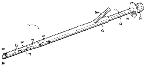
[010] Fig. 1 is a perspective view of an airway assembly described herein in a
collapsed
configuration;
[011] Fig. 2 is a side-elevational sectional view of the airway assembly of
Fig. 1;
[012] Fig. 3 is a perspective view of an airway assembly of Fig. 1 in an
expanded
configuration;
[013] Fig. 4 is a side-elevational sectional view of the airway assembly of
Fig. 3;
[014] Fig. 5 is an enlarged view of a portion of the airway assembly of Fig.
3;
[015] Fig. 6 is an elevational view of portions of the airway assembly of Fig.
3;
[016] Fig. 7 is an end view, taken along line 7-7 of Fig. 6;
[017] Fig. 8 is a perspective view of an airway assembly described herein in a
collapsed
configuration;
[018] Fig. 9 is a side-elevational sectional view of the airway assembly of
Fig. 8;
[019] Fig. 10 is a perspective view of the airway assembly of Fig. 8 in an
expanded
configuration;
[020] Fig. 11 is a side-elevational sectional view of the airway assembly of
Fig. 10;
[021] Fig. 12 is an enlarged view of a portion of the airway assembly of Fig.
10;
[022] Fig. 13 is an side-elevational view of a portion of the airway assembly
of Fig. 12;
[023] Fig. 14 is an end view, taken along line 14-14 of Fig. 13;
[024] Fig. 15 is a perspective view of an airway assembly described herein in
a
collapsed configuration;
[025] Fig. 16 is a side-elevational sectional view of the airway assembly of
Fig. 15;
[026] Fig. 17 is a perspective view of the airway assembly of Fig. 15 in an
expanded
configuration;
[027] Fig. 18 is a side-elevational sectional view of the airway assembly of
Fig. 17;
[028] Fig. 19 is an enlarged view of a portion of the airway assembly of Fig.
17;
[029] Fig. 20 is an elevational view of a portion of the airway assembly of
Fig. 19;
[030] Fig. 21 is an end view taken along line 21-21 of Fig. 20;
[031] Fig. 22 is a perspective view of an airway assembly described herein in
a
collapsed configuration;
[032] Fig. 23 is a side-elevational sectional view of the airway assembly of
Fig. 22;
- 4 -
CA 02710763 2010-06-23
WO 2009/086506
PCT/US2008/088420
[033] Fig. 24 is a perspective view of the airway assembly of Fig. 22 in an
expanded
configuration;
[034] Fig. 25 is a side-elevational sectional view of the airway assembly of
Fig. 24;
[035] Fig. 26 is an enlarged view of a portion of the airway assembly of Fig.
24;
[036] Fig. 27 is an end view taken along line 27-27 of Fig. 28;
[037] Fig. 28 is an elevational view of a portion of the airway assembly of
Fig. 26;
[038] Fig. 29 is a perspective view of an airway assembly described herein in
a
collapsed configuration;
[039] Fig. 30 is a side-elevational sectional view of the airway assembly of
Fig. 29;
[040] Fig. 31 is a perspective view of the airway assembly of Fig. 29 in an
expanded
configuration;
[041] Fig. 32 is a side-elevational sectional view of the airway assembly of
Fig. 31;
[042] Fig. 33 is an enlarged view of a portion of the airway assembly of Fig.
31;
[043] Fig. 34 is an elevational view of a portion of the airway assembly of
Fig. 31;
[044] Fig. 35 is an end view taken along line 35-35 of Fig. 34;
[045] Fig. 36 is a diagrammatic view of a portion of an airway assembly
described
herein used with a patient;
[046] Fig. 37 is a diagrammatic view of a portion the airway assembly of Fig.
36
located within a patient;
[047] Fig. 37A is a diagrammatic view of an embodiment of the airway assembly
described herein;
[048] Fig. 38 is a diagrammatic view of a portion the airway assembly of Fig.
36
located within a patient;
[049] Fig. 39 is a diagrammatic view of a portion of an airway assembly
described
herein used with a patient;
[050] Fig. 40 is a diagrammatic view of a portion the airway assembly of Fig.
36
located within a patient;
[051] Fig. 41 is a diagrammatic view of a portion of an airway assembly
described
herein used with a patient;
[052] Fig. 42 is a diagrammatic view of a portion the airway assembly of Fig.
36
located within a patient;
- 5 -
CA 02710763 2015-03-05
[053] Fig. 43 is a diagrammatic view of a portion of an airway assembly
described
herein used with a patient;
[054] Fig. 44 is a diagrammatic view of a portion the airway assembly of Fig.
36
located within a patient; and
[055] Fig. 45 is a diagrammatic view of a portion of an airway assembly
described
herein used with a patient.
DETAILED DESCRIPTION OF THE PREFERRED EMBODIMENTS
[056] Embodiments
described here relate generally to an airway assembly 10.
The airway assembly 10 can be used to intubate a patient. Structures common to
the
embodiments are provided with like reference numerals. As the embodiments are
related,
features, such as dimensions, materials and the like, may be shared.
Differences among the
embodiments are highlighted when present. Both structures of and methods of
use of the -
embodiments are described below. Some features of the embodiments may become
clear after
consideration of the entirety of this description.
[057] One embodiment of an airway assembly 10 is shown in Fig. 1.
[058] Drawing attention to Figs. 1 and 2, the airway assembly 10 comprises
an
inner tube 12 having a central lumen, an inner surface 16 and an outer surface
18, an outer tube
14 having a central lumen, having an inner surface 20 and an outer surface 22
and a diametrically
expansive seal 30. The inner tube 12 is disposed coaxially and reciprocally
moveable within the
outer tube 14. There is sufficient clearance provided between the inner
surface 20 of the outer
tube 14 and the outer surface 18 of the inner tube 12 to permit movement of
the inner tube 12
with respect to the outer tube 14. A connector 24 is joined to the inner tube
12, at a proximal end
thereof, so that a fluid, such as a gas, a liquid and the like, can flow
between the connector 24
and the inner tube 12. Typically, a ventilator (not shown) is connected to
connector 24 to
provide an airflow to the patient. A distal end 28 of the inner tube 12
opposite to the proximal
end thereof joined to the connector 24 is open to permit flow of fluid through
the inner tube 12.
- 6 -
CA 02710763 2010-06-23
WO 2009/086506
PCT/US2008/088420
A port 26 is formed on the outer tube 14 so that fluid may flow between the
port 26 and a space
between the outer surface 18 of the inner tube 12 and the inner surface 20 of
the outer tube 14.
In some embodiments, at least one perforation 72 is disposed on the outer tube
14. The at least
one perforation 72 passes between the inner surface 20 and the outer surface
22 of the outer tube
14. The at least one perforation 72 allows for secretions collecting in the
area above the
diametrically expansive seal 30 to flow and be aspirated from an airway, it
also allows for drug
infusion or the like, between the outer tube 14 and the airway. The at least
one perforation 72
may be positioned about 2 cm from the distal end 28 of the outer tube 14 and
may be of any
suitable shape, such as oblong, circular and the like, and any suitable size.
Further, more than
one perforation 72 may be included, and the more than one perforation 72 may
be distributed
along the length of the outer tube 14 in any desired manner.
[059] Distal end 28 of the outer tube 14 is configured to
facilitate introduction of
the airway assembly 10 to a patient. The distal end 28 may have a bevel to
facilitate passage
through the vocal chords. Distal end 28 may also have a tapered diametric
profile along its
length, within the range of about 5cm to about 7cm in length, of a distal
section of outer tube 14.
This distal taper may also be collapsible and allows for easier visualization
during the intubation
procedure. In some embodiments, the outer diameter of the outer tube 14 is
substantially within
the range of about 10mm to about 12mm at its proximal end and can be reduced
to an outer
diameter substantially within the range of about 6mm to about 8mm at the
distal end 28
[060] A diametrically expansive seal 30 is disposed at a distal end of the
inner tube 12
opposite to the end thereof attached to the connector 24. There is a
substantially smooth
transition between the inner tube 12 and the expansive seal 30. The expansive
seal 30 may
comprise a generally tubular member having walls, a proximal end fixedly
coupled to the inner
tube 12, and an uncoupled distal end which opens distally to the anatomical
airway. The
proximal end of the seal is coupled to a distal end of the inner tube 12 and
is in fluid flow
communication with the central lumen of the inner tube 12. The walls and the
distal end of the
seal 30 may expand diametrically such that the distal end forms a
diametrically enlarged distal
opening sealingly seated against and in fluid flow communication with the
airway. In some
embodiments, the expansive seal 30 is movable between a diametrically
collapsed position,
shown in Figs. 1 and 2, and a diametrically expanded position, shown in Figs.
3 through 7. The
expansive seal 30 has an inner surface 32 and an outer surface 34. The
diametrically expansive
- 7 -
CA 02710763 2010-06-23
WO 2009/086506
PCT/US2008/088420
seal 30 serves to seal the airway while exerting minimal pressure in the
mucosa sufficient to
prevent aspiration of secretions from the upper airways and trachea into the
lungs, while
preventing back leakage of air given during respiratory ventilation or while
ventilating anesthesia
gas.
[061] The expansive seal 30 may have many appropriate dimensions in length,
diameter, or in its general shape, all of which depend upon the patient
criteria or the anatomy of
the target airway, e.g., trachea or bronchus. For purposes of example, only,
one set of
dimensions are appropriate for pediatric patients and another set of
dimensions are appropriate
for a patient with a very large airways. In one embodiment, the expansive seal
30 has an
expanded outer diameter of about 25 mm while, in another embodiment, the
expansive seal 30
has an expanded outer diameter of about 20 mm. The outer diameter of expansive
seal 30 may
be within the range of about 18 to 20 mm for adult males and within the range
of about 16 to 18
mm for adult females. It is understood by those skilled in the art that as one
places the
expansive seal 30 further distally within the bronchial branches, the
anatomical diameter
decreases, necessitating smaller diameter expansive seals 30. It is
preferable, therefore, that the
outer diameter of the expansive seal 30 be between about 10 to 25 mm in order
to accommodate
a wide variety of variances in anatomical structures of the trachea and
bronchial branches.
[062] An aperture 36 is on the expansive seal 30 adjacent the inner tube 12.
The
aperture 36 permits fluid flow through the inner surface 32 of the expansive
seal 30. The
aperture 36 is fluidly associated with the inner tube 12 to permit fluid flow
between the inner
tube 12 and the expansive seal 30.
[063] The expansive seal 30 is preferably fabricated of a biocompatible
material, such
as silicone, which is suitable for use in the pulmonary system, particularly
the trachea and
bronchi. The expansive seal 30 may be fabricated using a single material,
wherein the seal is
formed as a single monolithic or unitary element, or of plural joined elements
formed of the
same biocompatible material. Alternatively, the expansive seal 30 may be
fabricated of plural
biocompatible materials may be joined as a composite. In either construct of
the expansive seal
30, but more preferably, in the case of a composite construction of the
expansive seal 30, at least
one reinforcing member 38 is operably associated with the expansive seal 30 to
facilitate
movement of the expansive seal 30 between its diametrically collapsed and
diametrically
expanded positions. In accordance with the illustrated embodiments, plural
reinforcing members
- 8 -
CA 02710763 2015-03-05
38 are associated with the expansive seal 30 and extend longitudinally along
the expansive seal
30 in a radially spaced apart relationship relative to each other. The at
least one reinforcing
member 38 may be coupled to the expansive seal 30 on either its luminal or
abluminal surfaces,
or may be embedded within expansive seal 30 such that it resides at least
partially within a wall
thickness of the expansive seal 30. Alternatively, the at least one
reinforcing member 38 may
comprise a relatively thickened region, such as a rib or a pattern or ribs, of
the same material
employed in fabricating the expansive seal 30. The at least one reinforcing
member 38 is
preferably an elastic, shape memory or superelastic material, such as
stainless steel, silicone,
nitinol, chromium-molybdenum alloys, or similar materials. In this manner the
expansive seal 30
is self-expanding upon being released from a constraining sheath or covering,
such as the outer
tube 14. For purposes of this application, when reference is made to expansive
seal 30, such
reference is intended to be inclusive of the at least one reinforcing member
38, where
appropriate. Those of ordinary skill in the art will understand that the at
least one reinforcing
member 38 may or may not be necessary, depending upon the construction and
materials
employed in fabricating the expansive seal 30, in order to provide for either
expansion or
collapse, or to facilitate or aid in apposition or sealing of the expansive
seal 30 against the
anatomical airway.
[064] When in its diametrically expanded position, the expansive seal 30 is
intended to achieve the size of the airway while exerting low pressure against
the tracheal
wall, thereby inhibiting passage of secretions beyond the expansive seal 30 to
areas of the
airway beyond the expansive seal 30, and improving clearance from secretions
deposited
distal of the expansive seal 30. The expansive seal 30 also reduces the
likelihood of
unintended fluid passage through the airway. In some embodiments, the
expansive seal 30
may include at least one radiopaque or fluoroscopic marker to facilitate
imaging the
position of the expansive seal 30 after placement. The expansive seal 30 may
take on any
appropriate shape, for instance, the expansive seal 30 can be substantially
elongated,
substantially rounded or substantially horseshoe shape in transverse cross
section. In
longitudinal aspect, expansive seal 30 preferably has an elongate generally
tubular shape
with a rounded taper at a proximal end thereof that connects with the distal
end of the inner
tube 12. The shape of the expansive seal 30 may be dictated by airway anatomy,
by
compatibility with the cough mechanism and by a need to reduce the likelihood
of
- 9 -
CA 02710763 2015-03-05
aspiration of secretions. In some embodiments, a distal portion of the
expansive seal 30,
sometimes measuring about 2 to about 3 mm in axial length, may be everted to
afford a
smoother circumferential surface area for tissue engagement. Everting a distal
portion of
the expansive seal 30 may reduce potential tissue growth around the expansive
seal 30, and
possibly facilitate advancement of the inner tube 12 with reduced risk of
trauma to the
patient.
[065] Another embodiment of the airway assembly 10 is illustrated in
Figs. 8
through 14. As elements of this embodiment are substantially similar to
elements of the
embodiment shown in Fig. 1 through 7, like reference numerals are used for
similar elements.
The modifications in the airway assembly 10 are intended to provide
independent ventilation to
each one of the lungs as commonly required for surgical procedures such us
lobectomies or in
cases in which independent or single lung ventilation is desired. The
following discussion
highlights elements not previously emphasized.
[066] The embodiments shown in Figs. 8 through 14 include
modifications to
provide both single and double lung ventilation. An inflatable member 40, such
as a balloon, is
disposed proximate the distal end 28 of the outer tube 14. The inflatable
member 40 has an inner
surface 42 and an outer surface 44 and is movable between a deflated position,
shown in Figs. 8
and 9, and an inflated position shown in Figs. 10 through 14. In one
embodiment, the inflatable
member 40 is intended to fully inflate at a pressure substantially within the
range of about 15 to
about 30 cm H20. At least one aperture 48 is disposed in the inner tube 12. It
is preferable
according to this embodiment to provide at least two apertures 48, as shown in
Figs. 9 and 11 to
permit the ventilation fluid to have sufficient flow to the second lung. The
at least one aperture
48 is movable between an open position and a closed position by axially moving
the inner tube
12 relative to outer tube 14, the at least one aperture 48 is exposed to an
open position or
retracted within the outer tube 14 to a closed position. The at least one
aperture 48 allows fluid
movement through the aperture 48 and passing between the interior and exterior
of the inner tube
12. However, it is to be noted that, because the inner tube 12 is moveable
with respect to the
outer tube 14, the tubes 12 and 14 may be positioned such that fluid flow
through the at least one
aperture 48 is restricted, i.e. the at least one aperture 48 is in a closed
position. Figs. 8 and 9
illustrate the relative position between inner tube 12 and outer tube 14
wherein the at least one
aperture 48 is in the closed position within the outer tube 14. Hence, it is
to be appreciated that
fluid flow through the at least one aperture 48 is dependent upon relative
position of the inner
tube 12 and the outer tube 14. It is to be noted that while the Figures show
that the inflatable
- 10 -
CA 02710763 2010-06-23
WO 2009/086506
PCT/US2008/088420
member 40 is in its inflated position when the expansive seal 30 is in its
expanded position, and
the inflatable member 40 is in its deflated position when the expansive seal
30 is in its collapsed
position, this does not always have to be the case. For example, the
inflatable member 40 may
be in its deflated position when the expansive seal 30 is in its expanded
position or the inflatable
member 40 may be in its inflated position while expansive seal 30 is in its
collapsed position.
[067] An inflation port 46 is disposed in communication with the outer tube
14
and communicates with the inner surface 42 of the inflatable member 40 so that
fluid can flow
between the port 46 and the inflatable member 40. A suitable conduit, not
shown for clarity, is
disposed on or in the outer tube 14 for conveying an inflation fluid between
the inflation port 46
and the inflatable member 40. In this manner, this fluid flow controls
inflation or deflation of
the inflatable member 40 between its inflated and deflated positions. Once the
endotracheal
tube is placed such that the distal end of the inner tube 12 is positioned at
a desired location in
the right or left bronchus, the outer tube 14 is retracted to release the
expansive seal 30
permitting expansive seal 30 to diametrically expand and sealingly conform
against the
bronchus. The outer tube 14 is retracted sufficiently to position the
inflation member 40 at a
desired location within the trachea and inflated into sealing conformity
against the trachea. If the
apertures 48 are exposed, ventilation will occur to both lungs, with one lung
being ventilated
through the expansive seal 30 and the other lung being ventilated through the
apertures 48. If the
apertures 48 are in their closed position, ventilation will only occur within
the lung
communicating with the bronchus in which the expansive seal 30 is positioned.
[068] Another embodiment of the airway assembly 10 is shown in Figs. 15
through 21. This embodiment is substantially similar to the embodiment shown
in Figs. 8
through 14, hence the like reference numerals for similar structures. However,
the embodiment
illustrated in Figs. 15 through 21 includes at least one aperture 48 and two
seals, including a first
expansive seal 30A and a second expansive seal 30B. Each of the seals 30, 30A
and 30B are
preferably similar construction and include at least one reinforcing member 38
as previously
described. Both seals 30A and 30B are carried on the inner tube 12 and
diametrically expand
independently between expanded and collapsed positions, depending on relative
position of the
inner tube 12 and the outer tube 14. While Figs. 15 and 21 show both seals 30A
and 30B being
simultaneously in the same position, either expanded or collapsed, it is to be
noted that the
expansive seal 30A may be in its expanded position while the expansive seal
30B is in its
- 11 -
CA 02710763 2010-06-23
WO 2009/086506
PCT/US2008/088420
collapsed position, depending upon the relative position of the inner tube 12
relative to the outer
tube 14. Significantly, as can be appreciated by considering Figs. 15, 16 and
18, when expansive
seal 30B is in its collapsed position, expansive seal 30B covers aperture 48
thereby restricting
fluid flow through the aperture 48.
[069] A further embodiment of the airway assembly 10 is shown in Figs.
22
through 28. This embodiment is substantially similar to the embodiments shown
in Figs. 8
through 14. However, in this embodiment, both the expansive seal 30 and the
inflatable member
40 are disposed on the inner tube 12 and in an order reversed from the order
of those items as
depicted in Figs. 8 through 14. This embodiment demonstrates that elements of
the airway
assembly 10 may be arranged in any appropriately desired way to arrive at an
airway assembly
10 that meets particular needs.
[070] Drawing attention to Fig. 22, the inflation port 46 is
associated with and
positioned at a proximal end of the inner tube 12. A suitable conduit, not
shown for clarity, is
provided in association with the inner tube 12 for conveying fluid between the
port 46 and the
inflatable member 40 that is disposed on the inner tube 12 as described
above.. The expansive
seal 30 is connected with the inner tube 12 at a position between the
inflatable member 40 and
the connector 24 relative to the longitudinal axis of the inner tube 12. The
at least one aperture
48 passes through the inner tube 12 and is positioned such that the expansive
seal 30, when in its
collapsed position, covers and closes the at least one aperture 48. As
discussed previously, the
positions of the inflatable member 40 and the expansive seal 30 can be changed
from what is
shown in Figs. 22 through 28. For example, the inflatable member 40 may be in
its collapsed
position while the expansive seal 30 is in its expanded position or the
longitudinal spacing of the
inflatable member 40 and expansive seal 30 along the longitudinal axis of the
inner tube 12 may
be altered.
[071] An additional embodiment of the airway assembly 10 is shown in
Figs. 29
through 35. This embodiment is similar to the embodiment illustrated in Figs.
8 through 14 in
that both include an expansive seal 30, an inflatable member 40 and at least
one aperture 48.
However, in this embodiment, the expansive seal 30, the inflatable member 40
and the at least
one aperture 48 are all disposed on the inner tube 12.
- 12 -
CA 02710763 2010-06-23
WO 2009/086506
PCT/US2008/088420
[072] The inflation port 46 is disposed at a proximal end of the inner tube
12
proximate the connector 24. As described above with reference to other
embodiments, a suitable
inflation conduit, not shown for clarity, is associated with the inner tube 12
for conveying an
inflation fluid between the inflation port 46 and the inflatable member 40
that is disposed on the
inner tube 12 as well. The expansive seal 30 is disposed on the inner tube 12
such that the
inflatable member 40 is located between the expansive seal 30 and the
connector 24. The at least
one aperture 48 passes through the inner tube 12 and is positioned between the
expansive seal 30
and the inflatable member 40. In this configuration, fluid flow through the at
least one aperture
48 is not dependent upon whether the expansive seal 30 is in its expanded or
collapsed position..
Fluid flow through the at least one aperture 48 is limited by appropriate
relative positioning of
the inner tube 12 and the outer tube 14, as shown in Figs. 31 through 34.
[073] With structure of the airway assembly 10 having been discussed with
reference to the foregoing embodiments now an exemplary method of use of an
airway assembly
will be explained. To ease understanding, the embodiment of the airway
assembly 10 similar to
that shown in Figs. 8 through 14 will be used. It is to be understood that any
of the embodiments
described herein can be used with this method with suitable modifications to
either the method or
to the assembly 10. Furthermore, additional features of the airway assembly 10
may become
apparent to those skilled in the art upon review of the following description.
[074] Beginning with Fig. 36, the airway assembly 10, including inner tube
12
and outer tube 14, is prepared for insertion into a patient to provide single
or double lung
ventilation. Positioning marks may be placed on the inner tube 12 to indicate
to the physician
the relative positions of the inner tube 12 and the outer tube 14 and whether
the airway assembly
10 is in a single lung ventilation mode or in a dual lung ventilation mode. A
first positioning
mark 50 and a second positioning mark 52 indicate the status of the expansive
seal 30 and the
condition of the at least one aperture 48. Specifically, the first positioning
mark 50 is provided
distally to indicate that an expansive seal 30 is collapsed and within the
outer tube 14, a first
intermediate mark (not shown), proximal to the distal positioning mark 50, may
indicate that the
expansive seal 30 is expanded and that the at least one aperture 48 is closed
and covered within
the outer tube 14, a second intermediate mark (not shown), proximal to the
first intermediate
mark, may indicate that the expansive seal 30 is expanded and that the at
least one aperture 48 is
exposed and uncovered by the outer tube 14, and the second positioning mark 52
is provided
- 13 -
CA 02710763 2010-06-23
WO 2009/086506
PCT/US2008/088420
proximally to indicate that the expansive seal 30 is expanded, the at least
one aperture 48 is open
and, where present, a proximal expansive seal is expanded. It will be
understood that depending
upon the specific configuration and number of expansive seals 30 and apertures
48, variations in
the number and positioning of the positioning marks 50, 52 are contemplated in
order to provide
the physician with an indicator of the status of the respective expansive
seals 30 or apertures 48.
[075] When a proximal end 53 of the outer tube 14 is located distally of the
first mark
50 (a first status of the airway assembly 10), the expansive seal 30 is in a
collapsed position and
the at least one aperture 48 is in its closed position. When a proximal end 53
of the outer tube 14
is adjacent the first mark 50 (a second status of the airway assembly 10), the
expansive seal 30 is
in its expanded position and the at lest one aperture 48 is in its close
position. When in the
second status of the airway assembly 10, ventilation of a single lung, through
the inner tube 12
and the aperture 36 in the expansive seal 30, is possible. Ventilation of both
lungs is
accomplished by positioning the proximal end 53 of the outer tube 14 adjacent
the second mark
52 (a third status of the airway assembly 10), the expansive seal 30 is in its
expanded position
and the at least one aperture 48 positioned in the inner tube 12 is in its
open position, and the
inflatable member 40 is inflated to seal the airway, thereby allowing an
operator, such as a doctor
and the like, of the airway assembly 10 to provide ventilation to both lungs.
Thus, it can be
appreciated that the first status of the airway assembly 10 corresponds to an
initial status of the
airway assembly 10, the second status of the airway assembly 10 corresponds to
a single lung
ventilation status of the airway assembly 10, and the third status of the
airway assembly 10
corresponds to a dual lung ventilation status of the airway assembly 10. In
some embodiments,
there may be more or less marks provided on the inner tube 12 or the outer
tube 14 or both,
thereby providing more airway assembly 10 status indicators. In operation, the
first mark 50 is
a distal mark that indicates that the outer tube 14 is pulled back to expose
the aperture 48, the
inflation member 40 is expanded, and double lung ventilation is being
performed. The second
mark 53 is a proximal mark that indicates that the outer tube 14 is positioned
to cover and close
the aperture 48, the inflation member 40 is deflated, and the expansive seal
30 is deployed in a
bronchi and single lung ventilation is being performed.
[076] As shown in Figures. 36 through 38, after the airway assembly 10 is
passed
through the vocal chords using a laryngoscope, an endoscope 54, such as a
bronchoscope and the
like, is placed coaxially through the central lumen of the inner tube 12 to
visualize distally the
- 14 -
CA 02710763 2010-06-23
WO 2009/086506
PCT/US2008/088420
airway assembly 10 and provide placement guidance for the airway assembly 10.
Once the
intended position for placement of the expansive seal 30 is identified, the
endoscope 54 acts like
a guidewire for the airway assembly 10 to permit placement of the expansive
seal 30 in the
patient's right or left bronchial tree to permit single lung ventilation to
the right or left lung
respectively.
[077] Fig. 37 illustrates portions of the airway assembly 10 and the endoscope
54
inserted into a patient. For ease of understanding, elements of the airway
assembly 10 are
represented transparently. A distal end 56 of the endoscope 54 is positioned
within a first
bronchus 58 of the patient. The first bronchus 58 is associated with a first
lung 60. Of course,
there is a second bronchus 62 associated with a second lung 64. The operator
positions the distal
end 56 of the endoscope 54 at a desired position in the first bronchus 58. The
airway assembly
10 is advanced along the endoscope 54 to the desired position. As shown in
Fig. 37A, some
embodiments of the airway assembly 10 include a narrowed distal region 66,
located adjacent
distal ends, where the diameter of the inner tube 12 and the diameter of the
outer tube 14 are
reduced from other more proximal portions of those elements. In this
embodiment, the expansive
seal 30 may have an outer diameter within the range of about 10 to 15 mm. In
some
embodiments, the reduced dimensions are outer diameters which, adjacent distal
ends, are
smaller than outer diameters adjacent proximal ends of the same element, such
as the inner tube
12, the outer tube 14 and the expansive seal 30. These reduced dimensions
facilitate introduction
of the airway assembly 10 into the patient by, for example, increasing ease of
moving the distal
end 28 of the outer tube 14 beyond vocal cords or glottic space of the
patient. In some
embodiments, the narrowed distal region 66 is substantially within the range
of about 5 to about
8 cm in axial length, and has a maximum outer diameter substantially within
the range of about 6
to about 10 mm. In some embodiments, when the expansive seal 30 is in its
collapsed position,
the expansive seal 30 has an outer diameter substantially equal to the outer
diameter of the inner
tube 12 adjacent the expansive seal 30.
[078] To further facilitate introduction and maneuvering of the airway
assembly 10,
portions of the inner tube 12 and the outer tube 14 may be comprised of
different materials
having different physical and/or material properties. For example, proximal
portions of the tubes
12 and 14 may be stiffer and more rigid than distal portions of the tubes 12
and 14. This
construction may ease the advancement of the airway assembly 10 in the patient
with reduced
- 15 -
CA 02710763 2015-03-05
deformation or curving of the tubes 12 and 14. Further, the relatively softer
and more malleable
material comprising the distal portions of the tubes 12 and 14 may allow for
deformation or
compression of distal ends of the tubes 12 and 14, and also may be more
accommodating to the
operator.
[079] In some embodiments, instead of having a tapered distal region 66, the
inner
tube 12 can have a substantially constant outer diameter similar to the outer
diameter of the
tapered distal region 66. This construction can reduce an outer diameter or
profile of the
airway assembly 10, and can facilitate aspiration through the space between
the outer
surface 18 of the inner tube 12 and the inner surface 20 of the outer tube 14.
In other
embodiments, both the inner tube 12 and the outer tube 14 can have
substantially constant
outer diameters, thereby making the region 66 unnecessary.
[080] As shown in Fig. 38, the airway assembly 10 is moved with respect to the
patient
to position the distal end 28 within the first bronchus 58. At this location,
it is desired to move
the expansive seal 30 from its collapsed position to its expanded position.
Related conditions of
a proximal end of the airway assembly 10 are shown in Fig. 39 (first location
with expansive seal
30 collapsed) and Fig. 41 (second location with seal expanded). Note the
relative locations of
the marks 50 and 52 and the end 53. The outer tube 14 is moved to allow the
expansive seal 30 to
diametrically expand from its collapsed position to its expanded position. The
endoscope 54 is
then removed from the airway assembly 10 as shown in Figure 40.
[081] In its 'expanded position, the outer surface 34 of the expansive
seal 30
contacts an inner surface of the first bronchus 58. The contact pressure
between the outer
surface 34 and the first bronchus 58 is sufficient to exclude secretions from
passing across
expansive seal 30 and into the first lung 60. However, that contact is
insufficient to harm the
first bronchus 58. With the expansive seal 30 in its expanded position, fluid
can flow among the
connector 24, the inner tube 12, the aperture 36, the first bronchus 58 and
the first lung 60. This
fluid flow is indicated generally by arrow 68 of Figs. 40 and 41; under this
condition the airway
assembly is providing single lung ventilation to the first lung 60. This
arrangement allows fluid
to flow among the connector 24, the inner tube 12, the aperture 36, the first
bronchus 58 and the
first lung 60 while limiting fluid flow to or from the second bronchus 62 and
the second lung 64.
This configuration permits single lung ventilation while excluding ventilation
to the other lung.
- 16 -
CA 02710763 2010-06-23
WO 2009/086506
PCT/US2008/088420
[082] It is not necessary to have the inflatable member 40 in its expanded
position to ventilate a single lung. During single lung ventilation, the
inflatable member 40 may
be either in its deflated or inflated positions. When the patient's condition
requires ventilation of
both lungs, the outer tube 14 is moved with respect to the inner tube 12 so
that the aperture 48 is
moved to its open position. The proximal end 53 is adjacent the second mark
52. This is the
third location (the at least one aperture 48 in its open position) and is
shown in Figs. 42 and 43.
The inflatable member 40 is moved to its inflated position. By doing this,
unintended back fluid
flow is limited.
[083] This status of the airway assembly 10 permits fluid flow among the
connector 24, the inner tube 12, the aperture 48, the second bronchus 62 and
the second lung 64.
This fluid flow is represented by arrow 70 of Fig. 42. Fluid flow 68 occurs as
well. The
inflatable member 40 is changed to its inflated position. The proximal end of
the airway
assembly 10 is shown in Fig. 43. This configuration permits both lungs to be
ventilated. .
[084] When the clinical condition does not require single lung ventilation,
such
as at the end of a surgical procedure, as shown in Fig. 44, the inner tube 12
is moved pulled back
proximally with respect to the outer tube 14 thereby capturing the expansive
seal 30 within the
outer tube 14 and collapsing the expansive seal 30. Fluid then flows to both
the first lung 60 and
the second lung 64. In this position, the airway assembly 10 is its delivery
configuration, as
shown in Fig. 45, and the airway assembly 10 may remain within the patient to
provide
continued intubation or the airway assembly 10 may be removed.
[085] Those of ordinary skill in the art will understand and appreciate that
the foregoing
description of the invention has been made with reference to certain exemplary
embodiments of
the invention, which describe airway assemblies suitable for single and/or
dual lung ventilation,
while excluding passage of secretions across the expansive seal 30. Those of
skill in the art will
understand that obvious variations in construction, materials, dimensions or
properties may be
made without departing from the scope of the invention which is intended to be
limited only by
the claims appended hereto.
- 17 -