Note: Descriptions are shown in the official language in which they were submitted.
CA 02710769 2010-06-25
WO 2009/085108 1 PCT/US2008/013553
NON CONTACT MAPPING CATHETER
Field of the Invention
The present invention relates generally to a catheter for.use
inside the human heart during medical procedures. The catheter can be
used for "non-contact" mapping of the electrical activity of the heart,
for locating and reporting the position of other procedure catheters
within the heart, and for other purposes. The catheter includes an
electrode array that can be deployed and retracted independently from
catheter articulation.
Background of the Invention
Cardiac arrhythmias are a leading cause of stroke, heart disease,
and sudden death. The physiological mechanism of arrhythmia
involves an abnormality in the electrical conduction of the heart. There
are a number of treatment options for patients with arrhythmia that
include medication, implantable devices, and catheter ablation of
cardiac tissue.
Traditionally, the arrhythmia is studied and diagnosed by
"electrically mapping" the heart with catheters inserted through the
vasculature into a heart chamber. Traditionally, the electrical activity of
the heart is acquired directly by "in- contact" mapping of the interior
wall surface of a heart chamber. In this technique electrodes are placed
in intimate contact with the heart wall and the voltage at that location is
recorded and plotted against time for display to the physician. The in-
contact catheters may be large and essentially fill the entire heart
chamber, or they may be smaller and moved around in the heart
chamber to sequentially map various areas of the heart. Mechanically,
the in-contact mapping catheters are "soft" so that they can conform to
the heart chamber. Softness is required so the electrodes come into
intimate contact with the heart wall while accommodating wall motion
of the beating heart.
CA 02710769 2010-06-25
WO 2009/085108 2 PCT/US2008/013553
For example, multiple electrode in-contact mapping catheters are
known from U.S. Patent No. 5,628,313 to Webster that shows a so-called
"basket" catheter. In use, this very flexible and conformal catheter is
deployed in the heart and presses individual electrodes against the
chamber wall for full chamber contact mapping of a beating heart.
Smaller multiple electrode catheters are known as well. For example,
the U.S. Patent No. 5,279,299 to Imran illustrates techniques for creating
smaller catheter arrays that are used to selectively contact map portions
of the cardiac chamber. This catheter is flexible and electrodes remain in
contact with the wall even when the catheter shaft is displaced slightly.
In each of these examples, the limbs of the catheter are very flexible and
gently contact the chamber wall while the wall of the heart is moving.
"Non-contact mapping," also known in the art, is an alternative
to in-contact mapping where a catheter array positioned within a
chamber is used to collect global electrical information. This global
information is then used to compute a solution to the so-called "inverse
problem". The inverse problem of electrophysiology is the calculation
of wall electrical potentials from the measured field voltages associated
with the wall potentials as measured within the blood pool remote from
the chamber wall. The mathematical "solution" displayed to the
physician is the computed wall surface voltages that can be used to
detect problems in electrical conduction in the heart wall.
Although in-contact and non-contact catheters are used for the
same patient indications, they have very different mechanical and
electrical requirements. Chief among the requirements of a non-contact
catheter is stability of the electrode array. The geometry and locations
of the electrodes are assumed for the inverse solution calculation and
need to be known with great precision. Small error in electrode position
can render large discrepancies in computed mathematical solution. In
addition, controlled movement of the electrode array within the
chamber of interest is necessary in order to improve the accuracy of the
non-contact map. Deployment of the electrode array into a repeatable
precisely known shape, while supporting controlled movement of the
CA 02710769 2010-06-25
WO 2009/085108 3 PCT/US2008/013553
catheter, pose particularly complex and novel requirement on the
catheter design.
Once the anatomic origin of problems in electrical conduction are
identified, the physician may proceed to ablate the offending tissue,
thus treating the arrhythmia. Catheter ablation procedures have
evolved in recent years to become an established treatment for patients
with a variety of supraventricular and ventricular arrhythmias. The
typical catheter ablation procedure involves mapping of the heart tissue
in order to identify the site of origin of the arrhythmia, followed by a
targeted ablation of the site with an RF catheter. The procedure takes
place in an electrophysiology laboratory and takes several hours most
of which is spent mapping the electrical conduction in the heart.
Although in-contact and non-contact mapping methods are
known in the art and various deflectable, displaceable and deployable
catheters are known as well, there is a continuing need to improve the
accuracy, stability and maneuverability of such devices so that they can
be more widely used, especially as an adjunct to cardiac ablation
procedures.
Summary of the Invention
The present invention is an intravascular catheter that may be
deployed within a heart chamber placing multiple electrodes in a
known spatial configuration. The catheter may be used to map electro-
anatomical characteristics of the heart and/or to locate and position
other catheters within the heart. Adoption of the inventive construction
of the present catheter provides a device that is smaller, less expensive
to manufacture, maneuverable, and stable in its deployed
configuration. Electrode stability makes the device much more accurate
and therefore, of more value to the physician. The design and
construction also make the device smaller in cross section than existing
designs and therefore, more easily used by a physician and better
tolerated by the patient. As set forth in detail hereafter, the distal array
of the catheter is fabricated as a flexible printed circuit. The deployment
CA 02710769 2010-06-25
WO 2009/085108 4 PCT/US2008/013553
and articulation functions of the catheter are very independent of each
other.
Two separate embodiments of the deployment mechanisms are
disclosed. In contrast to prior art devices both of these mechanisms
permit the deployment function to operate wholly independently from
the articulation or deflection feature of the catheter. The independence
of the deployment feature and the articulation feature together with
innovative structural features and materials create a non-contact
mapping catheter that is easily placed and used with a very stable
electrode geometry.
Brief Description of the Drawings
An illustrative embodiment of the invention is shown in the
several views of the figures. The use of identical reference numerals
throughout the several figures and views indicate the same element of
the device, wherein;
Fig. 1 is a schematic diagram showing the catheter in the context
of the system;
Fig. 2A is a schematic diagram showing the catheter;
Fig. 2B is a schematic diagram showing the catheter;
Fig. 2C is a schematic diagram showing the catheter;
Fig. 3A is a schematic diagram showing the distal portion of the
catheter;
Fig. 3B is a schematic diagram showing the distal portion of the
catheter;
Fig.4A shows a step in the construction of the distal portion;
Fig. 4B shows a step in the construction of the distal portion;
Fig. 4C shows a step in the construction of the distal portion;
Fig. 4D shows a step in the construction of the distal portion;
Fig. 5A shows a step in the manufacture of the distal portion;
Fig 5B shows a step in the manufacture of the distal portion;
Fig. 6A shows the flexible printed circuit in plan view;
Fig. 6B shows the flexible printed circuit in cross-section;
CA 02710769 2010-06-25
WO 2009/085108 5 PCT/US2008/013553
Fig. 7 shows a metallization layer of the flexible printed circuit;
Fig. 8A shows the spline assembly formed from a flexible printed
circuit in plan view;
Fig. 8B shows the spline assembly formed from a flexible printed
circuit in cross-section view;
Fig. 8C shows a distal array segment in projection view;
Fig. 8D shows a spline in cross section;
Fig. 8E depicts a portion of a spline of Fig.8D;
Fig. 9A shows the spline assembly formed from a flexible printed
circuit in plan view;
Fig. 9B shows the spline assembly formed from a flexible printed
circuit in cross-section view;
Fig. 9C shows a distal array segment in projection view;
Fig. 9D shows a spline in cross section;
Fig. 9E depicts a portion of the spline shown in Fig. 9D;
Fig. 10A shows a first embodiment of the deployment actuator;
Fig. 10B shows a first embodiment of the deployment actuator;
Fig. 11 shows a distal array segment in projection view;
Fig. 12 shows a distal array segment in projection view;
Fig. 13 shows a distal array segment in projection view
Fig. 14 shows a partial section of a distal segment with an
additional feature;
Fig. 15 shows simplified schematic of second embodiment of the
deployment actuator showing complimentary distal and proximal
springs;
Fig. 16A is a simplified schematic of the catheter;
Fig. 16B is a simplified schematic of the catheter;
Fig. 16C is a simplified schematic of the catheter; and,
Fig. 17 is a plot of force against displacement of several
structures in the catheter.
CA 02710769 2010-06-25
WO 2009/085108 6 PCT/US2008/013553
Description of the Invention
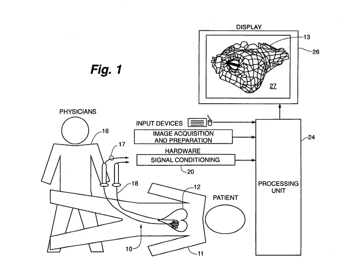
Fig. 1 depicts the context of the invention. The figure shows a
highly schematic view of the overall system that includes the physician,
patient, catheters, and related electrophysiology equipment located
within an operating room. The physician 16 introduces the catheter 10
into the vasculature of the patient 11 at the patient's leg and advances it
along a blood vessel ultimately, entering the patient's heart 12. Other
catheters that may be used in the procedure are represented by
companion catheter 18. Each catheter is coupled to signal conditioning
hardware 20 with appropriate catheter cabling typified by catheter
cable 17. The signal conditioning hardware 20 performs various
interface functions applicable to the mapping, tracking, and registration
procedures that are performed in conjunction with the workstation
class computer-processing unit 24. If the companion catheter 18 is an
ablation catheter, then conditioning hardware also forms an interface to
an RF ablation unit (not illustrated). Three patent applications all
published 12/27/2007 are incorporated by reference herein to further
explain the use of the catheter for non-contact mapping as follows:
20070299353;20070299352 and 20070299351.
In use, the physician looks at a computer display 26. Present on
the display is a substantial amount of information. A large window
presents an image of the heart chamber 13 along with an image of the
catheter 10. The physician will manipulate and control the catheter 10
based in part on the images an d other data presented on the display 26.
The image 27 seen in Fig. 1 is schematic and depicts the distal array of
the catheter 10 deployed, occupying a small portion of the heart
chamber 13 volume. The representation of the heart chamber 13 may
use color, wire frame, or other techniques to depict the structure of the
heart chamber 13 and to simultaneously portray electrical activity of the
patient's heart. It is regarded as useful to display chamber geometry,
catheter location, and electrical activity in an integrated fashion on the
display 26. In use, the physician will observe the display 26 and interact
CA 02710769 2010-06-25
WO 2009/085108 7 PCT/US2008/013553
with the workstation processing unit 24 and the catheters 10 and 18, to
direct the therapy as a medical procedure.
Fig. 2 A through Fig. 2 C depicts array deployment and catheter
articulation along with the associated positions of the handle controls.
Fig. 2A shows the catheter 10 in isolation. The catheter 10 has an
elongate body 31 with a distal end 37 and a proximal end 39. The
elongate body 31 includes a tubular sheath 35. The proximal end 39
connects to an assembly that includes a handle segment 30. The
physician may manipulate the handle segment 30 to selectively deflect,
deploy, and rotate the catheter to perform the medical procedure. The
handle segment 30 is coupled to an elongate intermediate section or
segment 32. The intermediate section is coupled to a deflection segment
34, which in turn is coupled to a distal array segment 36, located at the
distal tip or end 37. Not shown is the catheter cable 17 used to connect
the electrodes on the distal array segment 36 to the signal conditioning
hardware 20. In Fig. 2A the catheter 10 is in the undeflected and
undeployed state where the distal array segment 36 is collapsed and the
deflection segment 34 is straight. In this configuration, the catheter is
introduced into the body using the familiar Seldinger technique.
Fig. 2B shows the catheter 10 with the handle segment 30
manipulated to deploy the distal array segment 36 into the open or
deployed state. In one embodiment, the pommel 33 of the handle
assembly 30 is moved retrograde with respect to the handle assembly as
indicated by motion arrow 38 to deploy the distal electrode array
segment 36. In this embodiment, the pommel 33 will lock into position
to deploy the array 36. To set the lock, the pommel 33 will have to be
pulled enough to overcome a modest spring force to reach a detent
position. When deployed, the distal array segment 36 opens to place
electrodes into the operating position. In alternative embodiments the
deployment control may be turned or rotated to deploy the electrode
array.
Fig. 2C shows activation of the deflection segment 34. Antegrade
motion of the handle ferrule 42 of the handle segment 30 depicted by
CA 02710769 2010-06-25
WO 2009/085108 8 PCT/US2008/013553
motion arrow 40 deflects or articulates the deflection segment 34. Note
that the catheter 10 responds to this motion and the deflection segment
34 forms an arc confined to a single plane. In the figure, the articulation
or deflection motion lies in the plane of the page. The deflection
operation causes the distal array segment 36 to be' pointed up to 180
from the initial direction shown in panel 2A. The phantom dotted
position seen in the figure shows that this articulation may be
symmetrically "bi-directional". It should also be understood that the
articulation may also be asymmetrically bi-directional such that the arc
shape is different in each direction. In one embodiment, best depicted in
Fig. 15, articulation or deflection of the segment 34 moves a pull wire
from the center axis of the catheter and it moves off to the side within
the catheter body. This displacement of the pull wire reduces tension in
the pull wire and leads to the deflection.
Thus it is shown that the catheter 10 has an elongate body 31
having a distal end 37, and a proximal end 39, and an elongate central
axis. A proximal handle segment 30 having an articulation control 42
and a deployment control 33 are attached to the proximal end 39. There
is an intermediate segment 32 connected to the handle and a deflectable
segment 34 connected to the intermediate segment 32. The deflectable
segment 34 will articulate in a plane through an angle in response to the
articulation control. Also a distal array segment 36 is connected to the
deflectable segment 34. This distal array segment 36 includes a
deployable distal electrode array that can move from a first retracted
position depicted in Fig. 2A to a second deployed position depicted in
Fig 2B. The deployment mechanism coupled to said deployment control
couples the motion of the deployment control to operate the distal
electrode array segment which causes the distal array segment to
deploy into said second deployed position, independently of the
operation of said articulation control.
The physician can rotate the handle segment 30 and operate
ferrule 42 to position and "aim" the distal array segment 36 toward any
part of the cardiac anatomy within the heart chamber. When deployed,
CA 02710769 2010-06-25
WO 2009/085108 9 PCT/US2008/013553
the various splines typified by spline 50 carry various electrodes into
specific highly stable and reproducible spatial locations.
Fig. 3A and Fig. 3B depict the distal array segment 36 in the
deployed and undeployed states and serve to illustrate the location of
the electrodes. Fig. 3A shows the distal array segment 36 in isolation
and in the retracted or undeployed 43 state or condition. The drawing
shows a uniform and symmetrical distribution of the electrode sites as
typified by electrode 54 along the length of a typical spline 50. It may be
useful to place more of the sensing electrodes near the most distal end
or tip 37 of the distal array segment 36. An asymmetrical electrode
distribution may be advantageous for non-contact mapping functions.
In addition to multiple sensing electrodes, current injecting locator
electrodes, typified by locator electrode 55, may be placed at a location
along the spline 50. In general it is preferred to position locator
electrodes so that they are far apart in the deployed sate. Current
sourcing or sinking for the locator electrodes may also take place from
ring electrodes 57 and tip electrode 53. Tip electrode 53 may also be
provided for cardiac stimulation, ablation or as a locator electrode.
In summary, the splines 50 of the distal electrode array segment
36 may carry various sets of independent electrodes 54. Typically sixty-
four sensing electrodes will be distributed over and along the various
splines 50. Several locator electrodes may be positioned diametrically
opposed to each other as illustrated by example, on the meridian of
the deployed shape. Optionally other electrodes may occupy space in
the distal electrode array. In use, sets of the electrodes are used at
various times or concurrently during the medical procedure.
Fig. 3B shows the distal array segment 36 in the deployed state
41. Together Fig. 3A and 3B show the motion of the several splines that
make up the distal electrode array 36 as they move from the
undeployed state 43 to the deployed state 41. While in the undeployed
state 43, the splines lie together along side each other in a roughly
tubular shape seen in Fig. 3A. The splines typified by spline 50 deflect
and blossom moving outwardly in a radial direction as the array is
CA 02710769 2010-06-25
WO 2009/085108 10 PCT/US2008/013553
deployed to the deployed state 41 as seen in Fig. 3B. This spline motion
may be driven by a pull wire (Fig.15 element 52) in a pull wire
embodiment. Alternatively the spline motion may be driven by a
rotating screw 153 that moves the screw driven pull member 159 seen
within the array in Fig. 10 A and 10B. A rotatable member is used as a
torque transmitting device from the handle to the screw member in the
distal section. The rotatable member needs to be able to transfer torque
while in a curved environment. The rotatable member can be
implemented in the form of a torque transmitting wire, coil, braid
reinforced plastic tube or laser cut hypotube. The term rotatable
member is intended to describe all of these alternative
constructions. This alternative embodiment is called the rotary screw
embodiment.
In the pull wire embodiment, the pull wire 52 is pulled back into
the catheter body of the deflectable segment 34 and the splines deform
into a shape reminiscent of a bulb of garlic. The pommel control 33 and
the proximal spring 402 are connected to the pull wire 52 and motion of
the pommel control 33 moves the splines to the deployed state.
The individual splines may carry several types of electrodes. The
array of sensing electrodes typified by spline electrode 54 are used for
non-contact mapping and may also be used for assisting in the
detection and location of companion catheters in the heart chamber.
These non-contact electrodes are in the blood pool and they must
receive and detect very small voltages to perform the mapping
operation. Locator electrode 55 is typical of such a spline electrode used
for location purposes (also shown in Fig 3A). Typically locator
electrodes will lie on the greatest meridian of the deployed array 41 so
that once deployed they are quite far from each other as seen in Fig. 3B.
However not every spline need carry a locator electrode.
Each electrode on a spline is electrically connected to the cabling
in the handle. It is preferred that the signal from each individual
electrode be independently available to the hardware interface 20. This
may be achieved by passing a conductor for each electrode through the
CA 02710769 2010-06-25
WO 2009/085108 11 PCT/US2008/013553
connection cable 17. As an alternative, the electrical connections may be
multiplexed in the catheter device 10 to minimize conductors.
It is important that the high-density electrode array be deployed
into a known, reproducible, and relatively stiff shape. The number of
electrodes, their distribution and deployment shape, and stability in
shape determine the limits of system performance. As electrode number
and deployment volume increase, the performance is improved.
However it is both difficult and important to balance complexity, cost,
and performance with usability and patient benefit. An increase in
electrode number and deployment size increases catheter 10 complexity
and maneuverability of the catheter 10 is compromised. Experimental
work suggests that a typical catheter 10 should have sixty-four sensing
electrodes and deploy to a three dimensional somewhat spherical shape
with a diameter of 18mm. In order to know electrode locations for
analysis by the processing unit 24, the deployment shape must be
tightly controlled. Therefore, several critical design features must be
tightly controlled. The location of the electrodes 54 within the array
must be accurately placed. These electrodes 54 should also be placed in
a manner that facilitates their use in close proximity to the endocardial
surface when the array is deployed. This requirement may necessitate a
non-uniform distribution of the electrodes 54 as certain regions of the
.deployed array are more likely to be positioned closely to the
endocardium.
The deployed shape of the electrode array must be repeatable
through multiple deployment cycles. For example, electrode locations
need to be known to within 1mm between multiple deployments. The
array should be capable of deploying to a known shape and
subsequently closing to a low profile (e.g. 8 French) for retraction. This
shape change may be binary or continuous, but in either situation, the
shape must be repeatable and have a known geometry at the time of
data collection. The repeatable shape requirement is applicable to the
electrode array shape in both the circumferential and radial directions
and represent a significant design challenges. The inventive
CA 02710769 2010-06-25
WO 2009/085108 12 PCT/US2008/013553
combination of fabrication technology, structural design and material
choices cooperate together to achieve the design goal.
Also seen in Fig. 3B is a locator sensor 59. There are several
commercially available 3-D location systems available for use in
medical devices. In general location of the locator sensor 59 in space is
reported by a base station located near the patient. This technology is
widely used in robotic surgery and need not be described in detail.
Typically the locator sensor 59 would take the place of locator electrode
55.
Fig. 4A through Fig. 9D depict the formation of the array
structure from a flexible printed circuit.
Fig. 4A shows a step in a preferred construction methodology for
the distal array segment 36. The distal array segment 36 is
manufactured in part from a flexible printed circuit 60 ("FPC"). This
construction methodology has the advantage of repeatable high
accuracy and low manufacturing cost. To construct the FPC 60, the
material is initially fabricated in a planar form seen in Fig. 4A. In the
planar condition, a series of apertures 62 are cut through the FPC 60 at
one end typified by hole 62. Together the series of apertures 62 form a
bonding band 70. At the opposite more proximal end of the FPC 60
there is formed a termination band 106. The planar FPC 60 is also slit to
free the individual splines. Conventional laser processing is well suited
to this fabrication step.
Fig. 4B shows a process where the planar FPC 60 is wound
around a major axis 61 bringing first edge 63 toward second edge 65.
Fig. 4C shows the two edges juxtaposed with both ends fixed.
Together the bonding band 70 and the termination band or section 106
complete a cylindrical form. In general the distal bonding band 70 is
fixed by encapsulation and the termination band is fixed by anchoring
or bonding it to the deflection segment of the catheter.
Fig. 4D shows that with both ends fixed, the splines typified by
spline 50 may be moved radially with respect to the axis 61.
CA 02710769 2010-06-25
WO 2009/085108 13 PCT/US2008/013553
Fig. 5A shows that the ring of apertures 62 that together from a
bonding band 70. In the figure, the edges of the gap are seen in close
proximity at reference numeral 72.
Fig. 5B shows the use of the bonding band 70. Note that the
edges may be held together with a melted polymer or adhesive or other
plastic or thermoplastic material that is applied to the interior and
exterior of the tubular structure. This thermoplastic formed-in-place
plug 74 encapsulates the inside and outside of the FPC 60 providing an
unusually robust and durable structure that permits reliable
deployment of the splines.
Fig. 6A shows the FPC 60 in plan view. This view reveals the
several slits typified by slot or slit 108 which taken together form the
individual splines such as spline 50. These slits 108 extend from the
distal bonding section or band 70 to the termination section 106. Holes
62 appear in the bonding band 70 and additional slits 110 are formed
within the termination section 106 to facilitate attachment to the
deflectable section of the catheter.
The splines typified by spline 50 of the FPC 60 serve to position
the electrodes typified by electrode 54 along the length of the FPC 60.
The splines 50 also carry interconnecting metal traces (not shown) that
serve to electrically connect the electrodes to pads in the termination
section 106. The splines 50 are separated from each other using slits 108.
The slits are thin gaps that are cut in the FPC using one of many cutting
techniques that may include laser cutting, die cutting or chemical
etching. The slits 108 of the exemplary FPC are cut using a laser so as to
position slit location precisely.
The distribution of the electrodes 54 may, tightly controlled in
the design of the FPC 60. For example, in Fig 6A we note that electrodes
are distributed more densely in the distal tip area. It should be
appreciated that any desirable electrode distribution may be
accomplished using this method.
Fig. 6B shows the FPC 60 in cross-section. The various layers are
not to scale. Some layers described are very thin while other thick, not
CA 02710769 2010-06-25
WO 2009/085108 14 PCT/US2008/013553
all layers are depicted in the figure for clarity. In particular, very thin
layers are not shown explicitly in the drawings. The FPC is constructed
using a relatively thick core insulating layer 86. The core layer 86 of the
exemplary circuit is constructed of a 50um layer of polyimide.
Alternative materials and thickness core layers may be used to obtain
the desired mechanical and process characteristics. The core insulating
layer 86 is coated with a top metallization layer 88 and a bottom
metallization layer 90. Each of the exemplary metallization layers is
deposited by first sputtering a thin layer (- 0.1um) of titanium over the
core insulating layer 86. The titanium layer serves as an interface layer
to adhere additional metallization to the core insulating layer 86. The
metallization layers 88 and 90 can be added by further sputtering
and/or plating of additional metal over the titanium layers. The
exemplary metallization layers 88 and 90 are sputtered with a gold
layer over the titanium layer and then further plated with gold until the
total thickness of the metal layers measures 2um. It should be noted
that other conductors such as copper may also be used. It is also
necessary to provide electrical connection between metal layers 88 and
90 for the purpose of connecting circuit features that reside on each
layer. A connection can be formed by constructing a via 96 between the
two metallization layers. A via can be formed by creating a hole
through both metallization layers 88 and 90 and the core insulating
layer 86. Electrical connection is then made by plating the walls of the
hole between the two metallization layers forming a metal connection
96 between the metallization layers 88 and 90. The FPC is further
constructed by providing a top covercoat 92 over the top metallization
layer 90. The top covercoat 92 serves to insulate portions of the top
metal layer 88 from external contact. The top covercoat has openings 98
placed in regions where it is desired to have the top metal layer
exposed to external contact. For example a mapping electrode 54 may
have the covercoat above it exposed and be sputtered or plated onto the
top metal layer 88 as seen in Fig. 6B.
CA 02710769 2010-06-25
WO 2009/085108 15 PCT/US2008/013553
In the exemplary construction of Fig. 6B, the covercoat 92 of the
FPC is formed by a 25um layer of liquid photoimageable polyimide.
The photoimageable polyimide covercoat is exposed and developed to
precisely locate geometric features on the exterior surface to create
blood contacting electrodes, using similar registration and optical
techniques used to fabricate other features on the FPC.
A bottom covercoat 100 is applied to the bottom metal layer 90 in
order to insulate the bottom metal layer 90 from external contact. It may
be necessary in some applications to enable the bottom covercoat 100 to
have openings similar to the openings 98 of the top covercoat 92. Such
applications may require external contact to the bottom metal layer 90.
One important application for the mapping electrodes 54 is to sense low
voltage biological signals. The biological signals of interest are generally
in the tens of microvolts to several millivolt range in amplitude and are
time varying in the frequency range of 0.05 Hz to several kHz. The
detailed design of the Flexible Printed Circuit (FPC) layers and
electrodes in particular impact the noise level of the measurement
system. Reducing the impedance of the electrochemical interface
between the electrode and blood reduces overall system noise.
Although a wide range of materials may be used to reduce
impedance, our preferred electrode materials are selected from a small
group which have demonstrated to us that they are especially well
suited for this design. We prefer to select electrode materials for blood
contact from the group of gold, stainless steel, platinum, platinum-
iridium, titanium nitride, platinum black or iridium oxide (in order of
highest to lowest impedance). Electrode materials are applied using an
electroplating or sputtering process.
At present our preferred FPC 60 and electrode construction
includes an FPC with a polyimide core layer with gold metal layers.
The blood contacting electrodes are gold coated with iridium oxide.
In addition to material properties, electrode area has a profound
impact on impedance and in the design the electrode area may be
CA 02710769 2010-06-25
WO 2009/085108 16 PCT/US2008/013553
increased to a width limited by the dimension of the spline and further
limited by the presence of other metal features including traces.
It is also be possible to increase the surface area of electrodes
through surface finishing. Roughening of the electrode surface can be
accomplished through any one of several mechanical or chemical
surface treatments.
Fig. 6B also shows that a stiffener layer 102 may be applied over
the bottom covercoat 100 as seen in Fig. 6B. The stiffener layer 102 may
have various thickness and material compositions in order to achieve
the desired rigidity of the FPC in order to control the deployed shape.
The exemplary FPC of the invention is comprised of a 50um thick
polyimide stiffener 102 over the bottom covercoat 100. It should be
appreciated that other materials such as PEEK or Nitinol may be used
as a stiffener. The stiffener 102 is adhered to the to the bottom
covercoat using a polyimide adhesive layer. Other adhesives, and in
particular, pressure sensitive adhesives may also be used for this
purpose. Additional stiffener layers may be applied over stiffener layer
102. Stiffener layer 120 serves to increase the stiffness of the circuit in
selected areas.
The termination section 106 also serves to provide a region
where the FPC may be bonded to the outer catheter shaft during
installation.
Fig. 7 shows a metallization layer in plan view. The dark areas in
Fig. 7 are the metallization traces created by the processes described in
connection with Fig. 6A, but the core layer and other layers are not
shown for clarity. Subpanels seen in the figure are enlargements of the
metallization trace pattern to show various features. For example, the
termination section 106 of the FPC of Fig. 6A is shown as traces 108 in
this figure. The traces are metallic layers that serve to create a region
where the FPC can be connected to wire or cabling that serve to
electrically connect the FPC to circuitry or connectors in the proximal
section of the catheter. The wire or cabling may be attached to the FPC
at a series of termination lines as designated by reference numeral 112.
CA 02710769 2010-06-25
WO 2009/085108 17 PCT/US2008/013553
It should be appreciated that a number of metallization layers
ranging from 1 to 16 may be used. The addition of layers is helpful in
carrying additional signals given a width constraint such as the spline
width.
Fig. 8A shows how to increase the stiffness of the exemplary FPC
of Fig. 6 forming areas of high stiffness 124 and areas of lower stiffness
126.
Fig. 8B shows how to control the deployed shape of the array by
controlling the stiffness of the exemplary FPC forming areas of high
stiffness 124 and areas of lower stiffness 126.
Fig. 8C shows a representative shape where stiff zones 124 or
areas interspersed with less stiff areas 126 can create a complex array
shape upon deployment. In the figure, there is more stress in the thin
areas 126 which bend more readily than in the stiffer regions 124.
Fig. 8D shows thicker regions with additional stiffener layers
forming stiff zones 124 while less stiff material yields a less thick more
flexible area 126. The use of alternating stiffness areas helps to control
the distribution of stress as well as determine deployed shape. In this
embodiment the spline shape is segmented into relatively rigid
"straight" sections 124 followed by "hinged" areas 126. The detail
drawing of Fig. 8E shows the high stiffness area 124 next t o a lower
stiffness area 126.
Fig. 9A shows how to increase the stiffness of the exemplary FPC
of Fig. 6 forming areas of high stiffness 124 and areas of lower stiffness
126 that are spaced along the spline.
Fig. 9B shows that a stiffener layer 102 may be applied over the
bottom covercoat 100 as described in connection with Fig. 8B.
Fig. 9C shows a representative shape where stiff zones 124 or
areas combined with less stiff areas 126 can create a complex array
shape upon deployment. In the figure there is more stress in the thin
areas that bend more readily than in the stiffer regions 124. Together
the added material allows for a smoothly varying distribution of stress
along the spline.
CA 02710769 2010-06-25
WO 2009/085108 18 PCT/US2008/013553
Fig. 9D shows thicker regions with additional stiffener layers
forming stiff zones 124 while less stiffener material yields a less thick
more flexible area 126. The use of alternating stiffness areas helps to
control the distribution of stress as well as determine deployed shape
yielding a continuously curved spline having a smoothly varying
distribution of stress along the spline. The detail drawing in Fig. 9E
shows a stiff area 124 next to a less stiff area 126.
Thus it is shown that distal deployable electrode array segment
is formed from a multiple layer flexible printed circuit slit to form
splines and rolled about said longitudinal central axis to form said
distal electrode array The slits may be wider or narrower along the
length of the spline and this non-uniform shape characteristic results in
control of the shape of the electrode array in the deployed position. It
should also be appreciated that the stiffer elements along the splines
also create a non-uniform shape characteristic that results in control of
the final shape of the electrode array in the deployed position or state.
To provide the physician with visual feedback of the array state
(deployed or undeployed), the array needs to be visible on fluoroscopy.
This may be accomplished in several ways. The circuit may be made
from and enhanced with an additional layer made from materials that
are, in themselves, radiopaque such as gold, platinum, and/or
tungsten, including others. Alternatively, a radiopaque substrate can be
added to the array to enhance visualization upon deployment. This
substrate can be in the form of marker bands, coiled wire, or
radiopaque ink. In particular, the radiopaque ink may contain
suspended tungsten that has radiopaque properties. This type of ink
could be applied through a printing process on the undeployed
electrode assembly while in the FPC configuration.
Fig. 11, Fig. 12, and Fig. 13 show differing strategies to reduce
blood clotting on the array. It is conventional practice to administer
anticoagulants to a patient undergoing these procedures. However is
very useful to eliminate blood clotting on the catheter itself. Fig. 11,
Fig.12, and Fig.13 show several techniques that may be adopted to
CA 02710769 2010-06-25
WO 2009/085108 19 PCT/US2008/013553
achieve this goal. Continuous or episodic injection of saline or
heprinized saline are contemplated with the embodiments of Fig. 11
and Fig. 12. It should be noted that various coating such as hydrophilic
coatings, hepirnized coatings, and parylene may also be applied to
catheter surface alone or in combination with the techniques presented
in the figures in order to reduce clot.
Fig. 11 shows a distal segment having a fluid supply lumen
associated with the pull wire feature 52. Fluid 57 introduced into a hub
at the proximal end of the catheter emerges from aperture 53 and
aperture 55 to flood the array and prevent blood clots from adhering to
the splines.
Fig. 12 shows a porous membrane associated with the pull wire
feature location in the distal array segment to allow fluid introduced
into the catheter under pressure to emerge from the porous sheath 200
and flood the array to prevent blood clots from adhering to the splines.
Fig. 13 shows a collapsible corrugated section preventing blood
from entering the catheter opening in the distal array structures.
Fig. 14 shows a strategy for constraining the deployment
providing tight control over the final shape of the deployed array. For
example tether 300 may emerge from the central shaft in Fig. 14 to
restrain the motion of the splines or limbs.
As described previously, it is or great importance for the catheter
to support controlled articulation while keeping the deployed shape
known. Fig. 15 and Fig. 10 describe two different embodiments that
meet this requirement. The mechanism in Fig. 15 relies on a spring to
accomplish independence of the two mechanisms, while the mechanism
of Fig. 10 relies on threads in distal array segment 36 to accomplish the
same goals.
Fig. 15 is a simplified schematic diagram of the catheter that
serves to describe the interaction between the articulation and
deflection aspects of the catheter. The figure serves to explain the
operation of one embodiment of the array deployment construction. In
brief, the array is pulled open with a pull wire. The array is biased by a
CA 02710769 2010-06-25
WO 2009/085108 20 PCT/US2008/013553
spring 400 to return to the undeployed state. The pull wire 52 extends
from the handle 30 where it is anchored to a proximal spring 402 to the
distal tip 37 where it is anchored in the distal tip. The proximal spring
402 is in turn connected to the pommel or deployment control 33. As
the deployment control 33 is retracted the pull wire pulls the distal tip
37 toward the handle 30. The tip motion is guided by tube 406 sliding
over a bushing 408. This motion can continue until the tube bottoms out
on surface 404. This mechanical stop determines the amount of
shortening of the distal segment. As a consequence this stop also serves
to limit the deployed state of the deployable array. In this figure the
splines are not shown for clarity (for comparison see Fig. 16B). This
motion also compresses the distal spring 400. If tension of the pull wire
is eased then the distal spring 400 restores the array to the undeployed
state.
The pull wire 52 and the proximal compensator spring 402 have
a nominal length that gets longer or increases as the deployment control
moves into the locked position. The increase in length comes from the
tension supplied to the spring that increases spring length. This process
is seen clearly comparing Fig. 16A to Fig. 16B
Fig. 16C. corresponds to deflection or articulation of the catheter
deflectable segment 34. The deflection control causes the catheter to
deflect in the plane of the figure and this displaces the pull wire 52
within the elongate catheter body 32. As the pull wire moves from a
concentric to an offset position within the body 34 the relative length of
the pull wire compared to the length of the shaft changes. This is seen
most clearly at reference numeral 410.
The proximal spring 402 compensates for and takes up this
motion by contracting slightly while still providing enough tension in
the pull wire to keep the distal array fully deployed.
Fig. 17 shows the interplay of tension in the pull wire and displacement
of catheter components. As the control 33 is activated and moved
toward the deployed condition, tension rises in the wire as seen at
panel A. When the array is fully deployed the mechanical stop engages
CA 02710769 2010-06-25
WO 2009/085108 21 PCT/US2008/013553
the proximal spring and force preferably remains constant as the
control reaches the deployed state depicted in panel B. In this state, the
catheter is in the state depicted in Fig. 16B. During deflection, as seen in
Fig. 16C, the relative motion of the pull wire and its housing causes the
spring tension to fall off in the proximal spring as seen in panel C to D,
while the distal array remains against its stop. In this fashion, the distal
spring and its mechanical stop cooperate with the proximal spring force
to stabilize the array deployment during catheter deflection.Fig.10A
and Fig. 10B show an alternative embodiment for deploying the array
of the catheter. In this embodiment a screw 153 is positioned in the
distal segment of the catheter. This screw 153 is rotated by a rotatable
member or shaft 161 driven by a knob located in the handle which is
not illustrated in the figures. The rotatable member 161 is keyed to the
distal array segment 36 with the construction in section 155. The
construction provides the counter-force against which distal array
segment 26 is deployed and retracted. This construction also isolates
the screw 153 and prevents it from being influenced by tension in the
rotatable member 161. A complimentary nut forms a pull member 159
is free to slide over the stationary screw. The pull member 159 has an
end anchored in the distal tip of the array and the traction supplied by
the screw 153 causes the pull member 159 to move retrograde
deploying the splines 50 of the array as seen in Fig. 10B. This
construction renders the deployment function independent of the
articulation function of the catheter since the deployment function is
unaffected by the tension on rotatable member 161. In addition, this
embodiment permits the array to deploy to known continuous
intermediate states or positions between the fully retracted and fully
deployed states. These continuous intermediate positions are useful in
mapping operations where it is desirable to introduce the catheter into
structures smaller than its fully deployed diameter while maintaining
accurate knowledge of electrode locations. Electrode locations are
determined from the amount of deployment which can be derived from
CA 02710769 2010-06-25
WO 2009/085108 22 PCT/US2008/013553
the number of rotations employed by the rotatable member during
deployment.