Note: Descriptions are shown in the official language in which they were submitted.
CA 02710885 2010-07-22
METHODS AND DEVICES FOR REPAIRING AND ANCHORING DAMAGED TISSUE
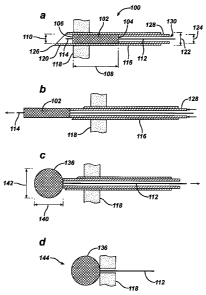
[0001] The present invention relates in general to devices, systems and
methods for repairing
and anchoring damaged tissue, and more particularly, to devices, systems and
methods for
anchoring suture to tissue.
BACKGROUND OF THE INVENTION
[0002] Injuries to tissue such as cartilage, skin, muscle, bone, tendon and
ligament, frequently
require surgical intervention to repair the damage and facilitate healing.
Surgical procedures to
repair tissue damage are often performed using sutures connected to one or
more anchoring
device (suture anchor) implanted in or adjacent to the damaged tissue. The
sutures can also be
passed through or around the tissue according to a variety of surgical
techniques to secure the
repair. The sutures can also interconnect two or more anchors used to perform
the repair. Suture
anchors have been fabricated with bodies formed from a variety of materials
including
nonabsorbable materials such as metals and durable polymers, as well as
bioabsorbable materials
such as absorbable polymers, bioceramics, absorbable composites and processed
bone.
[0003] Anchors can be designed for fixation with respect to tissue using
external screw threads
on an anchor body, an expandable body, toggling action, extendable components
such as barbs,
or other mechanical retention means. Sutures can be connected through or
around suture anchors
in a fixed or a sliding manner, for example, using eyelets or other passages
in an anchor body,
and can be secured using stationary or sliding knots, interference among
anchor components,
interference between an anchor and surrounding tissue, or other means. Some
suture anchors are
designed for suture to slide unidirectional through or around the anchor,
enabling a surgical
repair to be tightened by tensioning a portion of the suture with respect to
the anchor. Among
their many surgical applications, suture anchors are used with sutures to
reattach damaged
tendons or ligaments to bone, to tighten compromised tissue surrounding
articulating joints, and
to repair tears in cartilage, such as torn meniscal cartilage in a knee. In
some applications, two or
more anchors joined by an adjustable length of suture enable a tissue tear to
be cinched closed, or
compromised tissue to be stabilized.
[0004] Of great importance in suture anchor design is maximizing the retention
strength of the
anchor in tissue, to minimize the risk of anchor breakage or pullout from
tissue when an attached
CA 02710885 2010-07-22
suture is tensioned with respect to the anchor. One common approach to
maximizing anchor
retention strength is to use physically larger anchors than might be
preferable to minimize
surgical trauma caused by the procedure used to implant the anchor. Not only
does the
implantation of a larger anchor generally require a larger and therefore more
traumatic surgical
incision than would be required to implant a smaller anchor, but the tools
required to implant or
deploy a larger anchor may also be correspondingly larger. Compounding this
issue, the process
of deploying an anchor in tissue can require both substantially vertical
access to the tissue repair
site, and significantly deeper penetration into or through the tissue than the
depth required to
retain the anchor after deployment in tissue. In addition, many surgical
anchors have sharp
edges that can cause tissue damage when implanted in a patient. Addressing
these concerns is
particularly important in the development of minimally invasive surgeries such
as arthroscopic
procedures that restrict access to an operative site, at least in part to
reduce surgical trauma
relative to open surgical procedures.
[0005] There is a preference among some surgeons for using non-metallic suture
anchors rather
than metallic suture anchors. While some nonmetallic anchors can provide
advantages over
metallic anchors with respect to bioabsorbability or radiolucence, many
nonmetallic anchors
provide significantly lower mechanical strength than metallic anchors,
increasing the potential
for mechanical failure of the surgical repair during or post-surgery. For
example, suture may cut
through relatively soft materials used to fabricate a nonmetallic anchor, a
process often called
"cheese-wiring." With metallic suture anchors, the interface between suture
and the anchor must
also be carefully designed to protect attached suture from breakage. For
example, a metallic
suture anchor may require precision polishing to minimize suture failure where
suture contacts
the much harder metal. With any suture anchor, sharp bends of suture about
anchor components
are well-known stress points that can lead to failure of a surgical repair.
Post-surgical failure of
an anchor-based surgical repair during the healing period is of particular
concern because
uncontrolled fragments of a failed anchor have the potential to cause injury
to the patient.
[0006] Accordingly, there remains a need for improved suture anchoring
devices, systems and
methods for repairing damaged tissue that overcome the limitations and
disadvantages of known
suture anchors. A need also exists for suture anchors, deployment tools and
methods that
minimize the surgical trauma associated with the implantation of an anchor of
any given size.
-2-
CA 02710885 2010-07-22
SUMMARY OF THE INVENTION
[0007] The present invention generally provides devices, systems and methods
for anchoring
suture to tissue. One aspect of the present invention is a method for
anchoring a suture length to
human tissue. The method comprises the steps of providing a preformed knot
configuration to a
first portion of the suture length, positioning the preformed knot
configuration into an opening in
a portion of the human tissue, and expanding the preformed knot configuration
in at least one
physical dimension to form an anchoring knot, so as to engage the anchoring
knot against the
tissue. In one embodiment, the preformed knot configuration is reduced in
length and increased
in a cross-sectional dimension when reconfigured to form the anchoring knot.
The anchoring
knot can be formed behind a tissue wall, or within bulk tissue, which can be
soft tissue or bone.
[0008] The preformed knot configuration can be formed from a single line of
suture or from
joined lines of suture that can be of a single structure and material, or of
different structures and
materials. In various embodiments, the preformed suture configuration is
formed by intertwining
portions of suture using any of a variety of methods including, but not
limited to chain-knotting,
braiding and crocheting. In an embodiment, the step of reconfiguring the
preformed knot
configuration into an anchoring knot includes placing an abutment against the
preformed knot
configuration and moving the suture length relative to the abutment to cause
the preformed knot
configuration to bunch up and increase in cross sectional area as the
anchoring knot.
[0009] The preformed knot configuration can be delivered to tissue through an
inserter tube. In
an embodiment, the inserter tube is passed though a tissue wall, and the
anchoring knot is formed
behind the tissue wall. In one embodiment, the preformed knot configuration
does not protrude
more than 5 mm beyond the wall before fully forming the anchoring knot. In
another
embodiment, the diameter of the anchoring knot is at least twice the diameter
of the inserter tube.
[0010] Another aspect of the present invention is a suture unit for anchoring
in human tissue.
The suture unit includes a first preformed knot configuration along a portion
of a length of
suture. The first preformed knot configuration has a maximum diameter along
the suture length
and is reconfigurable into a first anchoring knot having a minimum diameter
that is at least five
times larger than the first preformed knot configuration maximum diameter. The
first preformed
knot configuration can be formed from a single line of suture or from joined
lines of suture that
-3-
CA 02710885 2010-07-22
can be of a single structure and material, or of different structures and
materials. In various
embodiments, the preformed suture configuration is formed by intertwining
portions of suture,
using methods including, but not limited to chain-knotting, braiding and
crocheting.
[0011 ] In an embodiment, the first preformed knot configuration includes a
portion of suture
formed into a loop closed with a sliding knot that in one embodiment is
positioned within the
first preformed knot configuration. The suture unit can include a second
preformed knot
configuration expandable into a second anchoring knot.
BRIEF DESCRIPTION OF THE DRAWINGS
[0012] The invention will be more fully understood from the following detailed
description
taken in conjunction with the accompanying drawings, in which:
[0013] FIG. 1 a through FIG. 1 d illustrate an embodiment of a suture-
anchoring device and its
deployment according to the present invention.
[0014] FIG. 2a through FIG. 2d illustrate a nonsliding embodiment of a suture
fixation device
according to the present invention comprising a twisted braid suture head.
[0015] FIG. 3a through FIG. 3c illustrate nonsliding embodiments of suture
fixation devices
according to the present invention comprising crocheted suture heads.
[0016] FIG. 4a through FIG. 4d illustrate sliding embodiments of suture
anchoring devices
according to the present invention.
[0017] FIG. 5a through FIG. 5d illustrate embodiments of suture anchoring
devices according to
the present invention comprising interpenetrating suture.
[0018] FIG. 6a and FIG. 6b illustrate an embodiment of a suture anchoring
device applicable to
repairing a meniscus in a knee according to the present invention.
[0019] FIG. 7a and FIG. 7b illustrate an embodiment of a delivery tool of the
present invention
that can be used to deliver the anchoring device illustrated in FIG. 6a and
FIG. 6b to tissue.
-4-
CA 02710885 2010-07-22
[0020] FIG. 8a and FIG. 8b illustrate an embodiment of a delivery tool of the
present invention
including a curved delivery needle.
[0021 ] FIG. 9a through FIG. 9k illustrate an embodiment of a surgical repair
procedure of the
present invention, for repairing a torn meniscus in a knee.
[0022] FIG. 1 Oa and FIG. I Ob illustrate an embodiment of a delivery tool of
the present
invention for single-location anchoring and for daisy-chaining anchoring
locations.
[0023] FIG. 11 a through FIG. 11 i illustrate an embodiment of a daisy-
chaining anchoring
procedure of the present invention.
[0024] FIG. 12a through FIG. l2e illustrate an embodiment of delivery of
suture anchoring
devices of the present invention into hard tissue.
[0025] FIG. 13 illustrates an embodiment of a sub-cortically dilated hole in a
bone for receiving
a suture anchoring device of the present invention.
[0026] FIG. 14a through FIG. 14d illustrate an alternative embodiment of a
delivery device of
the present invention.
[0027] FIG. 15a through FIG. 15d illustrate an embodiment of a suture
anchoring system of the
present invention wherein a suture head is used in conjunction with an
intermediate anchoring
implant.
[0028] FIG. 16a through FIG. 16d illustrate another embodiment of an anchoring
system of the
present invention wherein a suture head is deployed internally to an
intermediate anchoring
implant.
DETAILED DESCRIPTION
[0029] Certain exemplary embodiments will now be described to provide an
overall
understanding of the principles of the structure, function, manufacture, and
use of the devices,
systems and methods disclosed herein. Those skilled in the art will understand
that the devices
and methods specifically described herein and illustrated in the accompanying
drawings are
non-limiting exemplary embodiments and that the scope of the present invention
is defined
-5-
CA 02710885 2010-07-22
solely by the claims. The features illustrated or described in connection with
one embodiment
may be combined with the features of other embodiments. Such modifications and
variations are
intended to be included within the scope of the present invention. It should
be noted that the
figures are generally schematic and not drawn to scale, and are intended only
to facilitate the
description of specific embodiments of the invention.
[0030] The present invention generally provides devices, systems and methods
for anchoring
suture to tissue. The term "tissue" is used herein to refer to any natural
body tissue including,
but not limited to cartilage, skin, muscle, bone, tendon, ligament and organs,
as well as to
prosthetic materials such as grafts or other prosthetics that can be repaired
or attached to natural
body tissues with sutures and anchoring devices. Embodiments of suture
anchoring devices
fabricated substantially from surgical suture or any elongated, thread-like
materials that can be
used as medical devices (hereinafter, "suture") are disclosed herein. The
suture can comprise a
single filament or a plurality of interwoven filaments, and can have any cross-
sectional shape
including, but not limited to a substantially circular cross section, and a
flattened ribbon or tape-
like cross section. Further, the suture can be non-absorbable, bioabsorbable,
or partially
bioabsorbable. Without deviating from the intent or scope of the invention,
the suture material
can be mechanically or chemically modified, treated or coated to enhance
lubricity or knot-
holding ability, to elute a medicament, or for any combination of the
aforementioned or other
therapeutic purposes. Further, although various embodiments of anchoring
devices in
accordance with the invention can be constructed entirely of suture,
additional components such
as clips or adhesives can be included without deviating from the intent or
scope of the invention.
[0031 ] An anchoring device according to the present invention generally
comprises one or more
segment of suture (hereinafter, suture tail) extending from an anchoring
member having a
longitudinally elongated, small cross section initial configuration
(hereinafter, a suture head).
Upon deployment, the suture head is reconfigured (collapsed) to a
longitudinally compressed
configuration (an anchoring knot) of correspondingly larger cross-section than
the suture head.
That is, the anchoring knot has a larger cross sectional area and a larger
average cross sectional
dimension (hereinafter, cross-sectional dimension) than the corresponding
dimensions of the
suture head. In some embodiments, for delivery to tissue, the suture anchoring
device is
disposed in a cannulated delivery needle having an outer diameter
substantially smaller than the
-6-
CA 02710885 2010-07-22
cross-sectional dimension of the anchoring knot. In general, collapsing a
suture head to an
anchoring knot is accomplished by tensioning a specific one or more (collapse
tail) of the one or
more suture tail, with respect to the suture head.
[0032] FIG. 1 a schematically illustrates an embodiment of a suture-anchoring
device 100
according to the present invention. The suture anchoring device 100,
illustrated undeployed in
FIG. 1 a, comprises a suture head 102 having a first head end 104, a second
head end 106, a head
length 108 therebetween and an undeployed cross-sectional dimension 110 that
is smaller than
the head length 108. A first suture tail 112 is seen to extend substantially
from the first head end
104. In an embodiment, a second suture tail 114 extends substantially from the
second head end
106. In a further embodiment, the second suture tail 114 comprises a closed
loop of suture
extending from the second head end 106. In other embodiments, two or more
suture tails extend
from one or both of the first 104 and the second head end 106.
[0033] The suture head 102 comprises a longitudinally extended, preformed knot
configuration,
by which we mean any braided, crocheted, woven, knotted or otherwise
configured section of
suture that, for deployment and fixation with respect to tissue, can be
readily collapsed into a
longitudinally compressed, expanded cross-section form referred to herein as
an anchoring knot.
[0034] The suture head 102 is seen to be disposed in a cannulated delivery
needle 116 for
delivery into or through tissue 118. The delivery needle 116 has a distal
delivery end 120, an
outer diameter 122 and an inner diameter 124. Further, the delivery needle 116
can be straight or
curved along its length. In an embodiment, the delivery end 120 includes a
tissue-penetrating
point 126. In another embodiment (not illustrated), the delivery end 120 is
not pointed. A piston
128 having a longitudinal piston cannulation 130 therethrough is seen to be
slidingly disposed
within the delivery needle 116, proximal to the suture head 102. The first
suture tail 112 is seen
to pass proximally from the suture head 102 through the piston cannulation
130.
[0035] Now referring to FIG. lb, in one embodiment, the suture head 102 is
delivered from the
delivery needle 116 to the tissue 118 by pushing the piston 128 distally
against the suture head
102 to expel the suture head 102 from the delivery needle 116. The piston 128
is seen to abut the
expelled suture head 102. In an embodiment, the piston 128 is coupled to a
proximal handle (not
illustrated) that provides control of the longitudinal position of the piston
128 within the delivery
-7-
CA 02710885 2010-07-22
needle 116. In an alternate embodiment, the suture head 102 is delivered from
the delivery
needle 116 by distally pulling the second suture tail 114. In one embodiment,
the needle 116 is
straight. In another embodiment, the needle 116 is curved and the piston 128
is flexible so as to
enable the piston 128 to slide along the curve for delivery of the suture head
102 from the needle
116. As the suture head 102 substantially comprises suture, it is also
flexible for sliding through
a curved needle.
[0036] Now referring to FIG. 1 c, following or concurrently with delivery of
the suture head 102
from the needle 116, the suture head 102 is collapsed to form an anchoring
knot 136. In an
embodiment, the suture head 102 is collapsed to the anchoring knot 136 by
tensioning 138 the
first suture tail 112 (a collapse tail) with respect to the suture head 102
after the suture head 102
has been pushed entirely out of the distal end 120 of the needle 116 by the
piston 128. The
piston 128 abutting the suture head 102 provides a counter force to the
tensioning 138 of the first
suture tail 112 with respect to the suture head 102, to collapse the suture
head 102 to the
anchoring knot 136.
[0037] The term "collapse tail" is used herein to describe any suture tail
that, when tensioned
with respect to a suture head, can be used to collapse the suture head to an
anchoring knot. The
anchoring knot 136 has a knot length 140 that is shorter than the head length
108, and a
correspondingly increased cross section 142, determined substantially by the
volume of suture
originally comprising the suture head 102.
[0038] In another embodiment, the first suture tail 112 is tensioned
concurrently with the suture
head 102 being expelled from the distal end 120 of the needle 116, collapsing
the suture head
102 to the anchoring knot 136 as it emerges from the needle 116. In this
embodiment, the suture
head 102 does not extend distally from the delivery needle 116 the full head
length 108 during
deployment, but instead extends only the knot length 140. This shallower
extension can provide
deployment of the anchoring device 100 that minimizes surgical trauma to
tissue positioned
distally beyond, but in proximity to the distal end of the delivery needle.
[0039] FIG. 1 d illustrates a fully deployed anchoring device 144, wherein the
needle 116 is seen
to have been removed, and the anchoring knot 136 is anchored with respect to
the tissue 118,
leaving the first suture tail 112 available for connection to tissue or to
another anchoring device,
-8-
CA 02710885 2010-07-22
or for any other surgical step requiring a suture that is anchored to tissue.
According to the
requirements of a particular surgical repair, the anchoring knot 136 can be
deployed behind a
tissue wall as illustrated in FIG. Id, or within bulk tissue, for example,
within a bone for
anchoring suture to the bone. In other embodiments, two or more suture heads,
interconnected
by suture, are disposed in a needle for surgical procedures requiring two or
more tissue
anchoring points.
[0040] The anchoring knot 136 has a knot length 140 that is less than the head
length 108, and a
deployed cross sectional dimension 142 that is correspondingly greater than
the undeployed
cross sectional dimension 110, and greater than the outer diameter 122 of the
needle 116. In an
embodiment, the anchoring knot 136 is amorphous, that is, having an
incompletely
predetermined external shape following collapse from the suture head 102 to
the anchoring knot
136. In general, anchoring knots formed from suture heads according to the
various
embodiments of the present invention are amorphous. Although neither the
suture head 102 nor
the anchoring knot 136 have a completely predetermined shape, either can be
reasonably
described as having a length and a diameter transverse to the length, the
diameter approximately
defined by the average cross-sectional dimension transverse to the length.
[0041] By way of nonlimiting example, in one embodiment, the head length 108
is between
approximately ten and fifty times the undeployed cross-sectional dimension
110, and the
deployed cross-sectional dimension 142 is between three and ten times the
undeployed cross-
sectional dimension 110. The suture-anchoring device 100 can be fabricated
substantially from a
single continuous length of suture, or from a plurality of coupled lengths of
suture. The plurality
of coupled lengths can include a single type of suture or a combination of
suture types and sizes.
Further, the one or more suture tail can be fixedly coupled to the anchoring
knot, or slidably
coupled therethrough. In one embodiment, the inner diameter 124 of the needle
116 is less than
about six times a diameter of the suture material from which the suture head
is configured. In
another embodiment, the inner diameter 124 of the needle 116 is less than
about four times a
diameter of the suture material from which the suture head is configured.
[0042] In an anchoring device according to the present invention, the design
of the suture head
substantially determines the overall design and procedural details of delivery
and deployment.
-9-
CA 02710885 2010-07-22
The design of the suture head also determines the pull-out strength of the
deployed anchoring
knot, and the anchor density, that is, the mass of suture material in an
anchoring knot having a
given cross-sectional dimension. Many anchoring device designs are possible
within the scope
of the present invention. For nonlimiting descriptive purposes herein, these
designs are grouped
into two general categories respectively called non-sliding embodiments,
wherein all suture tails
extending from a fully deployed anchoring knot are fixed thereto substantially
without sliding
through the knot, and sliding embodiments, wherein at least one length of
suture slidingly passes
through the fully deployed anchoring knot. Sliding embodiments are
advantageous for some
surgical applications, for example, where it is desirable to tension suture
between a deployed
anchoring member and attached tissue, to draw two or more anchoring devices
together to close
a tissue tear, or to gather together intervening tissue between anchoring
devices.
[0043] In an illustrative sliding embodiment, the anchoring device comprises
at least a first
length of suture and a second length of suture. The first length of suture
generally comprises the
bulk of a suture head (and, following deployment, a corresponding anchoring
knot). The second
length of suture comprises two suture tails and is slidable through the
anchoring knot by
tensioning one or the other of the two tails individually with respect to the
anchoring knot. The
sliding embodiment can further include additional sliding sutures having
corresponding pairs of
suture tails. Sliding embodiments can also include one or more fixed suture
tail that can
comprise a portion of the first length of suture, or an additional length of
suture fixedly
connected, for example, tied, to the first length of suture. Further,
depending on the specific
design of the suture head, one or more suture tail can comprise one or both of
a collapse tail and
a sliding tail.
[0044] Nonsliding embodiments can comprise a single length of suture or a
plurality of suture
lengths that are fixedly joined together, for example, by one or more knot.
Nonsliding
embodiments include a suture head from which one or more suture tail extends,
at least one of
the one or more suture tail comprising a collapse tail.
[0045] A suture head according to the present invention can comprise any
preformed knot
configuration that can be collapsed from a longitudinally extended form to a
longitudinally
compressed, increased cross section anchoring knot. In various embodiments,
the suture head
-10-
CA 02710885 2010-07-22
includes a plurality of openings comprising loops, penetrations or other
openings formed along a
first longitudinal section of suture. A second longitudinal section of suture
comprising a collapse
tail is woven through two or more of the plurality of the openings. One or
more suture tails
extends from the suture head, at least one of the one or more suture tails
comprising the collapse
tail. In one embodiment, the first section of suture, the second section of
suture and the one or
more suture tail comprise a single continuous length of suture. In another
embodiment, the first
section of suture, the second section of suture and the one or more suture
tail comprise two or
more joined lengths of suture.
[0046] Anchoring devices of the present invention can include one or more of a
variety of types
of suture heads, and can be fabricated using a variety of methods. One type of
suture head
comprises a braided section of suture that is collapsible to an anchoring
knot. Any type of suture
braiding can be used to configure the suture head. An embodiment of a
nonsliding, twisted braid
suture-anchoring device is schematically illustrated in FIG. 2a and FIG. 2b.
FIG 2a illustrates a
first configuration step 200, wherein a length of suture 202 is seen to have
been formed into a
starting loop 204 having a first head end 206, a second head end 208 and a
head length 210
therebetween. A suture tail 212 extends from the second head end 208. FIG. 2b
illustrates a
configured suture head 214. As can be seen in FIG. 2b, the loop 204 has been
repetitively
twisted to provide a plurality of openings 216 along the head length 210.
Further, the suture tail
212 is seen to have been woven through the plurality of openings 216 from the
second head end
208 to the first head end 206, and extends from the first head end 206, where
it comprises a
collapse tail.
[0047] It should be noted that in FIG. 2b, as well as other figures herein
detailing suture head
configurations, the suture heads are generally illustrated in an expanded
schematic form to
support description of routing of suture therethrough. Any suture head
disclosed herein can be
readily compressed in cross section, for example, for disposition within a
cannulated delivery
needle, as illustrated in FIG. I a. By way of example, FIG. 2c illustrates the
suture head 214 in a
compressed cross section form 218, as for disposition in a delivery needle.
The suture head 214
can be collapsed to an anchoring knot 220, as illustrated in FIG. 2d, by
tensioning the suture tail
212 (collapse tail) with respect to the suture head 214.
-11-
CA 02710885 2010-07-22
[0048] By tensioning the first suture tail 212 with respect to the suture head
214, we mean
tensioning (pulling) the first suture tail 212 away from the suture head 214,
so that the portion of
the first suture tail 212 that is woven through the plurality of openings 216
is pulled further
through the plurality of openings 216 and through the first head end 206,
thereby gathering or
bunching the twisted suture along the head length 210 into the anchoring knot
220. For
tensioning 222 the first suture tail 212 with respect to the suture head 214,
an abutment to the
first head end, for example, the piston 128 of FIG. 1 is required to hold the
suture head in
position for collapsing to the anchoring knot 220. In some embodiments wherein
a suture head
is embedded in tissue or trapped behind a tissue wall before tensioning a
collapse tail, friction
with the tissue can also retain the suture head during collapse to an
anchoring knot.
[0049] Any type and diameter of suture, and any number of openings 216 for
braiding or
otherwise passing suture therethrough, can be used to configure a suture head
according to the
present invention. A larger number of openings generally provides a longer
suture head and,
upon deployment, an anchoring knot having a larger cross sectional dimension,
thereby
providing greater fixation strength of the anchoring knot with respect to
tissue. In one
embodiment, a 20 mm long suture head comprises between fifteen and thirty-five
openings
through which suture can be woven. In another embodiment, the plurality of
openings is
between twenty and thirty openings. In yet another embodiment, the suture head
is
approximately 25 millimeters (mm) in length, and upon deployment in tissue,
the suture head
collapses to a substantially amorphous anchoring knot approximately five mm in
diameter.
[0050] In one test embodiment, a suture head approximately 20 mm long was
configured from
partially absorbable, polyethylene-containing braided suture approximately 0.5
millimeters (mm)
in diameter (ORTHOCORDTM Orthopaedic Suture from DePuy Mitek, Raynham,
Massachusetts). Deployed through a 2-mm diameter hole into the cancellous
layer of artificial
bone having a 3-mm thick, 55-durometer cortex, the pullout strength of the
resulting anchoring
knot was approximately 45 pounds. Deployment of a similarly configured
anchoring device
through a 2-mm diameter hole in artificial bone having a 3-mm thick, 35-
durometer cortex
provided a pullout strength of approximately 22 pounds.
-12-
CA 02710885 2010-07-22
[0051 ] A person skilled in the art will appreciate that, within the scope of
the present invention,
many different methods can be used to configure a suture head having a
longitudinally extended
configuration that is collapsible to an anchoring knot. Braids, for example,
can be formed by a
variety of methods and with any number of suture sections braided together,
and a suture head
configured to include any braiding pattern is within the scope of the present
invention. Further,
braiding comprises only one of a variety of methods for configuring a suture
head according to
the present invention. Other methods for configuring a suture head can be
adapted, for example,
from other textile arts such as crocheting and weaving.
[0052] Another anchoring device of the present invention includes a suture
head configured
using a chain of suture loops. The chain of loops can comprise a plurality of
independent suture
loops, a physically connected chain of discrete loops, or a plurality of loops
formed along a
continuous length of suture using known textile arts such as crocheting, where
each of the
plurality of loops in a chain is formed by pulling a section of the suture
through a previously
formed loop in the suture. The plurality of loops provides a corresponding
plurality of openings
through which suture can be woven. Nonsliding embodiments of suture anchoring
devices
comprising suture heads configured using crocheted suture are schematically
illustrated in FIG.
3a through FIG 3c. FIG. 3a illustrates a crochet configuration step 230
wherein a length of
suture 232 has been crocheted to define a plurality of openings 234 along a
crocheted section 236
having a first crocheted end 238 and a second crocheted end 240. A first
suture tail 242 extends
from the first crocheted end 238 and a second suture tail 244 extends from the
second crocheted
end 240.
[0053] FIG. 3b illustrates a first embodiment 250 of a crocheted suture-
anchoring device. It
comprises a suture head 252 wherein the first suture tail 242 is seen to weave
through one or
more of the plurality of openings 234 along the crocheted section 236 from the
first crocheted
end 238 to the second crocheted end 240, and extends from the second crocheted
end 240. The
suture head 250 can be collapsed to an anchoring knot by tensioning the first
suture tail 242
(collapse tail) with respect to the suture head 252.
[0054] Figure 3c illustrates a second embodiment 260 of a crocheted suture-
anchoring device.
The second embodiment 260 resembles the first embodiment 250, with the
addition that in the
-13-
CA 02710885 2010-07-22
second embodiment 260, after being woven from the first 238 to the second
crocheted end 240,
the first suture tail 242 is returned through one or more of the plurality of
openings 234 from the
second crocheted end 240 to the first crocheted end 238, to extend from the
first crocheted end
238. The suture head 262 can be collapsed to an anchoring knot by tensioning
the first suture tail
242 (collapse tail) with respect to the suture head 262.
[0055] It should be appreciated that not all suture tails are collapse tails.
Referring to FIG. 3b
and FIG. 3c, in either the first 250 or the second embodiment 260, the second
suture tail 244 is
directly connected to and extends from the second crocheted end, and
tensioning the second
suture tail 244 with respect to the respective suture head 252, 262 (providing
an abutment against
second crocheted end 240 does not collapse the respective suture head to an
anchoring knot.
Thus the second suture tail 244 can be used, for example, to pull the
respective suture head 252,
262, into a cannulated needle, for delivery to tissue or through tissue,
without collapsing the
respective suture head to an anchoring knot.
[0056] FIG. 4a through FIG. 4d illustrate sliding embodiments of suture
anchoring devices
according to the present invention. FIG. 4a illustrates a sliding embodiment
of a twisted ring
suture-anchoring device 300. The twisted ring suture-anchoring device 300
comprises a suture
ring 302 that is a closed ring of suture repetitively twisted to form a
plurality of openings 304
between a first twist end 306 and a second twist end 308. A length of suture
310 having a first
suture tail 312 and a second suture tail 314 is woven through the plurality of
openings 304
between the first twist end 306 and the second twist end 308, and returning
through at least one
of the plurality of openings 304 near the first twist end 306, to configure a
suture head 316
having both the first 312 and the second suture tail 314 extending from the
first twist end 306.
The suture head 316 can be collapsed to an anchoring knot by simultaneous
tensioning of the
first 312 and the second suture tail 314 with respect to the suture head 316.
Thus the first 312
and the second suture tail 314 comprise collapse tails when tensioned
simultaneously with
respect to the suture head 316. As can be seen in FIG. 4a, the length of
suture 310 is not fixedly
connected to the suture ring 302, but woven therethrough to preserve
slidability of the length of
suture 310 through the anchoring knot. The length of suture 310 can slide
through the anchoring
knot by individually tensioning either the first 312 or the second suture tail
314 respectively,
with respect to the anchoring knot.
-14-
CA 02710885 2010-07-22
[0057] A suture ring used to configure a suture head according to the present
invention can
comprise suture formed as a continuous ring of suture material, or a length of
suture closed to
form the ring. Any method of closing the length of suture to a ring can be
used, including but
not limited to knotting, welding, gluing, or crimping with or without a
binding clamp or other
joining member. Further, the suture ring can include a plurality of
substantially parallel strands
of suture about its circumference, braided, crocheted, or otherwise
interlocked suture. In an
embodiment, the suture ring comprises a continuous ring of suture having a
first circumference,
that is doubled over to form a doubled suture ring having a second
circumference that is
substantially half the first circumference.
[0058] FIG. 4b illustrates a sliding embodiment of a suture-chain suture-
anchoring device 322.
The suture-chain suture-anchoring device 322 comprises a connected chain of
suture rings 324
defining a plurality of openings 326 between a first chain end 328 and a
second chain end 330.
A length of suture 332 having a first suture tail 334 and a second suture tail
336 is woven
through the plurality of openings 326 between the first chain end 328 and the
second chain end
330, then returning through at least one of the plurality of openings 326 near
the first chain end
328, to configure a suture head 338 having both the first 334 and the second
suture tail 336
extending from the first chain end 328. The suture head 338 can be collapsed
to an anchoring
knot by simultaneous tensioning of the first 334 and the second suture tail
336 with respect to the
suture head 338. Thus the first 334 and the second suture tail 336comprise
collapse tails when
tensioned simultaneously. As can be seen in FIG. 4b, the length of suture 332
is not fixedly
connected to the chain of suture rings 324, but woven therethrough to preserve
slidability of the
length of suture 332 through the anchoring knot. The length of suture 332 can
slide through the
anchoring knot by individually tensioning either the first 334 or the second
suture tail 336
respectively, with respect to the anchoring knot.
[0059] FIG. 4c illustrates a sliding embodiment of a wound-ring suture-
anchoring device 344
providing a sliding connection between suture and a deployed anchoring knot.
The wound-ring
suture-anchoring device 344 comprises a suture ring 346 that can be any type
of suture ring
described hereinabove. The wound-ring suture-anchoring device 344 also
comprises a first
length of suture 348 having a first suture tail 350 and a second suture tail
352. The first length of
suture 348 is wound substantially helically about the suture ring 346 to
configure a suture head
-15-
CA 02710885 2010-07-22
354, with the first 350 and the second suture tail 352 extending from the
suture ring 346
substantially adjacent to one another about the circumference of the suture
ring 346. In an
alternative embodiment, the first length of suture 348 is wound substantially
helically about a
second length of suture. The second length of suture, with the winding about
it, is subsequently
joined at a closure point 356 along its length to form the suture ring 346.
The suture head 354
can be collapsed to an anchoring knot by simultaneous tensioning of the first
350 and the second
suture tail 352 with respect to the suture head 354. Thus the first 350 and
the second suture tail
352 comprise collapse tails when tensioned simultaneously. The first length of
suture 348 can
slide through the anchoring knot by individually tensioning either the first
350 or the second
suture tail 352 respectively, with respect to the anchoring knot.
[0060] A sliding embodiment of a crochet-type suture-anchoring device 362 is
illustrated in FIG.
4d. The crochet-type suture-anchoring device 362 comprises a crocheted section
of suture 364
defining a plurality of openings 366 between a first crocheted end 368 and a
second crocheted
end 370. The crocheted section 364 is similar to the crocheted section 236
described in
association with FIG. 3a. The crochet-type suture-anchoring device 362 also
comprises a first
length of suture 372 having a first suture tail 374 and a second suture tail
376.
[0061] As can be seen in FIG. 4d, to configure a suture head 378, the first
length of suture 372 is
woven through one or more of the plurality of openings 366 between the first
crocheted end 368
and the second crocheted end 370, and returned to the first crocheted end 368
through at least
one of the plurality of openings 366 near the first crocheted end 368. The
first 374 and the
second suture tail 376 extend from the first crocheted end 368. In one
embodiment, the fraction
of the plurality of openings comprises approximately every third opening of
the plurality of
openings 366. In another embodiment, the first length of suture 372 is woven
through
substantially each of the plurality of openings 366. In other embodiments, the
interval varies
along the plurality of openings 366 between the first crocheted end 368 and
the second crocheted
end 370. In yet another embodiment, the first length of suture 372 passes
through a single one of
the plurality of openings 366, the single one of the plurality of openings 366
functioning as an
eyelet through which the first length of suture 372 passes.
-16-
CA 02710885 2010-07-22
[0062] A person skilled in the art will appreciate that any number of
additional lengths of suture
can be independently woven through one or more of the plurality of openings,
thereby providing
an anchoring device having a plurality of suture legs extending therefrom (a
multisuture
anchoring device). In various embodiments, two, three or four lengths of
suture are each woven
through one or more of the plurality of openings, providing anchoring devices
respectively
comprising four, six, or eight legs of suture extending therefrom. In some
surgical situations it
can be desirable to increase the number of sutures connected to a deployed
anchoring device. In
such circumstances, one or more suture needle with attached suture can be
passed through a
deployed anchoring knot to provide a multisuture anchoring device.
[0063] The suture head 378 can be collapsed to an anchoring knot by
simultaneous tensioning of
the first 374 and the second suture tail 376 with respect to the suture head
378. Thus the first
374 and the second suture tail 376 comprise collapse tails when tensioned
simultaneously with
respect to the suture head 378. The first length of suture 372 can slide
through the anchoring
knot by individually tensioning of either the first 374 or the second suture
tail 376 respectively,
with respect to the anchoring knot.
[0064] FIG. 5a through FIG. 5d illustrate embodiments of suture anchoring
devices comprising
interpenetrating suture. A suture head according to these embodiments
comprises a plurality of
longitudinally distributed, substantially transverse penetrations through the
material of a first
section of suture, and a second section of suture woven through the plurality
of penetrations.
The plurality of penetrations can comprise any type of penetrations. In one
embodiment, the
plurality of penetrations is defined using a sharp instrument such as a sewing
or suturing needle
connected to a suture tail. In another embodiment, the plurality of
penetrations comprises a
plurality of slits or bores is formed through the suture material to enable
weaving of a suture tail
therethrough. In yet another embodiment, a plurality of preformed penetrations
is provided
during fabrication of the section of suture. In still another embodiment, the
suture itself
comprises a braided material, for example, a braided suture, and the plurality
of penetrations pass
through the braid at a corresponding plurality of locations.
[0065] FIG. 5a and FIG. 5b illustrate interpenetrating sliding embodiments.
FIG. 5a illustrates a
first interpenetrating sliding embodiment 400 of a suture anchoring device
wherein a suture head
-17-
CA 02710885 2010-07-22
402 comprises a suture ring 404 having a plurality of penetrations 406
distributed around its
circumference. The suture ring 404 can be any type of suture ring disclosed
herein. The suture
head 402 further comprises a first length of suture 408 woven through the
plurality of
penetrations 406, and terminating in a first suture tail 410 and a second
suture tail 412, each
extending from the suture head 402 substantially adjacent to one another about
the circumference
of the suture ring 404. The suture head 402 can be collapsed to an anchoring
knot through which
the first length of suture 408 can slide after the anchoring knot is formed,
by simultaneously
tensioning the first 410 and the second suture tail 412 with respect to the
suture head 402. Thus
the first 410 and the second suture tail 412 comprise collapse tails when
tensioned
simultaneously. The first length of suture 408 can slide through the anchoring
knot by
individually tensioning either the first 410 or the second suture tail 412
respectively, with respect
to the anchoring knot.
[0066] In an alternate embodiment, the suture ring 404 comprises a second
length of suture that
is open at a point 418 on the circumference, and the ring shape is maintained
by the adjacent
penetrations 420, 422 of the second length of suture by the first length of
suture 408. In a further
alternate embodiment, the second length of suture penetrates the first length
of suture, instead of
the first length of suture 408 penetrating the second length of suture.
[0067] FIG. 5b illustrates a second interpenetrating sliding embodiment 424 of
a suture
anchoring device wherein a suture head 426 comprises a first length of suture
428 having a
plurality of penetrations 430 along a first portion 432 of its length, through
which a second
portion 434 of the length passes to define a corresponding plurality of
openings 436. The suture
head 426 further comprises a second length of suture 438 woven through the
plurality of
openings 436, and terminating in a first suture tail 440 and a second suture
tail 442, each
extending from the suture head 426. The suture head 426 can be collapsed to an
anchoring knot
through which the second length of suture 438 can slide, by simultaneously
tensioning the first
440 and the second suture tail 442 with respect to the suture head 426. Thus
the first 440 and the
second suture tail 442 comprise collapse tails when tensioned simultaneously.
The second length
of suture 438 can slide through the anchoring knot by individually tensioning
either the first 440
or the second suture tail 442 respectively, with respect to the anchoring
knot.
-18-
CA 02710885 2010-07-22
[0068] FIG. 5c and FIG. 5d illustrate interpenetrating suture nonsliding
embodiments of suture
anchoring devices. FIG. 5c illustrates a first interpenetrating nonsliding
embodiment 448
wherein a suture head 450 having a first head end 452 and a second head end
454 comprises a
first length of suture 456 having a plurality of penetrations 458 along a
first portion 460 of its
length between the first head end 452 and the second head end 454. A second
portion 462 of the
length passes through the plurality of penetrations 458 and extends from the
first head end 452 as
a suture tail 464. The suture head 450 can be collapsed to an anchoring knot
by tensioning the
suture tail 464 with respect to the suture head 450. In an embodiment, one of
the penetrations
468 of the second portion 462 through the first portion 454 is reinforced by
knotting or by
another means to prevent the suture head from unraveling during deployment.
The plurality of
penetrations 458 is seen to define a plurality of openings 470 along the
suture head 450 between
the first head end 454 and the second head end 456.
[0069] FIG. 5d illustrates a second interpenetrating nonsliding embodiment
472. The second
nonsliding embodiment 472 comprises a suture head 474 that resembles the
suture head 450 of
FIG. 5c, with the addition that in the second embodiment 472, the suture tail
464 is seen to be
reversed in direction and additionally woven through one or more of the
plurality of openings
470, to extend from the second head end 454. This additional pass of the
suture tail 464 provides
a larger volume of suture material in the suture head 474 of the second
embodiment 472,
compared with the volume of suture material in the suture head 450 of the
first embodiment 448.
A larger volume of suture in a suture head provides a correspondingly larger
anchoring knot
upon deployment in tissue. The suture head 474 can be collapsed to an
anchoring knot by
tensioning the suture tail 464 with respect to the suture head 474.
[0070] Although sliding and nonsliding embodiments are generally discussed
separately
hereinabove, various nonsliding embodiments can be converted to sliding
embodiments by
passing an additional length of suture through an opening in a nonsliding
suture head before
deployment. For example, with reference to FIG. 1, in an embodiment where the
second suture
tail 114 comprises a closed loop of suture, passing a length of suture through
the loop before
delivering the anchoring device 100 to tissue provides a sliding embodiment of
the suture
anchoring device, as the passed length of suture will be slidable with respect
to the anchoring
-19-
CA 02710885 2010-07-22
knot 136. In addition, various sliding embodiments can be converted to
nonsliding
embodiments, for example, by tying a knot in one or more sliding suture tail.
[0071 ] A person skilled in the art will appreciate that many variations of
the suture anchoring
devices disclosed herein are within the scope of the present invention,
including but not limited
to variations in suture material, size, number and combinations of suture
lengths used to
construct the anchoring device, the number of openings through which suture
comprising a
collapse tail passes along a suture head, and the number of sliding and
nonsliding suture tails
extending from a suture head. Further, any number of suture anchoring devices
can be coupled
together by suture to provide multi-point anchoring systems.
[0072] One application of the suture anchoring devices disclosed herein is the
repair of a
meniscal tear in a knee. FIG. 6a illustrates an embodiment of a dual anchoring
device 500
according to the present invention that in one embodiment is used for
repairing torn meniscal
tissue. The dual anchoring device 500 comprises a first suture head 502 having
a first distal end
504 and a first proximal end 506, and a second suture head 508 having a second
distal end 510
and a second proximal end 512. The second suture head 508 is seen to be
positioned proximal to
the first suture head 502. The first suture head 502 is a sliding suture head
that can be any type
of sliding suture head disclosed herein, or another sliding suture head. In
one embodiment, the
first suture head 502 is a crochet-type sliding suture head similar to the
suture head 378
described in association with FIG. 4d. The first suture head 502 comprises two
distal collapse
tails 514, 516 that are seen to extend form the first proximal end 506 toward
the second suture
head 508. The two distal collapse tails 514, 516 comprise a continuous length
of suture that
passes through the first suture head 502. Together, the two distal collapse
tails 514, 516
comprise a suture bridge between the first 502 and the second suture head 508.
[0073] The second suture head 508 is a non-sliding suture head that can
comprise any type of
nonsliding suture head disclosed herein, or another nonsliding suture head. In
one embodiment,
the second suture head 508 resembles the crochet-type nonsliding suture head
252 described in
association with FIG. 2c, with the addition of an integrated sliding knot 518
extending from the
second distal end 510. The second suture head 508 also comprises a proximal
collapse tail 520
extending proximally from the second proximal end 512. In one embodiment, the
second suture
-20-
CA 02710885 2010-07-22
head 508, the sliding knot 518, and the two distal collapse tails 514, 516
comprise a single
continuous length of suture. In another embodiment, the sliding knot 518,
rather than extending
from the second distal end 510, is disposed at a location 521 between second
distal end 510 and a
second proximal end 512 along the second suture head 508.
[0074] A partially expanded view 522 of the dual anchoring device 500 is shown
schematically
in FIG. 6b, illustrating the configuration of an embodiment of the sliding
knot 518. A first one
514 of the two distal collapse tails 514, 516 is seen to substantially
comprise one portion of the
sliding knot 518, and to connect to (or be continuous with) the second suture
head 508 at the
second distal end 510. The second distal collapse tail 516 is seen to pass
through the sliding knot
to extend proximally from the sliding knot 518, continuous with a tensioning
tail 524. The
second collapse tail 516 is also seen to pass through a suture loop 526
integral with the second
suture head 508 and extending distally therefrom.
[0075] An embodiment of a dual suture head delivery tool 530 for delivering
the dual anchoring
device 500 to tissue is schematically illustrated in FIG. 7a in a cross-
sectional view 532 and in
FIG 7b in an external view 534. The dual head delivery tool 530 is seen to
comprise a
cannulated delivery needle 536 having a distal needle end 538, a proximal
needle end 540 and a
longitudinal needle cannulation 542 therebetween. The delivery needle 536 is
proximally
coupled to a cannulated handle 544 having a handle wall 546 and a
substantially cylindrical
handle cannulation 548, the needle cannulation 542 being continuous with the
handle
cannulation 548. In an embodiment, the needle 536 is proximally reinforced by
a stiffening
member 550.
[0076] A cannulated piston 552 having a distal piston end 554, a proximal
piston end 556 and a
longitudinal piston cannulation 557 therebetween, is seen to be disposed
slidably within the
needle cannulation 542, and to extend proximally into the handle cannulation
548. In one
embodiment, each of the needle 536 and the piston 552 is straight. In another
embodiment, the
needle 536 comprises one or more curve between the distal needle end 538 and
the proximal
needle end 540, and the piston is flexible enough to be pushed and pulled
slidingly through the
needle cannulation 542. In one embodiment, the piston 552 comprises a flexible
tube. In
another embodiment, the piston 552 comprises a flexible, substantially helical
coil.
-21-
CA 02710885 2010-07-22
[0077] A piston positioning member 558 is fixedly connected to the piston 552,
and slidably
disposed within the handle cannulation 548. In one embodiment, the positioning
member 558 is
an annular member disposed about the piston 552. In an embodiment, the piston
positioning
member 558 substantially irreversibly locks in position longitudinally when
maximally advanced
distally within the handle cannulation 548. Any means of locking the piston
positioning member
558 can be used. In one embodiment, the piston positioning member 558 and the
handle 544
comprise interlocking latching members 560, 562 to lock the piston positioning
member 558
distally within the handle 544.
[0078] A control member 564 is connected to the positioning member 558, for
positioning the
piston 552 longitudinally within the dual head delivery tool 530, from outside
the delivery
device. For illustrative purposes, the control member is shown rotated ninety
degrees about a
longitudinal axis of the tool 530 in FIG 7a with respect to FIG. 7b. The
control member 564
extends laterally outward from the positioning member 558, through a slotted
opening 566 in the
handle wall 546. The slotted opening 566 can have any configuration that
accommodates the
requirements an anchoring device disposed in the delivery device, and
corresponding surgical
delivery requirements. In one embodiment, as illustrated in FIG. 7a and FIG.
7b for delivering
the dual anchoring device 500 to tissue, the slotted opening 566 is
substantially H-shaped. In an
embodiment, the H-shaped opening includes first and second longitudinal slots
812, 814 having
respective longitudinal positions that differ from one another along the
handle wall 546, each of
the two slots being adapted by its respective position for delivery of one of
the first 502 and the
second suture head 508 from the delivery needle 536. In other embodiments, the
slotted opening
is substantially T-shaped, L-shaped, longitudinally linear, or has another
configuration,
respective configurations being adapted to accommodate various suture head
delivery
requirements In another embodiment, the slotted opening comprises means for
locking the
piston positioning member 558 distally in the handle cannulation 548.
[0079] The control member 564 can comprise any means for communicating one or
both of
longitudinal and circumferential positioning force to the positioning member
558 and thereby to
the piston 552. In one embodiment, the control member 564 is a shaft fixedly
connected to the
positioning member 558 through the slotted opening 566. In another embodiment,
the control
member 564 includes means to releasable prevent the piston from moving within
the dual head
-22-
CA 02710885 2010-07-22
delivery tool 530. In one embodiment, the control member 564 is a thumbscrew
that is threaded
into the positioning member 558 through the slotted opening 566, such that
rotation of the
thumbscrew can be used to selectively lock and unlock the position of the
piston within the dual
head delivery tool 530. In another embodiment, the control member 564 is
resiliently loaded
with respect to the handle wall 546 to provide a predetermined resistance to
movement of the
control member 564 along the slotted opening 566.
[0080] The dual anchoring device 500 is seen to be disposed within the dual
head delivery tool
530, substantially within the needle cannulation 542, distal to the piston
distal end 554. The
second collapse tail 520 and the tensioning tail 524 pass proximally from the
dual anchoring
device 500 through the piston cannulation 557. Proximal to the piston proximal
end 556, the
tensioning tail 524 terminates at a tensioning tail end 568 and the second
collapse tail 520
terminates at a second collapse tail end 570. The second collapse tail 520
also comprises a
releasable holding member 572 disposed proximally to the proximal piston end
556, outside the
piston cannulation 557. The releasable holding member 572 prevents the second
collapse tail
520 from sliding distally through the cannulation of the piston 552. In one
embodiment, the
releasable holding member 572 is a releasable clamp attached to the second
collapse tail 520. In
another embodiment, the releasable holding member 572 is a releasable knot in
the second
collapse tail 520. In a further embodiment, proximally tensioning the second
collapse tail 520
releases the releasable knot. Upon release of the releasable holding member
572, the second
collapse tail 520 can slide distally through the cannulation of the piston
552.
[0081 ] The control member 564 controls the longitudinal position of the
distal end of the piston
552 within the delivery needle 536, and thereby controls the expulsion of the
dual anchoring
device 500 from the delivery needle 536. Depending on the configuration and
longitudinal
position of the slotted opening 566, the control member 564 can be used to
selectively expel only
the first suture head 502 from the delivery needle 536 for a predetermined
surgical step, and
selectively expel the second suture head 508 for a later surgical step, thus
enabling multipoint
anchoring procedures using devices and methods of the present invention.
[0082] It can be advantageous, particularly for a delivery device comprising a
curved delivery
needle, to be able to rotate the handle about a longitudinal axis with respect
to the curved needle,
-23-
CA 02710885 2010-07-22
to facilitate access to a control member on the handle for a particular
procedure, or to
accommodate differing preferences between left and right handed surgeons. An
embodiment of
a curved needle delivery tool 574 is illustrated in FIG. 8a. The curved needle
delivery tool 574
generally resembles the dual head delivery tool 530 associated with FIG. 7a
and FIG. 7b, which
comprises a straight delivery needle. The curved needle embodiment 574
includes a cannulated
curved delivery needle 576 having a distal delivery end 578 and a proximal end
580 that is
rotatably coupled to a handle 584 about a common longitudinal axis 586. A
control member 588
is accessible on an outside surface 590 of the handle 584. The curved delivery
needle 576
further comprises one or more curved portion 592, distal of which the curved
needle 576 deviates
from the axis 586.
[0083] In an embodiment, the curved needle delivery tool 574 comprises a
plurality of preferred
relative rotational orientations about the axis 586 between the handle 584 and
the curved needle
576. In a further embodiment, the plurality of preferred orientations
comprises a plurality of
detents 594 circumferentially distributed about the circumference of one or
both of the handle
590 and the curved needle 576. In another embodiment, angular markings are
provided on one
or both of the handle 584 and the curved needle 576 to indicate the relative
rotational orientation
between the curved needle 576 and the handle 584. FIG. 8a illustrates the
curved needle delivery
tool 574 in a first angular orientation 596 between the handle 584 and the
curved needle 576.
FIG. 8b illustrates the curved needle delivery tool 574 in a second angular
orientation 598
between the handle 584 and the curved needle 576.
[0084] FIG. 9a through FIG. 9k schematically illustrate a surgical repair
procedure for a torn
meniscus in a knee using the dual anchoring device 500 illustrated in FIG. 6
and the dual head
delivery tool 530 illustrated in FIG. 7. The procedure can be performed
arthroscopically or as an
open surgical procedure. Before beginning the procedure, the patient is
prepared according to
known preparatory and surgical techniques including the provision of access to
the torn
meniscus. The following description of the procedure references FIG. 6 and
FIG. 7 as well as
FIG. 9a through FIG. 9n. Each of FIG. 9a through FIG. 9n includes an
illustration of a surgical
step, in some of the figures accompanied by an illustration representing a
position of the control
member 564 in the substantially H-shaped slotted opening 566 as illustrated in
FIG. 7.
-24-
CA 02710885 2010-07-22
[0085] FIG. 9a illustrates a first step in which the delivery needle 536 of
the dual head delivery
tool 530 is seen to have been passed through a meniscus 802 that has suffered
a meniscal tear
804. The meniscus 802 is seen to have a first meniscal surface 806 which faces
toward a femur
(not shown) and a second meniscal surface 808 which faces laterally or
medially away from the
femur. It is preferred to minimize the protrusions on the first surface 806
facing the femur to
minimize irritation etc. of such surface which bears a load from the femur.
The delivery needle
536 is seen to have penetrated the meniscus 802 at a first location 810,
entering through the first
meniscal surface 806, and exiting through the second meniscal surface 808. The
first location
810 is determined by the surgeon performing the procedure, to optimize closure
of the meniscal
tear 804. In one embodiment, the delivery needle 536 penetrates through the
meniscus 802
across the meniscal tear 804.
[0086] The H-shaped slotted opening 566 is seen to comprise a first
longitudinal channel 812
and a second longitudinal channel 814, each having a respective proximal end
816, 818 and
distal end 820, 822. The H-shaped slotted opening 566 further comprises a
bridging channel 824
interconnecting the first 812 and the second channel 814. The control member
564 is seen to be
positioned at the proximal end 816 of the first channel 812. In this position
of the control
member 564, the dual anchoring device 500 is maximally retracted into the
delivery needle 536.
[0087] Now referring to FIG. 9b, in a second step, the control member 564 is
seen to have been
repositioned to the distal end 820 of the first channel 812, thereby expelling
the first suture head
502 distally from the delivery needle 536. The first channel 812 does not
extend distally far
enough to expel the second suture head 508 from the delivery needle 536. Now
referring to FIG.
9c, in a third step, the control member 564 is seen to have been retracted to
the proximal end 816
of the first channel 812. The retraction of the control member 564 collapses
the first suture head
502 to a first anchoring knot 830 as the proximal piston end 556 bears against
the releasable
holding member 572, transmitting tension via the proximal collapse tail 520,
the second suture
head 508, and the two distal collapse tails 514, 516 to the first suture head
502.
[0088] Now referring to FIG. 9d, in a fourth step, the delivery needle 536 is
seen to have been
retracted from the meniscus 802, leaving the two distal collapse tails 514,
516 bridging between
the first anchoring knot 830 and the second suture head 508 within the
delivery needle 536. In
-25-
CA 02710885 2010-07-22
an embodiment, retracting the delivery needle 536 positions the first
anchoring knot 830
proximally against the second meniscal surface 808. The control member can
move somewhat
distally in the first longitudinal channel 812 as the delivery needle 536 is
retracted from the
meniscus 802.
[0089] Now referring to FIG. 9e, in a fifth step, the control member 564 is
seen to have been
moved distally in the first longitudinal channel 812 and repositioned into the
second longitudinal
channel 814 via the bridging channel 824. Now referring to FIG. 9f, in a sixth
step, the delivery
needle 536 is seen to have been passed through the meniscus 802 at a second
location 838,
entering through the first meniscal surface 808, and exiting through the
second meniscal surface
808. The second location 838 is determined by the surgeon to optimize closure
of the meniscal
tear 804. In one embodiment, the delivery needle 536 penetrates through the
meniscus 802
across the meniscal tear 804.
[0090] Now referring to FIG. 9g, in a seventh step 840, the control member 564
is seen to have
been moved to the distal end 822 of the second longitudinal channel 814,
thereby expelling the
second suture head 508 from the delivery needle 536 and locking the
longitudinal position of the
positioning member 558 and the piston 552. Now referring to FIG. 9h, in an
eighth step 842, the
second collapse tail 520 is tensioned 844 proximally to collapse the second
suture head 508 to a
second anchoring knot 846. In addition, the releasable holding member 572 is
released in this
step. In one embodiment, the releasable holding member 572 is a knot in the
second collapse
tail, and tensioning the second collapse tail 520 releases the knot, leaving
the second collapse tail
end 570 free to pass through the piston cannulation 557.
[0091 ] Now referring to FIG. 9i, in a ninth step 848, the delivery needle 536
is seen to have been
retracted from the meniscus 802, leaving the second collapse tail 520 and the
tensioning tail 524
extending through the meniscus 802 and between the second anchoring knot 846
and the delivery
needle 536. Now referring to FIG. 9j, in a tenth step 850, the dual head
delivery tool 530 is seen
to have been removed entirely from the surgical site, leaving the second
collapse tail 520 and the
tensioning tail 524 extending from the second anchoring knot 846 and through
the first meniscal
surface 806. Further, proximally tensioning 852 the tensioning tail 524
transmits tension in turn
through the slipknot 518 (now part of the second anchoring knot 846) to the
second distal
-26-
CA 02710885 2010-07-22
collapse tail 516, through the first anchoring knot 830 and to the first
distal collapse tail 514,
thereby shortening the suture between the first 830 and the second anchoring
knot 846, to close
the meniscal tear 804. Now referring to FIG. 9k, in an eleventh step 854, the
second collapse tail
520 and the tensioning tail 524 are seen to have been trimmed to or below the
first meniscal
surface 806, resulting in a repaired meniscal tear 804.
[0092] In an alternate embodiment, one or both of the tensioning tail 524 and
the second collapse
tail 520 is left untrimmed, and is passed through a closed loop of suture
extending distally from
an additional suture head, to provide an additional "daisy-chained" suture
anchoring point when
the additional suture head is delivered to tissue. In one embodiment, the
additional suture head is
the suture head 102 of FIG. 1, wherein the second suture tail 114 comprises
the closed loop of
suture extending distally from the second head end 106. Any number of
additional anchoring
points can be provided by this daisy-chaining process, as each deployed
anchoring knot
comprises at least one suture tail that can be passed through a suture loop of
yet another suture
head, for anchoring to tissue.
[0093] One skilled in the art will appreciate that the embodiments illustrated
hereinabove of dual
suture anchoring devices, associated delivery tools and surgical methods, are
readily adapted for
single-point, and for other multi-point anchoring embodiments. In one
embodiment, a single
suture head is disposed in a delivery needle for deployment of a single
anchoring knot to tissue.
In another embodiment, three suture heads are disposed in a delivery needle,
for sequential
deployment to tissue as anchoring knots. In yet another embodiment, one or
more suture head is
disposed in each of two or more delivery needles, with at least one suture
bridge interconnecting
suture heads disposed in two or more of the delivery needles.
[0094] FIGS. 9a to 9k illustrate a tear 804 at least somewhat parallel to the
second surface 808.
However, tears may form in other locations and orientations and the location
of anchoring knots
830 and 846 and the path of the suture between can be altered as appropriate.
For instance, a tear
(not shown) may form in an orientation essentially orthogonal to the tear 804,
in which case the
first anchoring knot 830 could be placed on one side of such tear on the
second surface 808 the
suture being passed through the meniscus to the first surface 806 on the same
side of the tear and
then being passed along the first surface 806 across the tear and then back
through the meniscus
-27-
CA 02710885 2010-07-22
on the other side of the tear to the second anchoring knot 846 on the other
side of the tear and on
the second surface 808. Preferably all knots and other significant protrusions
on the suture are
kept on the second surface 808 and not on the first surface 806 facing the
femur or other surfaces
under load such as one which face towards the tibia (not shown).
[0095] An embodiment of a single suture head delivery tool 860 that can be
used for anchoring
suture to tissue at a single location, or for daisy-chaining suture anchoring
devices according to
the present invention, is schematically illustrated in FIG. 10a in a cross-
sectional view 862 and in
FIG. I Ob in an external view 864. The single head delivery tool 860 resembles
the dual head
delivery tool 530 disclosed hereinabove.
[0096] The single head delivery tool 860 is seen to comprise a cannulated
delivery needle 866
having a distal needle end 868, a proximal needle end 870 and a longitudinal
needle cannulation
872 therebetween. The delivery needle 866 is proximally coupled to a
cannulated handle 874
having a handle wall 876 and a substantially cylindrical handle cannulation
878, the needle
cannulation 872 being continuous with the handle cannulation 878. In an
embodiment, the
needle 866 is proximally reinforced by a stiffening member 880.
[0097] A cannulated piston 882 having a distal piston end 884, a proximal
piston end 886 and a
longitudinal piston cannulation 888 therebetween is seen to be disposed
slidably within the
needle cannulation 872, and to extend proximally into the handle cannulation
878. The needle
866 can be straight or curved as disclosed hereinabove for the dual head
delivery tool 530. A
piston positioning member 890 is fixedly connected to the piston 882, and
slidably disposed
within the handle cannulation 878. In one embodiment, the piston positioning
member 890 is an
annular member disposed about the piston 882. In an embodiment, the piston
positioning
member 890 substantially irreversibly locks in position longitudinally when
maximally advanced
distally within the handle cannulation 878, as disclosed hereinabove for the
dual head delivery
tool 530.
[0098] A control member 892 is connected to the positioning member 890, for
positioning the
piston 882 longitudinally within the delivery tool 860, from outside the
delivery tool 860. The
control member 892 can be of the same construction as the control member 564
disclosed
hereinabove, or of another construction. The control member 892 extends
laterally outward from
-28-
CA 02710885 2010-07-22
the positioning member 890, through a slotted opening 894 in the handle wall
876. The slotted
opening 894 comprises a longitudinal channel 896 through the handle wall. The
longitudinal
channel 896 is seen to have a distal channel end 898 and a proximal channel
end 900. In one
embodiment, as illustrated in FIG. 10, the slotted opening 894 also comprises
a transverse
channel 902 intersecting the longitudinal channel 896 at an intermediate
location between the
proximal channel end 898 and the distal channel end 900, providing a "T"
shaped opening.
[0099] A single head suture anchoring device 904 is seen to be disposed within
the single head
delivery tool 860, substantially within the needle cannulation 872 and distal
to the distal piston
end 884. The single head anchoring device 904 comprises a suture head 906 and
at least one
collapse tail 908 extending proximally from the suture head 906 through the
piston cannulation
888. The single head anchoring device 904 can comprise a nonsliding or a
sliding suture head.
In one daisy-chaining embodiment, the suture head 904 is a nonsliding suture
head that also
comprises a distal suture loop 910 disposed at the distal end of the suture
head 906, and a
releasable holding member 912 disposed proximally to the proximal piston end
886, the
releasable holding member 912 can be any type of releasable holding member
disclosed
hereinabove in association with the dual head delivery tool 530.
[00100] The releasable holding member 912 prevents the collapse tail 908 from
sliding
distally through the piston cannulation 888. Further, with the releasable
holding member 912 in
place, proximally retracting the piston 882 also retracts the suture anchoring
device 904, along
with the suture loop 910, into the needle cannulation 872 without collapsing
the suture head 906
to an anchoring knot. Upon release of the releasable holding member, the
collapse tail 908 can
slide distally through the piston cannulation 888.
[00101] FIG. 11 a through FIG 11 i schematically illustrate an embodiment of
surgical steps
for a daisy-chaining procedure using the single head delivery tool 860 and the
daisy-chaining
embodiment of the single-head suture anchoring device 904. The daisy-chaining
procedure can
be performed as part of an arthroscopic procedure or an open surgical
procedure wherein a suture
tail anchored to tissue is provided by earlier surgical steps. The following
description of the
procedure references FIG. 10 as well as FIG. 11 a through FIG. I I i. Each of
FIG. I 1 a through
FIG. 11 i includes an illustration of a surgical step, in some of the figures
accompanied by an
-29-
CA 02710885 2010-07-22
illustration representing a position of the control member 892 in the
substantially T-shaped
slotted opening 894 as illustrated in FIG. 10.
[00102] FIG. 11 a illustrates a first step in the daisy-chaining procedure. An
anchored
suture tail 922 is seen to have been anchored at a first location 924 to
tissue 926 having a first
surface 928 and a second surface 930. In one embodiment, the anchored suture
tail 922 is
anchored to the tissue 926 by a first anchoring knot 932 according to the
present invention. In
another embodiment, the anchored suture tail 922 is anchored to the tissue 926
by another type
of suture anchoring device. The anchored suture tail 922 is seen to have been
passed through the
suture loop 910 of the single head anchoring device 904 disposed in the single
head delivery tool
860. The control member 892 is seen to be disposed in the transverse channel
902 of the slotted
opening 894.
[00103] Now referring to FIG. I lb, in a second step, the control member 892
is seen to
have been moved from the transverse channel 902 to the longitudinal channel
896 and to the
proximal channel end 900, retracting the single-head suture anchoring device
904, including the
distal suture loop 910, into the distal end of the needle cannulation 872.
This retraction step
provides retention of the anchored suture tail 922 to the single head delivery
tool 860. Now
referring to FIG. I lc, in a third step, the needle 866 of the single head
delivery tool 860, along
with the retained, anchored suture tail 922, is seen to have been passed
distally through the tissue
926 at a second location 938. The anchored suture tail 922 is seen to bridge
the span of the
tissue 926 between the first 924 and the second location 938, to pass distally
through the tissue
926 alongside the needle 866, through the distal suture loop 910 (inside the
needle cannulation
872) and return proximally through the tissue 926 to extend proximally as a
tensioning tail 940.
[00104] Now referring to FIG. 11 d, in a fourth step, the control member 892
is seen to
have been moved along the longitudinal channel 896 to the distal channel end
898, thereby
expelling the suture head 906 distally from the delivery needle 866. Now
referring to FIG. 11 e,
in a fifth step, the collapse tail 908 is seen to have been tensioned 946
proximally, thereby
collapsing the suture head 906 to a second anchoring knot 948. In addition,
the releasable
holding member 912 is released in this step. Now referring to FIG. 11 If, in a
sixth step, the
delivery needle 866 is seen to have been retracted from the tissue 926,
leaving the collapse tail
-30-
CA 02710885 2010-07-22
908 extending from the second anchoring knot 948 and through the piston
cannulation 888. The
anchored suture tail 922 is seen to bridge the span of the tissue 926 between
the first 932 and the
second anchor knot 948, passing through the tissue 926 at the first 924 and
the second location
938. Further, the tensioning tail 940 extends proximally through the tissue
926 from the second
anchoring knot 948.
[00105] Now referring to FIG. 11g, in a seventh step, the single head delivery
tool 860 is
seen to have been removed entirely from the surgical site, leaving the
collapse tail 908 and the
tensioning tail 940 extending proximally from the second anchoring knot 948
and through the
tissue 926. Proximally tensioning the tensioning tail 940 transmits tension
through the distal
suture loop 910 (now part of the second anchoring knot 948) to the anchored
suture tail 922,
thereby shortening the suture between the first 932 and the second anchoring
knot 948. In an
embodiment, the tissue 926 comprises a tear between the first 924 and the
second location 938,
and tensioning 954 the tensioning tail 940 applies a closing force to the
tear.
[00106] Now referring to FIG. I lh, in an eighth step, a knot 958 is seen to
have been tied
between the tensioning tail 940 and the collapse tail 908 to prevent loosening
of the tension
applied to the tensioning tail 940 in the seventh step 952. The knot 958 can
be positioned
adjacent the second anchoring knot 948 using a knot pusher or another known
surgical
technique. Finally, referring to FIG. I li, in a ninth step, the collapse tail
908 and the tensioning
tail 940 are seen to have been trimmed to or below the first tissue surface
928. In an alternate
embodiment, one or both of the tensioning tail 940 and the collapse tail 908
is left untrimmed,
and is passed through a closed loop of suture extending distally from an
additional suture head,
to provide a starting point for additional daisy-chaining to other suture
anchoring locations.
[00107] FIG. 12a through FIG. 12e schematically illustrate an embodiment of an
alternative delivery system and method for anchoring suture to tissue
according to the present
invention. The alternative delivery system is particularly useful for surgical
procedures wherein
delivery of a suture head to relatively hard tissue such as cortical bone is
required, and for
procedures wherein full distal extension of the suture head from a delivery
needle is acceptable
before collapsing the suture head to an anchoring knot.
-31-
CA 02710885 2010-07-22
[00108] Referring to FIG. 12a, in a first step, a suture anchoring device 963
is seen to be
disposed in a cannulated delivery needle 964 having a longitudinal needle
cannulation 966. A
longitudinally slidable, cannulated piston 968 having a longitudinal piston
cannulation 970 is
seen to be positioned within the needle cannulation 966. In an embodiment, the
suture anchoring
device 963 is a sliding type device comprising a suture head 972 having a
distal head end 974, a
proximal head end 976 and two collapse tails 978, 980 extending from the
proximal head end
976. The collapse tails 978, 980 are seen to be disposed between an exterior
surface of the piston
968, and an interior surface of the delivery needle 964. A pusher rod 982
having a distal fork
984 is seen to be disposed through the piston cannulation 970 and distally
through or alongside
the suture head 972, to engage a portion of suture 986 from which the suture
head 972 is
configured, in proximity to the distal head end 974. The delivery needle 964
is seen to be
positioned in the entrance of an anchoring hole 988 prepared in tissue 990. In
the embodiment
illustrated in FIG. 12a through FIG. 12e, the tissue 990 is bone, the
anchoring hole 988
penetrating through a cortical layer 992 and into cancellous bone 994. The
anchoring hole 988
can be prepared using any surgical drilling or other bone-penetrating method.
[00109] Now referring to FIG. 12b, in a second step, the pusher rod 982 is
seen to have
been used to push the suture head 972 distally from the needle 964 into the
anchoring hole 988,
with the piston 968 following the distal motion of the suture head 972. In an
embodiment, the
piston 968 is used in concert with the pusher rod 982 to push the suture head
972 from the needle
964. Now referring to FIG. 12c, in a third step, the pusher rod 982 is seen to
have been removed,
leaving the suture head 972 positioned against the distal end of the piston
968. Now referring to
FIG. 12d, in a fourth step, the suture head 972 is seen to have been collapsed
to an anchoring
knot 1002 by proximally tensioning the two collapse tails 978, 980. Now
referring to FIG. 12e,
in a fifth step, the needle 964 and piston 968 therein are seen to have been
removed proximally
from the anchoring hole 988, leaving the two collapse tails 978, 980 anchored
to the tissue 994
and available for further use in a surgical procedure.
[00110] In particularly hard tissue, such as hard cancellous bone behind
overlying cortical
bone, tensioning of one or more collapse tail may be insufficient to fully
expand a suture head
against the walls of a receiving hole in the hard tissue to fully form an
anchoring knot. In
surgical situations where this may be the case, the diameter of the hole in
the cancellous bone is
-32-
CA 02710885 2010-07-22
preferably expanded without enlarging the entrance through the cortical bone,
before a delivery
needle is inserted to deliver the anchoring device. FIG. 13 illustrates a
modified anchoring hole
1010 in bone 1012 wherein a cancellous portion 1014 of the anchoring hole 1010
is seen to have
been dilated relative to a cortical portion 1016 of the anchoring hole 1010,
to accommodate
deployment of an anchoring knot 1018. Dilating the cancellous portion 1014 of
the anchoring
hole 1010 can be accomplished by any means, including but not limited to a
curved cutting tool
that can be inserted through the cortical portion 1016 of the anchoring hole
1010, a tool that can
be inserted into a drilled hole and deployed radially outward from an axis of
insertion, a drill bit
having a radially deployable cutting member near a distal end, and ultrasonic
or other powered
cutting tools.
[00111] FIG. 14a through FIG. 14d schematically illustrate an alternative
embodiment of a
suture anchoring device according to the present invention, and its delivery
to tissue, wherein a
cannulated delivery needle is not required to deliver a suture head to tissue.
Referring to FIG.
14a, a suture anchoring device 1020 is seen to comprise a nonsliding suture
head 1022 that can
comprise any type of nonsliding suture head disclosed herein. The suture head
1022 is seen to
have a first head end 1024 and a second end 1026. Extending from the first end
1024 is a
collapse tail 1028, and extending from the second end 1026 is a delivery
suture tail 1030
terminated with a tissue-penetrating tool 1032 that in various embodiments is
a suturing needle
or a surgical guidewire. The delivery tail 1030 is not configured as a
collapse tail, so tensioning
the delivery tail 1030 with respect to the suture head 1022 does not collapse
the suture head 1022
to an anchoring knot.
[00112] In an embodiment, the suture anchoring device 1020 is used in a
surgical
procedure performed from inside 1034 a patient's body, such as an arthroscopic
procedure, to
anchor suture to a first internal body tissue 1036 underlying the skin 1038 of
the patient. In an
embodiment, the first internal tissue 1036 also underlies a second internal
body tissue 1040
beneath the skin 1038. In one embodiment, the first internal tissue 1036 is a
meniscus of a knee,
and the second internal tissue 1040 is a joint capsule of the knee.
[00113] Now referring to FIG. 14b, illustrating a first surgical step, the
tissue-penetrating
tool 1032 is seen to have been passed from inside 1034 the patient through the
first body tissue
-33-
CA 02710885 2010-07-22
1036, the second body tissue 1040 and the skin 1038. In addition, the suture
head 1022 has been
pulled through the first body tissue 1036, disposing the suture head 1022
between the first body
tissue 1036 and the skin 1038. Referring now to FIG. 14c, in a deployment
step, the collapse
tail 1028 and the delivery tail 1030 are seen to have both been tensioned with
respect to the
suture head 1022, to collapse the suture head 1022 to an anchoring knot 1050.
Finally, now
referring to FIG. 14d, in a finishing step, the anchoring knot is seen to have
been anchored
against the first body tissue by tensioning the collapse tail 1028, and the
delivery tail 1030 is
seen to have been trimmed, leaving no suture protruding above the skin 1038.
The delivery tail
1030, anchored by the anchoring knot 1050, is then available for further use
in the surgical
procedure, for connection to another tissue or implantable device (not
illustrated in FIG. 14a -
FIG. 14d).
[00114] In addition to providing anchoring of suture directly to tissue, the
suture
anchoring devices of the present invention disclosed hereinabove can be used
to anchor suture to
an intermediate implant that itself can be fixed to tissue. The intermediate
implant can be any of
a variety of implant types including but not limited to a suture anchor, a
cannulated screw or
rivet, and another implanted object such as a bone plate. Fixation of the
intermediate implant to
tissue can be by any known means including but not limited to interference,
toggling, screw
threads, expandable or extendable members, adhesives and cements. FIG. 15a
through FIG. 15d
illustrate in cross-section, an embodiment of a suture anchoring system and
associated method
incorporating a suture head that can be any type of suture head disclosed
hereinabove, and an
intermediate implant for fixation to tissue.
[00115] First referring to FIG. 15a, a tissue-anchoring body 1060 is seen to
be provided
for fixation in a hole 1062 in tissue 1064 that in one embodiment is bone
having a cortical layer
1066 and a cancellous layer 1068. The tissue-anchoring body 1060 is seen to
have a proximal
end 1070, a distal end 1072 and a cannulation 1074 extending therebetween. In
an embodiment,
the cannulation 1074 has a substantially fixed cross-sectional dimension
(diameter, if the
cannulation has a circular cross section) along its length. The tissue-
anchoring body 1060 is seen
to further include one or more external fixation member 1076 for fixing the
tissue-anchoring
body 1060 in the hole 1062. Any known method can be used for preparing the
hole 1062 in the
tissue 1064 and for fixing the anchoring body 1060 therein.
-34-
CA 02710885 2010-07-22
[00116] In the present embodiment, the one or more fixation member 1076
comprises
external screw threads on the anchoring body 1060, for engagement with an
inner wall of the
hole 1062. The anchoring body 1060 also includes a tool-engaging feature 1078
for releasable
connection of a complementary insertion tool 1080 to the anchoring body 1060,
for rotationally
threading the anchoring body 1060 into the hole 1062. The tool-engaging
feature 1078 can
comprise a noncircular internal cross-section of the cannulation 1074 along at
least a portion of
its length between the proximal end 1070 and the distal end 1072. The
noncircular cross section
can be any type of cross section effective for rotational engagement, such as
a hexagonal internal
cross-section for receiving the insertion tool 1080, or other configurations
including but not
limited to square, star-shaped and other screwdriver-type fittings and the
like. In another
embodiment, the anchoring body 1060 comprises a self-drilling screw that can
be fixed into the
tissue 1064 without provision of a pre-drilled hole. FIG. 15b illustrates the
tissue-anchoring
body 1060 fixed to the tissue 1064.
[00117] Now referring to FIG. 15c, a delivery tool 1082 for delivering a
suture head 1084
comprising one or more proximally extending suture tail 1086, is seen to have
been disposed at
least partially through the cannulation 1074 from the proximal end 1070 toward
the distal end
1072 of the anchoring body 1060. The delivery tool 1082 preferably includes a
stop element
1088 that determines a maximum insertion depth of the delivery tool 1082 into
the cannulation
1074. With the delivery tool 1082 thus disposed in the cannulation 1074, the
suture head 1084 is
delivered distally from the delivery tool 1082 using methods and apparatus
disclosed
hereinabove. Now referring to FIG. 15d, the suture head 1084 is seen to have
been delivered
distally beyond the distal end 1072 of the cannulation 1074, and collapsed to
an anchoring knot
1090 that has a larger cross sectional dimension than that of the cannulation
1074, to resist
pullout of the anchoring knot 1090 proximally through the cannulation 1074
when the one or
more suture tail 1086 is tensioned. The delivery tool 1082 is also seen in
FIG. 15d to have been
removed from the cannulation 1074.
[00118] In various embodiments, the insertion tool 1080 is removed from the
anchoring
body 1060 following fixation of the anchoring body 1060 to the tissue 1064, as
illustrated in
FIG. 15b, or left connected to the anchoring body 1060 following fixation. In
an embodiment
wherein the insertion tool 1080 is left connected to the anchoring body 1060
following fixation
-35-
CA 02710885 2010-07-22
of the anchoring body 1060 to the tissue 1064, the insertion tool 1080 is
cannulated for receiving
the delivery tool 1082 therethrough and to the cannulation 1074. In this
embodiment, the
insertion tool 1080 also functions as a guide for assisting a surgeon in
locating the cannulation
1074 for deploying the suture head 1084.
[00119] Another embodiment of a suture anchoring system and associated method
incorporating a suture head and an intermediate implant for fixation to tissue
is illustrated in FIG.
16a through FIG. 16d. The embodiment of FIG 16a through FIG. 16d resembles the
embodiment of FIG. 15a through FIG. 15d, except that, in the embodiment of
FIG. 16a through
FIG. 16d, an anchoring knot is contained substantially within an intermediate
implant after
deployment, rather than distal to the intermediate implant.
[00120] Referring first to FIG. 16a, a knot-holding anchoring body 1094 for
fixation in the
hole 1062 in the tissue 1064 is provided. The knot-holding anchoring body 1094
is seen to have
a proximal end 1096, a distal end 1098 and a variable cross-section
cannulation 1100 extending
therebetween. The knot-holding anchoring body 1094 is seen to resemble the
tissue anchoring
body 1060 of FIG. 15a through FIG. 15d, except that the variable cross-section
cannulation 1100
is seen to include an enlarged diameter portion 1102 distally spaced from the
proximal end 1096.
In an embodiment, the variable cross-section cannulation is stepped 1104 in
cross-section along
its length, having a greater cross-section at the distal end 1098 than at a
proximal end 1096. In
another embodiment, the variable cross-section cannulation 1094 is
substantially closed at the
distal end 1098 of the knot-holding anchoring body 1094. FIG. 16b illustrates
the knot-holding
anchoring body 1094 fixed to the tissue 1064.
[00121] Now referring to FIG. 16c, the suture head 1084 is seen to be disposed
in the
delivery tool 1084 as described in association with FIG. 15c, but, as
illustrated in FIG. 16d,
delivered into the enlarged portion 1102 of the variable cannulation 1100 for
collapse to an
anchoring knot 1106 disposed substantially within the variable cannulation. In
an embodiment,
the anchoring knot 1106 conforms to an internal cross section of the enlarged
portion 1102 of the
variable cannulation 1100. Hybrid embodiments such as those of FIG. 15a
through FIG 16d,
comprising both a suture head and an intermediate implant, are useful, for
example, in surgical
-36-
CA 02710885 2010-07-22
procedures where a hole or a slot in the intermediate implant is or can be
provided for anchoring
suture in one or more location on the implant.
[00122] Following deployment through a preformed hole or cannulation of a
first
anchoring device according to the present invention, one or more suture leg
extends from a first
anchoring knot and proximally through the hole or cannulation. In a multi-
anchor embodiment,
a cross-sectional dimension of the preformed hole or cannulation is sized to
permit passage of a
delivery tool for a second anchoring device through the hole or cannulation,
alongside the one or
more suture leg. Thus sizing the hole or cannulation enables the deployment of
two or more
anchoring devices through the hole or cannulation, as determined by the
surgeon according to the
requirements of a surgical procedure.
[00123] Suture anchoring devices according to the present invention have many
advantages, including but not limited to advantages associated with their
structure and materials
of construction, versatility of application and delivery, reduced surgical
trauma, fixation strength,
and failure mode. With regard to materials of construction, suture-anchoring
devices according
to the present invention advantageously made substantially from suture present
no materials
compatibility issues between the materials used for an anchor body and suture
connected thereto.
Further, the present invention can provide suture anchoring devices and
associated sutures
constructed from a single continuous length of suture, advantageously
eliminating all interfaces
among anchor components.
[00124] In addition, suture-anchoring devices made substantially from suture
have no
sharp edges or corners that can damage the tissue in which they are implanted.
Further, the
materials of construction can be selected from among a broad variety of
available suture
materials, including materials that can provide one or more of great
mechanical strength,
excellent tissue compatibility, controlled bioabsorbability and tissue
ingrowth, and other
desirable properties suitable for a surgical repair procedure. Still further,
mechanical and
chemical treatment of sutures for controlling lubricity, knotting, or chemical
elution, is well
known in this art and readily applied to entire suture-anchoring devices of
the present invention,
or to sections thereof by treating only a portion of the suture used to
construct a suture anchoring
-37-
CA 02710885 2010-07-22
device. Yet further, knotted anchoring configurations themselves provided by
the present
invention are relatively open structures that can encourage tissue ingrowth
for enhanced healing.
[00125] Suture anchoring devices according to the present invention are
advantageously
delivered to tissue via a cannulated delivery needle having a diameter only a
few times the
diameter of the suture from which the anchoring device is constructed, thereby
minimizing
surgical trauma associated with the delivery method. Delivery can be to a
space behind a tissue
wall, or into bulk tissue, for example, into cartilage or into cancellous bone
behind a small-
diameter penetration through overlying cortical bone. In addition, the cross-
sectional size of a
deployed anchoring knot in tissue is determined primarily by the length of a
suture head disposed
in the delivery needle, so a wide range of anchor knot sizes, and therefore
deployed retention
strengths, can be delivered using a given delivery needle diameter. This
feature enables the
fixation strength of a repair to be tailored for a specific surgical
procedure, up to the failure
strength of the suture material, and without such issues as cheese-wiring of
anchor materials, or
breakage of suture at sharp corners of an anchor body. Also advantageously, if
a repair
performed using anchoring devices according to the present invention does
fail, no potentially
injurious sharp-edged or pointed fragments from a failed anchor body are
produced.
[00126] Suture anchoring devices according to the present invention are
particularly
advantageous for performing minimally invasive repairs of damaged tissue, such
as repairs of
soft tissue tears or separations from underlying bone. As one nonlimiting
example, a minimally
invasive repair of a partial thickness a rotator cuff tear could be performed
by passing a small
diameter delivery needle containing one or more suture head through tissue of
the damaged
rotator cuff and into underlying bone of the humerus, for anchoring suture in
the bone, for
reapproximating the rotator cuff tissue to the bone with minimum surgical
trauma.
[00127] Because the suture head is flexible, the delivery needle can be shaped
to include
one or more bend or a curve along its length, for optimizing access to a
surgical site and further
minimizing surgical trauma. Exemplary shapes of the delivery needle can
include a substantially
180 degree U-turn along the needle, as well as any other angle turn, and a
helical (corkscrew-
shaped) delivery needle. Further, the suture head can be collapsed to an
anchoring knot as it is
being expelled from the needle, thereby minimizing the penetration depth into
or through tissue
-38-
CA 02710885 2010-07-22
required for deployment. This feature is particularly advantageous, for
example, where
anchoring of suture is required to a small bone, or within a shallow space
behind a tissue wall.
For instance, FIG. lb shows the suture head 102 protruding in full length
beyond the needle 116.
However, by simultaneously tensioning the suture tail 112 while forcing the
suture head 102 out
with the piston 128 rather than the suture head 102 protruding in full length,
it just expands into
the anchoring knot 136 at the needle distal end 120.
[00128] Great breadth of applicability is provided by the present invention,
as various
embodiments of the suture anchoring devices disclosed herein include either or
both of fixed and
slidable coupling between an anchoring knot and suture. In addition, a
plurality of suture-
coupled anchoring devices can be delivered from a single delivery needle.
Further, the plurality
of anchoring devices in a single delivery needle can include more than one
type of anchoring
device. Yet further, an adhesive or cement can be injected with the anchoring
device, to
additionally stabilize a repair.
[00129] One skilled in the art will appreciate further features and advantages
of the invention
based on the above-described embodiments, and that the invention is applicable
to open,
minimally invasive and robotically-assisted surgery. Further, surgical steps
recited herein can be
varied in their details, order of execution, and necessity of inclusion in a
procedure, without
deviating from the intent and scope of the present invention. Accordingly, the
invention is not to
be limited by what has been particularly shown and described, except as
indicated by the
appended claims. All publications and references cited herein are expressly
incorporated herein
by reference in their entirety.
-39-