Note: Descriptions are shown in the official language in which they were submitted.
CA 02711029 2010-06-29
WO 2009/086920 PCT/EP2008/011146
Anti MIF antibodies
FIELD OF THE INVENTION
The present invention relates to monoclonal antibodies and antigen-binding
portions
thereof that specifically bind to the C-terminal or the center region of
macrophage
migration inhibitory factor (MIF). These anti-MIF antibodies and antigen-
binding portions
thereof further inhibit human MIF biological function. The invention also
relates to
isolated heavy and light chain immunoglobulins derived from anti-MIF
antibodies and
nucleic acid molecules encoding such immunoglobulins. The present invention
also
relates to a method of identifying anti-MIF antibodies, pharmaceutical
compositions
comprising these antibodies and a method of using these antibodies and
compositions
for the treatment of MI F-related conditions.
BACKGROUND
Macrophage migration inhibitory factor (MIF) is a cytokine initially isolated
based upon its
ability to inhibit the in vitro random migration of macrophages (Bloom et al.
Science
196G 1 50 2;
~ v I JJ, 8%-~, L a 1 el al. PNAS i 966, 56, 72-7). Although MIF has been
known since
1966 its precise function in the majority of cells is not known, but it seems
that MIF is a
critical upstream regulator of the innate and acquired immune response.
The human MIF cDNA was cloned in 1989 (Weiser et al., PNAS 1989, 86, 7522-6),
and
its genomic localization was mapped to chromosome 22. The product of the MIF
gene is
a amino acid protein of a molecular mass of 12.5 kDa. The protein is highly
conserved
with a sequence homology between human, mouse, rat, and bovine MIF between 90 -
96%. However, MIF has no significant sequence homology to any other protein.
The
three-dimensional structure of MIF is unlike any other cytokine or pituitary
hormone. The
protein crystallizes as a trimer of identical subunits. Each monomer contains
two
antiparallel alpha-helices that pack against a four-stranded beta-sheet. The
monomer
has an additional two beta-strands that interact with the beta-sheets of
adjacent subunits
to form the interface between monomers. The three beta-sheets are arranged to
form a
barrel containing a solvent-accessible channel that runs through the center of
the protein
along a molecular three-fold axis (Sun at al. PNAS 1996, 93, 5191-5196).
It was reported that MIF secretion from macrophages was induced at very low
concentrations of glucocorticoid (Calandra et at. Nature 1995, 377, 68-71).
However, as
a proinflammatory cytokine, MIF also counter-regulates the effects of
glucocorticoids and
1
CA 02711029 2010-06-29
WO 2009/086920 PCTIEP2008/011146
stimulates the secretion of other cytokines such as tumor necrosis factor TNF-
a and
interleukin IL-1 R (Baugh et al, Crit Care Med 2002, 30, S27-35) thus assuming
a role in
the pathogenesis of inflammatory and immune diseases. MIF is also directly
associated
with the growth of lymphoma, melanoma, and colon cancer (Nishihira et at. J
Interferon
Cytokine Res. 2000, 20:751-62).
MIF is a mediator of many pathologic conditions and thus associated with a
variety of
diseases including inflammatory bowel disease (IBD), rheumatoid arthritis
(RA), acute
respiratory distress syndrome (ARDS), asthma, glomerulonephritis, IgA
nephropathy,
cancer, myocardial infarct (MI), and sepsis.
Polyclonal and monoclonal anti-MIF antibodies have been developed against
recombinant human MIF (Shimizu et at., FEBS Lett. 1996; 381, 199-202;
Kawaguchi et
at., J. Leukoc. Biol. 1986, 39, 223-232, and Weiser et al., Cell. Immunol.
1985, 90, 167-
78).
Anti-MIF antibodies have been suggested for therapeutic use to inhibit TNF-a
release.
Calandra et al., (J. IMlamm. 1995. 47, 39-51) reportedly used anti-MIF
antibodies to
protect animals from experimentally induced gram-negative and gram-positive
septic
shock. Anti-MIF antibodies were suggested as a means of therapy to modulate
cytokine
production in septic shock and other inflammatory disease states.
US 6,645,493 discloses monoclonal anti-MIF antibodies derived from hybridoma
cells,
which neutralize the biological activity of MIF. It could be shown in an
animal model that
these mouse derived anti-MIF antibodies had a beneficial effect in the
treatment of
endotoxin induced shock. Some of the described anti-MIF antibodies (IIi.D.9,
XIV.14.3
and XIV.15.5) were used in the present invention for comparative experiments.
US 2003/0235584 discloses methods of preparing high affinity antibodies to M1F
in
animals in which the MIF gene has been homozygously knocked-out.
Glycosylation-inhibiting factor (GIF) is a protein described by Galat et al.
(Eur. J.
Biochem. 1994, 224, 417-21). MIF and GIF are now recognized to be identical.
Watarai
et al. (PNAS 2000, 97, 13251-6) described polyclonal antibodies binding to
different GIF
epitopes to identify the biochemical nature of the posttranslational
modification of GIF in
2
CA 02711029 2010-06-29
WO 2009/086920 PCT/EP2008/011146
Ts cells. Watarai et al (PNAS 2000, 97, 13251-6) reported that GIF occurs in
different
conformational isoforms in vitro. One type of isomer occurs by chemical
modification of a
single cysteine residue. The chemical modification leads to conformational
changes
within the GIF protein and changes its biological function.
Given the complexity of involvement of MIF in various diseases an elucidation
of the
function of epitope-specific anti-MIF antibodies and its use for therapeutic
approaches is
highly desirable. Therefore, there exists a need for epitope-specific anti-MIF
antibodies,
which inhibit human MIF biological function for the treatment of diseases and
conditions
io mediated by MIF.
SUMMARY OF THE INVENTION
The present invention relates to antibodies and antigen-binding portions
thereof that
specifically bind to the C-terminal or the center region of macrophage
migration inhibitory
factor (M1F).
The invention further relates to nucleic acid molecules encoding these
antibodies or
antigen-binding portions thereof, as well as to vectors comprising such a
nucleic acid and
to host cells comprising such a vector, as well as to methods for recombinant
production
of polypeptides encoded by nucleic acid molecules.
The invention also relates to pharmaceutical compositions comprising an anti-
MIF
antibody or an antigen-binding portion thereof. The pharmaceutical composition
may also
contain pharmaceutically acceptable carrier or other therapeutic agents.
The invention also relates to the use of an anti-MIF antibody or an antigen-
binding
portion thereof, in the manufacture of a medicament for the treatment of
immunological
diseases such as inflammatory diseases and hyperproliferative disorders.
The invention further relates to an anti-MIF antibody or antigen-binding
portion thereof,
for use in treating immunological diseases such as inflammatory diseases and
hyperproliferative disorders.
3
CA 02711029 2010-06-29
WO 20091086920 PCT/EP2008/011146
The invention also relates to methods for treating a variety of immunological
diseases
and conditions, such as inflammatory diseases and hyperproliferative disorders
with an
effective amount of an anti-MIF antibody, or an antigen binding portion
thereof.
The invention also relates to diagnostic methods. The anti-MIF antibody or
antigen-
binding portion thereof can be used to detect MIF in a biological sample.
The invention further relates to a process for the identification of an anti-
MIF antibody
capable of inhibiting active MIF and inducing a beneficial effect in an animal
model.
BRIEF DESCRIPTION OF THE DRAWINGS
Fig. 1: shows the amino acid sequence of the light chain variable region of
the human
anti-MIF antibody of the invention
Fig. 2: shows the amino acid sequence of the heavy chain variable region of
the human
anti-MIF antibody of the invention
Fig. 3: shows the DNA sequence and its translation of the light chain variable
region of
human anti-MIF antibodies of the invention
Fig. 4: shows the DNA sequence and its translation of the heavy chain variable
region of
human anti-MIF antibodies of the invention
Fig. 5: Competition experiment of marine III.D.9 against a control antibody
(C3) and anti-
MIF antibody Bax94. A clear competition by increasing amounts of antibody
Bax94 can
be observed.
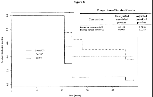
Fig. 6: Antibody Bax94 (dotted line) and antibody Bax152 (dashed line) showed
increased survival and delayed time to death in the peritonitis animal model
compared
with a control antibody (C3).
Fig. 7: Differential binding of antibody Bax94 to active MIF and non-active
MIF. Antibody
Bax94 binds active MIF in a direct ELISA format, whereas non-active-MIF does
not bind.
Fig. 8: Table summarizing in-vitro properties of human anti-MIF antibodies.
4
CA 02711029 2010-06-29
WO 2009/086920 PCT/EP2008/011146
Fig. 9: Pro-apoptotic effects of anti-MIF antibodies in a cell based assay.
Cellular
caspase-3 (effector caspase) activities are shown after antibody treatment of
PC-3 cells.
Assays are done in triplicate and data are presented as mean SD.
Fig. 10: Anti-invasive effects of anti-MIF antibodies. The invasion of PC-3
prostate
cancer cells through pores of matrigel-coated TranswellTM inserts is examined.
The
number of invaded cells per visual field are counted (microscopy at 400 fold
magnification). Data are presented as mean SD from 3-10 visual field counts
and
significant differences are shown.
DETAILED DESCRIPTION OF THE INVENTION
Definitions and General Techniques
Unless otherwise defined herein, scientific and technical terms used in
connection with
the present invention shall have the meanings that are commonly understood by
those of
ordinary skill in the art. Generally, nomenclatures used in connection with,
and
techniques of, cell and tissue culture, molecular biology, immunology,
microbiology,
genetics and protein and nucleic acid chemistry described herein are those
well known
and commonly used in the art. The methods and techniques of the present
invention are
generally performed according to conventional methods well known in the art
and as
described in various general and more specific references that are cited and
discussed
throughout the present specification unless otherwise indicated. See, e.g.,
Sambrook at
al., Molecular Cloning: A Laboratory Manual, 2d ed., Cold Spring Harbor
Laboratory
Press, Cold Spring Harbor, N.Y. (1989) and Ausubel at al., Current Protocols
in
Molecular Biology, Greene Publishing Associates (1992), and Harlow and Lane
Antibodies: A Laboratory Manual, Cold Spring Harbor Laboratory Press, Cold
Spring
Harbor, N.Y. (1990), which are incorporated herein by reference.
"MIF" or "macrophage migration inhibitory factor' refers to the protein, which
is known as
a critical mediator in the immune and inflammatory response, especially as a
counterregulator of glucocorticoids. MIF includes mammalian MIF, specifically
human
MIF (Swiss-Prot primary accession number: P14174), wherein the monomeric form
is
encoded as a 115 amino acid protein but is produced as a 114 amino acid
protein due to
cleavage of the initial Methionine. "MI" also includes "GIF' (glycosylation-
inhibiting
factor) and other forms of MIF such as fusion proteins of MIF. The numbering
of the
5
CA 02711029 2010-06-29
WO 2009/086920 PCT/EP2008/011146
aminoacids of MIF starts with the N-terminal Methionine (amino acid 1) and
ends with the
C-terminal Alanine (amino acid 115).
The term "active MIF' refers to naturally occurring conformational isoforms of
MIF, which
are relevant for its biological function. Active MIF includes isoforms that
can be observed
on the surface of cells (such as THP1 or the like). Active MIF also includes
MIF isoforms
that occur in serum of mammals after challenge with bacteria.
An "antibody" refers to an intact antibody or an antigen-binding portion that
competes
with the intact antibody for specific binding. See generally, Fundamental
Immunology,
Ch. 7 (Paul, W., ed., 2nd ed. Raven Press, N.Y. (1989)) (incorporated by
reference). The
term antibody includes genetically engineered forms such as chimeric or
humanized
antibodies.
The term "antigen-binding portion" of an antibody refers to one or more
fragments of an
antibody that retain the ability to specifically bind to an antigen (e.g.,
MIF). Antigen-
binding portions may be produced by recombinant DNA techniques or by enzymatic
or
chemical cleavage of intact antibodies. Antigen-binding portions include Fab,
Fab',
F(ab')2, Fv, and complementarity determining region (CDR) fragments, single-
chain
antibodies (scFv), chimeric antibodies, diabodies and polypeptides that
contain at least a
portion of an antibody that is sufficient to confer specific antigen binding
to the
polypeptide. From N-terminus to C-terminus, both the mature light and heavy
chain
variable domains comprise the regions FR1, CDR1, FR2, CDR2, FR3, CDR3 and FR4.
The assignment of amino acids to each domain is in accordance with the
definitions of
Kabat, Sequences of Proteins of Immunological Interest (National Institutes of
Health,
Bethesda, Md. (1987 and 1991)), Chothia et. al. J. Mol. Biol. 196:901-917
(1987), or
Chothia et al., Nature 342:878-883 (1989). An antibody or antigen-binding
portion thereof
can be derivatized or linked to another functional molecule (e.g., another
peptide or
protein). For example, an antibody or antigen- binding portion thereof can be
functionally
linked to one or more other molecular entities, such as another antibody
(e.g., a
bispecific antibody or a diabody), a detectable agent, a cytotoxic agent, a
pharmaceutical
agent, and/or a linking molecule.
The term "human antibody" refers to any antibody in which the variable and
constant
domain sequences are human sequences. The term encompasses antibodies with
6
CA 02711029 2010-06-29
WO 2009/086920 PCTIEP2008/011146
sequences derived from human genes, but which have been changed, e.g. to
decrease
possible immunogenicity, increase affinity, eliminate cysteines that might
cause
undesirable folding, etc. The term encompasses such antibodies produced
recombinantly in non-human cells, which might impart glycosylation not typical
of human
Cells.
The term "humanized antibody" refers to immunoglobulins, immunoglobulin chains
or
fragments thereof (such as Fv, Fab, Fab', F(ab')2, fragments, or other antigen-
binding
portions of antibodies), which contain sequences derived from a non-human
immunoglobulin.
The term "chimeric antibody' refers to an antibody that comprises regions from
two or
more different species.
The term "isolated antibody" or "isolated antigen-binding portion thereof'
refers to an
antibody or an antigen-binding portion thereof that has been identified and
selected from
an antibody source such as a phage display library or a B-cell repertoire.
The term "K0" refers to the equilibrium dissociation constant of a Fab portion
of a
particular antibody with the respective antigen.
The terms "center region" and "C-terminal region" of MIF refer to the region
of human
MIF comprising amino acids 35-68 and 86-115, respectively.
The term epitope" includes any protein determinant capable of specific
binding to an
immunoglobulin or an antibody fragment. Epitopic determinants usually consist
of
chemically active surface groupings of molecules such as exposed amino acids,
aminosugars, or other carbohydrate side chains and usually have specific three-
dimensional structural characteristics, as well as specific charge
characteristics.
The term "vector" refers to a nucleic acid molecule capable of transporting
another
nucleic acid to which it has been linked. In some embodiments, the vector is a
plasmid,
i.e., a circular double stranded DNA loop into which additional DNA segments
may be
ligated.
7
CA 02711029 2010-06-29
WO 2009/086920 PCT/EP2008/011146
The term "host cell" refers to a cell line, which is capable to produce a
recombinant
protein after introducing an expression vector. The term "recombinant cell
line", refers to
a cell line into which a recombinant expression vector has been introduced. It
should be
understood that "recombinant cell line" means not only the particular subject
cell line but
also the progeny of such a cell line. Because certain modifications may occur
in
succeeding generations due to either mutation or environmental influences,
such
progeny may not, in fact, be identical to the parent cell, but are still
included within the
scope of the term "recombinant cell line" as used herein.
The term "pharmaceutically acceptable carrier" refers to any and all solvents,
dispersion
media, coatings, antibacterial and antifungal agents, isotonic and absorption
delaying
agents, and the like that are physiologically compatible.
Anti-MIF Antibodies
In one embodiment, the invention relates to isolated monoclonal antibodies or
antigen-
binding portions thereof, which specifically bind to the C-terminal or the
center region of
human MIF and further inhibit human MIF biological function. In some
embodiments the
monoclonal antibodies, are human monoclonal antibodies. In other embodiments
the
monoclonal antibodies, are humanized monoclonal antibodies.
In some embodiments, the light chain of the anti-MIF antibody comprises the
amino acid
sequence that is the same as the amino acid sequence of the VLof antibody Bax8
(SEQ
ID NO: 1), antibody Bax69 (SEQ ID NO: 2), antibody Bax74 (SEQ ID NO: 3),
antibody
Bax94 (SEQ ID NO: 4), antibody Bax152 (SEQ ID NO: 5), antibody BaxA10 (SEQ ID
NO: 6), or an amino acid sequence which has 85 %, preferably 90% sequence
homology
to said amino acid sequences. In some embodiments, the light chain comprises
the
amino acid sequence from the beginning of the CDR1 to the end of the CDR3 of
any one
of said antibodies. In some embodiments, the light chain of the anti-MIF
antibody
comprises at least the light chain CDR1, CDR2 or CDR3 of the amino acid
sequences
shown in Figure 1.
In some embodiments, the heavy chain comprises an amino acid sequence of the
variable domain (VH) of antibody Bax8 (SEQ ID NO: 7), antibody Bax69 (SEQ ID
NO: 8),
antibody Bax74 (SEQ ID NO: 9), antibody Bax94 (SEQ ID NO: 10), antibody Bax152
(SEQ ID NO: 12), antibody BaxA10 (SEQ ID NO: 12), or an amino acid sequence
which
8
CA 02711029 2010-06-29
WO 2009/08692(} PCT/EP2008/011146
has 85 %, preferably 90% sequence homology to said amino acid sequences. In
some
embodiments, the heavy chain comprises the amino acid sequence from the
beginning
of the CDR1 to the end of the CDR3 of any one of said antibodies. In some
embodiments, the heavy chain of the anti-MIF antibody comprises at least the
heavy
chain CDR1, CDR2 or CDR3 of the amino acid sequences shown in Figure 2.
Class and Subclass of Anti-MIF Antibodies
The anti-MIF antibody of the invention is an isolated monoclonal antibody. The
anti-MIF
antibody can be an IgG, an igM, an lgE, an IgA, or an IgD molecule. In other
embodiments, the anti-MIF antibody is an IgG and is an lgG1, IgG2, IgG3 or
IgG4
subclass. In other embodiments, the antibody is either subclass IgG1 or IgG4.
In other
embodiments, the antibody is subclass IgG4. In some embodiments the IgG4
antibody
has a single mutation changing the serine (serine228, according to the Kabat
numbering
scheme) to proline. Accordingly, the CPSC sub-sequence in the Fc region of
IgG4
becomes CPPC, which is a sub-sequence in IgG1 (Angal et al. Mol Immunol. 1993,
30,
105-108).
MIF Epitopes Recognized by Anti-MIF Antibodies
In some embodiments, the invention relates to anti-MIF antibodies or antigen-
binding
portions thereof that specifically bind to the regions spanning from amino
acids 35-68 or
86-115 of human MIF, respectively, preferably the anti-MIF antibodies
specifically bind
to the regions spanning from amino acids 50 to 68, or 86 to 102, respectively,
and inhibit
human MIF biological function.
In other embodiments, the invention relates to anti-MIF antibodies, which
specifically
bind to active MIF and further inhibit human MIF biological function. In some
embodiments, active MIF is membrane-bound.
It was surprisingly found that anti-MIF antibodies of the invention had the
surprising
property of competing anti-MIF antibody III.D.9 in binding studies with human
MIF.
Competition of III.D.9 can be determined as described in Example 5.
Binding Affinity of Anti-MIF Antibodies to Human MIF
The invention relates to anti-MIF antibodies or antigen-binding portions
thereof, which
9
CA 02711029 2010-06-29
WO 2009/086920 PCT/EP2008/01.1146
bind to human MIF with a Kp of 5x10⁻⁷ M or less. In other embodiments, the
antibodies
bind to human MIF with a KO of 5x10⁻⁸ M or less, 5x10⁻⁹ M or less, or 5x10-M10
M or less.
The binding affinity of anti-MIF antibodies or antigen-binding portions
thereof to human
MIF can be determined by methods known in the art. The binding affinity for
example can
be measured by surface plasmon resonance (BIACORE). Example 10 exemplifies a
method for determining affinity constants of anti-MIF antibodies by BIACORE
technology.
In some embodiments, the invention further relates to anti-MIF antibodies or
antigen-
binding portions thereof, which bind to active MIF with a KDof less than 500nM
and
further inhibit human MIF function biological function. In some embodiments,
the anti-MIF
antibodies or antigen-binding portions thereof bind active MIF with a KD of
less than
5OnM.
Production of anti-MIF Antibodies
Ariii-M1F antibodies or antigen-binding portions thereof according to the
present invention
may be prepared by many methods known to the person skilled in the art, such
as
screening of phage display libraries of antibody fragments. Different formats
of phage
display libraries may be utilized, e.g. scFv or Fab fragments libraries or the
like. A phage
display library is screened for antibody fragments with desired affinities for
certain MIF
epitopes and the genetic material is recovered from the appropriate clone. In
consecutive
rounds of generating and screening libraries, antibody fragment can be
isolated with an
increased affinity compared to the affinity of the original antibody fragment
isolated. The
affinity of an identified anti-MIF fragment can be further enhanced by
affinity maturation.
Nucleic Acids, Vectors, Host Cells, and Recombinant Methods of Making anti-MIF
Antibodies
The invention further relates to nucleic acid molecules encoding anti-MIF
antibodies or
antigen-binding portions thereof according to the present invention, as well
as to vectors
comprising such nucleic acid and to host cells comprising such a vector, as
well as to
methods of recombinantly producing a polypeptide encoded by the nucleic acid
molecule.
In some embodiments, the DNA sequence encoding the VL region of the anti-MIF
antibody comprises the nucleotide sequence that is the same as the sequence of
the Vt
CA 02711029 2010-06-29
WO 2009/086920 PCT/EP2008/031146
of antibody Bax8 (SEQ ID NO: 13), antibody Bax69 (SEQ ID NO: 14), antibody
Bax74
(SEQ ID NO: 15), antibody Bax94 (SEQ ID NO:16), antibody Bax152 (SEQ ID NO:
17),
antibody BaxA10 (SEQ ID NO: 18) as shown in Fig 3, or a sequence, which has 85
%,
preferably 90% sequence homology to any of said nucleotide sequences.
In some embodiments, the DNA sequence encoding the VH region of the anti-MIF
antibody comprises the nucleotide sequence that is the same as the sequence of
the VH
of antibody Bax8 (SEQ ID NO: 19), antibody Bax69 (SEQ ID NO: 20), antibody
Bax74
(SEQ ID NO: 21), antibody Bax94 (SEQ ID NO: 22), antibody Bax152 (SEQ ID NO:
23),
antibody BaxA10 (SEQ ID NO: 24) as shown in Fig 4, or a sequence, which has 85
%,
preferably 90% sequence homology to any of said nucleotide sequences.
The production of the anti-MIF antibodies according to the present invention
include any
method for the generation of recombinant DNA by genetic engineering, e.g. via
reverse
transcription of RNA and/or amplification of DNA and cloning into expression
vectors.
In some embodiments, the vector is a viral vector, wherein additional DNA
segments
may be ligated into the viral genome. In some embodiments, the vector capable
of
autonomous replication in a host cell into which introduced (e.g., bacterial
vectors
having a bacterial origin of replication and episomal mammalian vectors). In
other
embodiments, the vector (e.g., non-episomal mammalian vectors) can be
integrated into
the genome of a host cell upon introduction into the host cell, and thereby
replicated
along with the host genome. Moreover, certain vectors are capable of directing
the
expression of genes to which they are operatively linked. Such vectors are
referred to
herein as "recombinant expression vectors" (or simply, "expression vectors").
Anti-MIF antibodies can be produced by means of conventional expression
vectors, such
as bacterial vectors (e.g., pBR322 and its derivatives), or eukaryotic
vectors. Those
sequences that encode the antibody can be provided with regulatory sequences
that
regulate the replication, expression and/or secretion from the host cell.
These regulatory
sequences comprise, for instance, promoters (e.g., CMV or SV40) and signal
sequences. The expression vectors can also comprise selection and
amplification
markers, such as the dihydrofolate reductase gene (DHFR), hygromycin-B-
phosphotransfe rase, and thymidine-kinase. The components of the vectors used,
such
as selection markers, replicons, enhancers, can either be commercially
obtained or
prepared by means of conventional methods. The vectors can be constructed for
the
11
CA 02711029 2010-06-29
WO 2009/086920 PCTIEP2008/011146
expression in various cell cultures, e.g., in mammalian cells such as CHO,
COS,
HEK293, NSO, fibroblasts, insect cells, yeast or bacteria such as E. cofi. In
some
instances, cells are used that allow for optimal glycosylation of the
expressed protein.
The anti-MIF antibody light chain gene and the anti-MIF antibody heavy chain
gene can
be inserted into separate vectors or both genes are inserted into the same
expression
vector. The antibody genes are inserted into the expression vector by standard
methods,
e.g., ligation of complementary restriction sites on the antibody gene
fragment and
vector, or blunt end ligation if no restriction sites are present.
The production of anti-MIF antibodies or antigen-binding portions thereof may
include
any method known in the art for the introduction of recombinant DNA into
eukaryotic cells
by transfection, e.g. via electroporation or microinjection. For example, the
recombinant
expression of anti-MIF antibody can be achieved by introducing an expression
plasmid
containing the anti-MIF antibody encoding DNA sequence under the control of
one or
more regulating sequences such as a strong promoter, into a suitable host cell
line by an
appropriate transfection method resulting in cells having the introduced
sequences stably
integrated into the genome. The lipofection method is an example of a
transfection
method which may be used according to the present invention.
The production of anti-MIF antibodies may also include any method known in the
art for
the cultivation of said transformed cells, e.g. in a continuous or batchwise
manner, and
the expression of the anti-MIF antibody, e.g. constitutive or upon induction.
The host cell type according to the present invention may be any eukaryotic
cell. In one
embodiment the cell is a mammalian cell with the ability to perform
posttranslational
modifications of anti-MIF antibodies. For example said mammalian cell is
derived from a
mammalian cell line, like for example a cell line selected from the group
consisting of
SkHep-, CHO-, HEK293-, and BHK-cells. In one embodiment, the anti-MIF antibody
is
expressed in a DHFR-deficient CHO cell line, e.g., DXB11, and the addition of
G418 as a
selection marker. When recombinant expression vectors encoding antibody genes
are
introduced into mammalian host cells, the antibodies are produced by culturing
the host
cells for a period of time sufficient to allow for expression of the antibody
in the host cells
or secretion of the antibody into the culture medium in which the host cells
are grown.
12
CA 02711029 2010-06-29
WO 2009/0869211 PCTJEP2008/011146
Anti-MIF antibodies can be recovered from the culture medium using standard
protein
purification methods.
Additionally, the production of anti-MIF antibodies may include any method
known in the
art for the purification of an antibody, e.g. via anion exchange
chromatography or affinity
chromatography. In one embodiment the anti-MIF antibody can be purified from
cell
culture supernatants by size exclusion chromatography.
Properties of Anti-MIF Antibodies
The invention relates to anti-MIF antibodies or antigen-binding portion
thereof, which
possess at least one of the following properties:
a) bind to the C-terminal or the center region of human MIF
b) inhibit glucocorticoid overriding (GCO) activity,
c) inhibit proliferation of cells lines such as fibroblasts or cancer cells
(e.g.
NIH/3T3 or PC-3)
d) bind to active MIF
e) does not bind to non-active MIF
f) compete mouse anti-MIF antibody III.D.9.
In some embodiments, active MIF is an isoform of active MIF that occurs by
treatment of
human MIF with mild oxidizing reagents, such as Cystine or by immobilizing
human MIF
on a support such as an ELISA-plate or beads. In other embodiments, active MIF
is an
isoform of active MIF that occurs in vivo after challenge of animals with
bacteria. In other
embodiments, active MIF is an isoform of active MIF that occurs in vivo on the
surface of
cells (e.g. THP1, CFB).
In some embodiments, non-active MIF is reduced MIF (e.g. as described in
Example 7)
or, intracellular stored MIF.
In other embodiments, the anti-MIF antibodies or antigen-binding portion
thereof bind
active MIF with a KD less than 500 nM.
Pharmaceutical Compositions of anti-MIF Antibodies and Methods of Treatment
The invention also relates to compositions comprising an anti-MIF antibody or
an
antigen-binding portion thereof, for the treatment of a subject in need of
treatment for
MIF-related conditions, specifically immunological diseases such as
inflammatory
diseases and hyperproliferative disorders.
13
CA 02711029 2010-06-29
WO 2009/086920 PCTIEP2008/011146
In some embodiments, the subject in need of treatment is a human.
Hyperproliferative
disorders, such as cancerous diseases, that may be treated by anti-MIF
antibodies of the
invention can involve any tissue or organ and include but are not limited to
brain, lung,
squamous cell, bladder, gastric, pancreatic, breast, head, neck, liver, renal,
ovarian,
prostate, colorectal, esophageal, gynecological, nasopharynx, or thyroid
cancers,
melanomas, lymphomas, leukemias or multiple myelomas. In particular, anti-MIF
antibodies of the invention are useful to treat carcinomas of the breast,
prostate, colon
and lung.
The invention also encompasses methods for the treatment of inflammatory
diseases
such as vasculitis, arthritis, sepsis, septic shock, endotoxic shock, toxic
shock syndrome,
acquired respiratory distress syndrome, glomerulonephritis, inflammatory bowel
disease,
Crohn's disease, ulcerative colitis, peritonitis, nephritis, atopic
dermatitis, asthma,
conjunctivitis, fever, Malaria or psoriasis in a subject, including a human,
comprising the
step of administering to said subject in need thereof a therapeutically
effective amount of
an anti-MIF antibody or antigen-binding portion thereof.
In other embodiments the composition comprising said anti-MIF antibody of the
invention
is used for the treatment of an inflammatory disease selected from the group
consisting
of glomerulonephritis, inflammatory bowel disease, nephritis and peritonitis.
The treatment may also involve administration of one or more anti-MIF antibody
of the
invention, or an antigen-binding fragment thereof, alone or with a
pharmaceutically
acceptable carrier. Some examples of pharmaceutically acceptable carriers are
water,
saline, phosphate buffered saline, dextrose, glycerol, ethanol and the like,
as well as
combinations thereof. In many cases, it will be preferable to include isotonic
agents, for
example, sugars, polyalcohols such as mannitol, sorbitol, or sodium chloride
in the
composition. Additional examples of pharmaceutically acceptable substances are
wetting
agents or minor amounts of auxiliary substances such as wetting or emulsifying
agents,
preservatives or buffers, which enhance the shelf life or effectiveness of the
antibody.
The anti-MIF antibody of the invention and the pharmaceutical compositions
comprising
them, can be administered in combination with one or more other therapeutic,
diagnostic
or prophylactic agents. Additional therapeutic agents include other anti-
neoplastic, anti-
14
CA 02711029 2010-06-29
WO 2009/086920 PCT/EP2008/011146
tumor, anti-angiogenic, chemotherapeutic agents or steroids, depending on the
disease
to be treated.
The pharmaceutical compositions of this invention may be in a variety of
forms, for
example, liquid, semi-solid and solid dosage forms, such as liquid solutions
(e.g.,
injectable and infusible solutions), dispersions or suspensions, tablets,
pills, powders,
liposomes and suppositories. The preferred form depends on the intended mode
of
administration and therapeutic application. Typical preferred compositions are
in the form
of injectable or infusible solutions, such as compositions similar to those
used for passive
immunization of humans, The preferred mode of administration is parenteral
(e.g.,
intravenous, subcutaneous, intraperitoneal, intramuscular). In a preferred
embodiment,
the antibody is administered by intravenous infusion or injection. In another
preferred
embodiment, the antibody is administered by intramuscular or subcutaneous
injection.
As will be appreciated by the skilled artisan, the route and/or mode of
administration will
vary depending upon the desired results.
The anti-MIF antibody may be administered once, but more preferably is
administered
multiple times. For example, the antibody may be administered from three times
daily to
once every six months or longer. The administering may be on a schedule such
as three
times daily, twice daily, once daily, once every two days, once every three
days, once
weekly, once every two weeks, once every month, once every two months, once
every
three months and once every six months.
The invention also encompass the use of an anti-MIF antibody or antigen-
binding
fragment thereof, in the manufacture of a medicament for the treatment of
immunological
diseases such as inflammatory diseases and hyperproliferative disorders.
The invention further encompass an anti-MIF antibody or antigen-binding
fragment
thereof, for use in treating immunological diseases such as inflammatory
diseases and
hyperproliferative disorders.
The invention also encompass an anti-MIF antibody or antigen-binding fragment
thereof,
for use in diagnostic methods. In one embodiment the anti-MIF antibody or
antigen-
binding portion thereof can be used to detect human MIF in a biological
sample.
CA 02711029 2010-06-29
WO 2009/086920 PCT/EP2008/011146
The anti-MIF antibodies or the antigen-binding portions thereof can also be
used to
determine the level of cell surface MIF in a tissue or in cells derived from
the tissue. In
some embodiments, the tissue is diseased tissue. The tissue can then be used
in an
immunoassay to determine, e.g., total MIF levels, cell surface levels of MIF,
or
localization of MIF.
The invention further relates to kits comprising an anti-MIF antibody or an
antigen-
binding portion of the invention or a pharmaceutical composition comprising
such an
antibody or portion. A kit may include, in addition to the antibody or
pharmaceutical
composition, diagnostic or therapeutic agents. A kit also can include
instructions for use
in a diagnostic or therapeutic method.
The invention further relates to a process for the identification of anti-MIF
antibodies
capable of inhibiting human MIF biological function and inducing a beneficial
effect in an
animal model by carrying out the following steps:
a) selecting an antibody that binds to active MIF and does not bind to non-
active
MIF
b) testing said antibody in in-vitro assays, such as glucocorticoid overriding
(GCO)
assay, or cell proliferation assays
c) selecting an antibody, which inhibits GCO and/or cell proliferation.
Results have shown that an anti-MIF antibody that only binds active MIF and
does not
bind non-active MIF and further inhibits GCO and/or cell proliferation induces
a beneficial
effect in an animal model (e.g. Example 6)
The present invention will be further illustrated by following examples,
without any
limitation thereto.
EXPERIMENTAL PART
Example 1: Antibody Selection
Phage display technology is used to generate human anti-MIF antibody
fragments.
Starting from a phage display library, different screening campaigns are
performed, three
of them by using full length MIF (human MIF coated / human MIF in solution /
human --
murine MIF alternating). The others by using six MIF derived peptides
alternating with
full length MIF, These six peptides are designed by dividing the MIF protein
into six
peptides of approximately 30 amino acids with overlapping stretches of
approximately
16
CA 02711029 2010-06-29
WO 20091086920 PCTIEP2008/011146
15-amino acids. After several selection rounds unique binders are identified,
all unique
binders are expressed and purified as human IgG4 antibodies. These antibodies
are
tested in several assays to demonstrate the in-vitro inhibition of MIF. An
epitope mapping
to determine the binding region within the MIF protein is carried. 193
antibodies are
tested and categorized according to their in-vitro activity of inhibiting MIF.
In-vitro assays
are described below. Three murine anti-MIF antibodies are used as control
(III.D.9,
XIV.14.3 and XIV.15.5).
Example 2: Inhibition of glucocorticoid overriding activity of MIF (GCO).
1o This method is based on the inhibition of endogenous MIF, i.e. MIF that is
produced by
the cell line used. This method is applied for antibody screening and for
determination of
dose response curves.
GCO-assay for antibody screening:
A THP1 suspension culture is centrifuged and cells are resuspended in fresh
full medium
to a cell density of 106 cells per ml. This culture is transferred into wells
of a 96-well
microplate (90 pi/well) and anti-MIF antibody is added to give a final
concentration of
75pg/ml. Each antibody is tested in triplicate. After o/n incubation at 37 C
dexamethasone is added to give a concentration of 2 nM and after one hour
incubation
at 37 C LPS is added (3 ng/ml final concentration). After further six hours
incubation at
37 C the supernatant is harvested and the IL-6 concentrations are determined
in an
ELISA (Cytoset kit, commercially available). The results of the triplicates
are averaged
and the percentage of IL-6 secretion is determined in comparison to the
control
antibodies. Antibodies that result in an IL-6 secretion of less than 75% are
evaluated as
positive.
Assay for determination of IC50 values
The experimental procedure is carried out as described for the screening assay
with the
exception that increasing amounts of antibody are used (typically from 1 - 125
nM). The
resultant dose response curve is expressed as % inhibition in comparison to a
negative
control antibody. This curve is used for calculation of the maximum inhibitory
effect of the
antibody (%Inh max) and the antibody concentration that shows 50% of the
maximum
inhibitory effect (IC50)
Results are summarized in Fig. 8, column 3 (IC50) and column 4 (maximum
inhibition).
For comparison, murine antibody XIV.14.3 shows 36% inhibition of GCO only
(data not
shown).
17
CA 02711029 2010-06-29
WO 2009/086920 PCT/EP2008/011146
Example 3: Inhibition of cell proliferation
Serum stimulates secretion of M1F in quiescent NIH/3T3 and MIF in turn
stimulates cell
proliferation. Antibodies inhibiting this endogenous MIF, therefore, decrease
the
proliferation of quiescent NIH/3T3 cells. The reduction of proliferation is
determined by
the incorporation of 3H-thymidine.
1000 NIH/3T3 cells per well are incubated in a 96 well plate over the weekend
at 37 C in
medium containing 10% serum. Cells are then starved over night at 37 C by
incubation
in medium containing 0.5% serum. The 0.5% medium is removed and replaced by
fresh
medium containing 10% serum, 75pglml antibody and 5pCi/ml of 3H-Thymidin.
After 16
hours incubation in a CO2 incubator at 37 C cells are washed twice with 150pi
of cold
PBS per well. Using a multi-channel pipette 150pi of a 5% (w/v) TCA solution
per well
are added and incubated for 30 minutes at 4 C. Plates are washed with 150pl
PBS. Per
well 75p1 of a 0.5M NaOH solution with 0.5% SDS are added, mixed and stored at
room
temperature. Samples are measured in a B-counter by mixing 5mi of Ultima Gold
(Packard) and 75p1 sample solution. Each determination is done in triplicate
and the
values are compared with the values of the control antibody by a t-test.
Antibodies that
significantly reduce proliferation (P<0.05) are evaluated as positive. Results
are
summarized in Fig.8, column 5.
Example 4: Binding studies: Epitope determination of anti-MIF antibodies
Each peptide is diluted in coupling buffer to give a peptide concentration of
typically 5
pg/mI, is added to microplates (NUNC lmmobilizerTM Amino Plate F96 Clear) and
incubated over night at 4 C (100pl/well). As controls recombinant full length
MIF and
PBS are used. The plate is washed 3 times with 200 pl PBST and antibodies (4
pg/ml in
PBS) are added (100 pl/well) and incubated for 2 hours at room temperature
with gentle
shaking. The plate is washed 3 times with 200 pl PBST and detection antibody (
e.g. Fc
specific anti-human IgG /HRP labeled , Sigma) is added (100 pl/well). After
incubation for
1 hour at room temperature with gentle shaking the plate is washed 3 times
with 200 pl
PBST. Each well is incubated with 100 pl TMB solution (T-0440, Sigma) for 30
minutes in
the dark. Staining reaction is stopped by adding 100 p1 of 1.8 M H2SO4-
solution per well.
Samples are measured at 450nm.
Example 5: Competition of human anti-MIF antibodies with murine anti-MIF
antibody
IIi.D.9
Antibody Bax94 is used for competition with mouse anti MIF antibodies III.D.9.
18
CA 02711029 2010-06-29
WO 2009/086920 PCTIEP2008/011.3.46
96 well plates (NUNC Maxisorp) are coated with recombinant human MIF. The
murine
anti-MIF antibody Il.D.9 and human anti-MIF antibodies are diluted in TBSTl2%
BSA and
mixed, whereas the final concentration of Iil.D9 is kept at 2pg/ml and the
concentration
of human anti-MIF antibodies is increased from Opg/ml to typically 32pg/ml.
After
washing of the microplate the antibodies are applied and incubated at room
temperature
for typically 2 hours. After washing, the plate is incubated with anti Mouse
lgG (Fc spec.)
peroxidase conjugate and incubated for 1 hour at room temperature. After
washing, the
plate is incubated with TMB-solution and the staining reaction is stopped by
adding
H2S04-solution. Fitting of the resultant competition curve enables the
calculation of the
maximum inhibition of the III.D.9 binding. The results are summarized in Fig.
8, column
6.
Example 6: Increased survival of anti-MIF antibodies in the live E.coli
peritonitis animal
model
The experiments are carried out according to Calandra et at. (Nature
Immunology, 2000)
using female NMRI mice (25-30g, 6-10 weeks of age) that are injected
intraperitoneally
with 6000 CFU of an E.coli 0111:64 suspension in 15% mucin and 4% hemoglobin.
Two
or three colonies (E.coli 0111:804) from a nutrient agar plate culture are
inoculated into
10 ml of TSB and incubated overnight at 36 C with shaking. The culture is
diluted in
physiological saline to the required concentration(s) - an overnight the
culture typically
reaches 2"109 CFU/ml - and mixed with mucin and hemoglobin (1 volume of
diluted
inoculum, 2 volumes of 15% mucin, 2 volumes of 4% hemoglobin). As the inoculum
mixture tends to sediment out, it is mixed between injections. A large (e.g.
23 gauge)
needle is used for injections to avoid blockage of the needle by particulates
in the
injection mixture. Antibody Bax94 (lgG4) and an isotype matching control
antibody are
given 2 hours prior to bacterial challenge interperitoneally. The antibody
dosage is
typically 800pg/mouse and 20 mice are used for each group. A statistically
significant
effect on survival/time to death could be shown for the IgGi and IgG4 isotypes
of human
anti-MIF antibodies. Figure 6 shows the results obtained for antibody Bax94
and
antibody Bax152 (lgG4). Kaplan-Meier statistics is used for evaluation of the
survival
curves.
Example 7: Binding specificity for active MIF
The anti-MIF antibodies described in this invention are able to discriminate
between
active and non-active MIF, which are generated by mild oxidation or reduction,
19
CA 02711029 2010-06-29
WO 2009/086920 PCT/EP2008/011.146
respectively. Discrimination between these conformers is assessed by ELISA or
surface
plasmon resonance.
ELISA for assessing differential binding of the antibodies:
= Transformation of MIF into its active conformation by mild oxidation.
Recombinant human MIF (0.5 mg/ml in PBS) is incubated for 3h at 37 C with a 3-
fold
excess (volume) of a saturated solution of L-Cystine in PBS (- 0.4-0.5 mM L-
Cystine). The MIF is then dialyzed two times against PBS in a Slide-A-Lyzer
Dialysis
Cassette with a molecular-weight cutoff of 7 kDa (Pierce),
= Transformation of MIF into its non-active conformation.
MIF is reduced at a concentration of 0.5 mg/ml by overnight incubation with 8-
16 mM
dithiothreitoi (final concentration) at 4 C.
= ELISA protocol.
The anti-MiF antibodies are coated into 96-well microplates (NUNC MaxisorpTM)
at a
concentration of 5 g/ml (dilution in coating buffer). After washing the plate
with
TBST (Tris-buffered saline with 0.1% Tween-20 (v/v)) and blocking with
TBST/2%BSA (TBST and 2% bovine serum albumin (w/v)), dilution series of either
active or non-active MIF are added and incubated at room temperature for 1-2
h.
Bound MIF is detected using a polyclonal rabbit anti-MIF antibody and a
horseradish
peroxidase labeled goat-anti-rabbit antibody (Biorad). TEST/2%BSA is used to
dilute
MIF, the rabbit anti-MIF antibody and the peroxidase conjugate to reduce
unspecific
binding. Figure 7 shows the ELISA results obtained with antibody Bax94.
Assesssing differential binding of the antibodies by Biacore.
Binding kinetics of active and non-active MIF to antibody Bax94 are examined
by
surface plasmon resonance analysis using a Biacore 3000 System. Therefore,
10000
Response Units of Bax 94 are immobilized on a sensor chip with a CM5
(=carboxymethylated dextran) matrix and incubated with active or non-active
MIF
huMIF in pro-reductive and pro-oxidative Glutathione redox buffers, ranging
from 4.8
mM GSH / 0.2 mM GSSG (GSSG = oxidized Glutathione) to 5 mM GSSG in HBS-EP
buffer (GE Healthcare). As a control, MIF is used for binding analysis in a
second
flow cell containing an immobilized isotype control antibody. Binding response
units
of control antibody and antibody Bax94 are subtracted for evaluation.
CA 02711029 2010-06-29
WO 2009/086920 PCT/EP2008/011146
Example 8: Detection of active MIF on the surface of THP-1 cells
Cells are incubated with anti-MIF antibody Bax94. Cells are washed with ice
cold PBS
and resuspended in cold cell lysis buffer (Cell Signaling Technologyo).
Magnetic Protein
G Dynabeads (Invitrogen) are blocked with TBST + 5% nonfat dried milk (w/v),
washed
and added to the lysed cells. Immunoprecipitation is carried out at 4 C
overnight. The
beads are then washed with cell lysis buffer and TBST and boiled in SDS PAGE
sample
buffer (without reducing agents). Samples are subjected to non-reductive SDS
PAGE for
Western Blot analysis.
Example 9: Binding of anti-MIF antibodies to membrane bound MIF
THP-1 cells are washed with ice cold PBS and resuspended in cold cell staining
buffer
(Biolegend) supplemented with 200 gglml mouse IgG. FITC- or TRITC-labeled anti-
MIF
antibodies are added to give a final concentration of typically 200-500 nM and
incubation
is done at 4 C. Cells are subsequently washed with ice cold cell staining
buffer and
resuspended in cell staining buffer supplemented with the Via-ProhPTM Cell
Viability
Solution (BD Biosciences). Cells are measured in an FACS CantoTM 11 Flow
Cytometry
System (BD Biosciences) and the median FITC-/TRITC-shift of the viable cell
populations are compared with the Dye-labeled isotype control antibody.
Example 10: Affinity determination of Fab fragments of anti-MIF antibodies by
Biacore
Typically 40RU Units of human recombinant MIF are immobilized on a sensor chip
with a
CM5 (=carboxymethylated dextran) matrix (Biacore). Fab fragments are injected
at a
concentration range of typically 6-100nM diluted in HBS-EP. After each cycle
the chip is
regenerated with 50mM NaOH + 1 M NaCl. Affinities are calculated according to
the 1:1
Langmuir model. The results are summarized in Fig. 8, column 7.
Example 11: Beneficial effect of anti-MIF antibodies in an animal model for
Crescentic
G lomerulonephritis
The anti-MIF antibodies are tested in a rat model of crescentic
glomerulonephtitis
described by Frederick W.K. Tam et. al. (Nephrol Dial Transplant, 1999, 1658-
1666).
Nephrotoxic nephritis is induced in male Wistar Kyoto rats by a single
intravenous
injection of anti-rat glomerular basement membrane serum. In the preventive
setup of the
experiment treatment with anti-MIF antibodies and an isotype matching control
antibody
is started at the time of induction of nephritis (day0) by interperitoneal
injection of the
antibody. Treatment is typically repeated every second day and animals are
culled on
21
CA 02711029 2010-06-29
WO 20091086920 PCT/EP2008/01.1146
day 7 for histological analyses. Urine is collected prior to the experiment
(baseline) and
on prior to the termination of the experiment (day 7). In a therapeutic setup,
treatment
with anti MIF antibody is started 4 days after induction of disease and
repeated every
second day. Rats are typically culled on day 8. Urine is collected prior to
the experiment
(baseline), prior to start of treatment (day 4) and prior to culling of the
animals (day 8).
Antibody dosage is typically 1 - 20 mg/kg per injection and 6 to 8 rats are
used for each
group. Disease severity is determined by measuring proteinuria, macrophage
infiltration
into the glomerulus and histological damage (crescent formation). In a
preventive
experiment treatment with anti-MIF antibody Bax69 (10 mg/kg per dose) for 7
days
results in a 47% reduction of proteinuria in comparison to control antibody
treated
animals. Treatment of established disease (therapeutic experiment) results in
a dose
dependent reduction of proteinuria by 16 % (10 mg/kg Bax69 per dose) and 34 %
(20
mg/kg Bax69 per dose) in comparison to control antibody treated animals.
Example 12: Beneficial effect of anti-MIF antibodies in an animal model for
Ulcerative
Cviius (Adoptive transfer of naive T cells in Rag -/- mice)
C578U6 mice were sacrificed and CD45RBhi cells (naive T cells) are isolated by
FACS
sorting of the spleen cell population. CD45RBhi cells (5x10⁵) are injected
i.p. in Rag-/-
C57BU6 mice (7-9 weeks old), which develop of Ulcerative colitis after approx.
2 weeks.
(de Jong et al., Nature immunology., 2001, 1061-1066). Anti-MIF antibodies and
the
isotype control antibody are injected intraperitoneally twice a week (1
mg/mouse/dose). in
a preventive setup treatment is started at the time of injection of T-cells.
In a therapeutic
setup, treatment is started 4 weeks after induction of the disease; Mice are
monitored
weekly for weight and disease development. Typically eight weeks after the
transfer of
CD4CD45RBhi cells into Rag-/- C57BU6 recipients the disease activity index
(DAI) is
calculated and colon sections are collected for histology index (HI) score.
Diseases
activity index (DAI) and histology index (HI) are determined at the end of the
animal
model (DAI is based on four parameters: hunching and wasting (scored 0 or 1),
colon
thickening (0-3) and stool consistency (0-3)). In a therapeutic experiment
anti-MIF
antibodies Bax69 and BaxA10 are used for treatment of established disease and
the
mean DAI is significantly reduced by approx. 60 % (Bax69) and approx 40 %
(BaxA10) in
comparison to isotype control treated mice. Furthermore, the mean HI score is
reduced
by approximately 33 % after treatment with Bax69.
Example 13: Beneficial effect of anti-MIF antibodies in an animal model for
Ulcerative
Colitis (Agonistic anti-CD40 model)
22
CA 02711029 2010-06-29
WO 2009/086920 PCT/EP2008/011146
This model is based on the activation of macrophages and dendritic cells by an
agonistic
anti-CD40 antibody, which induces intestinal pathology that resembles IBD in
Rag1-/-
mice.
Age / sex matched Rag-1-/- mice (4-5 wks) are purchased form Jackson
Laboratories
and kept for two weeks prior to the experiment in the animal facility. The
agonist-CD40
monoclonal antibody (FGK45, IgG2a) or Isotype control Rat IgG2a are dissolved
in PBS
at 1 mg/mi. Five groups (10 mice each group) are injected i.p with 200pg of
agonist anti-
CD40 monoclonal antibody and out of that tour groups are treated with anti-MIF
antibodies on day 0 and day 1 (2x1 mg/mouse ) The sixth group (10 mice) is
injected
only with isotype control (Rat IgG2a, healthy control). Mice are weighed for
the next 7
days. On Day 7, disease activity index (DAt) was calculated and colon sections
collected
for histology index (HI) score. The DAI score is based on : hunching (0-
1);wasting (0-1),
stool consistency( 0-3) and colon thickening (0-3). Histology score was based
on
thickness (0-3), crypt elongation, inflammation (0-3) and abscess (0-1).
Treatment with
anti-MIF antibodies Bax94, BaxA10 and Bax69 significantly reduces the DAI
score
(BaxA10: -48 % reduction ; Bax94 - 62 % reduction; Bax69- 73 % reduction )
compared
to isotype control treated mice. Furthermore, the mean HI scores is also
reduced by the
these antibodies.
Example 14: Inhibition of tumor growth in Mf1 nude mice by anti MIF antibodies
Human prostate adenocarcinoma cells (PC-3) are harvested from exponentially
growing
cultures and mixed with growth factor-depleted matrigel. 2*106 cells in 0.25
ml matrigel
are inoculated subcutaneously into the right flank of Mf1 nude mice. Treatment
with anti-
MIF antibody Bax94 and the isotype control C3 is started one day after
inoculation (0.6
mg antibody/mouse/day) and is repeated every second day. Measurement of the
sizes of
the tumors is typically started two weeks after cell injection and done every
second day.
The volumes are calculated using the formula V=0.5*a*b2 (where "a" is the
longest
diameter and "b" is the shortest diameter). Tumor growth of mice treated with
Bax94 is
significantly reduced and the mean volume of the tumors analyzed 28 days after
tumor
induction is 4.3 fold higher within the isotype control treated group in
comparison to the
Bax94 treated group.
In a therapeutic setup of the experiment antibody treatment was started one
week after
tumor engraftment. 50 mg/kg per dose of the isotype control antibody C3 and
the anti-
MIF antibody Bax69 are injected intraperitoneally every second day. After 22
days of
23
CA 02711029 2010-06-29
WO 2009/086920 PCTIEP2008/011.146
treatment the median of the tumor volume was determined to be 2.7 fold higher
within
the C3 treated group in comparison to the of Bax69 treated group.
Example 15: Pro-apoptotic effects of anti-MIF antibodies
Pro-apoptotic effects of anti-MJF antibody Bax94 are shown in a cell based
caspase-3
assay using the human prostate cancer cell line PC-3. PC-3 cells are seeded on
10 cm
culture dishes (-106 cells/ dish) in the presence of 10 % FCS. Fresh medium
containing
100 nM antibody Bax94 or 100 nM control antibody C3 is added after 24 h. After
another
incubation period of 48 h cellular lysates are prepared and caspase-3
activitly is
measured by adding a fluorescent labeled caspase substrate. (Figure 9).
Example 16: Inhibition of tumor cell invasion
Anti MIF antibodies Bax94 and Bax69 are tested in TranswellTM invasion assays,
using
the human prostate cancer cell line PC-3.
5'104 PC-3 cells are seeded per well in 24 well-TranswellT'" dishes (8 m pore
size),
which are coated with polyD-lysine on the bottom face of the polycarbonate
membrane
and with growth-factor depleted matrigel on the TranswelITM insert surface.
Cells are
allowed to attach for 4 h in the presence of 10 %FCS. Thereafter, the medium
was
changed to serum-free medium and cells are starved overnight (i.e. for 16h).
Subsequently, compounds (10 nM MIF, 500 nM antibodies) are added to the lower
chamber. Cells are allowed to migrate through the porous membrane for 24 h.
After this
incubation period, attached migrated cells of the lower face of the membrane
are stained
with Giemsa solution. The number of cells adhering to the lower face of the
membrane is
counted in independent visual fields at 400-fold magnification (Figure 10).
24
CA 02711029 2010-06-29
WO 2009/086920 PCT/EP20081011146
x x x x 1/14
H H H H H H
RT > > > > >
J CL (4 w ):. C) (4
a a a a a a
~ ai ~ N N 2 a
> a a oa a
J a d b co of d
U U U C) C) U
a a a ) C) C)
w C) o w a w
a Cl. a a a a
0 0 0 0 m 0
m C) cn tt a
H v)
f p. cn m
H H H H
H. H
a a a a a a
f+.
CC) w w C) W C
F E H C. 0 F
Cl) ul) En fn N Cl)
U t"IMUMMU)
C9 N C) C) CUR N 0
a: 4, a, r~ 41 1~ w
y a W W W Q a
J ~U C) 0 Cl) C5 0
N N N H H E'
a Of co cli > Q U) N ` y N N
J fJ a a c~ i~ o
H H H H H H
a a a a
a1 P. R, W C) a a
~- z 09 0 o)
a CC). 0 a a a
LL
:, a o of o a a
J 3 3 ) 3 3
Ea a U) H U)
N C7 N N to
Q H H H S N El
N N U
cNacyocycy
H H N a
> > F.
x cz C)
G R O U CC)C7 E.
> ; j a a .
IL
m N N Cl) Cl) N
U)) Cl) aa C m m
C))) Ua] C)) H r H
N cn Cn C) > C7
a a a c. a
r n C) cn cn 0 to
a E. 0 a 0
H 0 0 0 0 0
J o o C) A Q Q
O r c~ co v ~ c
z o 0 6 0 Z Z
z z Z Z
W a o a Q a o
co m
E
CD h 01 ~2
z m m m m m
CA 02711029 2010-06-29
WO 20091086920 N 0) m PCT/PP2008/011146
> 2/14
Q F a F H
E- 11 El
E
a s a a a
= 3 3 3 3 3 3
A to
!si Z7 G C7
KrS z
to H C H H E.
C., V. 2 0 0 to
v a N o a ~ a
a (0 0
a a u u a
< > H
El 0) 0) 4 a a
H
0) Q Q CC o CC
a
.0 a a a a a
CY v
a a o a to C C Y
a a a a
F zH H F E
4 D A CC 6
a a a CC a
LL H H H H N H
[[L,, H H H H
= K CY C CG fL G:
to to t K CO
to to to
L yxWr
N
N H H H H F
LL- U
M 0) to cn (0 (0 0 N N
U m n 09 H H H
= H m H H V H
V1 UJ N U) N 0
W u: 0) W CC
a a .a a a
CC CD Y x r x
N n. CO P. G P0. w
o~ ao a a to to
S C.> H H 0) H H
N N ill N VI N
F F F F F F
0 u C7 . C C7 .. u. w
C0 0 CO 0
(7
N N N 0 N N
a a a a
a 0) x a a CC
- a a
N y Vl V N N
0 C9 C7 C) C7 (0
a a eC a a
a > j a a C)
a a a
OG CO^. C) (07
N V) Cl) 0) U U
u7 u7 ul y W W
U. a a a a i
oa a O a a
2 m CC w w w
O m m o
z
0 z z Z z to Z
0 o 2 2 a d Q
w o o w w w wo
V) N N U) U) U)
47 1 Q) tt ~ N O
m a
E N x x K
z m m m in m" CO
CA 02711029 2010-06-29
WO 20(!91086920 PCT/EP2008/011146
3/14
EU CL u VIUG.Ua~ EUP UCOUaU 0C7
U C) CUB U 4 U F U C9
>CUE7 Y4 a,U0 00 >Cx4W UO~00
C
OYU>C) U C c 4 dU>C) U w
Q~OUC7CCn~ Ea QC) UCa C.~~E
M C E 4C 4 E C7 H 4C 4 U
ei
C ~~3Fd H~aE L) U
>Ed4HEC)a,u
CS1
CnUZF -I'EUEU E. Z4UIC)EU E , E ~ E 4C 4 E a E 4 44
E. L) 0
4
4lU :IF nM aCC)Cl)CF7 4CUa4=u~Uy0
C- E 4C U 4t U
E E U F E
cn E E cn E F E Cn E Cn Fu E 4 i E
C7 U U E C7 E 4 U E
7 U to 4 U[ U u1 4 I U F 4 4 U W E En 0
m ~ 41 U of cnUZC)>CcaE,ar4t
H C) G. E F C d U
Cn EUU" r-f E E u d U fn U
W Jx aH CE]U
< F
aUCn~H
El
C/1U Odd ~-7 Ecn c~.~;~=UU 4C Cn U 074,.7 F V) u>1 Cx
E. U U F E ti* E U U E E 4:
U F U 4 U U C7 E U U U U
co d cn 0 ~l E U C7 > E t 0 v v7 0 .] U 0 0 >+ H H C)
V 4L U C7 14
U 4i Ca E E U d F E
E. U
C) Q U x ~ r n C E U w 4 E U CO U v1C) EC) Wu
U U F U 4 C) C') C7 E U 4t C7
E t9 a U 0 C7 4C > H Z F a 0 P+ U 0 0 4t U > E
4C U U U C) C) 4C U U Ch C7 C7
C) U U H H C) (7 U V 4C F t7
M0UUF<Uco 0~`Cx 4: ~ODUEUleU) ty, 4: to L)
C) U F U E U U F 4t U H U
O H C H U ? C + E G 0 4 F U Z r - + F E U 9 P: E - KC FU
Z 4t 44 E C7 4C 4 It 4C E. C? 4C
U U C) C7 C9 U U 0 0 4 0
Q r-+ E+ U U [G 0 W h^ C9 C7 Q Q 4t H E 0 0 W 0 W 4 C7 0
C C7 C7 C7 C7 W CD 4 U 4 U 0
W (/j
Cn
H H H H H ri ei
CO lD N LS: '~ 0 CQ l0 N CO O
H r-1 N r+1 x H H N I')
m
CA 02711029 2010-06-29
WO 2009/086920 PCT/EP20081011146
4/14
HAP UtnEarU~~C C) H4 ~4 a4 VOVC C)
L) ri
~x40
> a OUC C) dCU~aU~E~ UU~
a>F IE-x~oaC)C) C) H
d U U U F d U C7 d H
H
pCUd7U 4:C C)CfC)E [S~~E HQcnU F
c9~>+Ev~4:, aU CSC) H CUU7HaaC
3 F O U H a U a U d C) 4 H U Cn U
n F v, a H d U H a Cn E F Cn a U a C)
H
d aE~ ~ E Cn Q ~7 E v1 UU Cn u H
HCC C)
aU EcnEE 4: EUH+ v1Htn4H G E~ H
h7UFUF C)G.HFC) .1 V C7UpCU9OU
WUC) 74UCCd7O4 HUCnCC)H4HUO'<
H C7 C7 U U d d H d U
E.
VIUH CF->+4t HUO'~ C)C F+~"HC'JCUJUC7
H d H FC U C) U d C3 E-
4 Cn~HUHdC7vUC7 avC7u L) FEEl
F C9 C9 F U d H 0 U 0 H
cn u O<4 a H co U x 4 Cn H O U U >~ x
H U U H E d
U H U d U U U H ) H CJ U
Od V10]F004 HH Odcn0C40cnu> EH
U FC U CJ H d U d d d C7 d
E- p
F V 4Ux C)CC)HU^4 HU4U 0, U0CU74Ui+74
d C7 ~ C7 d C) d C U 0 0 C7
0 U U U d 0 0 C) H H H 0
Hat7~ UC7O4:C)>H 04ULO 04H>H
d U U C7 C7 C) d d C) d H C7
U H U H F C7 C) U C9 U H C7
TOV UH rC u f U fi FXa ~U ~Uw E ^ 0
ZHFHUZ HC UC7QHU ZHE,cnCC)C)C' CC)C')4HU
a C) d
0 ~a H C) d H 0
U U FC C7 U C - U U F U H C)
('~^ar E-, 004C~^4Tuu ~jAd Ha^da000
C9 d C7 C9 i11 0 U U 07 U 0
W 0
co
co
r
W N W C O O) l0 N CO O
M
X ,-i --I N M N
m m
CA 02711029 2010-06-29
WO 20091086920 PCT/EP20081011146
5114
U FC U U ~ 4 C7
E U~ x 4: U W~ t7 ~ H~ x a a u (7 C) CS
U C7 U C~ U U C) U C7 U
ACS ci 4:~7UChC) ~~a JH~4 UC?U
x a U u x w a Of U u 0 w Q w H
c
w N HUm0E+U F EUMU4 EC)
u
3 H 0 +4 U C~C3H (H 4 70
O4 U4U CZ UHUWU aUFCUtzCHUV)U
U C7 4 4 H U U 4 a H
C n U . ] H Z U E Z U cnU ) H V ) 0 4 EEl040
E E ~ U ~ H H 4 U FC E- F
4 Er~cn&H1(SD E4 uo
a0 4: CC ) 0 U aUcn4uUQUaU
H 4: U E. E 4: 0 E. 4: 4? E H 4 O U
> H > U~ EH CCUU0 0U>U HHC7UUC~
F FF--~'
aUCnC7. F cny> a C.UCI<.] F V)UF+4
U <C U _H f-+ U C7 U E H
V)UaU4 H C?CS H cnH U U C) HCx
E U U C7 H E u u u E
C.7 H CI H U U C7 H 0 E 0 E+
a Q V) 0 Z U V ) O C' H H E Om 4 C C7 CG U V ) u> H H 4
u < 4C .: 0 xC u a - 4 CS
U U U U 4C 0 U U U U d
H U 4 u a U 0 0 Ft U w 4 H U el u P. U 0 0 IC U w
FC 0 U C) 0 u 4 0 U 0 C7
U CS H E E CC) 0 CI E H E
HfY.CE U V)C7m,El > E HP4C.^9UV)0W H>0
r.C d 4C 4 E C7 4 4 C7 4 H
U
0 U 0 U E U C7 U U U E
Q a U U H C Y U G.,EH ' Q C 4 7x 4`- 04 U 0 0,;4 t, E g 9 x ~a
a
Z u p U u ~yy U Z U U U 0 u
H H C n U C 3 C 9 C 4 C (4 18) H H V ) U u u f x U F 4H FE
4 H C7 ,2 u 4 - 9 H 0 .t 0 u
U U U U H 0 a u U H U H C7
wCa4.7HaUQ 4 aU0U JõJ,JQ9S PCuUQ4wU00
0 U U CC) U u t7 U U C7 U
CV O ~
-1 -4 _4 0
t0 N co 0 Q l0 N CD V' r)
ro .-1 ci N
co m
CA 02711029 2010-06-29
WO 2009/086920 PCT/EP2008/011146
6/14
E- <<u>~4 <0~ E FRCU314? aOf~
U U H H U FC U U H H U
a 0 d d cn u a U~ 1 U U a H H a U w U
U U H U < U U U U H
RC U H E. H U H H U U
aHZz DPUHUEn 00 ElZ0FU HUUUw0
H U FL < 4 U E. U FC FL U
F H H H U U H H H H U
wF>uZdZ FCUU~ wF>~FUZ <U~U
C7 rrU~~ U' H U H U U C7 U
U~3~U~x UCD 3 U UU3UUUx4UC7>H
r!)
E. H U H U E.
d H H H H U U H H H E-
0 n UFCU' UU)U H. H H UC7Z<UU' CnUH SHU
U U U F H < U C U E. H <
E 21 U H U H F H U H U H U
LLUZE-MU-~Z~ F...Cl U Z F F w U Z Hl< >u
U H H U U H U E. H U U U
UE`~ UC>UtuF CYUwHwERC4.7>C7Eiu
H U H 4 U H H U H U U
U,+ ~ w u O 4 U ~U FCU UN EU 0 9 U Q UH4
H H U F U H F F U H C.') CD
a H i V) F H U ^ w7 U H Q H H< V) E- U H U U U
H
I- F
UCH7cnU>+~ HE R~,Qv UC7CUwU}-tF ^ ~Ol
U H H U U U H H . U U
U U H U U U U H H U U
UUw F nUFUwldCnU UC7wFwUHUwaCUC7
U F H < C, F U H H 0 U
H H H U F U F F H U H U
UUHU>HCtiE ~CU3U UUHU>H4.HFCU3U
U K CI F U H U < U H U F
wU:+.E3UxUCLU^ d wUG.El 3Ulx CU.~QU> E
F H E. U FG C: H H H U u; U
U U F < F U U < U H < U
w U U w ca U a H P, H m U w U w U U a^ a
U U U U F H S U U U U 0
~t U U FC U H Ex.C U U U U
a CnFaH wfU aH F-. H El
U F F U FC U U c~ F F U F
aHFCUUU>E- Z tzU>H 4F4 UUU>HZ UU
H U U U < U U F U U U < U
O U x a U) U E. - H H U a d u x a,yy Ul U rU ?
U U H < E- fiC N U U FC H Ft H
0 o > U U U U U C U < > > E Z > H U U U U ^ 't cg < I Z C7 H U 0 U U U Q U H U
U U U
H H H U U U H H F U U
w r L w U U U E. >+ E. O F7 U R. U< U a H 3 U
(j U F U U H E< Q LU U F U U H E
U) U)
+ r r. i H C7) H H .) H
N U V' O ~O to t.D N co v c>
CA 02711029 2010-06-29
WO 20091086920 PCT/EP2008/011146
7114
d E H U dU U
H H U 0
aUdCH 4Ud VIE aU U~ EH>~ H <UCC)
xu ri Euc~U>U xL) UCaU>C) U
d U H H It u < U H H d U
E-Z 0HUHUZOHU aF SUE uE. u U UU
H U d d d d H U d d d U
H E+ E H U U E E H H U U
cnU>Uf E z~dU>U E>U E~d0 3
H C') U U H U H U U C7 H U
U U H U U x U H a U U U 3 E U U x U E
0 E. H C7 V U
U Ca ~ C7 U v) V H G~ ~
H H U 0
p E-1
H U H U H U F U H U F U
a U EU)UZ ~> UU wU HU)U2 > d F
U d H d H U U d H H d
F
ass aP4 9>FCl~ O'erdUaUa ~>FUC.~
U H U U U U U U U C7 U U
u E- H U H d U U
>HY <UUWx 4uoU > r~> >Ucx ~dCUj~ti U
F x dHE UMUFU 3 U IE HF~H V) UHU>+d
U d d F d H U d d E d H
H E U U U U F H E U U U
C~UUU3C!NH ) CZd C!)UC UUU -I EAd> d
U H H d U U U H U d U E
U U H E U U U U H E C7 U
U U (s H M U H dU W U C C.~ U U W H U E H U W Cr d > E
F E E U E H H E H U F
UUHU>Hk,H4U..7F UUEl U>EG H~^UUU
U d U H U U U d U H U U
H U 0 U 0 U F C! U U H U
U) U G.r H 3 0 CY. U W 0 C71 U U7 U ~+ E 3 U c4 U a E. U UZ C7
F E E U E E E F U d d
U d U F d U U d U F d H
G) U U C = ) U U .a E 2 r . ~ W C7 C ) C70 `" E U U V) E
d U C7 U F d U C7 U E U
E[nE 7 e x v)~C~U El ~EaE -' F dU>0
c H~CC H E. U C7 U
E. UUC!>FFFz UU a H lUUU> Uz >UEU
E U U U a:
Z>EHUUUUG) C7~<RQ Z>EUUUUCZ4C9<HEHU
C) H U U U U r) UtC H U U U Fd d
C CnUa U4UaEU11 Uui1 Cj W du)Uu Ua EU-F> E U
ui U F U U H U d w U H U U H U d
U) U)
(~ lD N co -V 0 kD 0) l0 N [D dN 0 ~D
N M M
r-i r-1 N M M x 1-4
co ETS
co
CA 02711029 2010-06-29
WO 2009/086920 PCT/EP2008/011146
8114
E F F U U U F F U U C7
~lUr~0 J F2dU0 .aUrC0 EY E-Z C70
z U U a U> a0 a U a U U x a U> 0
a U
d U F E d U ' U F F d U
aE~C~FUEUf~C700 7Etx0EUFUczC. 00
F U d d d U F U d Q d t7
C C> HZHy"' U3 znF> U 5 FC 3 E
H U U ~~C~ F U F 0 U F00 E+ U
U 3 E U x U > U j C7C dUF> u
E U U
F r E F U U F F E.
C~C~AdC7C~Ct)U dAFC C7C~AdUC~cnU7 RCA d
U U 0 F E 0 U 0 0 F H U
E C7 F U E Ca E U F U E C.
<E wU 4E4 P< E.
E F U C7 E U E F U C~ F
a~Co<C) d>EUCQ adrLU~uLId>EC7C~
0 U
U U U C7 C7 C9 U C7 U 0
F U F U U F U F d U U
C) E>0a'd ) E >C) F 4 0 F
E E U E C) U E C) U E U U
aE ~~ N FUHUS 2 aE C c u7UFC)F"C
U "C d E d F U F r F d E
F F E U U U F E F U U U
C9 O C U C~ C7 ;-; F A ~^ F "C L9 C) C U C~ t7 H E ~1 d F ~C
O E C7 'C C E C: F C7 d C7 F
U U F E' C) U U U E-
P C) U
U~ W CnUFUUW'CF< C7~G.~U]UE Idc 0y+h
E E. F U F FC E F F U F d
C7~EU> F 9w RCU00 C9~ F < >~ W
d C E El CUjU CU9
F U U U C7 E U
U~ C9
Ulu ~ + + C U - ' 3 C U 7 a l U 7 L Y , U7FUtl1U In U E ~ F 3 C 7 a 0M U
E E E- U d ' FC H F F U d d d
C'J d C: E 'C F d D FC C: 'C E d
u w c~ a u u LO u u U a F u U E-
d U U U F U d U (7 ~{ U F U
aEcnC- Fx m'C C) C) 4FyuaFxFCMUdu>
F F F d U E E F ~FFCCC d t7 C~
C7 E F F U C7 U 0 F cl u C'J U
a F d U C ~ C 7 > H Z r ~ > F F u a F d U 0 C ) > F z > F EU
F CJ U U C~ d F C7 C~ C7 u d
cli E.
r-I L)
Zn. f2l UuC -' (C)I- Z AC4C) U 'CF'C >C) U CU C) CD UH'E'C
dw~acnuauaua Fu dw c UIJ u Uu Cu CF>EE- EC)
0 F U C7 E C7 a C7 F u C7 E 0 d
U) U)
o
Uf) H H H H ri H H H ri H '-! r-4
r^ iD (~ CD C' O '0 4 D N N d' O 'O
X H H N M M X ri '-i N M M