Note: Descriptions are shown in the official language in which they were submitted.
CA 02711783 2010-08-10
An Apparatus for Sensing Arousal Onset
Field of the Invention
Generally, the invention relates to the field of therapeutic treatments. More
specifically, the invention relates to a method and apparatus for delivering
therapeutic
treatments to patients without adversely affecting their sleep.
Background of the Invention
Many therapeutic treatments are administered to a patient while they are
sleeping or are attempting to fall asleep. While these treatments may achieve
their
intended result, they also often severely affect the quality of sleep that the
patient gets
while undergoing these treatments. These treatments often interrupt the
patient's
normal progression of sleep, causing transient arousals. While these arousals
do not
result in the awakening of the patient, they often pull patients from deeper
stages or
higher quality states of sleep. Patients often do not reenter these deeper
stages of sleep
for a relatively long period of time.
In some instances, a therapeutic treatment may cause numerous arousals. This
fragments the patient's sleep and prevents the patient from reaching the
deeper stages
of sleep. Studies have shown that fragmented sleep results in excessive
daytime
sleepiness. This, in turn, is a direct contributor to many accidents, to a
general feeling
of lethargy, deterioration of cognitive performance, and/or daytime
sleepiness, in the
patient.
One example of therapeutic treatments causing sleep fragmentation is in the
treatment of sleep disorders. Continuous Positive Air Pressure (CPAP)
treatments are
a primary remedy for a number of sleep disorders such as sleep apnea,
hypopnea, and
snoring. CPAP treatments consist of delivering a constant positive airway
stream of
air pressure into a patient's airway during sleep in order to keep the
patient's airway
from collapsing upon itself State-of-the-art CPAP machines, often called auto-
titration PAP (APAP) machines, automatically adjust the pressure of the
delivered air
in order to accommodate a patient's respiratory pattern.
-1-
CA 02711783 2010-08-10
WO 2004/032719
PCT/US2003/032170
to the rapid changes of pressure in the patient's airway caused by the APAP
machines.
Another drawback of current state-of-the-art APAP machines is that they are
subject to either
false positives (such as when UAR and/or natural irregular breathing events
are not pre-
empted or do not occur, despite false detection of such and associated
treatment control
change) or false negatives (such as when genuine upper airway resistance (UAR)
and/or
related events are pre-empted or do occur but are not detected or responded to
with treatment
control change). This is due in part to the reliance of these machines on the
correct
interpretation of an inspiratory waveform and the inaccuracies related to the
interpretation of
the underlying waveform by the APAP machine. This can also be due to current
state of the
art gas delivery (or other treatment control such as pacemaker devices)
devices inability to
enable suitable algorithms to detect and adapt their computation detection
sufficiently to pre-
empt or predict the probability or onset likelihood of shallow breathing, UAR,
arousals, and
or associated sleep fragmentation or sleep quality deterioration.
The inspiratory waveform varies periodically for reasons not always associated
with
upper airway resistance. The use of inspiratory waveform as the primary or
only means of
detection of UAR-related events can cause remedial auto titration measures to
be taken when
none should be. This is particularly evident where the inspiratory waveform
analysis
technique does not employ an underlying time-course computational method. The
time-
course computational method refers to comparing a previous sequence of breaths
(prestored
from previous treatment session or stored from current session breathing data)
or the current
breath and comparing the variations or changes as an inferred measure of
arousal or sleep
fragmentation onset. Excessively rapid or excessively insensitive pressure
changes often
occur when an auto-CPAP machine tries to correct a normal non-UAR related
event, or
misses detecting the presence of subtle shallow breathing, hypopnea or UAR,
respectively. It
is believed that the primary cause of sleep fragmentation is the rapid
pressure changes in the
patient airway produced by the current APAP machines.
In addition to the above, studies have also suggested that some APAP machines
are
limited in their ability to accurately detect the onset or incidence of
shallow breathing, mild
hypopnea, or UAR events. This limitation is also possibly attributed to
limitations of the
machines in interpreting the wave form. Misdiagnosis of such mild hypopnea
events results
in increased UAR which in turn results in arousal and subsequent sleep
fragmentation.
-2-
CA 02711783 2010-08-10
WO 2004/032719
PCT/US2003/032170
Current state-of-the-art therapeutic devices do not optimally adapt to
minimize
arousals during therapy. Each patient's arousal threshold is affected by
varying parameters,
yet current state of the art devices do not have adaptive control algorithms
that can adapt their
treatment levels to accommodate a number of these varying parameters. These
varying
parameters include (but are not limited to) sleep history such as sleep
deprivation or sleep
propensity, physiological factors, psychological factors including (but not
limited to) stress or
anxiety, environmental factors including temperature; noise; lighting;
vibration, factors such
as varying threshold to arousals with changing age, drugs and alcohol effects
to arousal
thresholds and others.
Consequently, in light of the inherent drawbacks in current therapeutic
methods for
administering treatments to patients who are sleeping or are attempting to
sleep, there exists a
need for an apparatus and method of monitoring for patient arousal and for
adapting a
therapeutic treatment to minimize arousal.
Summary of the Invention
For the purposes of explanation only, the present invention is described
primarily in
the context of controlling delivery of gas to a patient. One skilled in the
art can readily
appreciate that the present invention is readily adaptable for use with other
therapeutic
treatments. The said therapeutic treatments can include ventilatory support or
assist devices,
oxygen therapy devices or pacemaker devices. As such, it is not intended that
this invention
be limited to the control of gas delivery.
The present invention is capable of maintaining the sleep quality of a patient
undergoing a therapeutic treatment by sensitizing the therapeutic device to
various
physiological indicators which predict the onset of arousal and using an
adaptive algorithm to
modify a patient's therapeutic treatment. The therapeutic control algorithm of
the present
invention has the capability to be adapted during real-time operation based on
any
combination of a) empirical clinical data, b) individual patient collected or
alternative (to
laboratory) collected data (from diagnostic study within sleep laboratory or
other alternative
site) or c) real-time monitored and analyzed data.
In one embodiment, the present invention has a capability to apply empirical
clinical
data to establish standard threshold configurations, which in turn determine a
therapeutic
device's response and performance given the current condition of the patient.
In the case of a
gas delivery device, parameters such as the rate of pressure change, the
absolute amount of
-3-
CA 02711783 2010-08-10
WO 2004/032719
PCT/US2003/032170
=
pressure change, the minimum delivered pressure values and the maximum
delivered
pressure values can be used. In order to minimize arousals while maintaining
the integrity of
the treatment, these rates and absolute pressure changes are adjusted in
accordance to various
patient states including (for example only) the patient's current sleep state
or the patient's
relative blood pressure or arrhythmia detection. The present invention can be
configured to
rely on a fixed set of reference data designed to predict the onset or detect
the occurrence of
arousal.
In one embodiment, the present invention is capable of operating with or
without any
previous patient data. In the case where a subject has no previous data or
threshold
indications, the present invention could commence operation with standardized
empirical
data threshold settings. During device generated pressure changes, or whenever
there is a
respiratory disturbance or prediction of onset of a respiratory disturbance,
the present
invention can adapt its control characteristics to minimize the respiratory
and arousal
disturbance. Control characteristics refer to the rate and absolute pressure
changes delivered
to a subject together with the devices sensitivity to detect subtle hypopnea,
shallow breathing,
or UAR. Respiratory disturbance, arousal or upper airway resistance can be
detected with an
airflow shape monitor, or more comprehensive combinations of physiological
monitored
channels. In the simplest configuration the present invention would record and
note the
likelihood of arousal or upper airway flow limitation by way of the shape
characteristics of
the airflow signal (as derived from a breathing mask circuit). This detection
of waveshape
characteristics could be achieved by detecting changes in the sequence (1 or
more) breathing
waveform shapes and then associating these changes with the onset probability
or actual
incidence of hypopnea, shallow breathing or UAR.
In one embodiment, the present invention includes an algorithm for detecting
variation in airflow shape that could be indicative of the incidence or
probable onset of upper
airway resistance (UAR) or variations of UAR, respiratory event related
arousals (RERA) or
treatment event related arousals (TERA). These airflow shape variations (and
others) can be
detected in the breathing mask of a patient undergoing CPAP, oxygen
concentration,
ventilation or other gas delivery or ventilation support. The detection
capability of airflow
shape variations enable the present invention to adopt analysis techniques
such as neural
networks or other methods that are capable of adopting self-learning and
algorithm adaptation
techniques.
-4-
CA 02711783 2010-08-10
WO 2004/032719
PCT/US2003/032170
In one embodiment, self-learning and adaptation techniques are specifically
applicable to the detection of RERA and TERA. RERA and TERA can be detected by
monitoring cortical or subcortical activity or by detecting airflow wave
shapes associated
with generation of such RERA's. Alternatively, airflow and shape only analysis
methods can
be adopted.
In one embodiment, the present invention is adapted to detect UAR, RERA, and
TERA in a patient using physiological parameters such as pulse transit time
(PTT) pulse
arterial tonometry (PAT), plethysmographic wave amplitude,
electroencephalogram (EEG),
electro-myogxam (EMG) and electro-oculogram (EOG), to name a few.
Utilizing these techniques, a gas delivery pressure device (oxygen
concentrator,
ventilator, VPAP, CPAP, APAP and others) can predict the UAR, RERA and TERA
events
or the onset of such events and adjust the treatment to avoid such events.
In one embodiment, the process of detecting and monitoring for arousals could
occur
simultaneously or in virtual real-time with automated gas delivery treatment
algorithms
which are able to adapt to reduce or eliminate both sleep breathing disorders
and sleep
fragmentation. The present invention is able to recognize when the pressure
adjustment of
the gas delivery device is either too severe and leading to the promotion of
RERAs or TERAs
or avoid the failure to compensate for less obvious (without comprehensive
shape analysis
and possibly patient specific calibration) or more subtle SBD such as UARs,
hypopnea
events, and shallow breathing.
Brief Description of the Drawings and Figures
For purposes of facilitating and understanding the subject matter sought to be
protected, there is illustrated in the accompanying drawings an embodiment
thereof. From an
inspection of the drawings, when considered in connection with the following
description, the
subject matter sought to be protected, its construction and operation, and
many of its
advantages should be readily understood and appreciated.
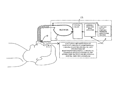
Fig. 1 is a schematic diagram of one embodiment of the present invention.
Fig. 2 is a schematic diagram of the arousal monitoring functions of the
present invention..
Fig. 3 is a flowchart of the airflow diagnostic process for the present
invention.
-5-
CA 02711783 2010-08-10
WO 2004/032719
PCT/US2003/032170
Fig. 4 is an example of a waveform for inspiration cycle with snoring.
Fig. 5 is an example of a waveform for an inspiration with a UAR.
Fig. 6 is a flow diagram of one embodiment of the present invention.
Fig. 7 is a schematic diagram of one embodiment of the present invention.
Detailed Description of the Preferred Embodiments
A. General Overview
The present invention is an apparatus and method for maintaining the sleep
quality of
a patient undergoing a therapeutic treatment. The present invention monitors
and interprets
physiological signals and spontaneous breathing events to detect the onset of
arousal. Once
the onset of arousal is determined, the present invention determines
adjustments that are
needed in the operation of a therapeutic device to avoid or minimize arousals.
As shown in Fig. 1, in one embodiment, the present invention includes one or
more
sensors 10 which detect a patient's physiological parameters, a controller 12
which monitors
and determines arousal based on the physiological variables received from the
sensor, and a
gas delivery apparatus 14 which is controlled by the controller 12. The sensor
10 can be a
combination of one or more devices which are able to monitor a physiological
parameter that
is used by the present invention to determine the onset of arousal or the
onset of a sleep
disorder. The sensors can be integrated into one unit or may operate
independent of the
others.
In one embodiment, the present invention is adapted to determine arousal using
physiological parameters such as pulse transit time (PTT) pulse arterial
tonometry (PAT),
plethysmographic wave amplitude, electroencephalogram (EEG), electro-myogram
(EMG)
and electro-oculogram (EOG), to name a few.
In one embodiment, the present invention is also adapted to monitor, analyze,
and
compute the sequence of airflow shape and sound. The breathing waveform
profiles or
sequence of waveform profiles or sounds of a patient are matched to various
templates which
are correlated to specific arousal events or Sleep Breathing Disorders.
-6-
CA 02711783 2010-08-10
WO 2004/032719
PCT/US2003/032170
In one embodiment the presence of SBD, UAR, shallow breathing or the onset of
the
same, can be analyzed and computed.
In one embodiment, the present invention receives a plurality of inputs from
sensors
and matches the inputs to values listed in a plurality of tables. The tables
identify various
breathing waveform profiles and physiological parameters or sequence of
waveforms and
physiological data and matches this information to a particular arousal event
or sleep
breathing disorder. Furthermore, a number of co-efficients and equations can
be applied to
the values stored in the table in order to accommodate variations which are
patient specific.
In one embodiment, the present invention has a capability of operating in
three
different modes. One mode is a default mode wherein empirical data establishes
thresholds
and reference data used to compute optimal therapeutic control. The present
invention also
includes a calibration mode wherein the present invention tests the response
of the patient to
various settings in order to determine patient tolerances. The present
invention also includes
an adaptation mode wherein the present invention utilizes optimal therapeutic
control in order
to minimize or eliminate arousal events or SBD.
B. System Configuration
In one embodiment, the present invention includes three main components, a
sensor
for monitoring a physiological parameter, a therapeutic device for
administering a therapeutic
treatment, and a controller for controlling the delivery of the therapeutic
treatment. The
present invention is described as having three main components for the
purposes of
explanation only. One skilled in the art can readily appreciate that the three
main
components of the present invention can be readily integrated into one or more
devices.
In one embodiment, the present invention includes a number of sensors, some of
which are used to detect upper airway resistance and airflow and some of which
detect
physiological parameters which are used to determine arousal. The sensor can
be any
apparatus known in the art which is capable of detecting, measuring, or
calculating a
physiological parameter which is used to determine arousal. The sensor can be
comprised of
a single integrated machine or a plurality of independent ones. The sensors
can communicate
with the controller by any known protocol.
-7-
CA 02711783 2010-08-10
WO 2004/032719
PCT/US2003/032170
In one embodiment, pressure transducers and a pneumotachograph are used in
cooperation or integration with an airtube or a patient mask to detect patient
airflow and
airway pressure. To detect physiological parameters, the present invention
uses sensors such
as, but is not limited to, EEG, EOG, EMG, ECG, pulse oximetry, blood pressure,
carbon
dioxide monitoring, bed transducers for monitoring patient position, video
processing
systems and microphones for breathing and breathing sounds.
Preferably, the sensors are all incorporated onto a single patient mask. A
suitable
mask is disclosed in International Publication Number WO 01/43804 entitled
"Bio-mask with
Integral Sensors," the contents of which are hereby incorporated by reference
in its entirety.
The mask has sensors integrated therein which are capable of detecting EMG,
EEG, EOG,
ECG, surface blood pressure, temperature, pulse oximetry, patient sounds, and
gas pressure
in the mask. The mask can include side-stream or full-stream gas sampling
capability for
monitoring in real-time, concentration of oxygen, CO2, nitric oxide and other
gases or any
combination of the aforesaid gases. In addition, the mask serves as the
conduit for gas
delivery to the patient.
In one embodiment, a mattress device is used to detect arousal. Currently,
there are
two commercially available mattresses which can perform the above functions.
One is
known as a Static Charge-sensitive Bed (SCSB) and the other is a polyvinlidene
fluoride
(PVDF-piezoelectric plastic) bed.
In one embodiment, eye activity is used to monitor arousal. An infrared video
monitoring system is employed as a sensor to determine eye activity via eyelid
position. The
image signal from the video monitoring is processed by graphic processing
program to
determine the status of the eyes.
In one embodiment, the present invention utilizes a unique multi-standard
wireless
interface system. Typically, two separate wireless bands are deployed to
separate
physiological wireless signals from control data. Furthermore, integrated
encryption and
security may be deployed to avoid unauthorized access to data.
An example of the typical embodiment could be where the 2.4 GHz ISM band is
applied for the interface of wireless based sensors interfaced to a
controller. Less critical
data, not effecting the patient therapy, such as user data viewing and
reports, could be
-8-
CA 02711783 2010-08-10
WO 2004/032719
PCT/US2003/032170
interfaced using W-LAN and even BluetoothTm wireless devices. The multi-radio
standard is
a particularly important consideration where operating with existent wireless
systems. A
further capability of the present invention is to detect interference from
similar radio band
system and switch the critical signal and other monitoring either to an
alternate band or
modify the system analysis adaptation without wireless signals. The present
invention can be
used with a range of wireless electrode devices to enable easy expansion and
access to
additional physiological signals.
In one embodiment, the sensors are battery powered for 1 or 2 days while
transmitting
signals to the controller. The wireless monitoring capability enables the
present invention to
monitor RERA, TERA, and SBD-related (sleep breathing disorder) signals during
a subject's
sleep. Furthermore, the ability to monitor these electrodes during a subject's
sleep might give
some augmented information (in addition to the respiratory airflow, pressure
and sound
signals normally derived from the subject's mask) and the ability of the SPAP
system to
provide optimal therapeutic pressure or gas delivery control to minimize RERA
or TERA,
while also minimizing obstructive sleep apnea-hypopnea (OSAH) and UARs.
This comparison of sleep efficiency during routine CPAP or enhanced
(additional
wireless signals applied, for example) CPAP operation can provide valuable
information to
the healthcare worker and patient in terms of sleep efficiency options for the
patient. In a
similar manner the patient may choose to utilize a wireless position sensor
which could be
attached to the therapeutic breathing mask or other parts of the patient
therapeutic equipment
or clothing.
The said wireless electrode contains several key functions enabling this
wireless
technology to be used with relative trouble-free ease within the patient's
home or the clinical
environment alike. The present invention electrodes can be packaged such that
the removal
of the disposable electrode outer package activates the battery. This
automatic wireless
electrode activation function enables automatic preservation of the battery
life, particularly
during storage. Use-by dating of the disposable electrode packaging ensures
that both the
electrode quality and battery life is used within a suitable period of time,
protecting the user
from battery age deterioration and electrode deterioration. The wireless
interfaced electrodes
of the present invention can be provided with self-gelled properties to
simplify electrode
-9-
CA 02711783 2010-08-10
WO 2004/032719
PCT/US2003/032170
attachment. The disposable self-adhesive (or reusable) electrode systems can
be attached by
the patient using simple visual guides.
The sensors input their physiological data to the controller (incorporates pre-
processing required for treatment control), which receives the data and
determines arousal or
onset of arousal. In one embodiment, the controller includes an analog
processing circuit
which converts analog signals from the sensors into a digital signal. The
analog processing
circuit utilizes known preamplifying, amplifying, conditioning, and filtering
configurations to
enable the analog sensor signal to be converted into a digital signal. In some
instances, the
sensor may directly input a digital signal.
In one embodiment, the controller also includes a processor which receives the
digital
signal and determines the patient state and an appropriate setting for the gas
delivery or other
therapeutic device. The processor employs a plurality of tables stored in a
database. The
tables include a plurality of entries which correlate the inputted signal from
a sensor with
arousal.
Typically, a number of different physiological parameters are inputted
simultaneously and all of the parameters are factored in determining arousal.
The processor
can employ a weighting system for each parameter and the appropriate action is
determined
by the derived index value. In another embodiment, the processor can link a
chain of
physiological values together and compare it to a table which correlates the
linked set of
values to arousal.
In one embodiment, the present invention includes memory devices containing
tables
holding stored profiles containing normal or acceptable limits for
physiological parameters
such as:
sleep fragmentation, apnea-hypopnea index (AHI), RERA, sleep architecture,
cortical arousals, sub-cortical arousals, PTT values, PAT values, HRV values,
central sleep apnea (CSA) occurrence, apnea occurrence, mixed apnea
occurrence, hypopnea occurrence, EEG spike occurrence, EEG spindle
occurrence, EEG K-complex occurrence, EEG seizure occurrence, bi-
coherence or bispectral index values, auditory evoked potential index, patient
posture optimal pressure values, patient sleep propensity, patient sleep
state.
-10-
CA 02711783 2010-08-10
WO 2004/032719
PCT/US2003/032170
The controller communicates with the therapeutic device to control the
treatment level
to the patient. The controller determines an appropriate instruction set by
using treatment
levels found in the table entry corresponding to the patient's physiological
condition. The
processor then communicates the instruction set to the gas delivery apparatus
which then
executes the instruction set.
B. Arousal Monitoring
As shown in Fig. 2, the present invention uses various physiological inputs to
determine arousal in a patient and to tailor the delivery of air to the
patient to minimize such
arousal. Due to the complex and varying states of sleep and broad range of
sleep disorders
that can be diagnosed, many different physiological parameters may be
monitored and
analyzed to determine arousal.
The minimization of arousals includes the capability to automatically adjust
the
therapeutic treatment while monitoring at least one physiological parameter or
signal where
the monitored physiological parameter(s), signal(s) or measures can include
(but are not
limited to):
Blood pressure, patient movement, patient vibration, patient tremor, patient
shake, Pulse oximetry, pulse-wave, EEG, EOG, EMG, patient position, patient
movement, breathing sounds, airflow signal, respiratory effort signal(s),
pharyngeal pressure signals, expired PCO2 signal, diaphragmatic EMG,
transthoracic impedance, electrocardiogram (ECG), reflective oximetry, pulse
oximetry, oxygen saturation, nasal pressure, airflow pressure, breathing mask
airflow, breathing mask pressure, breathing mask sound, breathing sound,
breathing pressure, respiratory inductive plethysmography, plethysmography-
wave, oesophageal pressure, nasal cannular sensor signals, nasal and oral
cannular sensor signals, oral cannular sensor signals, thermocouple sensor
signals, thermistor sensor signals, PVD temperature sensor signals, PVD
sound and vibration sensor signals, PVD breathing or airflow signals,
Pneumotach calibrated flow, or other routine or research application of
polysomnograph (PSG) monitoring sensors electrodes or signals.
In one embodiment, arousals are monitored using an EEG. Typically, the
forebrain is
-11-
CA 02711783 2010-08-10
WO 2004/032719
PCT/US2003/032170
monitored to determine cortical arousals and the brainstem is monitored to
measure
subcortical arousals. The onset of arousal is characterized by bursts of
higher frequency EEG
signals or a shift to alpha or theta activity from a slower background
frequency, and,
occasionally, transient increase in skeletal muscle tone. Standard EEG
electrode placements
and protocols may be used to measure arousals.
In one embodiment, the present invention includes the capability to
distinguish a
periodic leg movement (PLM) related arousal from a respiratory related
arousal.
Distinguishing PLM arousals from arousal associated with respiratory events
can be
important, particularly where optimum treatment control may not respond to a
PLM related
arousals but may need to respond to a respiratory event related arousal.
The present invention detects and distinguishes PLM and/or PLM arousals by
means
of comparing sub-cortical arousals inferred from blood pressure variations
with cortical
arousals (EEG). Cortical arousals are used to distinguish sleep-fragmentation
and
neurological related arousals versus sub-cortical arousals which generally
include both sleep-
fragmentation and neurological related arousals and PLM related arousals.
In one embodiment, the onset of arousal is determined using Pulse Transit Time
(PTT). Studies have shown that sleep disorders such as apnea, hypopnea or
upper airway
resistance result in an accompanying arousal, and this arousal is accompanied
by changes in
heart rate, a transient burst of sympathetic activity, and a surge in blood
pressure.
Obstructive sleep apnea can be correlated with an obvious and measurable
increase in
intrathoracic pressure associated with obstructive effort and
cardiobalistogram effect. The
cardiobalistogram effect is created when the lungs apply pressure to the
heart. This
compresses the heart and reduces the volume of blood pumped by the heart.
These
cardiovascular changes are recognizable by way of a transient but significant
dip in the
patient's baseline PTT value.
PTT is the time taken for the pulse wave to travel between two arterial sites.
The
blood pressure is directly proportional to the speed that the arterial
pressure wave travels. A
rise in blood pressure relates to faster pulse wave and thus shorter PTT.
Conversely, a drop
in blood pressure results in a slowing of the pulse wave and an increase in
PTT.
In one embodiment, PTT is obtained using sensors located on the above-
mentioned
bio-mask. A sensor receives input from the mask and generates a
plethysmography
waveform. A second sensor receives input from the mask and generates an ECG
signal. The
waveform and the signal are inputted into the controller and a PTT reading is
calculated.
-12-
CA 02711783 2010-08-10
WO 2004/032719
PCT/US2003/032170
=
The PTT is derived by utilizing a plethysmography waveform obtained by using
pulse
oximetry techniques in combination with an ECG signal. In one embodiment, the
ECG R or
Q wave can be used as the start point for the PTT measurement and the end
point of the PTT
measurement can be the point representing 25% or 50% of the height of the
maximum pulse
wave value.
In one embodiment, EMG measurements are used to detect levels of sleep in a
patient.
EMG monitoring enables the present invention to detect sleep¨related changes
in a patient's
muscle tonicity. Sleep states will typically be accompanied by changes of
tonicity in certain
muscles. Arousals will typically result in increased muscle tonicity.
In one embodiment, ECG and an EMG signal from the diaphragm are monitored in
combination to detect respiratory effort associated with central apnea versus
obstructive
apnea. The ECG electrodes are configured on the patient in order to
distinguish diaphragm
related respiratory effort from thoracic respiratory effort. During central
apnea, there will be
a cessation of breathing without respiratory effort. This is distinctly
different from
obstructive apneas wherein muscle activity increases as a result of increased
breathing effort
to overcome the obstructed airway.
In one embodiment, a patient's eye movements are monitored to assist in
determining
arousal. One technique involves the use of digital video recording and known
graphic
processing techniques to determine eye lid activity (i.e. whether the eye lids
are closed, open,
or degree of openness).
In one embodiment, arousals are detected by monitoring the presence of
waveform
signal disturbance evident on a high bandwidth analysis (DC to 200 Hz or
higher bandwidth)
of the airflow waveform and pressure waveform obtained within a breathing
mask. Apnea
events, shallow breathing, upper airway resistance and hypopnea events can
also be detected
and pre-empted by analysis of the change in shape of the high bandwidth
monitoring of the
airflow waveforms and pressure waveforms.
In addition to monitoring arousal, in one embodiment, other physiological
parameters
may be monitored to determine the patient's physical state. The present
invention can utilize
sensors in the biomask to determine heart rate, ECG, respiration rate, snoring
sounds, airflow,
air pressure, and 02 saturation. Conventional methods may also be incorporated
into the
present invention to monitor blood pressure, and CO2, The patient's sleeping
position may
also be monitored using pressure transducers or a mattress device.
-13-.
CA 02711783 2010-08-10
WO 2004/032719
PCT/US2003/032170
In one embodiment, wherein a patient is undergoing CPAP treatment, arousal
monitoring also includes monitoring pressure and airflow associated with a
patient's
breathing in order to determine UAR (which may induce RERA). To prevent RERA,
it is
necessary to detect a number of patterns which are indicative of sleep apnea
symptoms,
namely inspiratory flow limitation (flattening), snoring and flow amplitude
reduction
(hypopneas and apneas). As detailed in Fig. 3, the present invention analyzes
the airflow to
and from the patient in order to determine the existence of UAR.
A "Breath detection" component performs real time detection and
characterization of
individual breaths. Detection of inspiration and expiration peaks includes
"local" smoothing
(to separate real breaths from noise) and "global" detection of respiration
peaks based on
relatively long context which may include up to six consecutive breaths.
Breath analysis
includes accurate detection of inspiration interval and characterization of
flow during
inspiration, namely indices of flattening, snoring and inspiratory amplitude
as well as a few
others.
A "Time interval based processing" component performs analysis of pressure and
flow derived signals based on expiration of time intervals rather than
breaths. It is necessary
in cases when breaths are not discernible such as apneas or when the mask
comes off the
face.
The controller generates pressure adjustment signal on the basis of per breath
and per
time interval information provided by the two above components. The controller
is
implemented as a collection of rules which cover various combinations of
indicators of flow
limitation, snoring, breath amplitude, pressure leak and other parameters.
In one embodiment, the main strategy in breath detection is to use maximum and
minimum points (flow signal level) as the indicators of inspiration and
expiration intervals.
Inspiration is associated with positive flow signal deflection and expiration
is associated with
the negative deflection. However, the flow signal could be contaminated by
large amount of
noise, and it is necessary to smooth flow data before detecting actual breath
patterns (box
10050). For accurate detection of the inspiratory and expiratory peaks it is
also necessary to
use a relatively long context to prevent confusing them with local maxima and
minima in the
flow signal.
There are two main tasks in the local smoothing. First, all local maximum and
minimum points from the flow signal are detected, and each maximum point is
defined as an
initial candidate for the location of an inspiration peak, and each minimum
point for an initial
-14-
CA 02711783 2010-08-10
WO 2004/032719
PCT/US2003/032170
candidate for possible location of expiration peak. The second task is to
smooth some
maximum and minimum points with "relatively small amplitude", which are likely
to be
noise signal. As a result of local smoothing, only maximum and minimum points
with
"relatively large amplitude" are retained, and the flow signal is considered
to be sufficiently
smoothed. This sequence of local smoothing be described as follows:
1. Detect all local maximum points from a set of flow data.
2. For each maximum point, form a pattern called max-peak, in which the
maximum
point is located in the center, and data in its left side increase
monotonously, and decrease
monotonously in its right side. For the current flow data set, obtain a set of
max-peak
patterns.
3. For the same data set, detect all local minimum points and obtained a
serial of min-
peak patterns using the similar method.
4. Calculate a number of parameters such as signal variation and duration for
each
max-peak and mm-peak, and these parameters are used as the measurements to
test whether
some of detected max-peak and min-peak patterns are in fact noise.
5. Analysis sequences of adjacent max-peaks and min-peaks (in every sequence
the
number of max-peaks should exceed the number of min-peaks by one or
alternatively the
number of mm-peaks should exceed the number of max-peaks by one) and check if
a
sequence could be approximated by a single max-peak or mm-peak so that an
approximation
error is significantly less than variation and duration parameters of a
resulting max-peak or
min-peak
6. For the noise signal smoothing, use piecewise linear methods to approximate
the
flow signal.
7. The max-peaks with relative large amplitudes are retained, and for each
"retained"
max-peak, both 'increasing period' (left side) and 'decreasing period' (right
side) are not
shorter than a pre-defined threshold (0.75 s).
8. Same method is applied to mm-peak smoothing processing.
The local smoothing is basically designed for excluding noise signals that
have a
relatively small amplitude and short duration. As a result, a large amount of
maximum and
minimum points can be excluded from a list of "candidates" for inspiration and
expiration
peaks. The local smoothing processing can form separate "increasing periods"
or
"decreasing periods", and the signal within an "increasing period" or a
"decreasing period"
corresponds to a "likelihood" of the half duration of inspiration or
expiration. This "half-
-15-
CA 02711783 2010-08-10
WO 2004/032719
PCT/US2003/032170
duration" smoothing processing is one approach for deleting small noise
signal. On the other
hand, the "half-duration" approach lacks capability of smoothing flow data
containing some
relatively large noise and artifacts.
Another difficult problem in breath detection is related to the change of
respiration
patterns. The flow signal is often affected by patients that change their
"way" of breathing, in
other words, some periods of the increasing or decreasing signal level are
related to change of
patient's respiratory "behavior" rather than to inspiration or expiration.
Local smoothing is
unable to exclude these types of max-peaks or min-peaks. However, it is
possible to use
some global measurements to effectively detect these "unlikely" max-peaks or
min-peaks,
and this is the idea behind the global detection of respiratory peaks (global
smoothing) (box
10080). In the global smoothing, multiple consecutive breaths are checked to
further
disqualify some max-peaks or min-peaks from the list of candidates for
inspiration or
expiration peaks.
For a relatively long time interval (up to 3.5 minutes), conditions are tested
for pairs
of successive max-peaks (min-peaks as well), and a set of so-called max-pairs
(or min-pair) is
formed. Then a number of conditions for series of max-pairs to obtain a set of
max-pairs
with similar 'patterns', denoted as max-train are developed. The same
processing is carried
out to generate min-trains out of min-pairs. Therefore there are two main
parts in the global
smoothing, namely generation of max-pairs (min-pairs) (box 10100) and max-
trains (min-
trains) (box 10110). The following paragraphs outline max-pair and max-train
processing
briefly (min-pair and min-train generation employ the same respective
methods).
From the starting point of the max-peak set, a pair of max-peak patterns is
determined, which must meet the following conditions:
(1). The duration between two max-peaks must be longer than the minimum
duration of a breath (0.75 s).
(2). The duration between two max-peaks must be shorter than the maximum
duration of a breath (10 s).
(3). There is not any intermediate max-peak within a max-pair. An intermediate
max-peak pattern is defined as that the signal level of the maximum point in a
intermediate max-peak pattern is larger than 80% of signal level of the
maximum point in the max-pair itself.
This search processing is carried out through the whole max-peak pattern set
to obtain a
sequence of max-pairs.
-16-
CA 02711783 2010-08-10
WO 2004/032719 PCT/US2003/032170
(4). For each max-pair, a number of statistical measurements are calculated,
and
these measurements are based on the difference between the original flow
signal and the approximation lines within each max-pair.
There are two main outputs in this processing, one is a set of max-pairs which
is one
step closer to the final set of inspiratory peaks, and another is a number of
statistical
measurements which is used to represent the "shape" of the max-pair. In the
subsequent
max-train processing, we will rely on these statistical measurements to carry
out 'similarity'
test.
The main method used in the max-train processing is called "similarity" test,
i.e., we
measure the "similarity" within a sequence of max-pairs to form a sequence of
max-pairs
with "similar" pattern, denoted as max-train. Only the max-pair that passes a
"similarity test"
can be included into the max-train. The idea behind the max-train is that
i. A sequence of normal breaths over a successive period of time (3- 6
breath durations) should have similar shapes, and this pattern should
not be changed significantly over a short period of time as well.
ii. If a max-pair is not similar to this normal breath pattern, it could be
rather like a respiratory event (apnea or hypopnea), or some
noise/artifact pattern in flow signal.
The brief algorithm of max-train processing is then as follows:
1. Each candidate max-pair must first meet minimum duration requirement that
is
defined as the distance between candidate and the reference max-pair.
2. Starting from each single candidate, we calculate a number of parameters
such as
duration, variation of signal level, the shape of max-peak (or min-peak). We
then calculate
some statistical measurements for this group of candidates such as mean,
deviation, average
and maximum error for all the elements to check the similarity among of these
candidates.
3. If the condition of the similarity is met, the group of max-pair is formed
as a
max-train (box 10120). Otherwise, the processing is moved into next max-pair
until all max-
pair are checked.
4. The same method is applied to the mm-train processing.
5. Using max-train and min-train sets, we are now able to detect the locations
of
global maximum points of flow signal level that are related to the inspiration
periods, and a
number of minimum point that is associated with the expiration periods.
-17-
CA 02711783 2010-08-10
WO 2004/032719
PCT/US2003/032170
The global max-peak and min-peak arrays provide estimated locations of each
breath,
i.e., inspiration and expiration peaks. In order to detect respiratory events,
one needs to
closely look at these breaths, which includes:
1. Detect the start and end points of inspiration interval (box 10130).
2. Perform flow flattening (box 10150) and snoring analysis (box 10160) as
well as
calculation of other breath parameters.
During smoothing processing a linear approximation method is used to smooth
flow
data except of maximum and minimum points in max-train and min-train data
sets. However,
for purpose of breath analysis 'recover' raw flow data using maximum and
minimum points
as references is needed prior to carrying out breath analysis processing.
There are two steps involved in detecting inspiration, namely estimation and
fine-tune
processing. For the inspiratory interval estimation, the assumption is that
the amount of in-
taking flow during inspiration period should be same as that of 'expiring-out'
flow during
expiration period. Using the maximum and minimum points in flow signal as
references we
estimate the interval of inspiration, i.e., the start and end points of
inspiration based on
calculating the areas of flow data.
However, this method has inherently two problems that could effect the
accuracy in
inspiration detection. Firstly, when the flow is measured at the mask the
amounts of flow
during inspiration period and the followed expiration period may not be the
same, especially
when patients use their mouth to breathe, and we call this problem as "flow
imbalance".
Secondly, there may be "area insensitivity" problem. When patients start
inspiration the flow
signal level rapidly increases, but the measurement of flow area is an
integration processing
that is much slower than the change of flow signals itself In other words, the
change of flow
area is not sensitive enough to accurately measure the start point of
inspiration where flow
signal is changed rapidly.
The flow area is first calculated to estimate the inspiration interval, which
includes the
start point and the end point of an inspiration. The start point of an
expiration period is
simply defined as the end point of the previous inspiration period, and the
end point of the
expiration period is the start point of the following inspiration period or
can be ignored as this
point does not play any role in our control algorithms. Starting from the
estimated start point
inspiration period, linear approximation methods to detect the "break point"
during flow
signal increasing period, and this break point is then defined as the start
point of the
inspiration interval. The end point of the same inspiration period is simply
defined as a point
-18-
CA 02711783 2010-08-10
WO 2004/032719
PCT/US2003/032170
at which the signal level is the same as that of the start point of the
inspiration but it has
passed the maximum point.
As mentioned previously, the present invention needs to detect three types of
respiratory events, namely apneas and hypopneas, snoring, and inspiration flow
limitation.
The first type of events (apneas and hypopneas) is associated with reduction
of inspiration
flow and this can be resulted directly from the breath detection. Both snoring
and inspiration
flow limitation are more likely to occur during "abnormal" breath period. For
a "normal"
breath, the "shape" of signal on the top of inspiration flow appears "rounded"
and relatively
smooth. When snoring is present the high frequency flow signal is visible
during inspiration
as shown in Fig. 4. Inspiration flow limitation is defined as the event that
the patient is
unable to generate continuous flow increase during the first half of an
inspiration period. As
a result, the flow signal on the peak of inspiration flow becomes 'flat' as
shown in Fig. 5. In
flattening analysis, we determine a reference "flat" line which can be best
fitted for the flow
signal on the top of inspiration according the least square error (LSE), and
the difference of
the flow signal and the reference "flat" line during this period is then
calculated as a
flattening error. There are a number of flattening errors for different
selections of the
reference line. The flattening error with the smallest value is defined as a
flattening index.
The flattening index is then used to measure flow limitation, and the smaller
the flattening
index, the more severe is the inspiration flow limitation. A snoring index is
also utilized to
indicate the degree of the snoring. The snoring index is defined as
measurement of the
amount of high frequency signal on the top of inspiration flow.
C. Operation
An operational flow chart of one embodiment of the present invention is shown
in
Fig. 6. For the purposes of explanation only, the present invention will be
described in an
embodiment which is adapted for use with a CPAP machine. One skilled in the
art can
readily appreciate that the subject invention is easily adapted for use with,
or incorporated
within, other known therapeutic devices.
The present invention checks to make sure that it is receiving valid signals
from its
sensors. (box 2) Once the signal is verified, the signals are analyzed in
order to determine if
the onset of arousal has been detected. (boxes 5, 6, 7, and 8). The data used
in the analysis
is determined by the user.
-19-
CA 02711783 2010-08-10
WO 2004/032719
PCT/US2003/032170
In one embodiment, the present invention has the capability to use different
forced
oscillation treatment (FOT) to determine patient-specific threshold values for
arousals and
SBD. Results from the FOT are used to create templates which are used to
determine the
appropriate therapeutic response to avoid the onset of or eliminate the
incidence of CSA,
OSA, OSAHS, RERA, and TERA. (box 3) These templates or profiles are determined
from
patient-specific diagnostic studies or the appropriate FOT treatment at each
particular stage
in a subject's sleep or breathing status.
The present invention can obtain patient-specific FOT templates and profiles
by
utilizing forced oscillation of pressure, or changes in airflow pressure, to
determine whether
the changes in the airflow shape resulting from these subtle treatment changes
are able to
counteract the shape or profile characteristics indicative of the incidence or
on-set of arousals
(TERA or RERA) or OSAH and UAR. The present invention can vary the pressure
change
value and rate of change to countermeasure such events.
The present system provides a means to down-load from sleep laboratory studies
or
other types of previous sleep, respiratory and/or cardiac related
investigations. The specific
data is associated with a subject's breathing and sleep arousal parameters and
is used to
customize a gas delivery device to be more sensitive and accurate for both
minimizing
incidence of UARS, OSAHS, RERAs and TERAs, while still minimizing sleep
fragmentation
and optimizing sleep quality. (box 23) Each patient has a unique respiratory
breathing circuit
and associated pathways. Subsequently breathing waveforms during all stages of
sleep of a
patient will vary from patient to patient. The present invention ability to
accommodate the
patient's personal empirical data provides a means to produce more sensitive
and effective
treatment algorithm.
In one embodiment, if the onset of arousal has been determined (block 11), the
present invention then determines if the CPAP has caused a pressure change
(box 13) or if the
event is caused by the existence of UAR (box 14). If there was no pressure
change attributed
to the CPAP machine, the present invention will likely determine that the
onset of arousal
was caused by RERA or another form of arousal. If there was a CPAP related
pressure
change, the present invention would make a determination if the onset of
arousal was
attributed to the pressure change or some other event (box 15). An appropriate
remedy will
then be selected based on the based on the physiological signals and the
patient respiratory
flow. (box 18)
-20-
CA 02711783 2010-08-10
WO 2004/032719
PCT/US2003/032170
In one embodiment, the present invention is able to utilize the determination
to adapt
the the empirical data. (box 20) This enables the present invention to become
more acutely
sensitive to the physiological response of the patient.
The minimization of arousals includes the capability to automatically adjust
the
therapeutic treatment based on at least one index or derived data set wherein
the index or
derived data set can include the following:
Upper Airway Resistance (UAR), Respiratory Effort-Related Arousal
(RERA), Therapeutic-control Event-Related Arousal (TERA), Respiratory
Disturbance Index (RDI) Respiratory Arousal Index (RAI), Apnea-hypopnea
index (AHI), Arousals (Micro-arousals), Arousals (Cortical), Arousals
(subcortical), Arousals (Total), Total Arousal Time, Sleep stage, REM sleep,
Sleep on-set, Body movement, Percentage of arousal disrupted sleep
(breakdown of all disrupted stages), Sleep efficiency Index, Sleep
Fragmentation Index (new- SFI- Total Sleep Fragmenting arousals per hour),
Airflow Shape trend, Airflow Shape Template Type, Flattening Index, Forced
oscillation event, Pressure change event, Pressure change rate, Pressure
change event curve, Pressure change event maximal and minima, Mixed Sleep
Apnea events, Central Sleep Apnea events, Upper Airway Resistance
Syndrome (UARS) events, Obstructive sleep apnea and hypopnea syndrome
(OSAHS) events, Respiratory Effort-Related Arousals (RERA) with screen
linked qualification of associated respiratory effort arousals, Therapeutic
control Related Arousals (TERA) with screen linked qualification of pressure
changes and arousals, Sleep Quality Index (new- hourly sleep index factor)
Quality associated arousals, Oxygen Desaturation, Pulse Transit Time (PTT),
Pulse Arterial Tone (PAT), Pulse Wave Amplitude (PWA), desaturation
events and Sp02 artifacts ¨ accurate detection of cascaded desaturations,
desaturations with Sp02 artifacts inside, Sp02 artifact start and end
positions,
detection sequences of respiratory events with partial or short recovery,
classification of respiratory events with noisy or poor quality effort
signals,
detection episodes of Cheyne-Stokes breathing, Concordinance capability to
allow score comparisons between any two designated data sets, Pnetunotach
-21-
CA 02711783 2010-08-10
WO 2004/032719
PCT/US2003/032170
calibrated flow, Thermal sensor flow, Sum of respiratory effort signals, EEG
Arousals, PTT, Plethysmographic wave, Transthoracic impedance, Detection
and allowance screen grid highlighting for an expanded set of automatic
events to include the following events, Obstructive Sleep Apnea/Hypopnea
event or syndrome (OSA, OSH, OSAHS), Respiratory effort related arousal,
Central Sleep Apnea (CSA), Central Sleep Hypopnea (CSH), Cheyne-Stokes
breathing, Hypoventilation, Yawn, Unstable breathing related to sleep state
changes or onset of deeper stages of sleep, Swallowing, Coughing,
Spontaneous or irregular but normal shaped breathing signals, Derived tidal
volume (from nasal pressure or calibrated flow), Derived flow limitation index
(from nasal pressure or calibrated flow), Derived snoring (from nasal pressure
or calibrated flow), Derived diaphragmatic EMG amplitude, Derived upper
airway resistance (from mask pressure, pharyngeal pressure and calibrated
flow), Derived subcortical arousals (from PTT or pleth wave amplitude),
Breathing mask and/or airflow sound analysis with segmentation into various
breathing disorders such as cough, wheeze, strider, apnea and hypopnea.
For every selected event (combination of a set of expanded group of events
from
above and current set of events), a user is allowed to select the set of
measurement signals
and to set the parameters of detection for the event. This enables the present
invention to use
more than one signal at the same time to detect an event and more than one
scenario to detect
an event. The following are examples of defined events:
RERA ¨
1. Break in the flat inspiratory profile after a few flow limited breaths
2. Frequency shift in EEG, amplitude increase in EMG
3. Subsequent leg movement activity
4. No pressure augmentation (CTRL signal)
Leg movement related arousal-
1. Increase in leg movement activity
2. Frequency shift in EEG, amplitude increase in EMG
3. Break in the inspiratory profile not necessarily during inspiration
4. No pressure augmentation (CTRL signal)
-22-
CA 02711783 2010-08-10
WO 2004/032719
PCT/US2003/032170
Spontaneous arousal -
I. Frequency shift in EEG, amplitude increase in EMG
2. No increase (or after EEG/EMG changes) in leg movement activity
3. Break in the inspiratory profile not necessarily during inspiration
4. No pressure augmentation (CTRL signal)
Pressure augmentation related arousal -
1. Pressure increase according to the titration algorithm
2. Subsequent frequency shift in EEG, amplitude increase in EMG
3. No increase (or after EEG/EMG changes) in leg movement activity
4. Break in the inspiratory profile not necessarily during inspiration
The present invention significantly reduces arousal by restricting the
application of
pressure treatment until a patient is in a stage of sleep where this pressure
is not experienced
or causes no adverse patient discomfort. Pressure of air delivered to a
patient is ramped up or
down depending upon the patient's sleep state. Pressure is ramped up slowly
while
physiological parameters are monitored. Once the physiological parameters
indicate the
onset of arousal (microarousal) the pressure is maintained or reduced until
the patient is in
deeper levels of sleep enabling continued ramping up of pressure. Pressure is
also ramped
downwards accordingly.
The controller 12 is implemented as a combination of rules for pressure
change.
Every pressure change rule specifies the magnitude and sign of a pressure
change and the
allowed range of pressure values within which the pressure change can be
activated as well a
number of additional parameters including time constants, timeouts and forced
oscillation
logic. Every pressure change rule is activated if a respective logical
combination of its
conditions is true. In one embodiment, pressure change rules are combined via
logical OR ¨
pressure changes if any single rule in the set of rules is satisfied. If more
than one rule is
satisfied the rule with a higher priority takes precedence.
Conditions for various pressure change rules represent a number of
physiological
scenarios:
Flow limitation (flattening) over a number of subsequent breaths ¨ pressure
increase
-23-
CA 02711783 2010-08-10
WO 2004/032719
PCT/US2003/032170
Flow limitation (flattening) and snoring over a number of subsequent breaths ¨
pressure increase
Snoring over one or two breaths ¨ pressure increase
Hypopneas ¨ pressure increase (it is recommended to use additional
information such as PTT, band or mattress signals to discriminate obstructive
vs central hypopneas)
Detection of apnea start ¨ start forced oscillation
Low level of upper airway conductance with forced oscillation ¨ pressure
increase (obstructive apnea detected)
No flow limitation (rounded breath shape) ¨ gradual pressure reduction
Large leak ¨ pressure reduction to 4 cmH20
No airflow over 3 minutes ¨ pressure reduction to 4 cmH20
The present invention is capable of overcoming varying arousal dependent
factors by
applying adaptive algorithm techniques. The adaptive algorithm technique has
the capability
to apply empirical clinical data to establish standard threshold
configurations, which in turn
determine a device's response and performance in terms of gas delivery
characteristics. The
adaptive algorithm technique also has the capability to apply a set of
threshold characteristics.
In one embodiment, these threshold characteristics can vary parameters such as
the
rate of pressure change, the absolute amount of pressure change, the minimum
delivered
pressure values, the maximum delivered pressure values. These rates and
absolute pressure
changes can vary in accordance to various states of said patient including
(for example only)
the patient's current sleep state or the patient's relative blood pressure or
arrhythmia
detection. The present invention can be configured in a predetermined mode of
operation
where the algorithm adaptation function can be disabled and replaced by an
algorithm that
relies on a fixed set of reference data designed to predict the onset or
detect the occurrence of
TERA and RERA, while minimizing sleep breathing disorders.
In one embodiment, the present invention enables medical specialists to set
various
thresholds, which may prevent undesirable medical conditions for each
particular patient.
For example, if central sleep apnea is detected in combination with an
increase or undesirable
change or measure in ECG, pulse-wave or arrhythmia, the operation of the gas
delivery
device is augmented to stabilize the patient's condition. In some instances
this stabilization
may include the immediate cessation of pressure delivery. During events such
as central
-24-
CA 02711783 2010-08-10
WO 2004/032719
PCT/1JS2003/032170
sleep apnea (cessation of breathing activated by the brain commands versus
airway
obstruction), for example, forced pressure delivery without airway obstruction
may otherwise
aggravate the subject's blood pressure or cardiac function.
The present invention is capable of operating with or without any previous
patient
data (a specific airflow shape characteristics or various thresholds, for
example). In the case
where a subject has no previous data or threshold indications the present
invention could
commence operation with standard empirical data threshold settings. During
device
generated pressure changes or whenever there is a respiratory disturbance the
present
invention can adapt its control characteristics to minimize the respiratory
and arousal
disturbance.
In one embodiment, monitoring of arousals enables the subject invention to
augment a
CPAP's sleep disorder detection capabilities. False negatives often occur
during mild
hypopnea events. There is typically such minimal airflow limitation that CPAP
machines are
unable to detect the breathing disorder. However, such mild events often
create sufficient
UAR to cause arousal in a patient. The detection of the onset of such arousals
enables the
present invention to initiate a corrective response from the CPAP unit, even
though the CPAP
is unable to detect the event.
In one embodiment, a treatment mode includes utilizing breathing pattern
templates
stored in the table to augment current CPAP settings. These dynamically
allocated breathing
pattern templates supplement the CPAP algorithm by changing the control
characteristics of
the CPAP unit. The templates satisfy the particular patient's pressure
requirements while
optimizing the patient's sleep and minimizing patient arousal.
In one embodiment, the present invention enables the commencement of treatment
to
be determined by a pre-defined state of sleep, arousal activity level, and/or
pre-determined
sleep disordered breathing activity. One of the difficulties experienced by
patients with
existent state of the art gas delivery treatment devices is the discomfort
experienced from the
positive air pressure applied to a patient, while they are attempting to fall
asleep.
Existent state of the art devices have the capability to provide a delayed
start function.
This delay function provides a time delay before the treatment pressure slowly
increases or
ramps up to a prescribed value of start pressure. However, patients are not
always able to
predict their sleep onset time as drowsiness of a patient varies from one
night to the next.
The concept of a prescribed delay time can also provide a psychological
anxiety, as the
patient is always aware that if they do not succumb to a satisfactory sleep
state in an
-25-
CA 02711783 2010-08-10
WO 2004/032719
PCT/US2003/032170
=
appropriate amount of time, they risk experiencing the unpleasant sensation of
excessive
positive air pressure during their sleep preparation time.
The present invention enables the detection of sleep state and/or arousals as
a mean to
determine pressure activation. Treatments are applied only when the patient is
in a
preselected or deep state of sleep and subsequently is oblivious to the said
commencement of
treatment. The determination of sleep state can be the methods which are known
in the art,
including those methods disclosed in the U.S. Patent No. 6,397,845, the
contents of which are
hereby incorporated in its entirety.
In one embodiment, a present invention has an integrated diagnostic and
treatment
mode where adjustments to the delivered air are changed in real time (i.e.
changes are
instantaneously made depending upon the values of the monitored parameters).
The present invention's control algorithm has the capability to be adapted
during real-
time operation based on any combination of a) empirical clinical data, b)
individual patient
collected or collected data (from diagnostic study within sleep laboratory or
other alternative
site or c) real-time monitored and analyzed data.
D. Alternative Embodiments
In one alternative embodiment, as shown in Fig. 7, the present invention is
used to
deliver medication to a patient. Previous methods for determining sedative or
tranquilizer
dosage requirements for a subject are often estimated on a generalized patient
group or a
specific sample patient group.
A subject's sleep or vigilance propensity is highly complex and dependent on
many
parameters. It has been shown, for example that a person's sleep propensity
can be related to
sleep deprivation, alcohol, anxiety, stress, environmental factors, body mass
index, gender,
hereditary and other factors.
The consequence of over-sedation include increased recovery time, attention
deficit
risks associated with excessive drowsiness, increased costs of drugs, and
reduction in the
quality of life due to the extended drowsy state of a subject.
The drug administration can deliver a range of drugs utilizing methods such as
(but
not limited to) orally, transdermal, fluid drip delivery, vapor delivery and
gas delivery.
Utilizing the integration of a drug delivery system with the present
invention, a drug dosage
can be optimized for a predetermined level or an appropriate level of
drowsiness, vigilance or
attention state.
-26-
CA 02711783 2010-08-10
WO 2004/032719
PCT/US2003/032170
The user or health-care provider could adjust drug administration dosage in
consultation with the patient and the monitored patient data.
A further capability of the present invention is to contain sensors such as
sensitive
movement devices that together with signal analysis (such as but not limited
to spectral,
phase and amplitude) can detect shaking, tremors and other signs indicating
appropriate drug
usage. In the case of Parkinson's and other disease types the present system
could be
programmed to administer, for example, adequate drugs to minimize tremors and
shaking,
while at the same time provide the subject with a degree of vigilance during
the day and sleep
quality during the night that is most conducive to each individual's quality
of life
requirements or desires.
The present invention can be adapted in a number of configurations with
different
combination of physiological recording channels, sensors, analysis, storage
and display
capabilities. These capabilities could vary subject to the specific disease or
disorder being
treated, along with each subject's specific health-specialist requirements for
information.
In another alternative embodiment, as shown in Fig. 7, the present invention
includes
a pacemaker control algorithm which minimizes RERA and TERA while optimizing a
subject's heart pacing. The present invention enables the detection of RERA
and TERA from
the electrocardiogram or pulse-wave signals, for example. Alternatively, more
comprehensive signals can be deployed. The present invention enables the
conventional
optimization of ECG pacing while at the same time minimizing arousals and
sleep
fragmentation. Pacemaker control can also be utilized to assist in the
elimination of some
sleep disordered breathing. The present invention can provide important
feedback as to the
causation of sleep fragmentation such as inappropriate pacemaker control
causing promotion
of sleep fragmenting arousals.
The present invention is also able to monitor heart rate, blood-pressure
variations and
sleep fragmentation arousals throughout sleep and determine whether these said
variations
relate to normal sleep physiology, or whether these changes suggest
modification of
pacemaker control in order to optimize heart function, while at the same time
minimizing
sleep fragmentation.
In another embodiment, the present invention has the capability to provide
optimal
sleep during oxygen concentration treatment by utilizing cortical,
subcortical, airflow shape
or waveform characteristics as a marker for optimizing the treatment. The
present invention
can control the titration algorithm to minimize RERA and TERA while optimizing
a subject's
-27-
CA 02711783 2013-09-13
breathing therapy. The present invention enables the detection of RERA and
TERA by
monitoring any combination of breathing mask or hose sounds, airflow or
pressure signals.
Alternatively more comprehensive signals can be deployed. SOC enables the
conventional
optimization of blood-gas status of a subject, while minimizing arousals. An
inappropriate
mixture of oxygen and air, or rate of delivery of gas to a subject could
promote arousals, for
example.
Inappropriate gas delivery could in turn cause mechanical or chemical
receptors
within the patient's breathing anatomy to activate sleep fragmentation
arousals (TERA). The
monitoring of the airflow wave shape can be used to predict the onset or
incidence of TERA
or RERA and allow the gas treatment to be controlled in such a manner to
minimize such
arousals (while still optimizing breathing therapy).
In one embodiment, the present invention is utilized as purely a diagnostic
tool for
determining sleep disordered breathing and sleep quality. The present
invention is adapted to
record, meter, index or display, in real time or on a replay or review basis,
a number of sleep
or arousal related physiological data or statistics. Statistics and indexes
such as RAT, AHI,
RERA, RDI, arousals, sleep fragmentation, or sleep architecture index are
derived from the
monitored parameters and this information is stored for analysis.
In one embodiment, monitored physiological parameters monitored which were
utilized to determine the statistics and indexes may also be stored in order
to assist in the
analysis. The present invention can also include graphical and statistical
tools which are
known in the art to enable a user to manipulate and display raw data or
derived values in a
meaningful format. In one embodiment, the present invention has the capability
of
displaying raw data and then using visual clues to mark the occurrence of an
event such as
arousal in the raw data. The present invention can also link an event or
events to specific
index values or derived values which reflect the occurrence of the event.
The matter set forth in the foregoing description and accompanying drawings is
offered by way of illustration only and not as a limitation. While a
particular embodiment has
been shown and described, it will be apparent to those skilled in the art that
changes and
modifications may be made without departing from the broader aspects of
applicants'
contribution.
-28-