Note: Descriptions are shown in the official language in which they were submitted.
CA 02712824 2010-07-21
WO 2009/098701 PCT/IL2009/000161
SIVA 2 STABILIZATION
FIELD OF THE INVENTION
The present invention relates to modulation of SIVA2 stability in treatment or
prevention of diseases, disorders or conditions.
BACKGROUND OF THE INVENTION
Members of the TNF/NGF receptor family are expressed in almost all types
of cells and control a wide range of diverse cellular activities. They have
the ability
both to induce cellular changes that are protein-synthesis independent, the
best
known of which is caspase-mediated cell death (the extrinsic cell-death
pathway),
and to modulate gene-expression patterns both on the transcriptional and the
post-
transcriptional levels. These effects contribute to the control of practically
all
aspects of immune defense as well as some embryonic-development and tissue-
homeostatic processes. They vary, and depending on the type of cell and the
identity
of the activated receptor, as well as on numerous other determinants, some
effects
might even oppose others. This wide range of activities is mediated by a
rather
small number of signaling proteins, of which the best characterized are two
death-
domain-containing adapters, FADD/MORT 1 and TRADD, the inducer caspases
caspase-8 and -10, members of the TRAF ring-finger proteins, and cellular
inhibitor
of apoptosis protein 1 (cIAP 1) and cIAP2 (ring-finger proteins with IAP
motifs)(Wallach et al., 1999)'(Locksley et al., 2001). How this limited set of
proteins mediates the multiplicity of different effects of the receptors, and
how the
nature of the induced effect is adjusted to need, are still poorly understood.
SIVA, an additional protein suggested to participate in the proximal
signaling activities of members of the TNF/NGF receptor family, was identified
by
virtue of its binding to the receptor CD27 in the yeast two-hybrid test
(Prasad et al.,
1997). Some evidence was also presented for its association with several other
members of the TNF/NGF receptor family (Nocentini and Riccardi, 2005). The
existence of SIVA has been known for some years, and it was shown that when
1
CA 02712824 2010-07-21
WO 2009/098701 PCT/IL2009/000161
overexpressed for prolonged periods this protein kills cells (Prasad et at.,
1997).
However, whether this is its genuine and sole activity is not known. SIVA
shows no
close structural resemblance to any other known protein. One region within it
that
initially appeared to resemble the death domain does not contain the
structural
signatures by which that domain is characterized. C-terminally to that region
the
protein is relatively enriched in cysteine residues, which apparently
contribute to its
binding of several zinc ions (Nestler et al., 2006). The amino-acid sequence
in this
region, however, does not strictly conform to any of the known zinc-binding
motifs.
A central short a-helical region in the protein binds the anti-apoptotic
protein BCL-
XL(Xue et al., 2002), but the function served by the cysteine-rich region
(CRR) is
unknown.
SIVA it is known to exist as two alternative splice isoforms or splice
variants, SIVA 1 and SIVA2. SIVA1 is longer and contains a death domain
homology region (DDHR) with a putative amphipathical helix in its central
part.
SIVA2 is shorter and lacks the DDHR. Both isoforms contain a B-box-like ring
finger and a Zinc finger like domain in their C-termini. Enforced expression
of both
SIVAI and SIVA2 has been shown to induce apoptosis (Prasad et at., 1997, Yoon
et al., 1998, Spinicelli et al., 2003, (Py et at., 2004). SIVA1 induced
apoptosis is
suggested to be effected by its binding to and inhibition of the anti
apoptotic Bcl-2
family members through its amphipathic helical region (Chu et at., 2005; Chu
et al.,
2004; Xue et al., 2002). Consistent with its pro-apoptotic role, SIVA is a
direct
transcriptional target for tumor suppressors p53 and E2F1 (Fortin et at.,
2004).
Various point of evidence indicate that SIVA is a stress-induced protein and
is up-
regulated in acute ischemic injury (Padanilam et at., 1998), coxavirus
infection
(Henke et at., 2000), and also by cisplatin treatment (Qin et at., 2002), as
well as
TIP30 expression which induces apoptosis (Xiao et al., 2000). Recently, the
common N- and C-termini of SIVAI and SIVA2, yet not the death domain, have
been shown to be sufficient and capable to mediate apoptosis in lymphoid cells
through activation of a caspase dependent mitochondrial pathway (Py et at.,
2004).
2
CA 02712824 2010-07-21
WO 2009/098701 PCT/IL2009/000161
Recently, it was found that SIVA binds to NF-kB-inducing kinase (NIK) and
controls its function (Ramakrishnan et at., 2004), has ubiquitination-related
activity,
is capable of directly inducing self-ubiquitination, ubiquitination of TRAF2
(a TNF-
receptor associated adaptor protein 2), and that SIVA2 is an E3 ligase
(W02007080593).
Ubiquitylation, also termed ubiquitination, refers to the process particular
to
eukaryotes whereby a protein is post-translationally modified by covalent
attachment of a small protein named ubiquitin [originally ubiquitous
immunopoeitic
polypeptide (UBIP)]. Ubiquitin ligase is a protein which covalently attaches
ubiquitin to a lysine residue on a target protein. The ubiquitin ligase is
typically
involved in polyubiquitylation: a second ubiquitin is attached to the first; a
third is
attached to the second, and so forth. The ubiquitin ligase is referred to as
an "E3"
and operates in conjunction with an ubiquitin-activating enzyme (referred
herein as
"El") and an ubiquitin-conjugating enzyme (referred herein as "E2"). There is
one
major El enzyme, shared by all ubiquitin ligases, which uses ATP to activate
ubiquitin for conjugation and transfers it to an E2 enzyme. The E2 enzyme
interacts
with a specific E3 partner and transfers the ubiquitin to the target protein.
The E3,
which may be a multi-protein complex, is generally responsible for targeting
ubiquitination to specific substrate proteins. In some cases it receives the
ubiquitin
from the E2 enzyme and transfers it to the target protein or substrate
protein; in
other cases it acts by interacting with both the E2 enzyme and the substrate.
NIK, (MAP3K14) was discovered (Malinin et al., 1997) in a screening for
proteins that bind to TRAF2. The marked activation of NF-KB upon
overexpression
of NIK, and effective inhibition of NF-xB activation in response to a variety
of
inducing agents, upon expression of catalytically inactive NIK mutants
suggested
that NIK participates in signaling for NF-xB activation (Malinin et al.,
1997).
Assessment of the pattern of the NF-xB species in lymphoid organs indicated
that, apart from its role in the regulation of NF-KB complex(s) comprised of
Rel
proteins and IKB, NIK also participates in controlling the
expression/activation of
other NF-KB species. Indeed, NIK has been shown to participate in site-
specific
3
CA 02712824 2010-07-21
WO 2009/098701 PCT/IL2009/000161
phosphorylation of p 100, which serves as a molecular trigger for
ubiquitination and
active processing of p 100 to form p52. This p 100 processing activity was
found to
be ablated by the aly mutation of NIK (Xiao et al., 2001b).
NIK in thymic stroma is important for the normal production of Treg cells,
which are essential for maintaining immunological tolerance. NIK mutation
resulted
in disorganized thymic structure and impaired production of Treg cells in aly
mice
(Kajiura et al., 2004). Consistently, studies of NIK-deficient mice also
suggested a
role for NIK in controlling the development and expansion of Treg cells (Lu et
al.,
2005). These findings suggest an essential role of NIK in establishing self-
tolerance
in a stromal dependent manner. NIK also partakes in NF-KB activation as a
consequence of viral infection. Respiratory syncytial virus infection results
in
increased kinase activity of NIK and the formation of a complex comprised of
activated NIK, IKK 1, p 100 and the processed p52 in alveolus like a549 cells.
In this
case NIK itself gets translocated into the nucleus bound to p52 and
surprisingly,
these events precede the activation of canonical NF-KB pathway activation
(Choudhary et al., 2005). These findings indicate that NIK indeed serves as a
mediator of NF-KB activation, but may also serve other functions, and that it
exerts
these functions in a cell- and receptor-specific manner.
NIK can be activated as a consequence of phosphorylation of the `activation
loop' within the NIK molecule. Indeed, mutation of a phosphorylation-site
within
this loop (Thr-559) prevents activation of NF-KB upon NIK overexpression (Lin
et
al., 1999). In addition, the activity of NIK seems to be regulated through the
ability
of the regions upstream and downstream of its kinase motif to bind to each
other.
The C terminal region of NIK downstream of its kinase moiety has been shown to
be capable of binding directly to IKK 1 (Regnier et al., 1997) as well as to p
100
(Xiao et al., 2001b) and these interactions are apparently required for NIK
function
in NF-KB signaling. The N terminal region of NIK contains a negative-
regulatory
domain (NRD), which is composed of a basic motif (BR) and a proline-rich
repeat
motif (PRR) (Xiao and Sun, 2000).The N-terminal NRD interacts with the C-
terminal region of NIK in cis, thereby inhibiting the binding of NIK to its
substrate
4
CA 02712824 2010-07-21
WO 2009/098701 PCT/IL2009/000161
(IKK 1 and p 100). Ectopically expressed NIK spontaneously forms oligomers in
which these bindings of the N-terminal to the C terminal regions in each NIK
molecule are apparently disrupted, and display a high level of constitutive
activity
(Lin et al., 1999). The binding of the NIK C-terminal region to TRAF2 (as well
as
to other TRAF's) most likely participates in the activation process. However,
its
exact mode of participation is unknown.
Recently, a novel mechanism of NIK regulation has gained much attention.
This concerns the dynamic interaction of NIK and TRAF3 leading to proteasome
mediated degradation of NIK. Interestingly, inducers of the alternative
pathway of
NF-KB like CD40 and BLyS have been shown to induce TRAF3 degradation and
concomitant enhancement of NIK expression (Liao et al., 2004).
There is rather limited information yet of the downstream mechanisms in
NIK action. Evidence has been presented that NIK, through the binding of its C-
terminal region to IKK 1 can activate the IxB kinase (IKK) complex. It has
indeed
been shown to be capable of phosphorylating serine-176 in the activation loop
of
IKKI and thereby its activation (Ling et al., 1998).
It was suggested that NIK does not participate at all in the canonical NF-KB
pathway, but rather serves exclusively to activate the alternative one (see
(Pomerantz and Baltimore, 2002, for review). However, it was lately shown that
although the induction of IkappaB degradation in lymphocytes by TNF is
independent of NIK, its induction by CD70, CD40 ligand, and BLyS/BAFF, which
all also induce NF-kappaB2/plOO processing, does depend on NIK function
(Ramakrishnan et al. 2004). Both CD70 and TNF induce recruitment of the IKK
kinase complex to their receptors. In the case of CD70, but not TNF, this
process is
associated with NIK recruitment and is followed by prolonged receptor
association
of just IKKI and NIK. Recruitment of the IKK complex to CD27, but not that of
NIK, depends on NIK kinase function. These findings indicate that NIK
participates
in a unique set of proximal signaling events initiated by specific inducers,
which
activate both canonical and noncanonical NF-kappaB dimers.
5
CA 02712824 2010-07-21
WO 2009/098701 PCT/IL2009/000161
Yamamoto and Gaynor reviewed the role of NF-KB in pathogenesis of
human disease (Yamamoto and Gaynor 2001). Activation of the NF-KB pathway is
involved in the pathogenesis of chronic inflammatory disease, such as asthma,
rheumatoid arthritis (see Tak and Firestein, this Perspective series, ref.
Karin et al.
2000), and inflammatory bowel disease. In addition, altered NF-KB regulation
may
be involved in other diseases such as atherosclerosis (see Collins and
Cybulsky, this
series, ref. Leonard et al. 1995) and Alzheimer's disease (see Mattson and
Camandola, this series, ref. Lin et al. 1999), in which the inflammatory
response is
at least partially involved. Also, abnormalities in the NF-KB pathway are also
frequently seen in a variety of human cancers.
Several lines of evidence suggest that NF-KB activation of cytokine genes is
an important contributor to the pathogenesis of asthma, which is characterized
by
the infiltration of inflammatory cells and the deregulation of many cytokines
and
chemokines in the lung (Ling et al. 1998). Likewise, activation of the NF-KB
pathway also likely plays a role in the pathogenesis of rheumatoid arthritis.
Cytokines, such as TNF- , that activate NF-KB are elevated in the synovial
fluid of
patients with rheumatoid arthritis and contribute to the chronic inflammatory
changes and synovial hyperplasia seen in the joints of these patients (Malinin
et al.
1997). The administration of antibodies directed against TNF- or a truncated
TNF-
receptor that binds to TNF- can markedly improve the symptoms of patients with
rheumatoid arthritis.
Increases in the production of proinflammatory cytokines by both
lymphocytes and macrophages have also been implicated in the pathogenesis of
inflammatory bowel diseases, including Crohn's disease and ulcerative colitis
(Matsumoto et al. 1999). NF-KB activation is seen in mucosal biopsy specimens
from patients with active Crohn's disease and ulcerative colitis. Treatment of
patients with inflammatory bowel diseases with steroids decreases NF-KB
activity
in biopsy specimens and reduces clinical symptoms. These results suggest that
stimulation of the NF-KB pathway may be involved in the enhanced inflammatory
response associated with these diseases.
6
CA 02712824 2010-07-21
WO 2009/098701 PCT/IL2009/000161
Atherosclerosis is triggered by numerous insults to the endothelium and
smooth muscle of the damaged vessel wall (Matsushima et at. 2001). A large
number of growth factors, cytokines, and chemokines released from endothelial
cells, smooth muscle, macrophages, and lymphocytes are involved in this
chronic
inflammatory and fibroproliferative process (Matsushima et at. 2001). NF-KB
regulation of genes involved in the inflammatory response and in the control
of
cellular proliferation likely plays an important role in the initiation and
progression
of atherosclerosis.
Also, abnormalities in the regulation of the NF-KB pathway may be involved
in the pathogenesis of Alzheimer's disease. For example, NF-KB
immunoreactivity
is found predominantly in and around early neuritic plaque types in
Alzheimer's
disease, whereas mature plaque types show vastly reduced NF-KB activity
(Mercurio et al. 1999). Thus, NF-KB activation may be involved in the
initiation of
neuritic plaques and neuronal apoptosis during the early phases of Alzheimer's
disease. These data suggest that activation of the NF-KB pathway may play a
role in
a number of diseases that have an inflammatory component involved in their
pathogenesis.
In addition to a role in the pathogenesis of diseases characterized by
increases in the host immune and inflammatory response, constitutive
activation of
the NF-KB pathway has also been implicated in the pathogenesis of some human
cancers. Abnormalities in the regulation of the NF-KB pathway are frequently
seen
in a variety of human malignancies including leukemias, lymphomas, and solid
tumors (Miyawaki et al. 1994). These abnormalities result in constitutively
high
levels of NF-KB in the nucleus of a variety of tumors including breast,
ovarian,
prostate, and colon cancers. The majority of these changes are likely due to
alterations in regulatory proteins that activate signaling pathways that lead
to
activation of the NF-KB pathway. However, mutations that inactivate the I B
proteins in addition to amplification and rearrangements of genes encoding NF-
KB
7
CA 02712824 2010-07-21
WO 2009/098701 PCT/IL2009/000161
family members can result in the enhanced nuclear levels of NF-KB seen in some
tumors.
Apart from the contribution to the regulation of the development
and function of the immune system, NIK seems also to be involved in
the regulation of various non-immune functions such as mammary
gland development (Miyawaki et al., 1994). NIK has a role in lymphoid
organ development (Shinkura et al., 1999). In vitro studies implicated
NIK in signaling that leads to skeletal muscle cell differentiation
(Canicio et al., 2001), and in the survival and differentiation of
neurons (Foehr et al., 2000).
A need of a satisfactory treatment exists for numerous lethal and/or highly
debilitating diseases associated with disregulated activity of SIVA, NIK
and/or NF-
.K.B molecules, including malignant diseases and diseases associated with
pathological immune responses, such as autoimmune, allergic, inflammatory, and
transplantation-related diseases.
SUMMARY OF THE INVENTION
In one aspect, the invention provides a stability-improved SIVA2 or salt
thereof characterized by comprising the following post translation
modification(s)
(i) O-G1cNAcylation; (ii) phosphorylation at serine residues 5, 50, and 51
(iii)
ubiquitination on residues, K 17 and/or K99; or (iv) a combination of (i) to
(iii).
In one embodiment of the invention, the stability-improved SIVA2 is also
phosphorylated at serine residues 21, 26, and 35.
In another aspect, the invention provides a method of preparing a stability-
improved SIVA2 characterized by comprising the following post translation
modification(s) (i) O-G1cNAcylation; (ii) phosphorylation at serine residues
5, 50,
51 of SIVA2; (iii) ubiquitination on SIVA2 residues, K17 and/or K99 ; or (iv)
a
combination of (i) to (iii), the method comprising over-expressing in an
eukaryotic
cell recombinant or endogenous SIVA2 and increasing in said cell the levels of
(a)
8
CA 02712824 2010-07-21
WO 2009/098701 PCT/IL2009/000161
TRAF2, (b) a ring-finger mutant of cIAP 1, (c) a O-G1cNAc transferase, (d) an
inhibitor of O-G1cNAcase, (e) UDP-GlcNac (f) a combination of (a) to (e) or
(g)
increasing the levels of NIK and any one of (a) to (f).
In one embodiment of the invention, it is provided a method of preparing
stability-improved SIVA2 that is carried out ex-vivo, and includes culturing
said
cell under conditions allowing production of said stability-improved SIVA2 and
recovering the resulting stability-improved SIVA2 from the culture. Also, it
is
provided according to the invention a host cell comprising a stability-
improved
SIVA2 and an isolated stability-improved SIVA2 prepared according to the
method
of the invention.
In a further aspect, the invention provides a pharmaceutical composition
comprising a pharmaceutically acceptable carrier and a stability-improved
SIVA2
or salt thereof characterized by comprising one or more of the following post
translation modification(s) (i) O-G1cNAcylation; (ii) phosphorylation at
serine
residues 5, 50, and 51; (iii) ubiquitination on residues, K 17 and/or K99; or
(iv) a
combination of (i) to (iii). In one embodiment of the invention, the
pharmaceutical
composition can be used for treating a disease, disorder, or condition
associated
with low activity or level of SIVA2 or ameliorated by increasing the activity
or
level of a SIVA2 in cells; and/or for treating a disease, disorder or
condition in
which a signaling pathway activated by a member of the TNF/NGF receptor family
is associated with the pathogenesis or course said disease, disorder or
condition for
example, cancer, inflammatory diseases, and/or autoimmune diseases.
It is one aim of the invention to provide the use of a stability-improved
SIVA2
characterized by comprising one or more of the following post translation
modification(s) (i) O-G1cNAcylation; (ii) serine phosphorylation at serine
residues
5, 50, and 51; or (iii) ubiquitination on residues, K 17 and/or K99, for
treating, or in
the manufacture of a medicament for treating a disease, disorder, or condition
associated with low activity or level of SIVA2 or ameliorated by increasing
the
activity or level of a SIVA2 in cells; and/or treating a disease, disorder or
condition
in which signaling of a pathway mediated by a member of the TNF/NGF receptor
9
CA 02712824 2010-07-21
WO 2009/098701 PCT/IL2009/000161
family is associated with the pathogenesis or course said disease disorder or
condition such as cancer, inflammatory diseases, and/or an autoimmune
diseases.
It is another aim of the invention to provide a method for stabilizing SIVA2
comprising contacting SIVA2 with an O-G1cNac transferase, TRAF2, an inhibitor
of O-G1cNAcase , an inhibitor CIAP 1 activity, a ring-finger mutant of cIAP 1
such
as H588A or a combination thereof. Said contacting can be carried out in vivo,
in
vitro or ex-vivo.
It is a further aim of the invention to provide the use of an agent capable of
altering SIVA2 stability selected form (i) an agent capable of modulating 0-
G1cNacidation, (ii) an agent capable of modulating TRAF2 activity, (iii) an
agent
capable of modulatring CIAP 1 activity, and/or (iv) a ring-finger mutant of
CIAP 1
such as H588A for treating of a disease, disorder, or condition in which a
signaling
pathway by a member of the TNF/NGF receptor family is associated with the
pathogenesis or course of the disease, disorder, or condition.
In one embodiment of the invention, altering SIVA2 stability consists on
improving SIVA2 stability and the agent which can be used to improve SIVA2
stability is for example, O-GlcNac transferase, an inducer of O-GlcNac
transferase
such as UDP-GlcNac, TRAF2, an inhibitor of O-GlcNAcase, an inhibitor CIAP 1
activity, a ring-finger mutant of cIAP 1 such as H588A or a combination
thereof.
Improving SIVA2 stability can be used for treating cancer, an inflammatory
disease,
and/or an autoimmune disease.
In another embodiment of the invention, altering SIVA2 stability consists on
diminishing or reducing SIVA2 stability and the agent which can be used is an
inhibitor of O-GlcNac transferase, inhibitor of TRAF2, O-G1cNAcase , CIAP 1,
or a
combination thereof. Diminishing or reducing SIVA2 stability can be used for
treating an immune deficiency or ischemia/reperfusion.
In another aspect, the invention provides a complex of SIVA2 or stability-
improved SIVA2 with cIAP. In afurther embodiment of the invention, it is
provided
a complex of SIVA2 with cIAP 1.
CA 02712824 2010-07-21
WO 2009/098701 PCT/IL2009/000161
In afurther aspect, the invention provides a method for screening a molecule
capable of modulating signaling by a member of the TNF/NGF receptor family in
a
disease disorder or condition comprising contacting SIVA2 with cIAP and/or
TRAF2, monitoring the level of the complex of SIVA2 with clAP and/or TRAF2
in the presence and in the absence of a candidate molecule, wherein a change
in the
level of SIVA2-cIAP and/or SIVA2-TRAF2 complex in the presence of the
candidate molecule is indicative that the candidate molecule modulates
signaling by
said member of the TNF/NGF family.
In one embodiment of the invention, the method is for screening a molecule
capable of downregulating signaling by the member of the TNF/NGF receptor
family in a disease disorder or condition such as an automimmune disease,
disorder
or condition or in kidney ischemia and wherein the candidate molecule
increases the
level of the complex.
In another embodiment of the invention, the method is for screening a
molecule capable of prolonging signaling by the member the TNF/NGF receptor
family in a disease, disorder or condition such as a condition associated with
immunosuppression and wherein the candidate molecule decreases the level of
the
complex.
In a still further aspect of the invention, it is provided a method for
screening
a molecule capable of modulating signaling by a member of the TNF/NGF receptor
family in a disease, disorder or condition comprising inducing SIVA2 stability
in
the presence and in the absence of a candidate molecule, wherein a change in
the
level of stability-induced SIVA2 in the presence of a candidate molecule is
indicative that the candidate molecule can modulate signaling by the member of
the
TNF/NGF receptor family.
In one embodiment of the invention, the method is for screening a molecule
capable of downregulating signaling by the member of the TNF/NGF receptor
family in a disease, disorder or condition such as in automimmune disease,
disorder
or condition or in kidney ischemia and wherein the candidate molecule
increases the
level of stabilized SIVA2.
11
CA 02712824 2010-07-21
WO 2009/098701 PCT/IL2009/000161
In another embodiment, the method is for screening a molecule capable of
prolonging signaling by the member of the TNF/NGF receptor family in a
disease,
disorder or condition, for example, associated with immunopsuppression and
wherein the candidate molecule decreases the level of stabilized SIVA2.
The invention also provides, a method for treating a disease, disorder, or
condition in which a signaling pathway by a member of the TNF/NGF receptor
family is associated with the pathogenesis or course of the disease, disorder,
or
condition wherein the method comprises administration of a therapeutically
effective amount an agent capable of altering SIVA2 stability selected from
(i) an
agent capable of modulating O-G1cNacidation, (ii) an agent capable of
modulatring
TRAF2 activity, (iii) an agent capable of modulatring CIAP 1 activity, (iv) a
ring-
finger mutant of cIAP 1 such as H588A.
BRIEF DESCRIPTION OF THE FIGURES
Figure 1 shows that SIVA2 is stabilized by several ligands of the TNF
family. (A) Expression of SIVA 1 and SIVA2 in various cell lines. For each
cell
line, cellular protein (30 g) was resolved by 13.5% SDS-PAGE and probed with
anti-SIVA antibody. The two lanes at the right show SIVA 1 and SIVA2
overexpressed in HEK-293T cells (for each protein, 2 g cDNA/well in 6-well
plates). (B) Several SIVA isoforms are expressed in PBMCs. RT-PCR shows
expression of SIVA1, SIVA2, and SIVA3 in resting PBMCs. (C) Ligand activation
increases the amount of SIVA2 in resting PBMCs. Cells (2x 10⁶) were treated
with
the indicated ligands for 8 h and the cell lysates were analyzed by western
blotting.
(D) Ligand activation and proteasomal inhibition, but not genotoxic stress,
increase
SIVA2 levels in activated PBMCs. The cells were stimulated with PHA (1 g/ml)
for 48 h, washed twice with phosphate-buffered saline, and incubated for an
additional period of 18 h without PHA. Ligands and genotoxic agents
(camptothecin (CPT) 10 M and cisplatin (CIS) 50 M) were then applied for 18
h.
MG 132 was applied for the last 4 h of treatment. Except where otherwise
indicated,
MG 132 was applied in this study at a concentration of 25 M. `ns' denotes a
non-
12
CA 02712824 2010-07-21
WO 2009/098701 PCT/IL2009/000161
specific band serving as loading control. (E) SIVA2 message does not increase
after
ligand activation. PBMCs were activated as in Fig. Id. Ligands were applied to
the
indicated cell types for 18 h. Semi-quantitative RT-PCR for SIVA message was
performed as described in Methods. GAPDH was used as a basis for
normalization.
(F) Stabilization of transiently expressed SIVA2 by ligands of the TNF family
in
EcR-293-CD27 and EcR-293-CD40 cells. SIVA2 or SIVA1 plasmids (0.5 g) were
transfected, and 18 h later ligands were applied for 8 h. Total-cell lysates
were
analyzed by western blotting using anti-SIVA antibody. TNF-induced SIVA2
stabilization was assessed in EcR-293-CD27 cells. (G) Inducibly expressed
SIVA2
is stabilized by CD70. EcR-293-CD27-SIVA2 cells (see Methods) were treated
with ponasterone and CD70 as indicated. Total-cell lysates were analyzed by
western blotting using LDH as the loading control (bottom panel). (H)
Proteasomal
inhibition stabilizes transiently expressed SIVA2 and enhances accumulation of
the
polyubiquitinated protein. HEK-293T cells were transfected with FLAG-SIVA2
and analyzed 24 h after transfection. MG 132 was applied for the last 4 h of
treatment. TCL, total cell lysate. (I) Differential effects of genotoxic
agents on the
expression of SIVA 1 and SIVA2. Top: HepG2 cells were treated with CPT for 18
h.
When indicated, they were transfected with pSUPER SIVA 30 h prior to CPT
application. Cells lysates were analyzed by western blotting using anti-SIVA
antibody. Middle: HepG2 cells were exposed to UVC (20 J/m2) and levels of SIVA
proteins were determined after 18 h of treatment. Last lane ('Control') in the
top
and middle panels shows SIVA2 overexpressed with NIK and endogenous SIVA 1
in HEK-293T cells. Bottom: HEK-293T cells were transfected with 0.5 g of
FLAG-SIVA2. CPT and CD40L were applied, 8 h after transfection, for a further
18 h.
Figure 2 shows that TRAF2 and NIK, independently, contribute to ligand-
induced stabilization of SIVA2 while cIAP 1 facilitates its degradation (A)
NIK, but
not enzymatically inactive NIK, stabilizes SIVA. SIVA2 was cotransfected with
wild-type or enzymatically inactive NIK mutant, KD-NIK, in HEK-293T cells, and
lysates were analyzed for SIVA and NIK expression 24 h after transfection. (B)
13
CA 02712824 2010-07-21
WO 2009/098701 PCT/IL2009/000161
TRAF2 stabilizes SIVA2 independently of NIK. The plasmids were transfected
into
HEK-293T cells as indicated, and lysates were analyzed 24 h after
transfection. (C,
D) Both NIK and TRAF2 are essential for CD70-induced SIVA2 stabilization. (C),
Plasmids were transfected into EcR-293-CD27 cells 24 h after transfection of
TRAF2 siRNA. CD70 was applied for the last 18 h of the 48-h period of
treatment
starting from the time of the first transfection. (D) SIVA2 and KD-NIK were
transiently cotransfected into EcR-293-CD27 cells and treated with CD70 for
the
last 18 h of the 28-h transfection. (E) Both NIK and TRAF2 are essential for
CD40-
induced SIVA2 stabilization. EcR-293-CD40 cells were transfected with the
indicated plasmids, and after 8 h CD40L was applied for 18 h. Total plasmid
concentration in the transfection was maintained by the use of empty vectors.
Green
fluorescent protein (GFP) plasmid was used to monitor transfection uniformity.
(F)
TRAF2, but not NIK contribute to TNF-induced SIVA2 stabilization. HEK-293T
cells were transfected with SIVA2 and pSUPER NIK or TRAF2 siRNA, as
described above. TNF was applied at the indicated times before the cells were
harvested. (G) NIK stabilizes SIVA2 independently of TRAF2. The plasmids were
transfected into HEK-293T cells 24 h after transfection of TRAF2 siRNA.
Lysates
were prepared 48 h after the first transfection and analyzed for SIVA2 and
TRAF2.
(H) Effect of cIAPI and its H588A mutant on SIVA2 expression. HEK-293T cells
were seeded in 6 well plates and co-transfected with FLAG-SIVA2 and FLAG
cIAP 1 or FLAG cIAP 1 (H588A) plasmids. 28 h post transfection, the cells were
harvested and. SIVA2 and cIAP 1 levels were assessed by western blotting. The
arrow points to a modified form of SIVA2 that accumulates in cells transfected
with
cIAPI (H588A).
Figure 3 shows that SIVA is O-linked N-acetylglucosamine modified and
this kind of modification contributes to its stabilization by TRAF2 and
NIK.(A)
SIVA2 incorporates azido-G1cNAc in cells. HEK-293T cells cotransfected with
NIK and SIVA2 were metabolically labeled with azido-G1cNAc, in-vitro
biotinylated, and immunoprecipitated. The biotin-labeled G1cNAc moieties in
SIVA2 were detected with streptavidin horseradish peroxidase (HRP). (B) SIVA
14
CA 02712824 2010-07-21
WO 2009/098701 PCT/IL2009/000161
binds to wheatgerm-agglutinin. The plasmids for SIVA2, alone. or together with
NIK or TRAF2, were transiently expressed in HEK-293T cells. Lactacystin was
applied for the last 6 h of the 24-h treatment period. Lysates were analyzed
for
expression (right panel) and WGA binding (left panel) of SIVA2. N-acetyl-D-
glucosamine (0.5 M) was added as a competitor for WGA binding. (C) R-D-N-
acetyl hexosaminidase treatment abolishes binding of SIVA2 to WGA. FLAG-
SIVA2 was cotransfected with myc-NIK into HEK-293T cells and
immunoprecipitated with anti-FLAG-M2 beads. The immunoprecipitated beads
were boiled with 1% SDS and the eluted proteins were treated with (3-D-N-
acetyl
hexosaminidase as described (Whelan, 2006). The samples were collected after
treatment for 4, 8, and 20 h, diluted with WGA binding buffer, and lectin
binding
was assayed as described in Methods. Immunoprecipitation of the protein with
anti-
FLAG-M2 beads after treatment for 8 h as described above, followed by western
analysis, confirmed that despite having lost the ability to bind to WGA the
protein
remained intact. `Sup', SIVA protein remaining unbound after the reaction. (D)
Inhibition of O-glycosylation interferes with TRAF2-induced SIVA2
stabilization,
but does not affect MG132-induced stabilization. The plasmids were transiently
expressed in HEK-293T cells. DON (50 M) was applied for the last 16 h and
MG 132 for the last 6 h of the 28-h treatment period. (E) Inhibition of 0-
glycosylation blocks the NIK-mediated stabilization of wild-type SIVA2, but
has no
effect on the residual stabilization by NIK of the SIVA2 6SA mutant. The
experiment was performed as in (D). (F) Specific inhibition of O-G1cNAcylation
blocks NIK-induced SIVA2 stabilization. HEK-293T cells were cotransfected with
NIK and SIVA2 and then treated with 0.7, 1.4, or 2.0 mM BADGP for the last 16
h
of a 28-h treatment. The first lane shows expression of SIVA2 in cells treated
only
with the BADGP solvent.
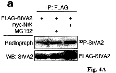
Figure 4 shows that SIVA2 is phosphorylated in mutiple serine residues at
its N-terminus and this phosphorylation as well seems to contribute to its
stabilization. (A) SIVA2 is phosphorylated in cells. HEK-293T cells
transiently
expressing myc-NIK and FLAG-SIVA2 were metabolically labeled with
CA 02712824 2010-07-21
WO 2009/098701 PCT/IL2009/000161
[32P]orthophosphate. MG132 was applied for the last 6 h of treatment. (B)
Assignment of the phosphorylation sites in SIVA2 isolated from cells
overexpressing NIK. The peptides comprising phosphorylated residues of SIVA2
were located by precursor ion scan in a negative ion mode at m/z -79.
Phosphorylated residues were later assigned by tandem nano-electrospray MS
analysis in a positive mode (see Table SI). Shown at the top is the amino-acid
sequence of SIVA2 with a schematic presentation of very likely (in bold) and
confirmed (in bold and designated pS") phosphorylated residues in SIVA2. (C)
SIVA2 in vitro phosphorylation. Effect of serine mutations. myc-NIK and FLAG-
SIVA2 and its indicated serine mutants (3SA, replacement of residues 5, 50 and
51
by alanines, and 6SA, replacement of residues 5, 21, 26,35, 50 and 51 by
alanines)
were cotransfected into HEK-293T cells and the immunoprecipitated SIVA was
subjected to an in-vitro kinase assay{Ramakrishnan, 20041. Bottom panel shows
normalized total amounts of SIVA2 and its mutants in the kinase reaction.
Western
blot analysis of the coprecipitated NIK confirmed that its amount in the
precipitate
was not decreased by the 3SA or the 6SA mutations. (D) NIK expression or
proteasomal inhibition stabilizes SIVA N-terminus. HEK-293T cells were
transfected with FLAG-SIVA2 (1-58) and myc-NIK as indicated. MG132 (25uM)
was applied for the last 6 hours of 24 h transfection. Cells were harvested,
lysed and
SIVA2 levels were assessed by anti-FLAG antibody. (E) NIK co-expression
enhances phosphorylation of SIVA2 (1-58). Phosphorylation of SIVA2 (1-58) in
cells was assessed by metabolic labeling with [32P]orthophosphate, 22 h after
transfection of the indicated plasmids. Okadaic acid (1 M) was added for the
last
45 min.
Figure 5 shows identification of amino acid residues in SIVA2 that
contribute to its stabilization by NIK and TRAF2. (A) Individual serine
mutations
do not interfere with NIK-induced SIVA2 stabilization. Different serine-mutant
SIVA2 plasmids were cotransfected with NIK into HEK-293T cells and the cell
lysates were analyzed 24 h after transfection. (B) Tyrosine 34 of SIVA2 does
not
participate in its phosphorylation or stabilization by NIK. The indicated
plasmids
16
CA 02712824 2010-07-21
WO 2009/098701 PCT/IL2009/000161
were transfected into HEK-293T cells, and 24 h later SIVA2 and NIK in the
lysates
were determined (top two panels). Bottom panel: phosphorylated SIVA2 from an
in-vitro kinase assay, performed as in Fig. 4C, with the SIVA and SIVA-mutant
proteins immunoprecipitated from cells co-expressing NIK.(C) Combined mutation
of several of residues in SIVA2 that can be phosphorylated interfere with the
protein's NIK-induced stabilization. Each of the indicated plasmids was
transfected
into HEK-293T cells, and the amount of SIVA2 and NIK in cell lysates was
determined 24 h after transfection. (D) TRAF2 and proteasomal inhibition
stabilize
the SIVA2 serine mutants that cannot be stabilized by NIK. Plasmids were
transfected as described above. MG132 was added 18 h later, and after a
further 6 h
the cellular proteins were extracted. (E) Combined serine mutation in SIVA2
compromises its stabilization by CD40L. EcR-293-CD40 cells were transfected
with 0.75 .tg of the SIVA2 plasmid or with 1.5 g of the SIVA2 6SA mutant
plasmid. CD40L was applied at the indicated times before cell harvesting,
which
was carried out 30 h after transfection. (F) Lysines in SIVA2 participate in
its
stabilization by TRAF2. The indicated plasmids were cotransfected into HEK-
293T
cells. Total-cell lysates were prepared 24 h after transfection and analyzed
by
western blotting. (G) The lysines contributing to SIVA2 stabilization by TRAF2
are
not involved in its stabilization by NIK. SIVA2 and NIK expression levels were
determined as above.
Figure 6 SIVA2 is recruited to receptors of the TNF/NGF family and binds
specifically to NIK, TRAF2, and cIAP1. (A) Transfected SIVA2 binds to
endogenous TRAF2. FLAG-SIVA2 or HIS-SIVA2 (control) was transfected into
HEK293T cells. SIVA2 was immunoprecipitated using anti-FLAG M2 beads and
the co-precipitated cellular TRAF2 was assayed by western blotting. The total
cellular level of TRAF2 is shown at the bottom. (B) SIVA2 binds TRAF2
inducibly. Treatment of the cells EcR293-CD27-SIVA2 with ponasterone for 2 h
to
induce SIVA2, and treated with CD70 as indicated was followed by
immunoprecipitation of TRAF2. (C) SIVA2 binds TRAF2 in vitro. FLAG-tagged
TRAF2 was immunoprecipitated from transfected HEK293T cells with anti-FLAG
17
CA 02712824 2010-07-21
WO 2009/098701 PCT/IL2009/000161
M2 beads, eluted from the beads using FLAG peptide and incubated with GST-
SIVA2 or its mutant, and then subjected to immunoprecipitation and western
blotting as indicated. (D) SIVA2 binds at its N-terminus to cIAPI. Left panel:
Binding in vitro. Recombinant cIAPI was incubated with GST-SIVA2 or its
mutant. Right and bottom panels depict binding in transfected HEK293T cells.
Right panel: Cells were transfected with HIS-SIVA2, HIS-SIVA2 (1-58), or
FLAG-TRAF2. After 28 h the endogenous cIAP 1 was immunoprecipitated. MG 132
was applied for the last 6 h of incubation. Bottom panel: Cells were
transfected with
FLAG-SIVA2, FLAG-SIVA2 (1-58) or, as a specificity control, FLAG-GST-BR3-
ICD* (in which the BAFF receptor intracellular domain is mutated at its TRAF3-
binding region (PVPAT>AVAAA)). After 28 h the transfected proteins were
immunoprecipitated and probed for co-precipitated endogenous cIAP I. MG 132
was
applied for the last 6 h of incubation. (E) Diagrammatic representation of the
deletion analyses of SIVA2 binding to cIAPI, NIK, and TRAF2 presented in
Figures 6D and H. Left: the deletion mutants used. Right: the binding
observed.
N/A, not analyzed. The asterisk denotes assessment in the yeast two-hybrid
test.
(All other tests were performed in transfected mammalian cells.) (F) Binding
of
NIK (upper panel) and of TRAF2 (lower panel) to SIVA2 involves the latter's C-
terminal region (the cysteine-rich region). The indicated plasmids were co-
transfected into HEK293T cells. Lysates were prepared 24 h after transfection
and
analyzed as indicated in the figure. To further increase SIVA2 expression in
cells
transfected with this plasmid alone, these cells were treated with MG132 (25
M)
during the last 4 h before harvesting. WB, anti-HIS. The deletion construct
corresponding to the CRR itself was rather poorly expressed and therefore
could not
be used to assess the binding of proteins to this region.
Figure 7 SIVA2 inhibits TRAF2- and NIK-mediated signaling. (A)
Induction of SIVA2 in Ramos T-REx-SIVA2 cells suppresses induction of both the
canonical and the alternative pathways by CD70 (middle and left panels,
respectively) and of the canonical NF-xB pathway by TNF (right panel). (B)
Induction of SIVA2 (left panels), but not of SIVA 1 (right panels), in EcR293-
18
CA 02712824 2010-07-21
WO 2009/098701 PCT/IL2009/000161
CD27-SIVA2 cells suppresses activation of the alternative NF-KB pathway by
CD70 (no IKB(x degradation or p65 translocation to the nucleus could be
discerned
in CD70-treated EcR293-CD27 cells). Western blot analysis of SIVA demonstrates
that the level of induction of SIVA2 is much lower than that of SIVAI. Unless
further enhanced by proteasomal inhibition, SIVA2 was below detection level.
(C)
Suppression of SIVA increases NIK expression, and also causes constitutive
activation of the alternative NF-KB pathway and increased responsiveness of
the
canonical pathway. Left panel: A mixture of pSUPER SIVA plasmids (2x pSUPER
275 + Ix pSUPER NC3) was transiently transfected into EcR293-CD27 cells
expressing retrovirally transduced NIK. After 40 h the cells were treated with
CD70
for 8 h, and cytoplasmic and nuclear extracts were then assayed for NF-xB
proteins
and NIK. Effective suppression of SIVA expression by the siRNAs was confirmed
in the experiments shown in panels C, D, and E by RT-PCR of SIVA message,
performed as described in Materials and Methods. Right panel: Ramos cells
stably
expressing lentivirally transduced SIVA shRNA NO (SIVA-knockdown) were
treated with CD70 for the indicated time periods, and nuclear extracts were
analyzed for NF-KB proteins. Octl served as the loading control. (D)
Suppression of
SIVA enhances CD70-induced NF-xB activation. HEK293T cells were transiently
co-transfected with CD27, a mixture of pSUPER-SIVA plasmids, and a luciferase
reporter plasmid. After 26 h the cells were treated with CD70 for 4 h. Lysates
were
analyzed in triplicate in two independent experiments; results represent the
mean
fold induction. (E) Suppression of SIVA enhances MAPK activation by CD70 and
TNF. Left: Control and SIVA-knockdown Ramos cells were treated as in the right
panel of C. Right: pSUPER SIVA was transiently expressed in HEK293T cells, and
48 h after transfection TNF was applied for the indicated durations. Total-
cell
lysates were analyzed for phosphorylated and total JNK and p38.
Figure 8 SIVA2, cooperatively with cIAPI, mediates ubiquitination and
degradation of TRAF2 in response to CD27. (A) SIVA2 facilitates ubiquitination
of TRAF2 in the CD27-receptor complex. Left panel: Suppression of the
recruitment of TRAF2 to the receptor complex as well as of its ubiquitination
by
19
CA 02712824 2010-07-21
WO 2009/098701 PCT/IL2009/000161
SIVA2 knockdown. EcR293-CD27 cells were transfected with the mixture of
pSUPER SIVA plasmids, and were treated 48 h later with CD70 for the indicated
time periods. Western blot analysis of CD27 in the immunoprecipitated receptor
complex serves as an internal control. The efficiency of SIVA suppression was
evaluated in this experiment and in C by RT-PCR of SIVA message, as described
in Materials and Methods. Other panels: Inducibly expressed SIVA2, but not
SIVA2 (C73A) or SIVA1, enhances TRAF2 ubiquitination in the receptor complex.
Ramos T-REx-SIVA2 cells, Ramos T-REx-SIVA2 (C73A) cells, or Ramos T-REx-
SIVA1 cells (5x10⁷ cells) were induced with doxycycline for 2 h, and CD70 was
then applied for the indicated time periods. Ubiquitin aldehyde (5 M) was
added to
the cell lysates in all experiments in which protein ubiquitination in cells
was
assayed. (B) Suppression of SIVA blocks CD70-induced TRAF2 degradation.
EcR293-CD27 cells were transfected as in the left panel of A and treated with
CD70 for the indicated time periods. (C) Effect of SIVA2 on the response of
NIK-
or NIK (K670A)-expressing cells to CD70. EcR293-CD27-SIVA2 cells
constitutively expressing retrovirally transduced NIK or NIK (K670A) mutant
were
treated with CD70 and, where indicated, also with ponasterone for 8 h. (D) K48-
linked ubiquitination of TRAF2 in the CD27-receptor complex of cells
expressing
SIVA2. EcR293-CD27-SIVA2 cells were transfected with the indicated HA-tagged
ubiquitin mutant plasmids and SIVA2 was induced with ponasterone for 2 h. CD70
was then applied for 15 min and the CD27-receptor complex was precipitated
through anti-FLAG. The immunoprecipitate was boiled with 1% SDS, diluted 20-
fold with lysis buffer, re-immunoprecipitated with anti-HA antibody, and
analyzed
with anti-TRAF2 antibody. (E) cIAP 1 is required for CD70-induced TRAF2
degradation. EcR293-CD27 cells were transfected with cIAPI siRNA or control
siRNA and treated, 48 h after transfection, with CD70 for the indicated time
periods.
Figure 9 SIVA2 mediates ubquitination of both TRAF2 and cIAP 1. (A)
cIAP-1 is required for SIVA2-mediated TRAF2 ubiquitination in cells. HEK293T
cells were transfected with clAP 1 siRNA and, 24 h later, with the other
plasmids as
CA 02712824 2010-07-21
WO 2009/098701 PCT/IL2009/000161
indicated. The extent of ubiquitination of the TRAF2 immunoprecipitated from
the
lysates of these cells, as well as the cellular levels of the endogenous cIAP
1 and
cIAP2 and the transfected ubiquitin, were determined by western blot analyses.
(B)
SIVA2, but not SIVA1, enhances K48-linked polyubiquitination of TRAF2 in
cells. HEK293T cells grown in 90-mm plates were transfected by the calcium
phosphate method with 4 g of FLAG-TRAF2 (C34A), together with 6 g of HA-
ubiquitin mutant plasmids and 6 g of HIS-SIVA2 or HIS-SIVA2 (C73A) or HIS-
SIVA 1. The cells were lysed 24 h after transfection and TRAF2 was
precipitated
and analyzed as indicated. Wild-type SIVA2 and SIVAI, as well as SIVA2 (C73A)
mutant, co-precipitated with TRAF2 (bottom panel). (C) SIVA2 ubiquitinates
cIAP-1 in vitro. Recombinant cIAPI was incubated with SIVA2 or the SIVA2
(C73A) mutant in a ubiquitination reaction with either UbcH5b or Ubc 13/Uev 1
a
used as the E2 enzyme. After the reaction the proteins were treated with SDS
as in
Figure 8 D, then immunoprecipitated and subjected to analysis by western
blotting
as indicated.
DETAILED DESCRIPTION OF THE INVENTION
The findings according to the invention show that SIVA2 is a feedback
regulator of TNF/NGF receptor signaling and that modulation of SIVA2 stability
can be used in therapy of disease disorder or conditions associated with the
activity
of these receptors.
The invention provides a stability-improved SIVA2 or salt thereof which can
be used in therapy wherein said stability-improved SIVA2 is characterized by
comprising one or more of the following post translation modification(s) (i) 0-
GlcNAcylation; (ii) phosphorylation at serine residues 5, 50, 51 of SIVA2;
(iii)
ubiquitination on SIVA2 residues, K17 and/or K99; or (iv) a combination of (i)
to
(iii). The present invention also relates to a stabilized SIVA2 mutein,
isoform, fused
protein, functional derivative, active fraction, fragment, circularly
permutated
derivative, collectively named herein stabilized SIVA2.
21
CA 02712824 2010-07-21
WO 2009/098701 PCT/IL2009/000161
Among the proteins known to participate in signaling by receptors of the
TNF/NGF family, it is possible to distinguish two functional groups: (i)
proteins
that mediate signaling, and (ii) those that regulate it, dictating which of
the
receptor's various activities will be turned on, at what intensity, and for
how long.
Proteins of the first group usually occur constitutively in the cells, ready
to be
recruited to the receptors upon ligand binding. Expression of those proteins
that
regulate signaling, however, is often itself signaling-dependent; their
cellular levels
are enhanced by TNF/NGF receptors, as well as by other agents that affect the
function of these receptors. Earlier studies of SIVA were interpreted as
suggesting
that this protein mediates signaling and that it acts specifically to promote
cell
death. This indeed seems to be the case with SIVA1. With respect to SIVA2, it
is
suggested according to the invention that this protein serves rather as a
regulator of
signaling, not necessarily in a way that promotes cell death; and indeed,
typically of
proteins that regulate receptor-induced signaling, its own levels in cells are
affected
by signals generated by TNF/NGF receptors. Both, in its function and in the
regulation of its formation, SIVA2 is shown according to the invention to
differ
from SIVA 1. The latter, unlike SIVA2, occurs constitutively in various cells
in
amounts much higher than those of SIVA2, and is further induced by cellular
stress.
Moreover, association of SIVA 1 with signaling complexes of receptors of the
TNF/NGF family was not detected, nor the effects on signaling displayed by
SIVA2.
SIVA2, is a short variant of SIVA1, is specifically recruited to receptors of
the TNF/NGF family and can both inhibit and enhance signaling for some of
their
nonapoptotic effects. It was found according to the present invention that:
(a) the
cellular content of SIVA2, is very low in the absence of stimulation and is
greatly
increased after these receptors are triggered; (b) that this increase reflects
its
enhanced stability contributed by TRAF2 and NIK, signaling proteins that bind
to
SIVA2, and (c) that said enhanced stability involves post-translational
modifications of SIVA2, including O-G1cNAcylation, ubiquitination in specific
lysines and phosphorylation in specific serines. Also, it was found according
to the
22
CA 02712824 2010-07-21
WO 2009/098701 PCT/IL2009/000161
invention that SIVA2 binds to and ubiquitinates the anti apoptotic protein
cIAP 1
and to TRAF2, triggering the latter's degradation. It was recently found by
the
inventors that SIVA2 also modulates ubiquitination and proteasomal processing
of
NIK and TRAF3 W02007080593. In all, these findings stress that SIVA2 is a
feedback regulator of TNF/NGF receptor signalling and that modulation of SIVA2
stability has a key role on signaling by receptors of the TNF/NGF family.
It was found according to the invention that the feedback loop is initiated by
the recruitment of SIVA2 to the receptors' signaling complexes, as well as the
dramatic stabilization of SIVA2, which can be induced by the activities of two
signaling proteins TRAF2 and the protein kinase NIK, to which SIVA2 binds.
Consequently, SIVA2 imposes ubiquitination of several of the signaling
proteins
that are recruited to the receptor and thus modulates their proteasomal
processing.
Up to now, the mechanisms reported to underlie an induced increase in the
cellular levels of proteins (such as A2019, TRAF 120, cFLIP21, or CYLD22) that
regulate signaling by receptors of the TNF/NGF family act on the
transcriptional
level. In contrast, it was found according to the invention that the increase
in SIVA2
level following ligand stimulation occurs post-transcriptionally. Post-
translational
modifications of SIVA2 demonstrated according to the present invention to
increase
the level of SIVA2 include phosphorylation of specific serine residues, 0-
GlcNAcylation, and ubiquitination, and as shown according to the invention
these
modifications contribute to the modulation of SIVA2 stability. At least two
signaling proteins seemed to participate in cytokine-induced SIVA2
stabilization:
NIK, in a way that depends on its protein kinase function, and TRAF2, by a
mechanism that involves its ubiquitin-ligase function.
More specifically, the following differences in SIVA 1 and SIVA2 were
found according to the invention; (a) SIVA2 is less expressed than SIVA 1
splice
variants in various cell lines and cytokines of the TNF/NGF family such as
CD70,
CD40L, TNF increased the amount of SIVA2 in resting PBMCs; (b) ligand
activation and proteasomal inhibition, but not genotoxic stress, increase
SIVA2
levels in activated PBMCs and the increase in SIVA2 levels were caused by
23
CA 02712824 2010-07-21
WO 2009/098701 PCT/IL2009/000161
increase in stability of SIVA2 and not by increase in SIVA2 expression; (c)
The
cytokine stabilization was specific for SIVA2 since that of SIVAI remained
unaltered; (d) SIVA2 is recruited to CD27 by treatment with CD70 while SIVA 1
is
not, in addition, SIVA2 was shown also to be recruited to CD40 and TNFR1; (e)
while genotoxic agents enhance SIVA1 expression they do not affect the
expression
of SIVA2; (f) stabilization of SIVA2 by CD40 in cells decreased when treated
with
the genotoxic agent CPT. These differences of SIVA1 and SIVA2 can be
advantageously used to specifically induce SIVA2 activity in therapy.
One of the modifications of SIVA2 that were found according to the
invention to contribute to its stabilization in cells is phosphorylation in
serine
residues, particularly in every serine residues 5, 50 and 51 (3S) and
especially in all
serine residues 5, 21, 26, 35, 50 and 51 (6S). For example, mutations in 3S
(e.g. in
SIVA3SA) significantly reduced stabilization of SIVA2 and mutations 6S
(SIVA6SA) almost completely reduced the stabilization of SIVA2 induced by NIK.
Of note, individual serine mutations did not interfere with NIK-induced SIVA2
stabilization and tyrosine 34 of SIVA2 did not participate in its
phosphorylation or
stabilization by NIK. SIVA2, but only some of SIVA3SA and almost none of
SIVA6SA mutants, were found to be phosphorylated also from an in-vitro kinase
assay, performed with SIVA proteins immunoprecipitated from cells co-
expressing
NIK. Unlike NIK, it was found that TRAF2 and proteasomal inhibition do
stabilize
SIVA6SA mutants. Of note, serine mutations of SIVA2 compromised its
stabilization by the cytokine CD40L. Also, the findings according to the
invention
show that lysines in SIVA2 participate in its stabilization by TRAF2. In
contrast, it
was found that lysines in SIVA2 are not involved in its stabilization by NIK.
Another modification of SIVA2 that was found according to the invention to
contribute to its stabilization in cells is O-GIcNAcylation. For example, it
was
found that (a) SIVA is a glycoprotein; (b) SIVA2 incorporates azido-G1cNAc in
cells cotransfected with NIK and SIVA2; (c) that the R-D-N-acetyl
hexosaminidase
treatment abolished binding of SIVA2, extracted from cells coexpressed with
NIK,
to Wheat Germ Agglutinin (WGA) which selectively binds to N-Acetyl
24
CA 02712824 2010-07-21
WO 2009/098701 PCT/IL2009/000161
glucosamine (G1cNAc) groups and to sialic acid; (d) that inhibition of 0-
glycosylation interfered also with TRAF2-induced SIVA2 stabilization, but did
not
affect MG 132-induced stabilization; (e) that inhibition of 0-glycosylation
blocks
the NIK-mediated stabilization of wild-type SIVA2, but had no effect on the
residual stabilization by NIK of the SIVA2 6SA mutant; and (f) that specific
inhibition of O-G1cNAcylation blocked NIK-induced SIVA2 stabilization.
In addition, it was found according to the invention that SIVA2
ubiquitination on SIVA2 residues, K17 and/or K99 contribute to SIVA2
stabilization. This stabilization by ubiquitination appears to be induced by
TRAF2.
Thus, the present invention provides the use of specific modulation of
SIVA2 stability in therapy. SIVA2 modulation can be carried out or induced in
vitro
e.g. in cell free system, or inside the cells e.g. in vivo or ex-vivo.
Modulation of
SIVA2 stability can be induced in diseased cells or in cells producing
unregulated
levels of cytokines. Examples of cells in which modulation of SIVA2 stability
can
be induced include but, are not limited to, mononuclear cells, lymphoid cells,
Treg
cells, endothelial cells, smooth muscular cells, macrophages, lymphocytes,
embryonic kidney cells, lymphoma cells, B-lymphoblastoma cell, hepatocellular
liver carcinoma cell, cells expressing unregulated levels of CD27, CD40,
and/or
TNF receptor. In one embodiment of the invention, modulation of SIVA2
stability
is induced in cells before during and/or after treatment with a genotoxic
agent such
as chemotherapy or irradiation.
In one embodiment of the invention, modulation of SIVA2 stability consists
on increasing the stability of SIVA2. Stabilized SIVA2 is characterized by
comprising one or more of the following post translation modification(s) (i) 0-
G1cNAcylation; (ii) phosphorylation at serine residues 5, 50, 51 of SIVA2;
(iii)
ubiquitination on SIVA2 residues, K17 and/or K99; or (iv) a combination of (i)
to
(iii).
Stabilized SIVA2 can be induced in a cell, for example, by over-expressing
in the same cell one or more of the following recombinant or endogenous
proteins
(see EGA below) such as NIK, TRAF2, cIAP 1 a ring-finger mutant of cIAP 1 such
CA 02712824 2010-07-21
WO 2009/098701 PCT/IL2009/000161
as H588A, a O-G1cNAc transferase, an inhibitor of O-GlcNAcase. Stabilizaed
SIVA2 can be induced in a cell by overexpressing SIVA2 together with said
protein(s) e.g. as shown in the examples below. Alternatively, an activator of
O-
GlcNac transferase, TRAF2, inhibitor of O-G1cNAcase, inhibitor CIAP 1
activity,
and/or a ring-finger mutant of clAP l such as H588A may be used.
Stabilized SIVA2 can be used for treating, or in the manufacture of a
medicament for treating a disease disorder or condition associated with low
activity
of SIVA2 or ameliorated by increasing the activity of SIVA2 in cells and/or in
a
disease; disorder; or condition in which signaling pathways activated towards
protein synthesis by several members of the TNF/NGF family are associated with
the pathogenesis or course of the disease disorder or condition such as e.g.
cancer,
an inflammatory disease, and/or an autoimmune disease. Said treating can be
carried out in vivo or ex-vivo.
The term "salts" herein refers to both salts of carboxyl groups and to acid
addition salts of amino groups of the polypeptide of the invention. Salts of a
carboxyl group may be formed by means known in the art and include inorganic
salts, for example, sodium, calcium, ammonium, ferric or zinc salts, and the
like,
and salts with organic bases as those formed, for example, with amines, such
as
triethanolamine, arginine or lysine, piperidine, procaine and the like. Acid
addition
salts include, for example, salts with mineral acids such as, for example,
hydrochloric acid or sulfuric acid, and salts with organic acids such as, for
example,
acetic acid or oxalic acid. Of course, any such salts must have substantially
similar
activity to the SIVA2.
As used herein, the term "fragment" refers to a part or fraction of the
polypeptide molecule, provided that the shorter peptide retains the desired
biological activity of SIVA2. Fragments may readily be prepared by removing
amino acids from either end of the polypeptide and testing the biological
activity of
the resulting fragment for example: binding to cIAPI, binding to TRAF2,
induction
of NIK degradation, and/or inhibition of NIK-mediated NFKB activation in
cells.
Proteases that remove one amino acid at a time from either the N-terminal or
the C-
26
CA 02712824 2010-07-21
WO 2009/098701 PCT/IL2009/000161
terminal of a polypeptide are known in the art, and fragments that retain the
desired
biological activity can be obtaining as a matter of routine experimentation by
employing such proteases.
As "active fractions" of the protein the present invention refers to any
fragment or precursor of the polypeptidic chain of the compound itself, alone
or in
combination with related molecules or residues bound to it, for example
residues
of sugars or phosphates, or aggregates of the polypeptide molecule when such
fragments or precursors show the same activity of SIVA2 as medicament.
"Precursors" are compounds which can be converted into the SIVA2 in the human
or animal body.
The definition "functional derivatives" as herein used refers to derivatives
which can be prepared from the functional groups present on the lateral chains
of
the amino acid moieties or on the terminal N- or C- groups according to known
methods and are comprised in the invention when they are pharmaceutically
acceptable i.e. when they do not destroy the protein activity or do not impart
toxicity to the pharmaceutical compositions containing them. Such derivatives
include for example esters or aliphatic amides of the carboxyl-groups and N-
acyl
derivatives of free amino groups or 0-acyl derivatives of free hydroxyl-groups
and
are formed with acyl-groups as for example alcanoyl- or aroyl-groups. SIVA2
may
be conjugated to polymers in order to improve the properties of the protein,
such as
the stability, half-life, bioavailability, tolerance by the human body, or
immunogenicity. Therefore, one embodiment of the invention relates to a
functional
derivative of SIVA2 comprising at least one moiety attached to one or more
functional groups, which occur as one or more side chains on the amino acid
residues. One embodiment of the invention relates to SIVA2 polypeptide linked
to
Polyethlyenglycol (PEG). PEGylation may be carried out by known methods, such
as the ones described in WO 92/13095, for example.
The term "circularly permuted derivatives" as used herein refers to a linear
molecule in which the termini have been joined together, either directly or
through a
linker, to produce a circular molecule, and then the circular molecule is
opened at
27
CA 02712824 2010-07-21
WO 2009/098701 PCT/IL2009/000161
another location to produce a new linear molecule with termini different from
the
termini in the original molecule. Circular permutations include those
molecules
whose structure is equivalent to a molecule that has been circularized and
then
opened. Thus, a circularly permuted molecule may be synthesized de novo as a
linear molecule and never go through a circularization and opening step. The
preparation of circularly permutated derivatives is described in W095/27732.
As used herein the term "muteins" refers to analogs of SIVA2. The present
invention also concerns analogs of the above SIVA2 protein of the invention,
which
analogs retain essentially the same biological activity of the SIVA2 protein
having
essentially only the naturally occurring sequences of SIVA2. Such "analogs"
may
be ones in which up to about 30 amino acid residues may be deleted, added or
substituted by others in the SIVA2 protein, such that modifications of this
kind do
not substantially change the biological activity of the protein analog with
respect to
the protein itself. Thus, one or more of the amino acid residues of the
naturally
occurring components of SIVA2 are replaced by different amino acid residues,
or
are deleted, or one or more amino acid residues are added to the original
sequence
of SIVA2, without changing considerably the activity of the resulting products
as
compared with the original SIVA2. These muteins are prepared by known
synthesis
and/or by site-directed mutagenesis techniques, or any other known technique
suitable therefore.
Any such mutein preferably has a sequence of amino acids sufficiently
duplicative of that of the basic SIVA2 such as to have substantially similar
activity
thereto. Thus, it can be determined whether any given mutein has substantially
the
same activity as the basic SIVA2 of the invention by means of routine
experimentation comprising subjecting such an analog to the biological
activity
tests set forth in Examples below e.g. monitoring binding to cIAPI, binding to
TRAF2, binding to NIK, induction of NIK degradation, ubiquitination of TRAF2,
self ubiquitination, ubiquitination of cIAP 1 or inhibition of NIK-mediated
NFxB
activation in cells.
28
CA 02712824 2010-07-21
WO 2009/098701 PCT/IL2009/000161
Muteins of the SIVA2 protein which can be used in accordance with the
present invention, or nucleic acid coding therefore, include a finite set of
substantially corresponding sequences as substitution peptides or
polynucleotides
which can be routinely obtained by one of ordinary skill in the art, without
undue
experimentation, based on the teachings and guidance presented herein. For a
detailed description of protein chemistry and structure, see Schulz, G.E. et
al.,
Principles of Protein Structure, Springer-Verlag, New York, 1978; and
Creighton,
T.E., Proteins: Structure and Molecular Properties, W.H. Freeman & Co., San
Francisco, 1983, which are hereby incorporated by reference. For a
presentation of
nucleotide sequence substitutions, such as codon preferences, see . See
Ausubel et
al., Current Protocols in Molecular Biology, Greene Publications and Wiley
Interscience, New York, NY, 1987-1995; Sambrook et al., Molecular Cloning: A
Laboratory Manual, Cold Spring Harbor Laboratory, Cold Spring Harbor, NY,
1989.
Preferred changes for muteins in accordance with the present invention are
what are known as "conservative" substitutions. Conservative amino acid
substitutions of those in the SIVA2 protein having essentially the naturally -
occurring SIVA2 sequences, may include synonymous amino acids within a group
which have sufficiently similar physicochemical properties that substitution
between members of the group will preserve the biological function of the
molecule, Grantham, Science, Vol. 185, pp. 862-864 (1974). It is clear that
insertions and deletions of amino acids may also be made in the above -defined
sequences without altering their function, particularly if the insertions or
deletions
only involve a few amino acids, e.g., under thirty, and preferably under ten,
and do
not remove or displace amino acids which are critical to a functional
conformation,
e.g., cysteine residues, Anfinsen, "Principles That Govern The Folding of
Protein
Chains", Science, Vol. 181, pp. 223-230 (1973). Analogs produced by such
deletions and/or insertions come within the purview of the present invention.
Preferably, the synonymous amino acid groups are those defined in Table I.
More preferably, the synonymous amino acid groups are those defined in Table
II;
29
CA 02712824 2010-07-21
WO 2009/098701 PCT/IL2009/000161
and most preferably the synonymous amino acid groups are those defined in
Table
III.
10
20
TABLE I Preferred Groups of Synonymous Amino Acids
Amino Acid Synonymous Group
Ser Ser, Thr, Gly, Asn
Arg Arg, Gln, Lys, Glu, His
Leu Ile, Phe, Tyr, Met, Val, Leu
Pro Gly, Ala, Thr, Pro
Thr Pro, Ser. Ala, Gly, His, Gln, Thr
Ala Gly, Thr, Pro, Ala
CA 02712824 2010-07-21
WO 2009/098701 PCT/IL2009/000161
Val Met, Tyr, Phe, Ile, Leu, Val
Gly Ala, Thr, Pro, Ser. Gly
Ile Met, Tyr, Phe, Val, Leu, Ile
Phe Trp, Met, Tyr, Ile, Val, Leu, Phe
Tyr Trp, Met, Phe, Ile, Val, Leu, Tyr
Cys Ser, Thr, Cys
His Glu, Lys, Gln, Thr, Arg, His
Gln Glu, Lys, Asn, His, Thr, Arg, Gln
Asn Gln, Asp, Ser, Asn
Lys Glu, Gin, His, Arg, Lys
Asp Glu, Asn, Asp
Glu Asp, Lys, Asn, Gln, His, Arg, Glu
Met Phe, Ile, Val, Leu, Met
Trp Trp
TABLE II More Preferred Groups of Synonimous Amino Acids
Amino Acid Synonymous Group
Ser Ser
Arg His, Lys, Arg
Leu Ile, Phe, Met, Leu
Pro Ala, Pro
Thr Thr
Ala Pro, Ala
31
CA 02712824 2010-07-21
WO 2009/098701 PCT/IL2009/000161
Val Met, Ile, Val
Gly Gly
Ile Ile, Met, Phe, Val, Leu
Phe Met, Tyr, Ile, Leu, Phe
Tyr Phe, Tyr
Cys Ser, Cys
His Arg, Gln, His
Gln Glu, His, Gln
Asn Asp, Asn
Lys Arg, Lys
Asp Asn, Asp
Glu Gln, Glu
Met Phe, Ile, Val, Leu, Met
Trp Trp
20
TABLE III Most Preferred Groups of Synonymous Amino Acids
Amino Acid Synonymous Group
Ser Ser
Arg Arg
Leu Ile, Met, Leu
Pro Pro
Thr Thr
Ala Ala
Val Val
32
CA 02712824 2010-07-21
WO 2009/098701 PCT/IL2009/000161
Gly Gly
Ile Ile, Met, Leu
Phe Phe
Tyr Tyr
Cys Ser, Cys
His His
Gln Gln
Asn Asn
Lys Lys
Asp Asp
Glu Glu
Met Ile, Leu, Met
Trp Trp
Examples of production of amino acid substitutions in proteins which can be
used for obtaining muteins of SIVA2 include any known method steps, such as
presented in US patents RE 33,653, 4,959,314, 4,588,585 and 4,737,462, to Mark
et
al; 5,116,943 to Koths et al., 4,965,195 to Namen et al; 4,879,111 to Chong et
al;
and 5,017,691 to Lee et al; and lysine substituted proteins presented in US
patent
No. 4,904,584 (Straw et al).
In another preferred embodiment of the present invention, any mutein of the
SIVA2 protein for use in the present invention has an amino acid sequence
essentially corresponding to that of the above noted SIVA2 protein of the
invention.
The term "essentially corresponding to" is intended to comprehend muteins with
minor changes to the sequence of the basic SIVA2 protein which does not affect
the
basic characteristics thereof, particularly insofar as its ability to SIVA2 is
concerned. The type of changes which are generally considered to fall within
the
"essentially corresponding to" language are those which would result from
conventional mutagenesis techniques of the DNA encoding the SIVA2 protein of
33
CA 02712824 2010-07-21
WO 2009/098701 PCT/IL2009/000161
the invention, resulting in a few minor modifications, and screening for the
desired
activity in the manner discussed above.
In one embodiment of the invention, any such mutein has at least 40%
identity with the sequence of SIVA2, more preferably, it has at least 50%, at
least
60%, at least 70%, at least 80% or, most preferably, at least 90% identity
thereto.
Identity reflects a relationship between two or more polypeptide sequences
or two or more polynucleotide sequences, determined by comparing the
sequences.
In general, identity refers to an exact nucleotide to nucleotide or amino acid
to
amino acid correspondence of the two polynucleotides or two polypeptide
sequences, respectively, over the length of the sequences being compared.
For sequences where there is not an exact correspondence, a"% identity" may
be determined. In general, the two sequences to be compared are aligned to
give a
maximum correlation between the sequences. This may include inserting "gaps"
in
either one or both sequences, to enhance the degree of alignment. A % identity
may
be determined over the whole length of each of the sequences being compared
(so-
called global alignment), that is particularly suitable for sequences of the
same or
very similar length, or over shorter, defined lengths (so-called local
alignment), that
is more suitable for sequences of unequal length.
The term "sequence identity" as used herein means that the amino acid
sequences are compared by alignment according to Hanks and Quinn (1991) with a
refinement of low homology regions using the Clustal-X program, which is the
Windows interface for the ClustalW multiple sequence alignment program
(Thompson et al., 1994). The Clustal-X program is available over the internet
at
ftp://ftp-igbmc.u-strasbg.fr/pub/clustalx/. Of course, it should be understood
that if
this link becomes inactive, those of ordinary skill in the art can find
versions of this
program at other links using standard internet search techniques without undue
experimentation. Unless otherwise specified, the most recent version of any
program referred herein, as of the effective filing date of the present
application, is
the one which is used in order to practice the present invention.
34
CA 02712824 2010-07-21
WO 2009/098701 PCT/IL2009/000161
If the above method for determining "sequence identity" is considered to be
nonenabled for any reason, then one may determine sequence identity by the
following technique. The sequences are aligned using Version 9 of the Genetic
Computing Group's GDAP (global alignment program), using the default
(BLOSUM62) matrix (values -4 to +11) with a gap open penalty of -12 (for
the first null of a gap) and a gap extension penalty of -4 (per each
additional
consecutive null in the gap). After alignment, percentage identity is
calculated by
expressing the number of matches as a percentage of the number of amino acids
in
the claimed sequence.
Muteins in accordance with the present invention include those encoded by a
nucleic acid, such as DNA or RNA, which hybridizes to DNA or RNA under
stringent conditions and which encodes a SIVA2 protein in accordance with the
present invention, comprising essentially all of the naturally-occurring
sequences
encoding SIVA2. For example, such a hybridizing DNA or RNA may be one
encoding the same protein of the invention having, for example, the sequence
of
SIVA2, but which nucleotide differs in its nucleotide sequence from the
naturally-
derived nucleotide sequence by virtue of the degeneracy of the genetic code,
i.e., a
somewhat different nucleic acid sequence may still code for the same amino
acid
sequence, due to this degeneracy.
The findings according to the invention allow the preparation of
stabilized SIVA2. For example, stabilized SIVA2 or stability improved SIVA2
may
be obtained by increasing O-GlcNAcylation in the protein, for example by
contacting the protein with O-G1cNAc transferase and/or by inhibiting the
activity
of O-GlcNAcase. Induction of G1cNAc transferase can be aimed, for example, by
increasing the levels of UDP-GlcNAc in cells (Slawson et al., Jounal of
cellular
Biochemistry 97:71-83, 2006). Also, stabilization of SIVA2 may be obtained or
further increased by inducing phosphorylation at serine residues 5, 50, and 51
of the
protein. Increased stabilization may be obtained by phosphorylation of serine
residues 5, 50, 51, 21, 26, and 35, for example by contacting SIVA2 with NIK.
In
addition, stabilization of SIVA2 may be obtained or further increased by
increasing
CA 02712824 2010-07-21
WO 2009/098701 PCT/IL2009/000161
ubiquitination of SIVA2, for example by contacting the protein with TRAF2.
Lysine residues involved in SIVA2 stabilization by ubiquitination are either
one of
two of the residues, K17 and K99. Mutations in these residues did not affect
the
stabilization of SIVA2 by NIK. Another way to stabilize SIVA2 is by contacting
SIVA2 with a ring-finger mutant of cIAP 1 such as H588A. Said contacting of
SIVA2 with the other mentioned proteins can be carried out in vivo (e.g.
inside
cells) and in vitro (e.g. in a cell free system). In one embodiment of the
invention,
for specifically increasing stability of SIVA2, cells can be manipulated to
overexpress one or more of the following proteins O-G1cNAc transferase, NIK,
TRAF2, or a ring-finger mutant of cIAP1 such as H588A. Overexpression of
endogenous protein can be carried out, for example, by endogenous gene
activation
(EGA, see bellow). Overexpression of exogenous protein can be carried out, by
introducing the gene encoding the protein into the cells, for example, by
using an
expression vector (see below). In a further embodiment of the invention SIVA2
is
co-overexpressed with the protein(s). If the stabilized SIVA2 is prepared ex-
vivo
the cells are cultured under conditions allowing production of said stability-
improved SIVA2 and recovering the resulting SIVA2 from the culture. For
example, cells stressed with a nutrient poor or nutrient-exessive environment
were
shown to elevate O-GlcNac levels and can be used to produce stabilized
SIVA2.Also, all forms of stress tested (osmotic, ethanolic, oxidative, and
heat
schok) to date rapidly raise O-GlcNac levels in cells (Slawson et al., 2006).
Furthermore, the invention provides a host cell comprising stabilized SIVA2
selected from eukaryotic cells, such as a mammalian, insect, and yeast cells.
In one
embodiment of the invention the cells are HeLa, 293 THEK or CHO cells.
Alternatively, the invention provides a method of producing s stabilized SIVA2
of
the invention comprising the generation of a transgenic animal and isolating
the
protein produced from the body fluids of the animal.
Stabilized SIVA2 can be produced in eukaryotic host cells transfected,
transformed or infected with vectors encoding SIVA2, or in transgenic animals.
36
CA 02712824 2010-07-21
WO 2009/098701 PCT/IL2009/000161
When using transgenic animals, it is particularly advantageous to produce
heterologous polypeptides in their milk.
Overexpression of a protein in a mammalian cell may be carried out by
inserting the DNA encoding the polypeptide into a vector comprising a
promoter,
optionally an intron sequence and splicing donor/acceptor signals, and further
optionally comprising a termination sequence and signal peptide for secretion,
by
well-known techniques (for example, as described in Current Protocols in
Molecular Biology, chapter 16).
Overexpression of a protein in a mammalian cell may be carried out by
inducing increase in expression of the endogenous gene which encodes e.g.
SIVA2
polypeptide and /or O-G1cNAc transferase, NIK, and TRAF2. Altering expression
of endogenous SIVA2 and /or O-G1cNAc transferase and /or O-G1cNAc transferase,
NIK, and TRAF2 can be also employed. If desired, a compound may increase the
level of expression of the gene or the activity of endogenous protein. Such
compound can be a vector for inducing the endogenous production of a protein
in a
cell which expresses amounts of the protein which are not sufficient. The
vector
may comprise regulatory sequences functional in the cells desired to express
the
protein. Such regulatory sequences may be promoters or enhancers, for example.
The regulatory sequence may then be introduced into the right locus of the
genome
by homologous recombination, thus operably linking the regulatory sequence
with
the gene, the expression of which is required to be induced or enhanced. The
technology is usually referred to as "Endogenous Gene Activation" (EGA), and
it is
described e.g. in WO 91/09955.
It will be understood by the person skilled in the art that it is also
possible to
shut peptide expression directly, in situations in which a peptide is over-
expressed
and results in excessive amounts of the polypeptide in a cell or when it is
desired to
shut the peptide expression. For example, to increase O-G1cNAcidated SIVA2 in
a
cell the expression of O-GIcNAcase may be ablated by EGA. To do that, a
negative
regulation element, like e.g. a silencing element, may be introduced into the
gene
locus of the protein, thus leading to down-regulation or prevention of protein
37
CA 02712824 2010-07-21
WO 2009/098701 PCT/IL2009/000161
expression. The person skilled in the art will understand that such down-
regulation
or silencing of protein expression has the same effect as the use of an
inhibitor.
The invention also provides a pharmaceutical composition comprising a
pharmaceutically acceptable carrier and a stability-improved SIVA2 or salt
thereof
characterized by comprising one or more of the following post translation
modification(s) (i) O-G1cNAcylation; (ii) phosphorylation at serine residues
5, 50,
51 of SIVA2; (iii) ubiquitination on SIVA2 residues, K17 and/or K99; or (iv) a
combination of (i) to (iii).
It was found according to the invention that SIVA2 binds to various other
proteins known to mediate signaling by receptors of the TNF/NGF family such as
TRAF2, cIAP 1 and NIK. TRAF2, like NIK binds to CRR in SIVA2 and cIAP 1 was
found according to the invention to bind to the N-terminal part of SIVA2
(upstream
of the CRR). It was also found that SIVA2 can inhibit TRAF2 and NIK mediated
signaling since induction of SIVA2 suppresses the activation of both the
alternative
and the canonical NF-KB pathways by CD70 as well as activation of the
canonical
pathway by TNF. SIVA1, on the other hand, although expressed at much higher
level than SIVA2, had no such effect. Conversely, cells in which SIVA
expression
has been knocked down displayed constitutive activation of the alternative NF-
KB
pathway and also displayed somewhat increased basal levels of canonical NF-KB
pathway and heightened responsiveness of this pathway to activation by CD70.
Knockdown of SIVA also enhanced the induction of JNK and p38 kinase
phosphorylation both by CD70 and by TNF. SIVA2, cooperatively with cIAPI,
mediated ubiquitination and degradation of TRAF2 in response to CD27. Also, it
was previously reported that TRAF2 molecules recruited to CD27 are massively
ubiquitinated (Ramakrishnan et al., 2004). Knockdown of SIVA attenuated the
CD70 ubiquitination of TRAF2. In contrast, induction of SIVA2 but not SIVA1,
enhanced it.
It was previously found that SIVA2 possesses intrinsic ubiquitin-ligase
activity and that, SIVA2 facilitated in-vitro ubiquitination of TRAF2
(W02007080593). It was found that cysteine residue at position 73 within the
CRR
38
CA 02712824 2010-07-21
WO 2009/098701 PCT/IL2009/000161
in SIVA2 was needed for the ubiquitination of TRAF2. It was demonstrated using
transfected cells, that over-expression of wild-type SIVA2, but not SIVA1,
markedly increased the K48-linked (though not the K63-linked)
polyubiquitination
of TRAF2 beyond that observed when TRAF2 was expressed alone, whereas
SIVA2 (C73A) hardly affected the ubiquitination. Self-ubiquitination of SIVA2
in
vitro was not affected by this mutation, but it was drastically reduced by
complete
deletion of the CRR. Expression of SIVA2 C73A mutant in cells was found to
ablate the enhancing effect of SIVA2 on the ubiquitination of TRAF2 in the
CD27
complex.
In addition to TRAF2, it was found according to the invention that cIAP 1
was also effectively ubiquitinated by SIVA2, and that this ubiquitination too
was
compromised by the SIVA2 (C73A) mutation. Knockdown of cIAP1, dramatically
reduced the ubiquitination of TRAF2 in response to SIVA2 expression. Thus,
although SIVA2 has the ability to directly ubiquitinate TRAF2 in vitro, its
facilitation of TRAF2 ubiquitination within cells is either mediated through
enhancement of the ability of cIAP 1 to do so, or requires cIAP 1 to play a
permissive role. Triggering of CD27 resulted in a significant decrease in the
cellular
amounts of TRAF2, suggesting that its ubiquitination within the receptor
complex
targets for degradation. The ubiquitin chains whose ligation to TRAF2 was
facilitated by SIVA2 were primarily K48-linked, as is generally the case with
ubiquitination that prompts proteosomal degradation, rising the possibility
that this
SIVA2 effect contributes to the induction of TRAF2 degradation by CD27.
Consistently, knockdown of SIVA expression prevented the downregulation of
TRAF2 by CD27, whereas induction of SIVA2 enhanced it.
As mentioned above, knockdown of SIVA also resulted in constitutive
activation of the alternative NF-KB pathway. Suppression of cIAP 1 expression
also
results in NF-KB activation ( Varfolomeev et al., 2007) (Vince et al., 2007);
in
addition, it compromises the downregulation of TRAF2 by TNF-RII, another
receptor of the TNF/NGF family ( Li et al., 2002). According to the present
invention knockdown of cIAP 1 (like knockdown of SIVA2) compromised the
39
CA 02712824 2010-07-21
WO 2009/098701 PCT/IL2009/000161
downregulation of TRAF2 by CD27 as well, along with constitutive activation of
the alternative NF-KB pathway. These findings suggested that SIVA2 and cIAP 1
play a shared role in the induction of TRAF2 degradation by CD27 and in the
regulation of NF-KB.
In view of these findings regarding the function of SIVA2, the specific
modulation of SIVA2 stabilization can be used in therapy (prevention or
treatment)
or diagnosis of situations associated with the level or activity of SIVA2
and/or in
situations in which signaling pathways activated towards protein synthesis by
several receptors members of the TNF/NGF family, and particularly those that
activate the alternative pathway, and are associated with the pathogenesis or
course
of the situation.
Thus, in one embodiment of the invention, stabilized SIVA2 is useful in
modulating the activity of NIK and NF-KB for example, wherein the disease,
disorder, or condition is characterized by inappropriate NIK-mediated activity
or
NIK-mediated NF-KB activity such as for example in developmental disorders,
cell
proliferative disorders and immune disorders. In one embodiment, the disease,
disorder, or condition is characterized by increased host immune, inflammatory
response and/or cell proliferation mediated by increased NIK and NF-KB
activity
and thus a stabilized SIVA2 may be used to treat said disease.
Such situations in which modulation of SIVA2 stability is beneficial may
include diseases disorders or conditions such as developmental disorders; cell
proliferative disorders for example neoplastic disorders, like cancer,
melanoma,
sarcoma, renal tumour, colon tumour; genetic disorders; nervous system
disorders;
metabolic disorders; infections and other pathological conditions; immune
disorders
such as osteoarthritis, autoimmune disease, rheumatoid arthritis, psoriasis,
systemic multiple sclerosis, and lupus erythematosus; inflammatory disorders
such
as glomerulonephritis, allergy, rhinitis, conjunctivitis, uveitis, digestive
system
inflammation, inflammatory bowel disease such as Crohn's disease and
ulcerative
colitis, myasthenia gravis, pancreatitis, sepsis, endotoxic shock, cachexia,
myalgia,
ankylosing spondylitis, asthma, airway inflammation; wound healing;
CA 02712824 2010-07-21
WO 2009/098701 PCT/IL2009/000161
dermatological disease; ageing; and infections, including plasmodium,
bacterial
infection and viral infection. Thus, stabilized SIVA2 may be used in the
manufacture of a medicament for the treatment of such situations.
In one embodiment of the invention, diseases, disorders or conditions
associated with decreased SIVA2, increased NIK activity and/or NF-KB activity
in
cells such as such as malignancies, including both primary tumor and
metastasis,
asthma, rheumatoid arthritis, atherosclerosis, inflammation may be treated by
administering stabilized SIVA2 of the invention capable of
downregulating/inhibiting the activity of NIK and/or NF-xB activity in cells.
The invention allows modulation of SIVA2 stability to modulate its activity
in cells and in order to modulate/mediate intracellular effects on the
inflammation,
cell death or cell survival pathways in which activity of SIVA2 is involved
directly,
or indirectly via other modulators/mediators of TNF/NGF pathways. To modulate
SIVA2 activity, cells can be treated by introducing into said cells said
stabilized
SIVA2 or by inducing modulation of SIVA2 within the cells. In one embodiment
of
the invention, a SIVA2 polynucleotide is carried in a suitable vector which is
capable of effecting the insertion of said polynucleotide into said host cells
in a way
that said sequence is expressed in said cells. The vector can be a virus
vector
carrying also a sequence encoding an enzyme capable of increasing G1cNAc
moiety
in proteins such as O-G1cNAc transferase, and/or NIK, , TRAF2, a ring-finger
mutant of cIAP1 (H588A. ), or an inhibitor of G1cNAcidase to stabilize the
expressed SIVA2. The treatment can be effected by infecting said cells with
said
vector. Of advantage is overexpressing SIVA2 in vivo in specific pathologies
or
under specific conditions in which O-GlcNac transferase (OGT) expression or
activity is high or O-GlcNacase expression or activity is reduced resulting in
proteins exhibiting increased O-GlcNac content. An example of such pathologies
in
which cells have increased O-G1cNac content is in Type II diabetes (Slawson et
al.,
2006). Of advantage is overexpressing SIVA2 in vivo together with OGT
induction
e.g. by stressing the cells to be treated e.g. by hypothermic conditions
(Slawson et
al., 2006).
41
CA 02712824 2010-07-21
WO 2009/098701 PCT/IL2009/000161
Reducing stabilization of SIVA2 can be achieved, to treat disease disorder or
conditions, associated with increased levels of SIVA, decreased activity of NF-
KB
or NIK, when increase of Levels of NF-KB or NIK in cells is desired, wherein
said
reducing in SIVA2 stabilization is carried out by decreasing the following
post
translation modification(s) of SIVA2 (i) O-G1cNAcylation; (ii) phosphorylation
at
serine residues 5, 50, 51 of SIVA2; (iii) ubiquitination; or (iv) a
combination of (i)
to (iii). Reducing stability of SIVA2 can be achieved by decreasing
phosphorylation in serine residues 5, 50 and 51 (3S) and especially in serine
residues 5, 21, 26, 35, 50 and 51 (6S) of SIVA2, e.g. by mutating this
residues such
as in SIVA3SA and SIVA6SA mutants; or by using these mutants to compete with
SIVA2 activity, inhibition of 0-glycosylation and specific inhibition of 0-
G1cNAcylation e.g. by reducing O-GlcNAcylation e.g. by 3-D-N-acetyl
hexosaminidase treatment, decreasing O-G1cNAc transferase activity, activation
of
O-G1cNAcase, reduction of the level of UDP-G1cNAc, and using cIAP I. Reducing
the stability of SIVA2 may be used e.g. for busting the immune response such
as in
immunocompromosed subjects. Also, reducing the stability of SIVA2 may be used
in ischemia/reperfision, since this condition is accompanied by increase in
levels of
SIVA (Padanilam et al., 1998).Decrease of SIVA2 stability may be used to when
decrease apoptosis is desired, for example to decrease apoptosis in normal
cells.
The invention provides a method and/or kit for diagnosing a disease in a
subject comprising assessing SIVA2 post translation modification(s) (i) 0-
G1cNAcylation; (ii) phosphorylation at serine residues 5, 50, 51 of SIVA2;
(iii)
ubiquitination on SIVA2 residues, K17 and/or K99; or (iv) a combination of (i)
in
tissue from said subject and comparing said level of post translation
modification to
a control level. The control level can be the level in a healthy individual. A
level of
SIVA2 post translation modification(s) in a subject that is different to that
of said
control level is indicative of disease. Also, the invention provides a similar
method
for monitoring the therapeutic treatment of disease in a patient by monitoring
the
level of post translation modification(s) in tissue from a patient before,
after and/or
during the therapeutic treatment. A level of SIVA2 post translation
modification(s)
42
CA 02712824 2010-07-21
WO 2009/098701 PCT/IL2009/000161
in a patient after therapeutic treatment that is different to that of the
patient before
therapeutic treatment is indicative of usefulness of the therapy. The SIVA2
post
translation modification(s) in the tissue can be measured, for example as
shown in
the examples below. Using specific methods and kits of the invention, SIVA2
post
translation modification(s) may be used to find association of the levels of
SIVA2
post translation modification(s) with a human disease, disorder or condition
that
may then be prevented, treated or alleviated by administrating an agent that
is
capable of regulating SIVA2 post translation modification(s).
A therapeutic or diagnostic or research-associated use of some of these tools
necessitates their introduction into cells of a living organism. For this
purpose, it is
desired. to improve membrane permeability of peptides, proteins and
oligonucleotides. Derivatization with lipophilic structures, may be used in
creating
peptides and proteins with enhanced membrane permeability. For instance, the
sequence of a known membranotropic peptide as noted above may be added to the
sequence of the peptide or protein. Further, the peptide or protein may be
derivatized by partly lipophilic structures such as the above-noted
hydrocarbon
chains, which are substituted with at least one polar or charged group. For
example,
lauroyl derivatives of peptides have been described by Muranishi et al.,
1991(Lipophilic peptides: synthesis of lauroyl thyrotropin-releasing hormone
and
its biological activity.Pharm Res. 1991 May;8(5):649-52.). Further
modifications of
peptides and proteins comprise the oxidation of methionine residues to thereby
create sulfoxide groups, as described by Zacharia et al. 1991 (Eur J
Pharmacol.
1991 Oct 22;203(3):353-7). Zacharia and co-workers also describe peptide or
derivatives wherein the relatively hydrophobic peptide bond is replaced by its
ketomethylene isoester (COCH2). These and other modifications known to the
person of skill in the art of protein and peptide chemistry enhance membrane
permeability.
Another way of enhancing membrane permeability is the use receptors, such
as virus receptors, on cell surfaces in order to induce cellular uptake of the
peptide
or protein. This mechanism is used frequently by viruses, which bind
specifically to
43
CA 02712824 2010-07-21
WO 2009/098701 PCT/IL2009/000161
certain cell surface molecules. Upon binding, the cell takes the virus up into
its
interior. The cell surface molecule is called a virus receptor. For instance,
the
integrin molecules CAR and AdV have been described as virus receptors for
Adenovirus, see Hemmi et al. 1998 (Hum Gene Ther. 1998 Nov 1;9(16):2363-73.),
and references therein. The CD4, GPR1, GPR15, and STRL33 molecules have been
identified as receptors/co-receptors for HIV, see Edinger et al. 1998
(Virology.
1998 Sep 30;249(2):367-78) and references therein.
Thus, conjugating peptides, proteins or oligonucleotides to molecules that are
known to bind to cell surface receptors will enhance membrane permeability of
said
peptides, proteins or oligonucleotides. Examples for suitable groups for
forming
conjugates are sugars, vitamins, hormones, cytokines, transferrin,
asialoglycoprotein, and the like molecules. Low et al., USP 5,108,921,
describes the
use of these molecules for the purpose of enhancing membrane permeability of
peptides, proteins and oligonucleotides, and the preparation of said
conjugates.
Low and co-workers further teach that molecules such as folate or biotin may
be used to target the conjugate to a multitude of cells in an organism,
because of the
abundant and unspecific expression of the receptors for these molecules.
The above use of cell surface proteins for enhancing membrane permeability
of a peptide, protein or oligonucleotide of the invention may also be used in
targeting said peptide, protein or oligonucleotide of the invention to certain
cell
types or tissues. For instance, if it is desired to target cancer cells, it is
preferable to
use a cell surface protein that is expressed more abundantly on the surface of
those
cells. Examples are the folate receptor, the mucin antigens MUC1, MUC2, MUC3,
MUC4, MUC5AC, MUCSB, and MUC7, the glycoprotein antigens KSA,
carcinoembryonic antigen, prostate-specific membrane antigen (PSMA), HER-
2/neu, and human chorionic gonadotropin-beta. The above-noted Wang et al.,
1998
(J Control Release. 1998 Apr 30;53(1-3):39-48. Review.), teaches the use of
folate
to target cancer cells, and Zhang et al. 1998 (Clin Cancer Res. 1998
Nov;4(11):2669-76. and Clin Cancer Res. 1998 Feb;4(2):295-302), teaches the
44
CA 02712824 2010-07-21
WO 2009/098701 PCT/IL2009/000161
relative abundance of each of the other antigens noted above in various types
of
cancer and in normal cells.
SIVA2 and/or proteins capable of modifying its stability may therefore,
using the above-described conjugation techniques, be targeted to certain cell
type as
desired. For instance, if it is desired to inhibit NIK in cells of the
lymphocytic
lineage, polypeptide or polynucleotide or compounds of the invention SIVA2 may
be targeted at such cells, for instance, by using the MHC class II molecules
that are
expressed on these cells. This may be achieved by coupling an antibody, or the
antigen-binding site thereof, directed against the constant region of said MHC
class
II molecule to the protein or peptide of the invention. Further, numerous cell
surface
receptors for various cytokines and other cell communication molecules have
been
described, and many of these molecules are expressed with in more or less
tissue- or
cell-type restricted fashion. Thus, when it is desired to target a subgroup of
T cells,
the CD4 T cell surface molecule may be used for producing the conjugate of the
invention. CD4-binding molecules are provided by the HIV virus, whose surface
antigen gp42 is capable of specifically binding to the CD4 molecule.
In one embodiment, peptides and polynucleotides may be introduced into
cells by the use of a viral vector. The use of vaccinia vector for this
purpose is
detailed in chapter 16 of Current Protocols in Molecular Biology. The use of
adenovirus vectors has been described e.g. by Teoh et al. (Blood. 1998 Dec
15;92(12):4591-601), Narumi et al, 1998 (Blood. 1998 Aug 1;92(3):822-33; and
Am J Respir Cell Mol Biol. 1998 Dec;19(6):936-41), Pederson et al, 1998 (J
Gastrointest Surg. 1998 May-Jun;2(3):283-91), Guang-Lin et al., 1998
(Transplant
Proc. 1998 Nov;30(7):2923-4), and references therein, Nishida et al., 1998
(Spine.
1998 Nov 15;23(22):2437-42; discussion 2443), Schwarzenberger et a11998 (J
Immunol. 1998 Dec 1;161(11):6383-9), and Cao et al., 1998 (Gene Ther. 1998
Aug;5(8):1130-6.). Retroviral transfer of antisense sequences has been
described by
Daniel et al. 1998 (J Biomed Sci. 1998 Sep-Oct;5(5):383-94.).
When using viruses as vectors, the viral surface proteins are generally used
to target the virus. As many viruses, such as the above adenovirus, are rather
CA 02712824 2010-07-21
WO 2009/098701 PCT/IL2009/000161
unspecific in their cellular tropism, it may be desirable to impart further
specificity
by using a cell-type or tissue-specific promoter. Griscelli et al., 1998 (Hum
Gene
Ther. 1998 Sep 1;9(13):1919-28) teach the use of the ventricle-specific
cardiac
myosin light chain 2 promoter for heart-specific targeting of a gene whose
transfer
is mediated by adenovirus.
Alternatively, the viral vector may be engineered to express an additional
protein on its surface, or the surface protein of the viral vector may be
changed to
incorporate a desired peptide sequence. The viral vector may thus be
engineered to
express one or more additional epitopes, which may be used to target, said
viral
vector. For instance, cytokine epitopes, MHC class 11-binding peptides, or
epitopes
derived from homing molecules may be used to target the viral vector in
accordance
with the teaching of the invention. SIVA2 and proteins capable of modulating
its
stability can be targeted by introducing a promoter capable of selective
expression
in specific cells.
The findings according to the invention show that when receptors of the
TNF/NGF family are triggered, SIVA2 is stabilized (by TRAF2 and NIK), and this
results in an increase in SIVA2 cellular level. When this increase in SIVA2
levels
occurs, SIVA2 binds to TRAF2, cIAP 1 and NIK. As a result of this binding and
SIVA2 E3 activity, TRAF2 is downregulated. As a result of downregulation of
TRAF2, signaling by the receptors (signaling for activation of both the
canonical
and alternative pathway, as well as signaling for JNK and p38 MAP kinases) is
arrested. Therefore, both molecules which block the stabilization of SIVA2,
and
molecules capable of blocking the interaction of SIVA2 with cIAP 1 or TRAF2
will
induce prolongation of signaling by receptors of the TNF/NGF family. A
possible
use of such prolongation of signaling by receptors of the TNF/NGF family is
for
potentiation of immune functions such as raising antibodies. Examples of
subjects
in which it may be desired to obtain such prolongation of signaling by
receptors of
the TNF/NGF family are AIDS patients, immunopsuppressed cancer patients, and
in elderly people. Conversely, molecules capable of facilitating the
stabilization of
SIVA2 or its interaction with cIAPI or TRAF2 will downregulate signaling by
46
CA 02712824 2010-07-21
WO 2009/098701 PCT/IL2009/000161
receptors of the TNF/NGF family. Examples in which it will be desired to
induce
downregulation of signaling by receptors of the TNF/NGF family are automimmune
diseases such as SLE RA etc., or kidney ischemia. Thus, the invention provides
complexes of SIVA2 with TRAF2 and SIVA2 with cIAP 1 and use of these
complexes for screening molecules capable of modulating signaling by receptors
of
the TNF/NGF family in a disease, disorder or condition.
In one embodiment, the invention provides a method for screening a
molecule capable modulating signaling by members of the TNF/NGF receptor
family in a disease, disorder or condition comprising contacting SIVA2 with
clAP
or TRAF2, monitoring the level of the complex of SIVA2 with clAP or TRAF2 in
the presence and in the absence of a candidate molecule, wherein a change in
the
level of SIVA2-cIAP or SIVA2-TRAF2 complex in the presence of a candidate
molecule is indicative that the candidate molecule modulates signaling by the
members of the TNF/NGF receptor family.
In another aspect, the invention provides a method for screening a molecule
capable modulating signaling by members of the TNF/NGF receptor family in a
disease, disorder or condition comprising inducing SIVA2 stability in the
presence
and in the absence of a candidate molecule, wherein a change in the level of
stabilized SIVA2 in the presence of a candidate molecule is indicative that
the
candidate molecule modulates signaling by the members of the of the TNF/NGF
receptor family.
Molecules screened in such method(s) and found to block the stabilization of
SIVA2, and found to be capable of blocking the interaction of SIVA2 with cIAP1
or TRAF2 will be useful in prolongation of signaling by members of the TNF/NGF
receptor family. Conversely, molecules screened in such assays and found to be
capable of facilitating the stabilization of SIVA2 or its interaction with
cIAP 1 or
TRAF2 will be useful to downregulate signaling by members of the TNF/NGF
receptor family.
Examples of assays monitoring the levels of SIVA2 with cIAP1 or TRAF2
and assays monitoring SIVA2 stability are provided in the Examples below.
47
CA 02712824 2010-07-21
WO 2009/098701 PCT/IL2009/000161
Examples of candidate molecules that can be screened in the screening
methods of the invention include, but are not limited to, small organic
molecules,
peptides (e.g. antibodies), nucleic acids, and molecules from natural
extracts,
carbohydrates or any other substance. Test agents include synthetic organic
compounds created e.g. by combinatorial chemistry. The compounds tested may be
obtained not only through combinatorial chemistry, but also by other high
throughput synthesis methods. Automated techniques enable the rapid synthesis
of
libraries of molecules, large collections of discrete compounds, which can be
screened. Producing larger and more diverse compound libraries increases the
likelihood of discovering a useful drug within the library. For high
throughput
screening robots can be used to test thousands of molecules.
The compositions according to the invention can be administered to a patient
in a variety of ways. Any suitable route of administration is envisaged by the
invention such as, but not limited to, intraliver, intradermal, transdermal
(e.g. in
slow release formulations), intramuscular, intraperitoneal, intravenous,
subcutaneous, oral, epidural, topical, and intranasal routes. The composition
can be
administered together with other biologically active agents.
The definition of "pharmaceutically acceptable" is meant to encompass any
carrier, which does not interfere with effectiveness of the biological
activity of the
active ingredient and that is not toxic to the host to which it is
administered. For
example, for parenteral administration, the substance according to the
invention
may be formulated in a unit dosage form for injection in vehicles such as
saline,
dextrose solution, serum albumin and Ringer's solution.
A "therapeutically effective amount" is such that when administered, the said
substances of the invention induce a beneficial effect in therapy. The dosage
administered, as single or multiple doses, to an individual may vary depending
upon
a variety of factors, including the route of administration, patient
conditions and
characteristics (sex, age, body weight, health, and size), extent and severity
of
symptoms, concurrent treatments, frequency of treatment and the effect
desired.
48
CA 02712824 2010-07-21
WO 2009/098701 PCT/IL2009/000161
Adjustment and manipulation of established dosage ranges are well within the
ability of those skilled in the art.
The term "dosage" relates to the determination and regulation of the
frequency and number of doses.
All references cited herein, including articles or abstracts, published or
unpublished patent application, issued patents or any other references, are
entirely
incorporated by reference herein.
The invention will be now illustrated by the following non-limiting
examples.
EXAMPLES
MATERIAL AND METHODS
Reagents. mCD70, hCD40L, were produced by large-scale transfection of human
embryonic kidney HEK-293T cells with the relevant expression constructs (see
below). Tumor necrosis factor (TNF), a gift from Dr. G. Adolf, Boehringer
Institute, Vienna, Austria, was applied to cells at a concentration of 100
ng/ml.
Phytohemagglutinin (PHA), 6-diazo-5-oxo-L-norleucine (DON), camptothecin
(CPT), N-acetyl-D-glucosamine and cisplatin (CIS) were purchased from Sigma.
MG 132, benzyl-a-GalNAc (BADGP), lactacystin and ponasterone were from
purchased from Calbiochem. Puromycin was from Invitrogen, Agarose-bound
wheat-germ agglutinin (WGA) was purchased from Vector Laboratories, and P-D-
N-acetyl hexosaminidase was purchased from V-Labs. El and E2 enzymes were
from Boston Biochem and from Alexis Biochemicals. [32P]orthophosphate was
from Amersham Biosciences, streptavidin HRP was from Pierce.
Cells. Peripheral-blood mononuclear cells (PBMCs) were isolated from buffy-
coat
samples and cultured as described (Ramakrishnan et al., 2004). Ecdysone-
inducible
EcR-293-CD27 cell lines expressing SIVA2 (EcR-293-CD27-SIVA2) or SIVA 1
(EcR-293-CD27-SIVA1) and EcR-293-CD40 were generated by transfection using
the calcium phosphate method according to the instructions of the manufacturer
(Invitrogen). All adherent cells-HEK-293T, EcR-293 (Invitrogen), HeLa, HeLa T-
49
CA 02712824 2010-07-21
WO 2009/098701 PCT/IL2009/000161
REx (Invitrogen), and HepG2-were cultured in Dulbecco's modified Eagle's
medium. Both culture media were supplemented with 10% fetal calf serum, 100
U/ml pencillin, and 100 g/ml streptomycin. The human lymphoblastoid lines
Ramos (Human Burkitt's lymphoma cell line) and BJAB (B-lymphoblastoma cell
line) were cultured in RPMI medium. Ecdysone-inducible EcR293-CD27 and
EcR293-CD40 cell lines were generated by their stable transfection with cDNAs
for
human CD27 and CD40, respectively. EcR293-CD27 cell lines expressing SIVA2
(EcR293-CD27-SIVA2) or SIVAI (EcR293 -CD27- SIVA 1) were generated by
transfection using the calcium phosphate method, and myc NIK and myc NIK
(K670A) were later introduced into these cells by retroviral transduction and
selection with 1 g/ml puromycin. Ramos cells constitutively expressing myc-
NIK
were generated by retroviral transduction, followed by selection with 1 g/ml
puromycin. BJAB cells stably expressing myc-NIK were generated by
electroporation and selection with 0.5 mg/ml G418. Later, SIVA2 was introduced
into these cells by retroviral transduction and selection with 1 g/ml
puromycin.
Ramos T-REx cells stably expressing the Tet repressor (Invitrogen) under
blasticidin selection were generated using pcDNA6/TR plasmid and Amaxa
nucleofection. These cells were transduced with SIVA2 (Ramos T-REx-SIVA2),
SIVA2 (C73A) (Ramos T-REx-SIVA2 (C73A)), or SIVAI (Ramos T-REx-SIVAI)
cDNAs under the tetracyline operator and CMV promoter of pcDNA4 vector
(Invitrogen) by the lentiviral system as described (Lois et al., 2002;
Ramakrishnan
et al., 2004). The T-REx cells were cultured in tetracycline-free serum
(Invitrogen).
SIVAI and SIVA2 were induced with ponasterone (5 g/ml) in EcR293 cells and
with doxycyline (1 gg/ml) in Ramos T-REx cells.
Yeast two-hybrid tests. The cDNAs of NIK, SIVA, and TRAF2 were expressed in
pGBKT7 or pGBT9 as bait and pGADT7 as prey vector. Binding was assayed in a
SFY526 reporter yeast strain according to the instructions of the supplier
(Clontech).
Mammalian expression vectors. SIVA2, SIVAI were cloned from ESTs by PCR.
The SIVA sequences were verified with the NCBI sequences NM_006427 (SIVA 1,
CA 02712824 2010-07-21
WO 2009/098701 PCT/IL2009/000161
SEQ ID NO. 10) and NM_021709 (SIVA2 SEQ ID NO: 11). The expression vectors for
the extracellular domains of mCD70 and hCD40L, for myc-tagged wild-type and
`kinase-
dead' NIK (KD-NIK), and for human CD27 have been previously described
(Ramakrishnan,2004). For retroviral transduction, myc-NIK were cloned into
pBABE-
5puro vector. pEGFP was purchased from Clontech. Point mutations in SIVA2,
TRAF2,
NIK, and Ubc l3 were generated by site-directed mutagenesis using Pfu turbo
DNA
polymerase (Stratagene). FLAG-GST-BR3-ICD* (the intracellular domain of the
BAFF
receptor [amino acids 100-184] with a mutation [PVPAT>AVAAA] that prevents the
binding of TRAF3 to it) was expressed in the same vector as that used for the
expression
10of the extracellular domains of mCD70 and hCD40L, except that in the latter
case the
leader sequence was removed. The cDNAs for the ubiquitin mutants have been
described
(Kovalenko et al., 2003). Enhanced green fluorescent protein plasmid (pEGFP)
was
purchased from Clontech. N-terminally FLAG-tagged cIAP 1 and cIAP 1 H588A
(cIAP 1
mut) were generated by subcloning from clAP expression vectors, kindly
provided by Dr.
15Gerry M. Cohen, University of Leicester.
Oligonucleotide sequences used for suppression of protein synthesis by RNA
interference. The following siRNA sequences were introduced into the pSUPER
vector
(Brummelkamp et al., 2002), with the sequence ttcaagaga(SEQ ID NO. 1) used as
a
spacer: for human SIVA-NC3, sense strand 5'-
20gatcccctgaataaacctctttatatttcaagagaatataaagaggtttattcatttttggaaa-3'(SEQ ID
NO. 2) and
antisense strand 5'-
agcttttccaaaaatgaataaacctctttatattctcttgaaatataaagaggtttattcaggg-
3'(SEQ ID NO. 3); for SIVA275, sense strand 5'-
gatccccactgcagtgacatgtacgattcaagagatcgtacatgtcactgcagttttttggaaa-3'(SEQ ID NO.
4) and
antisense strand 5'-
agcttttccaaaaaactgcagtgacatgtacgatctcttgaatcgtacatgtcactgcagtggg-
253'(SEQ ID NO. 5); for GFP, sense strand 5'-
gatccccgctacctgttccatggccattcaagagatggccatggaacaggtagctttttggaaa-3'(SEQ ID
NO.6) and
antisense strand 5'-
agcttttccaaaaagctacctgttccatggccatctcttgaatggccatggaacaggtagcggg-
3'(SEQ ID NO. 7).
Antibodies. A monoclonal antibody against human SIVA2 was raised in mice by
30 their immunization with bacterially produced GST-SIVA2 and was affinity-
purified
51
CA 02712824 2010-07-21
WO 2009/098701 PCT/IL2009/000161
with Trx-HIS-SIVA2. This antibody recognized both SIVA 1 and SIVA2. Anti-HIS,
anti-FLAG , anti-FLAG M2-beads, and anti-(3-actin were purchased from Sigma.
Anti-ubiquitin and anti-GST were from Covance, anti-GFP was purchased from
Roche , anti CD27 CD27, TNFR1, TRAF2, Oct-1, and HA was purchased from
Santa Cruz Biotechnology. The anti-NIK monoclonal antibody has been previously
described {Ramakrishnan, 2004}. The anti-HA monoclonal antibody that was used
for western analysis (clone-12CA5) and anti-myc monoclonal antibody (clone-
9E 10) were purified from mouse ascitic fluids on affinity columns to which
their
corresponding peptides were coupled.
Expression of recombinant proteins. For bacterial expression, GST-fusion
proteins of SIVA2 were cloned into pGEX2T vector and expressed, according to
the
GST Gene Fusion System protocol of the manufacturer (Pharmacia Biotech).
Transient transfections, total protein extractions, nuclear and cytoplasmic
protein
separations, immunoprecipitations, immunoblotting, and in-vitro kinase assays
were
carried out as described (Ramakrishnan et al., 2004). FLAG-SIVA2 was expressed
using the pET44 vector, and TRAF3 (Trx-HIS-TRAF3) and SIVA2 (Trx-HIS-
SIVA2) were expressed as Trx fusions using the pET32 vector (Novagen) in BL-
21(DE3)pLysS cells (Novagen). Induction of all proteins was carried out at OD
600
of 0.4-0.5 with 0.2 mM isopropyl-(3-D-thio-galactopyrano.
Luciferase assay HEK293T cells (2x 10⁵ cells) were seeded in 6-well plates and
transfected by the calcium-phosphate precipitation method. Luciferase cDNA
under
control of the human immunodeficiency virus long terminal repeat (HIV-LTR) NF-
KB promoter was used as the reporter plasmid. At the indicated times the cells
were
lysed in 120 l of lysis buffer as described ( Ausubel et al., 1996), and
lysates of
10-20 l were used for the assay with D-luciferin substrate in a Lumac
Biocounter
side for 4 h at 25 C.
In-vitro protein-binding assays The purified proteins were incubated at 30 C
for 1
h in 50 pl of buffer containing 30 mM HEPES pH 7.6, 5 MM MgC12, 150 mM
NaCl, and 0.5 mM dithiothreitol (DTT). The binding mixture was later diluted
to 1
52
CA 02712824 2010-07-21
WO 2009/098701 PCT/IL2009/000161
ml in the same buffer plus 1% Triton X-100 and 1 mM EDTA, and was then
subjected to immunoprecipitation.
siRNA and lentiviral transduction TRAF2, cIAP1, and siCONTROL nontargeting
siRNAs were purchased from Dharmacon. siRNA was stably expressed by
lentiviral transduction as previously described (Ramakrishnan et al., 2004).
siRNAs
were transiently transfected with Lipofectamine 2000 reagent (Invitrogen).
In-vitro ubiquitination. Ubiquitination in vitro was assayed in a 50- l
reaction
volume containing recombinant ubiquitin (8 g), E 1 enzyme (0.2 g), the
indicated
E2 enzyme (0.5 g), and 1-2 g of recombinant GST-SIVA2 or GST-SIVA2
(C73A) bound to glutathione agarose in a buffer containing 30 mM HEPES pH 7.6,
5 MM MgC12, 2 mM ATP, 0.5 mM DTT, 10 mM sodium citrate, 10 mM creatine
phosphate, 0.2 g/ml creatine kinase and 5 M ubiquitin aldehyde.. Reactions
were
incubated at 37 C for 2 h with intermittent agitation. Supernatants of the
reaction
were analyzed for free polyubiquitin chains formed in solution. For assay of
self-
ubiquitination of SIVA2 the glutathione beads were washed three times with a
buffer containing 20 mM HEPES pH 7.6, 250 mM NaCl, 1 mM DTT, 1% Triton X-
100 and Complete Protease Inhibitor Cocktail, boiled with LDS sample buffer
(Invitrogen), and analyzed by western blotting. For the in-vitro substrate
ubiquitination assay, 0.5-1 gg of recombinant GST-TRAF2, Trx-HIS-TRAF3,
cellular TRAF2 (C34A), or cIAP 1 was added in the reactions. After incubation,
reactions were terminated by boiling in sample buffer with 1% sodium dodecyl
sulfate (SDS). The boiled samples were diluted to 1 ml with buffer containing
20
mM HEPES pH 7.6, 150 mM NaCl, 0.2% NP-40, 1 mM EDTA, and Complete
Protease Inhibitor Cocktail. Specific proteins were immunoprecipitated for 2 h
at
4 C and assayed by western blotting, using 4-12% NuPAGE Novex Bis-Tris gels
(Invitrogen).
Semiquantitative RT-PCR and real-time PCR. RNA was prepared using the
RNeasy Mini Kit (Qiagen) according to the manufacturer's instructions.
Semiquantitative RT-PCR for SIVA2 message was performed with MMLV reverse
transcriptase and oligo dT primer (Promega). SIVA1, SIVA2, and SIVA3 were
53
CA 02712824 2010-07-21
WO 2009/098701 PCT/IL2009/000161
following primers: sense strand 5'- cgcggatccaacatgcccaagcggagctgcccc-3'(SEQ
ID
NO. 8), which contains a BamHI site, and antisense strand 5'-
ccgctcgaggccagcctcaggtctcgaacatgg-3'(SEQ ID NO.9), which contains a Xhol site.
In vivo [32P]orthophosphate labeling. Phosphorylation of SIVA2 in cells was
assessed
by metabolic labeling with [32P]orthophosphate in HEK-293T cells. Cells were
cultured, 22 h after transfection, in phosphate-free medium with 10% dialysed
serum.
Serum was dialysed against 10 mM tricine-buffered saline, pH 7.4, for 48 h.
Following
starvation for 90 min in phosphate-free medium, [32P]orthophosphate (0.4
mCi/ml) was
added for an additional 90-min period. MG132 was added to one sample for the
last 2
h. Cells were harvested 25 h after transfection, washed twice with phosphate-
free
medium, and lysed in kinase lysis buffer (Ramakrishnan et al., 2004). SIVA2
was
immunoprecipitated through the FLAG tag, and phosphate incorporation was
assessed
by autoradiography.
Electroelution of SIVA2 from Coomassie blue-stained gel. SIVA2 was isolated
from
extracts of HEK-293T cells cotransfected with FLAG-SIVA2 and NIK by
immunoprecipitation with anti-FLAG-M2 beads and, following SDS-PAGE, was
electroeluted in a GeBAflex-tube (Gene Bio Application) at 150 V for 2 h. The
elution
buffer contained 0.025% (w/v) SDS, 25 mM Tris buffer, and 250 mM Tricine
buffer
(pH 8.5). Following electroelution, SDS was removed by precipitation in cold
50%
(w/v) trichloroacetic acid in the presence of 0.5% (w/v) sodium deoxycholate
(Montigny, C., et al. Fe2+ -catalyzed oxidative cleavages of Ca2+ -ATPase
reveal
novel features of its pumping mechanism. JBiol Chem 279, 43971-43981 (2004).),
and
the samples were analyzed by mass spectrometry (MS).
In-gel digestion. Protein bands were excised from the SDS gel, stained with
Gel Code,
and destained by multiple washings with 50% acetonitrile in 50 mM ammonium
bicarbonate. The protein bands were subsequently reduced, alkylated, and
subjection to
in-gel digestion by bovine trypsin (sequencing grade, Roche), Lys C, and
endoproteinase Glu-C (V8) (both from Boehringer Mannheim) at a concentration
of
12.5 ng/pl in 50 mM ammonium bicarbonate at 37 C, as described
54
CA 02712824 2010-07-21
WO 2009/098701 PCT/IL2009/000161
(Shevchenko, A., Wilm, M., Vorm, O. & Mann, M. Mass spectrometric sequencing
of proteins silver-stained polyacrylamide gels. Anal Chem 68, 850-858 (1996)).
The
extracted peptide solution was dried for subsequent Matrix-Assisted Laser
Desorption Time-Of Flight-ionization (MALDI-TOF) and electrospray ionization -
mass spectrometric (ESI-MS) analyses.
Sample preparation. Aliquots of the extracted peptide mixture dissolved in
0.1%
trifluoroacetic acid were used for MALDI-TOF MS by means of either fast
evaporation (Jensen, O.N., Podtelejnikov, A. & Mann, M. Delayed extraction
improves specificity in database searches by matrix-assisted laser
desorption/ionization peptide maps. Rapid Commun Mass Spectrom 10, 1371-1378
(1996).) or dry droplet (Kussmann, M., Lassing, U., Sturmer, C.A., Przybylski,
M.
& Roepstorff, P. Matrix-assisted laser desorption/ionization mass
spectrometric
peptide mapping of the neural cell adhesion protein neurolin purified by
sodium
dodecyl sulfate polyacrylamide gel electrophoresis or acidic precipitation. J
Mass
Spectrom 32, 483-493 (1997)) methods. a-Cyano-4-hydroxy-cinnamic acid (HCCA)
or 2,5-dihydroxybenzoic acid or both were used as matrixes for analysis.
Samples
were purified and prepared for ESI-MS as described previously (Wilm, M.,
Neubauer, G. & Mann, M. Parent ion scans of unseparated peptide mixtures. Anal
Chem 68, 527-533 (1996). Microcolumns were prepared with R2 reverse phase
material (PerSeptive Biosystem, Framingham). Peptides were eluted with 60%
methanol/5% formic acid directly into a nano-electrospray capillary.
Intact mass measurement. This was done with a Reflex III MALDI-TOF mass
spectrometer (Bruker) equipped with a delayed extraction ion source, a
reflector, and
a 337-nm nitrogen laser. Electroeluted protein was dissolved in 1-2 l of 80%
formic acid and immediately diluted with MilliQ H20 to a final concentration
of
10%. Samples were sonicated for 5-10 min at 25 C. Part of the sample (5%-25%)
was used for the analysis. DHB was used as a matrix.
Protein identification by peptide mass mapping and nano-liquid
chromatography-tandem mass spectrometry (nano-LC-ESI-MS/MS). These
CA 02712824 2010-07-21
WO 2009/098701 PCT/IL2009/000161
procedures were performed with a Reflex III MALDI-TOF mass spectrometer and
an API Q-STAR Pulsar' Electrospray-Quadrupole TOF tandem mass spectrometer
with a quadrupole collision cell (MDS-Sciex) equipped with a nano-electrospray
source (MDS Proteomics).
Precursor ion scan and nano-ESI-MS/MS. Precursor ion scan experiments for
specific detection of phosphorylated serine, threonine, and tyrosine amino-
acid
residues at m/z of -67, -79 and -97 (Neubauer, G. & Mann, M. Mapping of
phosphorylation sites of gel-isolated proteins by nanoelectrospray tandem mass
spectrometry: potentials and limitations. Anal Chem 71, 235-242 (1999)), and
MS/MS sequencing of the phosphopeptides were performed on an API Q-STAR
Pulsar' Electrospray-Quadrupole TOF. Mass resolution was routinely obtained in
the range of 10,000 to 15,000 (for both conventional mass spectrometric and
MS/MS modes of operation), and a mass measurement accuracy of at least 0.02 Da
with external calibration was achieved. Approximately 2 pl of sample was
loaded
into a nanoelectrospray tip. For precursor ion-scan experiments the peptide
mixture
was desalted using a double alignment of desalting capillaries filled with
Poros R2
and Poros oligoR3 sorbent (PerSeptive Biosystems) prepared and operated
essentially as described 6. For identification of phosphorylation sites, the
data on all
the peptides that had accumulated during the multiple precursor ion-scan
experiments in negative mode were first analyzed and compared to ESI-MSTOF
spectra (recorded in positive ion mode, to assign the charge states of the
peptides.
The peptides that were hypothesized based on precursor ion scan experiments
(Kalkum, M., Lyon, G.J. & Chait, B.T. Detection of secreted peptides by using
hypothesis-driven multistage mass spectrometry. Proc Natl Acad Sci U S A 100,
2795-2800 (2003)) were subjected to further analysis by nano-ESI-MS/MS.
Nano-LC-ESI-MSIMS. This was carried out with a nano-liquid chromatography
system incorporating the Ultimate Capillary/Nano LC System, consisting of a
FAMOS Micro Autosampler and a Switchos Micro-Column Switching Module (LC
Packings, Dionex) on line with an API Q-STAR Pulsar' Electrospray-Quadrupole
56
CA 02712824 2010-07-21
WO 2009/098701 PCT/IL2009/000161
TOF tandem mass spectrometer. A C18 nanocolumn (internal diameter (i.d.) 75
m,
length 15 mm, particle size 5 m (LC Packings, Dionex)) was used. Flow rate
through the column was 150 nl/min. A methanol-acetonitrile gradient with a
mobile
phase containing 0.1% and 2% formic acid in buffers A and B, respectively, was
employed. The gradient used was 5%-50% acetonitrile over 45 min. The injection
volume was 5 l. In the nano-electrospray ionization source, the end of the
capillary
from the nano-LC column was connected to the emitter with pico-tip silica
tubing,
W. 20 m (New Objective), by a stainless steel union, with a PEEK sleeve for
coupling the nanospray with the on-line nano-LC. To produce an electrospray
the
voltage applied to the union in was 2 kV, and the cone voltage was 3 V. Argon
was
introduced as a collision gas at a pressure of 1 psi. The peptides retrieved
by nano-
ESI-MS/MS and nano-LC-ESI-MS/MS were identified, and the location of their
phosphorylated residues was determined from the detected collision-induced
dissociation products by Mascot software (Matrix Science), and confirmed by
manual inspection of the fragmentation series.
Metabolic glycoprotein labeling. In vivo O-G1cNAcylation of SIVA2 co-
expressed with NIK was detected with the Click-iT GlcNAz metabolic
glycoprotein
labeling reagent and Biotin Glycoprotein Detection Kit (Invitrogen) according
to
the manufacturer's instructions. Labeled proteins were detected by western
blotting
with streptavidin HRP.
Wheat-germ agglutinin-lectin binding assay. Cells were lysed in a buffer
containing 50 mM HEPES pH 7.6, 150 mM NaCl, 1% Triton X-100, and EDTA-
free Complete Protease Inhibitor Cocktail (Roche). WGA agarose beads were
washed twice with lysis buffer and added to the lysates. Binding of
glycoproteins to
the lectin was allowed to proceed at 4 C for 2 h in a rotator. To check the
specificity
of O-G1cNAcylated SIVA2 binding to WGA, 0.5 M N-acetyl-D-glucosamine was
added as a competitor in the lectin-binding assay. After incubation, the WGA
beads
were washed three times with the lysis buffer and bound SIVA2 was analyzed by
western blotting.
57
CA 02712824 2010-07-21
WO 2009/098701 PCT/IL2009/000161
Example 1: Cytokine-induced stabilization of SIVA2.
Although many cell types express the mRNAs for both SIVA 1 and SIVA2,
in our examination of various cell lines we were able to detect significant
amounts
of the SIVA1 protein only (Fig. 1A, left panel). Moreover, in transient-
transfection
experiments the SIVA2 cDNA was poorly expressed compared to SIVA1 (Fig. 1A,
right panel).
On closer examination of SIVA expression in peripheral-blood mononuclear
cells (PBMCs) transcripts of both splice variants were found, as well of a yet
shorter variant ('SIVA3') corresponding to exons 1 and 4, but very little of
the
proteins themselves (Fig. 113 and data not shown). However, treatment of the
cells
with CD70 (CD27 ligand), CD 154 (CD40 ligand, CD40L), or TNF, resulted in
extensive enhancement of SIVA2 expression but did not affect expression of
SIVA1 (Fig. 1C). An increase restricted to SIVA2 was also observed in cells
treated
by these cytokines following pre-activation with phytohemagglutinin (PHA). The
apparent molecular size of SIVA2 in the pre-activated cells, however, was
somewhat larger than that of the protein. produced by transfection of cells
with the
SIVA2 cDNA, probably as a consequence of some post-translational
modification(s) of the protein (Fig. 1D). This increase in the SIVA2 protein
was not
associated with any change in its transcript level (Fig. 1E), suggesting that
it occurs
post-transcriptionally. The above three ligands of the TNF family were also
found
to enhance the expression of SIVA2, but not of SIVA 1, in cells transfected
with the
corresponding cDNA (Fig. 1F) as well in cells that expressed constitutively
cDNA
constructs allowing inducible expression of either SIVA1 or SIVA2 (Fig. 1G and
data not shown). Blocking proteasomal function also caused a dramatic
enhancement in the expression of SIVA2, both in PBMCs (Fig. 1D) and in
transfected cell lines (Fig. 1H). It also increased the accumulation of
ubiquitinated
forms of the protein (Fig. 1H). Application of genotoxic agents and oxidative
stress,
which enhance SIVA 1 expression (Xue, 2002; Padanilam, 1998; Qin, 2002; Daoud,
2003; Fortin, 2004; Jacobs, 2007) and Fig. 11, top and middle panels), did not
58
CA 02712824 2010-07-21
WO 2009/098701 PCT/IL2009/000161
enhance the expression of SIVA2 and in fact antagonized its enhancement by
cytokines (Fig. 1D, and Fig. 11, bottom panel).
These findings suggested that ligands of the TNF family have the ability to
stabilize the SIVA2 protein.
Example 2: TRAF2 and NIK, independently, contribute to ligand-
induced stabilization of SIVA2, while cIAP1 facilitates SIVA2 degradation.
SIVA2 is recruited to the signaling complexes of several receptors of the
TNF family and it was found to bind specifically to three signaling proteins
that
these receptors employ: the ubiquitin ligases TRAF2 and cIAP 1 (see Examples
below) and the protein kinase NIK ({Ramakrishnan, 2004) and Ramakrishnan et
al., submitted). It was found by assessing the impact of these signaling
proteins on
SIVA2 that expression of this protein was dramatically upregulated when it was
co-
expressed with NIK (Fig. 2A), but not with the enzymatically inactive NIK
mutant,
KD-NIK (Fig. 2A). It was also strongly upregulated when co-expressed with
TRAF2 (Fig. 2B). Yet not with TRAF2 (C34A), a TRAF2 mutant deficient in
ubiquitin-ligase activity (Fig. 2B), suggesting that NIK and TRAF2 activity
contribute to its ligand-induced stabilization. In line with this notion,
stabilization of
SIVA2 by CD70 (Fig. 2C, D) or CD40L (Fig. 2E) was drastically reduced by
interference with the function or expression of either NIK or TRAF2; moreover,
its
stabilization by TNF, whose signaling activity does not employ NIK and does
not
induce its association with SIVA2 was decreased only by interference with the
function of TRAF2 (Fig. 2F).
Overexpression of the TRAF2 (C34A) mutant or transfection of the cells
with TRAF2 siRNA had no effect on NIK-induced stabilization of SIVA2 (Fig.
2G), nor did overexpression of KD-NIK or knockdown of NIK expression interfere
with SIVA2 stabilization by TRAF2 (Fig. 2B). These findings suggested that the
two proteins enhance SIVA2 stability by mechanisms that are, at least partly,
distinct.
Testing further the impact of cIAP1 binding on SIVA2 expression, it was
found that over-expression of clAP 1 resulted in dramatic reduction in the
amount of
59
CA 02712824 2010-07-21
WO 2009/098701 PCT/IL2009/000161
transiently expressed SIVA2 (Fig. 2H). In contrast, over-expression of a ring-
finger
mutant of cIAPI (H588A) that is devoid of ubiquitin-ligase activity enhanced
SIVA2 expression. It also facilitated accumulation of a modified form of the
protein
with a higher apparent molecular size (arrow in Fig. 2H). However siRNA-
mediated knockdown of cIAPI had no effect on SIVA2 expression (data not
shown). These findings suggested that, although cIAP 1 may in some situations
facilitate SIVA2 degradation, not this ubiquitin ligase but another one is
responsible
for the low stability of SIVA2 in our tested cells. The ability of the H588A
mutant
of clAP 1 to enhance SIVA2 expression may reflect competition between clAP 1
and
that other, yet unknown, ubiquitin ligase for a common binding site on SIVA2.
Example 3: SIVA is O-G1cNAcylated, and this modification seems to
contribute to SIVA2 stabilization by NIK and TRAF2.
Modulation of protein stability can be induced by various kinds of covalent
modifications, including serine, threonine, or tyrosine phosphorylation, O-
linked N-
acetylglucosamine modification (O-G1cNAcylation) of serine or threonine, and
linkage of ubiquitin or one of its homologues, mostly to lysine residues.
SIVA2 was
found to occur in cells in O-linked N-acetylglucosamine modified forms, as
assessed by in-vivo labeling (Fig. 3A), wheatgerm-agglutinin (WGA) binding
(Fig.
3B), and P-D-N-acetyl hexosaminidase treatment (Fig. 3C). Inhibition of 0-
GlcNAcylation decreased the stabilization of SIVA2 by TRAF2 (Fig. 3D) or NIK
(Fig. 3E, F), but not by a proteasomal inhibitor (Fig. 3D) suggesting that
this
modification is required for maintaining SIVA2 in a stable form.
Example 4: SIVA2 is phsosphorylated in mutiple serine residues at its
N-terminus and this phosphorylation seems also to contribute to its
stabilization.
Assessment of 32P incorporation into SIVA2 in cells cotransfected with NIK
disclosed that SIVA2 is subject to phosphorylation (Fig. 4A). In mass-
spectrometric
(MS) analysis of SIVA2 isolated from cells cotransfected with NIK, phosphate
was
found to be linked to several serine residues at its N-terminus (Fig. 4B,
Figs. S1 and
CA 02712824 2010-07-21
WO 2009/098701 PCT/IL2009/000161
S2 and Table Si), though not to the tyrosine at position 34, which was
previously
reported to be phosphorylated in response to oxidative stress {Cao, 2001 }.
Since only enzymatically-active NIK stabilizes SIVA2 (Fig. 2A), and since
NIK binds specifically to SIVA2, it seemed plausible that the stabilization of
SIVA2 by NIK occurs as a consequence of its direct phosphorylation by the
latter.
Indeed, it was found that, when immuopurified from cells that over-express
NIK,
SIVA2 preparations were effectively phosphorylated in vitro by some associated
protein kinase(s) (Fig. 4C). However, in deletion analysis of the NIK effect
on
SIVA2 it was found that NIK also enhances the phosphorylation and expression
of
SIVA2 (1-58), a deletion mutant of SIVA2 deficient of its C-terminal cysteine-
rich
region (CRR), which, as reported elsewhere, (Ramakrishnan et al. submitted) is
the
region in SIVA2 to which NIK binds (Fig. 4D, E). This finding suggested that
SIVA2 stabilization by NIK does not require their direct association. A
plausible
explanation for these observations was that NIK enhances the activity of
another
SIVA2-associated protein kinase, which in turn phosphorylates the N-terminal
part
of SIVA2 and thus enhances its stability.
Example 5: Identification of amino acid residues in SIVA2 that
contribute to its stabilization by NIK and TRAF2.
In view of the findings suggesting involvement of phosphorylation and
glycosylation of SIVA2, as well as involvement of the region in TRAF2 which is
required for protein ubiquitination by it, in the regulation of SIVA2
stability, it was
assessed the impact of mutations of potential target sites in SIVA2 for these
modifications on SIVA2 stability. Mutations of none of the individual serine
residues that were found to be phosphorylated in SIVA2 had any effect on the
extent of SIVA2 stabilization (Fig. 5A). Nor was SIVA2 stabilization by NIK
found
to be affected by mutation of tyrosine 34 (Fig. 5B). However, the combined
mutations of three of the serine residues that were found to be phosphorylated
(residues 5, 50, 51; `3SA'), and even more so of six (residues 5, 21, 26, 35,
50, 51;
`6SA'), effectively decreased the phosphorylation of SIVA2 in vitro (Fig. 4C),
and
also decreased its stabilization by NIK (Fig. 5C), while still allowing it to
be
61
CA 02712824 2010-07-21
WO 2009/098701 PCT/IL2009/000161
stabilized by proteasomal inhibitors (Fig. 5D). Unlike the wild-type protein,
the
6SA mutant could not be stabilized by CD40L (Fig. 5E). However, consistently
with our findings suggesting that TRAF2 and NIK stabilize SIVA2 by means of
different mechanisms, mutation of the above six serines did not compromise the
TRAF2-stabilizing effect (Fig. 5F).
Further examining the involvement of lysine residues in SIVA2 in its
stabilization, it was found that mutation of either one of two of the
residues, K17
and K99, abolished the protein's stabilization by TRAF2 (Fig. 5f). These
mutations
did not affect, however, the stabilization of SIVA2 by NIK (Fig. 5G).
Example 6: SIVA2 binds to NIK, Traf2 and cIAP.
On examining the binding of SIVA2 to various other proteins known to
mediate signaling by receptors of the TNF/NGF family, it was found that it
also
binds to TRAF2 (Fig. 6 (A-C) and cIAP 1 Fig. 6(D), but not to TRAF3 (not
shown).
Deletion analysis suggested that TRAF2, like NIK binds to CRR in SIVA2 (Fig. 6
(F), lower panel). On the other hand clAP 1 was found to bind to the N-
terminal part
of SIVA2, upstream of the CRR Fig. 6 D right and bottom panels, and Fig. 6
(E).
Example 7: SIVA2 inhibits TRAF2 and NIK mediated signaling.
In assessing the functional significance of the protein associations described
above , it was found that induction of SIVA2 suppresses the activation of both
the
alternative and the canonical NF-KB pathways by CD70 (Fig. 7A, left and middle
panels, and Fig. 7B upper panels) as well as activation of the canonical
pathway by
TNF (Fig. 7A, right panel). SIVA 1, on the other hand, although expressed at
much
higher level than SIVA2, had no such effect (Fig. 7B, right panel).
Conversely, cells in which SIVA expression has been knocked down
displayed constitutive activation of the alternative NF-KB pathway (Fig. 7C,
left
panel). They also displayed somewhat increased basal levels of canonical NF-KB
pathway and heightened responsiveness of this pathway to activation by CD70,
as
reflected both in the extent of translocation of p65 NF-KB protein to the
nucleus
(Fig. 7 (C, right panel) and luciferase reporter tests (Fig. 7D). Knockdown of
SIVA
62
CA 02712824 2010-07-21
WO 2009/098701 PCT/IL2009/000161
also enhanced the induction of JNK and p38 kinase phosphorylation both by CD70
and by TNF (Fig. 7E)
Example 8: SIVA2, cooperatively with cIAP!, mediates ubiquitination
and degradation of TRAF2 in response to CD27.
It was previously reported that TRAF2 molecules recruited to CD27 are
massively ubiquitinated (Ramakrishnan et al., 2004). To explore the mechanism
for
the effects of SIVA2 on CD27-induced signaling, it was assessed the impact of
SIVA2 on this ubiquitination. As shown in Fig. 8A, knockdown of SIVA
attenuated
the CD70 ubiquitination of TRAF2 (left panel). In contrast, induction of SIVA2
(middle panel) but not SIVA 1 (Right panel), enhanced it.
Example 9: SIVA2 mediates ubiquitination of both TRAF2 and cIAPl.
It was previously found that SIVA2 facilitate the self-polyubiquitination of
SIVA2 possesses intrinsic ubiquitin-ligase activity, and that it facilitated
in-vitro
ubiquitination of TRAF2 which was dependent on cysteine residue at position 73
within the CRR in SIVA2, although mutation in this residue does not affect
binding
of SIVA2 to TRAF2. The effect of mutation in residue 73 of SIVA2 was also
demonstrated in transfected cells (Fig. 9A), in which over-expression of wild-
type
SIVA2, but not SIVA1, markedly increased the K48-linked (though not the K63-
linked) polyubiquitination of TRAF2 beyond that observed when TRAF2 was
expressed alone, whereas SIVA2 (C73A) hardly affected the ubiquitination (Fig.
9B). Self-ubiquitination of SIVA2 in vitro was not affected by this mutation,
but it
was drastically reduced by complete deletion of the CRR (not shown).
To verify the physiological significance of this activity of SIVA2, its C73A
mutant in cells was expressed in an inducible manner. The mutation was found
to
ablate the enhancing effect of SIVA2 on the ubiquitination of TRAF2 in the
CD27
complex (Fig. 8A, right).
In addition to TRAF2, it was found that cIAP 1 was also effectively
ubiquitinated by SIVA2, and that this ubiquitination too was compromised by
the
SIVA2 (C73A) mutation (Fig. 9C). In an attempt to determine the causal
relationship between the effects of SIVA2 on cIAP 1 and on TRAF2, the effect
of
63
CA 02712824 2010-07-21
WO 2009/098701 PCT/IL2009/000161
cIAP 1 knockdown on the SIVA2 effect on TRAF2 was examined. As shown in Fig.
9A, knockdown of cIAP 1 dramatically reduced the ubiquitination of TRAF2 in
response to SIVA2 expression. Thus, although SIVA2 has the ability to directly
ubiquitinate TRAF2 in vitro, its facilitation of TRAF2 ubiquitination within
cells is
either mediated through enhancement of the ability of cIAP 1 to do so, or
requires
cIAP 1 to play a permissive role.
Measurement of the cytoplasmic level of TRAF2 in the tested cells revealed
that triggering of CD27 resulted in a significant decrease in the cellular
amounts of
TRAF2 (Fig. 8B, top left panel), suggesting that its ubiquitination within the
receptor complex targets for degradation. The ubiquitin chains whose ligation
to
TRAF2 was facilitated by SIVA2 were primarily K48-linked (Fig. 8D), as is
generally the case with ubiquitination that prompts proteosomal degradation,
rising
the possibility that this SIVA2 effect contributes to the induction of TRAF2
degradation by CD27. Consistently, knockdown of SIVA expression prevented the
downregulation of TRAF2 by CD27 (Fig. 8B, top right panel), whereas induction
of
SIVA2 enhanced it (Fig. 8C, left panel).
As mentioned above, knockdown of SIVA also resulted in constitutive
activation of the alternative NF-KB pathway (Fig. 7C, left panel, and Fig. 8B,
right
panel). Suppression of cIAP 1 expression also results in NF-KB activation
(Varfolomeev et al., 2007) (Vince et al., 2007); in addition, it compromises
the
downregulation of TRAF2 by TNF-RII, another receptor of the TNF/NGF family
(Li et al., 2002). As shown in Fig. 8E, knockdown of cIAP 1 (like knockdown of
SIVA2) compromised the downregulation of TRAF2 by CD27 as well, along with
constitutive activation of the alternative NF-KB pathway.
These findings suggested that SIVA2 and cIAP 1 play a shared role in the
induction of TRAF2 degradation by CD27 and in the regulation of NF-KB.
64
CA 02712824 2010-07-21
WO 2009/098701 PCT/IL2009/000161
REFERENCES
Brummelkamp, T.R., Bernards, R. & Agami, R. A system for stable
expression of short interfering RNAs in mammalian cells. Science 296, 550-553
(2002)
Canicio, J., Ruiz-Lozano, P., Carrasco, M., Palacin, M., Chien, K., Zorzano,
A., and Kaliman, P. (2001). Nuclear factor kappa B-inducing kinase and Ikappa
B
kinase-alpha signal skeletal muscle cell differentiation. J Biol Chem 276,
20228-
20233.
Cao, C., Ren, X., Kharbanda, S., Koleske, A., Prasad, K. V., and Kufe, D.
The ARG tyrosine kinase interacts with Siva-1 in the apoptotic response to
oxidative stress. J Biol Chem 276, 11465-11468. 2001
Chu, F., Barkinge, J., Hawkins, S., Gudi, R., Salgia, R., and Kanteti, P. V.
Expression of Siva-1 protein or its putative amphipathic helical region
enhances
cisplatin-induced apoptosis in breast cancer cells: effect of elevated levels
of BCL-
2. Cancer Res 65, 5301-5309.2005
Chu, F., Borthakur, A., Sun, X., Barkinge, J., Gudi, R., Hawkins, S., and
Prasad, K. V. The Siva-1 putative amphipathic helical region (SAH) is
sufficient to
bind to BCL-XL and sensitize cells to UV. 2004
Collins and Cybulsky, 2001. J Clin Invest. 108:255-64.
Daoud SS, Munson PJ, Reinhold W, Young L, Prabhu VV, Yu Q, LaRose J,
Kohn KW, Weinstein IN, Pommier Y.Impact of p53 knockout and topotecan
treatment on gene expression profiles in human colon carcinoma cells: a
pharmacogenomic study.Cancer Res. 2003 Jun 1;63(11):2782-93.
CA 02712824 2010-07-21
WO 2009/098701 PCT/IL2009/000161
Deng, L., Wang, C., Spencer, E., Yang, L., Braun, A., You, J., Slaughter, C.,
Pickart, C., and Chen, Z. J. Activation of the IkappaB kinase complex by TRAF6
requires a dimeric ubiquitin-conjugating enzyme complex and a unique
polyubiquitin chain. Cell 103, 351-361.98.2000
Dorsett, Y. and T. Tuschl (2004). "siRNAs: applications in functional
genomics and potential as therapeutics." Nat Rev Drug Discov 3(4): 318-29.
Fanslow, W. C., Clifford, K. N., Seaman, M., Alderson, M. R., Spriggs, M.
K., Armitage, R. J., and Ramsdell, F. Recombinant CD40 ligand exerts potent
biologic effects on T cells. J Immunol 152, 4262-4269. 1994
Foehr, E.D. et al., 2000. J Biol Chem. 275:34021-4
Fontanari Krause et al, Abstract 3152, Blood, Vol:102, 11, November 16,
2003.
Fortin, A., MacLaurin, J. G., Arbour, N., Cregan, S. P., Kushwaha, N.,
Callaghan, S. M., Park, D. S., Albert, P. R., and Slack, R. S. The
proapoptotic gene
SIVA is a direct transcriptional target for the tumor suppressors p53 and
E2171. J
Biol Chem 279, 28706-28714. 2004
Fred M. Ausubel, R. B., Robert E. Kingston, David D. Moore, J.G. Seidman,
John A. Smith, Kevin Struhl, ed. Current protocols in molecular biology. 1996
Glickman, M. H., and Ciechanover, A. The ubiquitin-proteasome proteolytic
pathway: destruction for the sake of construction. Physiol Rev 82, 373-428.
2002
Grech, A. P., Amesbury, M., Chan, T., Gardam, S., Basten, A., and Brink, R.
TRAF2 differentially regulates the canonical and noncanonical pathways of NF-
kappaB activation in mature B cells. Immunity 21, 629-642. 2004
66
CA 02712824 2010-07-21
WO 2009/098701 PCT/IL2009/000161
Henke, A., Launhardt, H., Klement, K., Stelzner, A., Zell, R., and Munder,
T. Apoptosis in coxsackievirus B3-caused diseases: interaction between the
capsid
protein VP2 and the proapoptotic protein siva. J Virol 74, 4284-4290. 2000
Hofmann, K., and Falquet, L. A ubiquitin-interacting motif conserved in
components of the proteasomal and lysosomal protein degradation systems.
Trends
Biochem Sci 26, 347-350. 2001
Hofmann, R. M., and Pickart, C. M. In vitro assembly and recognition of
Lys-63 polyubiquitin chains. J Biol Chem 276, 27936-27943. 2001
Jacobs SB, Basak S, Murray JI, Pathak N, Attardi LD. Siva is an apoptosis-
selective p53 target gene important for neuronal cell death. Cell Death
Differ. 2007
Jul;14(7):1374-85.
Kalkum, M., Lyon, G.J. & Chait, B.T. Detection of secreted peptides by using
hypothesis-driven multistage mass spectrometry. Proc Natl Acad Sci U S A 100,
2795-2800 (2003).
Kajiura, F., Sun, S., Nomura, T., Izumi, K., Ueno, T., Bando, Y., Kuroda, N.,
Han, H., Li, Y., Matsushima, A., et at. (2004). NF-kappa B-inducing kinase
establishes self-tolerance in a thymic stroma-dependent manner. J Immunol 172,
2067-2075.
Karin, M., and Ben-Neriah, Y. Phosphorylation meets ubiquitination: the
control of NF-[kappa]B activity. Annu Rev Immunol 18, 621-663.2000
Kim, D. H., M. A. Behlke, et al. (2005). "Synthetic dsRNA Dicer substrates
enhance RNAi potency and efficacy." Nat Biotechnol 23(2): 222-6.
Kovalenko, A., Chable-Bessia, C., Cantarella, G., Israel, A., Wallach, D.,
and Courtois, G. The tumour suppressor CYLD negatively regulates NF-kappaB
signalling by deubiquitination. Nature 424, 801-805. 2003
67
CA 02712824 2010-07-21
WO 2009/098701 PCT/IL2009/000161
Lee, Z. H., Lee, S. E., Kwack, K., Yeo, W., Lee, T. H., Bae, S. S., Suh, P.
G.,
and Kim, H. H. Caspase-mediated cleavage of TRAF3 in FasL-stimulated Jurkat-T
cells. J Leukoc Biol 69, 490-496. 2001
Leonard, W.J. et al., 1995. Immunol Rev. 148:97-114.
Liao, G., Zhang, M., Harhaj, E. W., and Sun, S. C. Regulation of the NF-
kappaB-inducing kinase by tumor necrosis factor receptor-associated factor 3-
induced degradation. J Biol Chem 279, 26243-26250. 2004.
Li, X., Yang, Y. and Ashwell, J.D. (2002) TNF-RII and c-IAP 1 mediate
ubiquitination and degradation of TRAF2. Nature, 416, 345-347.
Lin, X. et al., 1999. Immunity 10:271-80.
Ling, L. et al., 1998. Proc Natl Acad Sci U S A. 95:3792-7
Lois, C., Hong, E. J., Pease, S., Brown, E. J., and Baltimore, D. Germline
transmission and tissue-specific expression of transgenes delivered by
lentiviral
vectors. Science 295, 868-872. 2002.
Malinin, N. L., Boldin, M. P., Kovalenko, A. V., and Wallach, D. MAP3K-
related kinase involved in NF-kappaB induction by TNF, CD95 and IL-1. Nature
385, 540-544. 1997.
Matsushima, A. et al., 2001. J Exp Med. 193:631-6
Matsumoto, M. et al., 1999. J Immunol. 163:1584-91.
Mattson and Camandola, 2001. J Clin Invest. 107:247-54.
Mercurio F. and Manning A.M., 1999. Curr Opin Cell Biol. 11:226-32.
Miyawaki, S., Nakamura, Y., Suzuka, H., Koba, M., Yasumizu, R., Ikehara,
S., and Shibata, Y. (1994). A new mutation, aly, that induces a generalized
lack of
68
CA 02712824 2010-07-21
WO 2009/098701 PCT/IL2009/000161
lymph nodes accompanied by immunodeficiency in mice. Eur J Immunol 24, 429-
434.
Montigny, C., et al. Fe2+ -catalyzed oxidative cleavages of Ca2+ -ATPase
reveal novel features of its pumping mechanism. J Biol Chem 279, 43971-43981
(2004).
Nocentini, G., and Riccardi, C. GITR: a multifaceted regulator of immunity
belonging to the tumor necrosis factor receptor superfamily. Eur J Immunol 35,
1016-1022. 2005
Padanilam, B. J., Lewington, A. J., and Hammerman, M. R. Expression of
CD27 and ischemialreperfusion-induced expression of its ligand Siva in rat
kidneys.
Kidney Int 54, 1967-1975. 1998
Petit PX, P. B., Mrugala D, Biard-Piechaczyk M, Benichou S.SIVA: A new
intracellular ligand of the CD4 receptor modulating T lymphocyte apoptosis via
a
caspase-dependent mitochondrial pathway, Paper presented at: ISAC congress
XXII
(France: WILEY-LISS, DIV JOHN WILEY & SONS INC, 111 RIVER ST,
HOBOKEN, NJ 07030 USA). 2004
Pickart, C. M. Mechanisms underlying ubiquitination. Annu Rev Biochem
70, 503-533. 2001
Pomerantz, J. L., and Baltimore, D. (2002). Two pathways to NF-kappaB.
Mol Cell 10, 693-695.
Prasad, K. V., Ao, Z., Yoon, Y., Wu, M. X., Rizk, M., Jacquot, S., and
Schlossman, S. F. CD27, a member of the tumor necrosis factor receptor family,
induces apoptosis and binds to Siva, a proapoptotic protein. Proc Natl Acad
Sci U S
A 94, 6346-6351. 1997
Prasad, K. V., Ao, Z., Yoon, Y., Wu, M. X., Rizk, M., Jacquot, S., and
Schlossman, S. F. (1997). CD27, a member of the tumor necrosis factor receptor
69
CA 02712824 2010-07-21
WO 2009/098701 PCT/IL2009/000161
family, induces apoptosis and binds to Siva, a proapoptotic protein. Proc Natl
Acad
Sci U S A 94, 6346-6351.
Py, B., Slomianny, C., Auberger, P., Petit, P. X., and Benichou, S. Siva-1
and an alternative splice form lacking the death domain, Siva-2, similarly
induce
apoptosis in T lymphocytes via a caspasedependent mitochondrial pathway. J
Immunol 172, 4008-4017. 2004
Qin, L. F., Lee, T. K., and Ng, I. 0. Gene expression profiling by cDNA
array in human hepatoma cell line in response to cisplatin treatment. Life Sci
70,
1677-1690. 2002
Ramakrishnan, P., Wang, W., and Wallach, D. Receptor-specific signaling
for both the alternative and the canonical NF-kappaB activation pathways by NF-
kappaB-inducing kinase. Immunity 21, 477-489. 2004
Regnier, C.H. et al., 1997. Cell 90:373-83.
Rigaut, G., Shevchenko, A., Rutz, B., Wilm, M., Mann, M., and Seraphin, B.
A generic protein purification method for protein complex characterization and
proteome exploration. Nat Biotechnol 17, 1030-1032. 1999
Schreiber, E., Matthias, P., Muller, M. M., and Schaffner, W. Rapid
detection of octamer binding proteins with 'mini-extracts', prepared from a
small
number of cells. Nucleic Acids Res 17, 6419. 1989
Senftleben, U., Cao, Y., Xiao, G., Greten, F. R., Krahn, G., Bonizzi, G.,
Chen, Y., Hu, Y., Fong, A., Sun, S. C., and Karin, M. Activation by IKKalpha
of a
second, evolutionary conserved, NF-kappa B signaling pathway. Science 293,
1495-1499. 2001
Shinkura, R., Kitada, K., Matsuda, F., Tashiro, K., Ikuta, K., Suzuki, M.,
Kogishi, K., Serikawa, T., and Honjo, T. Alymphoplasia is caused by a point
CA 02712824 2010-07-21
WO 2009/098701 PCT/IL2009/000161
mutation in the mouse gene encoding Nf-kappa binducing kinase. Nat Genet 22,
74-
77.107. 1999
Spinicelli, S., Nocentini, G., Ronchetti, S., Krausz, L. T., Bianchini, R.,
and
Riccardi, C. GITR interacts with the pro-apoptotic protein Siva and induces
apoptosis. Cell Death Differ 9, 1382-1384. 2002
Sylla, B.S. et al., 1998. Proc Natl Acad Sci U S A. 95:10106-11
Soutschek, J., A. Akinc, et al. (2004). "Therapeutic silencing of an
endogenous gene by systemic administration of modified siRNAs." Nature
432(7014): 173-8.
Spinicelli, S., Nocentini, G., Ronchetti, S., Krausz, L. T., Bianchini, R.,
and
Riccardi, C. GITR interacts with the pro-apoptotic protein Siva and induces
apoptosis. Cell Death Differ 9, 1382-1384. 2002
Slawson et al., Jounal of cellular Biochemistry 97:71-83, 2006.
Varfolomeev, E., Blankenship, J.W., Wayson, S.M., Fedorova, A.V.,
Kayagaki, N., Garg, P., Zobel, K., Dynek, J.N., Elliott, L.O., Wallweber,
H.J.,
Flygare, J.A., Fairbrother, W.J., Deshayes, K., Dixit, V.M. and Vucic, D.
(2007)
IAP Antagonists Induce Autoubiquitination of c-IAPs, NF-kappaB Activation, and
TNFalpha-Dependent Apoptosis. Cell, 131, 669-681.
Vince, J.E., Wong, W.W., Khan, N., Feltham, R., Chau, D., Ahmed, A.U.,
Benetatos, C.A., Chunduru, S.K., Condon, S.M., McKinlay, M., Brink, R.,
Leverkus, M., Tergaonkar, V., Schneider, P., Callus, B.A., Koentgen, F., Vaux,
D.L. and Silke, J. (2007) IAP Antagonists Target cIAPI to Induce TNFalpha-
Dependent Apoptosis. Cell, 131, 682-693.
Wajant, H., and Scheurich, P. (2004). Analogies between Drosophila and
mammalian TRAF pathways. Prog Mol Subcell Biol 34, 47-72.
71
CA 02712824 2010-07-21
WO 2009/098701 PCT/IL2009/000161
Whelan SA, Hart GW.Identification of O-G1cNAc sites on proteins. Methods
Enzymol. 2006;415:113-33.
Xiao, G., Fong, A., and Sun, S. C. Induction of p100 processing by NF-
kappaB-inducing kinase involves docking IkappaB kinase alpha (IKKalpha) to
p100 and IKKalpha-mediated phosphorylation. J Biol Chem 279, 30099-30105.
2004
Xiao, G., and Sun, S. C. Negative regulation of the nuclear factor kappa B-
inducing kinase by a cis-acting domain. J Biol Chem 275, 21081-21085. 2000
Xu, L. G., Li, L. Y., and Shu, H. B. (2004). TRAF7 potentiates MEKK3-
induced AP 1 and CHOP activation and induces apoptosis. J Biol Chem 279, 17278-
17282.
Xue, L., Chu, F., Cheng, Y., Sun, X., Borthakur, A., Ramarao, M., Pandey,
P., Wu, M., Schlossman, S. F.,and Prasad, K. V. Siva-1 binds to and inhibits
BCL-
X(L)-mediated protection against UV. 2002
Yamamoto and Gaynor, 2001. J Clin Invest. 107:135-142
Yoon, Y., Ao, Z., Cheng, Y., Schlossman, S. F., and Prasad, K. V. Murine
Siva-1 and Siva-2, alternate splice forms of the mouse Siva gene, both bind to
CD27 but differentially transduce apoptosis. Oncogene 18, 7174-7179. 1999
25
72