Note: Descriptions are shown in the official language in which they were submitted.
CA 02712923 2010-07-22
WO 2009/094159 PCT/US2009/000402
MULTILUMEN BRACHYTHERAPY BALLOON CATHETER
FIELD OF THE INVENTION
[0001] This invention generally relates to devices and methods for treating
tissue
surrounding a body cavity, such as a site from which cancerous, pre-cancerous,
or
other tissue has been removed.
BACKGROUND OF THE INVENTION
[0002] In diagnosing and treating certain medical conditions, it is often
desirable
to perform a biopsy, in which a specimen or sample of tissue is removed for
pathological examination, tests and analysis. A biopsy typically results in a
biopsy
cavity occupying the space formerly occupied by the tissue that was removed.
As is
known, obtaining a tissue sample by biopsy and the subsequent examination are
typically employed in the diagnosis of cancers and other malignant tumors, or
to
confirm that a suspected lesion or tumor is not malignant. Treatment of
cancers
identified by biopsy may include subsequent removal of tissue surrounding the
biopsy site, leaving an enlarged cavity in the patient's body. Cancerous
tissue is
often treated by application of radiation, by chemotherapy, or by thermal
treatment
(e.g., local heating, cryogenic therapy, and other treatments to heat, cool,
or freeze
tissue).
[0003] Cancer treatment may be directed to a natural cavity, or to a cavity in
a
patient's body from which tissue has been removed, typically following removal
of
cancerous tissue during a biopsy or surgical procedure. For example, U.S. Pat.
No.
6,923,754 to Lubock and U.S. Pat. Application Serial No. 10/849,410 to Lubock,
the
disclosures of which are all hereby incorporated by reference in their
entireties,
describe devices for implantation into a cavity resulting from the removal of
cancerous tissue which can be used to deliver cancer treatments to surrounding
1
CA 02712923 2010-07-22
WO 2009/094159 PCT/US2009/000402
tissue. One form of radiation treatment used to treat cancer near a body
cavity
remaining following removal of tissue is "brachytherapy" in which a source of
radiation is placed near to the site to be treated.
[0004] Lubock above describes implantable devices for treating tissue
surrounding a cavity left by surgical removal of cancerous or other tissue
that
includes an inflatable balloon constructed for placement in the cavity. Such
devices
may be used to apply one or more of radiation therapy, chemotherapy, and
thermal
therapy to the tissue surrounding the cavity from which the tissue was
removed. The
device may be configured to receive a solid or a liquid radiation source or
both.
Radiation treatment is applied to tissue adjacent the balloon of the device by
placing
radioactive material such as radioactive "seeds" in a delivery lumen within a
distal
treatment location. Such treatments may be repeated if desired. While the
radiation
source is typically a solid radiation source, a radiation source such as a
miniature or
micro-miniature x-ray tube may also be used (e.g. U.S. Patent No. 6,319,188).
The
x-ray tubes are small, flexible and are believed to be maneuverable enough to
reach
the desired treatment location within a patient's body. The radiation source
is to be
removed following each treatment session, or remains in place as long as the
balloon remains within the body cavity. These inflatable treatment delivery
devices
and systems are useful to treat cancer in tissue adjacent a body cavity.
[0005] However, radiation, chemotherapy, thermal treatment, and other cancer
treatments often have deleterious effects on healthy tissue in addition to the
desired
effects on cancerous tissue. In such treatments, care must be taken to direct
the
maximum treatment effects to diseased tissue while minimizing its delivery or
effects
on healthy tissue. For example, radiation treatment may be most effective when
only
the portion of tissue requiring treatment receives the radiation and where
2
CA 02712923 2010-07-22
WO 2009/094159 PCT/US2009/000402
surrounding healthy tissue is unaffected. Tissue cavities typically are not
uniform or
regular in their sizes and shapes, so that differences in dosages applied to
different
regions of surrounding tissue, including "hot spots" and regions of relatively
low
dosage, often result from radiation treatment.
[0006] Features of a treatment delivery device for treating tissue adjacent a
body
cavity, included in the ConturaTM multilumen balloon catheter, have been
disclosed
in U.S. Patent No. 6,923,754. This patent describes applying a partial-vacuum
or
suction to bring tissue towards an inflated balloon around a radiation source
and
allows for uniform application of radiation to tissue surrounding a body
cavity.
Additional features are described in copending applications Serial No.
11/593,784
and Serial No. 11/593,789 relating to multilumen catheters which allow a
greater
degree of flexibility in asymmetric radiation source placement within the body
cavity.
However, some of these catheter constructions are complex and difficult to
manufacture.
SUMMARY OF THE INVENTION
[0007] This invention is generally directed to a balloon catheter for treating
a
patient's body cavity or other intracorporeal site (hereinafter collectively
referred to
as a body cavity) and methods for such treatments. The invention is
particularly
suitable for treating tissue adjacent to a body cavity formed by the removal
of tissue
such as from a patient's breast in a lumpectomy.
[0008] More specifically, a device embodying features of the invention has an
elongated shaft, a distal end, a distal shaft portion which has a treatment
location
and an inner lumen extending to the distal shaft portion. The elongated shaft
has a
shaft wall which at least in part defines an inner lumen and which has a
plurality of
lumens within the wall. The shaft wall in the distal shaft portion has a
plurality of
3
CA 02712923 2010-07-22
WO 2009/094159 PCT/US2009/000402
separated longitudinal wall segments having at least one lumen extending
therein. A
support member preferably extends within the distal shaft portion to support
the
separated longitudinal wall segments and preferably has a plurality of
recesses
configured to receive and support the separated longitudinal wall segments.
The
recesses of the support member preferably place the wall segments into a
convex
arcuate configuration away from a central longitudinal axis. The support
member
preferably has an inner lumen in fluid communication with the inner lumen
defined by
the shaft wall and preferably continues and is in fluid communication with the
inner
lumen of a tubular member within the inner lumen of the elongated shaft. The
distal
ends of the separated longitudinal wall segments are secured to the distal end
of the
support member or a distal tip of the elongated shaft with the separated wall
segments in convex arcuate configurations away from the longitudinal axis. A
distal
tip at the distal end of the shaft preferably closes off the inner lumen of
the support
member and one or more of the lumens which extend within the wall segments.
[0009] The inner lumen of the elongated shaft within the treatment location
and at
least one of the lumens extending within the separated longitudinal wall
segments
are configured to receive a radiation source so as to treat tissue adjacent to
the
distal shaft portion. The lumens may be coated with a lubricous material or
lined
with a tubular member with an inner lumen having suitable lubricous properties
(or a
lubricous material thereon) to allow radiation sources to be readily advanced
therein.
[0010] Preferably, the catheter has an enlarged or enlargeable cavity filling
member at the treatment location, such as an inflatable which at least in part
fills the
body cavity. The cavity filling member is mounted onto the distal shaft
portion
surrounding the separated longitudinal wall segments so that when expanded
will
hold tissue lining the body cavity in a desired configuration. The proximal
end of the
4
CA 02712923 2010-07-22
WO 2009/094159 PCT/US2009/000402
balloon is secured proximal to the arcuate separated longitudinal segments and
the
distal end is secured to the distal ends of the separated longitudinal wall
segments,
the distal tip or both.
[0011] The distal tip of the catheter preferably has proximally extending
plugs
members to close off the lumens within the wall segments and preferably, the
inner
lumen of the support member.
[0012] One or more of the lumens in the shaft wall may be used to deliver
inflation
fluid to the interior of a balloon surrounding the distal shaft portion. One
or more of
the lumens in the shaft wall may be connected to a vacuum source to provide a
vacuum to regions surrounding the distal shaft portion proximal and/or distal
to the
balloon through one or more vacuum ports therein, such as described in U.S.
Pat.
No. 6,923,754 and co-pending application Serial No. 10/849,410 filed on May
19,
2004, both of which are assigned to the SenoRx, Inc., present assignee.
Application
of a vacuum within the inner lumen aspirates fluid in the cavity through one
or more
vacuum ports and the vacuum within the body cavity pulls tissue defining the
cavity
onto the exterior of the cavity filling member deployed within the cavity.
[0013] The arcuate configuration of the longitudinal wall segments allows for
asymmetric deployment of radiation sources within the body cavity as described
in
co-pending applications Serial No. 11/593,784, and Serial No. 11/593,789, both
filed
on November 6, 2006, so as to be closer to a first portion of tissue
surrounding the
cavity than a second portion of tissue surrounding the cavity opposite the
first tissue
portion. This facilitates the radiation source to be offset or capable of
being offset
within the body cavity so that tissue of one portion of the cavity closer to
the source
receives more intense radiation treatment and tissue of the second portion
further
from the source receives less radiation.
CA 02712923 2010-07-22
WO 2009/094159 PCT/US2009/000402
[0014] The elongated shaft may also have one or more radiation shielding
components designed to reduce or minimize damaging irradiation of healthy
tissue
surrounding the body cavity while treating nearby areas having diseased tissue
with
radiation emitted from the radiation source. The radiation shielding
components
include one or more radiation shields disposed about a delivery shaft
containing the
radiation source. Suitable radiation shielding components are describe in co-
pending applications Serial No. 11/593,678 and 11/593,952, both filed on
November
6, 2006, both assigned to SenoRx, Inc. the present assignee.
[0015] A method for treating a body cavity or other intracorporeal site of a
patient
includes delivering a treatment agent such as a radiation source to a body
cavity to
treat the desired tissue while minimizing damaging irradiation of healthy
tissues.
More specifically, a method for treating a body cavity or intracorporeal site
includes
providing a device having an elongate shaft with a proximal end, a distal end,
and a
treatment location in a distal portion of the shaft. The method further
includes
providing a radiation source configured to be deposited in the treatment
location and
a radiation shielding component partially encircling the treatment location
which is
configured to control at least in part the emission of radiation emitted from
the
treatment location. The device is advanced within the patient until the
treatment
location of the device is deployed within the body cavity or site and the
radiation
source is positioned within the treatment location. The radiation shielding
component is positioned to shield portions of the body cavity from radiation
emitted
from the radiation source.
[0016] A patient's skin is susceptible to damage from radiation delivered by
isotopes (e.g. seeds) or x-ray catheters in a lumen of a radiation balloon
catheter if
the radiation source is too close to the skin. Generally, radiation treatments
using a
6
CA 02712923 2010-07-22
WO 2009/094159 PCT/US2009/000402
radiation balloon catheter is usually not performed on patients where the body
cavity
(e.g. from a lumpectomy) is less than 5 mm, sometimes less than 7 mm from the
patient's skin. Additionally, over inflation of the balloon can thin and
stretch the skin.
The application of a vacuum to the body cavity can help by pulling the tissue
to the
balloon and increased cavity to skin surface distances would result. However,
in
some instances it would still be too thin to treat. The number of potential
patient's
which are suitable candidates for treatments with the present device is
significantly
increased due to reducing the potential for skin tissue damage.
[0017] The surface (inside or outside) of the balloon or within the balloon
wall may
be provided with indicator marks for location or orientation detection during
the
procedures. For example, dots or lines to help place balloon in appropriate
position
under CT, x-ray or fluoroscopy. The indicator marks may be radiopaque.
Alternatively, or additionally, ultrasound indicators or MRI and direct visual
indicators
could be incorporated. The indicator marks may extend along the catheter shaft
to
help with placement of the catheter device during the treatment procedure and
the
orientation of the off-set lumen and shield.
[0018] One attractive additional feature for brachytherapy for breast
lumpectomy
sites is to provide heat to the tissue lining of the cavity either
simultaneously with or
sequentially to (before or after) irradiation. Suitable means to do this are
described
in U.S. Pat. No. 5,106,360 (Ishiwara et al.) wherein brachytherapy catheter is
provided with heating electrical coils in the treatment region of the
catheter. Other
means such as inflation fluid at elevated temperatures may be employed.
Generally,
tissue temperature must be less than 100 , preferably less than 60 C to avoid
excess damage to surrounding health tissue.
[0019] The present invention provides a brachytherapy catheter device which is
7
CA 02712923 2010-07-22
WO 2009/094159 PCT/US2009/000402
effective, easy to use and which is easy to manufacture. These and other
advantages of the present invention are described in more detail in the
following
written description and the accompanying exemplary drawings.
BRIEF DESCRIPTION OF THE DRAWINGS
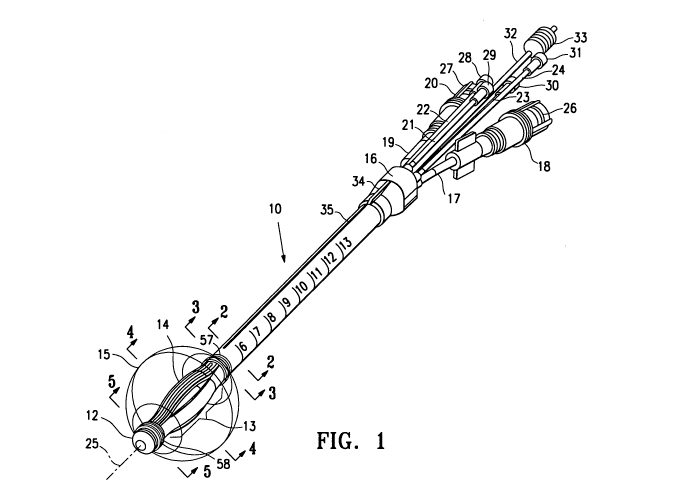
[0020] Figure 1 is a schematic perspective view, partially in section, of a
brachytherapy device embodying features of the invention.
[0021] Figure 2 is a transverse cross sectional view of the device shown in
Figure
1 taken along lines 2-2.
[0022] Figure 3 is a transverse cross sectional view of the device shown in
Figure
1 taken along the lines 3-3.
[0023] Figure 4 is a transverse cross sectional view of the device shown in
Figure
1 taken along the lines 4-4.
[0024] Figure 5 is a transverse cross sectional view of the device shown in
Figure
1 taken along the lines 5-5.
[0025] Figure 6 is a perspective view of the support member in the embodiment
shown in Figure 1.
[0026] Figure 7 is a perspective view of the distal tip of the device shown in
Figure
1.
[0027] Figure 8 is a longitudinal cross-sectional view of the distal tip shown
in
Figure 7.
[0028] Figure 9 is a perspective view of an alternative support member such as
shown in Figure 6 wherein the raised portions of the adjacent to the recess
are
provided with heating coils.
DETAILED DESCRIPTION OF THE INVENTION
[0029] The present invention is directed to devices and methods for treatment
of a
8
CA 02712923 2010-07-22
WO 2009/094159 PCT/US2009/000402
patient's body cavity, particularly to deliver asymmetrical radiation into a
body cavity
such as a cavity left after removal of tissue from the site. While the
detailed
description is directed to a device configured for treating a patient' breast
after tissue
removal such as in a lumpectomy, other body sites may also be treated with the
device.
[0030] Figures 1-8 illustrate a brachytherapy catheter device 10 embodying
features of the invention which has an elongated shaft 11, a distal tip 12, a
treatment
location 13 in a distal shaft portion 14 proximal to the distal tip. The
device 10 has a
balloon 15 on the distal shaft portion 14 which surrounds the treatment
location 13.
A hub 16 is mounted on the proximal end of the shaft 11 which has an inflation
line
17 with leur connection 18, a vacuum line 19 with a leur connection 20 and
four outer
delivery tubes 21, 22, 23, 24 for delivery of a radiation source through the
lumens
thereof to the treatment location 13 off set from a centrally location
longitudinal axis
25 to provide asymmetrical radiation of tissue surrounding the balloon 15. The
leur
connections 18 and 20 are provided with threaded caps 26 and 27 respectively
to
close off the connections. Each of the delivery tubes has a removable cap 28,
29,
30, and 31 respectively to close of the delivery tubes until use. A centrally
located
delivery tube 32 is provided for radiation source delivery along the central
longitudinal axis within the treatment location which also has a removable cap
33.
[0031] The hub 16 has a ridge 34 which is aligned with marker line 35 to
provide
the physician or other professional the orientation of the treatment location
13. The
elongated shaft 11 may also be provided with depth markings to help in the
placement of the balloon 15 within the cavity.
[0032] As shown best in Figure 2, the elongated shaft 11 has eight lumens,
four
lumens 36, 37, 38 and 39 equally spaced about the longitudinal axis 25 for
radiation
9
CA 02712923 2010-07-22
WO 2009/094159 PCT/US2009/000402
source delivery as described above and four equally spaced additional lumens
40,
41, 42 and 43, lumen 40 for vacuum application and lumen 42 for inflation
fluid
delivery to the interior of balloon 15. Lumens 41 and 44 are not used in this
embodiment, but may be used for a variety of functions. A proximal vacuum port
44
is provided in fluid communication with lumen 40 and distal vacuum port 45
(shown
best in Figures 5 and 7-8) is provided in the distal tip 12 which is in fluid
communication with lumen 40 through the annular space 46 between the central
delivery tube 32 and center lumen 47 of support member 48 shown in Figures 2-
4.
The support member 48, which is best shown in Figure 6.
[0033] As shown in Figures 1, 3 and 4, the distal shaft portion 14 is split
into four
separate longitudinal wall segments 49, 50, 51 and 52, with each wall segment
having one of the radiation source lumens 36-39 and being disposed within one
of
the recesses 53-56 in the exterior surface of support member 48. Recesses 55-
56
are not shown in Figure 6 but are on the opposite side of support member 48.
The
longitudinal wall segments 49-52 are slit through the lumens 40-43 as best
shown in
Figures 3-5. Lumens 40-43 are plugged off proximal to the split of the wall
segments
49-52. This wall segment structure facilitates the manufacture of the
catheter. The
elongated shaft may be extruded with all eight lumens 36-43 in place and the
distal
shaft portion 14 is segmented by cutting through lumens 40-43 by a cutting
blade or
other suitable cutting element. The support member 48 may be slid over the
central
delivery tube 32 with the proximal end of the support member secured within
the
central lumen of the shaft 11. The free ends of the slit wall segments 49-52
are
secured to the distal end of the support member 48. The balloon 15 may then be
secured to the shaft 11 with wound sutures 57 and 58 further securing the ends
of
the balloon to the shaft. The outer delivery tubes 21-24 may extend through
lumens
CA 02712923 2010-07-22
WO 2009/094159 PCT/US2009/000402
36-39 to the distal ends of the wall segments 49-52. Inflation line 17 and
vacuum
line 19 may likewise extend through lumens 40 and 41 to a location (not shown)
proximal to the split of the wall segments 49-52.
[0034] As best shown in Figures 5, 7 and 8, the distal tip 12 has the distal
vacuum
port 45 which is in fluid communication with the annular space 46 between
central
delivery tube 32 and lumen 47 of support member 48. The distal tip 12 is
provided
with outer source lumen plugs 60-63 for plugging lumens 36-39 and center
source
lumen plug 64 for plugging the distal end of central tube 32.
[0035] The brachytherapy catheter device 10 is readily manufactured. The
elongated shaft 11 is extruded, preferably with the lumens 36-43 within the
wall and
the central lumen 46. The distal shaft portion 14 is cut by a suitable cutting
member
such as a razor or knife like member to form the plurality of separated
longitudinal
wall segments 49-52. The support member 48 is preferably machined from an
extruded tubular polymeric product to form the recesses 53-56 and overall
shape
and centrally placed within the separated longitudinal wall segments. A
tubular
member 32 is positioned within the inner lumen of the elongated shaft 11 and
may
continue to the distal end of the shaft through the inner lumen of the support
member
48. The distal tip 12 is secured to the distal end of the shaft 11 and support
member
48 with plug members 60-63 inserted into the lumens within the wall segments
49-52
and central plug member 64 within the lumen of the centrally disposed tubular
member 32. The distal tip 12 is preferably preformed with the vacuum ports 45.
The
distal ends of the separated longitudinal wall segments are secured to the
distal end
of the device, preferably to the distal end of the support member. The balloon
15 is
mounted about the wall segments 49-52 and support member 48 with the distal
end
of the balloon secured to the distal end of the wall segments and support
member
11
CA 02712923 2010-07-22
WO 2009/094159 PCT/US2009/000402
and the proximal end of the balloon is secured to the elongated shaft proximal
to the
separated longitudinal wall segments. Preferably, strands or sutures are
wrapped
around each of the mounted ends of the balloon 15 to provide further support
to the
ends. The proximal end of the device 10 is similar to the brachytherapy
devices
previously described in copending applications 11/593,784 and 11/593,789
previously referred to herein.
[0036] A body cavity within a patient may be treated with the device 10 by
inserting the distal shaft portion 13 into the desired body cavity, inflating
the balloon
15 with inflation fluid to secure the device within the patient and applying a
vacuum
to either the distal or proximal vacuum ports or both to conform the tissue
lining the
cavity to the exterior of balloon 15. A radiation source is advanced through
one or
more of the source delivery lumens until the radiation source is properly
positioned
within the treatment location 13 (or prepositioned therein). The radiation
source (not
shown) is maintained at the treatment location 13 for a prescribe period of
time,
usually less than 30 minutes and typically a few (5-10) minutes. The radiation
source may be placed at several places within the treatment location with
within one
or multiple source lumens. At the end of the treatment time, the radiation
source
may be removed from device 10 or the entire device may be withdrawn from the
patient. Preferably, the device is left in place so that further radiation
treatments may
be performed.
[0037] The radiation source for the brachytherapy device 10 can include a
solid,
liquid or slurried radiation source. Suitable liquid radiation sources
include, for
example, a liquid containing a radioactive iodine isotope (e.g., 1125 or
1131), a slurry of
a solid isotope, for example, 198Au or 169Yb, or a gel containing a
radioactive isotope.
Liquid radiation sources are also commercially available (e.g., lotrex ,
Proxima
12
CA 02712923 2010-07-22
WO 2009/094159 PCT/US2009/000402
Therapeutics, Inc., Alpharetta, Ga.). The solid radiation source may be a
radioactive
microsphere available from 3M Company of St. Paul, Minn. A micro miniature x-
ray
source may also be utilized. The radiation source may be either preloaded into
the
device 10 at the time of manufacture or may be loaded into the device 10
before or
after placement into a body cavity or other site of a patient. Solid
radionuclides
suitable for use with a device 10 embodying features of the present invention
are
currently generally available as brachytherapy radiation sources (e.g., I-
Plant. TM.
Med-Tec, Orange City, Iowa.). Radiation may also be delivered by a device such
as
the x-ray tube of U.S. Patent No. 6,319,188. The x-ray tubes are small,
flexible and
are believed to be capable of being maneuverable enough to reach the desired
location within a patient's body.
[0038] The source delivery lumens of brachytherapy device 10 having features
of
the invention can be provided with a lubricious coating, such as a hydrophilic
material. The lubricious coating preferably is applied to the elongate shaft
12 or to
the cavity filling member, if one is present or both to reduce sticking and
friction
during insertion of a device 10. Hydrophilic coatings such as those provided
by AST,
Surmodics, TUA Systems, Hydromer, or STS Biopolymers are suitable.
[0039] A device 10 having features of the invention may also include an
antimicrobial coating that covers all or a portion of the device 10 to
minimize the risk
of introducing of an infection during extended treatments. The antimicrobial
coating
preferably is comprised of silver ions impregnated. into a hydrophilic
carrier.
Alternatively the silver ions are implanted onto the surface of the device 10
by ion
beam deposition. The antimicrobial coating preferably is comprised of an
antiseptic
or disinfectant such as chlorhexadiene, benzyl chloride or other suitable
biocompatible antimicrobial materials impregnated into hydrophilic coatings.
13
CA 02712923 2010-07-22
WO 2009/094159 PCT/US2009/000402
Antimicrobial coatings such as those provided by Spire, AST, Algon, Surfacine,
Ion
Fusion, or Bacterin International would be suitable. Alternatively a cuff
member
covered with the antimicrobial coating is provided on the elongated shaft of
the
delivery device 10 at the point where the device 10 enters the skin.
[0040] Figure 9 illustrates a modified support member 48 which is provided
with a
heating coil 70 to raise the temperature of tissue in the cavity lining either
simultaneously with or sequentially to irradiation of the cavity lining as
previously
described. While Figure 9 depicts a heating 70 on one raised portion of the
support
member 48, a heating element may be provided on a plurality of raised portions
of
the support member. Preferably, the heating coils are powered by RF energy and
are connected to a suitable high frequency generator. Voltage, current,
frequency
and duty factor may be adjusted to provide a suitable thermal treatment to
tissue
lining the cavity to augment the irradiation thereof. Other means may include
heating the inflation fluid within the balloon 15. The heating of the
inflation fluid may
be exterior to the device 10.
[0041] While particular forms of the invention have been illustrated and
described
herein, it will be apparent that various modifications and improvements can be
made
to the invention. Additional details of the brachytherapy catheter devices may
be
found in the patents and applications incorporated herein. To the extent not
otherwise disclosed herein, materials and structure may be of conventional
design.
[0042] Moreover, individual features of embodiments of the invention may be
shown in some drawings and not in others, but those skilled in the art will
recognize
that individual features of one embodiment of the invention can be combined
with
any or all the features of another embodiment. Accordingly, it is not intended
that
the invention be limited to the specific embodiments illustrated. It is
therefore
14
CA 02712923 2010-07-22
WO 2009/094159 PCT/US2009/000402
intended that this invention be defined by the scope of the appended claims as
broadly as the prior art will permit.
[0043] Terms such as "element", "member", "component", "device", "means",
"portion", "section", "steps" and words of similar import when used herein
shall not be
construed as invoking the provisions of 35 U.S.C 112(6) unless the following
claims
expressly use the terms "means for" or "step for" followed by a particular
function
without reference to a specific structure or a specific action. All patents
and all
patent applications referred to above are hereby incorporated by reference in
their
entirety.