Note: Descriptions are shown in the official language in which they were submitted.
CA 02713082 2010-07-22
WO 2009/103050
PCT/US2009/034213
METHOD OF QUANTITATION BY MASS SPECTROMETRY
INTRODUCTION
[0001] Quantitation by mass spectrometry is conventionally performed
with a
triple-quadrupole mass spectrometer using a multiple reaction monitoring (MRM)
method that selects certain product and precursor ion combinations to provide
the
best sensitivity and signal-to-noise. A linear dynamic range of three to five
orders
of magnitude can often be achieved by such a system. A triple-quadrupole mass
spectrometer with a time-of-flight mass spectrometer replacing the third
quadrupole (QqT0F) can also be used for quantitation, with the advantage that
much higher mass resolution can be achieved. However, intense product ions can
saturate the detector of a QqTOF mass spectrometer, limiting its linear
dynamic
range to only two to three orders of magnitude.
DRAWINGS
[0002] The skilled person in the art will understand that the drawings,
described
below, are for illustration purposes only. The drawings are not intended to
limit
the scope of the applicant's teachings in any way.
[0003] Figure 1 is a block diagram that illustrates a computer system,
upon which
embodiments of the present teachings may be implemented.
[0004] Figure 2 is a flowchart showing a method for improving
selectivity of a
measurement from a mass spectrometer, in accordance with the present
teachings.
[0005] Figure 3 is a flowchart showing a method for determining an
extracted ion
current (XIC) window to use for a mass spectrometer measurement, in accordance
with the present teachings.
1
CA 02713082 2010-07-22
WO 2009/103050
PCT/US2009/034213
[0006] Figure 4 is a flowchart showing a method for quantitation using
data from
a mass spectrometer, in accordance with the present teachings.
[0007] Figure 5 is a schematic diagram of a mass spectrometry system
that
includes a mass spectrometer and computer system, in accordance with the
present
teachings.
[0008] Figure 6 is an exemplary product ion mass spectrum from a urine
sample,
in accordance with the present teachings.
[0009] Figure 7 is an exemplary expanded view of a product ion mass
spectrum
from a urine sample, in accordance with the present teachings
[0010] Figure 8 is an exemplary expanded view of a product ion mass
spectrum
from a urine sample showing an XIC window with a width of 0.5 atomic mass
units (amu), in accordance with the present teachings.
[0011] Figure 9 is an exemplary plot of the XIC for five samples
injected about
three minutes apart using the XIC window shown in Figure 8, in accordance with
the present teachings.
[0012] Figure 10 is an exemplary expanded view of a product ion mass
spectrum
from a urine sample showing an XIC window with a width of 0.01 amu, in
accordance with the present teachings.
[0013] Figure 11 is an exemplary plot of the XIC for five samples
injected about
three minutes apart using the XIC window shown in Figure 10, in accordance
with
the present teachings.
[0014] Figure 12 is an exemplary plot of a mass peak of interest and an
interfering
mass peak, showing how it can be advantageous to select a position of the XIC
window that is not centered on the true center of the mass of interest, in
accordance with the present teachings.
2
CA 02713082 2010-07-22
WO 2009/103050
PCT/US2009/034213
[0015] Figure 13 is a table showing the linear ranges of the calibration
curves of
five product ions of an exemplary known compound, in accordance with the
present teachings.
[0016] Figure 14 is a table showing the intensities of five product ions
of an
exemplary known compound that are found in a sample, in accordance with the
present teachings.
[0017] Before one or more embodiments of the invention are described in
detail,
one skilled in the art will appreciate that the invention is not limited in
its
application to the details of construction, the arrangements of components,
and the
arrangement of steps set forth in the following detailed description or
illustrated in
the drawings. The invention is capable of other embodiments and of being
practiced or being carried out in various ways. Also, it is to be understood
that the
phraseology and terminology used herein is for the purpose of description and
should not be regarded as limiting.
DESCRIPTION OF VARIOUS EMBODIMENTS
[0018] The section headings used herein are for organizational purposes
only and
are not to be construed as limiting the subject matter described in any way.
COMPUTER IMPLEMENTED SYSTEM
[0019] Figure 1 is a block diagram that illustrates a computer system
100, upon
which embodiments of the present teachings may be implemented. Computer
system 100 includes a bus 102 or other communication mechanism for
communicating information, and a processor 104 coupled with bus 102 for
processing information. Computer system 100 also includes a memory 106,
which can be a random access memory (RAM) or other dynamic storage device,
coupled to bus 102 for determining base calls, and instructions to be executed
by
3
CA 02713082 2010-07-22
WO 2009/103050
PCT/US2009/034213
processor 104. Memory 106 also may be used for storing temporary variables or
other intermediate information during execution of instructions to be executed
by
processor 104. Computer system 100 further includes a read only memory
(ROM) 108 or other static storage device coupled to bus 102 for storing static
information and instructions for processor 104. A storage device 110, such as
a
magnetic disk or optical disk, is provided and coupled to bus 102 for storing
information and instructions.
[0020] Computer system 100 may be coupled via bus 102 to a display 112,
such
as a cathode ray tube (CRT) or liquid crystal display (LCD), for displaying
information to a computer user. An input device 114, including alphanumeric
and
other keys, is coupled to bus 102 for communicating information and command
selections to processor 104. Another type of user input device is cursor
control
116, such as a mouse, a trackball or cursor direction keys for communicating
direction information and command selections to processor 104 and for
controlling cursor movement on display 112. This input device typically has
two
degrees of freedom in two axes, a first axis (e.g., x) and a second axis
(e.g., y),
that allows the device to specify positions in a plane.
[0021] Computer system 100 can perform the present teachings. Consistent
with
certain implementations of the present teachings, results are provided by
computer
system 100 in response to processor 104 executing one or more sequences of one
or more instructions contained in memory 106. Such instructions may be read
into memory 106 from another computer-readable medium, such as storage device
110. Execution of the sequences of instructions contained in memory 106 causes
processor 104 to perform the process described herein. Alternatively hard-
wired
circuitry may be used in place of or in combination with software instructions
to
4
CA 02713082 2010-07-22
WO 2009/103050
PCT/US2009/034213
implement the present teachings. Thus implementations of the present teachings
are not limited to any specific combination of hardware circuitry and
software.
[0022] The term "computer-readable medium" as used herein refers to any
media
that participates in providing instructions to processor 104 for execution.
Such a
medium may take many forms, including but not limited to, non-volatile media,
volatile media, and transmission media. Non-volatile media includes, for
example, optical or magnetic disks, such as storage device 110. Volatile media
includes dynamic memory, such as memory 106. Transmission media includes
coaxial cables, copper wire, and fiber optics, including the wires that
comprise bus
102. Transmission media can also take the form of acoustic or light waves,
such
as those generated during radio-wave and infra-red data communications.
[0023] Common forms of computer-readable media include, for example, a
floppy disk, a flexible disk, hard disk, magnetic tape, or any other magnetic
medium, a CD-ROM, any other optical medium, punch cards, papertape, any
other physical medium with patterns of holes, a RAM, PROM, and EPROM, a
FLASH-EPROM, any other memory chip or cartridge, a carrier wave as described
hereinafter, or any other medium from which a computer can read.
[0024] Various forms of computer-readable media may be involved in
carrying
one or more sequences of one or more instructions to processor 104 for
execution.
For example, the instructions may initially be carried on the magnetic disk of
a
remote computer. The remote computer can load the instructions into its
dynamic
memory and send the instructions over a telephone line using a modem. A
modem local to computer system 100 can receive the data on the telephone line
and use an infra-red transmitter to convert the data to an infra-red signal.
An
infra-red detector coupled to bus 102 can receive the data carried in the
infra-red
CA 02713082 2010-07-22
WO 2009/103050
PCT/US2009/034213
signal and place the data on bus 102. Bus 102 carries the data to memory 106,
from which processor 104 retrieves and executes the instructions. The
instructions received by memory 106 may optionally be stored on storage device
110 either before or after execution by processor 104.
[0025] In accordance with various embodiments, instructions configured
to be
executed by a processor to perform a method are stored on a computer-readable
medium. The computer-readable medium can be a device that stores digital
information. For example, a computer-readable medium can include, but is not
limited to, a compact disc read-only memory (CD-ROM) as is known in the art
for
storing software. The computer-readable medium is accessed by a processor
suitable for executing instructions configured to be executed.
[0026] The following descriptions of various implementations of the
present
teachings have been presented for purposes of illustration and description. It
is
not exhaustive and does not limit the present teachings to the precise form
disclosed. Modifications and variations are possible in light of the above
teachings or may be acquired from practicing of the present teachings.
Additionally, the described implementation includes software but the present
teachings may be implemented as a combination of hardware and software or in
hardware alone. The present teachings may be implemented with both object-
oriented and non-object-oriented programming systems.
METHODS OF DATA PROCESSING
Selectivity
[0027] Triple quadrupole mass spectrometers are widely used to measure
the
amount or concentration of compounds such as, for example, pharmaceuticals in
plasma or urine samples. A precursor and product ion combination must be
6
CA 02713082 2010-07-22
WO 2009/103050
PCT/US2009/034213
selected in advance when using the multiple reaction monitoring (MRM) method
with a triple quadrupole mass spectrometer. Additionally, with a triple
quadrupole, the mass resolution (peak width) must be tuned and fixed in
advance
of the data acquisition. It is not possible after the acquisition to change or
select
the width of the XIC window with a triple quadrupole.
[0028] In contrast, when a triple-quadrupole mass spectrometer with a
time-of-
flight mass spectrometer replacing the third quadrupole (QqT0F) is used for
quantitation, a product ion or multiple product ions can be selected after
acquisition of a sample spectrum. There is no need to characterize the matrix
or
select the best MRM combinations in advance. There is no need to perform these
steps in advance because a QqTOF spectrometer can obtain a full product ion
spectrum.
[0029] The measurement of a concentration of an amount of a known
compound
in a sample is often performed, for example, by acquiring mass spectra
continuously during a time period in which the sample elutes from a liquid
chromatograph (LC) column. Alternatively, the compound can be injected into a
flowing liquid stream without an LC column, in a technique called flow
injection
analysis (FIA). Spectra are acquired continuously during a time period, which
can
be of several minutes in duration, commonly with a frequency of 1 spectrum per
second. In various embodiments, a plurality of spectra acquired during this
time
period forms a data set which can be processed by calculating an extracted ion
current (XIC) for each ion of interest.
[0030] Also, in various embodiments the mass-to-charge width, or width
of the
XIC window, for each product ion can be selected after the acquisition of a
plurality of sample spectra to provide the best signal-to-noise ratio (S/N).
For
7
CA 02713082 2010-07-22
WO 2009/103050
PCT/US2009/034213
example, a narrow XIC window that corresponds to less than the width of the
mass peak can be selected for processing if there is an improvement in the S/N
compared to selecting a wider XIC window. Both the center position and the
width of the selected window can be selected to provide maximum signal-to-
noise. For example, the center of the XIC window can be chosen to be on one
side of the actual mass value if there is an interfering mass peak that
overlaps on
the other side of the mass peak of interest. In order to generate a measurable
signal, the selected XIC window must overlap to some degree with the position
of
the true mass peak of interest. In various embodiments the selection of the
width
of the XIC window is selected after the acquisition of the plurality of sample
spectra, avoiding the necessity of tuning the mass spectrometer for a specific
mass
resolution before the analysis.
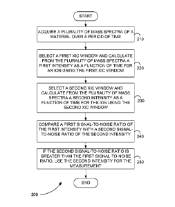
[0031] Figure 2 is a flowchart showing a method 200 for improving
selectivity of
a measurement from a mass spectrometer, in accordance with the present
teachings. The mass spectrometer can include, but is not limited to, a time of
flight mass spectrometer or an electrospray ionization time of flight mass
spectrometer. The measurement can be, for example, a quantitation measurement.
[0032] In step 210 of method 200, a plurality of mass spectra of a
material are
acquired over a period of time. The plurality of mass spectra can be, for
example,
product ion mass spectra.
[0033] In step 220, a first XIC window is selected and from the
plurality of mass
spectra a first intensity as a function of time is calculated for an ion using
the first
XIC window. The first XIC window includes a first width and a first center,
for
example. The ion can be, for example, a product ion. The first XIC window is
selected after acquisition of the plurality of mass spectra, for example.
8
CA 02713082 2010-07-22
WO 2009/103050
PCT/US2009/034213
[0034] In step 230, a second XIC window is selected and from the
plurality of
mass spectra a second intensity as a function of time is calculated for the
ion using
the second XIC window. The second XIC window includes a second width and a
second center, for example. The second XIC window is selected after
acquisition
of the plurality of mass spectra, for example.
[0035] In step 240, a first S/N of the first intensity is compared with
a second S/N
of the second intensity.
[0036] In step 250, if the second S/N is greater than the first S/N, the
second
intensity as a function of time is used for the measurement.
[0037] In various embodiments, the first width is larger than the second
width. In
various embodiments, the first width and the width have values less than 0.02
atomic mass units. In various embodiments, the first center and the second
center
are not equal.
[0038] Figure 3 is a flowchart showing a method 300 for determining an
XIC
window to use for a mass spectrometer measurement, in accordance with the
present teachings. The mass spectrometer can include, but is not limited to, a
time
of flight mass spectrometer or an electrospray ionization time of flight mass
spectrometer. The measurement can be, for example, a quantitation measurement.
[0039] In step 310 of method 300, a plurality of mass spectra of a
material are
acquired over a period of time. The plurality of mass spectra can be, for
example,
product ion mass spectra.
[0040] In step 320, an initial XIC window is selected. The initial XIC
window
can be selected after acquisition of the plurality of mass spectra.
9
CA 02713082 2010-07-22
WO 2009/103050
PCT/US2009/034213
[0041] In step 330, the XIC window is set equal to the initial XIC
window and
from the plurality of mass spectra an intensity as a function of time is
calculated
for an ion using the XIC window. The ion can be, for example, a product ion.
[0042] In step 340, the steps of changing a parameter of the XIC window
by an
increment, calculating from the plurality of mass spectra a next intensity as
a
function of time for the ion using the changed parameter of the XIC window,
and
calculating a next S/N from the next intensity are repeated until a stop
condition is
reached. The stop condition is, for example, the next S/N reaching a maximum
S/N. The XIC window at the maximum S/N can then be used for the
measurement.
[0043] In various embodiments, the stop condition is the next S/N
becoming
greater than or equal to a threshold. The threshold can be, for example, 3. A
parameter of the XIC window includes, but is not limited to, the width or the
center. Changing a parameter of the XIC window by an increment can include,
but is not limited to, decreasing the parameter by the increment or increasing
the
parameter by the increment. The increment can be, for example, 0.01 atomic
units.
[0044] In various embodiments, a mass spectrometry system includes a
mass
spectrometer and a computer system. The mass spectrometer can include, but is
not limited to, a time-of-flight mass spectrometer or an electrospray
ionization
time of flight mass spectrometer.
[0045] The computer system is in communication with mass spectrometer.
The
computer system can be, but is not limited to, computer system 100, shown in
Figure 1 and described above. The computer system acquires a plurality of mass
spectra of a material over a period of time, selects a first XIC window and
CA 02713082 2010-07-22
WO 2009/103050
PCT/US2009/034213
calculates from the plurality of mass spectra a first intensity as a function
of time
for an ion using the first XIC window, selects a second XIC window and
calculates from the plurality of mass spectra a second intensity as a function
of
time for the ion using the second XIC window, compares a first S/N of the
first
intensity with a second S/N of the second intensity, and, if the second S/N is
greater than the first S/N, uses the second intensity as a function of time
for the
measurement. The computer system can select the first XIC window and the
second XIC window after acquisition of the plurality of mass spectra, for
example.
Quantitation
[0046] When a triple-quadrupole mass spectrometer with a time-of-flight
mass
spectrometer replacing the third quadrupole (QqTOF) is used for quantitation,
a
full product ion spectrum can be obtained. The spectrum can include a range of
product ions, some more intense than others. In various embodiments, a wide
dynamic range can be obtained by using the most intense product ions for
quantitation at low concentrations, and using less intense product ions for
quantitation at high concentration where the larger-intensity ions are
saturated.
[0047] This wide dynamic range is possible with a QqTOF mass
spectrometer,
because the intensities of less intense ions are not affected by the
intensities of the
more intense ions. Use of a QqTOF mass spectrometer also allows a product ion
or multiple product ions to be selected for quantitation after acquisition of
a
sample spectrum. In contrast, the use of a conventional triple-quadrupole
spectrometer using a multiple reaction monitoring (MRM) method requires that a
product ion or multiple product ions be selected for quantitation before a
sample is
analyzed. Additionally, with a triple quadrupole, the mass resolution (peak
width)
must be tuned and fixed in advance of the data acquisition. It is not possible
after
11
CA 02713082 2010-07-22
WO 2009/103050
PCT/US2009/034213
the acquisition to change or select the width of the extracted ion current
(XIC)
window with a triple quadrupole.
[0048] Using a QqTOF mass spectrometer, a range of linear response for
each
product ion can be established from a calibration curve, and multiple product
ions
can be used to produce a linear calibration curve over a wide range of
concentrations. An internal standard can be used to compensate for matrix and
ionization suppression effects.
[0049] Other mass spectrometers can also be used to provide a full
product ion
spectrum and wide dynamic range. These spectrometers include, but are not
limited to, a linear ion trap mass spectrometer, an orbitrap mass
spectrometer, a
Fourier transform mass spectrometer, or a three-dimensional ion trap mass
spectrometer.
[0050] In various embodiments, calibration curves are constructed for
more than
one product ion of the same precursor, the acquired product ion spectra of a
sample is processed, and the concentration of the sample is measured by
selecting
the product ion or product ions that are still in the linear portion of the
response
curve. Multiple product ions can be used for the measurement of concentration
by
combining the measurements in an algorithm that assigns confidence or
precision
based on statistical criteria. For example, two product ions can be used to
measure the concentration, but if the signal-to-noise ratio (S/N) of one
product ion
is much lower than the other, then the two results can be combined in a
statistically relevant method. A statistically relevant method can include,
but is
not limited to, weighting the two results based on their S/Ns.
[0051] In various embodiments, a method of using a mass spectrometer for
quantitative measurement of an unknown concentration can include producing a
12
CA 02713082 2010-07-22
WO 2009/103050
PCT/US2009/034213
response curve from a standard material over a wide range of concentrations,
where full product ion spectra are acquired over the range of concentrations,
measuring the linearity of response for at least two product ions with
different
responses, and measuring the response to an unknown sample concentration by
acquiring the product ions spectrum, and determining the concentration from
the
intensity of the product ions that corresponds to a linear portion of the
response
curve.
[0052] Figure 4 is a flowchart showing a method 400 for quantitation
using data
from a mass spectrometer and involving the selection two or more ions within
their linear ranges, in accordance with the present teachings. The mass
spectrometer can include, but is not limited to, a time-of-flight mass
spectrometer,
a linear ion trap mass spectrometer, an orbitrap mass spectrometer, a Fourier
transform mass spectrometer, or a three-dimensional ion trap mass
spectrometer.
[0053] In step 410 of method 400, a plurality of calibration ion mass
spectra are
acquired for each of a plurality of known quantities of a material. The
plurality of
calibration ion mass spectra is, for example, a plurality of product ion mass
spectra.
[0054] In step 420, from the plurality of calibration ion mass spectra a
plurality of
ions that identify the material is determined and for each ion of the
plurality of
ions a linear range over which an intensity of each ion varies linearly with
quantity and a linear function for the linear range are determined.
[0055] In step 430, a plurality of sample ion mass spectra is acquired
for an
unknown quantity of the material. A sample ion mass spectrum is, for example,
a
product ion mass spectrum. In various embodiments, the resolution of a
quadrupole mass filter can be adjusted to transmit a mass window that includes
13
CA 02713082 2010-07-22
WO 2009/103050
PCT/US2009/034213
two or more isotopes of the compound. It is common that carbon-13 containing
isotopes provide precursor ions of less intensity than the first isotope.
Selection of
a mass window that includes two or more isotopes of the precursor ion can
result
in product ions that contain carbon-13 isotopes and are less intense than the
first
isotope. These less intense product ions can be used for quantitation.
Alternatively, the unfragmented precursor ion and its isotopes can be used for
quantitation. Alternatively, quantitation can be done in a time-of-flight mass
spectrometry (TOFMS) mode using the precursor ion and its isotopes to provide
a
range of ion intensities. In various embodiments, quantitation can be done in
a
TOFMS system using product ions that are created without selecting the
precursor
ions, for example, by fragmentation in the ion source, in the declustering
region,
or in the ion optics region before the time-of-flight (TOF).
[0056] In step 440, a sample intensity is measured for each ion of the
plurality of
ions from the sample spectra.
[0057] In step 450, after acquiring the sample spectra, one or more ions
from the
plurality of ions are selected such that a sample intensity of each selected
ion of
the one or more ions is within a linear range of each selected ion. In various
embodiments, the one or more ions are selected only if the signal to noise
ratio of
each ion of the one or more ions is greater than or equal to a threshold
value. A
S/N threshold value is 3, for example. Signal-to-noise can be determined by
methods that are known in the art. For example, signal-to-noise can be
determined by calculating the ratio of the peak height to the standard
deviation of
the background ion signal in a selected time window. Other measurements of
signal-to-noise can also be used.
14
CA 02713082 2010-07-22
WO 2009/103050
PCT/US2009/034213
[0058] In step 460, the unknown quantity is calculated from one or more
sample
intensities of the one or more ions and one or more linear functions of the
one or
more ions. In various embodiments, the one or more ions can include two or
more
ions. In various embodiments, calculating the unknown quantity can include
averaging two or more quantities of the two or more ions, where each quantity
of
the two or more quantities is obtained from a sample intensity and a linear
function of an ion of the two or more ions. In various embodiments,
calculating
the unknown quantity can also include summing two or more weighted quantities
of the two or more ions, wherein each weighted quantity of the two or more
weighted quantities is obtained from a signal-to-noise weighting factor, a
sample
intensity, and a linear function of an ion of the two or more ions.
[0059] In various embodiments, a sample intensity is measured by
measuring the
extracted ion current (XIC) as a function of time, and determining the area
under a
curve that corresponds to a time window that is characteristic of the sample
compound. For example, the characteristic window can be the time window in
which the compound elutes from a liquid chromatography system. The XIC is a
measurement of intensity consisting of the sum or integral of the ion current
measurement within a fixed mass-to-charge window in the mass spectrum. For
example, a mass peak of a product ion has a characteristic mass peak width
that is
determined by the resolution of the mass spectrometer. It is common to select
an
XIC window width that corresponds to all or a major portion of the mass peak,
and to select a center value for the XIC window that consists of the known
mass
value (which can be the peak top or centroid of the mass peak). In some cases
the
XIC can consist of a single mass peak point, corresponding to the minimum
width
of the mass scale. This minimum width in a time-of-flight mass spectrometer is
CA 02713082 2010-07-22
WO 2009/103050
PCT/US2009/034213
the minimum time resolution or bin size of the time measurement. The intensity
of the minimum width peak can also be the same as the peak height in the mass
spectrum.
[0060] Also, in various embodiments the width of the XIC window for each
product ion can be selected after the acquisition of a sample spectrum to
provide
the best signal-to-noise ratio (S/N). For example, a narrow XIC window that
corresponds to less than the width of the mass peak can be selected for
processing
if there is an improvement in the S/N compared to selecting a wider XIC
window.
Both the center position and the width of the selected window can be selected
to
provide maximum signal-to-noise. For example, the center of the XIC window
can be chosen to be on one side of the actual mass value if there is an
interfering
mass peak that overlaps on the other side of the mass peak of interest. In
order to
generate a measurable signal, the selected XIC window must overlap to some
degree with the position of the true mass peak of interest.
[0061] In various embodiments, the width and position of the XIC window
for
each mass in the spectrum known to be associated with the compound of interest
is selected in order to provide a maximum S/N. The XIC window width can be
different for each selected mass value. For each mass value, the XIC window
width selected to provide a maximum S/N can be used to calculate a
concentration
from a calibration curve that is generated from the calibration samples by
using
the same XIC window width. For example, if m/z 255.035 is a sample mass and
an XIC window width of 0.015 atomic mass units (amu) provides the best S/N for
a particular known sample of unknown concentration, then the concentration can
be calculated from a calibration curve for m/z 255.035 that uses the same
width of
0.015 amu for the XIC. The calibration curves of a different XIC window width
16
CA 02713082 2010-07-22
WO 2009/103050
PCT/US2009/034213
can be generated by the computer before the samples of unknown concentration
are run or after the samples are run.
[0062] Also in various embodiments, the position of the XIC window can
be
selected to provide the best S/N. For example, if the known exact m/z of the
sample ion is 255.035, then the XIC window for m/z 255.035 with a width of
0.015 amu can be selected by the computer, and the S/N of the peak at the
correct
retention time can be calculated. Next, the XIC window for m/z 255.045 can be
calculated with a width of 0.015 amu. If the S/N for a peak at the correct
retention
time is higher for m/z 255.045 than for m/z 255.035, then this XIC window can
be
used to measure the sample concentration from the calibration curve.
[0063] In various embodiments, after acquiring the sample spectrum, one
or more
ions are selected from the plurality of ions. For each selected ion the best
XIC
window width can be determined by measuring the S/N for a range of XIC
window widths. For each selected mass value, a calibration curve can be
generated for the selected XIC window width from calibration data. One or more
ions can be selected from the plurality of ions such that a sample intensity
of each
selected ion of the one or more ions is within a linear range of the
calibration
curve of each selected ion.
[0064] In various embodiments, the center position of the XIC window can
be
selected in order to provide the best S/N.
[0065] In various embodiments, a first XIC window and a second XIC
window
for at least one of the one or more ions are selected. The first XIC window
width
of the first XIC window is not equal to a second XIC window width of the
second
XIC window, for example. In various embodiments, a first XIC window center
position of the first XIC window is not equal to a second XIC window center
17
CA 02713082 2010-07-22
WO 2009/103050
PCT/US2009/034213
position of the second XIC window. A first sample intensity for the at least
one of
the one or more ions is calculated using the first XIC window and a second
sample intensity for the at least one of the one or more ions is calculate
using the
second XIC window. A first S/N of the first sample intensity is calculated and
a
second S/N of the second sample intensity is calculated. If the second S/N is
greater than the first S/N, the second sample intensity is used to calculate
the
unknown quantity.
[0066] In various embodiments, a first XIC window and a second XIC
window
for at least one of the one or more ions are selected. The first XIC window
width
of the first XIC window is not equal to a second XIC window width of the
second
XIC window, for example. In various embodiments, a first XIC window center
position of the first XIC window is not equal to a second XIC window center
position of the second XIC window. A first sample intensity for the at least
one of
the one or more ions is calculated using the first XIC window and a second
sample intensity for the at least one of the one or more ions is calculated
using the
second XIC window. A first relative contribution of a closely eluting compound
in the sample to the first sample intensity is calculated and a second
relative
contribution of the closely eluting compound in the sample to the second
sample
intensity is calculated. A relative contribution of a closely eluting compound
in
the sample to the sample intensity is a measure of interference between the
closely
eluting compound in the sample and the material of interest in the compound.
The
relative contribution of the closely eluting compound in the sample to the
sample
intensity is, for example, the proportion of the sample intensity due to the
closely
eluting compound relative to the proportion of the sample intensity due to the
materiel of interest. If the second relative contribution is less than the
first relative
18
CA 02713082 2010-07-22
WO 2009/103050
PCT/US2009/034213
contribution, the second sample intensity is used to calculate the unknown
quantity.
[0067] The linear range of the calibration curve, in terms of sample
concentration
or sample amount for each of the known ions in the sample, is not dependent on
the width of the XIC window selected for the calibration. For example, if the
linear range of calibration for the ion of m/z 255.035 is from 10 femto-grams
(fg)
to 10 pico-grams (pg) as determined from a calibration curve with an XIC
window
width of 0.02 amu, then the linear range of the calibration curve for an XIC
window width of 0.01 amu will still be from 10 fg to 10 pg. For any ion mass,
the
linear range for any selected XIC window width will all be the same. This is
because the non-linearity or curvature at the upper end of the range is due to
saturation of the ion detector, which is caused by the number of ions in the
entire
mass peak hitting the detector at that particular sample concentration.
Therefore
the range of linearity in sample concentration is determined by the number if
ions
in the mass peak. Selecting a different XIC window width changes the number of
ion counts associated with the sample concentration, and therefore changes the
absolute intensity value of the calibration curve, but does not change the
shape of
the calibration curve.
[0068] In various embodiments, an XIC window width can be selected for
each of
the known ions of the sample. Calibration curves can be determined for each of
the known ions, and linear ranges determined for each of the known ions. For
each unknown sample, the response for each known ion can be determined by
using the XIC window width. The ions that have a response within the linear
range can be determined. For those ions that are within the linear response
range,
the XIC window width and center value can be varied and selected according to
19
CA 02713082 2010-07-22
WO 2009/103050
PCT/US2009/034213
the methods described above in order to determine the best XIC window width.
If
a different XIC window width or center value is selected than that used for
the
calibration curve for that ion, a new calibration curve can be calculated by
using
the new selected XIC window width, and the concentration of the sample can be
calculated based on the new calibration curve. The improved S/N obtained from
the new selected XIC window can provide a more accurate measurement of the
sample concentration than was obtained from the original XIC window width.
[0069] In various embodiments the best XIC window can be selected before
the
linear range of the calibration is determined. After finding the best XIC
window
for each ion, the calibration curve can be produced by using that XIC window
to
process the data from the plurality of calibration ion mass spectra. For each
ion
and selected XIC window the linear range of response and a linear function can
be
determined. The unknown quantity is calculated from one or more sample
intensities of the one or more ions and one or more linear functions of the
one or
more ions.
[0070] Figure 5 is a schematic diagram of a mass spectrometry system 500
that
includes mass spectrometer 510 and computer system 520, in accordance with the
present teachings. Mass spectrometer 510 can include, but is not limited to, a
time-of-flight mass spectrometer, a linear ion trap mass spectrometer, an
orbitrap
mass spectrometer, a Fourier transform mass spectrometer, or a three-
dimensional
ion trap mass spectrometer.
[0071] Computer system 520 is in communication with mass spectrometer
510.
Computer system 520 can be, but is not limited to, computer system 100, shown
in Figure 1 and described above. Computer system 520 acquires a plurality of
calibration ion mass spectra for each of a plurality of known quantities of a
CA 02713082 2010-07-22
WO 2009/103050
PCT/US2009/034213
material, determines from the plurality of calibration ion mass spectra a
plurality
of ions that identify the material and for each ion of the plurality of ions a
linear
range over which an intensity of each ion varies linearly with quantity and a
linear
function for the linear range, acquires a plurality of sample ion mass spectra
for an
unknown quantity of the material, measures a sample intensity for each ion of
the
plurality of ions from the sample spectra, after acquiring the sample spectra,
selects one or more ions from the plurality of ions such that a sample
intensity of
each selected ion of the one or more ions is within a linear range of each
selected
ion, and calculates the unknown quantity from one or more sample intensities
of
the one or more ions and one or more linear functions of the one or more ions.
In
various embodiments, the width and center position of the XIC window is
selected
for each of the plurality of ions before the one or more ions is selected such
that
the sample intensity is within the linear range of the calibration curve.
EXAMPLES
[0072] Aspects of the applicant's teachings may be further understood in
light of
the following examples, which should not be construed as limiting the scope of
the present teachings in any way.
Selectivity
[0073] Figure 6 is an exemplary product ion mass spectrum 600 from a
urine
sample, in accordance with the present teachings. Spectrum 600 is a product
ion
spectrum of a precursor with mass-to-charge ratio (m/z) 237. Many peaks are
present, but only a few are associated with the drug of interest
(Carbamazapine).
[0074] Figure 7 is an exemplary expanded view of a product ion mass
spectrum
700 from a urine sample, in accordance with the present teachings. Spectrum
700
21
CA 02713082 2010-07-22
WO 2009/103050
PCT/US2009/034213
shows that even at m/z 192 product mass, there are several components present.
Only 192.0929, however, is due to Carbamazapine.
[0075] Figure 8 is an exemplary expanded view of a product ion mass
spectrum
800 from a urine sample showing an extracted ion current (XIC) window 810 with
a width of 0.5 atomic mass units (amu), in accordance with the present
teachings.
Selected XIC window 810 extends from 191.799 to 192.305 Daltons (Da) and has
a center at 192.052 Da. Selected XIC window 810 includes all components at m/z
192 product mass.
[0076] Figure 9 is an exemplary plot 900 of the XIC 910 for five samples
injected
about three minutes apart using XIC window 810 shown in Figure 8, in
accordance with the present teachings. Each sample is slightly different and
has
multiple peaks. Only one peak is Carbamazapine. The other peaks are potential
interferences that are separated by liquid chromatography (LC) in this case,
but
might not be in other cases.
[0077] Figure 10 is an exemplary expanded view of a product ion mass
spectrum
1000 from a urine sample showing an XIC window 1010 with a width of 0.01
atomic mass units (amu), in accordance with the present teachings. Figure 10
shows the same data as shown in Figure 8, but now using a narrower XIC
window. Selected XIC window 1010 extends from 192.079 to 192.089 Da and
has a center at 192.084 Da. Selected XIC window 1010 does not include all of
the
components at m/z 192 product mass.
[0078] Figure 11 is an exemplary plot 1100 of the XIC 1110 for five
samples
injected about three minutes apart using the XIC window shown in Figure 10, in
accordance with the present teachings. Comparing Figure 11 with Figure 9 shows
that selected narrower XIC window 1010, shown in Figure 10 and centered at
22
CA 02713082 2010-07-22
WO 2009/103050
PCT/US2009/034213
192.084 Da, provides a better S/N than selected XIC window 810, shown in
Figure 8 and centered at 192.052 Da. The width and center of XIC window 1010,
shown in Figure 10, and the width and center of XIC window 810, shown in
Figure 8, are, for example, chosen after sample data acquisition and selected
so
that each XIC window includes or is near correct mass value for Carbamazapine.
The correct mass value is known from a standard, for example.
[0079] Figure 12 is an exemplary plot 1200 of a mass peak of interest
1210 and an
interfering mass peak 1220, showing how it can be advantageous to select a
position of the XIC window that is not centered on the true center of the mass
of
interest, in accordance with the present teachings. If XIC window 1225 is
selected, the XIC will contain significant contributions from both mass peak
of
interest 1210 and interfering mass peak 1220. If XIC window 1215 is selected,
the signal from mass peak of interest 1210 is reduced, but the signal from
interfering mass peak 1220 is reduced even more, so the S/N is increased.
Quantitation
[0080] According to an exemplary method for quantitation using data
from a mass
spectrometer a series of standards are run over a range of concentrations.
From
the series of standards, a calibration curve is constructed for each product
ion over
the entire range of concentrations. For each calibration curve, the highest
concentrations are discarded until a linear function is regression fit to the
curve
within a threshold value. A threshold value for linearity can be determined
from a
coefficient of determination R2 > 0.995 where R is the coefficient of
variations,
determined using standard and well-known regression methods, for example. In
various embodiments, the threshold value can be greater than or less than
0.995.
23
CA 02713082 2010-07-22
WO 2009/103050
PCT/US2009/034213
[0081] Alternatively, other criteria can be used to determine where the
calibration
function becomes non-linear. For example, a threshold value can be used below
which the deviation from linearity is acceptable, and above which the
deviation
from linearity is unacceptable. In various embodiments a weighted linear
regression may be used. In one example of weighted linear regression, a
weighting factor of 1/x is applied to each data point where x is the sample
concentration. Weighted linear regression is known in the art.
[0082] As a result, a linear calibration curve that covers a certain
range of
concentrations is obtained for each ion. These calibration curves are
generated
and stored prior to running unknown samples. In various embodiments, the
calibration curves can be obtained after running the unknown samples and
before
processing the data to determine the concentrations of the unknowns. In
various
embodiments, calibration curves can be run before an unknown sample and after
running an unknown sample, and two or more calibration curves can be combined
in a statistically reasonable fashion, for example by averaging the
calibration
curves together.
[0083] After running a sample, the response for each of the known
product ions in
the sample is measured. The intensity of each product ion in the sample is
then
compared with its calibration curve to determine if the intensity is within
the
linear range of that calibration curve. If the intensity is within the linear
range, the
product ion can be used for quantitation.
[0084] Consider an exemplary known compound with a precursor mass-to-
charge
ratio (m/z) of 287 and product ions with m/z's of 59, 89, 122, 231, and 269.
Using the method described above, the linear ranges of the calibrations curves
for
24
CA 02713082 2015-12-17
WO 2009/103050
PCT/US2009/034213
each of the: product ions can be found and shown in terms of concentration and
intensity,
[0085] Figure 13 is a table 1300 showing the linear ranges of the
calibration
curves of five product ions of the exemplary known compound, in accordance
with the present teachings. The data shown. in table 1300 is consistent with a
mass
spectrometer that saturates (or becomes non-linear) above 4000 ions and has a
detection limit of 10 ions.
[00861 Again using the method described above, a sample is run and the
response
for each of the known prod-net ions in the sample is measured. Figure 14 is a
table
1400 showing the intensities of five product ions of an exemplary known
compound that are found in the Sample, in accordance with thepresent
teachings.
Table 1400 shows that product ions with mh's 59,. 89, and 269 are outside of
the
linear calibration ranges for those ions. Therefore the concentration of the
known
compound in the sample is calculated from the concentrations of the product
ions
with rnmz 122 (128 ions= 128 pico-grams (pg)) and rniz 231 (45 ions = 135 pg).
The concentration of the known compound can be found from the concentrations
of the two product ions,, for example. The concentration of the known compound
can also be found by combining the concentrations of the two product ions in a
statistically significant fashion..
[0087] An exemplary statistically significant method of combining the
concentrations of the two product ions includes Averaging the two
concentrations
together, or in various embodiments by using a weighting factor based on the
relative signal-to-noise ratios (S/N's) of the two sample intensity
measurements.
For example, if the S/N of mu z .122 is measured as 6 and the S/N of miz 23.1
is
CA 02713082 2010-07-22
WO 2009/103050
PCT/US2009/034213
measured as 20, then the calculated concentration is 128 multiplied by 6/26
plus
135 multiplied by 20/26, or 133.3 pg.
[0088] In another exemplary method, a known compound has a precursor
mass-
to-charge ratio (m/z) of 287 and product ions with pa/Zs of 59, 89, 122, 231,
and
269. The exemplary known compound has known exact m/z values of 287.135
for the precursor ion and 59.035, 89.088, 122.103, 231.145 and 269.201 for the
product ions. The analysis of the known compound at unknown concentrations in
complex biological samples can be made difficult by the presence of
interfering
compounds with the same or very similar precursor ion mass and the same or
very
similar product ion masses. For example, an exemplary interfering compound has
a precursor ion mass 287.155 with a plurality of product ions, one of which is
product ion of m/z 122.113. The interfering precursor ion can be transmitted
through the quadrupole mass filter even when it is set to transmit the sample
ion
mass of m/z 287.135 because the quadrupole mass filter resolution can only
separate ions that differ in mass by approximately 0.7 amu. A time-of-flight
mass
spectrometer with a mass resolution of 10,000 at half-height can partly but
not
fully separate the mass value of 122.103 from the sample and 122.113 from the
interfering compound. If the interfering compound elutes from the LC column
very close in time to the sample compound, the interfering compound can
interfere with the measurement of the sample. If the XIC window center value
is
122.103, and the XIC window width is 0.02 amu, then the XIC window will
include the integral of the ion signal between m/z 122.093 and 122.113. If the
interfering compounds are slightly separated by the LC, then two peaks can be
observed in the chromatogram and the calculation of sample concentration will
be
difficult due to the second peak in the chromatogram. If the interfering
compound
26
CA 02713082 2010-07-22
WO 2009/103050
PCT/US2009/034213
comprises a background ion signal that is constant in time, then the XIC of
the
sample ion will appear as a signal that is sitting on top of the constant
background
signal from the interfering ion. This will result in reduced S/N for the
sample
measurement. If the XIC window is reduced to 0.01 amu, then the XIC will
comprise the integral of the ion signal from m/z 122.098 to 122.108, and a
significant portion of the interfering ion signal from m/z 122.113 will be
eliminated. This will improve the S/N and the ability to identify the area of
the
peak in the LC chromatogram. After selecting the XIC window width of 0.01
amu, a calibration curve for the known compound can be measured from the
calibration data by using an XIC window width of 0.01 amu for the calibration
data, and the linear range of the calibration curve can be determined using
the
methods described above. The unknown concentration of the sample can be
determined by the intensity of the 122 peak with the selected XIC window width
if it falls within the linear range of the calibration curve. In other
embodiments,
the XIC window width for other product ions in the sample spectrum can be
selected by using this method, and the concentration of the sample determined
by
combining the measurements from linear calibration curves by using statistical
methods described above.
[0089] While the applicants' teachings are described in conjunction with
various
embodiments, it is not intended that the applicants' teachings be limited to
such
embodiments. On the contrary, the applicants' teachings encompass various
alternatives, modifications, and equivalents, as will be appreciated by those
of
skill in the art.
[0090] For example, the range of linearity for a known ion can be
selected by
determining the ratio of adjacent isotopic peaks, and determining when this
ratio
27
CA 02713082 2015-12-17
WO 2009/103050
PCT/US2009/034213
changes by an amount that indicates that the larger peak is saturating.
Alternatively, a predetermined ion count can be chosen as a threshold value
beyond which the response of the detector is not linear. In various
embodiments
only those ions with XIC or peak intensities that are less than this threshold
value
can be selected for quantitation. The threshold value .can be specified to
depend
on the XIC window width selected.
[0091] Further, in describing various embodiments, the specification May
have
presented a method and/or process as a particular sequence of steps. However,
to
the extent that the method or process does not rely on the particular order of
steps
set forth herein, the method or process should not be limited to the
particular
sequence of steps described. As one of ordinary skill in the art would
appreciate,
other sequences of steps may be possible. 'Therefore, the particular order of
the
steps set forth in the specification should not be construed as limitations on
the
claims.
28