Note: Descriptions are shown in the official language in which they were submitted.
CA 02713129 2010-07-23
,41
WO 2009/094640
PCT/US2009/032014
CRYOPROBE CLAMP HAVING TRANSMURALITY ASSESSMENT
WINDOW
FIELD OF THE INVENTION
[0001] The
present invention relates to a method and system for
ablating tissue. More
particularly the present invention relates to a
medical device having a pair of opposing blades, one blade having a
window to assess transmurality.
BACKGROUND OF THE INVENTION
[0002] It is
well documented that atrial fibrillation (AF), either alone or
as a consequence of other cardiac disease, continues to persist as the
most common type of cardiac arrhythmia. In the United States, AF
currently affects an estimated two million people, with approximately
160,000 new cases being diagnosed each year. The cost of treatment for
AF alone is estimated to be in excess of $400 million worldwide each year.
[0003] AF may
be treated using several approaches. Pharmacological
treatment is initially the preferred approach, first to maintain normal sinus
rhythm. Certain antiarrhythmic drugs, like quinidine and procainamide,
can reduce both the incidence and the duration of AF episodes but
typically fail to maintain sinus rhythm in the patient. Cardioactive drugs,
like digitalis, Beta blockers, and calcium channel blockers, are used to
control AF by restoring the heart's natural rhythm and limiting the natural
clotting mechanism of the blood. However, antiarrhythmic drug therapy
often becomes less effective over time. In addition, antiarrhythmic drugs
can have severe side effects, including pulmonary fibrosis and impaired
liver function.
[0004] A surgical
approach known as the 'MAZE" procedure was
developed, which effectively creates an electrical maze in the atrium and
precludes the ability of the atria to fibrillate. Utilizing the MAZE
procedure,
a surgeon makes strategically placed incisions through the wall of the
atrium with a scalpel and then sews the cuts back together, creating a scar
pattern. The scars interrupt the conduction routes of the most common
-1-
CA 02713129 2010-07-23
WO 2009/094640 PCT/US2009/032014
reentrant circuits and direct the sinus impulses from the sinoatrial node to
the atrioventricular node along a specified route. However, while effective
to ablate medically refractory atrial fibrillation, the MAZE procedure is
expensive and complicated to perform. Moreover, because the MAZE
procedure must be performed as an open-chest procedure, it significantly
increases the risk of complication and trauma to the patient.
[0005] Minimally invasive techniques were next developed to minimize
the long hospital stays associated with open-chest procedures. Typically,
these devices have an elongate, highly-flexible shaft with a steerable distal
end for negotiating a path through the body of a patient. Rigid shaft
devices are used in more invasive procedures where a more local opening
or direct access to a treatment site is available or created.
[0006] The foregoing devices are intended to ablate through the full
thickness of the cardiac wall, and thus create a risk associated with
damaging structures within or on the outer surface of the cardiac wall. To
address these problems ablation devices were developed which include
opposing blade members that ablate tissue from both sides of the cardiac
wall. For example, U.S. Pat. No. 5,443,463 to Stern et al., U.S. Pat. No.
5,733,280 to Avitall; U.S. Pat. No. 6,161,543 to Cox et al.; and U.S. Pat.
No. 6,517,536 to Hooven et al. all describe techniques for ablating tissue
of organs or vessels having opposing walls and also disclose ablation
devices having clamping members with opposing jaws that clamp a
treatment site therebetween.
[0007] Particularly, Stern et al. disclose a method and apparatus for
selectively coagulating blood vessels or tissue containing blood vessels
that involves the placement of the blood vessels or tissue between the
prongs of a forceps with the jaws of the forceps containing a plurality of
electrodes which are energized by radio-frequency power. A plurality of
sensors are associated with the electrodes and are in contact with the
vessels or tissue in order to measure the temperature rise of the tissue or
blood vessels and to provide feedback to the radio-frequency power in
-2-
CA 02713129 2010-07-23
WO 2009/094640 PCT/US2009/032014
order to control the heating and perform coagulation of the vessels or
tissue.
[0008] Avitall discloses probe devices suitable for epicardial mapping
and ablation. In one embodiment, the probes are designed to be used
directly in an open chest mode during cardiac surgery for the rapid
creation of linear lesions on an exposed heart. In another embodiment,
the probes are designed to capture myocardial tissue between parallel
probe members to create lesions through the tissue thickness. A first
probe member may be used to penetrate the myocardial tissue to the
inside of an atrial chamber. The first probe member may cooperate with a
second probe member disposed on the outer surface.
[0009] Cox et el. disclose a system for transmurally ablating heart
tissue that includes an ablating probe having an elongated shaft
positionable through the chest wall and into a transmural penetration
extending through a muscular wall of the heart and into a chamber thereof_
The shaft includes an elongated ablating surface for ablating heart tissue.
Furthermore, the system includes a sealing device fixable to the heart
tissue around the transmural penetration for forming a hemostatic seal
around the probe to inhibit blood loss therethrough.
[0010] Finally, Hooven et al. disclose a method and apparatus for
transmural ablation using an instrument containing two electrodes or
cryogenic probes. A clamping force is exerted on the two electrodes or
probes such that tissue is clamped therebetween. Bipolar RF energy is
then applied between the two electrodes, or the probes are cryogenically
cooled, thus ablating the tissue therebetween. As illustrated in Figure 9 of
Hooven et al., the electrodes or cryogenic probes are provided on the
center portion of solid jaw members. Consequently, the surgeon cannot
visualize the tissue that is clamped between these jaw members during
treatment. Therefore, a monitoring device is provided that measures a
suitable parameter, such as impedance or temperature, and indicates
when the tissue between the electrodes has been fully ablated.
-3-
CA 02713129 2010-07-23
WO 2009/094640 PCT/US2009/032014
[0011] Based on the foregoing, it is apparent that the systems
disclosed in Stern et al., Avitall, Cox et al., and Hooven et al. do not allow
the surgeon to assess transmurality of a lesion without relying on, for
example, temperature or impedance, or without having to first remove the
device from the tissue site.
[0012] One common element of the devices disclosed in Stern et al.,
Avitall, Cox et al., and Hooven et al. is that they include rigid
members/shafts to facilitate reaching the tissue treatment site. Although a
rigid shaft can be provided with a predetermined shape, one must select a
device with a rigid shaft that has the most appropriate shape for
positioning the working portion of the device in contact with the treatment
site in view of the particular anatomical pathway to be followed in the
patient. It will be appreciated that a large inventory of devices having rigid
shafts may be required to accommodate the various treatment sites and
patient anatomies. For example, Cox el al. describe a variety of rigid probe
shapes. Further, for a patient having a relatively uncommon anatomic
configuration and/or a difficult to reach treatment site, all rigid devices of
an existing set may have less than optimal shapes for positioning. This
may impair the prospects of successfully carrying out the treatment
procedure. For an ablation device which must bear against tissue at the
remote region to create lesions, the contour followed by the device in
reaching the target site will in general further restrict the direction and
magnitude of the movement and forces which may be applied or exerted
on the working portion of the device to effect tissue contact and treatment.
[0013] U.S. Publication No.
2004/0254606 to Wittenberger et al.
discloses a shaft assembly that has malleability such that the shaft
assembly retains a first shape until manipulated to a second shape thus
purportedly overcoming the problems associated with the foregoing
inventions. When positioned, the Wittenberger et al. device includes
sensor mechanisms that measure temperature and impedance that are
designed to help the surgeon assess transmurality. The resulting
temperature or impedance readings provide an indication to the surgeon of
the transmurality of the lesion. However, these electrode systems may be
-4-
CA 02713129 2010-07-23
WO 2009/094640 PCT/US2009/032014
prone to breaking down while in use and require an interpolation of
transmurality. For example, Wittenberger et al. disclose that transmurality
may be ascertained when the temperature sensor detects a temperature
of -40 degrees Centigrade for two minutes but that time and temperature
may be different for different types, conditions and thicknesses of tissue.
Therefore, the surgeon has to remove the clamp from the tissue to
visualize whether or not transmurality of the lesion has been achieved. If
not the clamp must then be positioned on the tissue again which may
result in improper placement with additional tissue being subjected to the
procedure, which tissue might not be fully ablated.
[0014] Therefore, a need exists for
a surgical ablation device that
includes a mechanism on the jaws that allows the surgeon to assess
transmurality of the lesion without relying on temperature or impedance
and without having to remove the clamp from the tissue site.
BRIEF SUMMARY OF THE INVENTION
[0015] The present invention
advantageously provides a surgical
clamp having a pair of opposing blade members that are movable relative
to one another from a first position, wherein the blade members are
disposed in a spaced apart relation relative to one another, to a second
position, wherein the blade members cooperate to grasp tissue
therebetween. A flexible ablation tool is connected to at least one of the
blade members, such that the blade members are capable of conducting
ablation energy through the tissue grasped therebetween.
[0016] In yet another exemplary
embodiment, a medical device for
ablating tissue is provided having a proximal blade and a distal blade, the
distal blade including a flexible ablation tool configured to circulate
cryogenic fluid therethrough and having at least one ablation segment.
The proximal blade includes a window that in operation allows the surgeon
to assess transmurality of the lesion. The medical device further includes
a handle assembly operably connected to the proximal and distal blades to
-5-
move the blades from a first position to a second position. An ablation
control system is operably connected to the ablation tool.
[0017] In an exemplary method, a
method of ablating tissue includes the
steps of: providing an ablating device having first and second opposing
blades positionable from a first position to a second position, wherein the
first of said opposing blades includes a flexible ablation tool situated in a
holding channel therewithin and the second opposing blade includes a
window longitudinally disposed along a length of the second opposing
blade; positioning the opposing blades in the first position such that the
opposing blades are in a spaced apart relation; placing the opposing blades
about the tissue to be treated; positioning the opposing blades in the second
position such that the opposing blades grasp the tissue to be treated;
ablating the tissue to be treated; and visualizing transmurality without
removing the opposing blades from the tissue to be treated.
[0018] In another exemplary method, a method for evaluating
transmurality of a lesion is provided and includes the steps of positioning a
pair of opposing blades about tissue to be treated; applying a cooling
element to at least one of the blades; clamping the opposing blades to
contact tissue to be treated; measuring temperature from a temperature
sensor associated with one of the blades of the ablating device; and
visualizing transmurality of the lesion without unclamping the opposing
blades.
[0019] In another exemplary embodiment, a medical device having
ablation and transmurality assessment capabilities is provided having a
surgical clamp with transmurality capability, and a flexible ablation tool
removably insertable within the surgical clamp.
[0019a] According to another
aspect, there is provided a cryoprobe
clamp comprising: a housing portion; a handle extending from the housing
portion; a clamp assembly including a distal blade and a proximal blade, the
distal and proximal blades structured for clamping a target tissue
-6-
CA 2713129 2017-09-14
therebetween, a trigger mechanism positioned adjacent the handle and
operably coupled to the clamp assembly; and an ablation tool extending
between the housing portion and the clamp assembly; wherein the distal
blade includes receiving means structured to receive a distal portion of the
ablation tool, and the proximal blade includes an outer frame surrounding an
open window portion; and wherein the distal portion of the ablation tool is
aligned substantially with the window portion of the proximal blade when the
target tissue is clamped between the distal and proximal blades, wherein
upon clamping the target tissue between the distal and proximal blades, a
lesion formed by the distal portion of the ablation tool as carried by the
distal
blade is visible through the window portion of the proximal blade.
[0019b] According to another aspect, there is provided a use of an ablating
device for forming a lesion in a tissue and assessing transmurality of the
lesion in a grasped tissue, wherein the ablating device comprises first and
second opposing blades moveable between a first spaced apart position and
a second spaced apart position for positioning about and grasping the tissue,
wherein the first opposing blade includes an ablation tool disposed within a
holding channel therewithin, the ablation tool constructed and arranged to
form the lesion in the tissue, wherein the lesion in the tissue is formable
solely
with the ablation tool of the first opposing blade; and wherein the second
opposing blade includes a window longitudinally disposed along a length of
the second opposing blade for visualizing and assessing the transmurality of
the lesion in the grasped tissue.
BRIEF DESCRIPTION OF THE DRAWINGS
[0020] FIG. 1
illustrates an exemplary ablation control system used with
the surgical clamp in accordance with the present invention.
[0021] FIG. 2 is a
cross-sectional view of one ablation tool in accordance
with the present invention.
-6a-
CA 2713129 2017-09-14
CA 02713129 2010-07-23
WO 2009/094640 PCT/US2009/032014
[0022] FIG. 3 is a cross-sectional view of a second ablation tool in
accordance with the present invention.
[0023] FIG. 4 is a sectional view of a segment of a third ablation tool
in
accordance with the present invention.
[0024] FIG. 5 is a perspective view of the cryoprobe clamp in
accordance with the present invention.
[0025] FIG. 6 is a side view of the cryoprobe clamp in accordance with
the present invention.
[0026] FIG. 7 is a perspective view showing detail of the opposing
blades including window.
[0027] FIG. 8 is a perspective view of the distal blade of the cryoprobe
clamp in accordance with the present invention.
[0028] FIG. 9 is a perspective view of the proximal blade with window
including flexible ablation tool.
[0029] FIG. 10 is a perspective partial view of the cryoprobe clamp in
accordance with the present invention showing flexible ablation tool.
[0030] FIG. 11A is a perspective view of a fully assembled cryoprobe
clamp illustrating opposing blades in the open position.
[0031] FIG. 11B is a perspective view of the cryoprobe clamp
illustrating opposing blades in the clamped position.
[0032] FIG. 12 is a rear perspective view of the cryoprobe of the
present invention wherein opposing blades engage treated tissue and
transmurality is visualized.
[0033] FIG. 13 is a side perspective view of the trigger drive
mechanism of the cryoprobe clamp in accordance with the present
invention.
-7-
CA 02713129 2010-07-23
WO 2009/094640 PCl/US2009/032014
[0034] FIG. 14 is a top perspective view of the trigger drive mechanism
of the cryoprobe in accordance with the present invention illustrating inlaid
track.
[0036] FIG. 15 is a perspective view of the trigger drive mechanism of
the cryoprobe in accordance with the present invention opposite that of
FIG. 14.
[0036] FIG. 16 is a side perspective view of trigger drive mechanism
illustrating operation.
[0037] FIG. 17 is a top perspective view of trigger drive mechanism in
the locked position.
[0038] FIG. 18 is a top perspective view of trigger drive mechanism
depicting the start position, lock pathway, locked position and unlock
pathway.
DETAILED DESCRIPTION OF THE INVENTION
[0039] The present invention provides a medical device having a
handle assembly for actuating a pair of opposing blade members. The
blade members are movable relative to one another from a first position,
wherein the blade members are disposed in a spaced apart relation
relative to one another, to a second position, wherein the blade members
cooperate to grasp tissue therebetween. A flexible ablation tool is
connected to at least one of the blade members, such that the blade
members are capable of conducting ablation energy through the tissue
grasped therebetween.
[0040] FIG. 1 illustrates an exemplary embodiment of an ablation
control system 10 in accordance with the present invention. Ablation
control system 10 generally includes a supply of cryogenic or cooling fluid
12 in communication with a cryoprobe clamp 14. A fluid controller 16 is
interposed or is in-line between the cryogenic fluid supply 12 and the
cryoprobe clamp 14 for regulating the flow of cryogenic fluid into the
-8-
CA 02713129 2010-07-23
WO 2009/094640 PCT/US2009/032014
cryoprobe clamp 14 in response to a controller command. Controller 16
commands may include programmed instructions, sensor signals, and
manual user input. For example, the
fluid controller 16 may be
programmed or configured to increase and decrease the pressure of the
fluid by predetermined pressure increments over predetermined time
intervals.
[0041] In another exemplary
embodiment, the fluid controller 16 may
be responsive to input from a user input device to permit flow of the
cryogenic fluid 12 into the cryoprobe clamp 14. In addition, one or more
temperature elements in electrical communication with the fluid controller
16 may be provided to regulate or terminate the flow of cryogenic fluid 12
into the cryoprobe clamp 14 when a predetermined temperature at a
selected point or points on or within an ablation segment of the cryoprobe
clamp 12 is/are obtained. For example, a
plurality of temperature
elements may be positioned at spaced intervals along an ablation tool
coupled to one of the blade members of the cryoprobe clamp 14.
[0042] In another exemplary
embodiment, one or more sensor
mechanisms, such as a ECG leads, in electrical communication with the
controller may be provided to regulate or terminate the flow of cryogenic
fluid 16 into the ablation tool of the cryoprobe clamp 14 depending on the
electrical activity in the tissue being treated. For example, the proximal and
distal blades (which will be discussed in more detail to follow) of the
cryoprobe clamp 14 may provide feedback that permits a user to gauge
the completeness of the ablation. Specifically, a lesion blocks electrical
signals because it is non-conductive scar tissue. The proximal and distal
elongated blades may be used to measure the ability of the lesion to block
an electrical signal. For example, an electrode may be affixed one each to
the distal ends of the proximal and distal blades and used to verify
electrical isolation of the lesion created by the ablation tool in the
cryoprobe clamp 14. An electrical signal may be transmitted from one
electrode, through the lesion, to the opposite electrode. The lesion may
be considered electrically isolated if the receiving electrode is electrically
silent to the signal. Alternatively, the electrical sensor mechanisms may
-9-
CA 02713129 2010-07-23
WO 2009/094640 PCT/US2009/032014
be replaced or supplemented with pressure sensors. The pressure
sensors may be used to determine when the ablation segment is in
physical contact with the tissue to be treated.
[0043] The cryogenic fluid
may be in a liquid or a gas state, or a
combination thereof. An extremely low temperature may be achieved
within the cryoprobe clamp 14, and more particularly at the ablation
segment, by cooling the fluid to a predetermined temperature prior to its
introduction into the cryoprobe clamp, by allowing a liquid state cryogenic
fluid to boil or vaporize, or by allowing a gas state cryogenic fluid to
expand. Exemplary liquids
include chlorodifluoromethane,
polydimethylsiloxane, ethyl alcohol, HFC's such as AZ-20 (a 50-50 mixture
of difluoromethane & pentafluoroethane sold by Allied Signal), and CFC's
such as DuPont's Freon. Exemplary gasses include argon, nitrous oxide,
and carbon dioxide.
[0044] FIG. 2 illustrates
one embodiment of an ablation tool 20 that
may be used in conjunction with the cryoprobe clamp in accordance with
the present invention. Referring to FIG. 2, the ablation tool 20 includes an
ablation segment 22 having a thermally-transmissive region 24, and
defining a fluid path having at least one fluid inlet 26 and at least out
fluid
outlet 28 to the ablation segment 22, wherein the fluid inlet 26 is in fluid
communication with a cryogenic fluid source. An orifice 30 at the distal
end of the fluid inlet 26 is structured to distribute fluid from the fluid
inlet 26
to the fluid outlet 28.
[0045] Even though many
materials and structures may be thermally
conductive or thermally transmissive if chilled to a very low temperature
and/or cold soaked, as used herein, a "thermally-transmissive region" is
intended to broadly encompass any structure or region of the ablation tool
20 that readily conducts heat. For example, a metal structure exposed
(directly or indirectly) to the cryogenic fluid path is considered a thermally-
transmissive region even if an adjacent polymeric or latex portion also
permits heat transfer, but to a much lesser extent than the metal. Thus, the
thermally-transmissive region 24 may be viewed as a relative term to
-10-
CA 02713129 2010-07-23
WO 2009/094640
PCT/US2009/032014
compare the heat transfer characteristics of different regions or structures,
regardless of the material.
[0046] As illustrated in FIG. 2, the thermally-transmissive region 24
may have a generally bellows-shaped configuration. In one alternative
embodiment, the thermally-transmissive region 24 may include, for
example, a single, continuous, and uninterrupted surface or structure. In
yet another alternative embodiment, the thermally-transmissive region 24
may include multiple, discrete, thermally-transmissive structures that
collectively define a thermally-transmissive region that is elongate or
linear.
[0047] Depending on the ability of the cryogenic system, or portions
thereof, to handle given thermal loads, the ablation of an elongate tissue
path may be performed in a single or multiple cycle process with or without
having to relocate the ablation tool 20 one or more times across tissue.
[0048] FIG. 3 illustrates a second exemplary embodiment of an
ablation tool. In particular, ablation tool 20A is similar to ablation tool 20
previously described above in reference to FIG. 2. However, instead of
having a single orifice 30 at the distal end of the fluid inlet 26, the
ablation
segment of ablation tool 20A includes a plurality of spaced apart orifices
32 structured to direct the fluid between the fluid inlet 26 and the fluid
outlet 28.
[0049] FIG. 4 illustrates a third exemplary embodiment of an ablation
tool. As shown in FIG. 4, ablation tool 20B includes an ablation segment
22 having a plurality of orifices 34 that enable the application of cryogenic
fluid directly onto the tissue to be treated.
[0050] The ablation tools described above in reference to FIGS. 2-4
are merely three embodiments of ablation tools that may be used in
conjunction with the cryoprobe clamp in accordance with the present
invention, and are presented herein for purposes of example and not
limitation. Thus, workers skilled in the art will appreciate that numerous
-11-
.
CA 02713129 2010-07-23
WO 2009/094640 PCT/US2009/032014
other types of ablation tools may be adapted for use with the cryoprobe
clamp without departing from the intended scope of the present invention.
[0051] FIGS. 5 and 6 are perspective and side views, respectively, of
the cryoprobe clamp 14 in accordance with the present invention.
Cryoprobe clamp 14 may generally include cryogenic fluid line 50,
proximal housing 52, distal housing 54, trigger member 56, offset slider 57,
outer tube 58, clamp assembly 59 having proximal blade 60 and distal
blade 62, and ablation tool 64 having thermally transmissive region 66.
[0052] Outer tube 58 may be coupled on a proximal end to offset slider
57 and on a distal end to proximal blade 60. Furthermore, outer tube 58
may be hollow and structured to slide in a longitudinal direction on an inner
tube coupled to distal blade 62. As will be discussed in detail to follow,
actuating trigger member 56 results in translational movement of offset
slider 57, which is therefore transferred to proximal blade 60 through outer
tube 58.
[0053] Ablation tool 64 may be similar to one of ablation tools 20, 20A,
or 20B described above. However, as stated previously, numerous other
embodiments of ablation tools may be used without departing from the
intended scope of the invention.
[0054] As shown in FIGS. 5 and 6, a bottom portion of proximal
housing 52 and distal housing 54 form a handle 68. Upon grasping handle
68, a surgeon preferably places his middle finger of the same hand on a
lower portion 70 of trigger member 56, and his index finger on an upper
portion 72 of trigger member 56. The surgeon is then able to comfortably
pull trigger member 56 in the proximal direction indicated by arrow 74 to
initiate translational movement of offset slider 57 and proximal blade 60
toward distal blade 62 in the distal direction indicated by arrow 76 in order
to grasp tissue between the blades.
[0055] FIGS. 7-10 illustrate perspective views of clamp assembly 59 in
various assembled and unassembled states. Outer tube 58 has also been
removed such that inner tube 78 is visible. Inner tube 78 may provide a
-12-
CA 02713129 2010-07-23
WO 2009/094640 PCT/US2009/032014
pathway for ablation tool 64 between distal blade 62 and distal housing 54.
Proximal blade 60 may include an outer frame 79 that surrounds and
defines an open window portion 80 structured to enable the surgeon to
view the tissue that is clamped between proximal blade 60 and distal blade
62 during a cryosurgery procedure. The distal side of outer frame 79 may
include a tissue engaging surface 81 that is structured to engage and
apply pressure to a target tissue clamped between proximal blade 60 and
distal blade 62.
[0056] As illustrated in
FIG. 7, window portion 80 of proximal blade 60
is surrounded on all sides by outer frame 79. Furthermore, window portion
80 is disposed along substantially the entire length of proximal blade 60.
However, those skilled in the art will appreciate that numerous other
window configurations, window shapes, and window sizes may be utilized
without departing from the intended scope of the present invention. For
example, in one alternative embodiment of proximal blade 60, a top
portion 83 of outer frame 79 may be removed such that outer frame 79 is
generally "U-shaped," In another alternative embodiment, proximal blade
60 is substantially as shown in FIG. 7, but further includes a transparent
cover over window portion 80. The transparent cover may be formed from
any suitable material, including but not limited to glass or plastic. In yet
another alternative embodiment, the transparent cover may be a
magnifying lens structured to provide the surgeon with a close-up, more
detailed view of the lesion formed by thermally transmissive region 66 of
ablation tool 64.
[0057] Distal blade 62 may
include a distal blade liner 82 coupled to
or formed integral with the distal blade and structured to serve as a
receiving means for ablation tool 64. As illustrated in FIG. 7, distal blade
liner 82 may structured to define a channel 85 along the length of distal
blade 62, and may include retaining means 87 for retaining ablation tool 64
within blade liner 82. Retaining means 87 may comprise, for example, a
pair of flanges extending over a portion of channel 85. However,
numerous other retaining means 87 are contemplated and within the
intended scope of the present invention.
-13-
CA 02713129 2010-07-23
=
WO 2009/094640 PCT/US2009/032014
[0058] As will be appreciated by those skilled in the art, distal blade
liner 82 may be structured to serve as a partial sheath that may cover, for
example, the back and a portion of the sides of ablation tool 64. One
benefit of providing a sheath is that it may limit the effective portion of
thermally transmissive region 66 available for creating lesions. This may
improve the surgeon's ability to more accurately form a lesion at the
intended treatment area.
[0059] Clamp assembly 59 is illustrated in FIGS. 7-10 with a "solid"
distal blade 62 and a proximal blade 60 with a window portion 80 merely
for purposes of example and not limitation. Thus, embodiments of clamp
assembly 59 that also include a window portion in distal blade 62 are also
possible. However, those skilled in the art will appreciate that because
distal blade 62 is positioned behind tissue during treatment and only
proximal blade 60 is generally visible, placing a window portion in distal
blade 62 may not provide any additional benefits. Furthermore, having a
solid distal blade 62 may be advantageous because it may provide
increased contact area and increased pressure when tissue is clamped
between distal blade 62 and tissue engaging surface 81 of proximal blade
60.
[0060] FIGS. 11A and 11B illustrate the translational movement of
proximal blade 60 with respect to distal blade 62 upon pulling trigger
member 56 as previously discussed. In particular, prior to actuating trigger
member 56 to initiate the "clamping" between the blades, proximal blade
60 and distal blade 62 are separated by a distance Dl. Then, upon
actuating trigger member 56, proximal blade 60 is pushed longitudinally
toward distal blade 62, and the blades are now separated by a distance
D2, which is less than distance Dl.
[0061] It should be noted, and those skilled in the art will appreciate,
that although embodiments of the present invention are described such
that proximal blade 60 is pushed longitudinally toward distal blade 62 upon
actuating trigger member 56, alternative embodiments may be structured
-14-
CA 02713129 2010-07-23
WO 2009/094640 PCT/US2009/032014
such that distal blade 62 is pulled longitudinally toward proximal blade 60
without departing from the intended scope of the present invention.
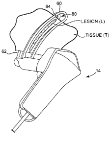
[0062] FIG. 12 is a rear perspective view of cryoprobe clamp 14
engaging tissue T for treatment. As will be appreciated by those skilled in
the art, when trigger member 56 is actuated to move proximal blade 60
toward distal blade 62, ablation tool 64 is aligned substantially with window
portion 80 of proximal blade 60. Consequently, when cryoprobe clamp 14
is operated so that thermally transmissive region 66 of ablation tool 64
creates a lesion L in tissue T, the lesion L may be visible to the surgeon
through window 80 in proximal blade 60. Thus, rather than waiting until
lesion L is so large that it is visible around the outer dimensions of
proximal blade 60, the surgeon is able to verify the formation of lesion L at
an earlier time by directly viewing lesion L through window 80 due to the
alignment of ablation tool 64 with window 80.
[0063] As will be appreciated by those skilled in the art, window portion
80 in proximal blade 60 may allow the surgeon to visually assess
transmurality of a lesion without having to remove clamp assembly 59 from
the tissue site. As will be further appreciated by those skilled in the art,
the
ability to perform a visual assessment in accordance with the present
invention may be combined with a system having a monitoring device that
measures a suitable parameter, such as impedance or temperature, in
order to indicate when a lesion has been fully formed. Providing a means
for visually assessing the formation of a lesion may allow the surgeon to
confirm the feedback from the monitoring device without having to remove
the clamp assembly.
[0064] FIG. 13 is a view illustrating a trigger drive mechanism 90 within
cryoprobe clamp 14. Drive mechanism 90 may generally include spring
rod 92, spring rod pin 94, pulley 96, guide pin 98, and extension spring
100. Spring rod 92 may be disposed within distal housing 54, may be
coupled on a distal end to offset slider 57, and may be slidable relative to
distal housing 54. Pulley 96 and guide pin 98 may be fixed relative to
distal housing 54. Finally, extension spring 100 may be disposed within a
-15-
CA 02713129 2010-07-23
WO 2009/094640 PCT/US2009/032014
channel 102 in a top portion of trigger member 56. As illustrated in FIG.
13, extension spring 100 may include first circular hook 104 and second
circular hook 106. Second circular hook 106 may be positioned over a
spring pin 108 coupled to trigger mechanism 56.
[0065] An elongate wire 110 may operably couple the various
components of trigger drive mechanism 90 together. Particularly, wire 110
may be coupled on a first end 112 to spring rod pin 94 and on a second
end 114 to first circular hook 104 of extension spring 100. As shown in
FIG. 13, wire 110 may extend from spring rod pin 94 around pulley 96,
then beneath guide pin 98 and to first circular hook 104.
[0066] As will be appreciate by those skilled in the art, as trigger
mechanism 56 is pulled backward in a proximal direction indicated by
arrow 74, second end 114 of wire 110 may also be pulled back in the
proximal direction due to the attachment to extension spring 100. Both
pulley 96 and guide pin 98 may help to guide wire 110 as second end 114
is being pulled by trigger mechanism 56. At the same time, first end 112
of wire 110 may be pulled in the distal direction, thereby causing spring
rod 92 and attached offset slider 57 to also move in the distal direction.
This movement of slider 57 causes the "clamping" movement of proximal
blade 60 as discussed above in reference to FIGS. 11A and 11B.
[0067] FIG. 14 is a top perspective view of trigger mechanism 56
illustrating an inlaid track 120. Track 120 may be designed to create a
pathway for bar 122. In particular, bar 122 may be coupled on a distal end
124 to distal housing 54. A proximal end 126 of bar 122 may be structured
to "ride" within track 120 as trigger member 56 is actuated. As will be
apparent from the following Figures and corresponding discussion, track
120 and bar 122 may function together to create a locking mechanism for
trigger member 56.
[0068] FIG. 15 is a perspective view of cryoprobe clamp 14 on the side
opposite that shown in FIG. 14, wherein both proximal and distal housings
52 and 54 have been removed to more clearly see the top portion of
trigger member 56. As shown in FIG. 15, a bar spring 128 may be
-16-
CA 02713129 2010-07-23
WO 2009/094640 PCT/US2009/032014
disposed between and engage inner tube 78 and bar 122. In particular, a
top portion 130 of bar spring 128 may engage the proximal end of inner
tube 78, while a bottom portion 132 of bar spring 128 may engage bar
122. Bar spring 128 may be structured to cause bar 122 to be spring
biased toward the center of the top portion of trigger mechanism 56, thus
biasing proximal end 126 of bar 122 to remain within track 120.
[0069] FIG. 16 is a view illustrating operation of the trigger drive
mechanism 90 within cryoprobe clamp 14. As shown in FIG. 16, when
trigger mechanism 56 is pulled backward in the proximal direction
indicated by arrow 74, second end 114 of wire 110 may also be pulled
back in the proximal direction indicated by arrow 74. As a result, first end
112 of wire 110 may be pulled in the distal direction indicated by arrow
134, thereby causing spring rod 92 and attached offset slider 57 and outer
tube 58 to also move in the distal direction. As a result, proximal blade 60
may be moved longitudinally toward distal blade 62 to allow tissue to be
clamped between the blades for treatment as previously illustrated in FIG.
12.
[0070] As shown in FIG. 16, trigger member 56 is now "locked." This
enables the surgeon to remove his fingers from trigger member 56 while
proximal and distal blades 60 and 62 remain clamped together with tissue
disposed therebetween. When trigger member 56 is locked, proximal end
126 of bar 122 is disposed within a "V-shaped" channel in track 120
positioned between the "lock pathway" for clamping together proximal and
distal blades 60 and 62, and the "unlock pathway" for releasing proximal
and distal blades 60 and 62 from their clamped position (see FIG. 18 for
an illustration of the "pathways"). This V-shaped channel in track 120
defines a "locked position" of proximal end 126 of bar 122 within track 120.
As will be appreciated by those skilled in the art, proximal end 126 of bar
122 slides to this locked position within track 120 while trigger member 56
is being pulled in the proximal direction illustrated in FIG. 16.
[0071] FIG. 17 is a top perspective view of trigger member 56
illustrating the position of bar 122 (shown in phantom lines) in the "locked"
-17-
CA 02713129 2015-09-15
trigger position. As clearly illustrated in FIG. 17, in the locked trigger
position,
proximal end 126 of bar 122 has slid within track 120 until it engages with
the "V-
shaped" channel. In this position, trigger member 56 is now locked, and if no
further action is taken, proximal blade 60 and distal blade 62 will remain
clamped
together.
[0072] When the surgeon desires to "unlock" the trigger member 56 and
clamp assembly 59, he once again pulls trigger member 56 in the proximal
direction as indicated by the direction of arrow 74. When trigger member 56 is
actuated in such manner, proximal end 126 of bar 122 slides into the "unlock
pathway" (see FIG. 18) and returns to the position illustrated by the cross-
hatched bar 122 in FIG. 17.
[0073] FIG. 18 is a top perspective view of track 120 of trigger member
56
illustrating the start position 140, lock pathway 142, locked position 144,
and
unlock pathway 146. As shown in FIG. 18, movement of proximal end 126 of bar
122 within track 120 throughout the entire process of locking and unlocking
trigger member 56 and clamp assembly 59 is also represented by a series of
arrows.
[0074] As illustrated in FIG. 18, there may be an elevation change in
the floor
of track 120 such that proximal end 126 of bar 122 "drops" down when it
approaches these elevation changes. In particular, a first change in elevation
may occur near area 150 such that, from start position 140, proximal end 126
of
bar 122 must travel down lock pathway 142 and cannot enter unlock pathway
146. A second change in elevation may occur near area 152 such that proximal
end 126 of bar 122 drops down into the locked position 144. This prevents
proximal end 126 of bar 122 from sliding back to start position 140 via lock
pathway 142. Thus, such changes in elevation help to ensure track 120 operates
only as a one-way path for proximal end 126 of bar 122.
[0075] Although the present invention has been described with reference
to
preferred embodiments, workers skilled in the art will recognize that changes
may
be made in form and detail without departing from the scope of the invention.
- 18-