Note: Descriptions are shown in the official language in which they were submitted.
CA 02713485 2010-07-28
WO 2009/101130 PCT/EP2009/051634
1
Sealing between a cannula part and a fluid path
The technical field
The invention relates to an infusion part comprising a cannula part and a
fluid path
for providing continuous administration of a therapeutically working
substance, such
as insulin. The infusion part can be connected to delivery means which means
provide e.g. controlled dosage of medication or nutrients.
Prior art
WO 2007/071258 describes a medical device for delivering fluid comprising an
injection part and a fluid delivery part where the fluid delivery part and the
injection
part can be separated and rejoined. The fluid delivery part comprises a
reservoir,
means for transport of liquid e.g. in form of a pump and a house in which the
active
units of the delivery part is placed. The injection part comprises: a base
plate, a
cannula part comprising a body with a through going opening provided with a
cannula extending past the proximal side of the base plate and means for
fixation of
the base plate to the skin of the user e.g. in the form of a mounting pad. The
cannula
part is provided with one or more openings leading fluid to a hollow in the
cannula
part and each opening is covered with a self closing membrane. The delivery
part
and the injection part is assembled through a connector comprising a fluid
path
leading fluid from the reservoir to the through-going opening in the cannula
part
which fluid path comprises means for blocking access to the injection part
when the
connector is disconnected from the delivery part and/or the injection part.
The
embodiments illustrated in this document are quite complex and not easy to
manufacture.
EP 652 027 discloses an infusion device to be placed on a patients skin for
delivering
of medication. This infusion device comprises a cannula device (10) carrying a
penetrating cannula of steel. The cannula device (10) is concentric i.e. all
parts of the
cannula device are rotational symmetric with respect to rotation around the
common
axis. The cannula device (10) can slide axially and has a channel (11) with an
inlet
opening in the cylindrical side surface which inlet opening corresponds to an
outlet
opening of a channel (7) through which medication or the like is entering.
Above and
below the outlet of the channel (7) is placed a first and a second O-ring (8).
Both 0-
rings (8) are placed in circular grooves in the inner surface of the
surrounding the
house (1). In this device the inserter and the cannula device are permanently
joined
CA 02713485 2010-07-28
WO 2009/101130 PCT/EP2009/051634
2
together and this allows the cannula device to be at least partly inserted
into a
cannula opening which fits tightly around the cannula device even before
insertion of
the cannula device has taken place i.e. this results in that there are
friction between
the cannula part and the inner surface of the house during the entire
insertion
procedure. Also there is no teaching in this document of how to adapt the use
of a
soft cannula to this device.
The invention
The object of the invention is to provide an infusion part allowing the use of
a soft
cannula which is safe and simple to manufacture and which reduces the friction
between the cannula part and the base part and therefore also the risk of
incorrect
positioning of the cannula part during insertion. This object is achieved by
reducing
the time where both the moving cannula part and the inner surface of the
opening for
receiving the cannula part are in contact with the gasket sealing of fluid
from the
surroundings. This can generally be achieved by creating a cannula part having
an
increasing diameter or by creating a sealing with a smaller area.
This object is achieved by an infusion part as described in claim 1 comprising
a cannula part and a fluid path, where
- the cannula part comprises a body formed by a hard material which body
has an inner through going opening which through going opening is in fluid
contact with a cannula, the cannula has an inner opening which provides fluid
contact with the patient, the body of the cannula part has an opening
corresponding to the inlet or outlet opening of the fluid path resulting in
fluid
contact between the fluid path and the cannula part and these two
corresponding openings do, when they are positioned opposite each other,
allow unrestricted flow,
- the fluid path comprises at least one inlet and one outlet opening through
which a fluid can enter and exit the fluid path, and
- a sealing is positioned between the cannula part and the inlet/outlet
opening
of the fluid path when the cannula part is in position for use in order to
keep
the fluid path to the cannula tight.
CA 02713485 2010-07-28
WO 2009/101130 PCT/EP2009/051634
3
The sealing is surrounding the inlet/outlet opening and/or the distance di
between a centre line c of the cannula part and a point on the outer surface
of
the cannula part positioned at or above the upper edge of the sealing is
larger
than the distance d2 between the centre line c of the cannula part and a point
on the outer surface of the cannula part positioned at or below the lower edge
of the sealing. "Upper edge of the sealing" defines the part of the sealing or
gasket which has the longest distance to the patient's skin, and "lower edge
of the sealing" defines the part of the sealing which has the shortest
distance
to the patient's skin when the infusion part according to the invention is
inserted in a use position.
According to one embodiment the body of the cannula part is provided with a
sealing before use or alternatively the opening of the fluid path or the
surface
surrounding the opening of the fluid path is provided with a sealing before
use. "Provided" means that the sealing or gasket is somehow attached to the
indicated surface, it might just be placed in a groove or a cavity as
indicated
in fig. 9 or 10.
According to one embodiment the penetrating member is provided with
attachment means assuring that the penetrating member is unreleasably
attached to the base part after insertion.
According to one embodiment the body of the cannula part is provided with a
sealing or gasket placed along the edge of the opening through which fluid
enters or exits the cannula part.
According to one embodiment the opening of the fluid path corresponding to
an opening of the cannula part is provided with a sealing placed along the
edge of the opening i.e. in a short distance from the opening. "A short
distance" is understood to be less than or equal to the distance equaling the
diameter of the opening and if the opening is not round: less than or equal to
the longest dimension of the opening.
CA 02713485 2010-07-28
WO 2009/101130 PCT/EP2009/051634
4
The sealing material according to any embodiment can be hydrophobic and
elastic e.g. the sealing material is made of silicone.
According to an embodiment the body of the cannula part has at least one
second opening to the inner through going opening and preferably this at
least one second opening to the inner through going opening is covered by a
self closing membrane which membrane can be penetrated by a blunt or
pointy needle and can be made of silicone.
This at least second opening can e.g. be used for insertion of the device if
the
cannula is a soft cannula not able to cut its way through the patients skin,
then a separate insertion needle can pass through the second opening, all
through the cannula and provide a cutting edge in front of the cannula. It can
also be used for supplying medication or nutrients which only are given to the
patient in smaller doses a few times a day.
According to an embodiment the infusion part comprises a base part which
can be fastened to a patient's skin.
According to one embodiment of such an infusion part the base part is
provided with an opening corresponding to the profile at the non-penetrating
end of the cannula part.
The "non-penetrating end" of the cannula part is the end opposite the cannula
i.e. the distal end of the penetrating member where "distal" indicates the end
is turned away from the patient. In the embodiment of the cannula part shown
in the figs. 4A, 4B and 4C the cannula part has a flat surface part on one
side
corresponding to a flat wall surrounding the opening of the fluid path, i.e.
that
the opening is "adapted" means that the surrounding walls correspond to the
cannula part and assures that the cannula part ends up in a well-defined and
CA 02713485 2010-07-28
WO 2009/101130 PCT/EP2009/051634
close fitted - preferably press-fitted - position. "Press-fitted" means that
it is
so close fitted that it requires a force to insert the cannula part.
According to this embodiment the opening can extend below the outer surface
5 of the base part providing walls which tightly fits around the cannula part
when the cannula part is inserted into the patient and preferably the inlet or
outlet opening of the fluid path opens into the wall of the opening fitting
around the cannula part and when the cannula part is inserted, an inlet or
outlet to the inner opening of the cannula part corresponds to the
inlet/outlet
opening of the fluid path.
According to one embodiment the distance di between a centre line c of the
cannula part and a point on the outer surface of the cannula part positioned
at
the upper edge of the sealing (18) is larger than the distance d2 between the
centre line c of the cannula part and a point on the outer surface of the
cannula part positioned at the lower edge of the sealing. The centre line c is
parallel to the direction of insertion.
According to one embodiment the angle d is the angle between the direction
of insertion of the cannula part and a plane being tangent to the surface
surrounding the opening opposite the sealing, and 0 < d <_ 90 , normally 45
d <_ 80 and most often 70 <_ d <_ 80 .
When a cannula part with a decreasing cross-section is inserted into a hollow
with a corresponding decreasing hollow then the cannula part can be press-
fitted into the hollow. This press-fitting both assures that the two
corresponding openings of respectively the fluid path and the cannula part are
pressed together thereby improving the fluid tight connection between them
and it can also lock the cannula part to the base part.
According to one embodiment the base part is formed at least partly of a hard
material. That a material is "hard" means that it can not be penetrated by a
CA 02713485 2010-07-28
WO 2009/101130 PCT/EP2009/051634
6
needle, and also that it is able to maintain a shape it is given during
production although it might be possible to flex the material due to the shape
it is given e.g. if it is formed as a thin plate or if it is very long but it
will not be
possible to compress it i.e. reduce it size.
According to one embodiment the fluid path is formed as an integrated part of
the base part fastened to the patient's skin. That the fluid path is formed as
an integrated part means that it is an unreleasable part of the device, i.e.
it is
permanently attached to the device at some time during the manufacturing
process of the base part and when the base part is in use it will not be
possible to separate the fluid path and the rest of the base part.
According to one embodiment the hard material is a molded plastic material
e.g. the plastic material is polypropylene.
According to one embodiment the base part comprises fastening means for
attaching delivery means to the base part. The delivery means can comprise a
connecting part provided with means corresponding to the means for
fastening of delivery means and provided with a tube for transferring
medication to the infusion part or the delivery means can comprise a reservoir
containing medication and means for transferring medication to the infusion
part. The means for transferring will normally be a pump and a programmable
part possibly combined with a sensor for assuring appropriate amounts of
medication to be delivered to the patient.
Embodiments of the invention will now be described with reference to the
figures in which:
Figure 1 shows a first embodiment of an infusion part according to the
invention.
Figures 2 and 2A shows a second embodiment of an infusion part
according to the invention.
CA 02713485 2010-07-28
WO 2009/101130 PCT/EP2009/051634
7
Figure 3 shows the same embodiment of an infusion part as figs. 2 and
2A.
Figure 4A, 4B and 4C show a cannula part which can be used in
connection with the invention.
Figure 5 shows a front view of an inserter which can be used in connection
with the invention.
Figure 6 shows a view from the proximal side of the inserter of fig. 5.
Figure 7 shows a connector part which can be part of an infusion part
according to the invention.
Figure 8 shows the same connector part as fig. 7 without the bubble
membrane covering the inlet.
Figure 9A and 9B show a cannula part having an inclined contact surface.
Figure 10A-10D show an enlargement of the contact between the cannula
part and the cannula opening of the connection part.
Figure 11A, B and C show an embodiment of a base part provided with a
fluid path mainly constructed of a tube.
Figure 12 shows an embodiment of an infusion part having an angle d =
90 between insertion direction and tangent to contact surface.
Figure 13 shows a cannula part which can be used in connection with the
invention.
Fig. 1 shows an embodiment of an infusion part comprising a cannula part
and a fluid path according to the invention. This embodiment comprises a
surface plate 1 attached to a contact surface. The surface plate 1 is in this
embodiment constructed of a molded plastic material and the contact surface
can be the proximal side of a mounting pad 2 which mounting pad 2 is
unreleasably fastened to the surface plate 1 during manufacturing of the
device. The mounting pad 2 of this embodiment has the same area as the
surface plate 1 but it could be of an area larger or smaller than the surface
plate 1.
A connector part 3 is position on the surface plate 1. The connector part 3
provides for the contact between the base part and some kind of delivery
CA 02713485 2010-07-28
WO 2009/101130 PCT/EP2009/051634
8
means. According to one embodiment the surface plate 1 and at least an
outer cover of the connector part 3 is simply molded in one piece during
manufacturing of the device. The internal parts of the connector part 3 forms
a fluid path between e.g. a reservoir of medication or a reservoir for liquid
collected from the patient and a cannula part 7. Therefore the connector part
3 is provided with at least two openings, one opening at each end of the fluid
path where the first opening 13 is an inlet or outlet opening receiving or
delivering fluid to a not shown reservoir and the second opening is an inlet
or
outlet opening 12 receiving or delivering fluid to a cannula part 7. The
connection part 3 might be provided with extra openings e.g. for inserting the
cannula part, for injection of a second medication or nutrient or for letting
the
fluid in the fluid path get in contact with a sensor.
In the following the first opening 13 will be referred to as "inlet" and the
second opening will be referred to as "outlet" although the direction of the
flow
through the fluid path is not significant for the invention.
The embodiment of fig. 1 is provided with two guiding means 4 in the form of
two right angled L-shaped profiles in the form: ~ F-, which profiles are
protruding from the surface plate 1 of a base part having a lower or proximal
side which is fastened to the skin of the patient. The guiding means 4
correspond to guiding means on a delivery part or a cover or connecting
means which are to be fastened to the base part during use. Such
corresponding means can e.g. be formed as one or more hooks having an L-
shaped profile in the form: L and J corresponding to the profiles on the base
part.
The fluid path of the connection part 3 of this embodiment is very short and
the inlet 13 of the connection part 3 is placed in a centre position in
relation to
the guiding means 4. The top of an inserted cannula part 7 is shown inserted
into the connection part 3.
The connection part 3 is further provided with a cannula cavity 12A which
accurately fits around a cannula part 7 i.e. the cannula cavity 12A has the
same 3-dimensional shape or profile as the cannula part 7 and is just big
CA 02713485 2010-07-28
WO 2009/101130 PCT/EP2009/051634
9
enough to let the cannula part 7 pass through and then fit into the opening.
In
fig. 1 the cannula part 7 is shown in a position where the cannula part 7 is
fully inserted. When the cannula part 7 is fully inserted, then the upper
surface i.e. the distal surface of the cannula part 7 is normally at level
with or
at a lower level than the outer surface of the connection part 3 around the
cannula cavity 12A.
When the cannula part 7 has been fully inserted into the connection part 3, an
opening 20 in a side surface of a body 24 of the cannula part 7 corresponds
to the opening 12 of the fluid path of the connection part 3 and fluid can
flow
from one part to the other. The opening 12 might in the following be referred
to as an "outlet" although the direction of the flow is not significant to the
invention.
Figs. 2 and 3 show a second embodiment of an infusion part according to the
invention. A delivery part corresponding to this embodiment could be joined to
the base part by pushing the delivery part down toward the guiding means 4
which in this case is a longitudinal raised platform having a magnet 5
fastened to the top surface. The delivery part would be provided with a
corresponding magnet e.g. of a smaller or different size than the magnet 5
which is placed in such a way e.g. in a track corresponding to the raised
platform 4, that the corresponding magnet of the delivery part can slide along
the magnet 5 on the raised platform 4 of the base part in the longitudinal
direction. When the delivery part arrives at its working position, two release
handles can engage respectively with two protruding parts 15 protruding from
the upper surface of the surface plate 1. When the delivery part is in its
working position it is locked in any horizontal direction by the release
handles
and in the direction perpendicular to the surface plate 1 by the two
corresponding magnets of respectively the delivery part and the base part.
These locking mechanisms make it possible to fasten and release the delivery
device from the base part as often as needed i.e. a single-use base part can
be combined with a multi-use delivery part.
In fig. 2 and 2A the base part is shown without the cannula part 7 and in fig.
3
the base part is shown having the cannula part 7 in a positioned reached just
CA 02713485 2010-07-28
WO 2009/101130 PCT/EP2009/051634
before insertion of the cannula part 7, normally the cannula part 7 would at
this stage of insertion still be placed inside an inserter and it would not be
visible.
5 Normally an inserter 10 holds the cannula part 7 before insertion and the
insertion can be initiated by pushing a handle 11. Fig. 5 and 6 shows the
direction the handle 11 has to be pushed in, in order to initiate insertion of
the
cannula part 7. After insertion a not shown insertion needle can be retracted
to the inside of the inserter 10 and the inserter 10 is removed from the base
10 part, leaving an inserted cannula 22 fastened to the surface plate 1. If
the
cannula 22 of the cannula part 7 is a hard self penetrating cannula there will
be no separate insertion needle and therefore no need to retract the insertion
needle.
In figs. 2 and 2a the connection part 3 is shown with an outer cover provided
by the molded surface plate 1. The outer cover shown in this embodiment is
not an independent unit but is attached unreleasably to or simply made as a
part of the surface plate 1 e.g. by a molding process. The outer cover is
provided with a cannula cavity 1 2A for the cannula part 7 and an access
opening 13 for e.g. a reservoir thereby allowing access to the fluid path of
the
connection part 3 by the reservoir and the cannula part 7. The cannula cavity
1 2A allows the cannula part 7 to be inserted sub- or transcutaneous into the
patient within the circumference of the hard surface plate 1 and the contact
surface 2 of the base part which in this embodiment is provided by a mounting
pad is also provided with an opening 12B which allows for the cannula to be
inserted (see fig. 7 and 8). This opening 12B is not necessary if the contact
surface 2 is constructed of such a material and thickness that it can be
penetrated by at least the cannula 22 of the cannula part 7.
In figs. 7 and 8 the connection part 3 is shown without the outer cover
provided by the molded surface plate 1. In order to secure a fluid tight
connection between the outlet opening 12 in the connection part 3 and the
cannula part 7 the outlet opening 12 of the connection part 3 is provided with
an elastic sealing 18 around the outlet opening 12. When the cannula part 7 is
inserted it will be press fitted into the cannula opening 12 and the elastic
CA 02713485 2010-07-28
WO 2009/101130 PCT/EP2009/051634
11
sealing 18 will provide a completely fluid tight gasket around the
corresponding openings 12 and 20. In order to improved the press-fitting and
thereby the fluid tight connection between the cannula part 7 and the outlet
of
the fluid path, the cannula cavity 12A can be provided with a decreasing
cross-section in a plane parallel to the cannula 22 when inserted and
perpendicular to the surface where the outlet of the fluid path is positioned.
The cannula part 7 will have a corresponding decreasing cross-section.
In order to secure a fluid tight connection between the inlet opening 13 in
the
connection part 3 and the reservoir 6, a bubble shaped membrane 17 has
been positioned around the first opening 13. The membrane 17 completely
covers the inlet opening 13 and prevents contamination of the internal of the
connection part 3. When a reservoir or connecting parts for a reservoir is
pressed towards the connection part 3, a connector needle 19 will penetrate
the membrane 17 and provide a completely fluid tight transfer of fluid between
the connection part 3 and the reservoir.
That the membrane 17 is bubble shaped means that it is attached around the
opening - normally around the edge of the opening - it protects and the
membrane 17 protrudes from the planed formed by the edge of the opening
and forms a dome in a distance from the edge which distance normally
corresponds to the length of a connector needle 19.
In fig. 8 the connector needle 19 is shown as being a part of the connection
part 3 i.e. it is attached to the connection part 3 but it might just as well
be a
part of the reservoir.
According to one embodiment the connection part 3 is provided with both a
connector needle 19 and a bubble shaped self closing membrane 17 and the
reservoir is also provided with a bubble shaped self closing membrane. As
both parts are provided with self closing membranes it will be possible to
separate the two units from each other and rejoin them at a later time without
the internal fluid path of the connection part 3 and thereby the patient being
contaminated.
CA 02713485 2010-07-28
WO 2009/101130 PCT/EP2009/051634
12
Fig. 4A, 4B and 4C shows an enlargement of a cannula part 7 which can be
used in connection with the invention. This embodiment comprises a body 24
provided with a cannula 22 and with a protruding front 25 having a flat
surface. The surface of the cannula part 7 having an opening need not be flat;
it can actually have any desired shape as long as it is possible to create a
corresponding surface on the connection part 3 facing the cannula part 7. In
one embodiment the front 25 is inclined in such a way that the cross-section
at the upper i.e. distal end is larger than the cross-section at the proximal
end, i.e. the enc closest to the patient after insertion, of the front in at
least
one dimension. The front 25 is provided with an opening 20 through which
liquid can exit or enter the cannula part 7. The body 24 is further provided
with a top opening 21 which opening can be covered with a self closing
membrane. The opening 21 need some kind of entrance protection as it is
facing an outer surface which is in contact with the surroundings. The top
opening 21 is primarily used when inserting the cannula part 7 if the cannula
22 is a soft cannula. That the cannula 22 is soft means that is made of a
relatively soft material which can not penetrate the patients skin, in this
case
it is necessary to use a pointy insertion needle of a relatively hard material
when inserting the cannula and this pointy needle can be inserted through the
top opening 21, pass through an inner through going opening in the body 24
of the cannula part and further pass through the full length of the cannula 22
in such a way that the pointy end of the insertion needle stick out of the
open
end of the hollow cannula 22. After insertion i.e. after the cannula 22 has
been placed sub- or transcutaneous in the patient, then the insertion needle
is
retracted and the cannula 22 is left inside the patient.
The cannula part 7 is also provided with fastening means 23 which fastening
means 23 lock the cannula part 7 to the base part at the time where it is
fully
inserted. The fastening means 23 of this embodiment comprises outward
hooks that can pivot around an axe close to the body 24 of the cannula part 7
in such a way that the diameter formed by the outermost edge of the hooks
can be reduced when the hooks are pressed inward i.e. towards the centre of
the cannula part 7. When the pressure is removed the hooks will return to
their original position due to the flexibility of the material. The hooks will
be
pushed inwards when they pass an opening such as e.g. the opening 12B or a
CA 02713485 2010-07-28
WO 2009/101130 PCT/EP2009/051634
13
corresponding opening in the surface plate having a cross-section which at
least in one dimension is smaller than the outer edge of the hooks and as the
hooks return to their original position after having passed through the
opening, the hooks will lock the cannula part 7 in the inserted position.
Figs. 5 and 6 show an inserter that can be used to position the cannula part 7
in the base part. The inserter comprises a housing 10 provided with an
internal opening where the cannula part 7 can be moved from a retracted
position to a forward position. In the retracted position the cannula 22 is
not in
contact with the patient and in the forward position the cannula 22 is
inserted
into the patient. The inserter further comprises an actuator handle 11 which
is
to be activated when the cannula part 7 is to be inserted and it comprises
fastening means 14 which means can lock the inserter to the base part before
and during insertion. Normally the inserter should be fastened to the base
part under sterile conditions or the joined base part and inserter should be
sterilized after fastening of the inserter in order to prevent contamination
of
the cannula cavity 12A, and in order to reduce the amount of material placed
on the patient's skin it is desirable to be able to remove the whole of or at
least part of the inserter after the cannula part 7 has been inserted.
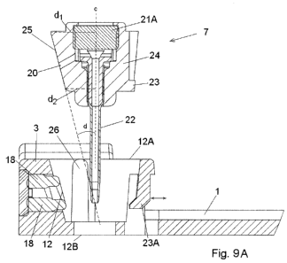
Figs. 9A and 9B show an enlargement of a second embodiment of a cannula
part 7. Fig. 9A shows the cannula part 7 in a state just before insertion and
fig. 9B shows the cannula part 7 inserted into the cavity 12A in the base
part.
This embodiment also comprises a body 24 provided with a cannula 22 and
with a protruding part 25 having a flat surface provided with an opening 20.
According to this embodiment the protruding part 25 is inclined in such a way
that the pressure between the opening 20 and the sealing 18 around the
second opening 12 of the connection part 3 is increased, also the sealing 18
is subjected to less tear during insertion. The inclination of the inclined
part
25 is defined by the angle d between the centre line c of the cannula 22 (the
centre line c is parallel to the insertion direction) and a line parallel to
the
surface around the opening 20. If the surface around the opening 20 is not
CA 02713485 2010-07-28
WO 2009/101130 PCT/EP2009/051634
14
straight, then the line parallel to the surface would be the tangent to the
surface around the opening 20. The angle d will be larger than 00 and smaller
than or equal to 90 , normally d e ]0 , 30 ] depending on the diameter or the
protrusion of the sealing 18 or [60 , 90 [. The distance d, measured at the
distal end of the surface of the protruding inclined part 25 where the distal
end is the end of the cannula part 7 which is furthest away from the patient
after insertion, between the surface of the protruding inclined part 25 and
the
centre c of the cannula part 7 is larger than the distance d2 between the
surface of the protruding part 25 at the proximal end i.e. the end closest to
the patient after insertion, and the centre c of the cannula part 7. Normally
the
distance d2 will be so small that the proximal end of the protruding inclined
part 25 does not touch the sealing 18 of the connection part 3 during
insertion.
In one embodiment (not shown) the angle d is close to 90 i.e. d = 90 , such
an embodiment would in a drawing corresponding to fig. 9A and 9B appear to
have an upward opening 12 of the connection part 3 fitting to a downward
opening 20 of the cannula part 7. This means that the force pushing the
cannula part 7 toward the sealing 18 will be close to perpendicular to the
contact surface of the sealing 18 and this will prevent that the sealing is
distorted during insertion of the cannula part 7 by the cannula part 7 sliding
along the sealing 18.
In another embodiment (shown in fig. 4A-C and in fig. 10A-B) d = 0 as the
protruding part 25 and the centre line c are parallel. According to this
embodiment the cannula part 7 will be in sliding contact with the protruding
sealing 18 which can cause the sealing to be distorted.
The protruding front 25 of the cannula part 7 need not be flat; it can
actually
have any desired shape e.g. partly spherical as long as it is possible to
create
a corresponding surface on the connection part 3 facing the cannula part 7.
Also the opening 20 of the protruding front 25 can behave as an inlet or an
outlet depending on the purpose of the cannula part 7. In fig. 9A and 9B which
is a cut-through view it is shown how the top opening 21 of the body 24 is
covered with a self closing membrane 21A. As according to the embodiment
CA 02713485 2010-07-28
WO 2009/101130 PCT/EP2009/051634
of fig. 4A-C the top opening 21 is primarily used when inserting the cannula
part 7 if the cannula 22 is a soft cannula but the top opening 21 can also be
used to inject medication or nutrients other than the primary medication which
could be e.g. insulin which the patient receive via the opening 20.
5
This embodiment of the cannula part 7 is also provided with fastening means
23 and in this embodiment the fastening means 23 has the form of a
protruding part 23 on the cannula part 7 which corresponds to a flexible part
23A on the stationary base part. The flexible part 23A can be pushed outward
10 as indicated with an arrow at fig. 9A when the protruding part 23 on the
cannula part 7 passes during insertion of the cannula part 7. After insertion
the upward surface of the protruding part 23 of the cannula part 7 will be
locked by the downward surface of the flexible part 23A of the base part and
it
will not be possible to detach the cannula part 7 from the base part.
The cannula part 7 of fig. 9A and 9B is provided with a soft cannula 22 which
soft cannula 22 together with a bushing 29 provides a cannula assembly. This
assembly is normally fastened inside the body 24 of the cannula part 7 by an
interference fit i.e. it is only the friction between the body 24 and the
cannula
assembly which keeps it in the correct position. In order to prevent the
cannula assembly from sliding back through the upper larger opening in the
body 24 of the cannula part 7, the body 24 of the cannula part 7 can be
provided with a ring shaped recess encircling the exit for the soft cannula
22.
As the recess creates an open space around the soft cannula 22, the soft
cannula 22 can form a small bulk i.e. a ring shaped bulk which prevents the
soft cannula from sliding back.
Fig. 10 illustrates how the unrestricted openings between the cannula part 7
having the body 24 and the fluid path having the inlet/outlet openingl2 slide
into place. Fig. 10A and 10B show an embodiment where d = 0 and fig. 10C
and 10D show and embodiment where d is around 15 , normally between 8-
22 . According to the embodiment of fig. 10A and 10B the body 24 of the
cannula part 7 is provided with an inclined edge in order to reduce distortion
or tearing of the sealing. In both embodiments the shown sealing 18 is a
circular or cylindrical silicone unit which is placed in a round track around
the
CA 02713485 2010-07-28
WO 2009/101130 PCT/EP2009/051634
16
inlet/outlet opening 12 in the connection part 3. The wall where the sealing
or
gasket 18 has been placed is provided with an adjacent expansion room 28.
After positioning of the cannula part 7 the sealing 18 can occupy this room.
In
the embodiment of fig. 10C and 10D is not only the sealing face angled, the
whole cylindrical sealing part 18 is angled in order to allow uniform sealing
deformation. The cylindrical sealing 18 does not form the walls of the
inlet/outlet opening 12, the wall or surfaces of this opening is formed by the
material which the connection part 3 is formed of in order to provide a pipe
which cannot be deformed. In order to create the necessary pressure between
the seal and the seal face i.e. the surface which the sealing 18 touches when
in a sealing position, the sealing face can be provided with a small
continuous
protrusion protruding from the sealing face and having the same shape as the
sealing which would e.g. be circular if the sealing has the cylindrical shape
shown in fig. 10A-D.
Figs. 11A-11C show one embodiment of a connection part 3. Fig. 11A show
the embodiment of the connection part 3 in an exploded view where the
internal holding parts 61 for a tube 60 providing a fluid path is shown. Fig.
11 B shows a cut through the internal holding part 61 according to which it is
possible to the position of the tube 60. Fig. 11 C shows an enlargement of the
encircled part of fig. 11 A.
According to the present embodiment the connection part 3 and the surface
plate 1 is molded in one piece of a plastic material, the connection part is
provided with several openings, one opening is the cavity 12A which is
prepared for fitting in the cannula part 7 and another opening is prepared for
fitting in the internal parts of the connection part 3. The internal parts of
the
connection part 3 according to this embodiment comprises one tube which at
two positions are bend in 90 i.e. both the inlet and the outlet end of the
tube
60 points in the same direction perpendicular to the connecting part of the
tube 60 where the connecting part of the tube 60 forms the fluid path between
the two bending parts.
At one end the tube 60 is protected by a bubble shaped membrane 17 and at
the other end the tube 60 is open and unprotected, but the open tube end is
CA 02713485 2010-07-28
WO 2009/101130 PCT/EP2009/051634
17
surrounded by a sealing 18 which is attached unreleasably to a holding part
61. When the internal parts have been placed in the corresponding opening in
the connection part 3 a cover 62 accurately fitting in the opening is placed
in
level with the surface of the connection part 3 in such a way that the user
experience a smooth surface which cannot be tampered with.
The embodiment of the base part shown in fig. 1 1A is provided with guiding
means 26 placed inside the cavity 12A of the connection part 3. The two
opposing ribs 26 which constitute the guiding means correspond to closely
fitting openings 27 in the cannula part 7. The guiding means 26 and the
corresponding parts 27 on the cannula part can have other forms, the
important feature is that they correspond to each other and make it possible
for the cannula part 7 to slide into use position.
Fig. 11 B shows an enlargement of the internal parts of the connection part 3.
The holding parts 61 comprise a single molded part which is providing a
stable embedment of the tube 60. The open end of the tube 60 opens into a
space surrounded by the sealing 18. The closed end of the tube 60 is
completely surrounded by a soft membrane. "Completely surrounded" means
that the there is no free access to the surroundings, "soft membrane" means
that the membrane can be penetrated by a needle, especially the connector
needle 19 which is provided by the end of the tube 60 and which is embedded
inside the soft membrane. The end of the tube 60 which constitutes the
connector needle 19 is in this embodiment not actually in touch with the
surrounding membrane 17. The connector needle 19 is surrounded by air, and
the internal space surrounding the connector needle 19 has a cylindrical or
conical shape i.e. a circular cross-section. The walls of the membrane 17 will
deform by bending inwards or outwards when the length of the membrane is
reduced as a result of the applied pressure.
Fig. 11C shows an enlargement of the enclosed field marked in fig. 11A.
Fig. 12 shows an embodiment of an infusion part where the angle d = 90 .
The inlet/outlet opening 12 is constructed as a pointy end of a tube 60 which
provide for the fluid path or connection between the reservoir 6 and the
CA 02713485 2010-07-28
WO 2009/101130 PCT/EP2009/051634
18
cannula part 7. A membrane e.g. self closing protects the entrance to the
reservoir 6 which means that micro organisms cannot access the reservoir 6
when the reservoir is removed from the connection part 3.
Fig. 13 shows yet an embodiment of a cannula part 7 which can be used with
an infusion part according to claim 1. The body 24 of the cannula part 7 has
the shape or profile of a truncated cone i.e. in each horizontal (according to
fig. 13) cross-section of the body it is round having varying diameters. The
body 24 is provided with two permanently attached circular sealings or
gaskets 18. Between these two gaskets 18 is the opening 20 positioned which
opening 20 allows for fluid to enter the inner through going opening of the
cannula part 7. The cannula part 7 is to be placed in a below illustrated
connection part 3 provided with a corresponding cavity 12A also having the
shape of a truncated cone. The cavity 1 2A has an inlet/outlet opening 12 for
fluid flowing to or from the cannula 22.