Note: Descriptions are shown in the official language in which they were submitted.
CA 02713545 2010-07-28
WO 2009/097511 PCT/US2009/032614
TWO STAGE ENRICHMENT OF CELL-FREE FETAL DNA
IN MATERNAL PLASMA
Though fetal DNA is present in maternal plasma, the levels are relatively low
to
enable routine prenatal genetic analysis. Without being bound to any theory,
we believe that
a portion of fetal DNA is packaged and thus protected from plasma endo-
nucleases. We
propose a novel enrichment technique that combines two distinct methods.
Separated plasma
is first treated with DNase to effectively decrease the percentage of
contaminating cell-free
maternal DNA fragments. Subsequent increase of fetal sequences is achieved
using a
modified Whole Genome Amplification (WGA) technique.
Methods: Plasma was isolated from maternal whole blood (n=24) then treated
with
increasing concentrations of DNase. DNase treated DNA was further processed
using the
Qiagen DNA extraction mini kit prior to a modified WGA protocol to enrich
smaller DNA
fragments. Presence of fetal sequences was confirmed using real-time Taqman
PCR (RT-
PCR) to measure (3-Globin and DYS 1 sequence levels.
Results: Fetal DNA sequences were detected following all DNase treatments.
Correct detection of male fetuses was achieved in all samples confirmed to
have a male fetus
(n=10) both before and after WGA. In addition to correct gender determination,
the samples
(n=7) that were subjected to the highest amount of DNase as well as the
modified WGA
protocol demonstrated significant enrichment of fetal sequences, attaining 50%
fetal DNA.
Conclusions: Results confirm that fetal DNA in plasma is protected and
resistant to
degradation from DNase treatment. These preliminary data also suggest that an
optimal level
of DNase treatment can be achieved that allows further enrichment using WGA.
The
observation that DNase eliminates predominantly maternal sequences suggests
that cell-free
fetal DNA is packaged differently than the maternal counterpart, allowing
preferential
enrichment of fetal sequences.
INTRODUCTION
Prenatal genetic diagnosis has relied on invasive procedures such as
amniocentesis or
chorionic villous sampling (CVS). While these procedures have provided
reliable results for
many years they still carry a slight risk to the fetus (1). Since the
discovery of amplifiable
fetal cell-free nucleic acids in maternal plasma (2), there have been numerous
studies aimed
at determining the potential for clinical non-invasive prenatal genetic tests.
While many of
1
CA 02713545 2010-07-28
WO 2009/097511 PCT/US2009/032614
these studies have shown great promise, the small existing quantities of fetal
DNA has made
it difficult to implement clinically. Therefore, we have focused on improving
methods of
fetal DNA isolation and enrichment. Without being bound to any theory, it is
believed that
these circulating nucleotides are the result of fetal cells undergoing
apoptosis (3). The
relative stability of cell-free DNA and RNA in plasma, which is known to
contain nucleases,
suggests that these nucleic acids are circulating within membrane bound
vesicles formed as a
result of the mechanism of programmed cell death (4). These fetal DNA
fragments are also
distinguishable from maternal fragments based on size, fetal fragments
generally smaller
(<300 bp) than maternal fragments (>500 bp) (5; Jorgez C et at., 2007).
We describe a novel two stage method for enrichment of fetal fragments. The
first
stage involves treatment of total maternal plasma (containing both maternal
and fetal DNA
fragments) with DNase. Given that fetal fragments are more stable and likely
packaged by
membrane bound apoptotic bodies, we hypothesize that DNase treatment would
deplete the
overall (unpackaged) maternally derived sequences. The second stage involves a
modified
whole genome amplification (WGA) protocol designed to amplify smaller
fragments,
presumably fetal.
METHODS
Following IRB approval and written informed consent, a total of 24 whole blood
samples (10 confirmed male pregnancies, mean gestational age 18 1/7 weeks
ranging from 11
4/7 to 25 2/7 weeks; 12 confirmed female pregnancies, mean gestational age 20
1/14 weeks
ranging from 9 6/7 to 37 4/7 weeks; and 2 non-pregnant controls, 1 male and 1
female) were
collected. For each, approximately 30 ml of blood drawn in ACD vacutainers was
processed
by an initial centrifugation at 800g for 10min to separate plasma from the
cellular fraction.
The plasma fraction was removed and centrifuged again at 16,000g for 10 min to
further
remove any contaminating cellular particles. This fraction was then frozen in
800 1 aliquots
and later thawed for simultaneous batch processing. Each 800 l plasma sample
was thawed
at room temperature then subjected to DNase (Promega, Cat #M6101) treatment at
various
concentrations (untreated, 1 L, 5 L, 10 L, 30 L of 1 unit/ l). Samples were
incubated at
37 C for 1 hour before adding stop solution. Samples were next subjected to
DNA extraction
using the Qiagen QiAamp Blood Mini Kit (Cat #51106) and eluted in a final
volume of
100 L. With slight modifications, the protocol for the GenomePlex Complete
Whole
Genome Amplification (WGA) Kit (Sigma, Cat #WGA2-50rxn) was followed on seven
maternal blood samples (4 confirmed males, and 3 confirmed females). We
modified the
2
CA 02713545 2010-07-28
WO 2009/097511 PCT/US2009/032614
procedure by first omitting the manufacturer suggested fragmentation
incubation due to the
anticipated size of target sequences. Second the cycle number in the
amplification step was
increased to 20 rather than the suggested 14. Amplified samples were stored at
4 C until RT-
PCR analysis for detection and quantification of (3-Globin (BGLO354F: GTG CAC
CTG
ACT CCT GAG GAG A, BGLO455R: CCT TGA TAC CAA CCT GCC CAG) (6) and
DYS1 (DYS1F: TCC TGC TTA TCC AAA TTC ACC AT, DYS1R: ACT TCC CTC TGA
CAT TAC CTG ATA ATT G) (7) (Applied Biosystems 7700, Foster City, CA). Level
of
enrichment was determined based on % fetal DNA which was calculated as a ratio
of DYS 1
to (3-Globin. (3-Globin represents the total amount of isolated DNA, maternal
and fetal, while
in the confirmed male pregnancies DYS 1 represents the amount of fetal DNA
present in the
sample.
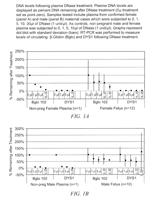
RESULTS
Following initial DNase treatments in control non-pregnant samples, both (3-
Globin
and DYS 1 levels decreased as a function of the amount of enzyme added. Each
was
effectively eliminated by DNase treatment. (Figure 1). In maternal samples,
although
detected levels of (3-Globin were decreased, these levels were persistent
despite harsher
DNase treatments (916.5 + 91.2 Geq/ml for male maternal cases and 610.5 +
389.62 Geq/ml
for female maternal cases) . No false positive DYS 1 levels were detected in
any of the
known female pregnancies. However, among pregnancies with known male fetuses
(n=10),
there was 100% detection of DYSl sequences at all treatment increments (O 1:
127.4 + 72.3
Geq/ml, l 1: 71.4 + 57.3 Geq/ml, 5 1: 71 + 58.6 Geq/ml, l0 1: 57.1 + 46.6
Geq/ml, 30 l: 154
+ 179.6 Geq/ml).
WGA was performed on seven DNase treated maternal samples (3 female and 4
male). Though fetal sequences are detected in all samples, increased levels of
fetal DYS1
sequences were only observed in the samples that were subjected to 30 l of
DNase (O 1:
1979 + 2083.9 Geq/ml, 1 l: 1.62 + 1.6 Geq/ml, 5 l: 723.5 + 875.7 Geq/ml, l0 1:
0.83 + 1.22
Geq/ml, 30 l: 7025 + 4381 Geq/ml). Thus indicating that more stringent DNase
treatments
were necessary to diminish maternal sequences (based on (3-Globin) to the
extent that they
were not present in levels that out-compete amplification of DYS 1 sequences.
In the samples
that were subjected to 30 1 of DNase followed by the modified WGA protocol, we
were able
to achieve a mean value of 49.96% fetal DNA. In the samples that were not
treated with
DNase and only subjected to the modified WGA, the mean percent fetal DNA among
samples was 11.16%. The samples that were treated with either 1, 5, l0 1 of
lunit/ l of
3
CA 02713545 2010-07-28
WO 2009/097511 PCT/US2009/032614
DNase all had mean values of 0.01 %, 3.5%, and 0.01 % fetal DNA respectively
after WGA.
Prior to WGA the samples had percentages ranging from 0.05% to 0.17%.
DISCUSSION
Our results demonstrate that cell-free fetal DNA is resistant to degradation
by DNase,
supporting the hypothesis that cell-free fetal DNA is packaged in membrane
bound vesicles.
We effectively removed maternal sequences as well as any contaminating
sequences that may
lead to false-positive results. The levels of cell-free fetal DNA persist in
patient samples
versus control samples. This finding confirms that fetal sequences are
resistant to
degradation and protected or packaged differently than maternal sequences. (3-
Globin levels
represent total DNA (maternal and fetal), thus failure to detect complete
digestion of 13-
Globin is not surprising given that a portion is likely to be fetal. Fetal
sequences appear to
have a unique molecular characteristic, differentiating it from maternal
sequences and
enabling enrichment. Detection of all male cases and no false positives
suggests that DNase
treatment has a novel application in eliminating unwanted maternal sequences
that have been
degraded further while in circulation.
The GenomePlex Complete Whole Genome Amplification Kit (Sigma-Aldrich)
amplifies genomic DNA by first randomly fragmenting the DNA and then attaching
a
common sequence on the end which is used to amplify all the fragments by
polymerase chain
reaction (PCR). We eliminated the fragmentation step from the procedure to
prevent larger
maternal sequences from fragmenting for subsequent amplification. This
potentially provides
small pre-existing fragmented fetal sequences an advantage during
amplification. WGA
increased the fetal to maternal ratio in samples that underwent the most
stringent DNase
treatments. Prior to WGA, the mean % of fetal DNA was <1% at all treatment
levels.
However, 50% fetal DNA was achieved in samples that underwent 30 1 of DNase
along
with the modified WGA protocol, suggesting that this is a viable method of
enrichment of
cell-free fetal DNA in maternal plasma. Based on (3-Globin levels, DNase
appears to reduce
the quantity of maternal sequences present in the sample, allowing fetal
sequences to be
amplified. The degradation of maternal nucleic acids prevents competition
during
amplification of fetal sequences in the PCR reaction. The samples that
underwent milder (1,
5, 10 i) DNase treatments displayed lower fetal to maternal ratios after WGA
than samples
that were not treated with DNAse. One possible explanation is that in these
samples the larger
maternal sequences were degraded but not completely eliminated, in effect
replacing the
4
CA 02713545 2010-07-28
WO 2009/097511 PCT/US2009/032614
fragmentation step of the original WGA protocol which allowed more efficient
amplification
of the maternal sequences.
We describe a combination of methods that allows us to overcome two major
complications that has limited non-invasive prenatal DNA genetic testing: low
fetal to
maternal ratio and small quantity of fetal DNA. Overall, this novel two stage
enrichment
process shows great potential in selective enrichment followed by
amplification.
ACKNOWLEDGMENTS
NIH grant
REFERENCES
1. Tabor A, Philip J, Madsen M, Banq J, Obel EB, Norgaard Pedersen B.
Randomised
controlled trial of genetic amniocentesis in 4606 low-risk women. Lancet
1986;8493:1287-
93.
2. Lo YM, Corbetta N, Chamberlain PF, Rai V, Sargent IL, Redman CW and
Wainscoat JS. Presence of fetal DNA in maternal plasma and serum. Lancet
1997;350:485-7.
3. Bischoff FZ, Lewis DE, Simpson JL. Cell-free fetal DNA in maternal blood:
kinetics, source and structure. Human Reproductive Update 2004;11:59-67.
4. Orozco AF, Bischoff FZ, Horne C, Popek E, Simpson JL, Lewis DE. Hypoxia-
induced membrane-bound apoptotic DNA particles: potential mechanism of fetal
DNA in
maternal plasma. Ann N Y Acad Sci. 2006;1075:57-62.
5. K.C. Allen Chan, Jun Zhang, Angela B.Y. Hui, Nathalie Wong, Tze K. Lau, Tse
N.
Leung, Kwok-Wai Lo, Dolly W.S. Huang, and Y.M. Dennis Lo. Size Distributions
of
Maternal and Fetal DNA in Maternal Plasma. Clinical Chemistry 2004;500:88-92.
Carolina J. Jorgez, Farideh Z. Bischoff. Improving Utility of Circulating DNA
for
Prenatal Genetic Testing: Fragment Size and Purity. 2007
6. Y M Lo, M S Tein, T K Lau, C J Haines, T N Leung, P M Poon, J S Wainscoat,
P J
Johnson, A M Chang, and N M Hjelm. Quantitative analysis of fetal DNA in
maternal plasma
and serum: implications for noninvasive prenatal diagnosis. Am J Hum Genet.
1998; 62:768-
75.
CA 02713545 2010-07-28
WO 2009/097511 PCT/US2009/032614
7. Tuangsit Wataganara, Erik LeShane, Antonio Farina, Geralyn M. Messerlian,
Thomas Lee, Jacob A. Canick, Diana W. Bianchi. Maternal Serum Cell-Free DNA
Levels are
Increased in Cases of Trisomy 13 but not 18. Hum Genet 2003;112:204-8
6