Note: Descriptions are shown in the official language in which they were submitted.
CA 02713813 2015-08-19
-1 -
INTRALYMPHATIC CHEMOTHERAPY DRUG CARRIERS
BACKGROUND OF THE INVENTION
The present invention claims the benefit of U.S. Provisional Application
Serial
No. 61/024,837, filed January 30, 2008.
Cancer is a class of diseases in which a group of cells display uncontrolled
growth
and division beyond the normal limits, invasion into and destruction of
adjacent tissues,
and sometimes metastasis that spreads the cancer to other locations in the
body via
lymphatics or blood vessels. These malignant properties of cancers
differentiate them
from benign tumors, which are self-limited, do not invade or metastasize. Most
cancers
is form a
tumor but some, like leukemia, do not. Cancer may affect people at all ages,
even
fetuses, but the risk for most varieties increases with age. Cancer causes
about 13% of all
deaths. According to the American Cancer Society, 7.6 million people died from
cancer
in the U.S. during 2007. Cancers can affect all animals.
Nearly all cancers are caused by abnormalities in the genetic material of the
transformed cells. These abnormalities may be due to the effects of
carcinogens, such as
tobacco smoke, radiation, chemicals, or infectious agents. Other cancer-
promoting
genetic abnormalities may be randomly acquired through errors in DNA
replication, or
are inherited, and thus present in all cells from birth. The heritability of
cancers are
usually affected by complex interactions between carcinogens and the host's
genome.
Diagnosis usually requires the histological examination of a tissue biopsy
specimen by a pathologist, although the initial indication of malignancy can
be symptoms
or radiographic imaging abnormalities. Most cancers can be treated and some
cured,
depending on the specific type, location, and stage. Once diagnosed, cancer is
usually
treated with a combination of surgery, chemotherapy, and radiotherapy. As
research
develops, treatments are becoming more specific for different varieties of
cancer. There
has been significant progress in the development of targeted therapy drugs
that act
specifically on detectable molecular abnormalities in certain tumors, and
which minimize
damage to normal cells. The prognosis of cancer patients is most influenced by
the type
of cancer, as well as the stage, or extent of the disease. In addition,
histological grading
and the presence of specific molecular markers can also be useful in
establishing
prognosis, as well as in determining individual treatments.
Cisplatin (i.e., cis-diamminedichloroplatinum or CDDP) has become an important
chemotherapeutic agent for many solid tumors. However, newer platinum drugs
have
CA 02713813 2010-07-29
-2-
been found to have fewer side-effects, and such drugs may become important
chemotherapeutic agents. One drawback to cisplatin as well as other
chemotherapeutics
or potential chemotherapeutics is significant toxicity.
Since organ toxicities hamper chemotherapy, oncologists have developed
procedures to confine chemotherapy to the diseased areas by temporarily
isolating the
affected tissues or organs from the systemic circulation and perfusing them
with the
chemotherapeutic. For example, intra-arterial percutaneous pelvic perfusion of
high-dose
chemotherapeutic can provide a therapeutic advantage in advanced uterine
cervical
carcinoma with low side effects. However, these treatments are highly invasive
and
require specialized skills and equipment usually restricted to large medical
research
centers. In addition, tissue isolation is not possible in many cases,
including locally
advanced breast cancer that has significant invasion into lymphatic tissues.
Treatment of locally advanced breast cancer may be improved if chemotherapy
were concentrated to the breast lymphatics, while maintaining adequate
systemic levels
for treatment of distant metastases. Neoadjuvant. systemic chemotherapy is
standard care
for locally advanced breast cancer (LABC), but after treatment cancer
typically spreads
first via the lymphatics with little stroma invasion before becoming a
systemic disease.
Surgical treatment for early stage breast cancer involves resection of the
primary tumor
along with the draining sentinel lymph node and further lymphatic resection if
warranted.
However, this procedure may miss nanoscopic metastases in the lymph nodes if
full
immunohistochemical analysis is not routinely performed on sentinel node
specimens,
which is estimated to double the risk of relapse (compared to truly node
negative cases).
Localized radiation to the breast and lymphatics along with systemic
chemotherapy
reduce the risks of relapse, but these treatments cause extensive damage to
healthy
tissues.
Regardless of their origin, many cancers metastasize by using the lymphatic
system (e.g. breast, ovarian, melanoma). The lymphatics are the body's
drainage system,
clearing waste from the tissues, and metastatic cancers follow this drainage
to "seed" first
in the local lymphatics. Surgery and chemotherapy can destroy many of these
early
metastases, but with great morbidity to the patient (e.g. toxicity side
effects and painful
lymphedema). Thus, it would be beneficial to have a chemotherapeutic that
avoids these
side effects by delivering chemotherapy directly to the tumor tissue in early
cancers.
Also, it would be advantageous for a chemotherapeutic to be preferentially
directed into
the lymphatics, and thereby avoiding side effects on normal cells elsewhere in
the body,
CA 02713813 2010-07-29
-3-
destroying the "seeds" that can cause recurrence after surgery and whole-body
chemotherapy.
BRIEF SUMMARY OF THE INVENTION
In one embodiment, the present invention includes a chemotherapeutic
composition configured for local administration by percutaneous injection.
The
composition can include a pharmaceutically acceptable carrier; and a
nanoconjugate
configured for preferential intralymphatic accumulation after percutaneous
(where
percutaneous refers to subcutaneous, intradermal, peritumoral, submucosal or
transdermal) administration.
In one embodiment, the present invention includes a nanoconjugate comprising:
a
nanocarrier configured for preferential intralymphatic accumulation after
percutaneous or
interstitial administration; and a plurality of chemotherapeutic agents
coupled to the
nanocarrier. The nanoconjugate can have a dimension of about 10 nm to about 80
nm.
Also, the nanoconjugate can be loaded with the chemotherapeutic agents from
about 10%
to about 50% w/w. The nanocarrier can be a hyaluronan polymer of about 20 kDa
to
about 150 kDa. Alternatively, the nanocarrier can be a dendrimer. The
chemotherapeutic
agents are selected from cisplatin, other platinum chemotherapeutic drugs,
melphalan,
withaferin A, mytomycin C, doxorubicin, epirubicin, docetaxel, daunorubicin,
combinations thereof, and the like.
In one embodiment, the chemotherapeutic agents are coupled to the nanocarrier
via a biodegradable linker. For example, the biodegradable linker is acid-
labile or
degradable.
In one embodiment, the chemotherapeutic composition and/or nanoconjugate is
substantially devoid of PEG, HPMA, polyglutames, and/or silver.
In one embodiment, the chemotherapeutic agent is present in a therapeutically
effective amount so as to provide a higher lymphatic AUC and a lower plasma
C..
compared to standard intravenous administration of the chemotherapeutic agent.
In one embodiment, the present invention includes a method for treating and/or
inhibiting cancer. Such a
method can include percutaneously administering a
composition having a pharmaceutically acceptable carrier, and a nanoconjugate
configured for preferential intralymphatic accumulation after subcutaneous
administration. The nanoconjugate can be any embodiment as described herein.
CA 02713813 2010-07-29
-4-
These and other embodiments and features of the present invention will become
more fully apparent from the following description and appended claims, or may
be
learned by the practice of the invention as set forth hereinafter.
BRIEF DESCRIPTION OF THE DRAWINGS
To further clarify the above and other advantages and features of the present
invention, a more particular description of the invention will be rendered by
reference to
specific embodiments thereof which are illustrated in the appended drawings.
It is
appreciated that these drawings depict only illustrated embodiments of the
invention and
are therefore not to be considered limiting of its scope. The invention will
be described
and explained with additional specificity and detail through the use of the
accompanying
drawings in which:
Figures 1A-1B include graphs that illustrate the tissue and plasma
concentration
of chemotherapeutic agents. Figure lA is a graph that shows the tissue
concentration of
platinum in ipsilateral (right) axillary nodes and contralateral (left)
axillary nodes after
intravenous injection of cisplatin or subcutaneous injection of HA-Cisplatin
(3.3 mg/kg
cisplatin-basis) into the right mammary fatpad. Figure 1B is a concentration
vs. time
pharmacokinetics graph that shows the plasma concentration of cisplatin after
either
intravenous injection of cisplatin (3.3 mg/kg) or subcutaneous injection of HA-
cisplatin
(3.3 mg/kg) in the right mammary fatpad.
Figures 2A-2B include graphs that illustrate the concentration of creatinine
in
urine over time after a single dose administration of subcutaneous HA-
cisplatin in the
right mammary fatpad. Figure 2A is a graph that shows the urine creatinine
concentration
of animals that received 3.3 mg/kg HA-cisplatin with or without silver. Figure
2B is a
graph that shows the urine creatinine concentration of animals that received
1.0 mg/kg
HA-cisplatin with or without silver. In figure 2A, lower urine creatinine is a
sign of renal
damage as seen with the high dose samples containing silver, whereas in figure
2B, there
was no significant difference between the two formulations at low doses.
Figures 3A-3F are images of kidney tissue 30 days post single injection with
drug
compound and stained with hematoxylin and eosin. Figure 3A shows that animals
receiving subcutaneous HA had normal tissues (control). Figure 3B shows that
animals
receiving 3.3 mg/kg intravenous cisplatin had degenerative changes such as
pyknotic
nuclei in corticomedullary tubular cells. Figure 3C shows that animals
receiving
subcutaneous 3.3 mg/kg HA-cisplatin without silver had fairly normal
appearance except
for minor foci of tubular cell necrosis at the corticomedullary junction
Figure 3D shows
CA 02713813 2010-07-29
- 5 -
that animals receiving 1.0 mg/kg subcutaneous HA-cisplatin with silver had
widely
spread pyknotic nuclei in medullary tubular epithelial cells. Figure 3E shows
that animals
receiving 1.0 mg/kg intravenous cisplatin had pyknotic nuclei in medullary
tubular
epithelial cells, increases in dark purple staining suggesting nuclear
staining and spread
apoptosis. Figure 3F shows that animals receiving 1.0 mg/kg subcutaneous HA-
cisplatin
had normal appearance except for minimal renal tubular cell swelling and
degeneration.
Figures 4A-4F are images of liver tissue 30 days post single injection with
drug
and stained with H&E. Figure 4A shows that animals receiving subcutaneous HA
had
normal tissue (control). Figure 4B shows that animals receiving 3.3 mg/kg
cisplatin had
moderate necrosis. Figure 4C shows that animals receiving 3.3 mg/kg
subcutaneous HA-
cisplatin had fairly normal appearance except for very mild degeneration.
Figure 4D
shows that animals receiving 1.0 mg/kg HA-cisplatin with silver had fairly
normal
appearance except for very mild degeneration. Figure 4E shows that animals
receiving
1.0 mg/kg intravenous cisplatin had fairly normal appearance except for very
mild =
degeneration. Figure 4F shows that animals receiving 1.0 mg/kg subcutaneous HA-
cisplatin had normal appearance.
Figures 5A-5F are images of brain tissue 30 days post single injection with
drug
and stained with H&E. Animals receiving subcutaneous injection of HA (control)
and all
study compounds (e.g., intravenous cisplatin 3.3 mg/kg, subcutaneous HA-
cisplatin 3.3
mg/kg, subcutaneous HA-cisplatin with Ag 1 mg/kg, intravenous cisplatin 1
mg/kg,
subcutaneous HA-cisplatin 1 mg/kg) had normal findings.
Figures 6A-6F are images of lymphoid tissue 30 days post injection and stained
with H&E. Animals receiving subcutaneous injection of HA (control) and all
study
compounds (e.g., intravenous cisplatin 3.3 mg/kg, subcutaneous HA-cisplatin
3.3 mg/kg,
subcutaneous HA-cisplatin with Ag 1 mg/kg, intravenous cisplatin 1 mg/kg,
subcutaneous
HA-cisplatin 1 mg/kg) had normal findings.
Figures 7A-7D are images of the underlying tissue at injection site 30 days
post
injection. Animals receiving subcutaneous injection of HA (control) and all
study
compounds (e.g., subcutaneous HA-cisplatin 3.3 mg/kg, subcutaneous HA-
cisplatin with
Ag 1 mg/kg, HA-cisplatin 1 mg/kg) had normal findings.
Figures 8A-8H are graphs illustrating tissue concentrations of platinum after
intravenous injection of cisplatin (3.3 mg/kg cisplatin basis) or subcutaneous
injection of
HA-cisplatin (3.3 mg/kg cisplatin basis) into right mammary fatpad. Figure 8A
is for the
bladder. Figure 8B is for the brain. Figure 8C is for the heart. Figure 8D is
for the
CA 02713813 2010-07-29
-6-
kidney. Figure 8E is for the liver. Figure 8F is for the lungs. Figure 8G is
for the
muscle. Figure 8H is for the spleen.
Figure 9 is a schematic representation of the synthesis of an intralymphatic
chemotherapeutic and its function in chemotherapy.
Figures 10A-10B are graphs that illustrate the total amount of agent after
subcutaneous injection. Figure 10A shows the tissue concentration of cisplatin
in right
axilla lymph nodes (RLN) and left axilla lymph nodes (LLN) after subcutaneous
injection
of cisplatin or cisplatin-HA (3.3 mg/kg cisplatin basis) into right mammary
fatpad.
Figure 10B shows the plasma concentration of cisplatin under the same
procedure. Of
note, serum Cmax for intravenous cisplatin is over 4 micrograms/mL whereas for
HA-
cisplatin it is less than 3 micrograms/mL. High Cmax with cisplatin has been
directly
linked with ototoxicity, nephrotoxicity and peripheral neuropathy associated
with this
drug. This data supports that HA-cisplatin may be less toxic than intravenous
cisplatin.
Figures 11A-11H are tissue concentration graphs for various tissue (e.g.,
Figure
11A is bladder, Figure 11B is brain, Figure 11C is heart, Figure 11D is
kidney, Figure
11E is liver, Figure 11F is lungs, Figure 11G is muscle, and Figure 11H is
spleen)
concentrations of cisplatin after subcutaneous injection of cisplatin-HA (10
mg/kg
cisplatin basis) into the right mammary fatpad.
Figure 12 is a graph that illustrates cell viability through the inhibition of
human
cancer cell growth by cisplatin and cisplatin-HA after 72 hrs. As a note, HA
by itself
showed no toxicity over the examined concentrations (up to 10 mg/mL, data not
shown).
This graph demonstrates that conjugating HA to CDDP did not adversely effect
the
anticancer effect of cisplatin in vitro as all of the cell lines demonstrated
similar IC50
levels.
Figures 13A-13C are photographs showing the localization of the intralymphatic
carrier after subcutaneous injection in nude mice bearing MDA-MB-468 breast
lymphatic
tumors expressing green fluorescent protein (GFP) Figure I 3A shows the breast
lymphatic tumor 4 at the time that the mice were subcutaneously injected with
Texas
Red-HA 6 in the left mammary fat pad. After 5 hrs and 18 hrs (Figure 13B and
Figure
13C, respectively), the photographs show that significant HA localized in the
draining
nodes and co-located with the tumor (GFP-channel in green in color and marked
with 4,
Texas Red channel in red and marked with 6, the blue arrow 2 is the injection
site).
Figures 14A-14C are schematic diagrams of the synthesis of nanoconjugates.
Figure 15A is a schematic diagram illustrating the synthesis of a dendrimer.
CA 02713813 2015-08-19
-7-
Figure 15B is a schematic diagram illustrating the conjugation of targeting
agents
to nanoconjugates.
Figure 16 shows PT concentration in relation to In vitro release of HA-
cisplatin.
Figures 17A-17B show that tumor growth was delayed by HA-cisplatin treatment
for 5 weeks compared to negative control group and 2 weeks compared to
conventional
cisplatin treatment.
Figure 18 shows the release of doxorubicin as a function of pH. The release
half-
life was found to be 167 hours at pH 7.4, 107 at pH 6.0, and 45 at pH 5Ø
Figure 19 shows the tumor growth was halted by nanocarrier-DOX treatment after
two weekly doses at 3rd and 5th week, a significant improvement in efficacy
compared to
standard intravenous doxorubicin (purple line).
Figure 20A illustrates a phosphoester-HA.
Figure 20B is a schematic diagram illustrating the synthesis of nanoconjugates
with phosphoester-HA.
Figure 21A is a graph that illustrates the in vivo efficacy of subcutaneous HA-
cisplatin administration.
Figure 21B-21C are graphs that illustrate standard cell viability vs. drug
concentration curves by MTS assay comparing in vitro antiproliferative
properties of
standard CDDP formulation (Figure 21B) with HA-Cisplatin (Figure 21C) against
two
human head and neck squamous carcinoma cell lines (JMAR and MDA-1986). Of note
the IC50 levels were very similar with both drugs indicating that HA
conjugation again
did not adversely effect the anticancer activity of CDDP in vitro.
Figures 22A-22F are photographs showing the distribution of HA-doxorubicin
after a single injection in the right mammary fat pad of a rat. Doxorubicin
has innate
fluorescence and the distribution and longevity of the drug-carrier conjugate
can been
well observed in this timed evaluation. Of note the bulk of drug-carrier is
transported to
the axillary lymph nodes where is slowly releases drug over a 9 day interval
with still
some residual activity even after 9 days. The oval marks the injection site in
the breast
and the darkest concentration (red) is in the axilla.
Figure 23 is a graph showing tumor response even after a single late term
peritumoral HA-Doxorubicin treatment in a considerably advanced breast cancer
tumor in
vivo.
Figures 24A-24E are photographs showing in vivo trafficking of HA-doxorubicin
as visualized on an Maestro multichannel fluorescent imaging system. There in
nice
CA 02713813 2010-07-29
- 8 -
uptake of drug and carrier into the locoregional tissues and lymph nodes of
the rat breast,
which stays well in the lymphatic's even 4 days post-injection.
DETAILED DESCRIPTION OF THE PREFERRED EMBODIMENTS
Generally, the present invention is related to new chemotherapeutic agents,
pharmaceutical compositions having the chemotherapeutic agents, methods of
making the
chemotherapeutic agents, and methods of administering the chemotherapeutic
agents in a
manner for preferential accumulation in the lymphatic system. The
chemotherapeutic
agent includes a nanocarrier that is preferentially routed into the lymphatic
system upon
administration into substantially any interstitial site within a body, such as
adjacent to a
tumor or subcutaneously, but is not systemically administered. That is, the
formulation is
not administered via intravenous administration. As such, the chemotherapeutic
agent
preferentially targets any sized tumors, cancerous cells, or other
malignancies within the
lymphatic system. Such an approach can effectively inhibit the spread of
cancerous cells
from an initial cancer to another part of the body.
The design of the chemotherapeutic agent, which is a nanoconjugate, allows for
translocation from the site of injection/administration through the lymphatic
system so as
to not produce elevated peak systemic concentrations that are toxic.
Traditional
administration routes, such as intravenous, produce high peak systemic
concentrations
usually via first-pass pharmacokinetics of the drug that are toxic and should
be avoided.
Therefore, the chemotherapeutic agent preferentially translocates into the
lymphatic
system, and can treat cancerous cells that are in the lymphatic system, which
include cells
that may be residing in a lymph node. This mode of translocation follows the
route many
cancerous cells follow when initially spreading from their primary focus, and
thereby can
be used to treat or inhibit the spread of cancerous cells through the
locoregional tissues.
The chemotherapeutic agent includes a nanocarrier that is optimized in size
and
composition to preferentially be translocated into the lymphatic system rather
than spread
and concentrate systemically. It has been found that hyaluronan polymeric
carriers and
some dendritic carriers, such as those described herein, have such as
selective
translocation characteristic into the lymphatic system. For example, the
chemotherapeutic agent can be deposited adjacent to a skin cancer and enter
the
lymphatic system similar to the cancerous cells.
Cisplatin is one of the most widely used chemotherapy agents for solid tumors;
however, its toxicity and resistance severely limits its dose and use in many
patients.
Penetration of systemic cisplatin into the lymphatics may be poor (less than 1-
5% of drug
CA 02713813 2010-07-29
=
-9-
injected), and alternatives treatments for localized cancers (e.g., surgical
removal or
radiation) can lead to serious side effects such as infections and lymphedema.
As such, a
method of treating or inhibiting the accumulation of cancerous cells in the
lymphatics is
desirable, and can now be accomplished through the subcutaneous or
interstitial
administration of a nanoconjugate that preferentially translocates to the loco-
regional
tissues and lymphatics. The nanoconjugate can include a nanocarrier of a FDA-
approved
biocompatible carrier coupled to a chemotherapeutic drug (e.g., cisplatin).
The
nanoconjugate can also decrease systemic toxicities compared to traditional
intravenous
therapy with cisplatin. This allows for the treatment, inhibition, and/or
prevention of
many cisplatin responsive cancers, such as breast, non-small cell lung,
ovarian, and head
and neck squamous cell carcinoma and other cisplatin-sensitive tumors, as the
lymphatics
are involved in early to late stages of the spread of the disease. Cisplatin
is also referred
to herein as CDDP and Pt, such as in the examples and figures.
Previously, targeting and nanocarrier strategies have been reported to
increase the
dosage of cisplatin reaching tumor-bearing tissues, while sparing normal
tissues from
toxic doses. These technologies can be categorized into untargeted or
passively targeted
carriers (e.g., non-receptor) and actively targeted carriers (e.g.,
antibodies). Among the
untargeted carriers such as polymeric micelles formulations (e.g., NC-6004)
have
demonstrated reduced nonspecific toxicities in preclinical studies and have
progressed to
early clinical trials. In phase I trials, an untargeted linear polymer
conjugate of N-(2-
hydroxypropyl)methacrylamide (HMPA) and platinum (e.g., AP5280) had
demonstrated
higher sustained plasma levels of platinum in humans with minimum toxicity
compared
to intravenous cisplatin. These untargeted carriers rely on the enhanced
permeability and
retention effect (EPR) to improve tumor accumulation of the drug, but in
tumors that are
not highly vascularized, the EPR effect is greatly reduced and untargeted
nanocarriers
have less advantages. Accordingly, passive targeting is not effective in the
treatment of
tumors with low vascularity, such cancerous cells that can be found in the
lymphatic
system.
Now, with the present invention cancerous cells that are not vascularized,
such as
those found within the lymphatic system, can be treated or inhibited by
selectively
accumulating a chemotherapeutic agent in the loco-regional lymphatics of the
tumor via a
subcutaneous injection at the site of the tumor, allowing the chemotherapeutic
drug to be
delivered along the lymphatic pathway where tumors are most likely to
initially
metastasize. This can be performed with a nanoconjugate of a biocompatible
nanocarrier
CA 02713813 2010-07-29
- 10 -
and a chemotherapeutic drug, where the nanocarrier provides for site selective
accumulation of a drug that is difficult to deliver without a carrier.
The subcutaneous administration of a nanoconjugate that can accumulate in
intralymphatic tissues is beneficial for treating many cancers, such as breast
cancers. For
example, breast cancers typically spread to regional lymph nodes once they
disseminate
to from the primary tumor, thus adequate evaluation and treatment of the
axillary lymph
nodes is important in early stage disease. One significant problem with
current therapy is
the side effects chemotherapy agents create systemically either alone or in
combination.
The nanoconjugates of the present invention are advantageous because they,
surprisingly
and unexpectedly, can accumulate in the cancerous cells that are present in
the lymphatic
system and/or intralymphatic tissues and thereby act on lymphatic metastases
without
undesirable systemic toxicities. The nanoconjugates can be subcutaneously
administered
for accumulation in intralymphatic tissue. As such, the nanoconjugates can be
used to
treat breast cancer to preferentially treat at-risk regional lymph nodes and
avoid systemic
toxicities.
The present invention can include a nanoconjugate of the polysaccharide
hyaluronan (HA) with a chemotherapeutic drug (e.g., cisplatin or other
platinum),
pharmaceutical compositions and methods related to the same, especially those
related to
subcutaneous injection for achieving therapeutic intralymphatic localization
and non-
toxic systemic concentrations. The HA nanoconjugate is formulated with a
molecular
weight/size of HA that is effective in concentrating cisplatin to the breast
lymphatics, and
reduce peak plasma concentrations that are toxic. While cisplatin has been
used herein as
a representative chemotherapeutic drug, other drugs that are shown to be
effective in
chemotherapy can be conjugated to the nanoconjugates of the present invention.
Also,
HA is used as a representative nanocarrier; however, other nanocarriers with
the same or
similar physiological delivery profiles and properties, such as dendrimers,
can be used.
Cisplatin was used and described herein because of the ease of determining
platinum
deposition in organs, tissues, and lymphatics by atomic absorption
spectroscopy. As
such, cisplatin deposition after administration is representative of other
chemotherapeutic
drugs that can be conjugated to the nanocarrier.
In one embodiment, the chemotherapeutic drug is cisplatin, which has been
shown
to be an excellent anticancer agent for many solid tumors, but the standard
formulation of
cisplatin has been shown to have significant systemic toxicity. Now, the
nanoconjugate of
cisplatin has been shown to capable of being administered subcutaneously as a
loco-
CA 02713813 2010-07-29
- -
regional delivery system to increase platinum levels in the lymphatics, where
early
metastasis is most likely to occur, while reducing systemic toxicities.
The cisplatin nanoconjugate surprisingly and, unexpectedly, also can provide
suitable
systemic concentrations that are therapeutically effective without significant
systemic
toxicity. As demonstrated in the figures in this application, HA-cisplatin is
able to
provide serum and systemic AUCs which are therapeutically effective but
without the
toxic high Cmax levels of standard intravenous CDDP. The combination of being
capable
of being delivered locally via subcutaneous administration (e.g., proximal the
tumor) for
therapeutically effective intralymphatic accumulation and systemic
concentrations and
having less toxic sustained release characteristics make it more advantageous
than
standard CDDP.
For example, cisplatin can be conjugated to a biocompatible polymer such as
hyaluronan, with a conjugation degree of approximately 20 w/w%. The
nanoconjugates
can be delivered via subcutaneous injection (e.g., into the breast tissue of
rats) for
therapeutically effective intralymphatic accumulation and systemic
concentrations. The
HA-cisplatin nanoconjugate demonstrated antiproliferative efficacy similar to
standard
cisplatin formulations in human breast cancer in vitro. The nanoconjugate
increased the
plasma area-under-the-curve (AUC) by 2.7-fold compared to normal cisplatin,
but with a
reduced peak plasma level (Cmax) which is beneficial for reducing systemic
toxicity. The
nanoconjugate increased the ipsilateral lymph node AUC by 3.8-fold compared to
cisplatin. Pathology studies of animals receiving the nanoconjugate treatment
showed
normal appearance of brain and lymph nodes, with less necrosis and
inflammation in the
kidneys and liver compared to intravenous administered cisplatin. Thus, the
nanoconjugate demonstrates that intralymphatic drug delivery with hyaluronan-
based
chemotherapeutic drugs may allow lower drug dosing levels with less toxicity
than
intravenous therapies while providing a "boost" dose of the chemotherapeutic
drug in the
loco-regional tissue basin where tumor burden is highest.
Generally, the nanocarrier may be conjugated to peptides, antibodies (both
monoclonal and polyclonal), interferon, other nitrogen mustard class drugs
besides
melphalan including chlorambucil, amiodarone, topotecan, withaferin A, HSP90
inhibitors including 17-AAG, VEGF inhibitors, histone deacetylase inhibitors,
and any of
the taxanes including taxol, paclitaxel, docetaxel and the like. Some examples
of drugs
that can be conjugated to the nanoconjugate include cisplatin, other platinum
drugs,
melphalan, mytomycin C, doxorubicin, epirubicin, docetaxel, daunorubicin,
CA 02713813 2010-07-29
- 12 -
chlorambucil, 5FU, paclitaxel, vincristine, Her2 antibodies and peptides, EGFR
antibodies and peptides, rapamycin, mTOR inhibitors, withaferin A, HDAC
inhibitors,
SAHA, Hsp90 inhibitors, 17-AAG, and I 7-DMAG.
In one embodiment, the nanocarrier is a hyaluronan (HA) polymer, which is a
highly biocompatible polymer that has now been found to follow lymphatic
drainage
from the interstitial spaces, such as from subcutaneous administration. The
nanoconjugates of HA and cisplatin can be formed by non-covalent conjugation
or
through biodegradable bonds, such as ester or hydrazine bonds. The
nanoconjugates can
be injected subcutaneously anywhere in the body. Examples include injection
into the
upper mammary fat pad of female subjects for treatment of breast cancer.
Hyaluronan (HA) polymer is a polysaccharide, of alternating D-glucuronic acid
and N-acetyl D-glucosamine, found in the connective tissues of the body and
cleared
primarily by the lymphatic system (12 to 72 hrs turnover half-life). After
entering the
lymphatic vessel, HA is transported to lymph nodes where it is catabolized by
receptor-
mediated endocytosis and lysosomal degradation. Several studies have
correlated
increased HA synthesis and uptake with cancer progression and metastatic
potential.
Breast cancer cells are known to have greater uptake of HA than normal
tissues, requiring
HA for high P-glycoprotein expression, the primary contributor to multi-drug
resistance.
Furthermore, invasive breast cancer cells overexpress CD44, the primary
receptor for HA,
and are dependent on high concentrations of CD44-internalized HA for
proliferation.
Thus, chemotherapeutic drug nanoconjugates with HA may be efficacious against
lymphatic metastases.
Accordingly, HA-drug nanoconjugates can be directed to the lymphatic system
and accumulate in lymph nodes by binding to CD-44 receptors on the lymph node
surface
and cancer cells where the CD-44 receptors are overexpressed. Hyaluronan is
also a
ligand for CD44 receptor and is cleared primarily by the lymphatic system
where it is
catabolized in the nodes by CD44 receptor-mediated endocytosis followed by
lysosomal
degradation. This allows the drug in the nanoconjugate to be delivered to the
site of initial
tumor spread, concentrating its effects in the lymph nodes. By haying
lymphatic uptake as
opposed to systemic absorption, the HA nanoconjugates provide for lower organ
and
systemic toxicity compared to current chemotherapy delivery technologies with
naked
drugs.
The molecular weight of the HA can be varied, but has a significant effect on
uptake into the lymph system and thereby affects the lymphatic drug
concentration. It has
CA 02713813 2010-07-29
- 13 -
been found that hyaluronan has superior performance at 35 kDa, but can also be
used at
75 and 150 kDa for administration. Due to inflammatory responses, less than 10
kDa
would may be feasible, and due to high viscosity more than 700 kDa may not be
practical.
Accordingly, the molecular weight of HA can be optimized to about 20 kDa to
about 150 kDa, more preferably from about 25 kDa to about 100 kDa, and most
preferably from about 30 kDa to about 75 kDa. These lower molecular weight HA
polymers can be further refined depending on the drug being loaded and the
accumulation
characteristics of the nanoconjugate in the lymphatic system. For example,
molecular
weights of 30 kDA to 50 kDa can be advantageous as well as about 35 kDa
polymers.
These HA polymers are sufficiently soluble so as to be capable of transporting
the drug
conjugated thereto into the lymphatic system.
Also, the nanocarrier can be a dendrimer. The dendrimer generation can be
selected to optimize the ratio of lymphatic to capillary uptake. Dendrimer
nanoparticles
have extremely well-defined size and surface charge depending on the
generation of
material and the termini group chemistry. An example includes PAMAM dendrimers
(polyamid o amine), phospho ester dendrimers, bis (3 -hydroxypro pyl)
phosphonate
dendrimers, Carboxy ester dendrimers, amino acid dendrimers, hyperbranched
polymers
(e.g. branched polyamino acids, branched polyesters, branched
polyphosphoesters),
polysaccarides (hyaluronan, dextran and its sulfonated derivatives,
cellulose), and the
like.
The nanoconjugate can be formulated for peritumor and subcutaneous injection
for preferential translocation into the lymphatic system so systemic exposure
is limited.
The nanoconjugate can be from about 10 to about 30 nm to avoid capillary
uptake with a
neutral or negative charge to maximize rapid lymphatic uptake, preferentially
about 15 to
25 nm, and most preferentially about 20 nm. There is an optimum size range for
lymphatic uptake of subcutaneously injected particles: particles larger than
100 nm will
remain largely confined to the site of injection, particles 10-80 nm are taken
up by the
lymphatics, and small particles and molecules (<20 kDa) will be absorbed by
the blood
capillary network into systemic circulation. Nanoconjugates larger than 100 nm
or less
than 5 nm are not very practical. Preferably, the nanoconjugates can be
between 10 and
80 nm, more preferably between 15 and 50, and most preferably between 20 and
40 nm.
Previous reports have demonstrated the ability of HA to form stable conjugates
with platinum drugs; however, the nanoconjugates of the present invention have
different
characteristics, such as molecular weight of HA, drug loading, and
formulations
CA 02713813 2010-07-29
- 14 -
configured for subcutaneous administrations. Never before have HA-drug
conjugates
been designed and formulated for subcutaneous administration for lymphatic
deposition
and retention as well as for suitable systemic concentrations. Furthermore,
subcutaneous
HA nanoconjugates have now been found to be drained to the axilla basin of
rats after
subcutaneous injection into the mammary fatpad. Thus, the compositions can be
configured for direct injection into a tumor and/or subcutaneous injection for
accumulation in the lymph system to treat metastasizes that may be found in
the lymph
system, such as in lymph nodes.
For example, subcutaneously injected cisplatin-HA nanoconjugates contained up
to 0.25 w/w of cisplatin and released drug with a half-life of 10 hours in
saline. Cisplatin-
FIA nanoconjugates had high anti-tumor activity in vitro similar to free
cisplatin:
cisplatin-HA IC50 7 .t.g/mL in MCF7 and MDA-MB-231 human breast cancer cells
(free
cisplatin 1050 7 mg/mL). Cisplatin-HA conjugates were well tolerated in
rodents with no
signs of injection site morbidity or major organ toxicity after 96 hours. The
AUC of
cisplatin in the axially lymph nodes after injection with cisplatin-HA
increased 74%
compared to normal cisplatin.
The systemic concentration of the chemotherapeutic drug delivered by the
nanoconjugates can achieve a high enough level to be effective in treating any
metastasized or systemic cancerous cells with a low enough level to be
substantially non-
toxic. Previously, the inclusion of naked platinum drugs in chemotherapeutic
regimens
has been associated with several toxicities including increased risk of
leukopenia, nausea,
hair loss, acute nephrotoxicity, chronic neurotoxicity, and anemia. As such, a
loco-
regional therapy approach for cancers confined to the breast and axilla may
greatly
improve the use of platinum drugs in breast cancer chemotherapy. For this
purpose,
hyaluronan may be an ideal carrier for localizing cisplatin to the lymph
nodes.
In one embodiment, the nanocarrier is conjugated to the chemotherapeutic drug
via a biodegradable linker. That is, the linker can be configure to degrade so
as to release
the chemotherapeutic proximal or within cancer cells. In the case of HA, the
linker can
be an acid-degradable linker. An acid-degradable linker can be utilized with
HA because
of the ability of HA to be internalized into a cell and translocated to a
lysosome, which
acidifies and degrades such a linker. Also, the hypoxic microenvironments
around
cancerous cells can degrade these linkers. This releases the drug directly
into the
cancerous cells that internalize the HA nanoconjugate.
CA 02713813 2010-07-29
- 15 -
Examples of acid-degradable linkers include hydrazone, esters, ketals,
biodegradable polymer linkers, polylactide, polyglycolide, copolymers thereof,
combinations thereof, and the like. In addition to acid degradable disulfides,
1,6
elimination linkers, phosphoester linkers, enzymatically cleavable linkers
including but
not limited to short peptide sequences recognized by enzymes found in tumors
and
surrounding tissues, lymphatics, and lymph nodes, which may be expressed at a
higher
level in these tissues than in most non-target tissues.
In one embodiment, the nanoconjugate is not pegylated. As such,
the
nanoconjugate can be substantially devoid of a PEG. Also, the nanoconjugate
can be
devoid of HPMA or polyglutames. The nanoconjugates can be formulated without
being
encapsulated.
The ability to subcutaneously administer the nanoconjugates and provide
localized
chemotherapy in the lymph system allows for the treatment of various cancers.
More
particularly, it allows for the the treatment of early stage cancers that have
begun to
translocate through the lymph system. Thus, this aministration route and
accumulation in
the lymph system allows the nanoconjugates to provide localized therapy to a
variety of
cancers, such as breast cancer, colon cancer, lung cancer, non-small cell
lung, melanoma,
head and neck cancers (e.g., head and neck squamous cell carcinoma), ovarian
cancer,
and lymphoma as well as others.
Direct injection into rat breast tissue of cisplatin with a silver-activated
nanoconjugate of cisplatin was studied even though local injection of
cisplatin is not
feasible due to tissue damage. Additionally, tumor studies showed the silver
activated
nanoconjugate to cause premature animal death. As such, the nanoconjugate of
the
present invention was developed and it does not require the use of silver
(i.e., the
nanocarrier is not silver activated), and thereby the nanoconjugate of the
present
invention does not have the toxic side effects associated with silver. The
localized
chemotherapy with silver-free nanoconjugate chemotherapeutics (e.g., HA-
cisplatin
nanoconjugates) after subcutaneous administration was compared to standard
intravenous
administered cisplatin with respect to the major organ pathologies in response
to the
different treatments. Subcutaneous administration of the nanoconjugates
provided
localized nanoconjugate chemotherapy with significantly increased lymphatic
tissue
concentrations over systemic therapy and reduced organ toxicities including
nephrotoxicity in rats.
CA 02713813 2010-07-29
- 16 -
Previously, hyaluronan was activated with silver nitrate prior to conjugation
with
cisplatin, as this has been reported to improve conjugation efficiency.
However, it has
now been found that it is extremely difficult to remove all traces of silver
from the
resulting conjugates, even after multiple rounds of extended dialysis against
water. The
presence of silver in the treatments resulted in a significant number of
animals
succumbing to silver-induced toxicity as determined by pathological
examination. This is
unacceptable in human treatment, and led to an alteration of the formulation
schema to
eliminate silver from the conjugation procedure. The coupling reaction was
then re-
engineered without silver activation in order to obtain the highest
conjugation efficiency,
which ultimately did not impair formation of the HA-platinum conjugates. This
is a
significant advancement in terms of clinical development as cisplatin and HA
are both
approved by the FDA for use in humans and no additional substances are
required for
formation of the complex. The resulting nanoconjugates still had excellent
antiproliferation activity against multiple breast cancer lines in vitro,
indicating the
change in formulation method does not affect cell-based drug efficacy.
The pharmacokinetics and tissue distribution for the nanoconjugate indicated
lymphatic delivery of the HA-cisplatin nanoconjugate improved drug levels in
the local
lymph basin compared to intravenous cisplatin dosing. At an equivalent dose of
platinum,
the HA-cisplatin carrier greatly increased lymph node basin concentrations,
suggesting
the carrier is able to deliver platinum to the lymph nodes through the
lymphatics much
more effectively than intravenous drug administration routes. In addition, the
HA-
cisplatin nanoconjugate appeared to maintain its stability in vivo long enough
to traffic or
localize into the lymphatics before releasing its conjugated drug.
The nanoconjugate preferential accumulation in the lymphatic system reduces
systemic tissue exposure to platinum compared to intravenously delivered
cisplatin, but
the HA-cisplatin nanoconjugate due to its sustained release properties (e.g.,
selective
translocation and selective degradation of the linker) actually increased
platinum AUC an
average of 200% in most tissues compared to intravenous cisplatin. This, is
likely due to
the accumulation of platinum (from the HA-cisplatin delivered subcutaneous)
over time
from a more sustained release profile compared to rapid decay and elimination
via an
intravenous bolus infusion. This increased tissue level also carries two
advantages: (1) a
lower dose of platinum being required to achieve the same tissue effect such
that the HA-
cisplatin dose may be reduced 50% and still maintain equivalent tissue levels;
and (2)
maintaining therapeutic systemic levels of drug is important for utilizing
this drug as an
CA 02713813 2010-07-29
=
- 17 -
adjuvant therapy since it is well known that most patients with breast cancers
which have
metastasized to the loco-regional lymph nodes have likely micro if not
macrometastases
systemically. Therefore, this treatment can be use in place of daily systemic
intravenous
therapy by now utilizing a less invasive and less-frequent dosing schedule,
e.g. weekly
subcutaneous dosing as compared to current therapy which is daily intravenous
infusion,
while simultaneously providing a local "boost" of drug delivery to the loco-
regional
tumor basin and lymphatics. The larger AUC of HA-cisplatin can also increase
rates of
tumor apoptosis since prolonged subtoxic levels of cisplatin can substantial
improve
tumor cell apoptosis compared to a single high dose.
Since the more severe side effects of cisplatin are likely due to the high
peak
plasma levels (C.) experienced immediately after intravenous administration,
recent
applications included metronomic dosing regimens have been shown to decrease
toxicity
although they increase inconvenience and costs to patients. As such, locally
administered
nanoconjugates prevent high peak levels both due to slow release of drug from
the
carriers, with a half-life in saline of around 10 hours compared to under an
hour for the
standard intravenous cisplatin formulation, and after its release from the
carrier, the
cisplatin takes time to diffuse from the tissues or lymph into the systemic
circulation.
Subcutaneous HA-cisplatin has a much lower peak plasma concentration compared
to
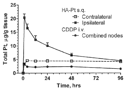
intravenous cisplatin (Fig. 1A-1B), although the overall plasma platinum AUC
is much
greater than with intravenous cisplatin. After subcutaneous HA-cisplatin
injection, there
was a 2 hr delay before plasma platinum peaked and plateaued-off, then
remaining at a
constant level. This release profile is consistent with a delayed and
sustained release
from the lymphoid tissues.
Figures 2A-2B show that creatinine levels did not indicate significant
differences
in renal toxicity between intravenous cisplatin and localized nanoconjugates
despite the
much higher AUC of nanoconjugate platinum. The high dose silver formulation
caused
decreased urine creatinine levels and animal death within the first week,
which is
unfavorable. The severe in vivo toxicity of silver formulations indicates that
the slightly
greater in vitro anti-proliferative activity of silver formulations (Table 1)
may be due to
non-specific silver toxicity. Although no damage was detected, creatinine
testing may not
be sensitive enough to detect minor renal damage after a single moderate dose,
so we
conducted pathological examinations for more direct evidence of platinum
toxicity. Thus,
in one embodiment, a nanoconjugate formulation is substantially devoid of
silver.
CA 02713813 2010-07-29
- 18 -
Pathological examination 30-days following a single dose injection of
cisplatin
revealed there were significant renal and hepatic differences between the
subcutaneous
HA-cisplatin and intravenous cisplatin formulations. Although the renal
platinum AUC of
HA-cisplatin was twice that of cisplatin, the lower incidence and less severe
nature of
renal cell necrosis in the HA-cisplatin treated animals indicated improved
tolerability
over intravenous cisplatin. The greater platinum AUC of HA-cisplatin in the
kidneys
would seem to contradict the lower toxicity observed, but may be due to its
lower peak
serum drug levels (Cit.) filtered by the kidney compared to intravenous
cisplatin.
Liver pathology indicated decreased toxicity of the HA-cisplatin compared to
cisplatin, and none of the animals had normal pathology in the 33 mg/kg
cisplatin arm.
Cisplatin-induced hepatotoxicity is known to occur due to the production of
reactive
oxygen species. Hyaluronan is metabolized in the liver and glycosaminoglycans
are
known antioxidants with hepato-protective effects, so the HA nanoconjugate may
protect
against platinum hepatotoxicity.
Accordingly, an embodiment includes a nanoconjugate of HA and cisplatin that
concentrates cisplatin in the breast lymphatics after subcutaneous injection
into the
mammary fatpad. These nanoconjugates have sustained release characteristics
resulting in
a higher lymphatic AUC and lower plasma Cmax compared to standard intravenous
cisplatin. The nanoconjugates do not cause substantial organ toxicities, such
as renal,
hepatic, neuro, or nephrotoxicity; and on pathological examination appear to
have lower
organ toxicity compared to the standard intravenous cisplatin. the
nanoconjugates do not
cause injection site or lymph node toxicities. The preferential intralymphatic
translocation
and accumulation of the nanoconjugates provides advantages for use in
combination
regimens for breast cancer with other chemotherapeutics.
In accordance with the present invention, the nanoconjugate can deliver
cisplatin
effectively to be use alone or as part of a combination therapy with
significantly less
toxicity. The intralymphatic delivery model using nanoconjugates not only
increases drug
concentrations in loco-regional nodal tissue significantly above the standard
cisplatin
formulation (74% greater AUC), but it also exhibits sustained release
kinetics, allowing
lower C. levels which lower organ toxicity over time. The only tissue level
that was
significantly different was the axillary lymph nodes ipsilateral to the drug
injection. This
translated into almost double the concentration of cisplatin penetrating the
loco-regional
nodes using nanoconjugates injected directly into the breast subcutaneously.
Therefore,
nanoconjugates that are preferentially translocated to the lymphatics
significantly boosts
CA 02713813 2010-07-29
- 19 -
the concentration of drug to treat and/or inhibit loco-regional tumor cell
development in
the lymphatics.
Also, the nanoconjugates can be successful in treating and/or inhibiting the
spread
of cancer because of the intralymphatic delivery of chemotherapeutics using
hyaluronan
as a targeted nanocarrier to the axilla. The preferential intralymphatic
delivery can
preferentially treat at-risk regional lymph nodes and avoid systemic
toxicities associated
with intravenous or oral drug administration. The preferential intralymphatic
delivery
reduces the systemic concentration but maintains a suitable level for also
treating and/or
inhibiting the spread of cancerous cells that are disseminated into the
systemic
circulation. As such, the nanoconjugates can provide a therapy for patients
with sentinel
nodes containing nanometastases which would not be offered lymph node
dissections
routinely. The nanoconjugate provides adequate systemic drug levels in a more
sustained-release manner than standard therapy, but it also provides a much-
needed boost
to the loco-regional nodal tissue, which is at risk for harboring tumor cells
not removed
by nodal dissection. Additionally, the nanoconjugate can be used as a
neoadjuvant for
locally advanced breast cancers, and can treat or inhibit regression.
In one embodiment, a pharmaceutical formulation having the nanoconjugate is
not
formulated for the following delivery routes: oral, systemic, transdermal,
intranasal,
suppository, intravenous, or intraluminal administration.
The pharmaceutical can be configured for percutaneous, intradermal, mucosal or
submucosal, subcutaneous, interstitial, intrafat, peritumoral, intramuscular
injection
mucosa, peritumorally, inhalation, and instillation.
The nanoconjugate of the present invention preferentially translocating to the
lymphatic system after subcutaneous or interstitial administration is
surprising and
unexpected. In part, this is due to the preferential translocation and
accumulation in the
lymphatic system after subcutaneous or interstitial injection, such as into
the breast tissue.
The nanoconjugate of HA or dendrimer with a chemotherapeutic such as cisplatin
additionally provides a therapeutic systemic dose with AUC similar or
sometimes higher
than standard intravenous agents (e.g., cisplatin and doxorubicin) but lower
Cmax
concentrations. This combination of findings allows these drugs to be used as
superior
adjuvant therapies for patients with loco-regional disease, with the
additional benefit of
being able to treat their systemic disease and with less toxicity since the
Cmax is lower
and this is associated with cisplatin and doxorubicin toxicity. It is also
surprising that the
HA nanoconjugates were substantially less toxic than standard cisplatin or
doxorubicin
CA 02713813 2010-07-29
- 20 -
with regard to local injection site, kidney, and ototoxicity evaluation by
OAEs and by
renal pathologic analysis. The lower toxicity allows for direct injection
into, adjacent, or
proximally into tissue or interstitial space without damaging the healthy
tissue. Thus, the
nanoconjugates can provide therapy to the primary tumor, the intralymphatic
cancerous
cells, and systemic cancerous cells.
In summary, the nanocarrier delivery system is superior to standard drug
formulations in (1) its efficacy and ability to treat cancers in animals, (2)
its lower toxicity
profile, and (3) its longer dosing interval (weekly or biweekly versus daily
with
intravenous agents). The subcutaneous injection offers patients a less
invasive treatment
option than being attached to intravenous infusion pumps which carry the risk
of drug
extravas ati on.
The nanoconjugate can be included in a pharmaceutical composition with an
acceptable carrier that formulates the nanoconjugate for suitable
administration, such as
subcutaneous. Suitable preparations for subcutaneous administration are
primarily
aqueous solutions of an active ingredient in water-soluble form, for example a
water-
soluble salt, and furthermore suspensions of the active ingredient, such as
appropriate oily
injection suspensions, using suitable lipophilic solvents or vehicles, such as
fatty oils, for
example sesame oil, or synthetic fatty acid esters, for example ethyl oleate
or
triglycerides, or aqueous injection suspensions which contain viscosity-
increasing
substances, for example sodium carboxymethylcellulose, sorbitol and/or
dextran, and, if
necessary, also stabilizers.
According to the methods of the present invention, the compositions of the
invention can be administered by injection by gradual infusion over time or by
any other
medically acceptable mode. Any medically acceptable method may be used to
administer
the composition to the patient. The particular mode selected will depend of
course, upon
factors such as the particular drug selected, the severity of the state of the
subject being
treated, or the dosage required for therapeutic efficacy. The methods of this
invention,
generally speaking, may be practiced using any mode of administration that is
medically
acceptable, meaning any mode that produces effective levels of the active
composition
without causing clinically unacceptable adverse effects.
For injection, the nanoconjugates can be formulated into preparations by
dissolving, suspending or emulsifying them in an aqueous or nonaqueous
solvent, such as
vegetable or other similar oils, synthetic aliphatic acid glycerides, esters
of higher
aliphatic acids or propylene glycol; and if desired, with conventional
additives such as
CA 02713813 2010-07-29
- 21 -
solubilizers, isotonic agents, suspending agents, emulsifying agents,
stabilizers and
preservatives. Preferably, the nanoconjugates can be formulated in aqueous
solutions,
preferably in physiologically compatible buffers such as Hanks's solution,
Ringer's
solution, or physiological saline buffer.
The nanoconjugates can be formulated for subcutaneous administration by
injection, e.g., by bolus injection or continuous infusion. Formulations for
injection may
be presented in unit dosage form, e.g., in ampules or in multidose containers,
with an
added preservative. The compositions may take such forms as suspensions,
solutions or
emulsions in oily or aqueous vehicles, and may contain formulator agents such
as
suspending, stabilizing and/or dispersing agents.
Sterile injectable forms of the compositions of this invention may be aqueous
or a
substantially aliphatic suspension. These suspensions may be formulated
according to
techniques known in the art using suitable dispersing or wetting agents and
suspending
agents. The sterile injectable preparation may also be a sterile injectable
solution or
suspension in a non-toxic parenterally-acceptable diluent or solvent, for
example as a
solution in 1,3-butanediol. Among the acceptable vehicles and solvents that
may be
employed are water, Ringer's solution and isotonic sodium chloride solution.
In addition,
sterile, fixed oils are conventionally employed as a solvent or suspending
medium. For
this purpose, any bland fixed oil may be employed including synthetic mono- or
di-
glycerides. Fatty acids, such as oleic acid and its glyceride derivatives are
useful in the -
preparation of injectables, as are natural pharmaceutically-acceptable oils,
such as olive
oil or castor oil, especially in their polyoxyethylated versions. These oil
solutions or
suspensions may also contain a long-chain alcohol diluent or dispersant.
The involvement of the lymphatic system in breast cancer metastasis is well
established, yet there are no effective non-surgical treatments to overcome
lymph node
metastases and disease progression. The greatest challenge with chemotherapy
of
lymphatic metastases is maximizing the amount of agent which actually is
retained in the
lymph nodes while avoiding systemic absorption and toxicity.
The lymphatically-localized chemotherapy provided by the present invention is
an
innovative leap in breast cancer therapy. The HA or dendrimer nanocarriers for
chemotherapeutic drugs can treat locally advanced breast cancer utilizing both
a targeted
and lymphatic delivery approach. The use of lymphatic targeted nanocarriers
for
intralymphatic drug delivery in breast cancer is highly innovative and has
never been
performed to date. By increasing drug loco-regional AUC over that achievable
by
CA 02713813 2010-07-29
- 22 -
standard chemotherapy drugs this technology provides significant neoadjuvant
therapy in
locally advanced breast cancer. Additionally having a targeted approach with
better
retention and sustained release of drug from the lymphatics should decrease
systemic
toxicity of the drugs leading to a combination of better tumor efficacy and
lower toxicity.
The present invention may be embodied in other specific forms without
departing
from its spirit or essential characteristics. The described embodiments are to
be
considered in all respects only as illustrative and not restrictive. The scope
of the
invention is, therefore, indicated by the appended claims rather than by the
foregoing
description. All changes which come within the meaning and range of
equivalency of the
claims are to be embraced within their scope. All references recited and/or
shown herein
are incorporated herein by specific reference.
EXPERIMENTAL
Hyaluronan (HA) from microbial fermentation was purchased from Lifecore
Biomedical (Chaska, MN) as sodium hyaluronate and used without further
purification.
Heparin solution was purchased from Abraxis Pharmaceutical Products
(Schaumburg,
IL). All other reagents were purchased from Fisher Scientific (Pittsburgh, PA)
or Sigma
Aldrich (St. Louis, MO) and were of ACS grade or better. Milli-Q water was
used in all
experiments. The MDA-MB-468LN cell line was kindly provided by Ann Chambers
(London Health Sciences Center, London, Ontario), while MCF-7 and MDA-MB-231
cells were obtained from the American Tissue Culture Collection (ATCC,
Manassas,
VA). Animal procedures were approved by the University of Kansas Institutional
Animal
Care and Use Committee. Sprague-Dawley rats were purchased from Charles River
Laboratories (Wilmington, MA).
1. Synthesis of HA-Cisplatin Conjugates
Cisplatin was conjugated to HA (35,000 g/mol) based on previously reported
procedures (S. Cali, Y. Xie, T. Bagby, M. S. Cohen, and M. L. Forrest.
Intralymphatic
chemotherapy using a hyaluronan-cisplatin conjugate. I Surg. Res. 147:247-252
(2008);
Y. I. Jeong, S. T. Kim, S. G. Jin, H. H. Ryu, Y. H. Jin, T. Y. Jung, I. Y.
Kim, and S. Jung.
Cisplatin-incorporated hyaluronic acid nanoparticles based on ion-complex
formation. I
Pharm Sci. (Epub.): (2008)), with and without the addition of silver nitrate
as an
activating agent. Typically, HA (100 mg) and cisplatin (45 mg) were dissolved
in H20
(20 ml) and stirred in the dark for four days under argon at ambient
temperature (ca. 25
C). The reaction mixture was filtered (0.2-inn nylon membrane) and dialyzed
against
CA 02713813 2010-07-29
-23-
H20 (10,000 MWCO; Pierce, Rockford, IL) for 48 hrs at 4 C protected from
light.
Following dialysis, the crude product was concentrated and stored at 4 C. The
degree of
cisplatin substitution was determined by atomic absorption spectroscopy (AAS)
(Varian
SpectrAA GTA-110 with graphite furnace). The furnace program was as follows:
ramp
25 to 80 C, hold 2 s, ramp to 120 C, hold 10 s, ramp to 1000 C, hold 5 s, ramp
to
2700 C, hold 2 s, cool to 25 C over 20 s. The graphite partition tube was
cleaned every
40 samples by baking at 2800 C for 7 s. Argon was used as the injection and
carrier gas.
The resulting conjugate is referred to as HA-cisplatin, cisplatin-HA, HA-CDDP,
HA-Pt,
although the conjugate is [PtC1(H20)(NH3)2]0000-HA (Figure 9).
The structure of cisplatin lends itself to complex formation with
polycarboxylic
polymers, since one or more of the chlorides can be displaced allowing
formation of a
labile ester linkage with the polymer. cisplatin was highly conjugated to HA
with typical
conjugations of 0.20 w/w platinum/complex (approximately 65% cisplatin
conjugation
efficiency). In previous studies, cisplatin conjugates were synthesized by
first activating
HA with AgNO3; however, it has now been found that eliminating this step does
not
significantly reduce conjugation and it reduces potential silver toxicity. The
AAS
produced a linear curve in the range of 10 to 450 ng/mL (R2=0.999) with a
limit of
detection of 5 ng/ml. Concentrated samples were diluted with water into the
linear
analytical range prior to analysis.
Fluorescent conjugates of HA were formed by condensation of Texas Red
hydrazide to HA. HA (35 000 MW, 100 mg) in 10 mL of 30% H20:Et0H was activated
with 2-chloro-1-methylpyridinim iodide (33 mg) and triethylamine (35 p.L).
After the
addition of Texas Red hydrazide (AnaSpec Inc., San Jose, CA) (2 mg in 0.4 mL
of
DMSO), the mixture was refluxed for 24 hrs. Workup proceeded by dialysis
against H20
for 48 hrs at ambient temperature, followed by lyophilization. Conjugation
efficiency was
determined using a molar extinction coefficient of 81 800 M-lcm-1 at X 588 nm.
Cisplatin was highly conjugated to HA, with typical conjugations of 0.25 w/w
cisplatin/complex using a starting ratio of 0.5 w/w cisplatin/HA. Up to 0.75
w/w
cisplatin/complex was attempted with decreasing efficiency (Table 4).
2. In Vivo Cell Toxicity
The lymphatically metastatic breast cancer cell line MDA-MB-468LN was
maintained in modified Eagle's medium alpha supplemented with 10% fetal bovine
plasma, 1% L-glutamine, and 0.4 mg/mL G418 (geneticin). Additional breast
cancer cell
CA 02713813 2010-07-29
- 24 -
lines MDA-MB-231 and MCF-7 were maintained according to protocols provided by
the
ATCC. Preceding proliferation studies, cells were trypsinized and seeded into
96-well
plates (5,000 cells/well). After 24 hrs, cisplatin, HA-cisplatin (with or
without silver
activation), or HA was added (n=12; 7 concentrations), and 72 hrs post-
addition,
resazurin blue in 10 pi of phosphate-buffered saline was added to each well
(final
concentration of 5 mM). After 4 hrs, well fluorescence was measured (X,, 560
nm, 2em
590 nm) using a fluorophotometer (SpectraMax Gemini; Molecular Devices,
Sunnyvale,
CA). IC50 was determined as the midpoint between saline (positive) and cell-
free
(negative) controls for each plate.
Cell toxicity was determined as the reduction in cell proliferation over 72
hrs. HA-
cisplatin conjugates with and without silver had similar cytotoxicity to free
drug in cell
culture (Table 1). No appreciable difference in toxicity was detected between
cisplatin
and HA-cisplatin using three different human breast cancer cell lines (Table
1). HA
showed no toxicity at 10 mg/ml, the upper limit of testing in all cell lines
compared with
saline controls (data not shown).
3. Pharmacokinetics and Tissue Distribution
Sprague-Dawley rats (female, 200-250 g) were cannulated in the left jugular
vein
under isoflurane and allowed to recover overnight. Animals were then injected
intravenous with cisplatin (1.0 or 3.3 mg/kg; n=5) or subcutaneous with HA-
cisplatin (1.0
or 3.3 mg/kg equivalent cisplatin; n=5) under isoflurane anesthesia
Subcutaneous
injections were given in the uppermost right mammary fatpad of the animal.
Whole blood
was withdrawn (100 Ill) from the cannula at 0, 5 mm, 0.5, 1, 2, 4, 6, 12, 24,
48 and 96 hrs
after dosing and placed into 2-ml centrifuge tubes pretreated with heparin.
The cannula
was washed before and after withdrawal with saline and then heparin locked.
The whole
blood was centrifuged at 17,000 x g for 5 mins, and the plasma was frozen at -
80 C until
analysis. Animals were euthanized 96 after treatment. The right ipsilateral
axilla nodes
(treated side), left contralateral axilla nodes (control side), and major
organs (liver,
kidneys, heart, spleen, lungs, brain, muscle, bladder) were excised; washed
with 0.9%
saline; and stored at -80 C until analysis. Tissue samples were prepared
using a
procedure reported previously (S. Cai, Y. Xie, T. Bagby, M. S. Cohen, and M.
L. Forrest.
Intralymphatic chemotherapy using a hyaluronan-cisplatin conjugate. J. Surg.
Res.
147:247-252 (2008)). Typically, 50 mg of tissue sample was digested using 1.5
ml of
6.7% nitric acid for 2 hours at 80 C. After digestion, samples were
homogenized (Tissue
CA 02713813 2010-07-29
- 25 -
Tearor; BioSpec Products Inc., Bartlesville, OK) and centrifuged. The
supernatant and
plasma samples were analyzed by AAS as described in the Synthesis section. The
pharmacokinetics of subcutaneous HA-cisplatin were compared to intravenous
cisplatin
in Sprague-Dawley rats. HA-cisplatin accumulated more preferentially in the
draining
ipsilateral axillary lymph nodes than did the intravenous cisplatin control
(Fig. 1A);
preferential accumulation was still evident at 48 hrs post-injection even
though the in
vitro disassociation half-life of cisplatin from HA is 10 hrs. The ipsilateral
axillary node
AUCo-96his of HA-cisplatin when injected locally was 3.8-fold greater than
intravenous
cisplatin (p<0.001), and the peak node concentration (Cmax) of HA-cisplatin
was 6.2-fold
greater than intravenous cisplatin.
The most significant, dose-limiting toxicities of cisplatin therapy are
nephrotoxicity followed by neurotoxicity, both of which are strongly
influenced by peak
plasma concentration. The peak plasma concentration intravenous cisplatin was
3.1-fold
greater than subcutaneous HA-cisplatin. The release of cisplatin into the
systemic
circulation was slow, and the resulting plasma AUC of HA-cisplatin was 3.9-
fold greater
than intravenous therapy with cisplatin, which is consistent with longer
lymphatic
retention of the nanocarrier HA-cisplatin (Fig. 1B, Table 2). Concentration
graphs for all
tissues are included in supplement (Fig. 8A-8H).
Additionally, Sprague-Dawley rats (200-250 g females, Charles River) were
placed under isoflurane anesthesia and injected subcutaneously (100 !IL) into
the right
mammary fat pad with cisplatin-HA or cisplatin in 0.9% saline (3.5 mg/kg
equivalent
cisplatin) (n=5). Animals were allowed to recover with access to food and
water. After 1,
4, 12, 24, 48 and 96 hrs post-injection, animals were euthanized by isoflurane
overdose.
Organs and tissues were washed with 0.9% saline and frozen (-80 C) until
analysis.
Plasma was separated by centrifugation from whole blood and frozen (-80 C).
Conjugation of cisplatin to HA impacted the local concentration of cisplatin
in draining
lymph nodes with a minor effect on systemic concentrations (Figures 10A-10B,
Table 5).
Over the experimental timeframe of 96 hrs, the area-under-the-curve (AUC) of
cisplatin-
HA conjugates in the right lymph node (RLN), which drains the injection site,
was 74%
greater than cisplatin in saline (p=0.0001), and the RLN had increased tissue
concentrations over the examined period (Figure 10A). The AUC of cisplatin-HA
in the
non-draining left lymph node (LLN) was not significantly different from
cisplatin in
saline (p=0.12).
CA 02713813 2010-07-29
- 26 -
A burst release of free cisplatin appeared in the plasma concentration profile
(Figure 10B), whereas cisplatin-HA demonstrated a longer, sustained release
into the
plasma. This is significant because dose-limiting toxicities of cisplatin
therapy are
strongly influenced by peak plasma concentration. There was not a significant
difference
in the plasma AUC between cisplatin-HA and cisplatin (p=0.13). Thus, localized
therapy
with cisplatin-HA may generate sufficient serum concentrations to treat
distant
metastases, while providing a boost therapy for the breast lymphatics. The
distribution of
cisplatin to other organs was not significantly different over the study
period (Figure 11,
Table 5).
4. Long-term Toxicology
Sprague-Dawley rats (35 females) were randomly divided into 7 study groups of
5
animals each: 1.0 mg/kg subcutaneous HA-cisplatin (with and without silver;
platinum
equivalent to 1.0 mg/kg cisplatin), 3.3 mg/kg subcutaneous HA-cisplatin (with
and
without silver), intravenous cisplatin at 1.0 and 3.3 mg/kg, and subcutaneous
HA (control;
HA equivalent to 3.3 mg/kg HA-Pt). Each animal was administered a single bolus
dose at
the beginning of the 30-day study period. Urine samples were collected every
day during
the first two weeks of the study and every four days during third and fourth
week of the
studies (except for the 3.3 mg/kg HA-cisplatin with silver group). In order to
reduce the
stress to animals, subjects were housed in metabolic cages for 12 hrs to
collect
approximately 5 ml of urine and then returned to cages with bedding until the
next
collection period. Urine samples were centrifuged at 17,000 x g for 5 mins and
stored in -
80 C freezer until creatinine analysis.
Urine creatinine was analyzed using the QuantiChromTM Creatinine Assay Kit
according to the manufacturer's instructions (BioAssay Systems, Hayward, CA).
Creatinine concentration of the sample was calculated as (ODsAmpLE 5 ¨
ODSAMPLE 1) /
(0DsTD 5 ¨ ODsTD 1) x [STD] (mg/dL). ODSAMPLE5, ODSAMPLE1, ODSTD5, and ODstni
are
OD510nm values of sample and standard at 5 min and 1 min, respectively.
The animals were euthanized at the end of the study (30 days) and the liver,
bilateral kidneys, spleen, lungs, heart, right (ipsilateral) and left
(contralateral) axillary
nodes, and brain were excised intact and stored in 80% alcoholic formalin
solution
overnight for fixation before slide mounting. Mounting using haematoxylin &
eosin
(H&E) staining were conducted by Veterinary Lab Resources (Kansas City, KS).
The
CA 02713813 2010-07-29
- 27 -
pathological examination was performed by a blinded board-certified
veterinarian
pathologist (University of Kansas Medical Center, Kansas City, KS).
Urine creatinine levels are an indirect indicator of renal function and renal
toxicity, with a decrease in creatinine excretion corresponding to decreased
renal function
and possible renal toxicity or damage. Significant renal toxicity was observed
in animals
given the high dose silver regimen (3.3 mg/kg), with a 30% decrease in
creatinine
excretion at 3 days and 70% decrease at 4 days. All animals in this group died
within 1
week of treatment due to drug-related cachexia. In contrast, the silver-free
high dose HA-
cisplatin did not demonstrate significant toxicity, and creatinine levels
remained near pre-
dosing levels throughout the study's duration. Similar renal function was
observed in both
groups when animals were administered low doses (1.0 mg/kg) of either HA-
cisplatin
with silver or HA-cisplatin without silver treatment (p>0.05, day 1 to 30)
(Fig. 2A-2B).
At the conclusion of the 30 day toxicity study, animals were euthanized and a
full
pathological examination performed. Brain tissue and underlying tissue of the
injection
site were normal with no microscopic changes for all study groups. Very mild
changes in
lymph nodes were detected for high dose intravenous cisplatin and subcutaneous
HA-
cisplatin formulated without silver. Lymphoid tissue from low dose
subcutaneous HA-
cisplatin with silver had normal appearance indicating by well-populated small
lymphocytes showing little or no follicular architecture (Fig. 6A-6F). Very
mild changes
were observed in the livers for animals receiving both low dose cisplatin
intravenous and
low dose HA-cisplatin subcutaneous indicated by the presence of mild
inflammation in
the sinusoids (Fig. 4A-4F). Mild degeneration with some sinusoidal necroses
were
observed for animals receiving high dose intravenous cisplatin and high dose
subcutaneous HA-cisplatin treatment; necroses were more severe in the
intravenous
cisplatin group. In addition, 60% of animals receiving low dose intravenous
cisplatin
were observed with mild renal necrosis including hemorrhage into the renal
tubules along
with tubular edema (Fig. 3A-3F). In contrast, none of the animals receiving
low dose
subcutaneous HA-cisplatin had renal necrosis. Similarly, 4 of 5 (80%) animals
receiving
high dose intravenous cisplatin compared to 1 of 5 (20%) animals receiving
high dose
subcutaneous HA-cisplatin were diagnosed with mild renal necrosis (Table 3).
Overall,
the pathology studies demonstrated that the silver-free HA-cisplatin
conjugates
demonstrated lower incidence of both renal and hepatic toxicity compared to
the
conventional intravenous cisplatin treatment at all dose ranges. Additionally
no
CA 02713813 2010-07-29
- 28 -
neurotoxicity in the brain or local injection site toxicity in the underlying
muscle tissue
was observed in the treated animals (Figs. 5A-5F and 7A-7D).
5. In vitro drug release
In vitro release rate of cisplatin from cisplatin-HA was determined in
phosphate
buffer with and without saline. Cisplatin-HA was added to 3,500 MWCO dialysis
bag
(Pierce) and placed in a phosphate-buffered water bath (pH 7.4, 37 C) or
physiological
saline (140 mM). Samples were taken from the dialysis bags at predetermined
time points
and remaining cisplatin concentration determined by AAS. The release rate of
cisplatin
from complexes was determined in both phosphate buffered saline and water. The
Cl" in
saline was expected to more rapidly displace cisplatin, increasing the release
rate. The
release of drug showed near first order release kinetics with a release half-
life of 42 h in
water and 10 h in physiological saline. The AAS produced a linear
concentration curve
from 10 to 450 ng/mL (R2---0.9998), with a limit of detection of 5 ng/mL and a
limit of
quantification of 10 ng/mL (5% standard deviation). Cisplatin recovery from
cisplatin-
HA spiked tissues was: plasma, 82+4% (+STD); lymph nodes, 92+2%; bladder, 88
1%;
brain, 94+0.3%; heart, 97 1%; kidneys, 98 1%; liver, 100+1%; lung, 94 1%;
muscle,
95 1%; spleen, 97 1%. cisplatin recovery from cisplatin-spiked tissues was:
plasma,
80+3%; lymph nodes, 92+6%; bladder, 86 3%; brain, 93+10%; heart, 93+5%;
kidneys,
100+2%; liver, 100+7%; lung, 95 8%; muscle, 100+5%; spleen, 96 9%.
Figure 16 illustrates the in vitro HA-cisplatin release rate at 37 C for water
and
PBS solutions. Table 6 shows the associated half life.
6. In Vitro Cell toxicity
Cell lines were seeded into 96-well plates (5 000 cells/well) in DMEM medium
supplemented with 5% FBS and 1% penicillin/streptomycin. After 24 hrs,
cisplatin,
cisplatin-HA, or HA was applied (n=12, 7 concentrations), and 72 hrs post-
addition,
reazurin blue in 10 pi PBS was applied to each well (final concentration of 5
mM). After
4 his, well fluorescence was measured (X.,õ 560 nm, ken, 590 nm) (SpectraMax
Gemini,
Molecular Devices), and the 1050 determined as the midpoint between saline
(positive)
and cell-free (negative) controls.
Cisplatin-HA conjugates had similar toxicities to free cisplatin in cell
culture.
Toxicity was evaluated in the highly metastatic human breast cancer cell lines
MCF7 and
MDA-MB-231 (Figure 12). In both cell lines, there was no appreciable
difference in
toxicity between cisplatin-HA (IC50 7 ii.g/mL, cisplatin basis) and cisplatin
(ICso 7
CA 02713813 2010-07-29
- 29 -
pig/mL). HA had no toxicity to human cells over the concentration range
examined (up to
mg/mL, data not shown). Table 7 shows the IC50 values.
7. Atomic Absorption Spectroscopy (AAS)
In vitro release samples (n=3) and plasma samples (n=5) were diluted 200-fold
and 10-fold, respectively, with 0.1% nitric acid for analysis. Tissue samples
(except for
to lymph nodes)
were prepared by digesting 50 mg of tissue in 1.5 mL of 6.7% nitric acid
for 1 hr at 80 C. Lymph nodes were processed similarly using 10 mg of tissue.
After
digestion, samples were homogenized (Tissue Tearor, BioSpec Products Inc.,
Bartlesville,
OK). All samples were centrifuged (17 000 x g, 20 min), and the supernate used
for
analysis.
Analysis was performed on a Varian SpectrAA GTA-110 with graphite furnace
and partition tubes. Samples (21 4) were injected using the autosampler,
followed by 19
pit of 0.1% nitric acid. Every 10 samples were bracked by calibration
standards at 150,
300, and 450 ng/mL, and a quality control sample (150 or 300 ng/mL) every 5
samples.
A full calibration curve was prepared from 1 to 450 ng/mL in 0.1% nitric acid
(10
concentrations). Cisplatin recovery was determined by spiking tissue blanks
with cisplatin
or cisplatin-HA (50 p.g/g) and processing as above. The furnace program was as
follows:
ramp 25 to 80 C, hold 2 s, ramp to 120 C, hold 10 s, ramp to 1000 C, hold 5 s,
ramp to
2700 C, hold 2 s, cool to 25 C over 20 s. The graphite partition tube was
cleaned every 40
samples by baking at 2800 C for 7 s. Argon was used as the injection and
carrier gas.
8. In vivo imaging
In order for nanocarriers to deliver anticancer drugs to nano- and
micrometastases
in the breast loco-regional lymphatics, carriers should drain from the breast
area to the
diseased lymph nodes. To verify anionic nanocarriers to do, we constructed a
fluorescent
anionic nanocarrier by coupling 35 kDa hyaluronan with Texas Red hydrazide
(AnaSpec,
San Jose, CA) using EDAC-mediated amide coupling followed by dialysis to
remove free
dye (0.1% w/w dye/carrier determined spectroscopically). We injected
fluorescent
nanoparticles (0.25 pig in 50 pIL of saline) subcutaneously beneath the nipple
of a
xenograft bearing lymphatic metastases.
Fluorescence was measured in 10-nm bandpass segments from 520 to 720 nm,
using a cooled CCD camera with autoexposure. Images were spectrally unmixed
using
the automatic deconvolution tools (Maestro ver. 2.4) to limit skin and
intestine
autofluorescence resulting from chlorophyll in food.
CA 02713813 2010-07-29
- 30 -
Lymphatic breast tumor metastasis were induced in nude mice according to the
procedure of Chambers and coworkers, who were kind enough to provide the
lymphatically metastatic breast tumor cell line MDA-MB-468LN. Nude mice (25-30
g
females, Charles River) were anesthesized with pentobarbital (50 mg/kg), and
100 111, of
MDA-MB-468LN (l 07 cells/mL) was injected orthotopically into the left second
thoracic
mammary fatpad through a small incision later closed with a wound clip. Tumors
were
palpable after 4-5 wks (100-300 mm3). Before imaging, mice were anesthesized,
and
Texas Red-HA (10 mg/mL in saline, 20 pit) was injected subcutaneously over the
left
mammary fatpad. The injection area was massaged gently for 5 min and
fluorescently
imaged after 5 and 18 hrs (CRI Maestro Flex, CRI Inc., Woburn, MA) using a 445-
to
490-nm filtered halogen excitation light and a 515-nm longpass emission
filter.
Four hours post injection, fluorescent imaging (kex 480, 2m 500-720 nm,
spectrally deconvoluted) shows that anionic nanoparticles accumulated in a
region
consistent with the axillary node group (Fig. 13A). At this early timepoint,
much of the
carrier is still in close proximity to the injection site, although some
trafficking to the
axillary lymph node tumor is evident. These images are in deconvoluted, so
overlapping
GFP and Texas Red signals do not appear yellow. The software includes a
colocalization
tool the confirmed Texas Red in the area of the tumor. Accumulation of Texas
Red-
labeled nanocarriers is more apparent in the coronal view (Fig. 13B).
After 18 hours post-injection, the nanocarrier has concentrated in the
axillary node
surrounding the tumor (Fig. 13C). Of note, no carrier fluorescence appears at
the injection
site, and there is no apparent spread to the liver, compared against the very
high axillary
node levels. Obvious areas of carrier localization around the tumor are
apparent in the
coronal view. Kobayashi et al. observed that cationic PAMAM nanoparticles did
not enter
lymphatic tumors, but trafficked around them. However, anionic carriers have a
much
longer residence time, possibly allowing the carrier and its anticancer drug
cargo to enter
tumors.
Figures 13A-13C are show the localization of the intralymphatic carrier after
subcutaneous injection in nude mice bearing MDA-MB-468 breast lymphatic tumors
expressing green fluorescent protein (GFP) Figure 13A shows the breast
lymphatic tumor
4 at the time that the mice were subcutaneously injected with Texas Red-HA 6
in the left
mammary fat pad. After 5 hrs and 18 hrs (Figure 13B and Figure 13C,
respectively), the
photographs show that significant HA localized in the draining nodes and co-
located with
CA 02713813 2010-07-29
- 31 -
the tumor (GFP-channel in green in color and marked with 4, Texas Red channel
in red
and marked with 6, the blue arrow 2 is the injection site).
9. Activity
Conjugation of doxorubicin to the nanocarrier did not significantly affect its
anticancer activity in breast cancer cells. The 10% w/w conjugate was applied
to cells,
and 72 hrs later, cell proliferation was determined using the Alamar blue
assay
(Invitrogen Corp.). There was no statically significant difference in
anticancer activity
between the conjugate and free doxorubicin in three aggressive human breast
cancer cell
lines. Similarly, cisplatin conjugates were applied to breast cancer cells in
culture. After
72 hrs, cisplatin conjugates were not significantly different from free
cisplatin. Overall,
conjugation of anticancer drugs to nanocarriers did not decrease the anti-
proliferative
effect in vitro. This may be due release of anticancer drugs from nanocarriers
into the
culture media, followed by uptake as with free drug. However, the microscopy
uptake
study suggested drugs may have activity despite conjugation to an anionic
carrier.
10. Synthetic Schemes
Figure 14A shows the synthesis of a dendrimer-cisplatin conjugate. The bis-
hydroxypropyl phosphate termini of the dendrimer are converted into carboxylic
acids,
which then form a complex with cisplatin by displacement of the chloride. The
resulting
complexes will slowly release the potent DNA crosslinker cis-
[Pt(NH3)2C1(H20)1+ at
physiological conditions. In preliminary studies, we formed cisplatin
conjugates to
hyaluronan carriers by displacement of the platinum chloride with a carrier
carboxylic
acid to form ¨C(=0)00-PtC1(NH3)2 conjugates. A similar scheme can be used to
conjugate cisplatin to the anionic dendrimer carrier. The bis-hydroxypropyl
phosphate
termini of dendrimers are be converted to carboxylic acids by mild reduction
with Dess-
Martin periodinane (DMP), IBX, or IBA, and ozonation into the carboxylic acid.
Alternatively, the hydroxyls can be converted to carboxylic acids by treatment
with
succinic anhydride/DMAP, although this would extend the termini by four
carbons and
enlarge the nanocarrier. Complexes will be purified by dialysis or Sephadex,
and the drug
quantity on nanocarriers will be determined by graphite furnace atomic
absorption
spectroscopy. The optimum amount of cisplatin conjugation can be between about
10%-
50% substitution, more preferably about 20%-40%, and most preferably about 25%-
35%.
For example, up to 25% w/w conjugation has allowed significant lymphatic
uptake of
Pt(II)-hyaluronan nanoparticles in vivo.
CA 02713813 2010-07-29
- 32 -
Figure 14B shows the synthesis of dendrimer-epirubicin conjugates. The bis-
hydroxypropyl phosphate termini of the dendrimer are converted into
hydrazides, which
then form a pH-sensitive hydrazone with the epirubicin. The resulting
complexes will
release epirubicin in response to decreased pH in endocytic vessels of cells
(e.g. tumor
cells). Nanocarrier conjugates can greatly increase the amount of
anthracyclines
accumulated in cancer cells.
The pH-sensitive conjugates are formed using a hydrazone linker between the
anthracycline 13-carbonyl and a grafted hydrazide on the nanocarrier. The
hydrazide
linker was formed by graphing an adipic dihydrazide to the carboxylic acid
residues using
literature procedures, but a more direct strategy is to convert the
nanocarrier hydroxyl
termini directly into hydrazides, which will retain the size of the dendrimer.
Amine
termini would be expected to increase the cationic nature of the carrier, but
the low pIC of
hydrazides (ca. pH 4.5) will minimize this effect extracellularly. In
addition, partial
conversion of the hydroxyl termini can create enough hydrazides to conjugate
the desired
drug load. After converting the bis-hydroxypropyl phosphate termini of the
dendrimer
into carboxylic acids, activate and then treat the termini with hydrazine,
forming
hydrazide termini. The epirubicin conjugate is formed by incubation of the
carrier and
epirubicin at pH 6.5 to form the reversible hydrazone linker followed by
dialysis or
Sephadex workup to remove the unconjugated drug. The optimum amount of
epirubicin
conjugation can be between about 5%-20%, more preferably about 8-15%, and most
preferably about 10%-12%.
Figure 14C shows the synthesis of dendrimer-docetaxel conjugates. The
docetaxel
is converted into a protected malic acid, and the new carboxylic acid is
directly linked
with the bis-hydroxypropyl phosphate termini of the dendrimer via an ester
linkage. The
resulting complex will protect tissues from docetaxel toxicity until
conjugates release
docetaxel in response to decreased pH in endocytic vessels and endogenous
esterases.
Docetaxel can be conjugated to the unmodified bis-hydroxypropyl phosphate
termini of
the dendrimer using by a malic acid linker grafted onto the C2 position of
docetaxel using
an ester bond. The docetaxel C2-OH can be condensed with 1, 2-0-isopropylidene-
malic
acid, forming a protected malic acid of docetaxel. Deprotection with acetic
acid will yield
the malic acid, which can be directly coupled onto the hydroxyl arms of the
dendrimer.
The resulting conjugate can release docetaxel in response to acidic pH or
endogenous
esterases in acidic vesicles. The expected half-life after injection is
expected to be greater
than 4 hours, allowing sufficient time for lymphatic uptake based on our
preliminary
CA 02713813 2010-07-29
- 33 -
studies of anionic lymphatic nanocarriers. The optimum amount of docetaxel
conjugation
can be between about 5%-20%, more preferably about 8-15%, and most preferably
about
10%42%.
Figure 15A shows the synthesis of a dendrimer in accordance with the present
invention, and demonstrates an overall synthetic strategy for our library,
although we are
to examining additional "arm" groups and substituents to further improve
uptake. From a
multi-arm core, we are building a several generation polyester core by
carbodiimide
coupling, followed by deprotection of the pendant hydroxyls. Additional
generations are
built from these pendant groups. After 2-4 generations, we switch to a
phosphite
branching unit (P3). Tetrazole is used to couple the P3 branches to the
pendant hydroxyls,
followed by selenium conversion to the selenophosphate. "Defect" branches (red
asterisks) are selectively included to yield ionizable groups when
deprotected, thus
integrating ionizable groups within the core to limit aggregation and increase
solubility.
The charge degree can be tailored by varying the ratio of defect branches.
After 2-3
generations, the poly-selenophosphoester branches are fully oxidize with
organic
peroxide, and terminal hydroxyls are converted to carboxylic acids for
cisplatin
conjugation or hydrazones for doxorubicin conjugation using standard methods.
Our preliminary studies determined that this dual core strategy can facilitate
synthesis. Phosphate groups can be selenium protected, otherwise the phosphate
is
sufficiently reactive to cleave the arm groups during conjugation; however, in
later
generations the inner selenophosphates become too hindered by subsequent
generations
for deprotective oxidation. To overcome these issues we introduce a carboxy
ester core,
which improves biodegradability and lessens steric crowding during oxidation
of the
outer phosphate groups. Fully carboxylic acid dendrimers have been developed,
but these
would not be water-soluble if highly substituted with hydrophobic drugs. We
use the
anticancer drugs cisplatin and doxorubicin as they are highly effective first-
line
treatments for multiple cancers including head and neck cancers (HNC) and
breast
cancer.
Figure 15B shows the synthesis of targeted dendrimers. The bis-hydroxypropyl
phosphate termini of the dendrimer are condensed with propiolic acid to form
the alkyne-
dendrimer. Alternately, the COOH or CONHNH2 can be condensed with propargyl to
form the alkyne-dendrimer. Using a "click" methodology, the dendrimer and the
azide of
EGF are coupled under aqueous conditions. This strategy can be applied to
other targeting
agents, e.g. HER2 antibody, EGFR antibody, PSMA, and the like.
CA 02713813 2010-07-29
- 34 -
The nanoconjugates can target breast cancer cells via the epidermal growth
factor
receptor (EGFR), which is highly overexpressed by aggressive breast cancers
with poor
prognosis. In addition, EGFR is endocytosed and can be used to internalize
nanocarriers
conjugated to EGF. Although EGFR is expressed at lower levels by other
tissues, passive
localization of the nanocarrier to the lymphatics will minimize nonspecific
interactions.
to Unlike systemic nanocarriers, the targeting moiety can improve cell
uptake, as physical
characteristics alone can localize anionic nanocarriers in the lymph nodes.
Epidermal growth factor (EGF) is a 6054-Da protein with lysine residues that
may
be used to link nanoparticles without decreasing interaction with cells. Since
the termini
group of the dendrimer is dependent on whether the dendrimer is used for
cisplatin
(COOH), docetaxel (OH) or epirubicin (C=ONHNH2), it is easiest to use a
"click" type
linker so all chemistries can be done in aqueous solution and as a final step.
EGF is
functionalized using azido-PEG-NHS ester (Quanta Biodesign Ltd., Powell, OH).
Alternatively, other targeting agents are easily added by forming the
respective azide.
Alkynes can be added to the dendrimer termini using DCC/DMAP chemistry and
either
propargyl alcohol (COOH termini) or propiolic acid (OH and C=ONHNH2 termini).
The
resulting targeted alkyne-dendrimer is mixed with the EGF-azide with a small
amount of
copper catalyst to form the conjugate, followed by Sephadex or dialysis
purification to
remove unbound EGF.
11. Tumor Response
Nude nu/nu mice were implanted with 1x10^6 cells (MDA-MB-468LN human
breast cancer cells) into the mammary fatpad, after approximately 4 weeks a
100-200
mm3 tumor had developed in the axillary lymph node package. Animals were
randomly
divided into four treatment groups of five animals each: intravenous saline,
s.q. HA,
intravenous cisplatin, or s.q. HA-cisplatin. At this point, doses were
administered to the
animals. In the HA-cisplatin and the HA groups, HA-cisplatin (3.3 mg/kg based
on Pt-
content) or HA (3.3 mg/kg) was dissolved in 100 mcL saline was injected
subcutaneously
peritumorally. In the cisplatin and saline groups, cisplatin (3.3 mg/kg, Pt-
basis) in 100
mcL saline or just saline was injected intravenously via the tail vein. In all
study groups,
a second dose was administered after 1 week. Tumor volumes were measured
weekly
using calipers and the formula: Volume = 0.52 x (width2 * length). Animals
were
euthanized when the tumor volume exceeded 1000 mm2 or the body score index
fell
below 2.
CA 02713813 2010-07-29
- 35 -
Figures 17A-17B show that tumor growth was delayed by HA-cisplatin treatment
for 5 weeks compared to negative control group and 2 weeks compared to
conventional
cisplatin treatment.
The HA-cisplatin formulation delayed tumor growth by 5 weeks compared to
cisplatin and no toxicity was observed in the HA-cisplatin treated animals.
While it was
expected that HA-cisplatin would likely have similar efficacy to intravenous
cisplatin,
what was unexpected and significant was that HA-cisplatin resulted in longer
delays in
tumor growth initially, indicating that the higher concentration of drug
reaching the tumor
allowed it to inhibit tumor growth more effectively during the initial
treatment period.
This provides convincing supportive evidence that HA-cisplatin subcutaneously
given,
carries at least the same if not better efficacy than standard cisplatin
formulations in this
= in vivo breast cancer model.
12. Doxorubicin Release
HA was condensed with adipic dihydrazide (ADH) at the carboxylic acid group of
the gluconic acid in HA. First, ADH and HA (1 eq. of ADH per eq. of HA
disaccharides)
were mixed with 2 eq. N-(3-Dimethylaminopropy1)-N'-ethylcarbodiimide
hydrochloride
(EDAC) in water adjusted to pH 4.7 with HC1 and a final HA concentration of
ca. 1%
w/w. After 20 min, the mixture was dialyzed against water using 13000 MWCO
dialysis
tubing for 2 days. The product was then conjugated to doxorubicin (DOX) in pH
6.5
phosphate buffer (2 eq. HA disaccharide per 1 eq. of DOX) for 24 hours in the
dark. The
mixture was then dialyzed against pH 6.5 phosphate buffer for 2 days. The
final material
was lyophilized and reconstituted in saline before use. For release
experiments,
reconstituted HA-DOX or DOX were placed in dialysis tubing and placed in a
bath
containing either pH 5, 6.0, or 7.4 phosphate buffer at 37 deg C. Samples were
taken from
the dialysis tubing the total drug remaining was determined by HPLC with a
fluorescent
detector, using a standard calibration curve.
Figure 18 shows the release of doxorubicin as a function of pH. The release
half-
life was found to be 167 hours at pH 7.4, 107 at pH 6.0, and 45 at pH 5Ø
The HA-DOX provided very sustained release, dependent on the pH of its
environment. They may offer substantial benefit, as the interior of solid
tumors is known
to be hypoxic and to have a reduced pH, thus DOX release from HA-DOX may be
accelerated in neoplastic tissues as compared to normal tissues.
To evaluate the efficacy of HA-doxorubicin conjugates injected subcutaneously
in
the rat breast compared with intravenous doxorubicin, the following experiment
was
CA 02713813 2010-07-29
- 36 -
performed. MDA-MB-468LN tumor cells into nude mice as described and divided
into
four groups of five animals each. After 3 weeks, HA-DOX was injected
peritumorally at
3.3 mg/kg (DOX basis), DOX was injected in saline via the tail vein, HA was
injected
peritumorally, and saline was injected via the tail vein. Animals were given a
second dose
at 5 weeks post-implantation.
Figure 19 shows the tumor growth was halted by nanocarrier-DOX treatment after
two weekly doses at 3rd and 5th week.
While it was expected that HA-doxorubicin would likely have similar efficacy
to
intravenous doxorubicin, what was unexpected and significant was that HA-
doxorubicin
resulted in complete arrest in tumor growth with treatment, indicating that
the higher
concentration of drug reaching the tumor allowed it to inhibit tumor growth
more
effectively during the treatment period. This provides convincing supportive
evidence
that HA-doxorubicin subcutaneously given, is clearly superior in efficacy
compared to
standard doxorubicin formulations in this in vivo breast cancer model.
HA-DOX substantially retard tumor growth in animals compared to intravenous
DOX or the HA and saline controls. After 6 weeks post-implantation, the DOX
and HA
and saline controls had tumor volumes greater than 300 mm3, whereas the HA-DOX
treatment group's tumor were less than 50 mm3. The effectivity was a surprise
as the
DOX-HA not only provided effective localized control of tumor growth but also
prevent
the appearance of new local, regional, or distant growths. HA-DOX animals did
not
exhibit any toxicity due to dosing. The HA-DOX may provide excellent
locoregional
control as a "boost" dose to the locoregional lymphatic and tissues of the
tumor, and slow
drug release may be able to replace the need for many multiple dosings. In
addition, the
HA-DOX may provide sustained plasma levels of drug compared to intravenous
DOX,
thus replaced the need for additional systemic therapy. Once again, sustained
drug release
may reduce the need for repeated dosing. This results were surprising as the
localized
chemotherapy was not expected to provide sufficient plasma levels of drug to
provide
distant tumor control.
13. Phosphoester-Hyaluronan Nanocartiers
Figure 20A illustrates a highly water-soluble and biodegradable phosphoester
hyaluronan (phHA) nanocarrier. These nanocarrier can be used as described
herein.
Figure 20B illustrates the synthesis of phHA-drug conjugates. The phi-IA can
be
functionalized to form labile conjugates (e.g. ester or hydrazone) with anti-
cancer drugs
cisplatin (top), epirubicin (middle) and docetaxel (bottom). The resulting
complexes will
CA 02713813 2010-07-29
-37-
protect tissues from toxicity until conjugates release drugs in response to
decreased pH in
endocytic vessels and endogenous esterases. Dephosphoration of phHA by AP in
the
lymphatics will promote conjugate accumulation and sustained release of drugs
within the
lymphatics.
The carboxyl groups of HA (30-50 kDa, corresponding to 10-20 nm can be
protected (e.g. with DMT added using NMM/CDMT). The primary alcohol of HA can
be
converted to a phosphoester (e.g. with 1:1 aq. H3PO4 and perrhenic acid). For
conjugation
with cisplatin, the DMT may be removed from phHA with acid giving the
carboxylic
acid. For epirubicin and docetaxel, DMT may be displaced with a strong
nucleophil such
as adipic dihydrazide (ADH) allowing formation of the pH-sensitive labile
hydrazone
with drugs.
Sodium phHA can be conjugated cisplatin using the same procedure as in
preliminary studies with HA, by overnight reaction with cisplatin (20-40%
w/w), and
purified by dialysis against water. The degree of conjugation can be
determined by
graphite-furnace AAS.
Anthracyclines (doxorubicin, epirubicin, and daunorubicin) can be used to form
pH-sensitive conjugates of the structurally similar doxorubicin, using a
hydrazone linker
between the anthracycline 13-carbonyl and a grafted hydrazide on HA. The
hydrazide
phHA (described above) can be conjugated to epirubicin by incubation at pH 5,
followed
by dialysis purification.
Taxanes including docetaxel and paclitaxel can now be prepared into a
polysorbate-free formulation. This can be accomplished by forming esters off
the C7
carbon that couple with the nanocarrier, with no detriment on anti-cancer
activity.
Docetaxel can be conjugated to the COOH of the phHA using a labile malic acid
linker
grafted onto the C2 position of docetaxel.
14. In Vivo Efficacy of HA-Cisplatin
The in vivo efficacy of HA-cisplatin was studied. All treatment began once
animal HNSCC tumors reached 100 mm3 volume. Treatment commenced for 3 weeks.
All animals were given 5x10^5 MDA-1986 cells in the buccal mucosa of the left
cheek
(which is a novel orthotopic HNSCC model I developed this year) and tumors
develop in
2-3 weeks.
Figure 21A includes efficacy graph of Nu/Nu female athymic nude mice treated
with HA carrier by itself (subcutaneous), control (saline subcutaneous), 5
mice with HA-
cisplatin weekly dose of 3.3 mg/kg subcutaneous for 3 weeks, and 3 mice with
CA 02713813 2010-07-29
- 38 -
intravenous cisplatin (weekly dose of 3.3 mg/kg intravenous for 3 weeks). This
graph
shows that HA-cisplatin delayed tumor growth in all 5 animals, 2 of which (HA-
cis 3 and
HA-cis 4) were similar to the rate at which i.v, cisplatin (control) delayed
growth, by
about 3 weeks. Whereas in the other 3 animals, 2 animals had a complete
response to
therapy and the third (HA-cis 1) had a partial response to therapy before it
was sacrificed.
Overall this shows an improved efficacy over standard intravenous cisplatin in
treating
head and neck squamous cell cancer in vivo.
To evaluate the anticancer, specifically antiproliferative, effects of HA-
cisplatin
compared to standard CDDP in vitro in two human head and neck squamous cell
carcinoma cell lines (JMAR and MDA-1986) a standard MTS assay was performed as
per
the manufacturer's specified protocol.
Once 75% confluent, cells were trypsinized (0.25% trypsin), counted and plated
in
96-well microtiter plates (Costar, Cambridge, MA, USA) (1X10³ cells/well) in
100 1.1,1_, of
growth media. After an overnight attachment period, cells were exposed to
varying
concentration of KU135 and 17-AAG, alone and in combination for 3 days. All
studies
were performed in triplicate and repeated at least three times independently.
After the 3-d
treatment period, the number of viable cells was determined using a
colorimetric Cell
Proliferation assay (CellTiter96 Aqueous Nonradioactive Cell Proliferation
assay;
Promega, Madison WI, USA), which measures the bioreduction of the MTS (344,5-
dimethylthiazol-2y1]-543-carboxymethoxypheny1]-244-sulfophenyl]-2H
tetrazolium) by
dehydrogenase enzymes of metabolically active cells into soluble formazon
product, in
the presence of the electron coupling reagent PMS (phenazine methosulfate). To
perform
the assay, 20 [IL of combined MTS/PMS solution containing 2 mg/mL MTS and 150
ji.M
PMS in buffer (0.2 g/L KC1, 8.0 g/L NaC1, 0.2 g/L KH2PO4, 1.15 g/L, 133 mg/mL
CaC12-2H20, 100 mg/mL, MgC12.6H20, pH 7.35) was added to each well and then
after
3 hr of incubation at 37 C in a humidified 5% CO2 atmosphere, absorbance was
measured at 490 nm in microplate reader. Triplicate wells with predetermined
cell
numbers were subjected to the above assay in parallel with the test samples to
normalize
the absorbance readings; this also provided internal confirmation that the
assay was linear
over the range of absorbance and cell numbers measured. Data was plotted as a
function
of % viability from controls (cell viability) vs. drug concentration (x-axis).
The
concentration of drug at which 50% of cells were inhibited from growth (IC50
level) was
determined as the point of inflection on a standard absorbance-concentration
curve.
CA 02713813 2010-07-29
- 39 -
Figure 21B demonstrates percent cell viability versus drug concentration
curves
for standard cisplatin against both cell lines after 72 hours drug treatment
at varying
concentrations of drug ranging from 10 nM to 100 micromolar concentrations.
Figure
21C demonstrates percent cell viability versus drug concentration curves for
HA-cisplatin
against both cell lines after 72 hours drug treatment at varying
concentrations of drug
ranging from 10 nM to 100 micromolar concentrations. ICSO concentrations were
calculated from these graphs as the point of inflection where 50% of cell
growth is
inhibited. Of note there is no significant difference in either cell line
between IC50 levels
for CDDP and HA-cisplatin indicating that in vitro, HA conjugation does not
adversely
effect cisplatin's ability to inhibit cancer cell growth and viability.
15. In Vivo Release of Doxorubicin
Figures 22A-22F are photographs showing the distribution of HA-doxorubicin
after a single injection in the right mammary fat pad of a rat. Doxorubicin
has innate
fluorescence and the distribution and longevity of the drug-carrier conjugate
can been
well observed in this timed evaluation. Of note the bulk of drug-carrier is
transported to
the axillary lymph nodes where is slowly releases drug over a 9 day interval
with still
some residual activity even after 9 days. The oval marks the injection site in
the breast
and the darkest concentration (red) is in the axilla.
Figure 23 is a graph showing tumor response even after a single late term
peritumoral HA-Doxorubicin treatment in a considerably advanced breast cancer
tumor in
vivo.
Figures 24A-24E are photographs in vivo trafficking of HA-doxorubicin as
visualized on an Maestro multichannel fluorescent imaging system. There in
nice uptake
of drug and carrier into the locoregional tissues and lymph nodes of the rat
breast, which
stays well in the lymphatic's even 4 days post-injection.
TABLES
Table 1 shows the IC50 values of cisplatin on human breast cancer cell lines
MDA-MB-468LN, MDA-MB-231, and MCF-7. IC5os for the HA carriers are given on a
Pt-basis equivalent to cisplatin.
CA 02713813 2010-07-29
3
- 40 -
TABLE 1
Cell lines / Cisplatin HA-Cisplatin IA-Cisplatin
10501-3g/m1-, (W) (without silver) (with silver)
MDA-MB-468LN 5 fig/mL 10 lag/mL 9 ps/mL
(17 pM) (33 JIM) (30 pM)
MDA-MB-231 6 g/mL 10 g/mL 4 lag/mL
(20 pM) (33 NM) (13 pM)
MCF-7 6 g/mL 11 g/mL 6 p.g/mL
(20 pM) (37 IIM) (20 pM)
Table 2 provides the tissue AUC (average SEM) and C. (average + SEM) of
3.3 mg/kg intravenous cisplatin and subcutaneous HA-cisplatin study groups.
Two-way
ANOVA analysis revealed study groups (cisplatin and HA-cisplatin) differed
significantly for all tissues. The first plasma sampling was a 5 mins.
TABLE 2
Tissue AUC0-96hrs, pg/g.h Cmax, nig
(Truax)
cisplatin, iv. HA-cisplatin, s.q. cisplatin, i. v.*
HA-cisplatin, s.q.
Heart 128 8 465 23 1.7 0.1 (1 hr) 14.7
1.1 (1 hr)
Lungs 139 7 347 13 2.2 0.1 (1 hr) 7.5
0.7 (1 hr)
Kidneys 291 8 669 28 4.6 0.4 (1 hr) 12.3
2.5 (4 hrs)
Brain 152 +11 344 22 2.3 0.1 (1 hr) 7.5 2.0
(4 hrs)
Liver 178 5 495 20 2.9 0.2 (4 hrs) 10.6
2.2 (4 hrs)
Spleen 201 7 384 15 3.0 0.4 (1 hr) 6.6 0.6
(4 hrs)
Muscle 151 9 262 15 2.2 02 (1 hr) 8.5 2.1
(4 hrs)
Bladder 162 10 194 6 3.1 0.4 (1 hr) 2.9 0.4
(4 hrs)
Ipsilateral 205 12 776 9 3.3 0.3 (1 hr) 20.4 1.4
(1 hr)
axilla nodes
CA 02713813 2010-07-29
-41 -
Contralateral 205 12 413 17 3.3 (1 hr) 4.6 0.3
(4 hrs)
axilla nodes
Plasma 17 3 67 10 3.1 0.2 (5 mins)
1.0 0.3 (2 hrs)
* The first tissue sampling was at 1 hr so a Cr,.. prior to this would not be
detected.
Table 3 shows the classification of tissue damage for each treatment group was
made according to the following scale: Kidneys, grade 0: normal (no symptoms);
grade 1:
minimal necrosis; grade 2: mild necrosis (includes degeneration and nuclear
pyknosis).
Liver, grade 0: normal (no symptoms); grade 1: inflammation (includes
granulomas,
microgranulomas and hepatitis) or inclusions; grade 2: degeneration or
necrosis. Axilla
lymph nodes, grade 0: normal (no symptoms); grade 1: mild lymphoid hyperplasia
or
depletion. Pathologies were graded by a blinded veterinarian pathologist, and
each
treatment group contained 5 animals.
TABLE 3
Treatment group Liver Kidneys Axilla lymph nodes
1.0 mg/kg cisplatin 40% Grade 0 20% Grade 0 100% Grade 0
40% Grade 1 20% Grade 1
20% Grade 2 60% Grade 2
1.0 mg/kg HA-Pt 20% Grade 0 20% Grade 0 60% Grade 0
(Pt refers to 60% Grade 1 80% Grade 1 40% Grade 1
cisplatin) 20% Grade 2
3.3 mg/kg cisplatin 60% Grade 1 20% Grade 1 80% Grade 0
40% Grade 2 80% Grade 2 20% Grade 1
3.3 mg/kg HA-Pt 20% Grade 0 20% Grade 0 60% Grade 0
60% Grade 1 60% Grade 1 40% Grade 1
20% Grade 2 20% Grade 2
Table 4 shows the conjugation efficiency of cisplatin to HA. Efficiency was
calculated as (cisplatin added/cisplatin incorporated) x 100%.
CA 02713813 2010-07-29
- 42 -
TABLE 4
Cisplatin added Conjugated Conjugation
w/w cisplatin/HA w/w cisplatin/HA Efficiency, %
0.03 0.022 73 %
0.08 0.040 50%
0.15 0.086 57%
0.20 0.119 60%
0.30 0.149 50%
0.40 0.210 53 %
0.50 0.254 51 %
0.60 0.263 44 %
0.70 0.241 34 %
Table 5 shows the tissue AUC of cisplatin and cisplatin-HA. Area-under-the-
curve (AUC) of cisplatin after subcutaneous administration of cisplatin or
cisplatin-HA
into the right mammary fatpad of female rats.
TABLE 5
Tissue AUC(0-96 hrs), i_tg=hr/g
Cisplatin Cisplatin-HA
Bladder 208.5 194.5
Brain 442.5 343.7
Heart 459.0 465.3
Kidneys 650.9 668.6
Liver 415.6 495.0
Lungs 409.8 347.3
CA 02713813 2010-07-29
- 43 -
Muscle 371.5 262.1
Spleen 349.1 383.4
Left lymph node (LLN) 349.5 413.4
Right lymph node (RLN) 446.0 I. 776.0
Plasma 174.1 186.1
p<0.05.
TABLE 6
Drug/Carrier Release half life, Release half
life,
(PBS), hrs (H20), his
Cisplatin-HA 10 42
Cisplatin control 0.6 0.9
TABLE 7
Drug/Carrier 1050: IC50:
Cisplatin Cisplatin-HA
MDA-MB-468LN 3 pg/mL * 3 pg/mL *
(10 pM) (10 pM)
MDA-MB-231 4 pg/mL * 7 pg/mL *
(13 pM) (23 pM)
MCF-7 7 pg/mL 7 pg/mL *
(23 pM) (23 pM)