Note: Descriptions are shown in the official language in which they were submitted.
CA 02714159 2010-08-31
NONINVASIVE METHOD AND SYSTEM FOR MONITORING
PHYSIOLOGICAL CHARACTERISTICS
Cross-Reference to Related Applications
[00011 This non-provisional application claims priority to U.S. Provisional
Application
No. 61/275,574, filed September 1, 2009, and U.S. Patent Application No.
12/869,578,
filed August 26, 2010, each of which is incorporated herein by reference in
its entirety.
Field of the Invention
[00021 The present invention relates generally to methods and systems for
monitoring
physiological and athletic performance characteristics of a subject. More
particularly, the
invention relates to improved methods and systems for determining a plurality
of
physiological and athletic performance characteristics, and characterizing
respiratory
activity and associated events, as well as spatial parameters, in real time.
The methods
and systems of the present invention can be applied in a variety of fields,
e.g., health care,
medical diagnosis and monitoring, and athletic monitoring and coaching.
Background of the Invention
100031 In medical diagnosis and treatment of a subject, it is often necessary
to assess one
or more physiological characteristics; particularly, respiratory
characteristics. A key
respiratory characteristic is respiratory air volume (or tidal volume).
Respiratory air
volume and other respiratory characteristics are also useful to assess
athletic performance,
for example, by aiding in detection of changes in physiological state and/or
performance
characteristics.
100041 Monitoring physiological and performance parameters of a subject can be
important in planning and evaluating athletic training and activity. A subject
may exercise
or otherwise engage in athletic activity for a variety of reasons, including,
for example,
maintaining or achieving a level of fitness, to prepare for or engage in
competition, and
for enjoyment. The subject may have a training program tailored to his or her
fitness level
and designed to help him or her progress toward a fitness or exercise goal.
Physiological
and performance parameters of a subject can provide useful information about
the
subject's progression in a training program, or about the athletic performance
of the
- I - Atty. Dkt. No. 3483.0010001
CA 02714159 2010-08-31
subject. In order to accurately appraise the subject's fitness level or
progress toward a
goal, it may be useful to determine, monitor, and record various physiological
or
performance parameters, and related contextual information.
[00051 Various methods and systems utilizing heart rate have been introduced
to
approximate effort and physiological stress during exercise. Convenient,
practicable, and
comfortable means of measuring pulmonary ventilation in non-laboratory
conditions,
however, have been scarce. While of good value, heart rate can only give an
approximation as to the true physiological state of an athlete or medical
patient, as it can
be confounded by external factors including, for example, sleep levels,
caffeine,
depressants, beta blockers, stress levels, hydration status, temperature, etc.
Furthermore,
accurate use of heart rate to gauge physiological performance requires
knowledge of the
amount of blood flowing to the muscles, which in turn requires knowledge of
the
instantaneous stroke volume of the heart as well as the rate of pumping. These
parameters
can be difficult to determine while a subject is engaging in a physical
activity.
[00061 Various conventional methods and systems have been employed to measure
(or
determine) tidal volume. One method includes having the patient or subject
breathe into a
mouthpiece connected to a flow rate measuring device. Flow rate is then
integrated to
provide air volume change.
[00071 As is well known in the art, there are several drawbacks and
disadvantages
associated with employing a mouthpiece. A significant drawback associated with
a
mouthpiece and nose-clip measuring device is that the noted items cause
changes in the
monitored subject's respiratory pattern (i.e., rate and volume). Tidal volume
determinations based on a mouthpiece and nose-clip are, thus, often
inaccurate.
[00081 A mouthpiece is difficult to use for monitoring athletic performance as
well as for
long term monitoring, especially for ill, sleeping, or anesthetized subjects.
It is
uncomfortable for the subject, tends to restrict breathing, and is generally
inconvenient
for the physician or technician to use. Monitoring respiratory characteristics
using a
mouthpiece is particularly impractical in the athletic performance monitoring
context.
During athletic activities, the mouthpiece interferes with the athlete's
performance. The
processing and collection accessories necessary to monitor the breathing
patterns captured
by the mouthpiece add further bulk to such devices. These systems also
typically require
an on-duty technician to set up and operate, further complicating their use.
-2- Atty. Dkt. No. 3483.0010001
CA 02714159 2010-08-31
[00091 Other conventional devices for determining tidal volume include
respiration
monitors. Illustrative are the systems disclosed in U.S. Patent No. 3,831,586,
issued
August 27, 1974 and U.S. Patent No. 4,033,332, issued July 5, 1977, each of
which is
incorporated by reference herein in its entirety.
[00101 Although the noted systems eliminate many of the disadvantages
associated with a
mouthpiece, the systems do not, in general, provide an accurate measurement of
tidal
volume. Further, the systems are typically only used to signal an attendant
when a
subject's breathing activity changes sharply or stops.
[00111 A further means for determining tidal volume is to measure the change
in size (or
displacement) of the rib cage and abdomen, as it is well known that lung
volume is a
function of these two parameters. A number of systems and devices have been
employed
to measure the change in size (i.e., A circumference) of the rib cage and
abdomen,
including mercury in rubber strain gauges, pneumobelts, respiratory inductive
plethysmograph (RIP) belts, and magnetometers. See, D.L. Wade, "Movements of
the
Thoracic Cage and Diaphragm in Respiration", J. Physiol., pp. 124-193 (1954),
Mead, et
al., "Pulmonary Ventilation Measured from Body Surface Movements", Science,
pp. 196,
1383-1384 (1967).
[00121 RIP belts are a common means employed to measure changes in the cross-
sectional areas of the rib cage and abdomen. RIP belts include conductive
loops of wire
that are coiled and sewed into an elastic belt. As the coil stretches and
contracts in
response to changes in a subject's chest cavity size, a magnetic field
generated by the wire
changes. The output voltage of an RIP belt is generally linearly related to
changes in the
expanded length of the belt and, thus, changes in the enclosed cross-sectional
area.
[00131 In practice, measuring changes in the cross-sectional areas of the
abdomen can
increase the accuracy of RIP belt systems. To measure changes in the cross-
sectional
areas of the rib cage and abdomen, one belt is typically secured around the
mid-thorax
and a second belt is typically placed around the mid-abdomen.
[00141 RIP belts can also be embedded in a garment, such as a shirt or vest,
and
appropriately positioned therein to measure rib cage and abdominal
displacements, and
other anatomical and physiological parameters, such as jugular venous pulse,
respiration-related intra-plural pressure changes, etc. Illustrative is the
VivoMetrics, Inc.
LifeShirt disclosed in U.S. Patent No. 6,551,252, issued April 22, 2003 and
U.S. Patent
-3- Atty. Dkt. No. 3483.0010001
CA 02714159 2010-08-31
No. 6,341,504, issued January 29, 2002, each of which is incorporated by
reference
herein in its entirety.
[0015] There are some drawbacks, however, to many RIP belt systems. For
example, RIP
belts are expensive in terms of material construction and in terms of the
electrical and
computing power required to operate them. In addition, the coils are generally
large and
tight on the chest and therefore can be cumbersome and uncomfortable for the
athlete.
[0016] Other technologies have been developed in an attempt to monitor
respiratory
characteristics of a subject while avoiding the drawbacks of RIP belt systems.
These
technologies generally work on a strain gauge principle and are often textile
based.
However, such technologies suffer significantly from motion interference that,
by and
large, renders them useless in athletic training applications where motion is
necessarily at
a relatively high level.
[0017] In an attempt to rectify the drawbacks of the RIP belt and strain gauge
systems,
various magnetometer systems have been recently developed to measure
displacements of
the rib cage and abdomen. Respiratory magnetometer systems typically comprise
one or
more tuned pairs of air-core magnetometers or electromagnetic coils. Other
types of
magnetometers sensitive to changes in distance therebetween can also be used.
One
magnetometer is adapted to transmit a specific high frequency AC magnetic
field and the
other magnetometer is adapted to receive the field. The paired magnetometers
are
responsive to changes in a spaced distance therebetween; the changes being
reflected in
changes in the strength of the magnetic field.
[0018] To measure changes in (or displacement of) the anteroposterior diameter
of the rib
cage, a first magnetometer is typically placed over the sternum at the level
of the 4th
intercostal space and the second magnetometer is placed over the spine at the
same level.
Using additional magnetometers can increase the accuracy of the magnetometer
system.
For example, to measure changes in the anteroposterior diameter of the
abdomen, a third
magnetometer can be placed on the abdomen at the level of the umbilicus and a
fourth
magnetometer can be placed over the spine at the same level.
[0019] Over the operational range of distances, the output voltage is linearly
related to the
distance between two magnetometers provided that the axes of the magnetometers
remain
substantially parallel to each other. As rotation of the axes can change the
voltage, the
-4- Atty. Dkt. No. 3483.0010001
CA 02714159 2010-08-31
magnetometers are typically secured to the subject's skin in a parallel
fashion and rotation
due to the motion of underlying soft tissue is minimized.
[00201 As set forth herein, magnetometers can also be embedded in or carried
by a
wearable garment, such as a shirt or vest. The wearable monitoring garment
eliminates
the need to attach the magnetometers directly to the skin of a subject and,
hence, resolves
all issues related thereto. The wearable monitoring garment also facilitates
repeated and
convenient positioning of magnetometers at virtually any appropriate (or
desired) position
on a subject's torso.
[00211 Various methods, algorithms, and mathematical models have been employed
with
the aforementioned systems to determine tidal volume and other respiratory
characteristics. In practice, "two-degrees-of-freedom" models are typically
employed to
determine tidal volume from RIP belt-derived rib cage and abdominal
displacements.
[00221 The "two-degrees-of-freedom" models are premised on the inter-related
movements by and between the thoracic cavity and the anterior and lateral
walls of the rib
cage and the abdomen, i.e., since the first rib and adjacent structures of the
neck are
relatively immobile, the moveable components of the thoracic cavity are taken
to be the
anterior and lateral walls of the rib cage and the abdomen. Changes in volume
of the
thoracic cavity will then be reflected by displacements of the rib cage and
abdomen.
100231 As is well known in the art, displacement (i.e., movement) of the rib
cage can be
directly assessed with an RIP belt. Diaphragm displacement cannot, however, be
measured directly. But, since the abdominal contents are essentially
incompressible,
caudal motion of the diaphragm relative to the pelvis and the volume it
displaces is
reflected by outward movement of the anterolateral abdominal wall.
[00241 The "two-degrees-of-freedom" model embraced by many in the field holds
that
tidal volume (VT) is equal to the sum of the volume displacements of the rib
cage and
abdomen, i.e.:
VT = aRC + (3Ab Eq. I
where RC and Ab represent linear displacements of the rib cage and abdomen,
respectively, and a and 0 represent volume-motion coefficients.
[00251 The accuracy of the "two-degrees-of-freedom" model and, hence, methods
employing same to determine volume-motion coefficients of the rib cage and
abdomen, is
limited by virtue of changes in spinal flexion that can accompany changes in
posture. It
-5- Atty. Dkt. No. 3483.0010001
CA 02714159 2010-08-31
has been found that VT can be over or under-estimated by as much as 50% of the
vital
capacity with spinal flexion and extension. See, McCool, et al., "Estimates of
Ventilation
From Body Surface Measurements in Unrestrained Subjects", J. Appl. Physiol.,
vol. 61,
pp. 1114-1119 (1986) and Paek, et al., "Postural Effects on Measurements of
Tidal
Volume From Body Surface Displacements", J. Appl. Physiol., vol. 68, pp. 2482-
2487
(1990).
[0026] There are two major causes that contribute to the noted error and,
hence,
limitation. A first contributing cause of the error is due to the substantial
displacement of
the summed rib cage and abdomen signals that occurs with isovolume spinal
flexion and
extension or pelvic rotation.
[0027] The second contributing cause of the error is due to posturally-induced
changes in
volume-motion coefficients. With isovolume spinal flexion, the rib cage comes
down
with respect to the pelvis and the axial dimension of the anterior abdominal
wall becomes
smaller. Therefore, less abdominal cavity is bordered by the anterior
abdominal wall.
[0028] With a smaller anterior abdominal wall surface to displace, a given
volume
displacement of the abdominal compartment would be accompanied by a greater
outward
displacement of the anterior abdominal wall. The abdominal volume-motion
coefficient
would accordingly be reduced.
[0029] It has, however, been found that the addition of a measure of the axial
motion of
the chest wall e.g., changes in the distance between the xiphoid and the pubic
symphysis
(Xi), provides a third degree of freedom, which, when employed to determine
tidal
volume (VT) can reduce the posture related error associated with the "two-
degrees-of-
freedom" model to within 15% of that measured by spirometry. See, Paek, et
al.,
"Postural Effects on Measurements of Tidal Volume From Body Surface
Displacements",
J. Appl. Physiol., vol. 68, pp. 2482-2487 (1990); and Smith, et al., "Three
Degree of
Freedom Description of Movement of the Human Chest Wall", J. Appl. Physiol.,
Vol. 60,
pp. 928-934 (1986).
[0030] Several magnetometer systems are thus adapted to additionally measure
the
displacement of the chest wall. Illustrative are the magnetometer systems
disclosed in
co-pending U.S. Patent Application No. 12/231,692, filed September 5, 2008,
which is
incorporated by reference herein in its entirety.
-6- Atty. Dkt. No. 3483.0010001
CA 02714159 2010-08-31
[00311 Various methods, algorithms and models are similarly employed with the
magnetometer systems to determine tidal volume (VT) and other respiratory
characteristics based on measured displacements of the rib cage, abdomen, and
chest
wall. The model embraced by many in the field is set forth in Equation 2
below:
VT = a(ARC) + R(AAb) + y(AXi) Eq. 2
where:
ARC represents the linear displacement of the rib cage;
AAb represents the linear displacement of the abdomen;
AXi represents axial displacement of the chest wall;
a represents a rib cage volume-motion coefficient;
R represents an abdominal volume-motion coefficient; and
y represents a chest wall volume-motion coefficient.
100321 There are, however, similarly several drawbacks and disadvantages
associated
with the noted "three-degrees-of-freedom" model. A major drawback is that
posture
related errors in tidal volume determinations are highly probable when a
subject is
involved in freely moving postural tasks, e.g., bending, wherein spinal
flexion and/or
extension is exhibited.
[00331 The most pronounced effect of spinal flexion is on the abdominal volume-
motion coefficient ((3). With bending, 0 decreases as the xiphi-umbilical
distance
decreases.
[00341 Various approaches and models have thus been developed to address the
noted
dependency and, hence, enhance the accuracy of tidal volume (VT)
determinations. In
co-pending U.S. Patent Application No. 12/231,692, a modified "three-degrees-
of-
freedom" model is employed to address the dependence of (3 on the xiphi-
umbilical
distance, i.e.:
VT = a(ARC) + ((3õ + CXi) x (AAb) + y(AXi) Eq.3
where:
ARC represents the linear displacement of the rib cage;
AAb represents the linear displacement of the abdomen;
AXi represents the change in the xiphi-umbilical distance from an upright
position;
a represents a rib cage volume-motion coefficient;
[3 represents an abdominal volume-motion coefficient;
-7- Atty. Dkt. No. 3483.0010001
CA 02714159 2010-08-31
Ru represents the value of the abdominal volume-motion coefficient ([3) in the
upright
position;
F, represents the linear slope of the relationship of 0 as a function of the
xiphi-umbilical
distance Xi;
(Bu + CXi) represents the corrected abdominal volume-motion coefficient; and
y represents a xiphi-umbilical volume-motion coefficient.
[00351 The "three-degrees-of-freedom" model reflected in Equation 3 above and
the
associated magnetometer systems and methods disclosed in co-pending U.S.
Patent
Application No. 12/231,692 have been found to reduce the posture related
error(s) in tidal
volume (VT) and other respiratory characteristic determinations. There are,
however,
several issues with the disclosed magnetometer systems and methods.
[00361 One issue is the placement of the coils or magnetometers. As indicated
above, to
maintain the desired parallel orientation of the paired coils, the coils are
typically secured
to a subject's skin. As will readily be appreciated by one having ordinary
skill in the art,
attaching coils or other magnetometers (or medical equipment) directly to the
skin of a
subject posses several potential problems. Among the problems are subject
discomfort,
subject sensitivity to the attaching medium (e.g., adhesive, tape, etc.)
dislodgement of the
coils or magnetometers, and dependence on the practitioner or technician to
accurately
position the coils and/or magnetometers on the subject.
[00371 Another issue is that ambulatory monitoring of respiratory and other
physiological
characteristics with the disclosed magnetometer systems can, and in many
instances, be
challenging.
BRIEF SUMMARY OF THE INVENTION
[00381 The present invention provides apparatuses and methods for improved
monitoring
of a subject's respiratory characteristics, which is of particular use in the
fields of athletic
performance monitoring and medical evaluation. The monitoring system for
noninvasively monitoring physiological parameters of a subject, in accordance
with one
embodiment of the invention, generally comprises (i) a wearable monitoring
garment that
is adapted to cover at least a portion of a subject's torso, and (ii) a
magnetometer system,
the magnetometer system being embedded in the monitoring garment, the
magnetometer
system including magnetometers that are responsive to changes in distance
therebetween,
-8- Atty. Dkt. No. 3483.0010001
CA 02714159 2010-08-31
the magnetometer system being adapted to generate at least one signal
representing
changes in the distance between the magnetometers. A variety of magnetometer
types can
be used in the magnetomer system, for example, coils or magnets.
[0039] In some embodiments of the invention, the monitoring system includes at
least
one physiological sensor system adapted to detect at least one physiological
characteristic
associated with the subject. For example, accelerometers, global positioning
systems
(GPS), and/or other orientation or movement monitoring devices can be included
in the
monitoring system to provide additional information regarding the subject's
physiological
state. In some embodiments of the invention, the physiological sensor system
is also
embedded in the wearable monitoring garment.
[0040] In accordance with another embodiment, there is provided a monitoring
system
for noninvasively monitoring physiological parameters of a subject, comprising
(i) a
wearable monitoring garment adapted to cover at least a portion of a subject's
torso, and
(ii) a magnetometer system including a first magnetometer and a second
magnetometer,
the magnetometer system being embedded in the monitoring garment, wherein the
first
magnetometer is configured to transmit a signal and the second magnetometer is
configured to receive a signal from the first magnetometer. One of the first
and second
magnetometers can be positioned on the front of the subject, preferably in an
area
corresponding to the subject's ribcage. The other of the first and second
magnetometers
can be positioned on the back of the subject, generally in the same plane as
the
magnetometer on the front of the subject. The first magnetometer can be
adapted to
generate a first magnetic field from a first position of the monitoring
garment (e.g., the
subject's chest area) and the second magnetometer can be adapted to receive
the first
magnetic field from a second position of the monitoring garment (e.g., the
subject's upper
back). The magnetometer system is responsive to changes in distance between
the first
magnetometer and second magnetometer.
[0041] The magnetometer system can also include additional magnetometers. For
example, the magnetometer system can include third and fourth magnetometers,
wherein
the third magnetometer is configured to transmit a signal and the fourth
magnetometer is
configured to receive a signal from the third magnetometer. The third
magnetometer can
be adapted to generate a second magnetic field from a third position of the
monitoring
garment (e.g., the subjects abdomen). The fourth magnetometer can be adapted
to receive
-9- Atty. Dkt. No. 3483.0010001
CA 02714159 2010-08-31
the first magnetic field from the first magnetometer and the second magnetic
field from
the third magnetometer. The fourth magnetometer can be located at a fourth
position of
the monitoring garment, (e.g., a position corresponding to the subject's
middle or lower
back). When the third and fourth magnetometers are included in the
magnetometer
system, the magnetometer system can be responsive to changes in distance
between the
third and fourth magnetometers and, in some embodiments, changes in distance
between
the first and fourth magnetometers. The magnetometer system can be further
adapted to
generate and transmit a first signal representing a change in the distance
between the first
and second magnetometers, at least a second signal representing a change in
distance
between the third and fourth magnetometers, and at least a third signal
representing a
change in distance between the first and fourth magnetometers. It is
understood that more
or less than four magnetometers can be used in embodiments of the present
invention.
[00421 In some embodiments of the invention, when the monitoring garment is
worn by
the subject, the first magnetometer position is proximate the subject's
abdomen and the
second magnetometer position is on the back of the subject proximate the same
axial
plane of the first magnetometer position, and the fourth magnetometer position
is on the
front of the subject proximate the base of the subject's sternum and the third
magnetometer position is on the back of the subject proximate the same axial
plane of the
fourth magnetometer position, whereby the first signal represents the
displacement of the
subject's abdominal region, the second signal represents the displacement of
the subject's
rib cage, and the third signal represents the displacement of the subject's
chest wall.
[00431 In one embodiment, the monitoring system includes processor means for
processing the first, second and third signals, and transmission means for
transmitting the
first, second and third signals from the magnetometer system to the processor
means.
[00441 In one embodiment, the processor means is also embedded in the
monitoring
garment.
[00451 In one embodiment, the transmission means includes a wireless
communication
link and associated protocol.
[00461 In some embodiments, other sensors can be included in the monitoring
system.
For example, heart rate monitors, accelerometers to detect movement and speed
of a
subject, global positioning systems (GPS), and/or other orientation or
movement
- 10 - Atty. Dkt. No. 3483.0010001
CA 02714159 2010-08-31
monitoring devices can be included in the monitoring system to provide
additional
information regarding the subject's physiological state.
BRIEF DESCRIPTION OF THE FIGURES
[00471 Further features and advantages will become apparent from the following
and
more particular description of the present invention, as illustrated in the
accompanying
drawings, and in which like referenced characters generally refer to the same
parts or
elements throughout the views.
[00481 FIG. I is a schematic illustration of a physiology monitoring system,
according to
one embodiment of the invention.
100491 FIG. 2 is a schematic illustration of a dual-paired electromagnetic
coil
arrangement, according to one embodiment of the invention.
[00501 FIG. 3 is a side view of a subject, showing the position of the dual-
paired
electromagnetic coil arrangement of FIG. 2 on the subject, according to one
embodiment
of the invention.
100511 FIG. 4 is a perspective view of the subject, showing the position of
electromagnetic coils on the front of the subject, according to one embodiment
of the
invention.
100521 FIG. 5 is a plane view of the subject's back, showing the position of
electromagnetic coils thereon, according to one embodiment of the invention.
[00531 FIG. 6 is an illustration of a wearable monitoring garment, according
to one
embodiment of the invention.
DETAILED DESCRIPTION OF THE INVENTION
100541 Before describing the present invention in detail, it is to be
understood that this
invention is not limited to particularly exemplified methods, apparatuses,
systems, or
circuits, as such may, of course, vary. Thus, although a number of methods and
systems
similar or equivalent to those described herein can be used in the practice of
the present
invention, the preferred methods, apparatus and systems are described herein.
- 11 - Atty. Dkt. No. 3483.0010001
CA 02714159 2010-08-31
100551 It is also to be understood that the terminology used herein is for the
purpose of
describing particular embodiments of the invention only and is not intended to
be
limiting.
[00561 Unless defined otherwise, all technical and scientific terms used
herein have the
same meaning as commonly understood by one having ordinary skill in the art to
which
the invention pertains.
[00571 As used in this specification and the appended claims, the singular
forms "a",
"an", and "the" include plural referents unless the content clearly dictates
otherwise.
[00581 Further, all publications, patents, and patent applications cited
herein, whether
supra or infra, are hereby incorporated by reference in their entirety.
100591 The publications discussed herein are provided solely for their
disclosure prior to
the filing date of the present application. Nothing herein is to be construed
as an
admission that the present invention is not entitled to antedate such
publication(s) by
virtue of prior invention. Further, the dates of publication may be different
from the actual
publication dates, which may need to be independently confirmed.
Definitions
100601 The terms "respiratory parameter" and "respiratory characteristic", as
used herein,
mean and include a characteristic associated with the respiratory system and
functioning
thereof, including, without limitation, breathing frequency (fB), tidal volume
(VT),
inspiration volume (V1), expiration volume (VE), minute ventilation (VE),
inspiratory
breathing time, expiratory breathing time, and flow rates (e.g., rates of
change in the chest
wall volume). The terms "respiratory parameter" and "respiratory
characteristic" further
mean and include inferences regarding ventilatory mechanics from synchronous
or
asynchronous movements of the chest wall compartments.
[00611 According to the present invention, flow rates and respiratory
accelerations can be
determined from a volume signal. Further, numerous inferences regarding
ventilatory
mechanics can be drawn from the degree of asynchrony in movement occurring
amongst
the discrete compartments that make up the chest wall.
100621 The terms "respiratory system disorder", "respiratory disorder", and
"adverse
respiratory event", as used herein, mean and include any dysfunction of the
respiratory
system that impedes the normal respiration or ventilation process.
-12- Atty. Dkt. No. 3483.0010001
CA 02714159 2010-08-31
[0063] The terms "physiological parameter" and "physiological characteristic",
as used
herein, mean and include, without limitation, electrical activity of the
heart, electrical
activity of other muscles, electrical activity of the brain, pulse rate, blood
pressure, blood
oxygen saturation level, skin temperature, and core temperature.
[0064] The terms "spatial parameter" and "spatial characteristic", as used
herein, mean
and include a subject's orientation and/or movement.
[0065] The terms "patient" and "subject", as used herein, mean and include
humans and
animals.
[0066] Pulmonary ventilation, tidal volume, respiratory rate, and other
associated
respiratory characteristics can provide a reliable and practical measure of
oxygen and
carbon dioxide transpiration in a living body. Respiratory characteristics are
directly
connected to exercise effort, physiological stress, and other physiological
characteristics.
One way to externally determine tidal volume is to measure the change in
thoracic
volume. Change in thoracic volume is caused by the expansion and contraction
of the
lungs. As the gas pressure in the lungs at the maxima and minima of the
pressure ranges
is equilibrated to surrounding air pressure, there is a very close and
monotonic
relationship between the volume of the lungs and the volume of air inspired.
[0067] Accurate measurement of the change in thoracic volume involves
measuring the
change in the diameter of the chest at the ribcage. Measurement of the change
in the
diameter of the chest below the ribcage can provide additional accuracy to the
measurement. Monitoring changes in the diameter of the chest below the ribcage
can
account for diaphragm delivered breathing where the contraction and relaxation
of the
diaphragm muscle causes the organs of the abdomen to be pushed down and
outwards,
thereby increasing the available volume of the lungs.
[0068] Monitoring and analyzing respiratory characteristics can be
particularly useful in
athletic applications, as there is a direct link between performance and an
athlete's
processing of oxygen and carbon dioxide. For example, in many athletic
training
situations, it is helpful to know when the athlete's body transitions between
aerobic
exercise and anaerobic exercise, sometimes referred to as the athlete's
ventilatory
threshold. Crossing over the ventilatory threshold level is an indicator of
pending
performance limitations during sport activities. For example, it can be
beneficial for
athletes to train in the anaerobic state for limited periods of time. However,
for many
- 13 - Atty. Dkt. No. 3483.0010001
CA 02714159 2010-08-31
sports, proper training requires only limited periods of anaerobic exercise
interrupted by
lower intensity aerobic exercises. It is difficult for an athlete to determine
which state,
anaerobic or aerobic, he or she is in without referencing physiological
characteristics such
as respiratory characteristics. Therefore, respiratory monitoring and data
processing can
provide substantial benefits in athletic training by allowing for accurate and
substantially
instantaneous measurements of the athlete's exercise state. Changes in an
athlete's
ventilatory threshold over time, as well as patterns of tidal volume during
post-exercise
recovery, can be valuable to measure improvements in the athlete's fitness
level over the
course of a training regime. Respiratory monitoring can further allow for
monitoring and
analyzing changes in a subject's resting metabolic rate.
[00691 A second ventilatory threshold exists at the point when the load on the
body is
such that the pulmonary ventilation is no longer sufficient to support life
sustainably.
Dwelling too long in this state will lead to collapse and so determination of
this point can
be of value in medical applications, and particularly to first responders and
other
emergency response personnel.
100701 The present invention is directed to noninvasive methods and associated
systems
for monitoring the physiological status of a subject; particularly, the status
of the subject's
respiratory system. As discussed in detail below, the monitoring systems of
the invention
include a wearable monitoring garment having coils or magnetometers embedded
in or
carried by the wearable garment. In some embodiments, the monitoring systems
include
additional physiological sensors, such as, for example, temperature sensors
and blood
oxygen sensors, and processing and monitoring means, which similarly are
embedded in
or carried by the wearable monitoring garment.
[00711 As will readily be appreciated by one having ordinary skill in the art,
the wearable
monitoring garments of the invention eliminate the need to attach
magnetometers (and
other physiological sensors) directly to the skin of a subject and, hence,
resolve all issues
related thereto. The wearable monitoring garments also facilitate repeated and
convenient
positioning of magnetometers at virtually any appropriate (or desired)
position on a
subject's torso.
[00721 The monitoring systems and methods also accommodate ambulatory
monitoring
of a subject and provide accurate, real-time determination of a plurality of
respiratory and
other physiological parameters and characteristics.
- 14 - Atty. Dkt. No. 3483.0010001
CA 02714159 2010-08-31
100731 Several embodiments of the physiology monitoring systems and associated
methods of the invention will now be described in detail. It is understood
that the
invention is not limited to the systems and associated methods described
herein. Indeed,
as will be appreciated by one having ordinary skill in the art, systems and
associated
methods similar or equivalent to the described systems and methods can also be
employed within the scope of the present invention.
[00741 Further, although the physiology monitoring systems and associated
methods are
described herein in connection with monitoring physiological parameters and
characteristics in a human body, the invention is in no way limited to such
use. The
physiology monitoring systems and associated methods of the invention can also
be
employed to monitor physiological parameters in non-human bodies. The
physiology
monitoring systems and associated methods of the invention can also be
employed in
non-medical contexts, such as determining volumes and/or volume changes in
extensible
bladders used for containing liquids and/or gasses.
[00751 Referring first to Fig. 1, there is shown a schematic illustration of
an exemplary
embodiment of a physiology monitoring system that is adapted to (i) monitor
and detect
changes in (or displacements of) the anteroposterior diameters of the rib cage
and
abdomen, and axial displacement of the chest wall, and (ii) determine
anatomical and
physiological information associated with the monitored subject as a function
of the
signals reflecting the noted anatomical displacements.
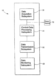
100761 As illustrated in Fig. 1, the physiology monitoring system 10
preferably includes a
data acquisition subsystem 20, a control-data processing subsystem 40, a data
transmission subsystem 50, a data monitoring subsystem 60, and a power source
70, such
as a battery.
[00771 As set forth in Figs. 2 and 3, the data acquisition subsystem 20 can
include paired
magnetometers that are positioned on a subject 100 and adapted to monitor and
detect
changes in (or displacements of) the anteroposterior diameters of the rib cage
and
abdomen, and axial displacement of the chest wall. As illustrated in Fig. 2,
the
magnetometers include first transmission magnetometer 22a, first receive
magnetometer
22b, second transmission magnetometer 24a, and second receive magnetometer
24b.
100781 Although the present invention is described herein in terms of
magnetometers and
magnetometer systems, it is understood that other types of sensor systems
capable of
- 15 - Atty. Dkt. No. 3483.0010001
CA 02714159 2010-08-31
measuring changes in distance between two or more sensors in the system can be
used in
place of, or in addition to, magnetometers. Specifically, the invention is not
limited to the
use of electromagnetic coils or magnetometers to acquire signals representing
measured
changes in the anteroposterior diameters of the rib cage and abdomen, and
axial
displacement of the chest wall. Various additional means and devices that can
be readily
adapted to measure the noted anatomical parameters can be employed within the
scope of
the invention. Such means and devices include, without limitation, Hall effect
sensors and
electronic compass sensors. Wireless sensors with the capability of measuring
time delay
in a signal sent from one sensor to another and thereby determine the distance
between
the two sensors can be substituted for or provided in addition to
magnetometers in
accordance with the present invention.
[00791 Control-data processing subsystem 40 includes programs, instructions
and
associated algorithms and parameters to control data acquisition subsystem 20
and, hence,
the paired magnetometers and the function thereof, and the transmission and
receipt of
signals, as well as data transmission subsystem 50 and data monitoring
subsystem 60.
[00801 Control-data processing subsystem 40 is further programmed and adapted
to
retrieve and process transmissions or signals reflecting changes in the
magnetometer
fields (and, hence, changes in spaced distances between the paired
magnetometers) and to
determine anatomical and physiological information associated with the
monitored
subject (as a function of the signals), including at least one respiratory
characteristic
(more preferably, a plurality of respiratory characteristics). Control-data
processing
subsystem 40 is also referred to herein as "processor subsystem," "processing
subsystem,"
and "data processing subsystem." The terms control-data processing subsystem,
processor
subsystem, processing subsystem, and data processing subsystem are used
interchangeably in the present application.
[0081] Data monitoring subsystem 60 is designed and adapted to display
physiological
and performance characteristics and parameters generated and transmitted by
control-data
processing subsystem 40.
[00821 Data transmission subsystem 50 is programmed and adapted to monitor and
control the communication links and, hence, transmissions by and between data
acquisition subsystem 20, control-data processing subsystem 40, and data
monitoring
subsystem 60.
- 16 - Atty. Dkt. No. 3483.0010001
CA 02714159 2010-08-31
100831 Further details of the noted physiological monitoring system are set
forth in U.S.
Provisional Application No. 61/275,575, filed September 1, 2009, and co-
pending U.S.
Application No. 12/869,582, filed August 26, 2010, each of which is
incorporated by
reference herein in its entirety.
100841 As will be readily appreciated by one having ordinary skill in the art,
the paired
magnetometers can be disposed in various anatomically appropriate positions on
a subject
to monitor and measure the change in distance (or displacement) between the
magnetometers. Referring now to Figs. 3-5, there is shown the position of
magnetometers
22a, 22b, 24a, 24b on a subject or patient 100, in accordance with the
inventions
disclosed in U.S. Provisional Application No. 61/275,575, co-pending U.S.
Application
No. 12/869,582, and co-pending U.S. Patent Application No. 12/231,692, filed
September
5, 2008, each of which is incorporated by reference herein in its entirety.
[00851 As illustrated in Figs. 3-5, first transmission magnetometer (i.e.,
first transmitter)
22a is preferably positioned on front 101 of subject 100 proximate the
subject's umbilicus,
and first receive magnetometer (i.e., first receiver) 22b is preferably
positioned proximate
the same axial position, but on back 102 of subject 100. Second receive
magnetometer
(i.e., second receiver) 24b is preferably positioned on front 101 of subject
100 proximate
the base of the sternum, and second transmission magnetometer (i.e., second
transmitter)
24a is positioned proximate the same axial position, but on back 102 of
subject 100.
[00861 As the subject or patient 100 breathes, displacement(s) of the rib cage
and
abdomen (i.e., changes in the distance between paired magnetometers 22a, 22b
and 24a,
24b, denoted, respectively, by arrow 29 and arrow 25), is determined from
measured
changes in the magnetic field between paired magnetometers 22a, 22b and 24a,
24b. The
axial displacement of the chest wall, denoted by arrow 23 (e.g.,
xiphiumbilical distance
(Xi)) is also determined from measured changes in the magnetic field between
magnetometers 22a and 24b. In such a case, magnetometer 24b can be a dual-
function
electromagnetic coil, where "dual function" refers to a coil capable of
receiving
transmissions from a plurality of different transmission coils (i.e.,
magnetometer 24b is
adapted to receive magnetic field transmissions from magnetometers 22a and
24a).
[00871 As indicated above, the measured displacements are typically employed
to
determine anatomical and physiological information associated with the
monitored
subject, including at least one or more respiratory characteristics. As set
forth in U.S.
- 17 - Atty. Dkt. No. 3483.0010001
CA 02714159 2010-08-31
Provisional Application No. 61/275,575, and co-pending U.S. Application No.
12/869,582, additional paired magnetometers can also be employed, and the
multiple
measured displacements can be employed to assess additional anatomical and
physiological characteristics, such as determining and characterizing the
relationship(s) of
chest wall movement(s) to respiratory activity and respiratory associated
events, such as
speaking, sneezing, laughing, and coughing.
[00881 As also set forth in U.S. Provisional Application No. 61/275,575, and
co-pending
U.S. Application No. 12/869,582, data acquisition subsystem 20 can additionaly
include
at least one additional physiological sensor (preferably a plurality of
additional
physiological sensors) adapted to monitor and record one or more physiological
characteristics associated with the monitored subject. The physiological
sensors can
include, without limitation, sensors that are adapted to monitor and record
electrical
activity of the brain, heart, and other muscles (e.g., EEG, ECG, EMG), pulse
rate, blood
oxygen saturation level (e.g., SP02), skin temperature, and core temperature.
Physiological parameters measured and/or calculated may include, for example,
heart
rate, respiration rate, blood oxygen level, blood flow, hydration status,
calories burned,
muscle fatigue, and/or body temperature.
[0089] Exemplary physiological sensors (and associated systems) are disclosed
in U.S.
Patent No. 6,551,252, issued April 22, 2003, U.S. Patent No. 7,267,652, issued
September
11, 2007, co-pending U.S. Patent Application No. 11/764,527, filed June 18,
2007, and
International Application No. PCT/US2005/021433, each of which is incorporated
by
reference herein in its entirety.
[0090] Data acquisition subsystem 20 can also include one or more audio
sensors, such
as, for example, a microphone, for monitoring sounds generated by a monitored
subject,
and a speaker to enable two-way communication by and between the monitored
subject
and a monitoring station or individual.
[0091] As indicated above, the monitoring systems of the invention include a
wearable
monitoring garments that can be comfortably worn by a monitored subject. In a
preferred
embodiment of the invention, the wearable monitoring garments include coils or
magnetometers, which are embedded in or carried by the wearable garment.
According to
the invention, the wearable monitoring garment can comprise various items that
are
-18- Atty. Dkt. No. 3483.0010001
CA 02714159 2010-08-31
adapted to cover at least a portion of a subject's body, such as a shirt,
vest, jacket, patch,
and the like.
[0092] In some embodiments of the invention, the aforementioned additional
sensors,
processing and monitoring systems (e.g., LDUs, if employed) associated wiring,
cabling,
and other power and signal transmission apparatus and/or systems are similarly
embedded
in or carried by the wearable garment.
[0093] Referring now to Fig. 6, there is shown one embodiment of a wearable
monitoring
garment 80 of the invention. As illustrated in Fig. 6, the wearable monitoring
garment 80
comprises a sleeveless shirt or vest, having magnetometers (e.g.,
magnetometers 22a,
24b) associated therewith.
[0094] Vest 80 preferably includes an overlapping front portion 72 having
closure means
that secures vest 80 to the subject's torso. According to the invention,
various
conventional closure means, such as a hook and pile system, e.g., VELCRO such
as that
manufactured by Velcro, Inc., snaps, zipper, etc., can be incorporated into
vest 80 to
facilitate closure thereof.
[0095] Additional suitable garments are also disclosed in U.S. Patent No.
7,267,652, U.S.
Patent No. 6,551,252, and U.S. Patent No. 6,047,203, issued April 4, 2000,
each of which
is incorporated by reference herein in its entirety.
[0096] According to the invention, the magnetometers, additional sensors,
processing and
monitoring systems, and other equipment can be arranged in or carried by the
garment,
for example, in open or closed pockets, or attached to the garment, for
example, as by
sewing, gluing, a hook and pile system, e.g., VELCRO such as that
manufactured by
Velcro, Inc., and the like. As indicated above, the magnetometers (e.g.,
magnetometers
22a, 22b, 24a, 24b) and additional sensors, if employed, can be disposed in or
carried by
the wearable garment at virtually any desired position, whereby, when the
garment is
worn by a subject the magnetometers and other sensors are positioned proximate
any
desired position on the subject's body.
[0097] The methods and systems of the invention, described above, thus provide
numerous significant advantages over conventional physiology monitoring
methods and
systems. A significant advantage is the provision of physiology monitoring
systems and
methods that accommodate ambulatory monitoring of respiratory and other
physiological
parameters and characteristics.
-19- Atty. Dkt. No. 3483.0010001
CA 02714159 2010-08-31
[00981 Additional advantages include the provision of physiology monitoring
systems
and methods that provide (i) accurate, real-time determination of a plurality
of respiratory
and other physiological parameters and characteristics, and (ii) real-time
determination
and characterization of a subject's orientation and movement.
100991 Additional advantages and applications of the present invention are
apparent with
reference to the systems and methods disclosed in U.S. Patent Application No.
12/869,582, filed August 26, 2010, U.S. Patent Application No. 12/869,576,
filed August
26, 2010, U.S. Patent Application No. 12/869,585, filed August 26, 2010, U.S.
Patent
Application No. 12/869,592, filed August 26, 2010, U.S. Patent Application No.
12/869,627, filed August 26, 2010, U.S. Patent Application No. 12/869,625,
filed August
26, 2010, and U.S. Patent Application No. 12/869,586, filed August 26, 2010,
each of
which is incorporated by reference herein in its entirety.
101001 Without departing from the spirit and scope of this invention, one of
ordinary skill
can make various changes and modifications to the invention to adapt it to
various usages
and conditions. As such, these changes and modifications are properly,
equitably, and
intended to be, within the full range of equivalence of the following claims.
-20- Atty. Dkt. No. 3483.0010001