Note: Descriptions are shown in the official language in which they were submitted.
CA 02714202 2010-09-01
ELECTROSURGICAL ELECTRODE WITH INSULATIVE COATING
BACKGROUND
Technical Field
100011 The present disclosure relates to an electrosurgical electrode and,
more
particularly, to an electrosurgical electrode including an insulative coating
configured to provide
a path for electrosurgical energy from the electrosurgical electrode to tissue
during an
electrosurgical procedure.
Back round of Related Art
[00021 Electrosurgical instruments have become widely used by surgeons in
recent years.
By and large, most electrosurgical instruments are hand-held instruments,
e.g., an electrosurgical
pencil, which transfer radio-frequency (RF) electrical or electrosurgical
energy to a tissue site via
an electrosurgical electrode. Typically, the electrosurgical energy is
returned to the
electrosurgical source via a return electrode pad positioned under a patient
(i.e., a monopolar
system configuration) or a smaller return electrode positionable in bodily
contact with or
immediately adjacent to the surgical site (i.e., a bipolar system
configuration). The waveforms
produced by the RF source yield a predetermined electrosurgical effect known
generally as
electrosurgical cutting and fulguration.
[00031 Typically, electrosurgical electrodes configured for electrosurgical
use are subject
to high temperatures at least where an electrosurgical arc emanates during the
electrosurgical
procedure, e.g., fulguration or coagulation. In some instances, the heat
generated by the
CA 02714202 2010-09-01
electrosurgical electrode during an electrosurgical procedure may cause
proteins in bodily fluids
and/or tissue to coagulate and adhere to the electrodes. To combat this
adhering of bodily fluids
and/or tissue to the electrosurgical electrodes, an insulative coating, e.g.,
a Teflon polymer, may
be applied to the electrosurgical electrode.
[0004] However, as can be appreciated by one skilled in the art, areas of the
electrosurgical electrode covered with an insulative coating cannot transmit
RF electrical or
electrosurgical energy to a tissue site.
SUMMARY
[0005] The present disclosure provides an electrode adapted to connect to an
electrosurgical instrument. The electrode includes a proximal end that is
adapted to connect to
an electrosurgical instrument and an electrosurgical energy source. The
electrode includes a
distal end configured for treating tissue. The distal end of the electrode
includes a first portion
having one or more edges and a second portion having a substantially blunt
profile. An
insulative material is disposed over at least the distal end of the electrode.
The insulative
material includes a first thickness at the first portion and a second
thickness at the second
portion, wherein upon activation, the insulative material disposed over the
first portion breaks
away from the first portion allowing energy to travel to tissue from the first
portion.
[0006] The present disclosure provides a method for performing an
electrosurgical
procedure. The method includes providing an electrosurgical system that
includes an electrode
that includes an insulative coating. A step of the method includes positioning
the electrosurgical
electrode adjacent a tissue site. The method includes transmitting an initial
command signal to
an electrosurgical generator in operative communication with the
electrosurgical system. The
2
CA 02714202 2010-09-01
method includes transmitting an RF pulse to the electrode in response to the
initial command
signal, such that at least a portion of the insulative coating is removed.
And, another step of the
method includes transmitting RF electrosurgical energy to the electrosurgical
electrode such that
an electrosurgical effect is achieved at the tissue site.
BRIEF DESCRIPTION OF THE DRAWINGS
[0007] Various embodiments of the present disclosure are described hereinbelow
with
references to the drawings, wherein:
[0008] FIG. 1. is a side, perspective view of an electrosurgical system
including an
electrosurgical electrode in accordance with an embodiment of the present
disclosure;
[0009] FIG. 2 is an enlarged view of the area of detail of the electrosurgical
electrode
illustrated in FIG. 1;
[0010] FIG. 3 is a cut-away, cross-sectional view taken along line segment 3-3
of FIG. 2;
[0011] FIG. 4 is an electrosurgical electrode configured for use with the
electrosurgical
system of FIG. 1 in accordance with an alternate embodiment of the present
disclosure;
[0012] FIG. 5 is a cut-away, cross-sectional taken along line segment 5-5 of
FIG. 4;
[0013] FIG. 6 is a flow chart illustrating steps for performing an
electrosurgical
procedure in accordance with an embodiment of the present disclosure; and
[0014] FIGS. 7A and 7B are cross-sectional views illustrating various
electrode
configurations.
3
CA 02714202 2010-09-01
DETAILED DESCRIPTION
[0015] Particular embodiments of the presently disclosed electrosurgical
electrode are
described in detail with reference to the drawing figures wherein like
reference numerals identify
similar or identical elements. As used herein, the term "distal" refers to
that portion which is
further from the user while the term "proximal" refers to that portion which
is closer to the user
or surgeon.
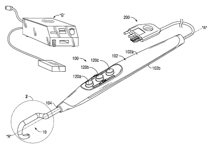
[0016] FIG. 1 sets forth a side, perspective view of an electrosurgical system
including
an electrosurgical pencil 100 including an electrosurgical electrode 10
constructed in accordance
with one embodiment of the present disclosure. While the following description
will be directed
towards electrosurgical pencils it is envisioned that the features and
concepts (or portions
thereof) of the present disclosure can be applied to any electrosurgical type
instrument, e.g.,
forceps, suction coagulators, vessel sealers, wands, etc.
[0017] As seen in FIG. 1, electrosurgical pencil 100 includes an elongated
housing 102
having a top-half shell portion 102a and a bottom-half shell portion 102b.
Electrosurgical pencil
100 includes a blade receptacle 104 disposed at a distal end of housing 102
configured to
operatively and removably connect to a replaceable electrosurgical electrode
10. Electrosurgical
pencil 100 may be coupled to a conventional electrosurgical generator "G" via
a plug assembly
200. Electrosurgical pencil 100 includes one or more activation switches
(three activation
switches 120a-120c are shown). Each activation switch 120a-120c controls the
transmission of
RF electrical energy supplied from generator "G" to electrosurgical electrode
10.
[0018] For a more detailed description of the electrosurgical pencil 100
including
operative components associated therewith, reference is made to commonly-owned
United States
Patent Publication No. 2006/0178667.
4
CA 02714202 2010-09-01
[00191 With reference now to FIGS. 2 and 3, and initially with reference to
FIG. 2,
electrosurgical electrode 10 (electrode 10) is shown. Electrode 10 may be
fabricated from a
conductive type material, such as, for example, stainless steel, or may be
coated with an
electrically conductive material. Electrode 10 may include any suitable
configuration including
but not limited to a hook, needle, loop, blade, wand, etc. In the embodiment
illustrated in FIGS.
1-5, electrode 10 includes a generally hook or "L" shape with a generally
circular cross-section
that extends from a proximal end 14 of electrode 10 to a distal end 12 of the
electrode 10.
[0020] Electrode 10 includes a layer of insulative coating 18 that coats
distal end 12
and/or proximal end 12. In embodiments, the layer of insulative coating 18 may
be applied
evenly over the entire surface of electrode 10. Conversely, insulative coating
may be applied in
a non-even fashion. More particularly, electrode 10 may include portions
(e.g., areas that are
intended to emanate electrosurgical energy to a tissue site) that have less
insulative coating 18
than other areas of the electrode 18 (e.g., areas that are not intended to
emanate electrosurgical
energy to a tissue site). More particularly, electrode 10 may include an
arcuate cutout 32 that
includes a thicker layer of insulative coating 18 and edges 32 that include a
thinner layer of
insulative coating 18. This configuration of electrode 10 includes an uneven
layer of insulative
coating 18 that facilitates and/or speeds up the breakdown of insulative
coating 18 at or near
edges 32. Insulative coating 18 may be made from any suitable material
including but not
limited to Teflon , Teflon polymers, silicone and the like.
[0021] As noted above, electrode 10 operatively and removably connects to
blade
receptacle 104. To this end, proximal end 14 is selectively retained by
receptacle 104 within the
distal end of housing 102. Reference is again made to commonly-owned United
States Patent
Publication No. 2006/0178667 for a more detailed description of the operative
electrical and/or
CA 02714202 2010-09-01
mechanical interfaces associated with proximal end 14 of electrode 10 and
receptacle 104. In
embodiments, an articulating portion 16 extends from proximal end 14 and
operably connects
distal end 12 and proximal end 14 to each other, see FIGS. 2 and 4. The
articulating portion 16
allows a user to substantially fix the distal end 12 of electrode 10 in a
desired position prior to
electrosurgically effecting tissue.
[0022] Distal end 12 of electrode 10 extends distally beyond receptacle 104.
Distal end
12 includes inner and an outer faces 12a and 12b, respectively. Distal end 12
includes an
elongated shaft portion 20 having a proximal end 22 that extends from a distal
end 24 of the
articulating portion 16. In embodiments, shaft portion 20 is disposed parallel
with respect to a
longitudinal axis "A" of the electrosurgical pencil 100, as best seen in FIG.
2.
[0023] Distal end 12 includes a curved portion 26 that extends from a distal
end 28 of the
shaft 20. Curved portion 26 includes a generally concave configuration. In
certain instances,
this concave configuration may facilitate manipulating tissue. Curved portion
26 includes a
generally circular cross-section. A distal end 40 of curved portion 26
includes a tip 30. In the
embodiments illustrated in FIGS. 1-3, tip 30 includes a generally rounded,
blunt tip
configuration. Conversely, tip 30 may include a generally pointed, sharp tip
configuration.
Specific tip configurations of tip 30 will depend on the contemplated uses of
a manufacturer.
[0024] An arcuate cutout 32 extends along the inner face 12a from the tip 30
of the
curved portion 26 to the distal end 28 of the shaft 20, as best seen in FIG.
2. Alternatively, the
arcuate cutout 32 may extend from the tip 30 to the distal end 24 of
articulating portion 16 (see
FIG. 4, for example). The specific configuration of arcuate 32 with respect to
the distal end 12
and/or shaft 20 will depend on the contemplated surgical purposes of the
instrument 10. For
6
CA 02714202 2010-09-01
example, in embodiments, the arcuate cutout 32 can be extended or reduced
along a length of the
inner face 12a such that a specific electrosurgical effect can be achieved at
a desired location
along the inner face 12a.
[0025] Arcuate cutout 32 extends along the inner face 12a and defines one or
more edges
34. In the embodiment illustrated in FIG. 2, arcuate cutout 23 defines two
relatively sharp edges
34. The combination of arcuate cutout 32 and edges 34 provides at least a
portion of the distal
end 12 of the electrode 10 that includes a region of insulative coating 18
that is configured to
provide a path for electrosurgical energy to flow from the distal end 12 of
the electrode 10 to
tissue during an electrosurgical procedure. More particularly, when
electrosurgical energy is
transmitted (e.g., in response to an initial command signal) to the distal end
12 of the electrode
10, the edges 34 provide an area of high concentration of electrosurgical
energy along the length
of the edge 34. This high concentration of electrosurgical energy breakdowns
or "blows off' the
layer of insulative coating 18 that electrically insulates the edges 34,
which, in turn, provides one
or more paths "P1" for RF energy to flow, see FIG. 3. The sharpness of edges
34 is directly
proportional to the concentration of electrosurgical energy at the edges 34
when electrosurgical
energy is transmitted to the electrode 10. That is, the sharper the edges 34
for a given amount of
transmitted electrosurgical energy the higher the concentration of
electrosurgical energy at the
edges 34 when electrosurgical energy is transmitted to the electrode 10. The
sharpness the edges
34 relative to the arcuate cutout 32 will depend on the contemplated uses of a
manufacturer.
[0026] Electrode 10 including distal end 12 and proximal end 14 may be formed
by any
suitable techniques, e.g., machining techniques. For example, in embodiments,
distal end 12
including arcuate cutout 32 and/or sharp edges 34 may be formed by known
milling techniques.
7
CA 02714202 2010-09-01
Alternatively, or in combination therewith, arcuate cutout 32 and/or sharp
edges 34 may be
formed by known etching techniques.
[0027] With reference to FIGS. 4 and 5, and initially with reference to FIG.
4, an
alternate embodiment of electrode 10 is shown designated 200. Electrode 200 is
substantially
similar to electrode 10. Accordingly, only those features and/or operative
components that are
unique or distinctive to electrode 200 will be described herein.
[0028] Unlike electrode 10, electrode 200 includes a pair of arcuate cutouts
232 that
extend along both an inner face 212a and outer face 212b from a tip 230 of the
curved portion
226 to the proximal end 222 of the shaft 220. More particularly, arcuate
cutouts 232 extend
along both the inner face 212a and outer face 212b and define one or more
edges 234. In the
embodiment illustrated in FIG. 4, arcuate cutouts 223 define four relatively
sharp edges 234.
The combination of arcuate cutouts 232 and edges 234 provides at least a
portion of the distal
end 212 of the electrode 200 that includes a region of insulative coating 218
that is configured to
provide a path for transmitting electrosurgical energy from the distal end 212
of the electrode
200 to tissue during an electrosurgical procedure. More particularly, when
electrosurgical
energy is transmitted to the distal end 212 of the electrode 200, the edges
234 provide an area of
high concentration of electrosurgical energy along the length of the edge 234.
This high
concentration of electrosurgical energy breakdowns or "blows off' the layer of
insulative coating
218 that electrically insulates the edges 234, which, in turn, provides one or
more paths "P2" for
RF energy to flow, see FIG. 5. As noted above with respect to edges 34, the
sharpness of edges
234 is directly proportional to the concentration of electrosurgical energy at
the edges 234 when
electrosurgical energy is transmitted to the electrode 200. That is, the
sharper the edges 234 the
8
CA 02714202 2010-09-01
higher the concentration of electrosurgical energy at the edges 234 when
electrosurgical energy
is transmitted to the electrode 200.
[0029] With reference to FIG. 6, a method 500 of use of electrode 10 will be
described in
terms of use with an electrosurgical system including an electrosurgical
pencil 100 coupled to a
conventional electrosurgical generator "G" via the plug assembly 200 (step
502). Electrosurgical
pencil 100 and/or generator "G" may be set to an initial insulation
"breakdown" mode setting. A
user may position the curved portion 26 of electrode 10 adjacent a tissue
site. One or more of the
activation switches 120a-120c may be employed to transmit an initial command
signal to the
generator "G" (step 504). In response to receiving the initial command signal,
generator "G"
may be configured to transmit an initial RF pulse that is configured to
breakdown or "blow-off'
the insulative coating 18 located at or adjacent the one or more edges 34
(step 506). As noted
above, only the insulative coating 18 located at or near the edges 34 is
broken-down, and the
insulative coating 18 located on the other areas (e.g., arcuate cutout 32) on
the electrode 10
remain in tact. In an embodiment, once the insulative coating 18 is broken-
down or "blown off',
one or more of the activation switches 120a-120c may be employed to transmit a
subsequent
command signal to the generator "G" (step 508). In response to the subsequent
command signal,
generator "G" may transmit RF electrosurgical energy to the electrode 10 which
emanates from
the one or more edges 34 such that an electrosurgical tissue effect may be
achieved at the tissue
site (step 510). It is contemplated that one skilled in the art will
appreciate other methods of use
for electrode 10.
[0030] From the foregoing and with reference to the various figure drawings,
those
skilled in the art will appreciate that certain modifications can also be made
to the present
disclosure without departing from the scope of the same. For example,
electrode 10 may include
9
CA 02714202 2010-09-01
other geometrical configurations. More particularly, FIGS. 7A and 7B are cut-
away views
illustrating other various electrode 10 configurations including their
associated paths "P3" and
"P4" for RF energy to flow.
[0031] While several embodiments of the disclosure have been shown in the
drawings, it
is not intended that the disclosure be limited thereto, as it is intended that
the disclosure be as
broad in scope as the art will allow and that the specification be read
likewise. Therefore, the
above description should not be construed as limiting, but merely as
exemplifications of
particular embodiments. Those skilled in the art will envision other
modifications within the
scope and spire of the claims appended hereto.