Note: Descriptions are shown in the official language in which they were submitted.
CA 02714375 2010-09-03
117.0037 US NP
PRESSURE MEASUREMENT OF A RESERVOIR FLUID IN A MICROFLUIDIC
DEVICE
CROSS-REFERENCE TO RELATED APPLCIATIONS
[0001] This patent application is a continuation-in-part of International
Patent
Application No. PCT/IB09/50500, filed February 7, 2009, which is incorporated
by
reference herein.
BACKGROUND OF THE INVENTION
Field of the Invention
[0002] This patent specification relates to an apparatus and method for
measuring thermo-physical properties of a reservoir fluid. More particularly,
the patent
specification relates to an apparatus and method for measuring pressure of a
reservoir
fluid flowing in a microfluidic device.
Description of Related Art
[0003] The measurement of reservoir fluid properties is a key step in the
planning and development of a potential oilfield. It is often desirable to
perform such
measurements frequently on a producing well to provide an indication of the
performance and behavior of the production process. Examples of such
measurements
are pressure, volume, and temperature measurements, often referred to as "PVT"
measurements, which are instrumental in predicting complicated thermo-physical
behavior of reservoir fluids. One important use of PVT measurements is the
construction of an equation of state describing the state of oil in the
reservoir fluid.
Other properties of interest that may be determined using PVT measurements
include
fluid viscosity, density, chemical composition, gas-oil-ratio, and the like.
Once a PVT
analysis is complete, the equation of state and other parameters can be input
into
reservoir modeling software to predict the behavior of the oilfield formation.
1
CA 02714375 2010-09-03
117.0037 US NP
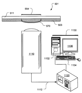
[0004] Conventional PVT measurements are performed using a cylinder
containing the reservoir fluid. A piston disposed in the cylinder maintains
the desired
pressure on the fluid, while the heights of the liquid and gaseous phases are
measured
using, for example, a cathetometer. International Patent Application No.
PCT/1609/50500, filed February 7, 2009, discusses microfluidic technique form
measuring thermo-physical properties of a reservoir fluid. The microfluidic
techniques
can provide certain advantages including: (1) providing a way to measure
thermo-
physical properties of a reservoir fluid with small amounts of reservoir
fluid; (2) providing
a way to perform pressure-volume-temperature analyses of a reservoir fluid in
a timely
fashion; and (3) providing a way to measure thermo-physical properties of a
reservoir
fluid using image analysis. However, in some cases the microfluidic based
measurements and analysis can benefit from pressure measurement at various
points
along the microchannel.
[0005] Pressure sensors based on deformation of a membrane have long
been developed. These membranes are usually micro-fabricated using SO[ or
silicone-
on-insulator wafers. For example, see, U.S Patents No. 5,095,401, 5,155,061,
5,165,282, and 5,177,661, each of which is incorporated by reference herein.
Numerous
techniques have been used to correlate deformation of the membrane with
pressure.
These techniques include piezo-resistive element (see, e.g., U.S. Patents No.
5,081,437, 5,172,205, and 6,843,121), optical fibers (See. e.g. U.S. Patents
No.
7,000,477, and 7,149,374; and U.S. Patent Publications No. 2005/0041905, and
2008/0175529), and capacitive sensors (See. e.g. U.S. Patents No. 7,254,008,
5,470,797, and 6,945,116, and PCT Patent Publications No. WO 96/16319, and WO
98/23934). Each of the foregoing patents and patent publications are
incorporated by
reference herein.
[0006] Most of these techniques have been developed for conventional
pressure sensors. Incorporating such tools inside a microchannel is either too
difficult or
otherwise impractical. Practical and cost effective measurement techniques for
microchannels are rare. To measure pressure inside a microfluidic channel,
some
2
CA 02714375 2010-09-03
117.0037 US NP
techniques have been described. For example, R. Baviere, F. Ayela, Meas. Sci.
Technol., 15, (2004), 377, incorporated by reference herein, discusses the use
of piezo-
resistive elements; and M. J. Kohl, S. I. Abdel-Khalik, S. M. Jeter, D. L.
Sadowski,
Sensors and Actuators a-Physical, 118, (2005), 212; and M. J. Kohl, S. I.
Abdel-Khalik,
S. M. Jeter, D. L. Sadowski, Int. J. Heat Mass Transfer, 48, (2005), 1518,
both
incorporated by reference herein, discuss the use of lasers.
[0007] However, there remains a need for simple non-invasive techniques to
measure pressure inside a microfluidic channel.
BRIEF SUMMARY OF THE INVENTION
[0008] According to embodiments, a system for measuring fluid pressure in a
microchannel is provided. The system includes a microchannel adapted to carry
a
fluid; a first flexible member adapted and positioned such that pressure of
the fluid in the
microchannel causes a deformation of the first flexible member; and an optical
sensing
system adapted and positioned to detect deformation of the first flexible
member.
[0009] The flexible member is preferably a membrane partially defining a
cavity that is in fluid communication with the microchannel at a first
location such that
deformation of the membrane is representative of the fluid pressure in the
microchannel
at the first location. According to some embodiments, second and third
membranes
also can be provided to provide detecting of pressure at second and third
locations on
the microchannel.
[0010] Additionally, according to some embodiments a method for measuring
fluid pressure in a microchannel is provided. The method includes providing a
microchannel adapted to carry a fluid, and a first flexible member adapted and
positioned such that pressure of the fluid in the microchannel causes a
deformation of
the first flexible member. Fluid is introduced under pressure into the
microchannel,
thereby causing a deformation of the first flexible member, and deformation of
the first
flexible member is optically detected. A value can be determined representing
the
3
CA 02714375 2010-09-03
117.0037 US NP
pressure at a location in the microchannel based at least in part on the
optically
detected deformation of the first flexible member.
[0011] Further features and advantages of the invention will become more
readily apparent from the following detailed description when taken in
conjunction with
the accompanying drawings.
BRIEF DESCRIPTION OF THE DRAWINGS
[0012] The present invention is further described in the detailed description
which follows, in reference to the noted plurality of drawings by way of non-
limiting
examples of exemplary embodiments of the present invention, in which like
reference
numerals represent similar parts throughout the several views of the drawings,
and
wherein:
[0013] Figure 1 is a stylized, exploded, perspective view of a first
illustrative
embodiment of a microfluidic device for measuring thermo-physical properties
of a
reservoir fluid;
[0014] Figure 2 is a stylized, schematic representation of a reaction of
reservoir fluid as the reservoir fluid flows through the microfluidic device
of Figure 1;
[0015] Figure 3 is a top, plan view of the microfluidic device of Figure 1
depicting three reservoir fluid flow regimes;
[0016] Figure 4 is a stylized, side, elevational view of a reservoir fluid
measurement system, including the microfluidic device of Figure 1 and a camera
for
generating images of the microfluidic device in use;
[0017] Figure 5 is a top, plan view of a second illustrative embodiment of a
microfluidic device for measuring thermo-physical properties of a reservoir
fluid;
4
CA 02714375 2010-09-03
117.0037 US NP
[0018] Figure 6 is a side, elevational view of the microfluidic device of
Figure
5;
[0019] Figures 7-9 depict exemplary microchannel constrictions of the
microfluidic device of Figure 5;
[0020] Figures 1OA and 1OB are schematic cross sections of an un-deformed
and deformed membrane respectively, according to some embodiments;
[0021] Figure 11 is a stylized, schematic representation a membrane
deformation measurement setup, according to some embodiments;
[0022] Figure 12 is a stylized, schematic representation a membrane
deformation measurement setup having multiple optical sensors, according to
some
embodiments;
[0023] Figure 13 shows plots of exemplary measurements of a membrane in
undeformed and deformed states, according to embodiments;
[0024] Figure 14 shows a plot of repeated measured deformations as a
function of hydrostatic pressure, according to embodiments; and
[0025] Figure 15 shows plots of the measured pressures in cavities for
different input pressures, according to embodiments.
[0026] While the invention is susceptible to various modifications and
alternative forms, specific embodiments thereof have been shown by way of
example in
the drawings and are herein described in detail. It should be understood,
however, that
the description herein of specific embodiments is not intended to limit the
invention to
the particular forms disclosed, but on the contrary, the intention is to cover
all
modifications, equivalents, and alternatives falling within the scope of the
invention as
defined by the appended claims.
CA 02714375 2010-09-03
117.0037 US NP
DETAILED DESCRIPTION OF PREFERRED EMBODIMENTS
[0027] Illustrative embodiments of the invention are described below. In the
interest of clarity, not all features of an actual implementation are
described in this
specification. It will be appreciated that in the development of any such
actual
embodiment, numerous implementation-specific decisions must be made to achieve
the
developer's specific goals, such as compliance with system-related and
business-
related constraints, which will vary from one implementation to another.
Moreover, it will
be appreciated that such a development effort might be complex and time-
consuming
but would nevertheless be a routine undertaking for those of ordinary skill in
the art
having the benefit of this disclosure. Further, like reference numbers and
designations
in the various drawings indicated like elements.
[0028] According to embodiments, systems and methods for measuring
pressure of a reservoir fluid in a microfluidic device are provided. For the
purposes of
this disclosure, the term "reservoir fluid" means a fluid stored in or
transmitted from a
subsurface body of permeable rock. Thus "reservoir fluid" may include, without
limitation, hydrocarbon fluids, saline fluids such as saline water, as well as
other
formation water, and other fluids such as carbon dioxide in a supercritical
phase.
Moreover, for the purposes of this disclosure, the term "microfluidic" means
having a
fluid-carrying channel exhibiting a width within a range of tens to hundreds
of
micrometers, but exhibiting a length that is many times longer than the width
of the
channel. Similarly the term "microchannel" means a fluid-carrying channel
exhibiting a
width within a range of tens to hundreds of micrometers. Although many of the
microchannels described herein are of rectangular cross section due to the
practicalities
of fabrication techniques, the cross section of a microchannel can be of any
shape,
including round, oval, ellipsoid, square, etc.
[0029] Figure 1 depicts a stylized, exploded, perspective view of a
microfluidic
device 101 in which pressure can be measured, according to some embodiments of
the
invention. In the illustrated embodiment, microfluidic device 101 comprises a
first
substrate 103 defining a microchannel 105, an entrance well 107 and an exit
well 109.
6
CA 02714375 2010-09-03
117.0037 US NP
Microchannel 105 extends between and is in fluid communication with entrance
well
107 and exit well 109. Microchannel 105 forms a serpentine pattern in first
substrate
103, thus allowing microchannel 105 to extend a significant length but occupy
a
relatively small area. According to one embodiment, microchannel 105 exhibits
a length
of one or more meters, a width of about 100 micrometers, and a depth of about
50
micrometers, although the present invention also contemplates other dimensions
for
microchannel 105. Microfluidic device 101 further comprises a second substrate
111
having a lower surface 113 that is bonded to an upper surface 115 of first
substrate 103.
When second substrate 111 is bonded to first substrate 103, microchannel 105
is
sealed except for an inlet 117 at entrance well 107 and an outlet 119 at exit
well 109.
Second substrate 111 defines an entrance passageway 121 and an exit passageway
123 therethrough, which are in fluid communication with entrance well 107 and
exit well
109, respectively, of first substrate 103. Also shown in Fig. 1 are a number
of cavities
such as cavity 150, each connected to the main microchannel 105 using a small
side
channel. As is explained in further detail below, each cavity such as cavity
150 is
partially defined by a deformable membrane that allows for pressure
measurement.
According to preferred embodiments substrate 103 is fabricated with circular
openings
and the cavities are defined on the sides by the walls of the openings in
substrate 103,
on the bottom with the deformable membrane, and on the top by the second
substrate
111.
[0030] In Figure 1, first substrate 103 is preferably made of silicon and is
approximately 500 micrometers thick, and second substrate 111 is made of
glass, such
as borosilicate glass, although the present invention contemplates other
materials for
first substrate 103, as is discussed in greater detail herein. According some
preferred
embodiments substrate 103 is a conventional silicon on insulator (SOI) wafer.
Exemplary borosilicate glasses are manufactured by Schott North America, Inc.
of
Elmsford, New York, USA, and by Corning Incorporated of Corning, New York,
USA.
[0031] In operation, pressurized reservoir fluid is urged through entrance
passageway 121, entrance well 107, and inlet 117 into microchannel 105. The
reservoir
7
CA 02714375 2010-09-03
117.0037 US NP
fluid exits microchannel 105 through outlet 119, exit well 109, and exit
passageway 123.
Microchannel 105 provides substantial resistance to the flow of reservoir
fluid
therethrough because microchannel 105 is very small in cross-section in
relation to the
length of microchannel 105. When fluid flow is established between inlet 117
and outlet
119 of microchannel 105, the pressure of the reservoir fluid within
microchannel 105
drops from an input pressure, e.g., reservoir pressure, at inlet 117 to an
output
pressure, e.g., atmospheric pressure, at outlet 119. The overall pressure drop
between
inlet 117 and outlet 119 depends upon the inlet pressure and the viscosity of
the
reservoir fluid. Fluid flow through microchannel 105 is laminar and, thus the
pressure
drop between inlet 117 and outlet 119 is linear when the reservoir fluid
exhibits single-
phase flow. For further details of microfluidic devices and method for
measuring
thermo-physical properties of reservoir fluid, see e.g. International Patent
Application
No. PCT/IB09/50500, filed February 7, 2009, which is incorporated by reference
herein,
and in co-pending U.S. Patent Application No. 12/533,305, Patent Application
Publication No. US 2009/0326827, entitled "PHASE BEHAVIOR ANAYSIS USING A
MICROFLUIDIC PLATFORM," Attorney Docket No. 117.0043 US NP, filed on even
date herewith, which is incorporated by reference herein. Once the flow is
established,
the membrane in each cavity, such as cavity 150, deforms due to the fluid
pressure and
the deformation can be optically detected, as is described more fully below.
[0032] Figure 2 provides a stylized, schematic representation of the reaction
of reservoir fluid 201 as the reservoir fluid flows through microchannel 105
in a direction
generally corresponding to arrow 202, according to some embodiments. When the
reservoir fluid enters inlet 117 of microchannel 105, the reservoir fluid is
at a pressure
above the "bubble point pressure" of the reservoir fluid. The bubble point
pressure of a
fluid is the pressure at or below which the fluid begins to boil, i.e.,
bubble, at a given
temperature. When the reservoir fluid exits outlet 119 of microchannel 105,
the
reservoir fluid is at a pressure below the bubble point pressure of the
reservoir fluid.
Thus, a "first" bubble 203 forms in the reservoir fluid at some location,
e.g., at 205 in
Figure 2, within microchannel 105 where the reservoir fluid is at the bubble
point
pressure. Downstream of location 205, multi-phase flow, e.g., gas and liquid
flow, of
8
CA 02714375 2010-09-03
117.0037 US NP
reservoir fluid 201 occurs in microchannel 105. Previously-formed bubbles,
e.g.
bubbles 207, 209, 211, 213, 215, and the like, grow in size as reservoir fluid
201 flows
within microchannel 105 beyond the location corresponding to the formation of
the first
bubble due to decreased pressure in this portion of microchannel 105 and more
evaporation of the lighter components of reservoir fluid 201. The bubbles are
separated
by slugs of liquid, such as slugs 217, 219, 221, 223, 225, and the like.
Expansion of the
bubbles, such as bubbles 207, 209, 211, 213, 215, results in an increased flow
velocity
of the bubbles and liquid slugs, such as slugs 217, 219, 221, 223, 225, within
microchannel 105. The mass flow rate of reservoir fluid 201 is substantially
constant
along microchannel 105; however, the volume flow rate of reservoir fluid 201
increases
as reservoir fluid flows along microchannel 105. The reservoir fluid also
enters cavity
150 through small channel 152. According to some embodiments the width of
small
side channel 152 is approximately 50 micrometers, or about half of the width
of
microchannel 105, and is about 50 micrometers deep.
[0033] Thermo-physical properties of the reservoir fluid, such as reservoir
fluid
201 of Figure 2, for example gas-oil-ratio, phase envelope, and equation of
state, can
be determined by measuring the size and concentration of bubbles within
microchannel
105. Referring now to Figure 3, the flow of the reservoir fluid through
microchannel 105
is depicted in three regimes. A first bubble, such as first bubble 203 of
Figure 2, is
formed at 301 along microchannel 105. From inlet 117 of microchannel 105 to
location
301 of the first bubble, indicated in Figure 3 as a first region 303, the
pressure of the
reservoir fluid is above the bubble point. No bubbles are observed within
first region
303. In first region 303, the flow of the reservoir fluid is laminar due to a
low Reynolds
number and the pressure drops linearly therein. Once bubbles are formed, the
bubbles
move along within microchannel 105 toward outlet 119 and the volumes of the
bubbles
increases. In a second region 305, the void fraction, i.e., the volume of gas
to total
volume, of the reservoir fluid is less than one. In a third region 307, the
flow of the
reservoir fluid is dominated by high-speed gas flow. The gas bubbles are
separated by
small droplets of liquid, such as water. The pressure of the reservoir fluid
within third
region 307 decreases rapidly. Gas bubbles flow within second region 305 at a
slower
9
CA 02714375 2010-09-03
117.0037 US NP
rate than in third region 307, where they are often nearly impossible to
follow with the
naked eye.
[0034] Once a stabilized flow of reservoir fluid is established in
microchannel
105, a camera 401 is used to capture snapshots of the flow, as shown in Figure
4. Note
that the flow of reservoir fluid into inlet 117 (shown in Figures 1 and 3) is
represented by
an arrow 403 and that the flow of reservoir fluid from outlet 119 (shown in
Figures 1 and
3) is represented by an arrow 405. In one embodiment, camera 401 is a charge-
coupled device (CCD) type camera. The images produced by camera 401 are
processed using image analysis software, such as ImageJ 1.38x, available from
the
United States National Institutes of Health, of Bethesda, Maryland, USA, and
ProAnalyst, available from Xcitex, Inc. of Cambridge, Massachusetts, USA, to
measure
the size and concentration of the bubbles in the reservoir fluid disposed in
microchannel
105. Using this technique, many thermo-physical properties of the reservoir
fluid, such
as gas-oil-ratio, phase envelope, and equation of state, can be determined.
[0035] Figures 5 and 6 depict a microfluidic device 501, according to some
embodiments. As in microfluidic device 101 of Figure 1, microfluidic device
501
comprises a first substrate 503 defining a microchannel 505, an entrance well
507, and
an exit well 509. Microchannel 505 extends between and is in fluid
communication with
entrance well 507 and exit well 509. In the illustrated embodiment, first
substrate 503 is
made from silicon; however, first substrate 503 may be made from glass.
Microchannel
505, entrance well 507, and exit well 509 are, in one embodiment, first
patterned onto
first substrate 503 using a photolithography technique and then etched into
first
substrate 503 using a deep reactive ion etching technique. As in the first
embodiment
shown in Figure 1, in a preferred embodiment, microchannel 505 exhibits a
length of
one or more meters, a width of about 100 micrometers, and a depth of about 50
micrometers, although the present invention also contemplates other dimensions
for
microchannel 505. A number small side channels, such as side channels 552 and
556
lead from the main microchannel 505 to circular cavities such as cavities 550
and 554.
Also shown in a side channel 560 that leads to cavity 558. According to some
CA 02714375 2010-09-03
117.0037 US NP
embodiments, twelve cavities are spaced out along the length of microchannel
505 and
each of the cavities are about 2mm in diameter, although the present invention
also
contemplates other numbers of cavities and diameters for each cavity.
[0036] Microfluidic device 501 further comprises a second substrate 511
defining an entrance passageway 513 and an exit passageway 515 in fluid
communication with entrance well 507 and exit well 509. Second substrate 511
is made
from glass, as discussed herein concerning second substrate 111 (shown in
Figure 1).
In one embodiment, entrance passageway 513 and exit passageway 515 are
generated
in second substrate 511 using a water jet or abrasive water jet technique.
First
substrate 503 and second substrate 511 are preferably fused using an anodic
bonding
method after careful cleaning of the bonding surfaces of substrates 503 and
511. The
cavities can be fabricated using a verity of techniques. According to some
embodiments, a deep ion reaction (DRIE) etching process is used.
[0037] The present invention contemplates microfluidic device 501 having any
suitable size and/or shape needed for a particular implementation. In one
embodiment,
microfluidic device 501 exhibits an overall length A of about 80 millimeters
and an
overall width B of about 15 millimeters. In such an embodiment, passageways
513 and
515 are spaced apart a distance C of about 72 millimeters, cavities 558 and
550 are
spaced apart a distance D of about 3 millimeters, and cavities along the
serpentine
section of microchannel 505, such as cavities 550 and 554 are spaced apart by
a
distance E of about 5 millimeters. It should be noted that microfluidic device
101 may
also exhibit dimensions corresponding to microfluidic device 501. However, the
scope
of the present invention is not so limited.
[0038] Referring to Figure 7, one or more portions of microchannel 505
include zones of reduced cross-sectional area to induce the formation of
bubble nuclei
in the reservoir fluid. For example, as shown in Figures 7 and 8, a micro-
venturi 701 is
incorporated into an inlet of microchannel 505. Micro-venturi 701 includes a
nozzle
opening 801 having a width W1, which is smaller than a width W2 of
microchannel 505.
The contraction provided by micro-venturi 701 causes a substantial pressure
drop in the
11
CA 02714375 2010-09-03
117.0037 US NP
reservoir fluid at nozzle opening 801 along with an increased velocity of
reservoir fluid
flow. The combined effect of the pressure drop and the increased velocity
induces
formation of bubble nuclei in the reservoir fluid. Preferably, microchannel
505 further
includes one or more additional constrictions 703, as shown in Figures 7 and
9.
Constrictions 703 exhibit widths W3, which are smaller than a width W4 of
microchannel
505. Preferably, width IN, of nozzle opening 801 and widths W3 of
constrictions 703 are
about 20 micrometers, whereas the preferred width W2 and W4 of microchannel
505 is
100 micrometers. These restrictions increase the velocity of the reservoir
fluid by up to
about 500 percent.
[0039] Figures 10A and 10B are schematic cross sections of an un-deformed
and deformed membrane respectively, according to some embodiments. Cavity 554
is
shown defined on the sides by the first substrate 503, on the top by a second
substrate
511, and on the bottom by deformable membrane 570. According to some
embodiments, membrane 570 is micro-fabricated in the device 501 using
conventional
SOI (Silicon one insulator) wafers. According to some embodiments, the
membranes,
such as membrane 570 are not separate parts from the first substrate 503.
Rather they
are formed the same material as substrate 503. According to such embodiments,
starting with substrate 503 is a 500 micrometer thick silicon wafer. The
cavities, such as
cavity 554 are etched down to about 400 micrometers. This leaves a 100
micrometer
wall at the bottom of each cavity, which forms the flexible membrane, such as
membrane 570.
[0040] In Figure 10B, membrane 570 is shown in a deformed state. Once the
pressure inside the microchannel 505 (not shown) and inside cavity 554 exceeds
that of
the atmosphere, the membrane 570 will expand outward. Membrane 570 is designed
such that deformation of the membrane 570 is linear within the expected
pressure range
for the device 501. It has been found that for many downhole applications a
membrane
diameter of about 2mm in diameter and about 100 micrometers in thickness,
although
other membrane dimensions, including thickness, are contemplated. According to
12
CA 02714375 2010-09-03
117.0037 US NP
some embodiments, modeling such as finite element modeling can be used to
ensure
the membrane will behave linearly within the expected range of pressures.
[0041] Figure 11 is a stylized, schematic representation a membrane
deformation measurement setup, according to some embodiments. The setup
includes
a microfluidic device 501, confocal sensor 1110, spectrometer 1120, and a
computer
system 1130. Due to changing pressure inside the microchannel of device 501,
the
pressure changes in cavity 554 and membrane 570 deforms. The deformation is
detected and measured by the sensor 1110. To measure deformation of the
membrane
570, according to some embodiments, a confocal chromatic sensor, or an optical
pen, is
used. Suitable sensors include the chromatic confocal distance sensors made by
STIL
(Sciences et Techniques Industrielles de la Lumiere) SA, of France. The
confocal
sensor uses the wide spectrum of the white light. It then disperses the white
light into
monochromatic light using a series of lenses. The distance of the object from
the
sensors is measured by spectroscopy of the reflected light using spectrometer
1120
which receives optical signals from the sensor via fiber optic connection
1112. The
setup is controlled by and the results are interpreted and displayed using
computer
system 1130. Computer system 1130 includes a one or more processors, a storage
system 1132 (which includes one or more removable storage devices that accept
computer readable media), display 1136, and one or more human input devices
1134,
such as a keyboard and/or a mouse. Computer system 1130 also includes a data
acquisition system for collecting data from the spectrometer 1120.
[0042] According to one embodiment, the microfluidic device 501 is mounted
on a chip holder perpendicular to the main axis of the confocal sensor 1110.
The sensor
is also mounted on a holder that can move the sensor in two orthogonal
directions using
two micro-stages. In this way, the sensor 1110 can be focused, one at a time,
on any of
the other membranes of the other cavities located on device 501.
[0043] Figure 12 is a stylized, schematic representation a membrane
deformation measurement setup having multiple optical sensors, according to
some
embodiments. As in the case of Figure 11, the setup includes a microfluidic
device 501,
13
CA 02714375 2010-09-03
117.0037 US NP
spectrometer 1120, and a computer system 1130. The setup in Figure 12 includes
a
plurality of optical sensors 1210 with one optical sensor focused on each
membrane of
device 510. For example, sensor 1212 is focused on the membrane of cavity 558,
and
sensor 1214 is focused on the membrane of cavity 550. The signals form the
sensors
1210 that represent various states of deformation of the membranes are fed to
spectrometer 1120 and then stored, evaluated and/or displayed by computer
system
1130. According to some embodiments, the sensor 1210 are mounted on a micro-
stage
such that each optical sensor can be positioned to focus on several points
with respect
to the membrane. For example, the micro-stage can be programmed such that each
optical sensor focuses on three points corresponding to points A, B and C on
the curves
1310 and 1320 of Figure 13, which is described more fully below.
[0044] Figure 13 shows plots of exemplary measurements of a membrane in
undeformed and deformed states, according to embodiments. To measure the
deformation of the membrane, the optical sensor was moved across the membrane
using a micro-stage. Curve 1310 is the membrane profile under no (i.e.
atmospheric)
pressure and curve 1320 is the membrane profile under 400psi pressure. It can
be seen
that the flat membrane assumes a bell-shape under the applied pressure. Two
reference points "A" and "B" were selected on either side of the membrane an
the line
1312 represents the device plane in the case of curve 1310 and the line 1322
represents the device plane in the case of curve 1320. From curve 1310, it can
be seen
that approximately 1 micrometer offset exists between the device plane and the
undeformed membrane surface. The deformation of the center point "C" of the
membrane is used as a measure of the applied pressure. According to curve 1320
the
deformation from the device plane is slightly more than 4 micrometers.
[0045] To calibrate membrane deformation, a series of hydrostatic tests were
performed. The exit port of the microfluidic device was plugged to prevent any
flow in
the system. Then, the input pressure was varied from 0 psig up to 800 psig.
This
guaranteed a uniform hydrostatic pressure throughout the channel. Figure 14
shows a
plot of repeated measured deformations as a function of hydrostatic pressure,
according
14
CA 02714375 2010-09-03
117.0037 US NP
to embodiments. As shown by curve 1410, good linearity was achieved for the
designed range. Reasonable repeatability and reproducibility is achieved as
shown by
the standard deviation bars at various points along curve 1410. Thus curve
1410
indicates that the described techniques can be reliably used to measure
pressure inside
a microchannel.
[0046] The accuracy and reliability of the described techniques is further
demonstrated by the following experiment. In a microchannel where Reynolds
number
is extremely low, the pressure drop is linear. In other words, if a fluid is
injected at a give
pressure and the output pressure is atmospheric, the pressure inside the
channel
maintains a linear relationship with the length of the channel. In such a
system, flow rate
is calculated using:
_ A P
1
R O
[0047] where Q , AP, and R represent flow rate, pressure drop,
and channels resistance respectively. For a rectangular microchannel R can be
calculated using the teachings of D. J. Beebe, G. A. Mensing, G. M. Walker,
Annual
Review of Biomedical Engineering, 4, (2002), 261, which is incorporated herein
by
reference, namely:
-1
R=12," 1_ h 192 1 tam n/T co
3 5 5 (2)
CO h CO )T n=1,3,5 n 2h
[0048] where CV is the channel width'and h is the channel height.
The above equations show that there is a linear relationship between pressure
inside
CA 02714375 2010-09-03
117.0037 US NP
the channel and the length. Therefore, it can be expected that there is a
linear pressure
drop along the channels.
[0049] The membranes were calibrated using the data shown in Figure 14.
Then the fluid (water) was injected into the channel. The injection pressure
was varied
from 600psig down to 100 psig. The deformations of the membranes were measured
at
each pressure. Then, the deformations were converted into pressure using the
calibration curve shown in Figure 14. Figure 15 shows plots of the measured
pressures
in the cavities for different input pressures, according to embodiments. The
injected fluid
is water. Each data point shows the pressure at the corresponding cavity. The
input
pressure was varied from 600psi (curve 1510) down to 100psi (curve 1520). From
the
curves, a linear pressure distribution is evident in the channel, which is in
accord with
the above analysis.
[0050] Although many embodiments have been described herein with respect
to analysis of reservoir fluids, the present invention is also applicable to
the analysis of
many other types of fluids. According to some embodiments analysis of one or
more
types of biomedical fluids is provided including but not limited to bodily
fluids such as
blood, urine, serum, mucus, and saliva. According to other embodiments
analysis of
one or more fluids is provided in relation to environmental monitoring,
including by not
limited to water purification, water quality, and waste water processing, and
potable
water and/or sea water processing and/or analysis. According to yet other
embodiments, analysis of other fluid chemical compositions is provided.
[0051] Whereas many alterations and modifications of the present invention
will no doubt become apparent to a person of ordinary skill in the art after
having read
the foregoing description, it is to be understood that the particular
embodiments shown
and described by way of illustration are in no way intended to be considered
limiting.
Further, the invention has been described with reference to particular
preferred
embodiments, but variations within the spirit and scope of the invention will
occur to
those skilled in the art. It is noted that the foregoing examples have been
provided
merely for the purpose of explanation and are in no way to be construed as
limiting of
16
CA 02714375 2010-09-03
117.0037 US NP
the present invention. While the present invention has been described with
reference to
exemplary embodiments, it is understood that the words, which have been used
herein,
are words of description and illustration, rather than words of limitation.
Changes may
be made, within the purview of the appended claims, as presently stated and as
amended, without departing from the scope and spirit of the present invention
in its
aspects. Although the present invention has been described herein with
reference to
particular means, materials and embodiments, the present invention is not
intended to
be limited to the particulars disclosed herein; rather, the present invention
extends to all
functionally equivalent structures, methods and uses, such as are within the
scope of
the appended claims.
17