Note: Descriptions are shown in the official language in which they were submitted.
CA 02714401 2016-07-15
COMPOSITION COMPRISING EXTRACTS OF Biota Orientalis AND USE
THEREOF FOR TREATING CARTILAGE INFLAMMATION
FIELD OF THE INVENTION
The present invention relates generally to nutraceutical compositions and
methods
of administering them for the treatment of inflammation or inflammation
associated
disorders.
The present invention also relates to nutraceutical compositions extracts from
a
plant capable of treating inflammation or inflammation associated disorders.
DESCRIPTION OF THE PRIOR ART
In this specification, where a document, act or item of knowledge is referred
to or
discussed, this reference or discussion is not an admission that the document,
act
or item of knowledge or any combination thereof was at the priority date: part
of
common general Knowledge, or known to be relevant to an attempt to solve any
problem with which this specification is concerned.
The use of non-steroidal anti-inflammatory drugs (NSAID), such as aspirin and
ibuprofen, for the treatment of pain, inflammation and fever is well known.
Adverse
reactions from such drugs are widespread and increasingly prevalent resulting
in
over 100,000 hospitalisations in the US in 2001. Some of the newer NSAID's
have
been shown to increase a patients risk of myocardial infarction by 80%.
Moreover, there have been a number of increased adverse drug reactions (ADR),
particularly when the NSAID was taken in combination with a COX-2 inhibitor.
Some common gastrointestinal ADR's observed include, nausea, vomiting,
dyspepsia, gastric ulceration and diarrhoea, other more severe ADR's have also
been observed to include hypertension, interstitial nephritis, acute renal
failure and
photosensitivity.
NSAID's work primarily as a COX inhibitor, and certain NSAID's were developed
as specific COX-1 or COX-2 inhibitors.
In 2004, the US FDA issued a public health advisory on the safety of VioxxTM,
a
selective COX-2 inhibitor, on the basis that there was an increase in
cardiovascular events observed in those taking the drug.
CA 02714401 2010-08-04
WO 2009/073931
PCT/AU2008/001834
2
In 2005, the US FDA issued an alert for practitioners in relation to the
safety of the
NSAID Celebrexn' again on the basis of the observed increase in cardiovascular
events in patients taking the drug.
As a result of the above there has been a general reluctance to prescribe
known
NSAID's in many situations, or to prescribe reduced dosages in an attempt to
combat the adverse side effects currently being observed.
NSAID's have long been used in the treatment of joint inflammation as a form
of
pain relief.
Shark cartilage provides significant improvement in joint health in an
experimental
model of immune-mediated arthritis (Pivnenko et al., 2005), and may improve
sulfate uptake into new proteoglycan molecules.
Similarly, there is clinical evidence for the efficacy of perha mussel as a
treatment
for degenerative joint disease in dogs (Pollard et al., 2006; Bui and Bierer
2003).
Likewise abalone has potential benefits in alleviating and treating joint
disease. It
has a high concentration of n-3 polyunsaturated fatty acids (Su and Antonas
2004)
which are known to reduce the formation of inflammatory eicosanoids (Mesa
Garcia et al., 2006) and at least in part account for the inhibition of nitric
oxide
production (Pearson et al., 2007).The latter being linked with
chondroprotective
and analgesic properties (Pearson et al., 2007).
OBJECT. OF THE INVENTION
It is an object of the invention to provide a nutraceutical composition . for
the
treatment of inflammation or inflammation associated disorders.
It is an object of the present invention to overcome, or at least
substantially
ameliorate, the disadvantages and shortcomings of the prior art.
Other objects and advantages of the present invention will become apparent
from
the following description, taking in connection with the accompanying
drawings,
wherein, by way of illustration and example, an embodiment of the present
invention is disclosed.
SUMMARY OF THE INVENTION
CA 02714401 2010-08-04
WO 2009/073931 PCT/AU2008/001834
3
In a first aspect of the invention, although this should not be seen as
limiting the
invention in any way, there is provided a method of modulating inflammation in
an
organism, the method including administering to an organism a composition
including a therapeutic amount of an extract from the plant Biota orientalis.
In a typical method, administering a composition a composition including a
therapeutic amount of an extract from the plant Biota orientalis to an
'organism
decreases inflammation in the organism.
In One embodiment, a composition for modulating inflammation including a B.
orientalis extract as described herein further includes an additional extract
such as
mussel extract, abalone extract or powder, shark cartilage powder or
combinations
thereof.
In one embodiment, the B. orientalis extract can be produced from a simulated
digest mimicking gastrointestinal functioning/processing.
In a further aspect of the invention there is a provided a method of
inhibiting cox
expression in an organism, the method including administering to an organism a
therapeutic or prophylactic amount of an extract from the plant Biota
oriental/s.
In preference, the cox is cox 1.
In preference, the cox is cox 2.
In preference, the cox expression is inhibited by greater than 70%"(e.g., 75,
80,
85, 90, 95%). =
A further aspect of the invention resides in the provision of a method of
inhibiting =
IL-1-Induced INO8 expression In an organism, the method including
administering
to an organism a therapeutic or prophylactic amount of an extract from the
plant
Biota orientalis.
In yet a further form of the invention, there is a therapeutic composition
including a
synergistic combination of an extract from the plant Biota orientalis, with
one or
more of shark cartilage, perna mussel extract or powder and abalone extract or
powder.
CA 02714401 2010-08-04
WO 2009/073931
PCT/AU2008/001834
4
In a further embodiment, the composition comprises an extract, from the plant
Biota orientalis at a concentration of 5-30% by weight, shark cartilage at a
concentration of 10-30% by weight, abalone extract at a concentration of 10-
30%
by weight, and mussel extract at a concentration of 40-60% by weight_
In yet a further form of the invention there is a use of a composition
including at
least one of the compounds selected from the group consisting of (9Z,13S,15Z)-
12,13-epoxyoctadeca-9,11,15-trienoic acid, cis, cis, cis-9,12,15-
octadecatrienoic
acid (ALA), cis, cis, cis-6,9,12-octadecatrienoic acid (GLA), cis, cis-9,12-
octadecadienoic acid and 9-Octadeoenoic acid for the manufacture of a
medicament for the therapeutic and/or prophylactic treatment of anti-
inflammatory
conditions.
In preference, the medicament includes an additional extract such as perna
mussel extract, abalone extract or powder, shark cartilage powder or
combinations
thereof.
A further form of the invention resides in a method of treatment for anti-
inflammatory conditions in a mammal, which includes administering to the
mammal a therapeutically effective amount of a polyunsaturated fatty acid.
In preference, the polyunsaturated fatty acid is selected from the group of
omega-
3, omega-6, omega-9 and conjugated fatty acids or mixtures thereof.
In preference, the omega-3 fatty acid is selected from the group including:
cis,cis, cis-7, 10, 13-hexadecatrienoic acid; cis, cis, cis-9, 12,15-
octadecatrienoic acid;
cis,cis,cis, cis-6, 9,12,15,-octadecatetrae-noic acid; cis,
cis, cis-11, 14, 17-
eicosatrienoic acid; cis,cis,cis,cis-8,11,14,17-eicosatetraenoic
acid;
cis,cis,cis,cis,cis-5,8,11,14,17-eicosapentaenoic acid;
cis,cis,cis,cis,cis-
7,10,13,16, 19-docosapentaenoic acid;
cis,cis,cis,cis,cis,cis-4, 7,10,13, 16,19-
docosahexaenoic acid; cis,ciS,cis,cis-9,12,15, 18,21-tetracosapentaenoic acid;
and
cis,cis,cis,cis,cis,cis-6,9,12,15,18,21-tetracosahexaenoic acid or mixtures
thereof.
In preference, the omega-6 fatty acid is selected from the group including:
cis,cis-
9,12-octadecadienoic acid; cis,cis,cis-6,9,12-octadecatrienoic acid; cis,cis-
11,14-
eicosadienoic acid; cis,cis,cis-8,11,14-eicosatrienoic acid; cis,cis,cis,cis-
5,8,11,14-
eicosatetraenoic acid; cis,cis-13,16-docosadienoic acid; cis,cis,cis,cis-
7,10,13,16-
CA 02714401 2010-08-04
WO 2009/073931 PCT/AU2008/001834
docosatetraenoic acid; and cis,cis,cis,cis,cis-4,7,10,13,16-docosa-pentaenoic
acid
or mixtures thereof.
In preference, the omega-9 fatty acid is selected from the group including:
cis-9-
octadecenoic acid; cis-11-eicosenoic acid; cis,cis,Cis-5,8,11-eicosatrienoic
acid;
cis-13-docosenoic acid; and cis-15-tetracosenoic acid or mixtures thereof.
In preference, the conjugated fatty acid is selected from the group including:
9Z, 11E-octadeca-9, 11-d ienoic acid; 10E, 12Z-
octadeca-9,11-dienoic acid;
8E, 10E,12Z-octadecatrienoic acid; 8E,10E, 12E-
octadecatrienoic acid;
8E, 10Z,12E-octadecatrienoic acid; 9E, 11E, 13Z-octadeca-9,11,13-trienoic
acid;
9E ,11E,13E-octadeca-9,11,13-trienoic acid; 9Z, 11Z, 13E-octadeca-9,11,13-
trienoic
acid; 9Z, 11E,13Z-octadeca-9,11, 13-trienoic acid; 9E,11Z,15E-octadeca-9,
11,15-
trienoic acid; 9E,11Z,13Z,15E-octadeca-9,11,13,15-trienoic acid;
trans,trans,trans,trans-octadeca-9,11,13,15-trienoic acid; (9Z,138,15Z)-12,13-
epoxyoctadeca-9, 11, 15-trienoic acid; and 5Z,8Z,10E,12E,14Z-eicosanoic acid
or
mixtures thereof.
In preference, the fatty acid(s) are/is in a form of a salt.
Another form of the invention resides in a pharmaceutical preparation anti-
inflammatory conditions in a mammal, which includes a therapeutically
effective
amount of a polyunsaturated fatty acid.
BRIEF DESCRIPTION OF THE DRAWINGS
By way of example, an employment of the invention, is described more fully
hereinafter with reference to the accompanying drawings, in which:
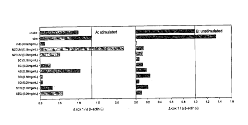
Figure 1: Relative expression of cox 1 RNA in IL-1 stimulated (A) and
unstimulated
(B) cartilage explants. =
Figure 2: Relative expression of cox 2 RNA in IL-1 stimulated (A) and
unstimulated
(B) cartilage explants.
Figure 3: Relative expression of iNOS RNA in IL-1 stimulated (A) and
unstimulated (B) cartilage explants.
CA 02714401 2010-08-04
WO 2009/073931
PCT/AU2008/001834
6
Figure 4: Relative expression of aggrecan RNA in IL-1 stimulated (A) and
unstimulated (B) cartilage -explants. =
Figure 5: Prostaglandin E2 (PGE2) production by IL-1 stimulated (A) and
unstimulated (B) cartilage explants. 10-4110 represents treatments
significantly
different from stimulated (A) or unstimulated (B) controls. Indo,irn, SEQsim
(both
doses) and BOall (0.18mg/mL) resulted in significantly lower PGE2 in
stimulated
explants compared with stimulated controls. Indosim and SEQsim lowered -PGE2 =
production in unstimulated explanfs relative to unstimulated controls.
Figure 6: Timeline of injections and sample collection; Sample collection
consisted
of synovial fluid arthrocentesis from left and right intercarpal joints, and
jugular
venous blood. Dietary supplementation began on day 0 and continued for the
duration of the experiment.
Figure 7: Synovial fluid [PGE2] from intercarpal joints of control horses
injected
with IL-1 (long on inj-1, 10Ong on inj-2) or saline in CON (A) and SEQ (B)
horses.
Healthy horses received a diet containing placebo (CON) or Sasha's EQ (SEQ)
for
28 days. Intra-articular IL-1 (long in 6004 sterile saline) was injected into
the
intercarpal joint, and sterile saline (5004) was injected into the
contralateral joint
14 days after c,ommencement of supplementation (inj-1). A second intra-
articular
injection of IL-1 (10Ong in 5004 sterile saline) or saline (5004) was injected
the
same joints 24 h later (inj-2). Approximately 1.5mL synovial fluid was
aspirated
from the intercarpal joints on days pre (before commencement of
supplementation), inj-1 and inj-2 (prior to injections), inj-2-2 (8h after 2nd
IL-1
injection), and 1, 3, 7 and 14 days after 2nd IL-1 injection. * denotes
significant
change from inj-1 within treatments. Letters denote significant differences
between saline and IL-1 within treatments. Changes are significant when
ps0_05.
Figure 8: Synovial fluid [GAG] from intercarpal joints injected with IL-1 (1
Ong on
inj-1, 10Ong on inj-2) or saline in CON (A) and SEQ (B) horses. Healthy horses
received a diet containing placebo (CON) or Sasha's E0 (SEQ) for 28 days.
Infra;
articular IL-1 (long in 5004 sterile saline) was injected into the intercarpal
joint,
and sterile saline (5004) was injected into the contralateral joint 14 days
after
commencement of supplementation (inj-1). A second intra-articular injection of
IL-
1 (10Ong in 5004 sterile saline) or saline (5001it) was injected the same
joints 24
CA 02714401 2010-08-04
WO 2009/073931
PCT/AU2008/001834
7
h later (inj-2). Approximately 1.5mL synovial fluid was aspirated from the
intercarpal joints on days pre (before commencement of supplementation), inj-1
and inj-2 (prior to injections), inj-2-2 (8h'after 2nd IL-1 injection), and 1,
3, 7 and 14
days after 2nd IL-1 injection. * denotes significant change from inj-1
within
treatments. Letters denote significant difference between IL-1 and saline
within
treatments. SEQ horses had significantly higher synovial fluid [GAG] than CON
horses. Differences were significant when ps0.05.
Figure 9: Synovial fluid [protein] from intercarpal joints of control horses
injected
with IL-1 (10ng on inj-1, 10Ong on inj-2) or saline in CON (A) and SEQ (B)
horses.
Healthy horses received a diet containing placebo (CON) or Sasha's Ea (SEQ)
for
28 days. Intra-articular 1L-1 (I Ong in 5001.L sterile saline) was injected
into the
intercarpal joint, and sterile saline (5004) was injected into the
contralateral joint
14 days after commencement of supplementation (inj-1). A second intra-
articular
injection of IL-1 (10Ong in 5004 sterile saline) or saline (5004) was injected
the
same joints 24 h later (inj-2) Approximately 1.5mL synovial fluid was
aspirated
from the intercarpal joints on days pre (before commencement of
supplementation), inj-1 and inj-2 (prior to injections), inj-2-2 (8h after 2nd
IL-1
injection), and 1, 3, 7 and 14 days after 2'1 IL-1 injection. * denotes
significant
change from inj-1 within treatments. Letters denote significant differences
between IL-1 and saline within treatments. Differences were significant when
p0.05.
Figure 10: Circumference of intercarpal joints injected with IL-1 (long on inj-
1,
10Ong on inj-2) or saline in CON (A) and SEQ (B) horses. Healthy horses
received a diet containing placebo (CON) or Sasha's'EQ (SEQ) for 28 days.
Infra-
articular IL-1 (10ng in 5001AL sterile saline) was injected into the
intercarpal joint,
and sterile saline (5004) was injected into the contralateral joint 14 days
after
commencement of supplementation (inj-1). A second intra-articular injection of
IL-
1 (10Ong in 5001AL sterile saline) or saline (5001.tL) was injected the same
joints 24
h later (inj-2). Approximately 1.5mL synovial fluid was aspirated from the
intercarpal joints on days pre (before commencement of supplementation), inj-1
,and inj-2 (prior to injections), inj-2-2 (8h after 2nd IL-1 injection), and
1, 3, 7 and 14
days after 2nd 1L-1 injection. " denotes significant change from inj-1
within
treatments. Letters denote significant differences between 1L-1 and saline
within
8
treatments. Joint circumference of IL-1 -injected joints was significantly
lower in SEQ horses than CON
horses (p<0.001). Differences were significant when p5 0.05.
Figure 11: Table 1 showing the primers for aggrecan and 13-actin.
Figure 12: Table 2 showing the composition of Sasha's EQ powder prepared by
combining Abalone (AB),
New Zealand Green Lipped Mussel (NZGLM), Shark cartilage (SC) and BO
(Interpath Pty Ltd, Australia).
Figure 13: Table 3 showing the nutrient composition of Sasha's EQ for feeding
to horses.
Figure 14: Chromatographic spectrum of the extract of Biota orientalis oil.
Figure 15: Shows the concentration of NO of each of the isolated fractions in
the cell culture assay.
Figure 16: Shows the induced PGE2 level of the isolated fractions Fri and Fl.
Figure 17: Shows the induced PGE2 level of the isolated fractions FV and Vi.
Figure 18: Shows the reduction of IL-113 induced PGF2a levels on fractions Fri
and Fri.
Figure 19: Shows the reduction of IL-113 induced PGF2a levels on fractions FrV
and FrVi.
DETAILED DESCRIPTION OF THE INVENTION
To facilitate an understanding of the invention various terms and
abbreviations are used and defined
below:
"SEQ" means a blend of New Zealand Green Lipped Mussel, abalone, shark
cartilage powder and Biota
oil.
"BO" means "Biota oil" being an extract of the seeds of the plant Biota
orientalis. BO was purchased from
Interpath Pty Ltd, Australia. The BO was obtained using the separation process
described in
W003/089399 (published October 30, 2003) and employing supercritical carbon
dioxide.
"NZGLM" means New Zealand Green Lipped Mussel,
"sim" means a simulated digest or simulated digestion.
CA 2714401 2019-05-08
CA 02714401 2010-08-04
WO 2009/073931
PCT/AU2008/001834
9
'COX" or "cox" means the enzyme cyclooxygenase.
"iNOS" means inducible nitric oxide (NO) synthase. -
Biota is an herb native to Western China and North Korea and is known by a
number of other names, such as Thuja or/entails, Platycladus striae, and
Platycladus oriental's.
Simulated digests of shark cartilage, NZGLM and abalone have been previously
reported to have anti-inflammatory effects in a cartilage explant model of
arthritis
by reducing PGE2, GAG and/or nitric oxide (Pearson et al., 2007).
=The following data reports alterations in gene expression associated with
conditioning cartilage explants with simulated digests of the combination of
all four
constituents (SEQ; SEQ64õ), and , to characterize their effects on IL-1-
induced
PGE2, GAG, NO, cell viability, and genetic expression of cox 1, cox 2, iNOS
and
aggrecan.
Methods
Explant cultures
Front legs of market weight pigs (5-7 months old, 200-250Ibs) were obtained
from
a local abattoir. Legs were chilled on crushed ice until dissection. Using
aseptic
technique, the intercarpal joint was opened and the cartilage surfaces
exposed. A
4mm dermal biopsy punch was used to take explants .(-0.5mm thickness; 11-
15mg/explant) of healthy cartilage from: the weight-bearing region of both
articulating surfaces of the intercarpal joint. Cartilage pieces were washed 3
times
in DMEM supplemented with NaHCO3. Two cartilage discs were placed into each
well of 24-well tissue culture plates containing DMEM supplemented with amino
acids, sodium selenite, manganese sulfate, NaHCO3 and ascorbic acid (TCM ¨
tissue culture medium). Plates were incubated at 37 C, 7% CO2 in a humidified
atmosphere for up to 144h_ Every 24 h media was completely aspirated into 1mL
microcentrifuge tubes and immediately replaced with control, conditioned
and/or
stimulated media (described below) before being returned to the incubator. The
collected media was stored at -80 C until analysis. Cartilage was harvested at
the
end of each experiment with one explant per well stained for cytotoxicity and
the
remaining cartilage immediately frozen at -80 C.
CA 02714401 2010-08-04
WO 2009/073931 PCT/AU2008/001834
=10
Simulated digestion and ultrafiltration
A simulated digestion procedure was developed to mimic the gastrointestinal
processing of ingested dietary supplements. This type of approach has
previously
been used to improve the bio-assessment of putative nutraceuticals (Rininger
et
al., 2000; Pearson et al., 2007).
Simulated digests were prepared using SEQ (0.859), BO [2.5mL (0.859) and indo
(0.074g - a positive anti-inflammatory control). Each test substance was
individually suspended in 35mL of simulated gastric fluid (37mM NaCI, 0_03N
NCI,
3.2mg/mL pepsin), and shaken at 37 C for 2 h (Rininger et al_, 2000). After
this,
solution acidity was neutralized by adding an equinormal volume of 2.2 N NaOH
(1.15mL). To this was added 36.15mL of simulated intestinal fluid (Rininger et
at..
2000 - 30mM K2HPO4, 160mM NaH2PO4; 20mg/mL pancreatin; pH adjusted to
7.4) and the resultant mixture shaken in a 37 C incubator for a further 2 h. A
."blank" was prepared using identical methodology but without including any
test
substance. Appropriate volumes of gastric and intestinal fluid were derived
from .
those approximated in a human stomach-(Marciani et al., 2005).
Upon completion .of the 4-hour incubation, simulated digests of SEQ (SEQ5i,õ)
BO
(BO) and indomethacin (indosirn) were centrifuged at 3,000 x g for 25 min at 4
C. =
The supernatant was decanted and centrifuged a second time at 3,000 x g for 15
min at 4 C. The resulting supernatant was warmed to room temperature and
filtered (0.24m) to remove particulates. This filtrate was further fractioned
with an
ultrafiltration centrifuge unit with a 50kDa molecular weight cut-off,
(AmiconUltra,
Millipore, Mississauga ON), spinning at 3,000 x g for 25 min (room
temperature).
Filtered simulated digest was stored at 4 C until use for a maximum of 7 days.
Effect of SEQshi, and BOsin, on IL-1-induced inflammation
=
SEQsim was prepared as explained above. Explants from 12 pigs were prepared
as previously described, and maintained in unconditioned media for the initial
24 h.
At 24 hours post-culture, SEQ,im, BOsin, (0, 0_06 or 0.18 mg/mL) or indosin,
(0.02mg/mL) Was added to TCM (conditioned media). Conditioned media was
refreshed every 24 hours for the duration of the experiment. At 72 hours post-
culture, and every 24 hours thereafter, explants were stimulated with 11,1 (0
or
10ng/mL, Medicorp, Montreal, Quebec; CaL #PHC0813). Explants from each
CA 02714401 2010-08-04
WO 2009/073931
PCT/AU2008/001834
11
animal were exposed to each treatment in duplicate. Explants were cultured for
a
total of 120 h. Media was analyzed for [PGE2], [GAG], [NO]. One explant per
treatment was collected into sterile phosphate buffered saline (PBS) and
immediately stained for cell viability (see below). The second explant was
frozen
at -130 C for RNA extraction (see below).
PGE2 analysis:
PGE2 concentration of TCM was determined using a commercially available PGE2
ELISA kit (The kit has 7% cross-reactivity with PGE1) (Amershan, Bale D'Urfe,
Quebec). Plates were read using a Victor 3 microplate reader (Perkin Elmer,
Woodbridge ON) with absorbance set at 405nm. PGE2 standard curves were
developed for each plate, and a best-fit 3rd order polynomial equation with
R20.99
was used to calculate PGE2 concentrations for standards and samples from each
plate.
NO analysis:
NO concentration of tissue culture media was determined by the Griess Reaction
(Shen et al., 2005). Plates were read using a Victor 3 microplate reader with
absorbance set at 530nm. Sodium nitrite standard curves were developed for
each plate, and a best-fit linear regression equation with R2X199 was used to
calculate NO concentrations, which were compared with the nitrite standard.
Isolation of total RNA and synthesis of cDNA
Total RNA was extracted from cartilage explants using a modified TRIzol
procedure (Chan et al., 2006). Frozen cartilage from each animal was pooled
according to conditioning and stimulation, and homogenized in Tri-Reagent
(100mg tissue/mL; Sigma, Mississauga ON). Chloroform was added to extract
RNA followed by vigorous agitation and 2-min incubation at room temperature.
Sample was then centrifuged (12,000.x g, 15 min) and RNA was precipitated with
an equal volume of 70% ethanol (DEPC). RNA precipitate was applied to an
RNeasy mini column (Qiagen, Valencia CA, USA) and RNA was purified according
to manufacturer instructions.
For each pooled sample, 1pg total RNA was converted to single stranded cDNA
using Moloney Murine Leukemia Virus (MMLV) reverse transcriptase (Invitrogen,
CA 02714401 2010-08-04
WO 2009/073931
PCT/AU2008/001834
12
Burlington ON) according to manufacturer instructions. Single-strand cDNA was
quantified by UV spectrophotometry and diluted with DEPC-I-120 to a final
concentration of lOng/pL.
Quantitative real time RT-PCR
Primers for porcine iNOS (Granja et al., 2006), Cox1/2 (Blitek et al., 2006),
aggrecan (Fehrenbacher et al., 2003) and f3-actin (housekeeping gene;
Nishimoto
et al., 2005) (Table 1) were prepared (Laboratory Services Division,
University of
Guelph) and stored at -20 C until use. Cartilage samples from SEQ,h, and BOsi,
were evaluated for changes in gene expression, together with cartilage
cultured
under identical conditions previously with the other 3 components of SEQ (see
Pearson et al., 2007 for detailed culture conditions). Twenty five microliter
PCR
reactions were performed in triplicate using an ABI Prism 7000 sequence
detection
system (Perkin-Elmer). Amplification of 5Ong of each cDNA sample was detected
using SYBR-Rox (invitrogen, Burlington ON) and compared to a standard curve of
pooled cDNA containing equal amounts of cDNA from each sample. A 1.5%
agarose electrophoresis gel was used to confirm PCR products. Expression of
each gene of interest (G) in each sample was compared to amplification of I3-
actin
(3), and calibrated to unstimulated control explants (ie. fold change for
calibrator =
1). Fold change in expression (AG / Ai3) is presented in arbitrary units.
Cytotoxicity Staining
Cell viability was determined using a commercially available viability
staining kit
(Invitrogen; Burlington ON) (Pearson et al., 2007). Briefly, explants were
washed
in 500uL PBS and placed into a 96-well microtitre plate (one explant per
well), and
were incubated in 200uL of stock stain (411M C-AM; 8pM EthD-1) for one hour at
room temperature. The plate was read from the bottom of each well using 10
horizontal steps, 3 vertical steps, and a 0.1mm displacement. C-AM and EthD-1
fluorescence in live and killed explants were obtained with
excitation/emission
filters of 485/530nm and 530/685nm, respectively. '
Data analysis
=
=
Data from analysis of tissue culture media and viability are presented as
means
standard error. Means of replicates from each treatment/animal were analyzed
CA 02714401 2010-08-04
WO 2009/073931
PCT/AU2008/001834
13
using two-way repeated measures analysis of variance comparing each treatment
with unconditioned controls and indomethacin- conditioned controls. Viability
data
.were analyzed using the Student's Mest, individually comparing stimulated
controls with all other treatments. When = a significant F-ratio was obtained,
the
Holm-Sidak post-hoc test was used to identify significant differences between
treatment and/or time. Significance was accepted if 00.05.
= Due to low cellularity of cartilage explants, it was necessary to pool
RNA from
explants exposed to the same conditioning and stimulation in order to extract
sufficient RNA for a reverse transcription reaction_ Thus, PCR data are
presented
in the text as a mean change in gene expression (calibrated to controls)
relative to
13-actin coefficient of variation for the assay. A calibrated fold
expression change
a 2 is considered to be biologically relevant (Yang at al., 2002; Schena at
al.,
1995) and are discussed in the text as significant differences.
Results
PCR
=
Cox / (Figure 1, A and B): IL-1 stimulation of control explants resulted in a
35%
increase in cox 1 expression compared with unstimulated controls. Cox 1
expression was decreased by exposure to indosim by 98 and 91.5% in
unstimulated
and stimulated explants, respectively.
All constituents of SEQ reduced cox 1 expression in unstimulated explants
(range:
76 ¨ 95% inhibition). Importantly, it was observed that BO sim (0.06mg/mL) was
the
most effective cox 1 inhibitor, reducing cox 1 expression by 95% in both
unstimulated and stimulated explants.
In addition, it was observed that SEasi. (0.06 and 0.18mg/mL) reduced cox 1
expression in unstimulated explants by 90 and 80%, respectively. In IL-1
stimulated explants, Saishy, (0_06 and 0.18mg/mL) inhibited cox 1 expression
by
57 and 76%, respectively. The least effective cox 1 inhibitor in IL-1-
stimulated
explants was NZGLM (0.18mg/mL), which increased cox 1 expression by 62%.
Fold change in cox 1 for all samples was > 2 and therefore not considered
significant.
CA 02714401 2010-08-04
WO 2009/073931
PCT/AU2008/001834
14
Cox 2 (Figure 2, A and B): Stimulation of Control explants resulted in a
significant
4.3-fold increase in cox 2 expression. Indosim reduced expression of cox 2 by
44
and 47% in unstimulated and stimulated explants, respectively: Fold increase
in
cox 2 for indoso-conditioned, IL-1-stimulated explants was significant (2.3).
Abalone (0.18mg/mL) significantly increased cox 2 expression in unstimulated
explants, showing similar effect on cox 2 (3.7-fold) as IL-1. All other
constituents
decreased Cox 2 expression in unstimulated explants (range: 56 ¨ 90%).
IL-1-stimulation resulted in a significant increase in cox 2 expression in
those
explants conditioned with indosim (2.3401d), SEQsim (0.06mg/mL; 2.0-fold),
NZGLMsim (0_18mg/mL; 28.2-fold), and AB sim (0.18mg/mL; 41.5-fold). All other
constituents prevented a significant increase in IL-1-induced cox 2
expression; the
most effective inhibitor was BOsirn (0.06mg/mL) which inhibited cox 2
expression by
92%.
iNOS (Figure 3, A and B): Stimulation of control explants by IL-1 resulted in
a 287-
fold increase in iNOS expression. lndosim conditioning had no effect on iNOS
in
unstimulated explants. In IL-1-stimulated explants, indoor conditioning
augmented
the effect of IL-1 on 1NOS expression (725-fold increase).
SEQ and all of its individual constituents significantly increased iNOS
expression in
unstimulated explants (range: 39 ¨ 2486-fold increase). IL-1-stimulation
resulted
in a significant increase in iNOS expression in all conditioned explants.
However,
compared with IL-1-stimulated controls, INOS was significantly inhibited by
both
doses of SEQ,i, in a dose-dependent manner (60 and 89% inhibition for 0.06 and
0.18mg/mL, respectively). BOsim (0.06mg/mL) and ABsim (0.18mg/mL) also
significantly inhibited IL-1-induced iNOS expression by 55 and 12%,
respectively.
Aggrecan (Figure 4, A and B): Stimulation of control explants with IL-1
resulted in
a slight, non-significant decline in aggrecan expression. Conditioning
of
unstimulated explants with indosim resulted in 58-fold increase in aggrecan.
This
increase was completely abolished by stimulation of indosim-conditioned
explants
with IL-1.
SEQ and all of its constituents significantly increase aggrecan expression in
unstimulated explants. SEQsim
increased aggrecan expression in unstimulated
CA 02714401 2010-08-04
WO 2009/073931
PCT/AU2008/001834
explants in a dose-dependent manner (42.8 and 215.7-fold increase for 0.06 and
0.18mg/mL, respectively).
Stimulation of conditioned explants with. IL-1 rebutted in significant
increase in
aggrecan expression in SEO and all of its constituents, with the exception of
SC,Irn
(0.18mg/mL; 1.4-fold increase).
=
Tissue culture experiments:
PGE2 (Figure 5,A and B): Stimulation of control explants with IL-1 (10ng/mL)
resulted in a significant increase in media [PGE2] over the 48h stimulation
period,
resulting in a significant difference between stimulated and unstimulated
controls
(p=0_03). indosim (0.02mg/mL) significantly reduced media [PGE2] in IL-1
stimulated and unstimulated explants compared with stimulated and ustimulated
controls, respectively. There was no IL-1-induced increase in media [PGE2] in
explants Conditioned with indosim.
=
Stimulation with IL-1 of explants conditioned with SEQsi, (0.06 and 0.18mg/mL)
did not increase media [PGE21. Media [PGE2] was significantly lower in these
explants compared with stimulated and unstimulated control explants (Figure 5,
A).
In unstimulated explants media [PGE2] was significantly lower in explants
conditioned with SR:41m (0_06 and 0.18mg/mL) than in unstimulated controls
(Figure 5, B). There was no significant difference in media [PGE2] between
SEOsin, (0.06 and 0.18mg/mL) and indosim in both IL-1-stimulated and
unstimulated
explants.
=
_
There was no increase in media [PGE2] subsequent to IL-1 exposure in explants
conditioned with BOsiff, (0.06 and 0.18mg/mL) (Figure 5, A). Conditioning of
1L-1-
stimulated explants with BO sim (0.18mg/mL) resulted in a significantly lower
media
[PGE2] than stimulated controls. There was no significant effect of BOsirn on
unstimulated explants (Figure 5, B).
NO: There was no significant change in media [NO] in unstimulated control
explants. Exposure of control explants to IL-1 (10ng/mL) resulted in a
significant
elevation of media [NO] at 24 (1_21 0.1 pg/mL) and 48 h (1.06 0_1 pg/m14.
There was no significant effect of indosim on [NO] in stimulated or
unstimulated
explants (Figure 7).
CA 02714401 2010-08-04
WO 2009/073931
PCT/AU2008/001834
16
Discussion
These experiments assist in describing effects of the simulated digest of SEQ
on
cox 1, cox 2, iNOS, and aggrecan gene expression. The gene expression data can
then be used to make predictions about the mechanism of action of SEQ.
Alterations in gene expression observed in IL-1-stimulated control explants
showed a pattern consistent with an inflammatory response. IL-1 stimulation
resulted in a small, non-significant increase in cox 1 expression coupled with
a
significant increase in cox 2 expression, as has been reported by other
authors
(Kydd et al., 2007).
As shown, indosim showed a cox 1:cox 2 inhibition profile of about 2:1, which
is
consistent with its classification as a cox 1/2 inhibitor (Gerstenfeld et al.,
2003).
We have also shown that indosim does not inhibit IL-1-induced iNOS expression,
consistent with reports by other authors (Palmer et al., 1993). Nor did it
influence
IL-1-mediated aggrecan expression in ILA-stimulated explants, an effect that
has
been reported in mechanically stressed cartilage explants (limoto et at.,
2005).
These data characterize indomethacin as an effective anti-inflammatory
predominately through cox inhibition: Its inability to reduce IL-1-mediated
aggrecan expression and its augmenting effect on 1L-1-mediated iNOS
expression,
however, suggest that cartilage exposed to indomethacin would continue to
degenerate through decline in matrix formation and would suffer from increased
==nitric oxide-mediated cell death. Indeed these adverse effects have been
reported
in arthritic dogs using prophylactic indomethacin (Hungin and Kean 2001), and
indornethacin is associated with worsening of some pathophysiological
indicators
of arthritis in humans (Rashad et al., 1989; Huakinsson at al., 1995). When
indosi,
was applied to cartilage explants in the current study, there was an increase
in IL-
1-mediated NO production, but this was not coupled with a decrease in cell
viability.
The relative inhibitory profile of SEQ,,,, on cox 1:cox 2 expression was
approximately 1:1 at both doses. In the experiments described herein,
SECIsiff, at
the lower dose was comparable to indo,im as a cox 2 inhibitor, whereas the
higher
dose was a more effective inhibitor of cox .2 than inclosim. It is therefore
predicted
that SEQ sbil should effectively inhibit PGE2 production by IL-1-stimulated
explants.
CA 02714401 2010-08-04
WO 2009/073931
PCT/AU2008/001834
17
This inhibition was observed in the tissue culture explant experiment.
Inhibition of
IL-1-mediated PGE2 production by SEOsim-conditioned cartilage explants was
significant at both doses, and was not statistically different from PGE2
inhibition by
indosim. This provides an explanation for the observed clinical benefit of SEQ
in
relieving pain in arthritic patients (Rukwied et al., 2007; Zhao et al.,
2007).
Earlier publications have reported that SC3irn and NZGLMsi, inhibit PGE2
production by IL-1-stimulated cartilage explants (Pearson et al., 2007), and
the
data in this application shows that BOsi, also has this effect. However, it is
of
interest that, with the exception of SCsim (0.18mg/mL), cox 2 inhibition by
the most
effective dose of SEasim is stronger than any single constituents alone. This
points
to a synergistic relationship between the constituents.
Given the effective PGE2-inhibiting, and related cox-inhibiting properties of
SEQ,,,,,
the effects of SEQ,in, on iNOS were investigated. With a standard 'NSAID-like'
mechanism it is predicted that SEQ would also augment iNOS expression in IL-1-
stimulated explants. In fact, the opposite was true, and SEO,iõ, was found to
significantly and strongly inhibit iNOS expression.
The effect of IL-1 on cellular expression of iNOS and cox 2 is differentially
regulated through activation of at least 2 Mitogen Activated Protein Kinases
.(MAPKs) (LaPointe and Isenovi 1999). Net expression of iNOS and cox 2 are at
least partially dependent on the relative amounts of pericellular NO and PGE2
(Shin et at., 2007). Thus, products which increase pericellular NO can
effectively
downregulate expression of cox 2, and vice versa (Shin et al., 2007; Kim et'
al.,
2005). This provides some explanation as to why SEC1sim showed a significant
inhibitory effect on iNOS while many of the individual constituents, including
shark
cartilage, Biota and NZGLMsim (0.18mg/mL), actually upregulated expression of
'NOS.
Conclusions
= =
SEQ is capable of effectively downregulating RNA for iNOS and cox 2. Its
effect
on iNOS and cox 2 appears to be due to synergy between its four constituents,
but
it may be related to post-translational inhibition of NO production (Pearson
et al.,
2007).
CA 02714401 2010-08-04
WO 2009/073931
PCT/AU2008/001834
18
Models of cartilage inflammation in horses are widely reported, and include
intra-
articular challenges such as lipopolysaccharide (Jacobsen et al., 2006),
Freunds
Complete Adjuvant (Toutain and Coster 2004) or Na-monoiodoacetate (Welch et
al., 1991); or surgical disruptions including creation of osteochondral
fragments
(Friable et al., 2007), focal contusion impact injuries (Bolam et at., 2006)
and
ligamentous tanssection (Simmons et al.,. 1999). While these models capably
demonstrate maximal activation of a complexity of inflammatory mechanisms
within cartilage and associated subchondral bone and soft tissues, they
represent
a predominately traumatic inflammatory response. They are less representative
of
the more subtle biochemical, functional and pathophysiological changes in
incipient or sub-acute articular inflammation that characterize most cases of
lameness in racing horses (Steel et al., 2006).
While non-steroidal anti-inflammatory drugs (NSAIDs) and corticosteroids
remain
important therapeutic resources for treatment of overt clinical lameness,
nutraceuticals are becoming widespread as a therapeutic and prophylactic
management strategy,for horses with low-grade, sub-acute articular damage and
for those at risk of developing articular problems (Trumble 2005; Neil et al.,
2005). =
Most research reported on the efficacy and/or safety of these products in
arthritis
uses in vitro models (Pearson et al., 2007; Chan et t, 2006), or traumatic
injury or
clinical in vivo research in non-equine species (McCarthy et al., 2006; Cho et
al.,
2003). Though useful as screening tools, in vitro models cannot account for
the
systemic effects of a dietary product which may influence outcomes in the
articular
space
The objectives of this section are to a) produce and characterize a
reversible, sub-
clinical model of IL-1-induced intra-articular inflammation in the horse with
respect
to PGE2 and NO production, and GAG release from cartilage; and b) to apply
this
model to the evaluation of SEQ in mammals, particularly in horses.
Method
Diets: SEQ powder was prepared by combining Abalone (AB), New Zealand
Green Lipped Mussel (NZGLM), Shark cartilage (SC) and Biota oil (Interpath Pty
Ltd, Australia) according to the composition provided in Table 2. SEQ mixed
ration was prepared by combining SEQ powder (10g/kg), molasses (29g/kg) and
flavoring (Essential Sweet Horse Essence D 2344. Essentials Inc. Abbotsford,
CA 02714401 2010-08-04
WO 2009/073931
PCT/AU2008/001834
19
BC.) (19/kg) to a sweet feed horse ration (Table 2), and blending in a diet
mixer in
5kg batches until fully mixed. Control ration (CON) was prepared using the
same
sweet feed diet blended with molasses (-20g/kg) and flavoring (1g/kg).
Horses: 11 healthy horses without signs of articular inflammation (3
thoroughbred,
8 standardbred; age 5¨ 12 years; 10 geldings, 1 mare) were randomly allocated
to
either Group A (SEQ; 1.5kg/day; n=6) or Group B (CON; 1.5kg/day; n=5). The 28-
day experiment consisted of two phases - Phase 1: pretreatment (14 days);
Phase
2: treatment (14 days)_ Supplementation began on Day 0 and continued for the
duration of the experiment (Figure 6). Sample collection occurred on days 0
(pre),
14 (inj-1), 15(2 samples: inj-2 - taken inimediately before injection; inj-2-2
¨ taken
8h post-injection), 16 (day 1), 18 (day 3), 21 (day 7) and 28 (day 14); on
these
days blood was collected from the jugular vein, and synovial fluid was sampled
from both intercarpal joints by aseptic arthrocentesis (see below). An
inflammatory
challenge ¨ recombinant interleukin-113 (IL-1) ¨ was injected into the left or
right
intercarpal joint on day 14 (inj-1; long in 500pL sterile saline) and 15 (inj-
2; 10Ong
in 500pL sterile saline). An equal volume of sterile saline was injected into
the
cohtralateral intercarpal joint. Joint circumference as an indicator of joint
effusion
was measured with a tape measure at each sampling of joint fluid.
All horses were turned out in paddocks during the day and housed in box-stalls
overnight. They were bedded on wood shavings and offered hay, water, and
mineral salts ad libitum. All procedures were approved by the University of
Guelph
Animal Care Committee in accordance with guidelines of the Canadian Council on
Animal Care.
Arthrocentesis: The knees of both the. left and right legs were shaved, and
the
area aseptically prepared using chlorhexadine (4%), and rinsed with 70%
isopropyl
alcohol. A sterile 22 gauge, 1.5" needle was inserted into the lateral aspect
of the
left intercarpal joint. A 3 cc sterile syringe was then attached, and
approximately
1.5 ¨ 2 mL of synovial fluid was aspired and immediately injected into a
sterile K2-
heparin vacutainer. The procedure was then repeated for the right intercarpal
joint. On days 14 (inj-1) and 15 (inj-2), IL-1 (500pL) was injected into
either the
right or left intercarpal (500pL saline injected into oontralateral joint)
after
aspiration of synovial fluid and before removal of the needle hub.
Approximately
1.5mL of synovial fluid was removed from the vacutainer and placed into a
CA 02714401 2010-08-04
WO 2009/073931
PCT/AU2008/001834
microcentrifuge tube and spun at 11,000 x g for 10 minutes to remove cellular
debris. Supernatant was placed into another microcentrifuge tube containing
10pg
indomethacin, and frozen at .-80 C until analyzed for PGE2, GAG and NO.
lndomethacin was added to synovial fluid after it was collected in order to
prevent
further formation of PGE2 during storage of samples. The remaining ¨0.5mL
synovial fluid was sent to the Animal Health Laboratory. (University of
Guelph) for
cytological analysis.
Synovial fluid cytology
=
1,0 ¨ 1.5mL of fluid was removed from the vacutainer for PGE2, NO and GAG
analysis (see below), and approximately 0.5mL was analyzed. for total
nucleated
cell count (Coulter 72 counter: Beckman Coulter Canada Inc. Mississauga ON),
protein (refractometer) and cell differential (on 100 nucleated cells) at the
Animal
Health Laboratory.
=
Synovial fluid [PGE21:
Synovial fluid was thawed to room temperature then incubated with 20i1
hyaluronidase (10mg/mL) on a 'tube rocker for 30 minutes at 37 C to digest
hyaluronic acid. Sample was then diluted 1:2 with formic acid (0.1%), and
centrifuged 12,000 x g for 10 minutes. The supernatant was decanted and
analyzed for PGE2 by a commercially available ELISA kit (GE Amersham, Bale
D'Urfe, Quebec). PGE2 was extracted from the sample using provided lysis
reagents to dissociate PGE2 from soluble membrane receptors and binding
proteins, and then quantified according to kit protocol. Plates were read
using a
Victor 3 microplate reader (Perkin Elmer, Woodbridge ON) with absorbance 'set
at
450nm. A best-fit 3rd order polynomial standard curve was developed for each
plate (R2.?.Ø99), and these equations were used to calculate PGE2
concentrations
for samples from each plate.
Synovial fluid [GAG]:
Hyaluronic acid in synovial fluid samples were digested with hyaluronidase as
described above. GAG concentration of synovial fluid was determined using a
1,9-
DMB spectrophotometric assay as described by Chandrasekhar et al. (1987)_
Samples were diluted 1:3 with dilution buffer and placed into a 96-well
microtitre
CA 02714401 2010-08-04
WO 2009/073931
PCT/AU2008/001834
21
plate. Guanidine hydrochloride (275g/L) was added to each well followed
immediately by addition of 150pL DMB reagent. Plates were incubated in the
dark
for 10 minutes, and absorbance was read on a Victor 3 microplate reader at
530nm. Sample absorbance was compared to that of a bovine chondroitin sulfate
standard (Sigma, Oakville ON). A best-fit linear standard curves was developed
for each plate (R20.99), and these equations were used to calculate GAG
concentrations for samples on each plate.
Synovial fluid [NO]:
=
Nitrite (NO2.), a stable oxidation product of NO, was analyzed by the Griess
reaction (Fenton et al., 2002). Undiluted TCM samples were added to 96 well
plates. Sulfanilamide (0.01g/rnL) and N-(1)-Napthylethylene diamine
hydrochloride
(1mg/mL) dissolved in phosphoric acid (0.085g/L) was added to all wells, and
absorbance was read within 5 minutes on a Victor 3 microplate reader at 530
nm.
Sample absorbance was compared to a sodium nitrite standard.
Data analysis and presentation
Two-way repeated measures (RM) analysis of variance (ANOVA) was used to
detect differences between treatments. When a significant F-ratio was
obtained,
the Holm Sidak post-hoc test Was used to identify differences between
treatments.
One-way RM ANOVA was used to detect 'differences within treatments with
respect to time. For blood and synovial fluid data, one-way comparisons of
data
were made against pre- and inj-1 data, as each represented baseline for diet
and
IL-1 injections, respectively. Data are presented as means SEM. Graphs for
biochemistry and hematology data are scaled to physiological reference
intervals
unless otherwise stated. Reference intervals are those published by the Animal
Health Laboratory, University of Guelph
(http://www.labservices. uog uelp h. ca/units/ah I/flles/AHL-use rg uide.
pdf).
Results =
Synovial fluid =
=
=
PGE2:
CA 02714401 2010-08-04
WO 2009/073931
PCT/AU2008/001834
22
CON horses: There was no significant change in synovial fluid [PGE2] in saline-
injected joints at any time (Figure 7, A). Relative to pre-injection
concentrations,
[PGE2] was significantly increased at inj-2-2 (321.3 161.8 pg/mL; p-=0.04)
in IL-1-
injected joints, at which time synovial fluid [PGE2] was significantly higher
in IL-1-
injected joints than in saline-injected joints (p<0.001).
SEQ horses: Data represent n=5, as one outlier horse was removed from the
analysis. PGE2 did not change in saline-injected joints of SEQ horses. Like
CON
horses, there was a spike in [PGE2] increased at inj-2-2 (176.4 89.2 pg/mL)
in IL-
1-injected joints of SEQ horses (Figure 7, B). However, this increase was not
significant when compared with pre-injection concentrations. PGE2 response to
saline injection was not different in . SEQ horses compared with CON horses.
There was no significant difference in PGE2 response to IL-1 injection
compared
with saline in SEQ horses.
Although mean [PGE2] at inj-2-2 in SEQ horses was approximately 55% that of
CON horses, variability about the means resulted in no significant difference
between diets.
GAG:
CON horses: Synovial fluid [GAG] increased in saline-injected joints between
inj-1
(18.3 6.8 pg/mL) and day 1 (48.1 9.6 pg/mL) (Figure 8, A). Injection of IL-
1
(long). caused a rapid and significant increase in synovial fluid [GAG]
between inj-
1 (24.5 7.3 pg/mL) and inj-2 (77.6 4.4 pg/mL). Synovial fluid [GAG]
remained
significantly elevated in IL-1-injected joints at inj-2-2 (66.0 9.6 pg/mL)
and day 1
(53.3 11.4 .pg/mL) compared with pre-injection concentrations_ The magnitude
of
increase in synovial fluid [GAG] was significantly higher in IL-1-injected
joints than.
in saline-injected joints (p=0.003).
SEQ horses: Synovial fluid [GAG] tended to increase (p=0.09) in both saline-
and
IL-1-injected joints between pre (saline: 29.3 5.9 pg/mL; IL-1: 27.0 10.8
pg/mL)
and inj-1 (saline: 85.5 28.0 pg/mL; IL-1; 83.2 27.9 pg/mL), suggesting an
effect
of diet on synovial fluid [GAG] (Figure 8, B). There was no change in synovial
fluid
[GAG] in saline- OF IL-1-injected joints over the course of the experiment.
There
was no significant difference in synovial fluid [GAG] of IL-1-injected and
saline-
injected joints.
CA 02714401 2010-08-04
WO 2009/073931
PCT/AU2008/001834
23
Synovial fluid [GAG] in IL-1- and saline-injected joints was significantly
higher in
SEQ horses than CON horses (p<0.001). This difference. was mainly an effect of
diet, and not an effect of IL-1, as evidenced by the fact that the majority of
the
increase occurred prior to any IL-1 injection.
NO:
CON horses: Synovial fluid [NO] was low and variable over the course of the
experiment in both saline- and IL-1-injected joints. There was no significant
effect
of either saline or IL-1 injection on NO levels in CON horses over time (data
not
shown). The magnitude of synovial fluid [NO] was not different between 1L-1-
and
saline-injected joints.
SEQ horses: There was no change in synovial fluid [NO] in IL-1- or saline-
injected
joints at any time over the course of the experiment. There was no significant
difference between IL-1 or saline at any time
There was no significant effect of diet on synovial fluid [NO] in IL-1- or
saline-
injected joints.
Synovial fluid cytology:
CON horses: Pre-injection total cell count (0.61 t 0.1 x 10⁹/L) was
significantly
elevated by provision of exogenous IL-1 (10 ng) at inj-2 (40.17 16.1 x
109/14
Cell count was not further increased following the 2nd IL-1 injection (100
rig), but
remained slightly (but not significantly) elevated through day I. Inj-1
celf,count in
saline-injected joints (0.6 0.2 x 10⁹/L) increased mildly, reaching a
maximum at
day 1 (6_0 2.6 x 10⁹/L), but this increase was not significant. Total cell
counts of
saline- and IL-1 injected joints were significantly different from each other
at inj-2
[le. 24 h after the 1st IL-1 injection (10 ng)]. The increase in cell count
was due
mainly to an increase in the relative percentage of neutrophils. Percent
neutrophils
significantly increased in both IL-1- and saline-injected joints after the
first injection.
Neutrophil counts significantly declined in both IL-1- and saline-injected
joints
between day 1 and 3 without further increase for the remainder of the
experiment.
There was no difference in c/o neutrophils between IL-1- and saline-injected
joints
(data not shown).
CA 02714401 2010-08-04
WO 2009/073931
PCT/AU2008/001834
24
SEQ horses: Pre-injection total cell count (0.4 0.03 x'109/L) was
significantly
elevated. by provision of exogenous IL-1 (10 ng) by inj-2 (27.5 8.7 x
109/L). Cell
count was not further increased by inj-2-2, but remained signtficantly
elevated
through day 1. lnj-1 total cell count in saline-injected joints (0.4 0.1 x
1014
increased mildly, reaching a maximum at inj-2-2 (4.0 I 2.6 x 10⁹/L), but this
increase was not significant. Total cell counts of saline- and IL-1 injected
joints
were significantly different from each other at inj-2 (ie. 24 h after the 1st
IL-1
injection of 10 rig), inj-2-2 (ie. 8 h after the 2nd IL-1 injection of 10Ong),
and day 1
(ie. 24 h after the 2nd IL-1 injection Of 10Ong). Percent neutrophils
significantly
increased in both IL-1- and saline-injected joints after the first injection.
Increase in
neutrophil concentration of saline-injected joints may have been attributable
. to
minor inflammation being caused by injection trauma. Neutrophil counts (%)
significantly declined in both IL-1- and saline-injected joints between day 1
and 3
with a second significant spike on day 7. There was no difference in %
neutrophils
between IL-1- and. saline-injected joints.
There was no significant difference in the effect of SEQ and CON diets on
total
cells counts or % neutrophils in IL-1- or saline-injected joints.
CON horses: Synovial fluid [protein] was significantly increased by injection
of 10
ng IL-1 (20 0.0 g/L to 39.4 t 4.0 g/L) (Figure 9, A). [Protein] was not
further
increased by injection of 10Ong IL-1, and significantly declined 24 h after
the 10Ong
injection. Injection of saline also resulted in a significant increase in
[protein]
immediately after the first injection, returning to .baseline concentrations
by day 1
(25.5 1.5 g/L). The magnitude of increase in [protein] over the course of
the
experiment was significantly higher in IL-1-injected= than saline-injected
joints
(3=0.01).
SEQ horse's: Injection of 10 ng IL-1 resulted in a significant increase in
synovial
fluid protein on inj-2 (38.7 4.9 g/L), inj-2-2 (36.2 4.4 g/L), and day 1
(27.8 3.8
g/L) compared with inj-1 (20 0 g/L) (Figure 9, B). There was no further
effect of
the 2nd IL-1 injection of 100 ng on [protein]. Saline injection also resulted
in a
significant increase in [protein] on inj-2-am (27_5 3.0 g/L) and inj-2-pm
(25.8 2.5
g/L) compared with nj-1 (20.6 0.6 g/L). The magnitude of increase in
synovial
fluid [protein] was significantly higher in IL-1-injected joints than in
saline-injected
joints (pfr-0.003).
CA 02714401 2010-08-04
WO 2009/073931
PCT/AU2008/001834
There was no significant difference in the effect of SEQ and CON diets on
synovia
fluid fproteinj in IL-1- or saline injected joints.
Joint circumference:
CON horses: There was no significant change in circumference over time in IL-1-
or saline-injected joints, and there was no significant difference in joint
circumference between IL-1- and saline-injected joints (Figure 10, A).
SEQ horses: There was a significant increase in joint circumference in IL-1-
injected joints between inj-1 (31.1 0.2 cm) and inj-2 (31.9 0.5 cm) in SEQ
horses (Figure 10, B). Joint circumference remained significantly elevated at
inj-2-
2 (31.7 0.4 cm) before declining to pre-injection levels. Exactly the same
pattern
was shown in the saline-injected joints of SEQ horses.
Joint circumference of IL-1-injected joints was significantly lower in SEQ
horses
than CON horses (p<0.001).
Discussion
This data shows a minimally invasive, reversible model of early stage
articular
inflammation that can be used to evaluate putative anti-inflammatory
nutraceuticals. =
The double IL-1 injection protocol resulted in a statistically significant
increase in
PGE2 at 8h after the 2'd injection. None of the CON horses were overtly lame
at
the walk or brief trot at any time during the experiment, despite mean peak
synovial fluid [PGE2] (498 pg/mL) being commensurate with that associated with
lameness in horses (488 pg/mL; de Grauw et al., 2006). The increase in PGE2
was not accompanied by a concomitant increase in NO. This provides a possible
explanation as to why these horses were not lame, as transmission and
perception
of nociceptive pain occurs predominately as a result of combined effect of
elevated
PGE2 and NO. CON horses may have demonstrated a low-grade lameness had
they been subjected to moderate exercise, but this was not undertaken due to
the
confounding effect of exercise on synovial fluid [PGE2] (van den Boom et al.,
2005). The observed increase in synovial fluid [PGE21 in CON horses provides
good evidence for a low-grade IL-1-induced inflammation within the joint. We
CA 02714401 2010-08-04
WO 2009/073931
PCT/AU2008/001834
26
hypothesized that this increase would be blunted by dietary provision of an
efficacious anti-inflammatory nutraceutical.
Trafficking of inflammatory cells and release of glycosaminoglycan into the
synovial fluid were more sensitive to stimulation with IL-1 than production of
PGE2,
as an increase in synovial fluid [GAG] and [neutrophils] was observed 24 h
after
the initial 10 ng IL-1 injection. Synovial fluid [protein] was .also
elevated
immediately after the 1" IL-1 injection. These parameters were not further
increased by provision of a higher IL-1 challenge. These responses are
consistent
with a 'pre-arthritic' inflammatory state (Adarichev et at., 2006). Genes
turned on
in the early stage of arthritis are predominately those associated with
transcription
of chemokines, cytokines (notably, IL-1), and metalloproteinases, notably, MMP-
13
and MMP-9. Chemokines are potent signals for inflammatory cell migration into
the synovial space. As synoviocytes and endothelial cells of the synovial
membrane become activated to express cell adhesion molecules and produce
chemokines, neutrophil extravasation into the joint space greatly increases,
as was
observed in the studies described herein as a steep increase in synovial fluid
[neutrophils]. Cells of the synovial membrane also become more permeable to
serum proteins (Middleton et al_, 2004) resulting in the observed rapid
increase in
synovial fluid [protein]. MMP-13 (Yammani et al., 2006) and MMP-9 (Soder et
at.,
2006) are key degradative enzymes in articular cartilage, and the increase in
IL-1-
induced synovial fluid [GAG] observed in the current study support studies
demonstrating substantial upregulation of .genes encoding these enzymes in
early
arthritis (Adarichev et al., 2006; Kydd et al., 2007). Micro-array analysis of
pre-
arthritic cartilage in PG-stimulated mice revealed that genes encoding for
phospholipase C2, the enzyme catalyzing release of arachidonic acid from
nuclear
membranes, was not elevated (Adarichev et al., 2006). This may explain, at
least
in part, why PGE2 required 'a longer time course for elevation subsequent to
IL-1
stimulation than cell migration and release of GAGs.
Intra-articular challenge with IL-1 did not result in a consistent increase in
synovial
fluid nitric oxide. IL-1-induced nitric oxide has been frequently reported in
cartilage
explant models (Pearson at al., 2007; Petrov et at. 2005), cells taken from
animal
models of acute articular inflammation (Kumar et al., 2006) and clinical cases
of
articular inflammation (Karatay et al., 2005). This data provides support for
evidence that genes encoding inducible nitric oxide =synthase are not
upregulated
CA 02714401 2010-08-04
WO 2009/073931
PCT/AU2008/001834
27
in early stage arthritis (Kydd et al., 2007), which delays IL-1-induced
formation of
nitric oxide.
SEQ provided protection to IL-1-stimulated joints as evidenced by: 1) no
significant
increase in synovial fluid [PGE2]; 2) increased [GAG] in the synovial fluid
prior to
IL-1 challenge, then preventing IL-1-induced increase in GAG; and 3) limited
effusion into the joint space subsequent to IL-1 challenge.
As part of the diet for 2 weeks prior to an intra-articular IL-1 challenge,
SEQ
prevented significant elevation in IL-1-induced PGE2.. Similar to CON horses,
PGE2 response to IL-1 in SEQ horses peaked at 8h after the second IL-1
injection,
but the peak was lower, and did not result in statistically significant
changes over
time or significant differences between IL-1 and saline injection. This shows
that
SEQ reduces inflammation and pain associated with elevated PGE2 in horses with
early stage arthritis, and implies that feeding SEQ to horses prior to
articular
damage may impede progression of the disease to a more advanced stage.
The observed increase in synovial fluid [GAG] of SEQ horses in both saline-
and
IL-1-injected joints between pre and inj-1 ¨ ie. before inflammatory challenge
¨
provides evidence for the post-absorptive accumulation of dietary GAGs within
the
synovial space.
The effectiveness of SEQ in preventing biochemical indicators of early-stage
arthritis results from a synergistic effect of its four ingredients.
< .
Published reports have reported significant improvement in arthritic signs in
dogs
provided with dietary NZGLM (Pollard et al., 2006), and significant protection
by
glucosamine and chondroitin ¨ the major bioactive constituents of SC ¨ of
cartilage
explants against degradation by IL-1 (Dechant et at, 2005). However, the in
vitro
PGE2-inhibitory effect of SEQ is greater than that of any of its four
constituents
alone, per gram of product (Pearson et al. unpublished), suggesting a level of
synergism between the ingredients.
Fractionation of Biota Oil
Chromatography
CA 02714401 2010-08-04
WO 2009/073931
PCT/AU2008/001834
28
Oil from the seeds of Biota Orientalis = was fractionated using an Agilent
1200
Preparative HPLC equipped with a diode array detector and an automated
fraction
collector. The column used was an Agilent Prep C18, 10pm (30 x 250 mm) with
the following gradient at a flow rate of 20m1/minute with a 900pL injection of
Constituent 4. 0-5 minutes 80% water 20% Acetonitrile. 5-7 minutes Gradient
change to 10% water 90% Acetonitrile, 7-25 minutes isocratic 10% water 90%
Acetonitrile. Fraction detection was achieved at 254nm.
Mass Spectrometry:
The mass spectrometry detection was performed on an Agilent 6210 MSD Time of
Flight mass spectrometry in both positive and negative ion mode. The following
electrospray ionization conditions were used, drying gas: nitrogen (7mL min-1,
350 C); nebuliser gas: nitrogen (15psi); capillary voltage: 4.0 kV;
vaporization
temperature: 350 C and cone voltage: 60V
Figure 14 shows the chromatographic spectrum of the oil, and various fractions
were collected and numbered as shown.
(B) Anti-inflammatory potential of fractions from Biota Oil
To study the anti-inflammatory activities, assays Fr 1, Fr i, Fr V and Fr Vi
were
selected and tested at a concentration of s 64pg/ml. The assays carried out to
measure the 1) Nitric Oxide (NO) levels, 2) prostaglandin PGE2 levels, 3)
prostaglandin PGF2a levels. NHAC cells at passage 3, were stimulated first
with,
proinflammatory cytokine IL-l3 at a predetermined concentration 1Ong/ml
overnight, NHAC Cells were then treated with fractions in the presence of IL-
113
long/m1 for 24 hours and cell culture supernatant was collected to measure NO,
PGE2 and PGF2a levels. Griess Reagent Kit for Nitrite Determination (Molecular
Probes, Invitrogen) was used as per kit instructions. For estimation of PGs,
High
Sensitivity PGE2 & PGF2a EIA kits (Assay Designs Inc.) were used.
As shown in Figure 15, fractions 1 (Fr 1), Fr I, and Fr V reduced the NO
levels
(highly significant) in a dose dependent manner. Fri was found to be the most
effective among all the four fractions with FT Vi the least effective,
although still
showing some effect_
CA 02714401 2016-07-15
29
The non steroidal anti inflammatory drug lndomethacin used as a positive
control
significantly reduced the IL-16 induced PGE2 levels. All the four fractions
had no
effect on these levels at any of the concentrations tested (Figure 16 & 17).
Indomethacin significantly reduced the IL-113 induced PGF2a levels. Fr 1
showed
no effect at all on the PGF2a levels, while Fr i, Fr V and Fr Vi reduced these
levels,
in a dose dependent manner (64-32pg/m1) (Figure 18 & 19).
The effectiveness of the biota oil extract fractions has until now not been
known.
The use of the compounds of F1.1-1.4 either separately or as a mixture with
one or
more of the other fractions provides for a remarkable improvement in the
treatment
of conditions, such as osteoarthritis.
Any improvement may be made in part or all of the method steps and systems
components. The scope of the claims should not be limited by the preferred
embodiments, statement herein as to the nature or benefits of the invention or
exemplary language (e.g., "such as") set forth in the examples, but should be
given the broadest interpretation consistent with the description as a whole.
More
generally, no language in the specification should be construed as indicating
any
non-claimed element as being essential to the practice of the invention. This
invention includes all modifications and equivalents of the subject matter
recited as
permitted by applicable law. Moreover, any combination of the above-described
elements in all possible variations thereof is encompassed by the invention
unless
otherwise indicated herein or otherwise clearly contraindicated by context.
CA 02714401 2010-08-04
WO 2009/073931
PCT/AU2008/001834
References
Adarichev VA, Verrnes C, Hanyecz A, Ludanyi K, Tunyogi-Csapa M, Finnegan A,
Mikecz K, Giant U. (2006) Antigen-induced differential gene expression in
lymphocytes and gene expression profile in synovium prior to the onset of
arthritis. Autoimmunity; 39(8):663-73.
Aoyama T, Liang B, Okamoto T, Matsusaki T, Nishijo K, lshibe T, Yasura K,
Nagayama S, Nakayama T, Nakamura T, Toguchida J. (2005) PGE2 signal
through EP2 promotes the growth of articular chondrocytes. J Bone Miner Res;
20(3):377-89.
Blitek A, Ziecik AJ. (2006) Role of tumour necrosis factor alpha in
stimulation of
prostaglandins F(2a1pha) and E(2) release by cultured porcine endometrial
cells. Reprod Domest Anim; 41(6):562-7.
Bolam CJ, Hurtig MB, Cruz A, McEwen BJ. (2006) Characterization of
experimentally induced post-traumatic osteoarthritis in the medial
femorotibial
joint of horses. Am J Vet Res; 67(3):433-47. =
Bui LM, Bierer TL. (2003) Influence of green lipped mussels (Perna
canal/cu/us) in
alleviating signs of arthritis in dogs. Vet Ther; 4(4):397-407.
Chan PS, Caron JP, Rosa GJ, Orth.MW. (2006) Glucosamine and chandroitin
sulfate regulate gene expression and synthesis of nitric oxide and
prostaglandin E(2) in articular cartilage explants. Osteoarthritis Cartilage;
13(5):387-94.
Chandrasekhar S, Esterman MA, Hoffman HA. (1987) Microdetermination of
proteoglycans and glycosaminoglycans in the presence of guanidine
hydrochloride. Anal Biochem; 161(1):103-108.
Cho SH, Jung YB, Seong SC, Park HB, Byun KY, Lee DC, Song EK, Son JH.
(2003) Clinical efficacy and safety of Lyprinol, a patented extract from New
CA 02714401 2010-08-04
WO 2009/073931
PCT/AU2008/001834
31
Zealand green-lipped mussel (Perna Canaliculus) in patients with
osteoarthritis
of the hip and knee: a multicenter 2-month clinical trial. Allerg Immunol
(Paris);
35(6):212-6.
Dechant JE. Baxter GM, Frisbie DD, Trotter OW, Mcl!wraith CW. (2005) Effects
of
glucosamine hydrochloride and chondroitin sulphate, alone and in combination,
on, normal and interleukin-1 conditioned equine articular cartilage explant
metabolism, Equine Vet J, 37, 227-31.
Fehrenbacher A, Steck E, Rickert M, Roth W, Richter W. (2003) Rapid regulation
of collagen but not metalloproteinase 1, 3, 13, 14 and tissue inhibitor of
metalloproteinase 1, 2, 3 expression in response to mechanical loading of
cartilage explants in vitro. Arch ElioChem Biophys; 410(1):39-47
Fenton JI, Chiebek-Brown KA, Caron JP, Orth MW. (2002) Effect of glucosamine
on interleukin-1-conditioned articular cartilage. Equine Vet J Suppl; (34):219-
23.
Frisbie DD, Kawcak CE, Werpy NM, Park RD, Mcl{wraith CW. (2007) Clinical,
biochemical, and histologic effects of intra-articular administration of
autologous conditioned serum in horses with experimentally induced
osteoarthritis. Am J Vet Res; 68(3):290-6. =
Gerstenfeld LC, Thiede M. Seibert K, Mielke C, Phippard D, Svagr 13, Cullinane
D,
Einhorn TA. (2003) Differential inhibition of fracture healing by non-
selective
and cyclooxygenase-2 selective non-steroidal anti-inflammatory drugs. J
Orthop Res; 21(4):670-5_
Granja AG, Sabina P, Salas ML, Fresno M, Revilla Y. (2006) Regulation of
inducible nitric oxide synthase expression by viral A2313L-mediated inhibition
of
p65/RelA acetylation and p300 transactivation. J Virol; 80(21):10487-96.
CA 02714401 2010-08-04
WO 2009/073931
PCT/AU2008/001834
32
HuakinSSon EC, Berry H, Gishen P. (1995) Effects of anti-inflammatory 'drugs
on
- the progression of osteoarthritis of the knee. J Rheumatol, 22:1941-1946.
Hungin AP, Kean VVF. (2001) Nonsteroidal anti-inflammatory drugs: overused or
underused in osteoarthritis? Am J.Med; 110(1A):8S-11S.
limoto S, Watanabe S. Takahashi T, Shimizu A, Yamamoto H. (2005) The
influence of Celecoxib on matrix synthesis by chondrocytes under mechanical
stress in vitro. Int J Mol Med; 16(6)1 083-8.
Jacobsen S, Niewold TA, Halling-Thomsen M, Nanni S, Olsen E, Lindegaard C,
Andersen PH. (2006) Serum amyloid A isoforms in serum and synovial fluid in
horses with lipopolysaccharide-induced arthritis . Vet Immunol Immunopathol;
110(3-4):325-30
Karatay S, Kiziltunc A, Yildirim K, Karanfil RC, Senel K. (2005) Effects of
different
hyaluronic acid products on synovial fluid NO levels in knee osteoarthritis.
Clin
Rheumatol; 24(5):497-501.
Kida Y, Kobayashi M, Suzuki T, Takeshita A, Okamatsu Y, Hanazawa S, Yasui T,
Hasegawa K. (2005) Interleukin-1 stimulates cytokines, prostaglandin E2 and
matrix metalloproteinase-1 production via activation of MAPK/AP-1 and NF-
kappaB in human gingival fibroblasts_Cytokine; 29(4):159-68.
Kim SF, Hun DA, Snyder SH. (2005) Inducible nitric oxide synthase binds, S-
- nitrosylates, and activates cyclooxygenase-2. Science; 310(5756):1966-70.
Kumar DA, Raju KV, Settu K, Kumanan K, Puvanakrishnan R. (2005) Effect of a
derivatized tetrapeptide from lactoferrin on nitric oxide mediated matrix
metalloproteinase-2 production by synovial fibroblasts in collagen-induced
arthritis in rats. Peptides; 27(6)1434-42.
Kusano S, Igarashi N, Sakai S, Toida T. [Effect of orally administered
chondrosine
on uptake of 35S sulfate into rat cartilage] Yakugaku Zasshi; 126(4):297-300.
CA 02714401 2010-08-04
WO 2009/073931
PCT/AU2008/001834
33
Kydd AS, Reno CR, Tsao HW, Hart DA. (2007) Early inflammatory arthritis in the
rabbit: the influence of intraarticular and systemic corticosteroids, on mRNA
levels in connective tissues of the knee. J Rheumatol; 34(1):130-9_
LaPointe MC, lsenovic E. (1999) Interleukin-1beta regulation of inducible
nitric
oxide synthase and cyclooxygenase-2 involves the p42/44 and p38 MAPK
signaling pathways in cardiac myocytes. Hypertension; 33(1 Pt 2):276-82.
Marciani L, Bush D, Wright P, Wickham M, Pick B, Wright J, Faulks R, Fillery-
Travis A,.Spiller RC, Gowland PA. (2005) Monitoring of gallbladder and gastric
coordination by EP1. J Magn Reson Imaging; 21(1):82-85.
McCarthy G, O'donovan J, Jones B, McAllister H, Seed M. Mooney C. (2006)
Randomised double-blind, positive-controlled trial to assess the efficacy of
glucosamine/chondroitin sulfate for the treatment of dogs with osteoarthritis.
Vet J. 2006 Apr 27;
Mesa Garcia MD, Aguilera Garcia CM, Gil Hernandez A. (2006) importance of
lipids in the nutritional treatment of inflammatory diseases. Nutr Hosp; 21
Suppl
2:28-41, 30-43.
Middleton J, Americh L, Gayon R, Julien D, Aguilar.L, Arnaldo F. Girard JP.
(2004)
Endothelial cell phenotypes in the rheumatoid synovium: activated, angiogenic,
apoptotic and leaky_ Arthritis Res Ther; 6(2):60-72.
Neil KM, Caron JP, Orth MW. (2005) The role of glucosamine and chondroitin
sulfate in treatment for and prevention of osteoarthritis in animals. J Am Vet
Med Assoc; 226(7)1079-88.
Nishimoto S, Takagi M, Wakitani 5, Nihira T, Yoshida T. (2005) Effect of
chondroitin sulfate and hyaluronic acid on gene expression in a three-
dimensional culture of chondrocytes. J BioSci Bioeng; 100(1)123-6.
CA 02714401 2010-08-04
WO 2009/073931
PCT/AU2008/001834
34
Palmer RM, Hickery MS, Charles IG, Moncada S. Bayliss MT. (1993) Induction of
nitric oxide synthase in human chondrocytes. Biochem Biophys Res Commun;
193(1):398-405. =
Pearson W, Orth MW, Karrow NA, MacLusky N, Lindinger MI (2007) Anti-
inflammatory and chondroprotective effects of. nutraceuticals in a cartilage
explant model of inflammation. Mol Nutr Food Res: 2007, 51, 1020-1030.
Petrov R, MacDonald .MH, Tesch AM, Benton HP. (2005) Inhibition of adenosine
kinase attenuates interleukin-1- and lipopolysaccharide-induced= alterations
in
articular cartilage metabolism. Osteoarthritis Cartilage; 13(3):250-7.
Pivnenko TN, Sukhoverkhova Glu, Epshtein LM, Somovalsachkova LM,
Timchenko NF, Besednova NN. (2005) [Experimental morphological study of
the therapeutic effect of shark cartilage preparation in a model of infective
allergic arthritis] Antibiot Khimioter; 50(5-6):20-3.
Pollard B, Guilford WG, Ankenbauer-Perkins KL, Hedderley D. (2006) Clinical
efficacy and tolerance of an extract of green-lipped mussel (Perna
canaliculus)
in dogs presumptively diagnosed with degenerative joint disease. N Z Vet J;
54(3)114-8.
Rashad S, Revell P, Hemingway A, Low F. Rainsford K, Walker F. (1989) Effect
of
non-steroidal anti-inflammatory drugs on the course of osteoarthritis. Lancet;
2(8662):519-22.
Rininger JA, Kickner 8, Chigurupati P, McLean A, Franck Z. (2000)
Immunopharmacological activity of Echinacea preparations following simulated
digestion on murine macrophages and human peripheral blood mononuclear
cells. J .Leukoc Biol; 68(4):503-10.
CA 02714401 2010-08-04
WO 2009/073931
PCT/AU2008/001834
Rukwied R, Chizh BA, Lorenz U, Obreja 0, Margarit S, Schley M, Schmelz M.
(2007) Potentiation of Nociceptive Responses to Low pH Injections in Humans
by Prostaglandin E2. J Pain. 2007 May;8(5):443-51.
Schena M, Shalon D; Davis RW, Brown PO. (1995) Quantitative monitoring of
gene expression patterns with a complementary DNA microarray. Science;
270:467-470.
Shen CL, Hong KJ, Kim SW (2006) Comparative effects of ginger root (Zingiber
officinale Rosc.) on the production of inflammatory mediators in normal and
osteoarthrotic sow chondrocytes. J Med Food; 8(2)149-53.
Shin JI, Lee YK, Kim YM, Hwang JT, Park 01 (2007) Possible link between NO
concentrations and COX-2 expression in systems treated with soy-isoflavones.
=
Ann N Y Acad Sci; 1095:564-73.
Simmons EJ, Bertone AL, Weisbrode SE. (1999) Instability-induced
osteoarthritis
in the metacarpophalangeal joint of horses. Am J Vet Res; 60(11:7-13.
Soder S, Roach HI, Oehler S, Bau B, Haag J, Aigner T. (2006) MMP-9/gelatinase
B is a gene product of human adult articular chondrocytes and increased in,
osteoaithritic cartilage. Clin Exp Rheumatol; 24(3):302-4.
Steel CM, Hopper BJ, Richardson JL,Alexander GR, Robertson ID. (2006) Clinical
_ findings, diagnosis, prevalence and predisposing factors for lameness
localised
to the middle carpal joint in young Standardbred racehorses. Equine Vet .1;
38(2):152-7.
=Su XQ, Antonas KN, Li D_ (2004) Comparison of n-3 polyunsaturated fatty acid
contents of wild and cultured Australian abalone. Int J Food Sci Nutr;
55(4149-54.
CA 02714401 2010-08-04
WO 2009/073931
PCT/AU2008/001834
= 36
Toutain PL, Cester CC. (2004) Pharmacokinetic-phannacodynamic relationships
and dose response to meloxicam in horses with induced arthritis in the right
carpal joint. Am J Vet Res; 65(11):1533-41_
Trumble TN. (2005) The use of nutraceuticals for osteoarthritis in horses_ Vet
Clin
North Am Equine Pract; 21(3):575-97, v-vi.
van den Boom R, van de Lest CH, Bull S, Brama RA, van Weeren PR, Barneveld
A. (2005) Influence of repeated arthrocentesis and exercise on synovial fluid
concentrations of nitric oxide, prostaglandin E2 and glycosaminoglycans in
healthy equine joints. Equine Vet J; 37(3):250-6.
Welch RD, Watkins JP, DeBowes"RM, Leipold HW. (1991) Effects of intra-
articular
administration of dimethylsulfoxide on chemically induced synovitis in
immature
horses. Am J Vet Res; 52(6):934-9.
Yammani RR, Carlson CS, Bre-snick AR, Loeser RE. (2006) increase in
production of matrix metalloproteinase 13 by human articular chondrocytes due
to stimulation with S100A4: Role of the receptor for advanced glycation end
products. Arthritis Rheum; 54(9):2901-11.
Yang IV, Chen E, Hasseman JP, Liang W, Frank BC, Wang S. Sharov V. Saeed
Al, White J, Li J, Lee NH, Yeatman TJ, Quackenbush J. (2002) Within the fold:
assessing differential expression measures and reproducibility in microarray
assays. Genome Biol; 3(11):0062.
Zhao P, Waxman SG, Hains BC. (2007) Extracellular signal-regulated kinase-
regulated *microglia-neuron signaling by prostaglandin E2 contributes to pain
after spinal cord injury. J Neurosci; 27(9):2357-68.