Note: Descriptions are shown in the official language in which they were submitted.
CA 02714889 2015-07-14
N
1
INSUFFLATING ACCESS SYSTEM
[0001]
BACKGROUND
Technical Field
[0002] This application is generally directed to surgical
instruments, and more
particularly, to a first entry, insufflating access system.
Description of the Related Art
[0003] In laparoscopic procedures in which a patient's abdomen is
insufflated or
inflated with gas, placing a device through which the abdomen is insufflated,
also referred to as a
first entry device, is often problematic. Because the peritoneum directly
contacts the organ bed, a
device puncturing the peritoneum can also damage the underlying organ bed.
Placing subsequent
devices is less dangerous because the insufflating the abdomen lifts the
peritoneum above a gas-
fill space or cavity above the organ bed, thereby reducing the risk of
inadvertent damage thereto.
[0004] Several techniques are used to achieve pneumoperitoneum in
laparoscopic
surgery. A first technique uses a Veress needle, which is a sharp needle
placed blindly through
the abdominal wall into the abdominal cavity. An insufflation gas, for
example, CO2, is then
pumped through the hollow Veress needle and into the abdominal cavity, thereby
insufflating the
peritoneal cavity. The Veress needle technique, also known as a controlled
stab, is capable of
damaging organs such as the intestinal tract. The technique provides little or
no feedback to the
surgeon that any damage to an anatomic structure has occurred.
[0005] A second technique is known as the Hassan technique in which a
surgeon
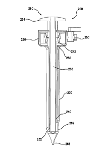
performs a mini-laparotomy through the abdominal layers into the abdominal
cavity, through
which a trocar is inserted and the abdomen insufflated. The Hassan technique
is a cut-down
technique that results in larger abdominal defects and increased patient
scarring. The technique is
also difficult to perform on obese patients with very thick abdominal walls.
CA 02714889 2010-07-08
WO 2009/094644 PCT/US2009/032026
2
[0006] In a third technique, the surgeon places a trocar optically,
visualizing the
abdominal layers as the trocar is placed through the abdominal wall through a
laparoscope
disposed within the obturator of the trocar. The tip of the obturator can
penetrate about 2 cm
(about 0.75") into the organ bed of the abdominal cavity when placing the
cannula and
establishing pneumoperitoneum.
[0007] In a fourth technique, the abdominal layers are visualized while
the trocar
is advanced though the abdominal wall. As soon as the tip of the obturator
punctures the
peritoneum, gas is pumped through the trocar system into the abdominal cavity
through vent
holes disposed at the tip of the obturator. The fourth technique uses a vacuum
release, which
causes the organs to fall away from the abdominal wall, thereby creating a
space in the
abdominal cavity for the obturator tip. Accordingly, the abdominal cavity can
be inflated with
minimal penetration into the space. As soon as the tip of the obturator
punctures the
peritoneum, gas enters the abdominal cavity through the vent holes in the tip
of the obturator,
thereby reducing the negative pressure caused by the surgeon's lifting of the
abdominal wall,
and in turn, creating a space above the organ bed into which the trocar system
is fully inserted
into the cavity. A seal is disposed within the obturator that provides a gas
tight seal both with
and without the laparoscope in place. The vent holes at the tip of the
obturator allow moisture
and tissue to enter the obturator, however, which obscure the field of view
within the
obturator tip. Gas flowing directly past the laparoscope within the obturator
can cool the
laparoscope, thereby fogging of the lens thereof.
SUMMARY OF THE INVENTION
[0008] Systems, devices, and methods permit insufflation of a body
cavity prior to
the insertion of a carmula into the body cavity. Some embodiments of an access
system
comprise an obturator, a trocar, and a fluid flow channel. The access system
has a closed
configuration, in which a distal end of the access system is fluidly isolated
from the fluid
flow channel, and an open configuration, in which the distal end of the access
system is
fluidly connected to the fluid flow channel, thereby permitting fluid flow,
for example, an
insufflation gas into a body cavity.
[0009] Accordingly, some embodiments provide an insufflating surgical
access
system and a method for insufflating a body cavity using the insufflating
surgical access
CA 02714889 2010-07-08
WO 2009/094644 PCT/US2009/032026
3
system. Some embodiments of the insufflating surgical access system comprise:
a trocar
comprising: a proximal end and a distal end; a trocar seal assembly disposed
at the proximal
end of the trocar, the trocar seal assembly comprising an instrument seal; an
elongate cannula
disposed at the distal end of the trocar, the cannula comprising a tubular
wall defining a
lumen, an open proximal end, and an open distal end; an access channel
defining a
longitudinal axis, extending through the trocar seal assembly and the lumen of
the cannula,
from the proximal end of the trocar to the distal end of the trocar; a fluid
port disposed at the
proximal end of the trocar; and a fluid flow seal disposed in the access
channel; an obturator
comprising: an elongate body comprising a proximal end and a distal end; a
tissue
penetrating tip disposed at the distal end; and a handle disposed at the
proximal end, wherein
the obturator is slidably insertable into the proximal end of the access
channel, and the tip of
the obturator extends out of the distal, open end of the cannula when fully
inserted
therethrough; and a fluid flow channel fluidly connected to the fluid port of
the trocar, and
extending to a distal end of the insufflating access system. The obturator in
the access
channel has a closed position, in which the body of the obturator sealing
contacts the fluid
flow seal, thereby preventing gas flow through the fluid flow channel, and an
open position,
in which the body of the obturator does not sealing contact the fluid flow
seal, thereby
allowing fluid flow through the fluid flow channel.
[0010] In some embodiments, the trocar seal assembly further comprises
a zero
seal.
[0011] In some embodiments, the distal end of the cannula comprises an
angled
tip.
[0012] In some embodiments, the fluid port is disposed on the trocar
seal
assembly.
[0013] In some embodiments, the fluid flow seal is integrated with a
cannula tip
disposed at the distal end of the cannula. In some embodiments, the fluid flow
seal is
disposed proximal of the distal end of the cannula. In some embodiments, the
fluid flow seal
is substantially perpendicular to the longitudinal axis of the axis channel.
In some
embodiments, the fluid flow seal is not perpendicular to the longitudinal axis
of the axis
channel.
CA 02714889 2010-07-08
WO 2009/094644 PCT/US2009/032026
4
[0014] In some embodiments, the obturator further comprises an
instrument well
open at a proximal end of the obturator, extending longitudinally through the
body of the
obturator, terminating at the tip of the obturator, and dimensioned to receive
a laparoscope
therein, wherein at least a portion of the tip of the obturator is
transparent. Some
embodiments further comprise a laparoscope.
[0015] In some embodiments, the fluid flow channel comprises a space
defined
by the lumen of the cannula and the body of the obturator. In some
embodiments, the fluid
flow channel comprises an instrument well disposed in the body of the
obturator. In some
embodiments, the fluid flow channel comprises at least one proximal opening
and at least one
distal opening disposed in the body of the obturator. In some embodiments, the
fluid flow
channel comprises a slot disposed in the body of the obturator.
[0016] In some embodiments, in the closed position, the obturator is
displaced
distally in the access channel compared with the open position. In some
embodiments, in the
closed position, the obturator is displaced proximally in the access channel
compared with
the open position. In some embodiments, in the closed position, the obturator
is rotated in the
access channel compared with the open position.
[0017] Some embodiments of the method for insufflating a body cavity
comprise:
positioning the obturator in the closed position; fluidly connecting the fluid
port with a source
of insufflation gas; positioning the tissue penetrating tip at a desired
position; advancing the
tissue penetrating tip through a body wall until the tip enters a body cavity;
and positioning
the obturator in the open position, fluidly connecting the fluid port with the
body cavity
through the fluid flow channel, thereby insufflating the body cavity.
[0018] Some embodiments further comprise visually monitoring the
position of
the penetrating tip through a laparoscope.
[0019] Some embodiments provide an insufflating surgical access system
comprising: a trocar comprising: a proximal end and a distal end; a trocar
seal assembly
disposed at the proximal end of the trocar, the trocar seal assembly
comprising an instrument
seal; an elongate cannula disposed at the distal end of the trocar, the
cannula comprising a
tubular wall defining a lumen, an open proximal end, and an open distal end;
an access
channel defining a longitudinal axis, extending through the trocar seal
assembly and the
CA 02714889 2010-07-08
WO 2009/094644 PCT/US2009/032026
lumen of the cannula, from the proximal end of the trocar to the distal end of
the trocar; a
fluid port disposed at the proximal end of the trocar; an obturator
comprising: an elongate
body comprising a proximal end and a distal end; a tissue penetrating tip
disposed at the
distal end; and a handle disposed at the proximal end, wherein the obturator
is slidably
insertable into the proximal end of the access channel, and the tip of the
obturator extends out
of the distal, open end of the cannula when fully inserted therethrough; and a
gas flow
channel fluidly connected to the fluid port of the trocar, and extending to a
distal end of the
insufflating access system; and means for modulating gas flow through the gas
flow channel.
BRIEF DESCRIPTION OF THE DRAWINGS
[0020] FIG. 1A is a perspective view of an embodiment of an
insufflating access
system. FIG. 1B is a perspective cutaway view of a trocar of the insufflating
access system
illustrated in FIG. 1A. FIG.1C is a side cutaway view of the trocar
illustrated in FIG. 1B.
FIG. 1D is a side cutaway view of the insufflating access system illustrated
in FIG. 1A in the
closed configuration. FIG. 1E is a side cross-sectional view of the
insufflating access system
illustrated in FIG. 1A in a closed configuration. FIG. if is a cross-sectional
view of the
insufflating access system illustrated in FIG. 1A in an open configuration.
[0021] FIG. 2A is a perspective view of another embodiment of an
insufflating
access system. FIG. 2B is a perspective view of an embodiment of an obturator
from the
insufflating access system illustrated in FIG. 2A. FIG. 2C is a side cutaway
view of an
embodiment of the insufflating access system illustrated in FIG. 2A in a
closed configuration.
FIG. 2D is a side cutaway view of the embodiment of the insufflating access
system
illustrated in FIG. 2C in an open configuration.
[0022] FIG. 3A is a perspective view of another embodiment of an
insufflating
access system in an open configuration. FIG. 3B is a side cross section of the
insufflating
access system illustrated in FIG. 3A in a closed configuration. FIG. 3C is a
side see-through
view of another embodiment of an insufflating access system in an open
configuration.
[0023] FIG. 4A is a side cross section of another embodiment of an
insufflating
access system in a closed configuration. FIG. 4B is a side cross section of
the insufflating
access system illustrated in FIG. 4A in an open configuration. FIG. 4C is a
perspective view
CA 02714889 2010-07-08
WO 2009/094644 PCT/US2009/032026
6
of an embodiment of an obturator of the insufflating access system illustrated
in FIGS. 4A
and 4B.
[0024] FIG. 5A-5C schematically illustrate an embodiment of a method
for
placing the embodiment of the access device illustrated in FIGS. 1A-1F.
DETAILED DESCRIPTION OF CERTAIN EMBODIMENTS
[0025] FIG. 1A is a perspective view of an embodiment of an
insufflating access
system 100 comprising a trocar 110 and an obturator 160 slidably insertable
into the trocar
110. The insufflating access system 100 also comprises a fluid flow channel,
which is
discussed in greater detail below. The trocar 110 and obturator 160 comprise
suitable
biologically compatible materials.
[0026] FIG. 1B is a perspective cutaway view and FIG. 1C is a partial
side cross
section of the trocar 110. The trocar 110 comprises a proximal end 112 and a
distal end 114.
A trocar seal assembly 120 is disposed at the proximal end 112 of the trocar,
and an elongate
cannula 130 extends from the trocar seal assembly 120 and is disposed at the
distal end 114
of the trocar. An access channel 116 extends through the trocar seal assembly
120 and the
cannula 130, from the proximal end 112 to the distal end 114 of the trocar.
The access
channel 116 defines a longitudinal axis. In some embodiments, the trocar seal
assembly 120
and the cannula 130 are integrated, while in other embodiments, the trocar
seal assembly 120
and the cannula 130 are separate components, and in some embodiments,
releasably coupled.
[0027] In the illustrated embodiment, the trocar seal assembly 120
comprises a
first seal 122 and a second seal 124 disposed on the access channel 116 within
a trocar seal
housing 126. The first seal 122 is an instrument seal, which forms a
substantially fluid tight
seal with an instrument extending therethrough, thereby preventing fluid from
escaping from
the proximal end 112 of the trocar. In some embodiments, the first seal 122
comprises a
septum seal. The second seal 124 is a zero seal, which forms a fluid tight
seal with no
instrument extending therethrough, preventing fluid from escaping from the
proximal end
112 of the trocar. In some embodiments, the second seal 124 comprises a
duckbill valve, a
double duckbill valve, and/or a flap valve. The second seal 124 is optional in
some
embodiments. For example, in some embodiments, the first seal 122 provides
both an
CA 02714889 2010-07-08
WO 2009/094644 PCT/US2009/032026
7
instrument seal and a zero seal, for example, a valve comprising a gel
material. Other
embodiments do not comprise a zero seal. In some embodiments, the first seal
122 and the
second seal 124 comprise an elastomer, for example rubber, synthetic rubber,
silicone,
ethylene propylene diene monomer (EPDM), ethylene-propylene copolymer (EP
rubber),
polyisoprene, polybutadiene, polyurethane, styrene-butadiene, ethylene vinyl
acetate (EVA),
polychloroprene (Neoprene ), perfluorelastomer (Kalrezt), and the like.
[0028] The cannula 130 comprises a proximal end, at which the trocar
seal
assembly 120 is disposed, and a distal end terminating in a tip 132. In the
illustrated
embodiment, the tip 132 of the cannula is angled with a beveled edge. The
angled tip 132
facilitates insertion through tissue. In other embodiments, the tip 132 is not
angled. The
cannula 130 comprises a hollow tube open at the proximal and the distal ends.
The hollow
tube defines a lumen 134, through which the access channel 116 extends. One or
more
optional vents 136 perforate the cannula 130 at or near the distal end
thereof. In the illustrated
embodiment, the cannula 130 has a generally circular cross section, although
those skilled in
the art will understand that other embodiments have other suitable cross
sections, for
example, oval, elliptical, diamond, square, polygonal, and the like.
[00291 A fluid flow seal 140 is disposed within the lumen of the
cannula 130, on
an inner wall of the hollow tube. The fluid flow seal 140 is positioned,
dimensioned, and
configured for sealing contacting the body 162 of the obturator, as discussed
in greater detail
below. In the illustrated embodiment, the fluid flow seal 140 is disposed near
the tip 132 or
distal end of the cannula. The fluid flow seal 140 is substantially normal or
perpendicular to
the longitudinal axis of the trocar 110, and consequently, is generally
circular in the
illustrated embodiment. In other embodiments, the fluid flow seal 140 is
disposed at another
location. For example, in some embodiments, the fluid flow seal 140 is
disposed at or
integrated with the tip 132 of the cannula, or spaced adjacent to or just
slightly inward from
the tip 132 at the distal-most end. In some of these embodiments in which the
tip 132 is not
perpendicular to the longitudinal axis of the trocar 110, the fluid flow seal
140 also subtends
a non-normal angle with the longitudinal axis, and consequently, is elliptical
or oval rather
than circular. In some embodiments, the fluid flow seal 140 and the tip 132
subtend about the
same angle with the longitudinal axis, while in other embodiments, the fluid
flow seal 140
CA 02714889 2010-07-08
WO 2009/094644 PCT/US2009/032026
8
and the tip 132 subtend different angles with the longitudinal axis. Some
embodiments of the
fluid flow seal 140 comprise a plurality of sub-seals, which are disposed at
about the same
location in some embodiments, and disposed in a plurality of locations in
other embodiments.
The fluid flow seal 140 comprises a suitable elastomer for example, at least
one of rubber,
synthetic rubber, silicone, ethylene propylene diene monomer (EPDM), ethylene-
propylene
copolymer (EP rubber), polyisoprene, polybutadiene, polyurethane, styrene-
butadiene,
ethylene vinyl acetate (EVA), polychloroprene (Neoprene ), perfluorelastomer
(Kalreze),
and the like.
[0030] A fluid port 150 is disposed on the housing 126 of the trocar
seal
assembly, fluidly connected with the access channel 116 distal of the first
122 and second
124 seals. The fluid port 150 comprises a stopcock in the illustrated
embodiment, and
terminates in a fitting that permits coupling to a fluid and/or suction
source, for example, a
Luer fitting. In other embodiments, the fluid port 150 has another location,
for example, on
the cannula 130 or the obturator 160. Embodiments of the fluid port 150 are
useful for
introducing and/or venting an insufflation gas, for example, carbon dioxide,
therethrough.
Other fluids are introduced and/or vented in other embodiments.
[0031] The trocar 110 is typically manufactured in a range of sizes to
accommodate instruments of different diameters, for example, up to about 5 mm,
up to about
8 mm, up to about 11 mm, up to about 12 mm, or up to about 15 mm. Embodiments
of the
trocar 110 have working cannula lengths of about 55 mm, about 75 mm, about 100
mm, or
about 150 mm.
[0032] As best seen in FIG. 1D, which is a side cutaway view of the
insufflating
access system 100, the obturator 160 comprises an elongate body 162 comprising
a proximal
end terminating in a handle 164 and a distal end terminating in a tissue
penetrating tip 166. A
diameter of the tip 166 converges from a proximal end to a distal end thereof.
The body 162
and tip 166 of obturator is slidably insertable into and removable from the
access channel 116
through the proximal end of the trocar 110. In a fully inserted configuration,
the tip 166 of the
obturator extends out of the distal end or tip 132 of the cannula 130. The
first seal 122 of the
trocar seal assembly forms an instrument seal with the body 162 of the
cannula, thereby
substantially preventing fluid leaking from the proximal end of the access
channel 116. In the
CA 02714889 2010-07-08
WO 2009/094644 PCT/US2009/032026
9
illustrated embodiment, the body 162 comprises a hollow, instrument well 168,
which is
open at the handle 162 at the proximal end of the obturator, and which extends
to the tip 166
of the obturator. The instrument well 168 is dimensioned to receive a
laparoscope through the
proximal opening thereof. When fully inserted, an end of the laparoscope
extends through the
body 162 of the obturator into or proximal to the tip 166. In the illustrated
embodiment, the
tip 166 comprises at least a transparent or windowed portion through which the
laparoscope
images tissue proximal to the tip 166, for example, for monitoring the
position of the tip 166
during the insertion of the access system 100 into a body cavity. Some
embodiments of the
tip 166 comprise markers or another type of visually enhancing or facilitating
features, which
assist in viewing the tissue and body cavity, and thus, traversal of the tip
166 through the
body. In some embodiments, the laparoscope is fully inserted in the instrument
well 168,
thereby preventing or reducing fogging thereof. Accordingly, some embodiments
comprise at
least one of a distal laparoscope seal, membrane, or lock in that provides at
least one of
holding the laparoscope in a fully or nearly fully inserted position,
preventing or reducing
fogging, and preventing or reducing other types of interference of the viewing
area of the
laparoscope.
[0033] FIG. 1E is a side cross section of the insufflating access
system 100 in a
closed configuration and FIG. 1F is a side cross section of the insufflating
access system 100
in an open configuration. The lumen 134 of the cannula and the body 162 of the
obturator
together define a fluid flow channel 180 therebetween, which extends
longitudinally in the
access channel 116. A proximal end 182 of the fluid flow channel is fluidly
connected to the
fluid port 150. A distal end 184 of the fluid flow channel extends to the
distal end of the
access system 100, which in the illustrated embodiment, comprises the tip 132
of the cannula.
In the illustrated embodiment, the lumen 134 of the cannula and the body 162
of the obturator
are both generally circular and define a fluid flow channel 180 with a
generally annular cross
section. In other embodiments, cross-sectional shapes of the lumen 134 of the
cannula and
the body 162 of the obturator are different from each other, and the cross
section of the fluid
flow channel 180 has another shape.
[0034] A size of a space or gap between the tip 166 of the obturator
and the tip
132 of the cannula is selected to prevent or reduce coring of tissue as the
access system is
CA 02714889 2010-07-08
WO 2009/094644 PCT/US2009/032026
advanced. In some embodiments, the gap between the tip 166 of the obturator
and the tip 132
of the cannula provides sufficient gas flow for insufflation, which is
discussed in greater
detail below. In some embodiments, a gap between the body 162 of the obturator
and the
lumen 134 of the cannula is not uniform longitudinally, for example, wider at
the proximal
end 112 and narrower at the distal end 116.
[0035] In the closed configuration illustrated in FIG. 1E, the tip 166
of the
obturator extends from the tip 132 of the cannula in a configuration suitable
for inserting the
access system 100 through a body wall and into a body cavity. The position of
the obturator
160 in this configuration is referred to as a closed position. In the closed
configuration, the
fluid flow seal 140 sealing contacts the body 162 of the obturator, thereby
cooperating
therewith to prevent fluid flow through the fluid flow channel 180, from the
fluid port 150,
and out through the tip 132 of the cannula. Accordingly, the fluid port 150 is
not fluidly
connected with the distal end of the fluid flow channel 184.
[0036] In the open configuration illustrated in FIG. 1F, the obturator
160 is
partially withdrawn from access channel 116, that is translated proximally
along the
longitudinal axis compared with the configuration illustrated in FIG. 1E. The
position of the
obturator 160 in this configuration is referred to as an open position. In the
illustrated
embodiment, the body 162 of the obturator is proximal of the fluid flow seal
140, and
consequently, does not make contact and form a seal therewith. Accordingly, in
the open
configuration, the distal end 184 of the fluid flow channel is fluidly
connected with the fluid
port 150. In some embodiments, the fluid flow seal 140 contacts a portion of
the body 162 of
the obturator in the open configuration, but does not sealing contact
therewith. For example,
in some embodiments, at least one of the fluid flow seal 140, and a transition
between the
body 162 and tip 166 of obturator are not normal or perpendicular to the
longitudinal axis. In
some of these embodiments, in some positions of the obturator 160 in the
access channel, the
body 162 of the obturator contacts only a portion of the fluid flow seal 140
rather than the
entire sealing surface thereof, and consequently, does not form a seal
therewith.
[0037] Some embodiments of the access system 100 comprise an indicator
of the
configuration thereof. For example, some embodiments comprise indicia on the
obturator 160
and/or the trocar 110 that indicate the position of the obturator 160 in the
open and/or closed
CA 02714889 2010-07-08
WO 2009/094644 PCT/US2009/032026
11
position. Some embodiments comprise an audio and/or visual indicator of fluid
flow through
the fluid flow channel 180 and/or fluid port 150, for example, a spinning
disk, a spinning
ball, a lamp, a whistle, and/or an alarm.
[0038] Some embodiments comprise one or more mechanical features that
indicate the state of and/or lock the access system 100 into at least one of
the open
configuration and the closed configuration, for example, detents, latches,
stops, and the like.
[0039] FIG. 2A is a perspective view of another embodiment of an
insufflating
access system 200 generally similar to the embodiment illustrated in FIGS. 1A-
1F,
comprising a trocar 210, an obturator 260 inserted in the trocar 210, and a
laparoscope 290
inserted into the obturator 260. FIG. 2B is a perspective view of an
embodiment of the
obturator 260, which is similar to the embodiment of the obturator 160
described above, and
comprises an elongate body 262, a handle 264 disposed at the proximal end, a
tip 266
disposed at the distal end, and an instrument well 268 extending
longitudinally from an
opening in the handle 264 at the proximal end of the obturator to the tip 266
at the distal end
thereof.
[0040] The obturator 260 further comprises a slot 272 that extends
longitudinally
on the body 262 thereof. As will be apparent from the description below, the
slot 272
incorporates the instrument well 268 into the fluid flow channel 280 in the
illustrated
embodiment. Because the fluid flow channel 280 comprising the instrument well
268 has a
larger cross-sectional area compared with the fluid flow channel 180 of
embodiment
illustrated in FIGS. 1A-1F, embodiments of the access system 200 exhibit
increased fluid
flow. A proximal end of the slot 272 is positioned such that the slot 272 does
not interfere
with the instrument seal between the body 262 and the trocar seal assembly 220
when the
obturator 260 is in the open position or the closed position. A distal end of
the slot 272 is
positioned such that fluid does not flow from the slot 272 in the closed
configuration, that is,
the distal end of the slot 272 is fluidly isolated from the distal end of the
access system 200.
The open configuration permits fluid flow from the distal end of the slot 272,
that is, the
distal end of the slot 272 is fluidly connected to the distal end of the
access system 200. In the
illustrated embodiment, the slot 272 perforates the body 262 of the obturator
into the
instrument well 268. Accordingly, the illustrated embodiment comprises a seal
between the
CA 02714889 2010-07-08
WO 2009/094644 PCT/US2009/032026
12
proximal end of the obturator 260 and the laparoscope 290, thereby preventing
or reducing
fluid flow therefrom. The seal is disposed on at least one of the laparoscope
290 and the
obturator 260. In some embodiments, the slot 272 does not perforate the body
262 of the
obturator, for example, comprising one or more longitudinal grooves disposed
on an outer
surface of the body 262. The seal between the proximal end of the obturator
260 and the
laparoscope 190 is optional in these embodiments. Some embodiments of the
fluid flow
channel 280 comprise one or more longitudinal grooves disposed on an inner
wall of the
cannula 230.
[0041] FIG. 2C is a side cutaway view of an embodiment of the
insufflating
access system illustrated in FIG. 2A in a closed configuration. In the closed
position, the
obturator 260 is inserted into the access channel of the trocar 210 with the
tip 266 extending
from the tip 232 of the cannula in a tissue penetrating position. The proximal
end of the slot
272 is disposed below the instrument seal of the seal assembly 220, in fluid
communication
with the fluid port 250. The distal end of the slot 272 is disposed proximal
to the fluid
channel seal 240, which seals with the body 262 of the obturator, thereby
fluidly isolating the
slot 272 from the tip 232 of the cannula and the distal end of the access
system 200.
[0042] FIG. 2D is a side cutaway view of the embodiment of the
insufflating
access system illustrated in FIG. 2C in an open configuration in which the
obturator 260 is
advanced distally in the access channel, for example, using the handle 264,
thereby advancing
the distal end of the slot 272 past the fluid channel seal 240, and thereby
releasing the seal
between the body 262 of the obturator and the fluid channel seal 240. In the
illustrated
embodiment, the slot 272 has about the same length as the cannula 230,
extending from about
the position of the fluid inlet to past a proximal portion of the angled tip
232 of the cannula,
with which the slot 272 is aligned. Accordingly, alignment of the distal end
of the slot 272
with the proximal portion of the angled tip 232 exposes the distal end of the
slot 272, thereby
providing a fluid flow channel 280 that permits insufflation gas to exit
directly out the slot
272 and into the body cavity. In some embodiments, the obturator 260 and
trocar 210 are
keyed or otherwise configured to prevent rotation therebetween, thereby
locking the slot 272
in the exposed condition when the access system 200 is in the open
configuration. Those
skilled in the art will understand that some embodiments in which the slot 272
is rotated
CA 02714889 2010-07-08
WO 2009/094644 PCT/US2009/032026
13
relative to the configuration illustrated in FIG. 2D such that the distal end
of the slot 272 and
the proximal portion of the angled tip 232 are not aligned, also permit fluid
flow
therethrough, but at reduced flow rates.
[0043] In other embodiments, the access system 200 has an open
configuration, as
illustrated in FIG. 2A, with a fluid flow seal 240 disposed in the cannula 230
at or near the
angled tip 232 thereof. As discussed above, in some of these embodiments, the
fluid flow seal
240 subtends the same or a similar angle as the tip 232. Rotating the
obturator 260 positions
the entirety of the slot 272 within the cannula 230, proximal of the fluid
flow seal 240,
thereby converts the open configuration to the closed configuration in which
the distal end of
the slot 272 is fluidly isolated from the distal end of the access system 200,
and preventing
fluid flow therefrom. In some of these embodiments, rotation of the handle 264
of the
obturator is restricted such that the device 200 is in the open configuration
at a first limit of
the rotation and in a closed configuration at a second limit thereof.
[0044] FIG. 3A is a perspective view of another embodiment of an
insufflating
access system 300 in an open configuration. FIG. 3B is a side cross section of
the insufflating
access system 300 illustrated in FIG. 3A in a closed configuration. The
embodiment of the
access system 300 illustrated in FIGS. 3A and 3B is similar to the embodiments
described
above. Like the embodiment illustrated in FIGS. 2A-2D, a gas flow channel in
the illustrated
embodiment incorporates an instrument well, thereby increasing the cross
sectional area
thereof The access system 300 comprises a trocar 310 and an obturator 360.
[0045] As best seen in FIG. 3B, the obturator 360 comprises at least
one proximal
opening 372 and at least one distal opening 374, both of which perforate the
body 362 of the
obturator into the instrument well 368. In the illustrated embodiment, the at
least one
proximal opening 372 and the at least one distal opening 374 are both
generally circular or
oval, but in other embodiments, independently have other suitable shapes.
[0046] A fluid flow seal 340 is disposed at or integrated with the
cannula tip 332
in the illustrated embodiment, as described above. Some embodiments of the
trocar 310
further comprise a second fluid flow seal, either in addition to or instead of
the fluid flow seal
340. Some embodiments of the second fluid flow seal comprise a tubular member,
disposed
in the seal assembly 320, through which the obturator extends, wherein the
tubular member
CA 02714889 2010-07-08
WO 2009/094644 PCT/US2009/032026
14
comprises at least one opening that is aligned with the at least one proximal
opening 372
when the obturator is in an open position, thereby permitting fluid flow
therethrough. The at
least one opening in the tubular member is not aligned with the at least one
proximal opening
372 when the obturator is in a closed position, thereby preventing fluid flow
therethrough.
[0047] In the illustrated embodiment, the access system 300 is
converted from the
open configuration illustrated in FIG. 3A to the closed configuration
illustrated in FIG. 3B by
rotating the obturator 360, for example, using the handle 362. In the
illustrated embodiment,
the obturator 360 is rotated about 1800 between the views illustrated in FIGS.
3A and 3B,
although those skilled in the art will understand that other rotational angles
are used in other
embodiments. The particular rotational angle depends on factors including the
size and shape
of the distal opening 374, the location of the distal opening 374, the
location of the fluid flow
seal 340, the angle of the fluid flow seal 340. In the illustrated embodiment,
fluid flows from
the fluid port 350, into the proximal opening 372, into the instrument well
368, and out of the
distal opening 374. In the illustrated embodiment, the distal opening 374 is
exposed in the
open configuration. In the closed configuration, the distal opening 374 is
positioned proximal
of the fluid flow seal 340, which forms a seal with a portion of the body 362
of the obturator
distal of the distal opening 374, thereby preventing fluid flow therefrom.
[0048] FIG. 3C is a see-through view of another embodiment of an
insufflating
access system 300 in an open configuration. In the illustrated embodiment, the
trocar 310 is
similar to the embodiment illustrated in FIGS. 1A-1F in which the fluid flow
seal 340 is
disposed in the lumen 334 of the cannula proximal to the tip 332. In the
illustrated
embodiment, the access system 300 is converted from the illustrated open
configuration to a
closed configuration by withdrawing the obturator longitudinally toward the
proximal end,
thereby positioning the distal opening 374 proximal of the fluid flow seal
340. The fluid flow
seal 340 seals with a portion of the body 362 of the obturator distal of the
distal opening 374,
thereby preventing fluid flow therefrom.
[0049] FIG. 4A is a side cross section of another embodiment of an
insufflating
access system 400 in a closed configuration. FIG. 4B is a side cross section
of the insufflating
access system 400 illustrated in FIG. 4A in an open configuration. The
insufflating access
system 400 is generally similar to the insufflating access systems described
above, and
CA 02714889 2010-07-08
WO 2009/094644 PCT/US2009/032026
comprises a trocar 410 and an obturator 460. In the illustrated embodiment,
the trocar 410 is
generally similar to the embodiment illustrated in FIGS. 1A-1F and described
above. The
trocar 410 comprises a fluid flow seal 440 disposed in the lumen 434 of a
cannula 430
thereof, proximal of the tip 432 of the cannula. In the illustrated
embodiment, the fluid flow
seal 440 is generally perpendicular to a longitudinal axis of the trocar 310.
[0050] As best seen in FIG. 4C, which is a perspective view of an
embodiment of
an obturator 460, the obturator 460 comprises a plurality of openings 472
disposed
longitudinally and circumferentially on the body 462 of the obturator, which
extend into the
instrument well 468. The illustrated embodiment comprises a plurality of
proximal openings
472a, a plurality of distal openings 472b, and a plurality of optional
intermediate openings
472c.
[0051] In converting the access system 400 from the closed
configuration
illustrated in FIG. 1A to the open configuration illustrated in FIG. 4B, the
obturator 460 is
translated proximally along the longitudinal axis. In the closed configuration
illustrated in
FIG. 4A, the fluid flow seal 440 seals with a portion of the body 462 of the
obturator distal of
the distal openings 472b, thereby preventing fluid flow out of the tip 432 of
the cannula at the
distal end of access system 400. In the open configuration illustrated in FIG.
4B, because the
body 462 of the obturator is proximal of the fluid flow seal 440, the body 462
and fluid flow
seal 440 do not cooperate in forming a seal in the fluid flow channel 480.
Accordingly, fluid
flow is possible from the fluid port 450 into and through the fluid flow
channel 480. In the
illustrated embodiment, the fluid flow channel 480 comprises both the
instrument well 468
and a space between the lumen 434 of the cannula and the body 462 of the
obturator. As best
seen in FIG. 4B, in the illustrated embodiment, the proximal openings 472a are
disposed
proximal to the cannula 430 within the trocar seal assembly 420, thereby
increasing a cross
sectional area around the proximal openings 472a and increasing fluid flow
therethrough.
Fluid continues flowing longitudinally towards the distal end of the access
system 400
through both the instrument well 468 and the space between the lumen 434 of
the cannula
and the body 462 of the obturator. At the distal end of the obturator 460,
fluid exits the
instrument well 468 through the distal openings 472c and continues distally in
the space
CA 02714889 2010-07-08
WO 2009/094644 PCT/US2009/032026
16
between the lumen 434 of the cannula and the body 462 of the obturator. The
fluid exits the
access system 400 through the tip 432.
[0052] Although embodiments of the insufflating access system are
applicable to
any endoscopic application using insufflation, a prototypical application is
in laparoscopic
procedures. Consequently, for purposes of illustration only, the following
describes an
embodiment of a method for inserting an endoscopic port or trocar of an
insufflating access
system, and establishing pneumoperitoneum in laparoscopic surgery with
reference to the
embodiment illustrated in FIGS. 1A-1F. Those skilled in the art will
understand that the
method is also applicable to other embodiments of the access system disclosed
herein.
[0053] The obturator 160 is inserted into the trocar 110 and positioned
in the
closed configuration illustrated in FIG. 1E. A laparoscope is inserted into
the instrument well
168 of the obturator and the laparoscope coupled with an imaging system, for
example, a
camera and a video monitor. The fluid port 150 is fluidly coupled to a source
of pressurized
insufflation gas.
[0054] The user positions the tissue penetrating tip 166 in an incision
made at a
desired location on the patient's abdomen 500 and advances the insufflating
access system
100 through the abdominal wall 502 as illustrated schematically in FIG. 5A.
The user
monitors the position of the tip 166 through the laparoscope and imaging
system. When the
user observes the tip 166 penetrating the peritoneum 504, as illustrated in
FIG. 5B, the user
converts the access system 100 to the open configuration. In the illustrated
embodiment, the
user urges the obturator 160 proximally, for example, pulling on the handle
162 to convert
the access system 100 to the open configuration illustrated in FIG. 1F. As
discussed above, in
some embodiments, the obturator 160 and/or trocar 110 comprise one or more
marks or
indicia that indicate the position of the obturator 160 in the open position.
With the access
system 100 in the open configuration, insufflation gas flows from the source
of insufflation
gas, into the fluid port 150, into the proximal end 182 of the fluid flow
channel,
longitudinally through the fluid flow channel 180, past the fluid flow seal
140, past the
partially withdrawn tip 166 of the obturator, and out the tip 132 of the
cannula. Some gas
may also flow out of the vents 136, particularly if the tip 132 of the cannula
is fully or
partially blocked. The gas flows through the opening in the peritoneum into
the abdominal
CA 02714889 2010-07-08
WO 2009/094644 PCT/US2009/032026
17
cavity, thereby insufflating the abdominal cavity 506 and establishing
pneumoperitoneum as
illustrated in FIG. 5C.
[0055] Accordingly, embodiments of the access system 100 and method
provide
an accurate and simple method for gaining access to the abdominal cavity for
laparoscopic
surgery. In other embodiments, the insufflating access system 100 provides
access to the
abdominal cavity through another surface adjacent to the peritoneal lining of
the abdominal
cavity, for example, the cul-de-sac of the vagina, any point along the gastro-
intestinal tract
from the diaphragm to the anus, or one of the great vessels such as the
abdominal aorta or
vena cava. Embodiments of the access system 100 and method provide access to
other
internal structures, for example, the kidney, the stomach, and/or the third
ventricle of the
brain, or any hollow organ for which accurate and shallow entry and the
subsequent flow of
gas or liquid is desired.
[0056] Disposing a fluid flow seal 140 between the obturator 160 and
the distal
end 114 of the cannula permits the device 100 to be fluidly coupled with a
source of CO2
while the device 100 is advanced through the abdominal wall 502. As soon as
the peritoneum
504 is punctured by the tip 166 of the obturator, the surgeon stops forward
movement of the
device 100 and dislocates the fluid flow seal 140 between the obturator 160
and the cannula
130, thereby allowing the gas to flow through the gas flow channel 180 and out
of the device
100. The gas, following a path of least resistance, flows between the tip 166
and the
abdominal wall 502, through the opening made in the peritoneum 504, and
finally into the
abdominal cavity 504. Consequently, pneumoperitoneum is established with
reduced
penetration into the organ bed because the tip 166 of the obturator does not
extend as far
beyond the peritoneum as in embodiments in which the tip comprises vent holes
through
which gas insufflates the abdominal cavity. Embodiments of the device 100 also
do not
comprise at least one of: seals within the obturator and gas channels around
the laparoscope.
Some embodiments eliminate or reduce the possibility of fluid and/or tissue
entering the
visual field within the obturator tip 166. In some embodiments, the peritoneum
504 is
punctured and the abdominal cavity 504 insufflated without further penetration
of the tip 166
beyond the peritoneum 504 and into the abdominal cavity 506 or organ bed.
CA 02714889 2015-07-14
18
[0057]
While certain embodiments have been particularly shown and described with
reference to exemplary embodiments thereof, it will be understood by those of
ordinary skill in
the art that various changes in form and details may be made therein without
departing from the
scope thereof as defined by the following claims.