Note: Descriptions are shown in the official language in which they were submitted.
CA 02715254 2010-08-05
WO 2009/100454 PCT/US2009/033596
COMPOSITIONS AND METHODS FOR DISTRACTION OSTEOGENESIS
CROSS REFERENCE TO RELATED APPLICATIONS
[0001] This application claims the benefit under 35 U.S.C. 119(e) of U.S.
Provisional
Application Serial No. 61/026,934, filed February 7, 2008, the contents of
which are
incorporated herein by reference in their entirety.
FIELD OF THE INVENTION
[0002] The present invention relates to compositions and methods for use in
osteodistraction
procedures.
BACKGROUND OF THE INVENTION
[0003] Osteodistraction or distraction osteogenesis is the process by which
the slow,
incremental distraction of fracture callus is used to stimulate and prolong
active bone formation,
thereby providing a means of bridging what would otherwise be a large bony
defect. Distraction
osteogenesis is used in the reconstruction of skeletal deformities and the
lengthening of bones,
such as in the treatment of pediatric limb length inequality.
[0004] In osteodistraction procedures, the bone is surgically split in two
segments, and the two
ends of the bone are gradually moved apart (distraction phase). The rate at
which the two bone
segments are moved apart is slow enough so that new bone can form in the gap.
When the
desired length has been reached, a consolidation phase follows.
[0005] Whether in the long bones or in the craniofacial skeleton, distraction
osteogenesis takes
place primarily through intramembranous ossification. Histologic studies have
identified 4
stages that result in the eventual formation of mature bone.
[0006] Stage I: The intervening gap initially is composed of fibrous tissue
(longitudinally
oriented collagen with spindle-shaped fibroblasts within a mesenchymal matrix
of
undifferentiated cells).
-1-
CA 02715254 2010-08-05
WO 2009/100454 PCT/US2009/033596
[0007] Stage II: Slender trabeculae of bone are observed extending from the
bony edges.
Early bone formation advances along collagen fibers with osteoblasts on the
surface of these
early bony spicules laying down bone matrix. Histochemically, significantly
increased levels of
alkaline phosphatase, pyruvic acid, and lactic acid are noted.
[0008] Stage III: Remodeling begins with advancing zones of bone apposition
and resorption
and an increase in the number of osteoclasts.
[0009] Stage IV: Early compact cortical bone is formed adjacent to the mature
bone of the
sectioned bone ends, with increasingly less longitudinally oriented bony
spicules; this resembles
the normal architecture.
[0010] Bone remodeling begins during the consolidation phase and continues
over 1-2 years,
eventually transforming the regenerated bone into a mature osseous structure
similar in size and
shape to the adjacent bone. Although the volume of new bone is comparable to
that of adjacent
bones, animal studies show that mineral content and radiodensity is
approximately 30% less, as
is the tensile strength of the regenerated segment.
[0011] Moreover, delayed consolidation following distraction is a troubling
complication.
When delayed consolidation occurs, removal of bone fixators is postponed and
the risk of other
complications, such as infection, is increased.
[0012] In view of the problems associated with consolidation and the resulting
new bone
structure, it would be desirable to provide alternative osteogenic
regeneration systems for use in
osteodistraction procedures. It would additionally be desirable to provide
methods of using
alternative osteogenic regeneration systems in osteodistraction procedures.
SUMMARY OF THE INVENTION
[0013] The present invention provides compositions and methods for stimulating
osteogenesis
during and/or following bone distraction. The present compositions facilitate
and, in some
embodiments, accelerate the bone consolidation phase following bone
distraction.
[0014] In one aspect, a composition of the present invention for stimulating
osteogenesis
during and/or following bone distraction comprises a solution comprising
platelet derived growth
-2-
CA 02715254 2010-08-05
WO 2009/100454 PCT/US2009/033596
factor (PDGF) and a biocompatible matrix, wherein the solution is disposed in
the biocompatible
matrix. In some embodiments, PDGF is present in the solution in a
concentration ranging from
about 0.01 mg/ml to about 10 mg/ml, from about 0.05 mg/ml to about 5 mg/ml,
from about 0.1
mg/ml to about 1.0 mg/ml, or about 0.3 mg/ml. The concentration of PDGF within
the solution
may be within any of the concentration ranges stated above.
[0015] In some embodiments of the present invention, PDGF comprises PDGF
homodimers
and heterodimers, including PDGF-AA, PDGF-BB, PDGF-AB, PDGF-CC, PDGF-DD, and
mixtures and derivatives thereof. In some embodiments, PDGF comprises PDGF-BB.
In
another embodiment PDGF comprises a recombinant human (rh) PDGF such as
recombinant
human PDGF-BB (rhPDGF-BB).
[0016] In some embodiments of the present invention, PDGF comprises PDGF
fragments. In
some embodiments rhPDGF-B comprises the following fragments: amino acid
sequences 1-3 1,
1-32, 33-108, 33-109, and/or 1-108 of the entire B chain. The complete amino
acid sequence (1-
109) of the B chain of PDGF is provided in Figure 15 of U.S. Patent No.
5,516,896. It is to be
understood that the rhPDGF compositions of the present invention may comprise
a combination
of intact rhPDGF-B (1-109) and fragments thereof. Other fragments of PDGF may
be employed
such as those disclosed in U.S. Patent No. 5,516,896. In accordance with a
embodiment, the
rhPDGF-BB comprises at least 50% of intact rhPDGF-B (1-109).
[0017] A biocompatible matrix, according to some embodiments of the present
invention,
comprises a bone scaffolding material. In some embodiments, a bone scaffolding
material
comprises calcium phosphate. Calcium phosphate, in some embodiments, comprises
J3-
tricalcium phosphate. In some embodiments, a bone scaffolding material
comprises collagen or
other biocompatible polymeric materials.
[0018] In another aspect, the present invention provides a composition for
stimulating
osteogenesis during and/or following bone distraction comprising a PDGF
solution disposed in a
biocompatible matrix, wherein the biocompatible matrix comprises a bone
scaffolding material
and a biocompatible binder. The PDGF solution may have a concentration of PDGF
as
described above. A bone scaffolding material, in some embodiments, comprises a
calcium
phosphate. In some embodiments, a calcium phosphate comprises a (3-tricalcium
phosphate.
-3-
CA 02715254 2010-08-05
WO 2009/100454 PCT/US2009/033596
Moreover, in some embodiments, a biocompatible binder comprises a material
operable to
promote adhesion between combined substances. A biocompatible binder, for
example, can
promote adhesion between particles of a scaffolding material in the formation
of a biocompatible
matrix. In some embodiments, for example, a biocompatible binder comprising
collagen can
promote adhesion between (3-TCP particles of a scaffolding material.
[0019] In some embodiments, biocompatible matrices include calcium phosphate
particles
with or without biocompatible binders or bone allograft such as demineralized
freeze-dried bone
allograft (DFDBA), mineralized freeze-dried bone allograft (FDBA), or
particulate
demineralized bone matrix (DBM). In another embodiment, biocompatible matrices
comprise
bone allograft such as DFDBA, DBM, or other bone allograft materials including
cortical bone
shapes, such as blocks, wedges, cylinders, or particles, or cancellous bone
particles of various
shapes and sizes.
[0020] Moreover, a biocompatible binder, according to some embodiments of the
present
invention, comprises proteins, polysaccharides, nucleic acids, carbohydrates,
synthetic polymers,
or mixtures thereof. In some embodiments, a biocompatible binder comprises
collagen. In
another embodiment, a biocompatible binder comprises collagen, such as bovine
collagen or
human collagen.
[0021] In some embodiments of the present invention, compositions for
stimulating
osteogenesis during and/or following bone distraction further comprise at
least one contrast
agent. Contrast agents, according to some embodiments of the present
invention, are substances
operable to at least partially provide differentiation of two or more bodily
tissues when imaged
and can assist in placement of compositions described herein in sites of
distraction. Contrast
agents, according to some embodiments, comprise cationic contrast agents,
anionic contrast
agents, nonionic contrast agents, or mixtures thereof. In some embodiments,
contrast agents
comprise radiopaque contrast agents. Radiopaque contrast agents, in some
embodiments,
comprise iodo-compounds including (S)-N,N'-bis[2-hydroxy-1- (hydroxymethyl) -
ethyl] -2,4,6-
triiodo-5-lactamido isophthalamide (lopamidol) and derivatives thereof.
[0022] The compositions of the invention may be for use in treating a bone
during an
osteodistraction procedure and/or for use in the manufacture of a medicament
useful in treating a
-4-
CA 02715254 2010-08-05
WO 2009/100454 PCT/US2009/033596
bone during an osteodistraction procedure. It is to be understood that the use
of the composition
may involve any of the compositions and/or methods as described herein. In one
embodiment of
the invention is the use of a composition comprising a PDGF solution and a
biocompatible
matrix, wherein the solution is disposed in the biocompatible matrix, in the
preparation of a
medicament useful for stimulating osteogenesis in an osteodistraction
procedure. In one
embodiment of the invention is the use of a composition comprising a PDGF
solution and a
biocompatible matrix, wherein the solution is disposed in the biocompatible
matrix, in the
preparation of a medicament useful for accelerating bone consolidation in an
osteodistraction
procedure.
[0023] In another aspect, the present invention provides a kit comprising a
biocompatible
matrix in a first package and a solution comprising PDGF in a second package.
In some
embodiments, the solution comprises a predetermined concentration of PDGF. The
concentration of PDGF can be predetermined according to the nature of the
osteodistraction
procedure being performed. Moreover, the amount of biocompatible matrix
provided by a kit
can be dependent on the nature or classification of the osteodistraction
procedure being
performed. A syringe can facilitate disposition of the PDGF solution in the
biocompatible
matrix for application at a surgical site, such as a site of bone distraction.
[0024] The present invention additionally provides methods for producing
compositions for
promoting osteogenesis. In some embodiments, a method for producing a
composition
comprises providing a solution comprising PDGF, providing a biocompatible
matrix, and
disposing the solution in the biocompatible matrix.
[0025] The present invention additionally provides methods of treating a bone
during an
osteodistraction procedure, and methods of promoting and/or accelerating
osteogenesis during
and/or following bone distraction.
[0026] In some embodiments, a method for stimulating and/or accelerating
osteogenesis
comprises providing a composition comprising a PDGF solution disposed in a
biocompatible
matrix and applying an effective amount of the composition to at least one
site of bone
distraction. In some embodiments, the composition comprising a PDGF solution
disposed in a
biocompatible matrix is applied during bone distraction. When applied during
the bone
-5-
CA 02715254 2010-08-05
WO 2009/100454 PCT/US2009/033596
distraction, the composition may be applied to the site one or more (e.g. two,
three, four, five,
six, seven, eight, nine, ten or more) times during bone distraction. In other
embodiments, the
composition is applied after bone distraction. When applied after bone
distraction, the
composition may be applied to the site one or more (e.g. two, three, four,
five, six, seven, eight,
nine, ten or more) times after bone distraction. In some embodiments, an
effective amount of the
composition is applied during and after bone distraction.
[0027] In some embodiments, a method of stimulating osteogenesis comprises:
applying an
effective amount of a composition comprising a platelet-derived growth factor
(PDGF) solution
disposed in a biocompatible matrix to at least one site of bone distraction.
In some embodiments,
the composition is applied during the distraction phase of the
osteodistraction procedure. In some
embodiments, the composition is applied during the consolidation phase of the
osteodistraction
procedure. In some embodiments, the composition is applied during the
distraction and
consolidation phases of the osteodistraction procedure. In some embodiments,
the method
comprises accelerating bone consolidation following bone distraction. In some
embodiments, the
composition is applied to the site at least twice. In some embodiments, the
biocompatible matrix
comprises a porous calcium phosphate. In some embodiments, the calcium
phosphate comprises
(3-TCP. In some embodiments, the calcium phosphate has interconnected pores.
In some
embodiments, the calcium phosphate has a porosity greater than about 40%. In
some
embodiments, the calcium phosphate consists of particles in a range of about
100 microns to
about 5000 microns in size. In some embodiments, the calcium phosphate
consists of particles in
a range of about 100 microns to about 300 microns in size. In some
embodiments, the
biocompatible matrix is resorbable such that at least about 80% of the calcium
phosphate is
resorbed within about one year of being implanted. In some embodiments, the
calcium phosphate
is capable of absorbing an amount of the PDGF solution that is equal to at
least about 25% of the
weight of the calcium phosphate. In some embodiments, the biocompatible matrix
comprises
collagen. In some embodiments, the PDGF is present in the solution at a
concentration of about
0.1 mg/ml to about 1.0 mg/ml. In some embodiments, the PDGF is present in the
solution at a
concentration of about 0.3 mg/ml. In some embodiments, the solution comprises
a buffer. In
some embodiments, the biocompatible matrix has a porosity that facilitates
cell migration into
-6-
CA 02715254 2010-08-05
WO 2009/100454 PCT/US2009/033596
the composition. In some embodiments, the biocompatible matrix comprises
collagen and (3-TCP
in a ratio of about 20:80. In some embodiments, the composition is injectable.
[0028] In a further embodiment, a method for stimulating osteogenesis
comprises accelerating
bone consolidation following bone distraction, wherein accelerating comprises
providing a
composition comprising a PDGF solution disposed in a biocompatible matrix and
applying the
composition to a site of bone distraction.
[0029] The present invention also provides methods of accelerating bone union
following bone
distraction. In some embodiments, a method for accelerating bone union
following bone
distraction comprises providing a composition comprising a PDGF solution
disposed in a
biocompatible matrix and applying an effective amount of the composition to at
least one site of
bone distraction.
[0030] Moreover, the present invention provides methods of performing
osteodistraction
procedures. In some embodiments, a method of performing an osteodistraction
procedure
comprises (a) partitioning a bone into a first bone segment and a second bone
segment, (b)
moving at least one of the first and second bone segments to produce a space
between the first
and second bone segments, and (c) stimulating osteogenesis in the space,
wherein stimulating
osteogenesis comprises providing a composition comprising a PDGF solution
disposed in a
biocompatible matrix and at least partially applying an effective amount of
the composition in
the space. In some embodiments, steps (b) and (c) can be repeated as many
times as necessary to
lengthen the bone any desired amount.
[0031] In some embodiments, a method of performing an osteodistraction
procedure
comprises: (a) partitioning a bone into a first bone segment and a second bone
segment; (b)
moving at least one of the first and second bone segments to form a space
between the first and
second bone segments; and (c) stimulating osteogenesis in the space, wherein
stimulating
osteogenesis comprises applying an effective amount of a composition
comprising a PDGF
solution disposed in a biocompatible matrix to the space. In some embodiments,
the method
further comprises repeating steps (b) and (c) a number of times necessary to
lengthen the bone a
desired amount.
-7-
CA 02715254 2010-08-05
WO 2009/100454 PCT/US2009/033596
[0032] In various embodiments, the bone may be lengthened a total of at least
about 1 mm, at
least about 2 mm, at least about 3 mm, at least about 4 mm, at least about 5
mm, at least about 6
mm, at least about 8 mm, at least about 10 mm, at least about 12 mm, at least
about 15 mm, at
least about 20 mm, at least about 25 mm, at least about 30 mm, at least about
35 mm, at least
about 50 mm, at least about 75 mm, at least about 100 mm, at least about 125
mm, at least about
150 mm, at least about 175 mm, at least about 200 mm. In various embodiments,
the first and
second bone segments are separated by at least about 0.1 mm, at least about
0.2 mm, at least
about 0.3 mm, at least about 0.4 mm, at least about 0.5 mm, at least about 0.6
mm, at least about
0.7 mm, at least about 0.8 mm, at least about 0.9 mm, at least about 1.0 mm
per distraction step
(e.g. during step (b) above). In some embodiments, the first and second bone
segments are
separated by about 0.5 mm to about 1.5 mm per distraction step. In some
embodiments, the first
and second bone segments are separated by about 0.8 mm to about 1.2 mm per
distraction step.
In some embodiments, the first and second bone segments are separated by about
1 mm per
distraction step. In various embodiments, at least 1, at least 2, at least 3,
at least 4, at least 5, at
least 10, at least 15, at least 20, at least 25 distraction steps may be
performed.
[0033] In some embodiments of methods of the present invention, applying a
composition
comprising a PDGF solution disposed in a biocompatible matrix comprises
injecting the
composition in a site of bone distraction. In some embodiments, injecting
comprises
percutaneous injection of the composition in the distraction site. In another
embodiment, the
composition is injected into an open or surgically exposed site of bone
distraction. In a further
embodiment, applying the composition comprises disposing the composition in a
site of bone
distraction with a spatula or other device.
[0034] In some embodiments of the methods of the invention, the composition is
applied to the
distraction site once. In various embodiments, the composition is applied to
the distraction site at
least twice, at least three times, at least four times, at least five times,
at least six times, at least
eight times, at least ten times during the distraction and/or consolidation
phases. In various
embodiments, the composition may be administered to the distraction site more
than once daily,
daily, every other day, every third day, every fourth day, every fifth day,
every six day, every
week, or less than once per week during the distraction and/or consolidation
phases.
-8-
CA 02715254 2010-08-05
WO 2009/100454 PCT/US2009/033596
[0035] In some embodiments of methods of the present invention, the
biocompatible matrix
comprises a bone scaffolding material. In some embodiments, the biocompatible
matrix
comprises a bone scaffolding material and a biocompatible binder.
[0036] As provided herein, in some embodiments, a composition of the present
invention is
applied to at least one site of bone distraction during the distraction phase
of an osteodistraction
procedure. In other embodiments, a composition of the present invention is
applied to at least
one site of bone distraction during the consolidation phase following bone
distraction. In a
further embodiment, a composition of the present invention is applied to at
least one site of bone
distraction during the distraction and consolidation phases.
[0037] As provided herein, osteodistraction procedures, according to some
embodiments of the
present invention, comprise those used in the treatment of bilateral
mandibular hypoplasia,
hemifacial microsomia, congenital short femur, fibular hemimelia, hemiatrophy,
achondroplasia,
neurofibromatosis, bow legs, growth plate fractures, bone defects,
craniofacial applications,
osteomyelitis, septic arthritis, and poliomyelitis. Moreover, osteodistraction
procedures,
according to some embodiments of the present invention, comprise those used in
the treatment of
various traumas. Traumas requiring osteodistraction procedures can comprise
fractures to long
bones of the body including the femur, tibia, fibula, humerous, and/or radius.
Traumas requiring
osteodistraction procedures can also include fractures to the craniofacial
bones. In some
embodiments, for example, osteodistraction procedures can be used to lengthen
bone in
preparation for use with a prosthesis.
[0038] Accordingly, it is an object of the present invention to provide
methods and
compositions comprising PDGF disposed in a biocompatible matrix useful in
facilitating and, in
some embodiments, accelerating osteogenesis at sites of bone distraction.
[0039] It is another object of the present invention to provide kits for
constructing
compositions comprising PDGF disposed in a biocompatible matrix, the
compositions being
useful in facilitating and, in some embodiments, accelerating bone
consolidation following bone
distraction.
-9-
CA 02715254 2010-08-05
WO 2009/100454 PCT/US2009/033596
[0040] These and other embodiments of the present invention are described in
greater detail in
the detailed description which follows. These and other objects, features, and
advantages of the
present invention will become apparent after review of the following detailed
description of the
disclosed embodiments and claims.
BRIEF DESCRIPTION OF THE DRAWINGS
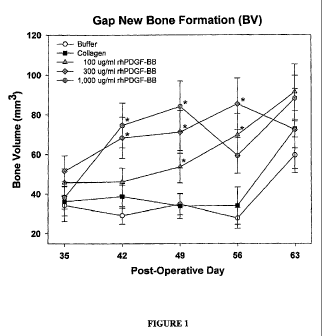
[0041] Figure 1 illustrates volume of new bone formation in a distraction
procedure as a
function of composition administered and healing time according to one
embodiment of the
present invention.
[0042] Figure 2 illustrates fraction of new bone formation in a distraction
procedure as a
function of composition administered and healing time according to one
embodiment of the
present invention.
DETAILED DESCRIPTION
[0043] The present invention provides compositions and methods for stimulating
and/or
accelerating osteogenesis during and/or following bone distraction. The
present compositions
facilitate and, in some embodiments, accelerate the bone union and the bone
consolidation phase
following bone distraction. In some embodiments, a composition comprises a
solution
comprising PDGF and a biocompatible matrix, wherein the solution is disposed
in the
biocompatible matrix. In another embodiment, a composition comprises a PDGF
solution
disposed in a biocompatible matrix, wherein the biocompatible matrix comprises
a bone
scaffolding material and a biocompatible binder.
[0044] Without wishing to be bound by theory, it is hypothesized that
distraction osteogenesis
is distinct from treating fractures, in that the bone segments are gradually
moved apart during the
distraction phase, thus re-injuring the site repeatedly, and thus keeping the
new tissue in early
stage bone healing during the distraction phase. During distraction, it is
hypothesized that the
state of the new tissue is soft, fibrous tissue callus (Phases I-III above),
with some bone
formation at the ends of the bones being distracted, and treatment of the
distraction gap during
the distraction phase of the procedure involves treating tissue during Phases
I, II, and perhaps
Phase III. Additionally, without wishing to be bound by theory, it is
hypothesized that normal
-10-
CA 02715254 2010-08-05
WO 2009/100454 PCT/US2009/033596
fracture healing occurs by endocortical ossification (which includes a
cartilage intermediate),
whereas distraction osteogenesis healing involves primarily intramembranous
ossification (no
cartilage intermediate).
[0045] Turning now to components that can be included in various embodiments
of the present
invention, compositions of the present invention comprise a solution
comprising PDGF.
PDGF Solutions
[0046] PDGF plays an important role in regulating cell growth and migration.
PDGF, as with
other growth factors, is operable to bind with the extracellular domains of
receptor tyrosine
kinases. The binding of PDGF to these transmembrane proteins activates the
kinase activity of
their catalytic domains located on the cytosolic side of the membrane. By
phosphorylating
tyrosine residues of target proteins, the kinases induce a variety of cellular
processes that include
cell growth and extracellular matrix production.
[0047] In one aspect, a composition provided by the present invention
comprises a solution
comprising platelet derived growth factor (PDGF) and a biocompatible matrix,
wherein the
solution is disposed in the biocompatible matrix. In some embodiments, PDGF is
present in the
solution in a concentration ranging from about 0.01 mg/ml to about 10 mg/ml,
from about 0.05
mg/ml to about 5 mg/ml, or from about 0.1 mg/ml to about 1.0 mg/ml. PDGF may
be present in
the solution at any concentration within these stated ranges. In various
embodiments, PDGF is
present in the solution at any one of the following concentrations: about 0.05
mg/ml; about 0.1
mg/ml; about 0.15 mg/ml; about 0.2 mg/ml; about 0.25 mg/ml; about 0.3 mg/ml;
about 0.35
mg/ml; about 0.4 mg/ml; about 0.45 mg/ml; about 0.5 mg/ml, about 0.55 mg/ml,
about 0.6
mg/ml, about 0.65 mg/ml, about 0.7 mg/ml; about 0.75 mg/ml; about 0.8 mg/ml;
about 0.85
mg/ml; about 0.9 mg/ml; about 0.95 mg/ml; about 1.0 mg/ml; or about 3.0 mg/ml.
In some
embodiments, PDGF is present in the solution in a concentration ranging from
about 0.2 mg/ml
to about 2 mg/ml, from about 0.3 mg/ml to about 3 mg/ml, from about 0.4 mg/ml
to about 4
mg/ml, from about 0.5 mg/ml to about 5 mg/ml, from about 0.25 mg/ml to about
0.5 mg/ml, or
from about 0.2 mg/ml to about 0.75 mg/ml. It is to be understood that these
concentrations are
simply examples of particular embodiments, and that the concentration of PDGF
may be within
any of the concentration ranges stated above.
-11-
CA 02715254 2010-08-05
WO 2009/100454 PCT/US2009/033596
[0048] Various amounts of PDGF may be used in the compositions of the present
invention.
Amounts of PDGF that could be used include amounts in the following ranges:
about 1 ug to
about 50 mg, about 10 ug to about 25 mg, about 100 ug to about 10 mg, and
about 250 ug to
about 5 mg.
[0049] The concentration of PDGF or other growth factors in embodiments of the
present
invention can be determined by using an enzyme-linked immunoassay as described
in U.S.
Patent Nos. 6,221,625, 5,747,273, and 5,290,708, or any other assay known in
the art for
determining PDGF concentration. When provided herein, the molar concentration
of PDGF is
determined based on the molecular weight of PDGF dimer (e.g., PDGF-BB; MW
about 25 kDa).
[0050] In embodiments of the present invention, PDGF comprises PDGF homodimers
and
heterodimers, including PDGF-AA, PDGF-BB, PDGF-AB, PDGF-CC, PDGF-DD, and
mixtures
and derivatives thereof. In some embodiments, PDGF comprises PDGF-BB. In
another
embodiment PDGF comprises a recombinant human PDGF, such as rhPDGF-BB. In some
embodiments, PDGF comprises mixtures of the various homodimers and/or
heterodimers.
Embodiments of the present invention contemplate any combination of PDGF-AA,
PDGF-BB,
PDGF-AB, PDGF-CC, and/or PDGF-DD
[0051] PDGF, in some embodiments, can be obtained from natural sources. In
other
embodiments, PDGF can be produced by recombinant DNA techniques. In other
embodiments,
PDGF or fragments thereof may be produced using peptide synthesis techniques
known to one of
ordinary skill in the art, such as solid phase peptide synthesis. When
obtained from natural
sources, PDGF can be derived from biological fluids. Biological fluids,
according to some
embodiments, can comprise any treated or untreated fluid associated with
living organisms
including blood.
[0052] Biological fluids, in another embodiment, can also comprise blood
components
including platelet concentrate (PC), apheresed platelets, platelet-rich plasma
(PRP), plasma,
serum, fresh frozen plasma (FFP), and buffy coat (BC). Biological fluids, in a
further
embodiment, can comprise platelets separated from plasma and resuspended in a
physiological
fluid.
-12-
CA 02715254 2010-08-05
WO 2009/100454 PCT/US2009/033596
[0053] When produced by recombinant DNA techniques, a DNA sequence encoding a
single
monomer (e.g., PDGF B-chain or A-chain), in some embodiments, can be inserted
into cultured
prokaryotic or eukaryotic cells for expression to subsequently produce the
homodimer (e.g.
PDGF-BB or PDGF-AA). In other embodiments, a PDGF heterodimer can be generated
by
inserting DNA sequences encoding for both monomeric units of the heterodimer
into cultured
prokaryotic or eukaryotic cells and allowing the translated monomeric units to
be processed by
the cells to produce the heterodimer (e.g. PDGF-AB). Commercially available
cGMP
recombinant PDGF-BB can be obtained commercially from Novartis Corporation
(Emeryville,
CA). Research grade rhPDGF-BB can be obtained from multiple sources including
R&D
Systems, Inc. (Minneapolis, MN), BD Biosciences (San Jose, CA), and Chemicon,
International
(Temecula, CA). In some embodiments, monomeric units can be produced in
prokaryotic cells
in a denatured form, wherein the denatured form is subsequently refolded into
an active
molecule.
[0054] In embodiments of the present invention, PDGF comprises PDGF fragments.
In some
embodiments rhPDGF-B comprises the following fragments: amino acid sequences 1-
31, 1-32,
33-108, 33-109, and/or 1-108 of the entire B chain. The complete amino acid
sequence (1-109)
of the B chain of PDGF is provided in Figure 15 of U.S. Patent No. 5,516,896.
It is to be
understood that the rhPDGF compositions of the present invention may comprise
a combination
of intact rhPDGF-B (1-109) and fragments thereof. Other fragments of PDGF may
be employed
such as those disclosed in U.S. Patent No. 5,516,896. In accordance with one
embodiment, the
rhPDGF-BB comprises at least about 60% of intact rhPDGF-B (1-109). In another
embodiment,
the rhPDGF-BB comprises at least about 65%, 75%, 80%, 85%, 90%, 95% or 99% of
intact
rhPDGF-B (1-109).
[0055] In some embodiments of the present invention, PDGF can be purified.
Purified PDGF,
as used herein, comprises compositions having greater than about 95% by weight
PDGF prior to
incorporation in solutions of the present invention. The solution may be any
pharmaceutically
acceptable solution. In other embodiments, the PDGF can be substantially
purified.
Substantially purified PDGF, as used herein, comprises compositions having
about 5% to about
95% by weight PDGF prior to incorporation into solutions of the present
invention. In some
embodiments, substantially purified PDGF comprises compositions having about
65% to about
-13-
CA 02715254 2010-08-05
WO 2009/100454 PCT/US2009/033596
95% by weight PDGF prior to incorporation into solutions of the present
invention. In other
embodiments, substantially purified PDGF comprises compositions having about
70% to about
95%, about 75% to about 95%, about 80% to about 95%, about 85% to about 95%,
or about 90%
to about 95%, by weight PDGF, prior to incorporation into solutions of the
present invention.
Purified PDGF and substantially purified PDGF may be incorporated into
scaffolds and binders.
[0056] In a further embodiment, PDGF can be partially purified. Partially
purified PDGF, as
used herein, comprises compositions having PDGF in the context of platelet
rich plasma (PRP),
fresh frozen plasma (FFP), or any other blood product that requires collection
and separation to
produce PDGF. Embodiments of the present invention contemplate that any of the
PDGF
isoforms provided herein, including homodimers and heterodimers, can be
purified or partially
purified. Compositions of the present invention containing PDGF mixtures may
contain PDGF
isoforms or PDGF fragments in partially purified proportions. Partially
purified and purified
PDGF, in some embodiments, can be prepared as described in U.S. Patent
Application Serial No.
11/159,533 (Publication No: 20060084602).
[0057] In some embodiments, solutions comprising PDGF are formed by
solubilizing PDGF in
one or more buffers. Buffers suitable for use in PDGF solutions of the present
invention can
comprise, but are not limited to, carbonates, phosphates (e.g. phosphate
buffered saline),
histidine, acetates (e.g. sodium acetate), acidic buffers such as acetic acid
and HC1, and organic
buffers such as lysine, Tris buffers (e.g. tris(hydroxymethyl)aminoethane), N-
2-
hydroxyethylpiperazine-N'-2-ethanesulfonic acid (HEPES), and 3-(N-morpholino)
propanesulfonic acid (MOPS). Buffers can be selected based on biocompatibility
with PDGF
and the buffer's ability to impede undesirable protein modification. Buffers
can additionally be
selected based on compatibility with host tissues. In some embodiments, sodium
acetate buffer
is used. The buffers may be employed at different molarities, for example
about 0.1 MM to
about 100 mM, about 1 mM to about 50 mM, about 5 mM to about 40 mM, about 10
mM to
about 30 mM, or about 15 mM to about 25 mM, or any molarity within these
ranges. In some
embodiments, an acetate buffer (e.g. sodium acetate) is employed at a molarity
of about 20 mM.
-14-
CA 02715254 2010-08-05
WO 2009/100454 PCT/US2009/033596
[0058] In another embodiment, solutions comprising PDGF are formed by
solubilizing
lyophilized PDGF in water, wherein prior to solubilization the PDGF is
lyophilized from an
appropriate buffer.
[0059] Solutions comprising PDGF, according to embodiments of the present
invention, can
have a pH ranging from about 3.0 to about 8Ø In some embodiments, a solution
comprising
PDGF has a pH ranging from about 5.0 to about 8.0, more preferably about 5.5
to about 7.0,
most preferably about 5.5 to about 6.5, or any value within these ranges. In
some embodiments,
the pH is about 6Ø The pH of solutions comprising PDGF, in some embodiments,
can be
compatible with the prolonged stability and efficacy of PDGF or any other
desired biologically
active agent. PDGF is generally more stable in an acidic environment.
Therefore, in accordance
with one embodiment the present invention comprises an acidic storage
formulation of a PDGF
solution. In accordance with this embodiment, the PDGF solution preferably has
a pH from
about 3.0 to about 7.0, and more preferably from about 4.0 to about 6.5. The
biological activity
of PDGF, however, can be optimized in a solution having a neutral pH range.
Therefore, in a
further embodiment, the present invention comprises a neutral pH formulation
of a PDGF
solution. In accordance with this embodiment, the PDGF solution preferably has
a pH from
about 5.0 to about 8.0, more preferably about 5.5 to about 7.0, most
preferably about 5.5 to about
6.5. In accordance with a method of the present invention, an acidic PDGF
solution is
reformulated to a neutral pH composition, wherein such composition is then
used to promote
bone growth at distraction sites in osteodistraction procedures. In accordance
with one
embodiment of the present invention, the PDGF utilized in the solutions is
rhPDGF-BB.
[0060] In some embodiments, the pH of the PDGF containing solution may be
altered to
optimize the binding kinetics of PDGF to a matrix substrate or linker. If
desired, as the pH of the
material equilibrates to adjacent material, the bound PDGF may become labile.
[0061] The pH of solutions comprising PDGF, in some embodiments, can be
controlled by the
buffers recited herein. Various proteins demonstrate different pH ranges in
which they are
stable. Protein stabilities are primarily reflected by isoelectric points and
charges on the proteins.
The pH range can affect the conformational structure of a protein and the
susceptibility of a
-15-
CA 02715254 2010-08-05
WO 2009/100454 PCT/US2009/033596
protein to proteolytic degradation, hydrolysis, oxidation, and other processes
that can result in
modification to the structure and/or biological activity of the protein.
[0062] In some embodiments, solutions comprising PDGF can further comprise
additional
components, such as other biologically active agents. In other embodiments,
solutions
comprising PDGF can further comprise cell culture media, other stabilizing
proteins such as
albumin, antibacterial agents, protease inhibitors [e.g.,
ethylenediaminetetraacetic acid (EDTA),
ethylene glycol-bis (beta- aminoethylether)-N,N,N',N'-tetraacetic acid (EGTA),
aprotinin, c-
aminocaproic acid (EACA), etc.] and/or other growth factors such as fibroblast
growth factors
(FGFs), epidermal growth factors (EGFs), transforming growth factors (TGFs),
keratinocyte
growth factors (KGFs), insulin-like growth factors (IGFs), hepatocyte growth
factors (HGFs),
bone morphogenetic proteins (BMPs), or other PDGFs including compositions of
PDGF-AA,
PDGF-BB, PDGF-AB, PDGF-CC and/or PDGF-DD.
[0063] In addition to solutions comprising PDGF, compositions of the present
invention also
comprise a biocompatible matrix in which to dispose the PDGF solutions and may
also comprise
a biocompatible binder either with or without a biocompatible matrix.
Biocompatible Matrix
Scaffolding Material
[0064] A biocompatible matrix, according to embodiments of the present
invention, comprises
a scaffolding material. The scaffolding material, according to embodiments of
the present
invention, provides the framework or scaffold for new tissue and/or bone
growth to occur. A
scaffolding material, in some embodiments, comprises multi-directional and
interconnected
pores of varying diameters. In some embodiments, a scaffolding material
comprises a plurality
of pockets and non-interconnected pores of various diameters in addition to
the interconnected
pores.
[0065] A scaffolding material, in some embodiments, comprises at least one
calcium
phosphate. In other embodiments, a scaffolding material can comprise a
plurality of calcium
phosphates. Calcium phosphates suitable for use as a scaffolding material, in
some embodiments
of the present invention, have a calcium to phosphorus atomic ratio ranging
from 0.5 to 2Ø In
-16-
CA 02715254 2010-08-05
WO 2009/100454 PCT/US2009/033596
some embodiments, the biocompatible matrix comprises allograft such as
demineralized freeze-
dried bone allograft (DFDBA), particulate demineralized bone matrix (DBM),
mineralized bone
matrix, or combinations thereof.
[0066] Non-limiting examples of calcium phosphates suitable for use as
scaffolding materials
comprise amorphous calcium phosphate, monocalcium phosphate monohydrate
(MCPM),
monocalcium phosphate anhydrous (MCPA), dicalcium phosphate dehydrate (DCPD),
dicalcium
phosphate anhydrous (DCPA), octacalcium phosphate (OCP), a-tricalcium
phosphate, f3-
tricalcium phosphate, hydroxyapatite (OHAp), poorly crystalline
hydroxyapatite, tetracalcium
phosphate (TTCP), heptacalcium decaphosphate, calcium metaphosphate, calcium
pyrophosphate dihydrate, carbonated calcium phosphate, calcium pyrophosphate,
hydroxyapatite, or derivatives thereof.
[0067] In some embodiments, a scaffolding material comprises a polymeric
material. A
polymeric scaffold, in some embodiments, comprises collagen, polylactic acid,
poly(L-lactide),
poly(D,L-lactide), polyglycolic acid, poly(L-lactide-co-glycolide), poly(L-
lactide-co-D,L-
lactide), polyacrylate, polymethacrylate, polymethylmethacrylate, chitosan, or
combinations or
derivatives thereof.
[0068] In some embodiments, a scaffolding material comprises porous structure.
Porous
scaffolding materials, according to some embodiments, can comprise pores
having diameters
ranging from about 1 m to about 1 mm. In some embodiments, a scaffolding
material
comprises macropores having diameters ranging from about 100 m to about 1 mm
or greater.
In another embodiment, a scaffolding material comprises mesopores having
diameters ranging
from about 10 m to about 100 m. In a further embodiment, a scaffolding
material comprises
micropores having diameters less than about 10 m. Embodiments of the present
invention
contemplate scaffolding materials comprising macropores, mesopores, and
micropores or any
combination thereof.
[0069] A porous scaffolding material, in some embodiments, has a porosity
greater than about
25% or greater than about 40%. In another embodiment, a porous scaffolding
material has a
porosity greater than about 50%, greater than about 60%, greater than about
65%, greater than
about 70%, greater than about 80%, or greater than about 85%. In a further
embodiment, a
-17-
CA 02715254 2010-08-05
WO 2009/100454 PCT/US2009/033596
porous scaffolding material has a porosity greater than about 90%. In some
embodiments, a
porous scaffolding material comprises a porosity that facilitates cell
migration into the
scaffolding material.
[0070] In some embodiments, a scaffolding material comprises a plurality of
particles.
Scaffolding particles may be mm, m, or submicron (nm) in size. Scaffolding
particles, in some
embodiments, have an average diameter ranging from about 1 m to about 5 mm.
In other
embodiments, particles have an average diameter ranging from about 1 mm to
about 2 mm, from
about 1 mm to about 3 mm, or from about 250 m to about 750 m. Scaffolding
particles, in
another embodiment, have an average diameter ranging from about 100 m to
about 300 m. In
a further embodiment, scaffolding particles have an average diameter ranging
from about 75 m
to about 300 m. In additional embodiments, scaffolding particles have an
average diameter less
than about 25 m, less than about 1 m, or less than about 1 mm. In some
embodiments,
scaffolding particles have an average diameter ranging from about 100 m to
about 5 mm or
from about 100 m to about 3 mm. In other embodiments, scaffolding particles
have an average
diameter ranging from about 250 m to about 2 mm, from about 250 m to about 1
mm, or from
about 200 m to about 3 mm. Particles may also be in the range of about 1 nm
to about 1 m,
less than about 500 nm, or less than about 250 nm.
[0071] Scaffolding materials, according to some embodiments, are moldable,
extrudable
and/or injectable. Moldable, extrudable, and/or injectable scaffolding
materials can facilitate
efficient placement of compositions of the present invention in and around
sites of bone
distraction. In some embodiments, moldable, extrudable, and/or injectable
scaffolding materials
are applied to sites of bone distraction with a spatula or equivalent device.
In some
embodiments, scaffolding materials are flowable. Flowable scaffolding
materials, in some
embodiments, can be applied to a site of bone distraction through a syringe
and needle or
cannula. In some embodiments, the flowable scaffolding materials can be
applied to a site of
bone distraction percutaneously. In other embodiments, flowable scaffolding
materials can be
applied to a surgically exposed site of bone distraction. Moreover, in some
embodiments,
scaffolding materials are provided as blocks or particles.
-18-
CA 02715254 2010-08-05
WO 2009/100454 PCT/US2009/033596
[0072] In some embodiments, scaffolding materials are bioresorbable. A
scaffolding material,
in some embodiments, can be at least about 30%, 40%, 50%, 60%, 70%, 75%, 80%,
85%, or
90% resorbed within one year subsequent to in vivo implantation. In another
embodiment, a
scaffolding material can be resorbed at least about 5%, 10%, 20%, 30%, 40%,
50%, 60%, 70%,
75%, 80%, 85% or 90% within about 1, 3, 6, 9, 12, or 18 months of in vivo
implantation. In
some embodiments, scaffolding materials are greater than 90% resorbed within
about 1, 3, 6, 9,
12, or 18 months of in vivo implantation. Bioresorbability will be dependent
on: (1) the nature of
the matrix material (i.e., its chemical make up, physical structure and size);
(2) the location
within the body in which the matrix is placed; (3) the amount of matrix
material that is used; (4)
the metabolic state of the patient (diabetic/non-diabetic, osteoporotic,
smoker, old age, steroid
use, etc.); (5) the extent and/or type of injury treated; and (6) the use of
other materials in
addition to the matrix such as other bone anabolic, catabolic and anti-
catabolic factors.
Scaffolding Comprising /3-Tricalcium Phosphate
[0073] A scaffolding material for use as a biocompatible matrix, in some
embodiments,
comprises (3-tricalcium phosphate ((3-TCP). (3-TCP, according to some
embodiments, can
comprise a porous structure having multidirectional and interconnected pores
of varying
diameters. In some embodiments, (3-TCP comprises a plurality of pockets and
non-
interconnected pores of various diameters in addition to the interconnected
pores. The porous
structure of (3-TCP, in some embodiments, comprises macropores having
diameters ranging from
about 100 m to about 1 mm or greater, mesopores having diameters ranging from
about 10 m
to about 100 m, and micropores having diameters less than about 10 m.
Macropores and
mesopores of the (3-TCP can facilitate tissue in-growth including
osteoinduction and
osteoconduction while macropores, mesopores and micropores can permit fluid
communication
and nutrient transport to support tissue and bone regrowth, throughout the R-
TCP biocompatible
matrix.
[0074] In comprising a porous structure, (3-TCP, in some embodiments, can have
a porosity
greater than about 25%, or greater than about 40%. In other embodiments, (3-
TCP can have a
porosity greater than about 50%, greater than about 60%, greater than about
65%, greater than
about 70%, greater than about 75%, greater than about 80%, or greater than
about 85%. In a
-19-
CA 02715254 2010-08-05
WO 2009/100454 PCT/US2009/033596
further embodiment, (3-TCP can have a porosity greater than about 90%. In some
embodiments,
(3-TCP can have a porosity that facilitates cell migration into the (3-TCP.
[0075] In some embodiments, a scaffolding material comprises (3-TCP particles.
(3-TCP
particles, in some embodiments, can individually demonstrate any of the pore
diameters, pore
structures, and porosities provided herein for scaffolding materials.
[0076] (3-TCP particles, in some embodiments have an average diameter ranging
from about 1
m to about 5 mm. In other embodiments, (3-TCP particles have an average
diameter ranging
from about 1 mm to about 2 mm, from about 1 mm to about 3 mm, from about 100
m to about
mm, from about 100 m to about 3 mm, from about 250 m to about 2 mm, from
about 250
m to about 750 m, from about 250 m to about 1 mm, from about 250 m to about
2 mm, or
from about 200 m to about 3 mm. In another embodiment, (3-TCP particles have
an average
diameter ranging from about 100 m to about 300 m. In some embodiments, (3-
TCP particles
have an average diameter ranging from about 75 m to about 300 m. In some
embodiments, f3-
TCP particles have an average diameter of less than about 25 m, less than
about 1 m, or less
than about 1 mm. In some embodiments, (3-TCP particles have an average
diameter ranging
from about 1 nm to about 1 m. In a further embodiment, (3-TCP particles have
an average
diameter less than about 500 nm or less than about 250 nm.
[0077] A biocompatible matrix comprising (3-TCP particles, in some
embodiments, can be
provided in a shape suitable for implantation (e.g., a sphere, a cylinder, or
a block). In other
embodiments, a (3-TCP scaffolding material is moldable, extrudable, and/or
injectable thereby
facilitating application of the matrix to sites of bone distraction. Flowable
matrices may be
applied through syringes, tubes, cannulas, or spatulas.
[0078] A (3-TCP scaffolding material, according to some embodiments, is
bioresorbable. In
some embodiments, a (3-TCP scaffolding material can be at least about 30%,
40%, 50%, 60%,
65%, 70%, 75%, 80%, or 85% resorbed about one year subsequent to in vivo
implantation. In
another embodiment, a (3-TCP scaffolding material can be greater than about
90% resorbed about
one year subsequent to in vivo implantation.
-20-
CA 02715254 2010-08-05
WO 2009/100454 PCT/US2009/033596
Scaffolding Material and Biocompatible Binder
[0079] In another embodiment, a biocompatible matrix comprises a scaffolding
material and a
biocompatible binder.
[0080] Biocompatible binders, according to some embodiments, can comprise
materials
operable to promote cohesion between combined substances. A biocompatible
binder, for
example, can promote adhesion between particles of a scaffolding material in
the formation of a
biocompatible matrix. In certain embodiments, the same material may serve as
both a
scaffolding material and binder. In some embodiments, for example, polymeric
materials
described herein such as collagen and chitosan may serve as both a scaffolding
material and a
binder.
[0081] Biocompatible binders, in some embodiments, can comprise collagen,
elastin,
polysaccharides, nucleic acids, carbohydrates, proteins, polypeptides, poly((x-
hydroxy acids),
poly(lactones), poly(amino acids), poly(anhydrides), polyurethanes,
poly(orthoesters),
poly(anhydride-co-imides), poly(orthocarbonates), poly((X-hydroxy alkanoates),
poly(dioxanones), poly(phosphoesters), polylactic acid, poly(L-lactide)
(PLLA), poly(D,L-
lactide) (PDLLA), polyglycolide (PGA), poly(lactide-co-glycolide (PLGA),
poly(L-lactide-co-
D,L-lactide), poly(D,L-lactide-co-trimethylene carbonate), polyglycolic acid,
polyhydroxybutyrate (PHB), poly(E-caprolactone), poly(8-valerolactone), poly(y-
butyrolactone),
poly(caprolactone), polyacrylic acid, polycarboxylic acid, poly(allylamine
hydrochloride),
poly(diallyldimethylammonium chloride), poly(ethyleneimine), polypropylene
fumarate,
polyvinyl alcohol, polyvinylpyrrolidone, polyethylene, polymethylmethacrylate,
carbon fibers,
poly(ethylene glycol), poly(ethylene oxide), poly(vinyl alcohol),
poly(vinylpyrrolidone),
poly(ethyloxazoline), poly(ethylene oxide)-co-poly(propylene oxide) block
copolymers,
poly(ethylene terephthalate)polyamide, and copolymers and mixtures thereof.
[0082] Biocompatible binders, in other embodiments, can comprise alginic acid,
arabic gum,
guar gum, xantham gum, gelatin, chitin, chitosan, chitosan acetate, chitosan
lactate, chondroitin
sulfate, N,O-carboxymethyl chitosan, a dextran (e.g., a-cyclodextrin, (3-
cyclodextrin, y-
cyclodextrin, or sodium dextran sulfate), fibrin glue, lecithin,
phosphatidylcholine derivatives,
glycerol, hyaluronic acid, sodium hyaluronate, a cellulose (e.g.,
methylcellulose,
-21-
CA 02715254 2010-08-05
WO 2009/100454 PCT/US2009/033596
carboxymethylcellulose, hydroxypropyl methylcellulose, or hydroxyethyl
cellulose), a
glucosamine, a proteoglycan, a starch (e.g., hydroxyethyl starch or starch
soluble), lactic acid,
pluronic acids, sodium glycerophosphate, glycogen, a keratin, silk, and
derivatives and mixtures
thereof.
[0083] In some embodiments, a biocompatible binder is water-soluble. A water-
soluble binder
can dissolve from the biocompatible matrix shortly after its implantation,
thereby introducing
macroporosity into the biocompatible matrix. Macroporosity, as discussed
herein, can increase
the osteoconductivity of the implant material by enhancing the access and,
consequently, the
remodeling activity of the osteoclasts and osteoblasts at the implant site.
[0084] In some embodiments, a biocompatible binder can be present in a
biocompatible matrix
in an amount ranging from about 5 weight percent to about 50 weight percent of
the matrix. In
other embodiments, a biocompatible binder can be present in an amount ranging
from about 10
weight percent to about 40 weight percent of the biocompatible matrix. In
another embodiment,
a biocompatible binder can be present in an amount ranging from about 15
weight percent to
about 35 weight percent of the biocompatible matrix. In a further embodiment,
a biocompatible
binder can be present in an amount of about 20 weight percent of the
biocompatible matrix. In
another embodiment, a biocompatible binder can be present in a biocompatible
matrix in an
amount greater than about 50 weight percent or 60 weight percent of the
matrix. In some
embodiments, a biocompatible binder can be present in a biocompatible matrix
in an amount up
to about 99 weight percent of the matrix.
[0085] A biocompatible matrix comprising a scaffolding material and a
biocompatible binder,
according to some embodiments, can be flowable, moldable, and/or extrudable.
In such
embodiments, a biocompatible matrix can be in the form of a paste or putty. A
biocompatible
matrix in the form of a paste or putty, in some embodiments, can comprise
particles of a
scaffolding material adhered to one another by a biocompatible binder.
[0086] A biocompatible matrix in paste or putty form can be molded into the
desired implant
shape or can be molded to the contours of the implantation site. In some
embodiments, a
biocompatible matrix in paste or putty form can be injected into an
implantation site with a
syringe or cannula.
-22-
CA 02715254 2010-08-05
WO 2009/100454 PCT/US2009/033596
[0087] In some embodiments, a biocompatible matrix in paste or putty form does
not harden
and retains a flowable and moldable form subsequent to implantation. In other
embodiments, a
paste or putty can harden subsequent to implantation, thereby reducing matrix
flowability and
moldability.
[0088] A biocompatible matrix comprising a scaffolding material and a
biocompatible binder,
in some embodiments, is bioresorbable. A biocompatible matrix, in such
embodiments, can be
resorbed within about one year of in vivo implantation. In another embodiment,
a biocompatible
matrix comprising a scaffolding material and a biocompatible binder can be
resorbed within
about 1, 3, 6, or 9 months of in vivo implantation. In some embodiments, a
biocompatible matrix
comprising a scaffolding material and a biocompatible binder can be resorbed
within about 1, 3,
or 6 years of in vivo implantation. Bioresorbablity will be dependent on: (1)
the nature of the
matrix material (i.e., its chemical make up, physical structure and size); (2)
the location within
the body in which the matrix is placed; (3) the amount of matrix material that
is used; (4) the
metabolic state of the patient (diabetic/non-diabetic, osteoporotic, smoker,
old age, steroid use,
etc.); (5) the extent and/or type of injury treated; and (6) the use of other
materials in addition to
the matrix such as other bone anabolic, catabolic and anti-catabolic factors.
Biocompatible Matrix Comprising, /3-TCP and Collate
[0089] In some embodiments, a biocompatible matrix can comprise a (3-TCP
scaffolding
material and a biocompatible collagen binder. (3-TCP scaffolding materials
suitable for
combination with a collagen binder are consistent with those provided
hereinabove.
[0090] A collagen binder, in some embodiments, comprises any type of collagen,
including
Type I, Type II, and Type III collagens. In some embodiments, a collagen
binder comprises a
mixture of collagens, such as a mixture of Type I and Type II collagen. In
other embodiments, a
collagen binder is soluble under physiological conditions. Other types of
collagen present in
bone or musculoskeletal tissues may be employed. Recombinant, synthetic and
naturally
occurring forms of collagen may be used in the present invention.
[0091] A biocompatible matrix, according to some embodiments, can comprise a
plurality of
(3-TCP particles adhered to one another with a collagen binder. In some
embodiments, (3-TCP
-23-
CA 02715254 2010-08-05
WO 2009/100454 PCT/US2009/033596
particles for combination with a collagen binder have an average diameter
ranging from about 1
m to about 5 mm. In other embodiments, (3-TCP particles have an average
diameter ranging
from about 1 mm to about 2 mm, from about 1 mm to about 3 mm, from about 100
m to about
mm, from about 100 m to about 3 mm, from about 250 m to about 2 mm, from
about 250
m to about 750 m, from about 250 m to about 1 mm, from about 250 m to about
2 mm, or
from about 200 m to about 3 mm. In another embodiment, (3-TCP particles have
an average
diameter ranging from about 100 m to about 300 m. In some embodiments, (3-
TCP particles
have an average diameter ranging from about 75 m to about 300 m. In some
embodiments, f3-
TCP particles have an average diameter of less than about 25 m, less than
about 1 m, or less
than about 1 mm. In some embodiments, (3-TCP particles have an average
diameter ranging
from about 1 nm to about 1 m. In a further embodiment, (3-TCP particles have
an average
diameter less than about 500 nm or less than about 250 nm.
[0092] (3-TCP particles, in some embodiments, can be adhered to one another by
the collagen
binder so as to produce a biocompatible matrix having a porous structure. In
some
embodiments, the porous structure of a biocompatible matrix comprising (3-TCP
particles and a
collagen binder demonstrates multidirectional and interconnected pores of
varying diameters. In
some embodiments, a the biocompatible matrix comprises a plurality of pockets
and non-
interconnected pores of various diameters in addition to the interconnected
pores.
[0093] In some embodiments, a biocompatible matrix comprising (3-TCP particles
and a
collagen binder can comprise pores having diameters ranging from about 1 m to
about 1 mm.
A biocompatible matrix comprising (3-TCP particles and a collagen binder can
comprise
macropores having diameters ranging from about 100 m to about 1 mm or
greater, mesopores
having diameters ranging from about 10 m to 100 m, and micropores having
diameters less
than about 10 m.
[0094] A biocompatible matrix comprising R-TCP particles and a collagen binder
can have a
porosity greater than about 25%, or greater than about 40%. In various
embodiments, the
biocompatible matrix can have a porosity greater than about 50%, greater than
about 65%,
greater than about 70%, greater than about 75%, greater than about 80%, or
greater than about
85%. In a further embodiment, the biocompatible matrix can have a porosity
greater than about
-24-
CA 02715254 2010-08-05
WO 2009/100454 PCT/US2009/033596
90%. In some embodiments, the biocompatible matrix can have a porosity that
facilitates cell
migration into the matrix.
[0095] In some embodiments, the (3-TCP particles, can individually demonstrate
any of the
pore diameters, pore structures, and porosities provided herein for a
biocompatible matrix
comprising the (3-TCP and collagen binder.
[0096] A biocompatible matrix comprising (3-TCP particles, in some
embodiments, can
comprise a collagen binder in an amount ranging from about 5 weight percent to
about 50 weight
percent of the matrix. In other embodiments, a collagen binder can be present
in an amount
ranging from about 10 weight percent to about 40 weight percent of the
biocompatible matrix.
In another embodiment, a collagen binder can be present in an amount ranging
from about 15
weight percent to about 35 weight percent of the biocompatible matrix. In a
further embodiment,
a collagen binder can be present in an amount of about 20 weight percent of
the biocompatible
matrix. In another embodiment, a collagen binder is present in an amount of
about 20 weight
percent of the biocompatible matrix, and (3-TCP is present in an amount of
about 80 weight
percent of the biocompatible matrix. In some embodiments, the collagen is
soluble bovine type I
collagen. In some embodiments, the (3-TCP comprises granules having a diameter
of about 100
to about 300 microns.
[0097] In some embodiments, the biocompatible matrix is composed of 20%
soluble bovine
type I collagen and 80% (3-TCP granules (100-300 micron particle diameter
range) by mass. In
some embodiments, the matrix is combined with a liquid formulation of 0.3
mg/ml rhPDGF-BB
in 20 mM sodium acetate solution, pH 6.0, and the two components mixed to
generate a paste
that can be injected or spread over a bone surface.
[0098] A biocompatible matrix comprising R-TCP particles and a collagen
binder, according to
some embodiments, can be flowable, moldable, and/or extrudable. In such
embodiments, the
biocompatible matrix can be in the form of a paste or putty. A paste or putty
can be molded into
the desired implant shape or can be molded to the contours of the implantation
site. In some
embodiments, a biocompatible matrix in paste or putty form comprising (3-TCP
particles and a
collagen binder can be injected into an implantation site with a syringe or
cannula. In various
-25-
CA 02715254 2010-08-05
WO 2009/100454 PCT/US2009/033596
embodiments, the biocompatible matrix comprising (3-TCP particles and a
collagen binder can be
injected into an implantation site through e.g. a 10, 11, 12, 13, 14, 15, 16,
17, 18, 19, or 20 gauge
needle.
[0099] In some embodiments, a biocompatible matrix in paste or putty form
comprising 13-
TCP particles and a collagen binder can retain a flowable and moldable form
when implanted.
In other embodiments, the paste or putty can harden subsequent to
implantation, thereby
reducing matrix flowability and moldability.
[0100] A biocompatible matrix comprising (3-TCP particles and a collagen
binder, in some
embodiments, can be provided in a predetermined shape such as a block, sphere,
or cylinder.
[0101] A biocompatible matrix comprising (3-TCP particles and a collagen
binder can be
resorbable. In some embodiments, a biocompatible matrix comprising (3-TCP
particles and a
collagen binder can be at least about 75% resorbed about one year subsequent
to in vivo
implantation. In another embodiment, a biocompatible matrix comprising (3-TCP
particles and a
collagen binder can be greater than about 90% resorbed about one year
subsequent to in vivo
implantation.
[0102] In some embodiments, a solution comprising PDGF can be disposed in a
biocompatible
matrix to produce a composition for use in osteodistraction procedures.
Disposing a PDGF Solution in a Biocompatible Matrix
[0103] The present invention provides methods for producing compositions for
stimulating
osteogenesis during and/or following bone distraction. In some embodiments, a
method for
producing such compositions comprises providing a solution comprising PDGF,
providing a
biocompatible matrix, and disposing the solution in the biocompatible matrix.
PDGF solutions
and biocompatible matrices suitable for combination are consistent with those
described
hereinabove.
[0104] In some embodiments, a PDGF solution can be disposed in a biocompatible
matrix by
soaking the biocompatible matrix in the PDGF solution. A PDGF solution, in
another
embodiment, can be disposed in a biocompatible matrix by injecting the
biocompatible matrix
-26-
CA 02715254 2010-08-05
WO 2009/100454 PCT/US2009/033596
with the PDGF solution. In some embodiments, injecting a PDGF solution can
comprise
disposing the PDGF solution in a syringe and expelling the PDGF solution into
the
biocompatible matrix to saturate the biocompatible matrix.
[0105] In some embodiments, the PDGF is absorbed into the pores of the
biocompatible
matrix. In some embodiments, the PDGF is adsorbed onto one or more surfaces of
the
biocompatible matrix, including surfaces within pores of the biocompatible
matrix.
[0106] In some embodiments, the biocompatible matrix is capable of absorbing
an amount of
liquid comprising PDGF that is equal to at least about 25% of the weight of
the biocompatible
matrix. In various embodiments, the biocompatible matrix is capable of
absorbing an amount of
liquid comprising PDGF that is equal to at least about 50%, at least about
200%, at least about
300% of the weight of the biocompatible matrix.
[0107] The biocompatible matrix, according to some embodiments, can be in a
predetermined
shape, such as a brick or cylinder, prior to receiving a PDGF solution.
Subsequent to receiving a
PDGF solution, the biocompatible matrix can have a paste or putty form that is
flowable,
extrudable, and/or injectable. In other embodiments, the biocompatible matrix
can demonstrate a
flowable, extrudable, and/or injectable paste or putty form prior to receiving
a solution
comprising PDGF.
Compositions Further Comprising Biologically Active Agents
[0108] Compositions of the present invention, according to some embodiments,
can further
comprise one or more biologically active agents in addition to PDGF.
Biologically active agents
that can be incorporated into compositions of the present invention, in
addition to PDGF, can
comprise organic molecules, inorganic materials, proteins, peptides, nucleic
acids (e.g., genes,
gene fragments, small-interfering ribonucleic acids [si-RNAs] gene regulatory
sequences,
nuclear transcriptional factors, and antisense molecules), nucleoproteins,
polysaccharides (e.g.,
heparin), glycoproteins, and lipoproteins. Non-limiting examples of
biologically active
compounds that can be incorporated into compositions of the present invention,
including, e.g.,
anti-cancer agents, antibiotics, analgesics, anti-inflammatory agents,
immunosuppressants,
enzyme inhibitors, antihistamines, hormones, muscle relaxants, prostaglandins,
trophic factors,
-27-
CA 02715254 2010-08-05
WO 2009/100454 PCT/US2009/033596
osteoinductive proteins, growth factors, and vaccines, are disclosed in U.S.
Patent Application
Serial No. 11/159,533 (Publication No: 20060084602). Biologically active
compounds that can
be incorporated into compositions of the present invention include
osteostimulatory factors such
as insulin-like growth factors, fibroblast growth factors, or other PDGFs. In
accordance with
other embodiments, biologically active compounds that can be incorporated into
compositions of
the present invention preferably include osteoinductive and osteostimulatory
factors such as bone
morphogenetic proteins (BMPs), BMP mimetics, calcitonin, or calcitonin
mimetics, statins,
statin derivatives, fibroblast growth factors, insulin-like growth factors,
growth-differentiating
factors, small molecule or antibody blockers of Wnt antagonists (e.g.
sclerostin, DKK, soluble
Wnt receptors), and parathyroid hormone. In some embodiments, factors also
include protease
inhibitors, as well as osteoporotic treatments that decrease bone resorption
including
bisphosphonates, teriparadide, and antibodies to the activator receptor of the
NF-kB (RANK)
ligand.
[0109] Standard protocols and regimens for delivery of additional biologically
active agents
are known in the art. Additional biologically active agents can be introduced
into compositions
of the present invention in amounts that allow delivery of an appropriate
dosage of the agent to
the implant site. In most cases, dosages are determined using guidelines known
to practitioners
and applicable to the particular agent in question. The amount of an
additional biologically
active agent to be included in a composition of the present invention can
depend on such
variables as the type and extent of the condition, the overall health status
of the particular patient,
the formulation of the biologically active agent, release kinetics, and the
bioresorbability of the
biocompatible matrix. Standard clinical trials may be used to optimize the
dose and dosing
frequency for any particular additional biologically active agent.
[0110] A composition of the present invention, according to some embodiments,
can further
comprise the addition of additional grafting materials with PDGF including
autologous bone
marrow, autologous platelet extracts, allografts, synthetic bone matrix
materials, xenografts, and
derivatives thereof.
[0111] In some embodiments of the present invention, compositions for
stimulating
osteogenesis during and/or following bone distraction further comprise at
least one contrast
-28-
CA 02715254 2010-08-05
WO 2009/100454 PCT/US2009/033596
agent. Contrast agents, according to embodiments of the present invention, are
substances
operable to at least partially provide differentiation of two or more bodily
tissues when imaged.
Contrast agents, according to some embodiments, comprise cationic contrast
agents, anionic
contrast agents, nonionic contrast agents, or mixtures thereof. In some
embodiments, contrast
agents comprise radiopaque contrast agents. Radiopaque contrast agents, in
some embodiments,
comprise iodo-compounds including (S)-N,N'-bis[2-hydroxy-1- (hydroxymethyl) -
ethyl] -2,4,6-
triiodo-5-lactamidoisophthalamide (lopamidol) and derivatives thereof.
Methods of Stimulating Osteogenesis
[0112] In some embodiments, a method for stimulating and/or accelerating
osteogenesis
comprises providing a composition comprising a PDGF solution disposed in a
biocompatible
matrix and applying an effective amount of the composition to at least one
site of bone
distraction. In some embodiments, the composition comprising a PDGF solution
disposed in a
biocompatible matrix is applied during bone distraction. In other embodiments,
the composition
is applied after bone distraction. In some embodiments, an effective amount of
the composition
is applied during and after bone distraction.
[0113] The present invention also provides methods of accelerating bone union
following bone
distraction. In some embodiments, a method for accelerating bone union
following bone
distraction comprises providing a composition comprising a PDGF solution
disposed in a
biocompatible matrix and applying an effective amount of the composition to at
least one site of
bone distraction.
[0114] The present invention additionally provides methods of performing
osteodistraction
procedures. In some embodiments, a method of performing an osteodistraction
procedure
comprises (a) partitioning a bone into a first bone segment and a second bone
segment, (b)
moving at least one of the first and second bone segments to produce a space
between the first
and second bone segments, and (c) stimulating osteogenesis in the space,
wherein stimulating
osteogenesis comprises providing a composition comprising a PDGF solution
disposed in a
biocompatible matrix and at least partially disposing an effective amount of
the composition in
the space. In some embodiments, steps (b) and (c) can be repeated as many
times as necessary to
lengthen the bone any desired amount.
-29-
CA 02715254 2010-08-05
WO 2009/100454 PCT/US2009/033596
[0115] In some embodiments of methods of the present invention, applying the
composition
comprises injecting the composition in a site of bone distraction. In some
embodiments,
injecting comprises percutaneous injection of the composition in the
distraction site. In another
embodiment, the composition is injected into an open or surgically exposed
site of bone
distraction. In a further embodiment, applying the composition comprises
disposing (e.g.
spreading) the composition in a site of bone distraction with a spatula or
other device.
[0116] In various embodiments, the composition can be injected into the
implantation site
through e.g. a 10, 11, 12, 13, 14, 15, 16, 17, 18, 19, or 20 gauge needle.
[0117] In some embodiments of methods of the present invention, the
biocompatible matrix
comprising a bone scaffolding material. In some embodiments, the biocompatible
matrix
comprises a bone scaffolding material and a biocompatible binder.
[0118] In some embodiments, a composition of the present invention is applied
to at least one
site of bone distraction during the distraction phase of an osteodistraction
procedure. In other
embodiments, a composition of the present invention is applied to at least one
site of bone
distraction during the consolidation phase following bone distraction. In a
further embodiment, a
composition of the present invention is applied to at least one site of bone
distraction during the
distraction and consolidation phases.
[0119] In various embodiments, the bone may be lengthened a total of at least
about 1 mm, at
least about 2 mm, at least about 3 mm, at least about 4 mm, at least about 5
mm, at least about 6
mm, at least about 8 mm, at least about 10 mm, at least about 12 mm, at least
about 15 mm, at
least about 20 mm, at least about 25 mm, at least about 30 mm, at least about
35 mm, at least
about 50 mm, at least about 75 mm, at least about 100 mm, at least about 125
mm, at least about
150 mm, at least about 175 mm, at least about 200 mm. In various embodiments,
the first and
second bone segments are separated by at least about 0.1 mm, at least about
0.2 mm, at least
about 0.3 mm, at least about 0.4 mm, at least about 0.5 mm, at least about 0.6
mm, at least about
0.7 mm, at least about 0.8 mm, at least about 0.9 mm, at least about 1.0 mm
per distraction step
(e.g. during step (b) above). In some embodiments, the first and second bone
segments are
separated by about 0.5 mm to about 1.5 mm per distraction step. In some
embodiments, the first
and second bone segments are separated by about 0.8 mm to about 1.2 mm per
distraction step.
-30-
CA 02715254 2010-08-05
WO 2009/100454 PCT/US2009/033596
In some embodiments, the first and second bone segments are separated by about
1 mm per
distraction step.
[0120] In some embodiments of the methods of the invention, the composition is
applied to the
distraction site once. In various embodiments, the composition is applied to
the distraction site at
least twice, at least three times, at least four times, at least five times,
at least six times, at least
eight times, at least ten times during the distraction and/or consolidation
phases. In various
embodiments, the composition may be administered to the distraction site more
than once daily,
daily, every other day, every third day, every fourth day, every fifth day,
every six day, every
week, or less than once per week during the distraction and/or consolidation
phases.
[0121] In various embodiments, there is a significant increase in bone volume
(mm3) and/or
bone volume fraction (BV/TV) in the new tissue within about 1 week, within
about 2 weeks,
within about 3 weeks, within about 4 weeks, within about 5 weeks, within about
6 weeks, within
about 7 weeks, within about 8 weeks, within about 9 weeks, or within about 10
weeks of
beginning dosing with the composition, as compared with untreated and matrix
controls. In
various embodiments, there is a significant increase in bone volume (mm3)
and/or bone volume
fraction (BV/TV) in the new tissue within about 1 week, within about 2 weeks,
within about 3
weeks, within about 4 weeks, within about 5 weeks, within about 6 weeks,
within about 7 weeks,
within about 8 weeks, within about 9 weeks, or within about 10 weeks of
cessation of dosing
with the composition, as compared with untreated and matrix controls.
[0122] As provided herein, osteodistraction procedures, according to
embodiments of the
present invention, comprise those used in the treatment of bilateral
mandibular hypoplasia,
hemifacial microsomia, congenital short femur, fibular hemimelia, hemiatrophy,
achondroplasia,
neurofibromatosis, bow legs, growth plate fractures, bone defects,
craniofacial applications,
osteomyelitis, septic arthritis, and poliomyelitis.
[0123] In some embodiments, a methods of the present invention further
comprise providing at
least one pharmaceutical composition in addition to the composition comprising
a PDGF
solution disposed in a biocompatible matrix and administering the at least one
pharmaceutical
composition locally and/or systemically. The at least one pharmaceutical
composition, in some
embodiments, comprises vitamins, such as vitamin D3, calcium supplements, or
any osteoclast
-31-
CA 02715254 2010-08-05
WO 2009/100454 PCT/US2009/033596
inhibitor known to one of skill in the art, including bisphosphonates. In some
embodiments, the
at least one pharmaceutical composition is administered locally. In such
embodiments, the at
least one pharmaceutical composition can be incorporated into the
biocompatible matrix or
otherwise disposed in and around a site of bone distraction. In other
embodiments, the at least
one pharmaceutical composition is administered systemically to a patient. In
some
embodiments, for example, the at least one pharmaceutical composition is
administered orally to
a patient. In another embodiment, the at least one pharmaceutical composition
is administered
intravenously to a patient.
[0124] The following examples will serve to further illustrate the present
invention without, at
the same time, however, constituting any limitation thereof. On the contrary,
it is to be clearly
understood that resort may be had to various embodiments, modifications and
equivalents
thereof which, after reading the description herein, may suggest themselves to
those skilled in the
art without departing from the spirit of the invention.
EXAMPLE 1
Preparation of a Composition Comprising a Solution of PDGF and a Biocompatible
Matrix
[0125] A composition comprising a solution of PDGF and a biocompatible matrix
was
prepared according to the following procedure.
[0126] A pre-weighed block of biocompatible matrix comprising (3-TCP and
collagen was
obtained. The (3-TCP comprised (3-TCP particles having an average size ranging
from about 100
m to about 300 m. The (3-TCP particles were formulated with about 20 weight
percent soluble
Type I bovine collagen binder. Such a 0-TCP/collagen biocompatible matrix can
be
commercially obtained from Kensey Nash (Exton, Pennsylvania).
[0127] A solution comprising rhPDGF-BB was obtained. rhPDGF-BB is commercially
available from Novartis Corporation at a stock concentration of 10 mg/ml
(i.e., Lot # QA2217) in
a sodium acetate buffer. The rhPDGF-BB is produced in a yeast expression
system by Novartis
Corporation and is derived from the same production facility as the rhPDGF-BB
that is utilized
in the products REGRANEX, (Johnson &Johnson) and GEM 21S (BioMimetic
Therapeutics)
-32-
CA 02715254 2010-08-05
WO 2009/100454 PCT/US2009/033596
which has been approved for human use by the United States Food and Drug
Administration.
This rhPDGF-BB is also approved for human use in the European Union and
Canada. The
rhPDGF-BB solution was diluted to 0.3 mg/ml in the sodium acetate buffer. The
rhPDGF-BB
solution can be diluted to any desired concentration according to embodiments
of the present
invention.
[0128] A ratio of about 3 ml of rhPDGF-BB solution to about 1 g dry weight of
the (3-
TCP/collagen biocompatible matrix was used to produce the composition. The
rhPDGF-BB
solution was expelled on the biocompatible matrix with a syringe, and the
resulting composition
was blended and molded into a thin strand for insertion into a syringe for
injection into a site of
bone distraction.
EXAMPLE 2
Preparation of a Composition Comprising a Solution of PDGF and a Biocompatible
Matrix
[0129] A composition comprising a PDGF solution disposed in a biocompatible
matrix was
prepared according to the following procedure.
[0130] A dry matrix of soluble bovine collagen weighing about 50 mg was
obtained from
Kensey Nash of Exton, PA. The collagen matrix was added to a 1.5 ml microfuge
test tube. 1.0
ml of a rhPDGF-BB solution in 20 mM sodium acetate buffer (pH 6.0) was added
to the test tube
containing the collagen matrix. The concentration of the rhPDGF-BB buffer
solution was 0.3
mg/ml rhPDGF-BB. However, any desired concentration of rhPDGF-BB can be used.
The
collagen matrix soaked in the rhPDGF-BB buffer solution for about 10 minutes.
After 10
minutes, the collagen matrix was removed from the test tube, inverted, and
replaced in the test
tube to assist in the hydration procedure. The collagen matrix was left in the
test tube containing
the rhPDGF-BB solution for an additional five minutes.
[0131] The hydrated collagen matrix and any remaining rhPDGF-BB solution in
the test tube
were placed in a sterile Petri dish. The hydrated collagen matrix and any
remaining rhPDGF-BB
solution was mixed with a sterile spatula to complete the hydration procedure.
The hydrated
collagen matrix was disposed in a first 3 ml syringe. Once in the first
syringe, the hydrated
collagen matrix was extruded into a second 3 ml syringe. The hydrated collagen
matrix was
-33-
CA 02715254 2010-08-05
WO 2009/100454 PCT/US2009/033596
subsequently extruded back into the first 3 ml syringe. The back and forth
extrusion of the
hydrated collagen matrix between the first and second syringes was performed 3
times to convert
the hydrated collagen matrix into a flowable putty. Extrusion between the
first and second
syringes occurred through the open bores of the syringes with no needles
attached.
[0132] After three cycles, a 16 gauge needle was added to the 3 ml syringe
containing the
hydrated collagen matrix, and the hydrated collagen matrix was extruded
through the 16 gauge
needle. The hydrated collagen matrix was subsequently extruded through a 20
gauge needle and
loaded in to a 1 ml syringe for disposition at a site of bone distraction.
EXAMPLE 3
Method of Performing an Osteodistraction Procedure
[0133] 83 male Sprague Dawley rats (age about 6 months old, weight 400-500 g)
were
randomly divided into five treatment groups as provided in Table 1. Each rat
underwent a
unilateral mid-diaphyseal femoral lengthening (See Moore et al. J. Orthop.
Res. 2003, 21:489-
496). A custom, distractable four-pin monolateral fixator was applied to the
right femur,
followed by a periosteal-sparing mid-diaphyseal corticotomy to allow femoral
lengthening. The
wounds were closed in layers and the animals were returned to their cages and
allowed
unrestricted weight bearing. Following a seven day latency period, the femurs
were lengthened
0.17 mm two times per day for 21 days, for a total lengthening of 7 mm.
Table 1 - Treatment Strategy
Treatment Groups Number of Animals
1) Buffer (0.2 M sodium acetate) 17
2) Injectable Collagen-Buffer (0.2 M sodium acetate) 16
3) Injectable Collagen- /PDGF (0.1 mg/ml PDGF) 16
4) Injectable Collagen-/PDGF (0.3 mg/ml PDGF) 17
5) Injectable Collagen-/PDGF (1.0 mg/ml PDGF) 17
Total 83
-34-
CA 02715254 2010-08-05
WO 2009/100454 PCT/US2009/033596
[0134] rhPDGF-BB solution or buffer was mixed with injectable, soluble bovine
collagen in
the concentrations delineated in Table 1. The soluble bovine collagen was
obtained from Kensey
Nash of Exton, PA and combined with the PDGF solution or sodium acetate buffer
according to
the procedure in Example 2.
[0135] On post-operative days 7, 14, 21, and 28, as set forth in the treatment
and data
acquisition scheme of Table 2, 50 1 of the assigned composition was injected
in to the
distraction gap of each animal in each treatment group. Administering 50 1 of
the assigned
composition resulted in the animals of Group 3 receiving 5 g of rhPDGF-BB
while the animals
of Groups 4 and 5 received 15 g and 50 g of rhPDGF-BB respectively. Healing
was followed
every two weeks with radiographs taken with a high-resolution cabinet x-ray
system (Faxitron
MX 20 X-Ray, Faxitron X-Ray Corporation, Wheeling, IL).
Table 2 - Treatment and Data Acquisition Scheme for All Groups
Day 1 7* 14* 21* 28* 35 42 49 56 63
Injection: T T T T
Faxitron X-ray T T T T T T T
CT & Histology (n=3/pt) T T T T T
*Distraction Phase
[0136] Animals (n=3) from each group were humanely sacrificed on post-
operative days 35,
42, 49, 56, and 63. At sacrifice, the femurs were removed en bloc and placed
in formalin. High-
resolution 3-D images (16 m isometric voxel size) were generated of a 16.5 mm
region at the
mid-diaphysis via micro-computed tomography ( CT40, Scanco Medical AG,
Bassersdorf, CH).
The original scanned images were segmented using a low-pass noise reducing
filter (a=1,
support=1.0) and fixed threshold (316), and volume renderings were generated
for visualization.
New bone formation (BV) and bone volume fraction in the callus (BV/TV) were
then calculated
from a 6.4 mm (400-slice) segment centered in the distraction gap using the
scanner system's
built-in image processing software. Union was assessed by inspection of the
volume renderings
for bone bridging.
-35-
CA 02715254 2010-08-05
WO 2009/100454 PCT/US2009/033596
[0137] All data was analyzed using SAS version 9.1.3 (SAS Institute, Inc.,
Cary, NC). Post
hoc comparisons were performed using the Holm test, with alpha maintained
<_0.05. Prior to
analysis, the BV data was logarithmically transformed to reduce positive
skewness (Shapiro-
Wilk p=0.28 after transform). Afterwards geometric means were back-translated
for
presentation.
[0138] The radiographs revealed new bone formation within the distraction gap
in each of
Groups 3-5 receiving a rhPDGF-BB-collagen composition. On day 28, new bone
formation in
control Groups 1 and 2 ranked significantly lower than new bone formation in
Groups 3-5.
Moreover, new bone formation in Group 4 (0.3 mg/ml rhPDGF-BB) ranked
significantly higher
than new bone formation in Group 5 (1.0 mg/ml rhPDGF-BB) and control Groups 1
and 2
(p<0.05 for all). On day 42, Group 4 (0.3 mg/ml rhPDGF-BB) ranked
significantly higher in
new bone formation than control Groups 1 and 2 (p<0.05).
[0139] Mixed linear analysis of the BV and BV/TV data revealed statistically
significant
differences between control Groups 1 and 2 and Groups 3-5 (p<0.0001 for both
BV and BV/TV)
at various sacrifice time points. Figures 1 and 2 illustrate BV and BY/TV
values for Groups 1-5
at each sacrifice time point. New bone formation was lowest in control Groups
1 and 2, which
were not significantly different from one another at any time point. BV in
Group 3 (0.1 mg/ml
rhPDGF-BB) was greater than that in control Groups 1 and 2 on day 56 (p<0.05),
and BV in
Group 5 (1.0 mg/ml rhPDGF-BB) was greater than that in control Groups 1 and 2
on days 42 and
49 (p<0.05 for both). BV in Group 4 (0.3 mg/ml rhPDGF-BB) was greater than
that in control
Groups 1 and 2 on days 42, 49 and 56 (p=0.0002, p=0.0002 and p<0.0001,
respectively).
[0140] Moreover, BV/TV in Group 3 (0.1 mg/ml rhPDGF-BB) was greater than that
in control
Groups 1 and 2 on day 49 (p=0.0009). BV/TV in Group 5 (1.0 mg/ml rhPDGF-BB)
was greater
than that in control Groups 1 and 2 on days 42 and 49 (p=0.0019 and p<0.0001,
respectively),
and BV/TV in Group 4 (0.3 mg/ml rhPDGF-BB) was greater than that in control
Groups 1 and 2
on days 42, 49 and 56 (p=0.0007, p<0.0001 and p<0.0001, respectively).
[0141] Inspection of the CT images suggested there was a general increase in
the rate of bone
bridging in Groups 3-5 receiving a rhPDGF-BB composition. As provided in Table
3, the union
-36-
CA 02715254 2010-08-05
WO 2009/100454 PCT/US2009/033596
rate of the animals in Groups 3-5 at the time of sacrifice was significantly
greater than that of the
combined controls (40.3% vs. 4.55%, respectively, p=0.0127)
Table 3 - CT Assessed Bone Union
Treatment Grou
Sacrifice Acetate Acetate+ Acetate+ Acetate+ Acetate+
Time point Buffer Collagen Collagen + Collagen + Collagen +
(days) 0.1 mg/mL 0.3 mg/mL 1.0 mg/mL
rhPDGF-BB
35 0/3 0/1 0/3 0/3 0/3
42 0/3 0/3 1/3 2/3 1/3
49 0/3 0/2 1/3 1/3 1/3
56 0/2 0/1 2/3 2/3 0/3
63 0/3 1/1 2/3 4/4 2/4
Total 1 / 22 (4.55%) 19 / 47 (40.43%)
[0142] From the results of the present study, the administration of
compositions comprising
rhPDGF-BB to distraction sites significantly increases new bone formation
during the distraction
procedure and accelerates bone union.
[0143] All patents, publications and abstracts cited above are incorporated
herein by reference
in their entirety. It should be understood that the foregoing relates only to
preferred
embodiments of the present invention and that numerous modifications or
alterations may be
made therein without departing from the spirit and the scope of the present
invention as defined
in the following claims.
-37-