Note: Descriptions are shown in the official language in which they were submitted.
WO 2009/109368 - 1 - PCT/EP2009/001525
Identification of antigen- or ligand-specific binding proteins
Field of the Invention
The present invention discloses novel methods for the
generation, expression and screening of diverse collections of
binding proteins in vertebrate cells in vitro, allowing the
identification and isolation of ligand- or antigen-reactive
binding proteins. In particular, the present invention relates
to methods for the retroviral expression, isolation and
identification of at least one nucleotide sequence encoding a
binding protein such as an antibody or fragment thereof
specific for a desired antigen or ligand.
Background
Display technologies have played an important role in the
isolation of specific high-affinity binding proteins for
diagnostic and therapeutic applications in a vast number of
disorders and diseases. These technologies extend into the
broad field of antibody engineering, synthetic enzymes,
proteomics, and cell-free protein synthesis. Biomolecular
display technologies, which allow the construction of a large
pool of modularly coded biomolecules, their display for
property selection, and rapid characterisation (decoding) of
their structures, are particularly useful for accessing and
analyzing protein diversity on a large scale. Recently, in
vitro display technologies have come to prominence due to the
isolation of antibodies by phage display, ribosome display and
microbial display, which have now become mainstream antibody
and protein engineering platforms. However, microbial
expression and display systems suffer from limitations in
particular for the expression of large, dimeric vertebrate
proteins, like antibodies. This is due to the general
inability to express full-length antibodies in such expression
systems, which requires the display of engineered antibody
CONFIRMATION COPY
CA 2715341 2018-05-09
CA 02715341 2010-08-12
WO 2009/109368 - 2 - PCT/EP2009/001525
fragments, but also due to the lack of glycosylation, absence
of chaperone proteins, lack of subcellular compartments and
eukaryotic cell specific protein trafficking, that
individually and collectively result in protein folding
artefacts in microbially expressed mammalian proteins.
Recently, in vitro display methods have also been developed
employing eukaryotic host cells, including yeast, plants and
mammalian cells. Yeast and plant cell expression systems also
suffer from a lack of glycosylation and specific vertebrate
and mammalian cell-specific chaperones, so that the same
limitations with regard to protein folding apply for the
expression of vertebrate proteins in such systems. Expression,
proper protein folding and posttranslational modification of
large recombinant proteins, like antibodies, can only be
expected to occur with reasonable efficiency and quality in
vertebrate expression systems, ideally expressing proteins in
the phylogenetically most closely related cell system.
Therefore, therapeutically interesting proteins, like
antibodies from rodents or humans, are ideally expressed in
rodent or human cells, and it is not surprising that only
expression systems from such species are approved by
regulatory authorities for the production of clinically-grade
full-length therapeutic antibodies. However, vertebrate and
mammalian cell based expression systems are laborious, require
long-time frames to establish stably producing cell lines and
clones, and an efficient and controlled genetic modification
of such cells is often not trivial and therefore makes these
systems less attractive for screening and display methods. For
instance, DNA transfection methods cannot be controlled for
the number of DNA constructs that are either transiently or
stably incorporated into transfected cells, which precludes
clonal expression of protein libraries and therefore a clean
gene to phenotype screen. The alternative viral systems either
lack a proper control of clonal expression, a stable
CA 02715341 2015-12-30
4-Antibody AG - 3 -
2133
maintenance of the genetic constructs, and/or suffer from the
fact that such systems often cause cytopathic effects in the
target cells (e.g. vaccinia virus expression), such that
protein clones either cannot be displayed and/or sequentially
enriched for a particular phenotype, like e.g. specific
binding to an antigen.
It is thus an object of the present invention to provide a
method that clearly overcomes all of the above-mentioned
limitations and drawbacks of prior art prokaryotic and
eukaryotic gene expression and selection systems. The method
according to the invention utilises stable retroviral
expression of antibodies in precursor B lymphocytes such that
stable and preferably clonal expression of antibody proteins
is achieved in the presence of proper glycosylation, chaperone
proteins and protein trafficking, ensuring proper protein
folding and allowing efficient and, if desired, repeated
screening for antigen-binding antibody clones. Since the
method according to [he invention is based on the retroviral
expression of antibodies or fragments thereof in precursor B
lymphocytes the technology disclosed herein is termed
'Retrocyte Display' (for retroviral preB lymphocyte display).
Summary of the Invention
The present invention generally relates to the provision of
therapeutic or diagnostic antibodies or fragments thereof. In
particular, it relates to the identification and selection of
antigen-reactive antibodies with fully human amino acid
sequences that are of interest for therapeutic applications.
The embodiments of the invention involve retroviral expression
vectors enabling the expression of diverse collections of
antibodies or fragments thereof in vertebrate precursor B
lymphocytes and methods for the
CA 02715341 2015-12-30
4-Antibody AC - 4 -
2133
efficient isolation of antigen-reactive molecules. The present
invention provides novel methods for the generation of diverse
collections of antibodies or fragments thereof by three
alternative methods. First, by chain shuffling of at least one
heavy or light chain molecule against a diverse collection
(library) of light or heavy chains, (chain-shuffling
approach), or second, by diversification of at least one
combination of an antibody heavy and light chain after
retroviral transduction into vertebrate cells in situ by
somatic mutation of retrovirally transduced expression
constructs (somatic mutation approach), or third, by V(D),.7
recombination of retrovirally transduced expression constructs
containing the coding regions for variable binding domains of
antibodies in "quasi-germline- configuration, i.e. still
separated into V, optionally D and J gene segments (V(D)J
recombination approach). It is to be understood that diverse
collections of antibodies or fragments thereof can also be
generated by any combination of the above-mentioned methods.
According to the invention said antibodies or fragments
thereof are displayed on the surface of precursor B
lymphocytes.
The present invention particularly provides methods allowing
the stable, and optionally clonal, expression of diverse
collections of antibodies in vertebrate cells using retroviral
transduction, which greatly facilitates the amplification,
isolation, and cloning of antibody encoding genes, in
comparison to alternative, plasmid-based or non-integrating
virus-based vertebrate expression systems known in the art.
The retroviral transduction of (m1;rine) precursor
lymphocytes that endogenously express Iga and Igp molecules
facilitating membrane deposition of said antibody or fragment
thereof and are incapable of expressing endogenous antibody
polypeptides and at least one surrogate light chain component
is disclosed, such that only heterclogous, recombinant
CA 02715341 2015-12-30
4-Antibody AC - 4a -
2133
antibodies are expressed in the host cells as membrane-bound
antibodies. Furthermore, the invention
CA 02715341 2015-12-30
4-Antibody AG - 5-
2133
illustrates how cells that express antigen-reactive antibodies
or fragments thereof can be isolated and optionally expanded
in vitro, in order to iteratively enrich for a population of
antigen-reactive binder cells, from which genes encoding
antigen-reactive antibodies or fragments thereof can
subsequently be cloned and sequenced by standard molecular
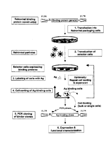
biology procedures known in the art (Fig. 1).
Although a preferred embodiment of the method according to the
invention is directed to the retroviral expression of
preferably human, full-length antibodies as binding proteins,
it can likewise be used for the expression of any fragment
thereof (e.g. single chain F, or Fab fragments of antibodies).
Retroviral transduction protocols are disclosed which
optionally allow (i) delivery of single binding protein
encoding constructs into single target cells, in order to
ensure clonal expression of binding proteins in the host
cells; (ii) shuffling of at least one expression construct
encoding a first polypeptide chain with at least one
expression construct encoding a second polypeptide chain,
thereby generating a functional multimeric binding protein
(e.g. an antibody molecule); (iii) somatic mutation of at
least one expression construct encoding at least one binding
protein upon transduction of vertebrate cells in situ; and
2 (iv) generation of binding protein expression from at least
one expression construct by the mechanism of V(D)J
recombination upon retroviral transduction into vertebrate
cells in situ.
In order to achieve somatic mutation of binding protein
encoding constructs in situ, retroviral expression vectors and
their utilization are disclosed, wherein said vectors contain
cis-regulatory genetic elements targeting somatic
hypermutation to protein encoding sequences, preferably via an
activation-induced cytidine deaminase (AID) pathway
CA 02715341 2015-12-30
4-Antibody AG - 6 -
2133
(Papavasiliou & Schatz, 2002), or by using other enzymes
targeting somatic mutations to binding protein encoding
sequences. For the generation of diverse collections of
binding proteins, i.e. antibodies or fragments thereof, by
V(D)J recombination in situ, retroviral vector constructs and
their utilization are disclosed, wherein said constructs
contain variable (V), optionally diversity (D), and joining
(J) gene segments arranged in "quasi-germline" configuration
allowing assembly of coding regions for immunoglobulins via
recombination activating gene (RAG)-mediated rearrangement of
the gene segments by the process known as V(D)J recombination
(Grawunder et a/., 1998).
According to a further aspect, the present invention further
illustrates how retrovirally transduced cells stably
expressing diverse collections of recombinant binding proteins
are subsequently labelled by binding to at least one ligand or
antigen of interest, and how cells binding to the
aforementioned ligand or antigen of interest are detected by
appropriate secondary reagents. Methods for the specific
labelling of ligand- or antigen-reactive cells and their
enrichment or isolation, preferably by high-speed fluorescent
activated cell sorting (FACS), are described. Due to the
stable expression phenotype of retrovlrally transduced cells,
it is described how antigen-reactive cells may optionally be
isolated and again expanded in tissue culture, such that
optionally iterative cycles of antigen labelling, antigen-
directed enrichment, and expansion of ligand or antigen-
reactive cells can be performed, until subcloning of the cells
is performed allowing the identification of the nucleotide
coding region for antigen-reactive antibodies by standard PCR
cloning methods (Fig. 1).
CA 02715341 2015-12-30
4-Antibody AG - 7 -
2133
The methods disclosed herein allow the expression of diverse
collections of antibody chains or fragments thereof from at
least one vector construct, which optionally can give rise to
collections of diverse binding proteins upon transfer and
expression into vertebrate cells in situ. Expression of
antibody chains in vertebrate cells is mediated by retroviral
transduction.
As such, a first embodiment of the present invention refers
to a method for the isolation and identification of at least
one nucleotide sequence encoding an antibody or fraament
thereof specific for a desired antigen or ligand, comprising
the steps of:
(a) transducing at least one retroviral expression construct
encoding an antibody or fragment thereof into vertebrate host
cells by using replication incompetent retroviral particles,
wherein the at least one construct stably integrates into the
host cell genome such that the transduced host cells are
capable of expressing and displaying said antibody or fragment
thereof on their cell surface, and wherein the vertebrate host
cells are precursor B lymphocytes that endogenously express
Iga and Igp molecules facilitating membrane deposition of said
antibody or fragment thereof, and are unable to express
endogenous antibody polypeptides and at least one surrogate
light chain component;
(b) stably expressing said antibody or fragment thereof in
said vertebrate host cells and displaying the same on their
cell surface;
(c) enriching vertebrate host cells expressing said antibody
or fragment thereof on the basis of their ability to bind to
said desired antigen or ligand by separating cells that
exhibit specific antigen binding from a non-binding cell
population thus selectively isolating strong antibody binders
having high affinity to said desired antigen or ligand; and
CA 02715341 2015-12-30
4-Antibody AG - 75 -
2133
(d) isolating and identifying said at least one nucleotide
sequence encoding said antibody or fragment thereof from the
retrovirally transduced and enriched vertebrate host cells.
In addition to the aforementioned steps, step (d) may be
preceded by a step of expanding the enriched vertebrate host
cells in tissue culture. Furthermore, step (c) may be followed
by a step of expanding the enriched vertebrate host cells in
tissue culture, after which step (c) is repeated at least once
before step (d) is carried out.
To achieve clonal expression of at least one antibody it is
preferable to control the retroviral transduction such that
the majority of retrovirally transduced cells are genetically
modified by only one recombinant retroviral construct per
CA 02715341 2010-08-12
WO 2009/109368 - 8 - PCT/EP2009/001525
antibody chain integrating in to the host cell genome.
Therefore, in one embodiment of the present invention,
retroviral transduction is performed at a multiplicity of
infection (MOI) of equal to or less than 0.1.
An antibody according to a method of the present invention is
preferably a full-length antibody. A fragment of an antibody
may be selected from the group consisting of: a heavy chain, a
light chain, a single VH domain, a single VL domain, a scFv
fragment, a Fab fragment, and a F(ab')2 fragment. The antibody
or fragment(s) thereof may have a naturally occurring amino
acid sequence, an artificially engineered amino acid sequence
or a combination thereof.
Whilst the method of the present invention is used preferably
for the isolation and identification of at least one
nucleotide sequence encoding an antibody chain, it would be
apparent to a person skilled in the art that the method of the
present invention can also be used for the isolation and
identification of at least one nucleotide sequence encoding
any monomeric or multimeric cell surface receptor belonging to
the Ig-superfamily, and any functional fragment thereof, or a
monomeric or multimeric cell surface receptor belonging to the
TNFa-receptor superfamily, or any fragment thereof.
Furthermore, where the binding protein is a full-length
antibody, the full-length antibody is selected from the group
consisting of a fully human antibody, a humanized antibody, in
which CDR regions of a non-human antibody or antibodies have
been grafted onto a human antibody framework, and a chimeric
antibody, in which variable region domains from one vertebrate
species are combined with constant region domains of another
vertebrate species, with the constant domain of the chimeric
antibody preferably being derived from a human antibody or
antibodies.
CA 02715341 2015-12-30
4-Antibody AG - 9 -
2133
In an embodiment of the methods disclosed herein, the
vertebrate host cells may be derived from a group of species
comprising cartilaginous fish, bony fish, amphibians,
reptilia, birds and mammals. The group of species of mammals
may include pigs, sheep, cattle,, horses and rodents. The group
of rodents may further comprise mice, rats, rabbits and guinea
pigs. In a preferred embodiment of the present invention, the
vertebrate host cell species is mouse (Mus muscu/us).
The vertebrate host cells for use in a method of the present
invention are of the B cell lineage, because these cells
express antibody-specific chaperone proteins, and because
accesscry molecules, like Igo and Igp required to mediate cell
surface anchoring of antibodies are expressed in these cells,
namely precursor B lymphocytes, as preB cells can be found
that do not express any endogenous antibody chains. In fact,
the precursor B lymphocytes as utilised in the present
invention are unable to express endogenous antibody
polypeptides including components of the so-called surrogate
lighe chain, encoded by the genes lambda-5, VpreB1 and VpreB2.
Therefore, these lymphocytes express accessory membrane
proteins facilitating membrane deposition of antibody
molecules, such as the B cell specific Igo and Ig13 molecules,
but they lack expression of any endogenous antibody
pclypeptide or surrogate light chain component. However, it
shall be noted that it may be possible to express Iga and Ig0
molecules ectopically, by methods known in the art, e.g.
stable transfection with expression vectors for these
proteins. In an embodiment of the present invention, antibody
molecules are anchored to the cell membrane of lymphocytes via
endogenously expressed Iga and Ig(S proteins, which are
naturally expressed in murine pre-B lymphocytes.
CA 02715341 2015-12-30
4-Antibody AG - 10 -
2133
The methods disclosed herein include procedures allowing the
isolation of cells displaying desired binding characteristics
for a ligand or antigen of interest and the isolation of genes
encoding a desired binding protein of interest. The method of
retroviral expression of an antibody in vertebrate cells
disclosed herein allows for stable and preferably clonal
expression of antibodies, which greatly facilitates the
amplification, isolation, and cloning of antibody encoding
genes, in comparison to alternative, plasmid-based or non-
integrating virus-based vertebrate expression systems known in
the art. The disclosed methods allow for efficient generation
of diverse collections of antibody molecules in vitro by
either:
(i) shuffling of at least one expression construct encoding at
least one polypeptide chain (like e.g. a heavy chain) of a
multimeric antibody, with at least one expression construct
encoding at least one matching polypeptide chain (like e.g. a
light chain of an antibody) generating a functional multimeric
antibody;
(ii) somatic mutation of at least one expression vector
encoding at least one antibody molecule upon transfer into
vertebrate cells in situ;
(iii) somatic recombination of V (variable), optionally D
(diversity), and J (joining) gene segments encoding variable
binding domains of immunoglobulins contained in at least one
expression vector upon transfer into vertebrate cells in situ,
by the process known as V(D)J recombination; or
(iv) by any combination of procedures (i), (ii), and (iii).
According to a preferred embodiment, the at least one
nucleotide sequence is a plurality of nucleotide sequences
that comprise an antibody heavy chain sequence and multiple
antibody light chain sequences, or - in the alternative -
CA 02715341 2010-08-12
WO 2009/109368 - 11 - PCT/EP2009/001525
comprise an antibody light chain sequence and multiple
antibody heavy chain sequences.
According to another preferred embodiment, the antibody or
fragment thereof comprises a variable binding domain encoded
by the at least one retroviral expression construct enabling
V(D)J recombination in order to generate a coding sequence for
a variable binding domain upon retroviral transduction or
In a further preferred embodiment, step (b) of the above
method is performed under mutagenizing conditions, preferably
via the expression of activation induced cytidine deaminase
(AID) which is either endogenously or ectopically expressed,
wherein the ectopic expression of AID is performed under
inducible conditions.
In one aspect of the above method, the at least one retroviral
expression construct encoding said antibody or fragment
thereof contains a combination of cis-regulatory promoter and
enhancer elements allowing the targeting of AID mediated
somatic mutation to a variable binding domain encoded by the
expression construct, wherein the promoter and enhancer
elements are selected from the group consisting of
(a) immunoglobulin heavy chain promoter, intron enhancer
(EpH)and 3'a enhancer elements,
(b) immunoglobulin K light chain promoter, K intron enhancer
(KiE) and 3'K enhancer (3'KE) elements,
(c) immunoglobulin A light chain promoter, A2-4 and A3-1
enhancer elements, and
(d) any functional combination thereof.
Description of the Figures
Fig.1: This figure illustrates the principle of 'Retrocyte
Display' allowing the identification and isolation of a
binding protein such as an antibody, specific for a desired
antigen or ligand. In a first step, at least one retroviral
CA 02715341 2010-08-12
WO 2009/109368 - 12 - PCT/EP2009/001525
expression construct that can give rise to expression of a
diverse collection of binding proteins is stably transduced
into suitable vertebrate host cells ("selector cells"). This
is accomplished by transfecting at least one retroviral vector
encoding at least one binding protein into retroviral
packaging cells (step 1), which may either constitutively or
transiently express retroviral proteins Gag, Pol and Env.
Packaging cells transfected with the at least one retroviral
binding protein construct will then produce recombinant
retroviral particles within 24-72 hours post transfection,
containing the at least one retroviral expression construct.
The resulting retroviral particles accumulate in the cell
culture supernatant of the retroviral packaging cells, and can
be used to transduce suitable vertebrate host cells ("selector
cells") (step 2), which then express the binding protein. In
the preferred method, the binding proteins such as antibodies
or fragments thereof are expressed on the cell surface of the
"selector cells" and the cells then are labelled with a
desired antigen or ligand (step 3). Antigen- or ligand binding
cells are then preferably analyzed by fluorescent activated
cell sorting (FACS) and cells that exhibit specific antigen
binding, are separated from the non-binding cell population
preferably by preparative, high-speed FACS (step 4). Antigen-
or ligand reactive cells may optionally be expanded in tissue
culture again, and due to the stable expression phenotype of
retrovirally transduced cells, cycles of antigen-directed cell
sorting and tissue culture expansion may be repeated, up to
the point that a detectable antigen- or ligand reactive cell
population is obtained. This antigen- or ligand reactive cell
population may be subjected to a final, preferable, single-
cell sorting step, or may directly be used for cloning of
binding protein encoding genes on a population basis. In the
next step (step 5), the coding regions of relevant binding
domains are cloned from the antigen- or ligand-selected cell
pools or cell clones, by RT-PCR or genomic PCR using primer
CA 02715341 2015-12-30
4-Antibody AG - 13 -
2133
pairs binding to sequences specific for the binding protein
library and/or specific for other vector sequences, by
standard methods known in the art. Cloned and sequenced coding
regions for binding proteins may then optionally be expressed
as recombinant proteins in any expression system of choice for
further functional characterization and to confirm antigen- or
ligand binding specificity (step 6).
Fig.2: (a) This figure illustrates the schematic structure of
antibodies or immunoglobulins and fragments thereof, which are
the binding proteins according to the disclosed invention.
Fig. 2a) shows the schematic structure of an IgG antibody
(left), which is characterized by a characteristic Y-shaped
structure and is composed of two identical immuneglobulin (Ig)
heavy and light chains, comprising four )VH-CH1-CH2-CH3) and two
immunoglobulin domains (VL-CL), respectively. The V-domains are
the highly variable antigen binding regions of IgH and IgL
chains, whereas the CH and CL domains represent the constant
region domains. The variable region domains of IgH chains are
encoded by V, t and J gene segments, whereas the variable
region domains of IgL chains are encoded by only V and J gene
segments, which need to be assembled from germline
immunoglobulin gene loci (Figs. 2b) and 2c) during early
lymphopoiesis, by the process known as V(D)J recombination.
Antibody IgH and IgL chains are covalently held together by
disulphide bridges, which couple the identical IgH chains
together at a location close to the flexible hinge region,
i.e. between the CH1 and CH2 domains, whereas additional
disulphide bridges between the CH1 and CL domains, as depicted,
are covalently coupling IgH and IgT, chains (Fig. 2a left).
Fab fragments are univalent fragments of full-length
antibodies only containing VH-CE11/VL-CL domains coupled by a
natural disulphide bridge, which can either be derived by
ak 02715341 2010-08-12
WO 2009/109368 - 14 - PCT/EP2009/001525
enzymatic papain cleavage from full-length antibodies, or
which can be expressed as recombinant proteins by expressing
CH2-CH3 deleted IgH chains together with IgL chains. Additional
fragments of fully human antibodies are single chain variable
domain fragments (scFv-fragments), which only comprise the
variable region domains of IgH and IgL chains that are coupled
by a synthetic linker or an artificial disulphide bridge. The
expression of either full-length antibodies, or antibody
fragments, as the depicted Fab and scFv fragments, may also be
expressed as binding proteins in order to realize the
invention.
Fig. 2b) schematically depicts the process of V(D)J
recombination occurring on a germline IgH chain allele,
resulting in the assembly of the coding regions of antibody VH
domains. The variable domains of IgH chains in vertebrate
species are encoded by a multitude of V, D and J gene
segments, which are separated in germline configuration.
During V(D)J recombination occurring during early B
lymphopoiesis, one selected V, D and J gene segment is site-
specifically rearranged to generate a unique coding region for
an antibody VH domain. V(D)J recombination in the IgH chain
locus is an ordered process and starts with rearrangement of a
selected D to a selected J gene segment, usually on both IgH
chain alleles. Only after D to J gene rearrangement, one
selected V region is site-specifically joined to the already
assembled DJ region, thereby generating a V-D-J ORF encoding
the VH domain. The process of V(D)J recombination is dependent
on the expression of precursor lymphocyte specific
recombination activating genes (RAG) 1 and 2.
Fig. 2c) schematically depicts the process of V(D)J
recombination occurring on a germline IgL chain allele,
resulting in the assembly of the coding regions of antibody VL
domains. The variable domains of IgL chains in vertebrate
species are encoded only by V and J gene segments, which are
CA 02715341 2010-08-12
WO 2009/109368 - 15 - PCT/EP2009/001525
separated in germline configuration, similar to the gene
segments in the IgH chain locus. The generation of an antibody
VL domains requires only one site-specific V(D)J recombination
event, as depicted.
Fig. 3: This figure schematically illustrates the principle of
stable genetic modification of target cells for the expression
of a binding protein of interest (BPOI) such as an antibody
(alternatively labelled "X") by retroviral transduction.
Fig. 3a) depicts the schematic organization of a wild-type
retroviral genome (upper left), in which the genes for the
structural and functional proteins Gag, Pol and Env are
located in between so-called 5' and 3' long-terminal repeat
(LTR) sequences flanking the retroviral genome. The 5'LTRs are
important for the expression of the retroviral genes and also
for the replication of the retroviral genome in the infected
host cell. Another important region in the retroviral genome
is the * (Psi) packaging signal, which is required for the
packaging of the retroviral RNA during replication and/or
production of retroviral particles.
For the generation of recombinant retroviral particles, the
gag, pol and env genes may be removed from a wild-type
retroviral genome, so that only 5' and 3' LTRs and the * (Psi)
packaging signal remains. For the construction of recombinant
retroviral vectors it is then convenient to introduce a
multiple cloning site (MCS) containing several unique and
convenient restriction enzyme sites. This design, as depicted
on top/right, represents the simplest retroviral transfer
vector.
For the expression of recombinant retroviruses allowing the
expression of a recombinant protein (e.g. a binding protein of
interest (BPOI) "X") such as an antibody, minimally an open
reading frame (ORF) of a BPOI needs to be inserted into an
CA 02715341 2010-08-12
WO 2009/109368 - 16 - PCT/EP2009/001525
"empty" retroviral transfer vector, as the 5'LTR region has a
promoter activity able to drive expression of any downstream
positioned gene. However, in order to improve expression
levels, expression of a gene of interest (e.g. a BPOI-"X") may
optionally be driven by an additional heterologous promoter
(Prom.), and optional addition of a marker gene, e.g.
downstream of the 5'LTR promoter and * packaging signal, as
depicted here, may allow selection and/or tracking of
retrovirally transduced constructs.
Fig. 3b) schematically illustrates the procedure of retroviral
transduction of target cells resulting in the stable
expression of a BPOI-"X" such as an antibody. For this, first,
a recombinant retroviral construct containing an expression
cassette for a BPOI-"X" is transiently transfected into a
retroviral packaging cell line (PCL), expressing structural
and functional retroviral proteins Gag, Pal and Env of a wild-
type retrovirus (left). A retroviral PCL can be generated by
either stably or transiently transfecting expression
constructs for the Gag, Pol and Env proteins into a suitable
and easy to transfect cell line (e.g. standard human 293 HEK
cells, or mouse NIH 3T3 fibroblasts). Two to three days post
transfection, the recombinant retroviral genomes, containing
the BPOI-"X" gene are packaged into replication incompetent
retroviral particles, which accumulate in the cell culture
supernatant of the PCL. The retroviral particles are
replication incompetent, because they lack the genes for the
functional retroviral Gag, Pol and Env proteins and therefore,
they can deliver their genetic payload into a target cell only
once, a process that is called retroviral transduction, or
single round infection. During retroviral transduction the
packaged RNA of a recombinant retrovirus is introduced into
the target cells, where it is reverse transcribed into cDNA,
which is then stably integrated into the target cell genome.
Two to three days after retroviral transduction, a gene of
CA 02715341 2010-08-12
WO 2009/109368 - 17 - PCT/EP2009/001525
interest, like the BPOI-"X", is then permanently expressed by
the target cells, due to the integration of the cDNA
retroviral construct into the host cell genome.
Fig. 4: This figure shows the schematic design of preferred
types of retroviral expression constructs that can be used to
realize the invention. The drawing depicts the schematic
design of retroviral vectors contained in a standard DNA
cloning plasmid backbone (closed black line); the relevant
genes and regions for the retroviral genome are highlighted.
One preferred vector generation, depicted in panel (a), whose
detailed cloning is described in Figs 5 and 6, and provided in
Example 1, contains the cDNA coding regions for human
chains and IgKL chains driven by a strong constitutive CMV
promoter (Prom) and flanked up- and downstream by the Ig K
intron enhancer (KiE) and 3'K enhancer (3'KE) elements,
promoting somatic hypermutation to the V coding regions of the
IgH and IgL chains. The retroviral IgH and IgL chain
expression constructs additionally contain open reading frames
for the antibiotic resistance markers hygromycinB (hygroR) and
puromycin (puroR), respectively, allowing the selection of
stable integration of the IgH and IgL chain constructs
applying respective antibiotic drug selection to cultures of
retrovirally transduced vertebrate cells. In addition,
convenient, unique restriction enzyme sites are highlighted,
allowing the straightforward replacement of V coding regions
with HindIII and Eco47III, or the replacement of the entire
IgH and IgL chain coding regions by using the restriction
enzymes HindIII and NotI. This way, from one existing IgH or
IgL chain expression construct different V regions and even
entire collections of V regions can easily be cloned into the
disclosed expression vectors.
(b) In this panel, another class of preferred vectors is
described, which carry a replacement of the variable coding
region by a DNA fragment, in which the variable coding region
CA 02715341 2010-08-12
WO 2009/109368 - 18 - PCT/EP2009/001525
is still separated into V, D and J gene segments (for the IgH
construct) and V and J gene segments (for the IgL chain
construct) in "quasi-germline" configuration. While otherwise
identical to the retroviral expression vectors provided in (a)
these V(D)J-recombination competent retroviral vectors first
need to undergo site-specific rearrangement of the V,
optionally D and J gene segments, in order to generate a
coding region for a variable binding domain of a IgH or IgL
chain. The detailed cloning of such a vector allowing the
expression of IgH chains after V(D)J recombination is
described in Fig. 11.
A unique feature of these constructs is their capability to
generate diverse V domain coding regions in V(D)J
recombination active cells in situ, e.g. in precursor
lymphocytes expressing endogenous RAG1 and RAG2 proteins.
Because the process of V(D)J recombination is not precise, a
diverse collection of variable coding region sequences may
result from one individual retroviral vector within a given
set of V, D and J gene segments for IgH, or a given set of V
and J gene segments for IgL. The diversity in the joining of
V, optionally D and J gene segments is due to a combination of
exonuclease activity, TdT mediated N-region addition, and P
nucleotide generation, which may all contribute individually
or jointly to coding joint diversification. As the V, D and J
gene segments have been cloned in a fashion that different V,
D and J gene segment family members can be easily replaced by
unique restriction enzyme sites, a limited number of
constructs generated and introduced into V(D)J recombination
competent host cells, can result in an enormous diversity of
in situ generated binding protein diversity. As these vectors
contain additional xiE and 3'KE elements, conferring somatic
hypermutation to an V(D)J-rearranged V domain coding region, a
primary in situ generated collection of diverse binding
proteins can optionally further be mutagenized by an AID-
CA 02715341 2010-08-12
WO 2009/109368 - 19 - PCT/EP2009/001525
dependent somatic hypermutation process. This way, the entire
process of generation of antibody diversity in vivo, can be
recapitulated in situ and in vitro using the disclosed
retroviral constructs and host cells exhibiting V(D)J
recombination activity (e.g. precursor lymphocytes), and in
which AID mediated somatic hypermutation is active, or can be
activated.
(c) This panel schematically depicts yet another design of
retroviral constructs that can be used to realize the
invention. Here, the expression of the IgH and IgL coding
regions is driven by the 5'LTR promoter of the retroviral
backbone and the expression of IgH and IgL chains is coupled
to the expression of GFP and YFP autofluorescence markers,
respectively, allowing the tracking and isolation of IgH and
IgL expressing cells simply by analyzing the transduced cells
for green and yellow fluorescence. These constructs are very
useful for controlling the multiplicity of infection of
"selector cells" without further labelling procedures.
A legend of symbols used in Figs 4a) to c) for important DNA
sequences included in the construct is provided. The
subdivision of the IgH and IgL coding regions into variable
domains (VH and VL) all containing endogenous leader (L)
sequences, hinge (H), constant (CH1, CH2, CH3, CL), and
membrane-spanning coding regions (M1/2, because this region is
encoded by two exons) is provided for a better understanding
of the illustrations.
Fig. 5a-e illustrates the cloning strategy for the
construction of a retroviral IgH (human Igyl isotype)
expression vector, disclosed in detail in Example 1, and in
the basic design provided in Fig. 4(a). The cloning of
expression constructs for both membrane bound IgG as well as
secreted IgG is depicted, as detailed in Example 1 - unique
restriction enzyme sites in the plasmid maps are provided for
CA 02715341 2010-08-12
WO 2009/109368 - 20 - PCT/EP2009/001525
general reference purposes. Based on the final retroviral IgyiH
chain expression construct, as disclosed in Fig. 5e herein,
any other VH domain coding region, or a collection (library) of
diverse VH domain coding regions can be introduced into the
vectors using the unique HindIII and Eco47III restriction
enzyme sites, by replacing the existing VH region with said any
other VH domain coding regions. Fig. 5a depicts a first
preparatory cloning step, in which an Eco47III restriction
site (circled) is removed from the commercially available
pLHCX vector backbone by site-directed mutagenesis, as
described in Example 1. This generates the retroviral vector
backbone pLHCXml, in which the Eco47III restriction enzyme
site can later be re-introduced for the cloning and
replacement of VH domain coding regions. The advantages of
using Eco47III for this purpose is based on the fact that
Eco47III is the only restriction enzyme site that can be
introduced directly at the border between human VH and CY1
coding regions, without changing the amino acid composition of
expressed human IgyiH chains. Fig. 5a further illustrates, how
cloned fragments of the human Vi constant region genes, either
with, or without membrane spanning exons M1/M2 are cloned into
the pLHCXml backbone using unique HindIII and ClaI restriction
enzyme sites present in the MCS of pLHCXml. The fragments were
designed to contain additional flanking Eco47III and NotI
restriction enzyme sites for later cloning purposes, as
detailed in Example 1. Fig. 5b) shows the plasmids maps of the
cloning intermediates without VH domain coding regions, and it
is shown, how a particular VH coding region flanked by HindIII
and Eco47III sites is cloned into the constructs. These
constructs, which are thus generated, are depicted in Fig. 5c,
and would in principle be sufficient to confer the expression
of human IgyiH chains in any recipient cell line. However, the
possibility to additionally mutagenize VH coding regions in an
AID-dependent manner, is an aspect of this invention and two
additional cloning steps are disclosed, in which the core KiE
CA 02715341 2010-08-12
WO 2009/109368 - 21 - PCT/EP2009/001525
element with additional flanking sequences is cloned into a
unique BglII site, upstream of the CMV promoter of the
expression cassette (Fig. 5c bottom and Fig. 5d) and in which
the 3'KE element with some flanking DNA sequence is cloned
into the unique ClaI site downstream of the expression
cassette for human Igy1H chains. This results in the final
expression vector for either membrane bound or secreted human
IgyLH chains, for which the plasmid maps are provided in Fig.
5e. These constructs correspond to the schematic plasmid maps
that have already been disclosed in Fig. 4a, but here with
precise restriction enzyme maps and drawn to scale.
Fig. 6a-d illustrates the detailed cloning strategy provided
in Example 1, for the construction of retroviral IgL (human
IgKL isotype) expression construct, whose basic design was
already provided in Fig. 4(b). Based on the final retroviral
IgKL chain expression construct, as disclosed in Fig. 6d
herein, any other VL domain coding region, or a collection
(library) of diverse VL domain coding regions can be introduced
into the vectors using the unique HindIII and Eco47III
restriction enzyme sites, by replacing the existing VL region
with said any other VL domain coding region(s). The cloning
strategy for the retroviral IgL chain expression vectors
required preparatory cloning steps, in order to generate a
modified retroviral vector backbone, into which the desired
elements could be cloned using convenient restriction enzyme
sites as depicted. In a first step, from commercial plasmid
pLPCX an undesired Eco47III site was removed from the IV (Psi)
packaging signal by site-directed mutagenesis as described in
Example 2, resulting in modified plasmid pLPCXml (Fig. 6a). In
a second step a novel pLPCXm2 backbone was generated by
ligating a large, AscI-BlpI digested fragment from commercial
plasmid pLHCX with an AscI-NcoI fragment from pLPCXml (Fig.
6b). For both fragments the non-compatible BlpI and NcoI DNA
ends needed to be filled up with nucleotides using Klenow
CA 02715341 2010-08-12
WO 2009/109368 - 22 - PCT/EP2009/001525
fragment as described in Example 1. Into the resulting pLPCXm2
backbone the constant region for a human KL chain (CK) has
been inserted via HindIII and ClaI as shown (Fig. 6b). Similar
to the cloning strategy for human IgH chains, the human OK
fragment was further flanked by Eco47III and NotI sites to
facilitate additional cloning procedures. After insertion of
the human OK fragment, one selected human VK element was
cloned into the construct via unique HindIII and Eco47III
sites (Fig. 6c). This construct would in principle be
sufficient to confer the expression of human IgKL chains in
any recipient cell line. However, like in the case for the IgH
chain expression constructs (Fig. 5a-e), additional KiE and
3'KE elements were cloned into the construct into the unique
BglII and ClaI sites upstream and downstream of the IgKL chain
expression cassette, identical to the cloning strategy of the
IgH chain constructs (Fig. 6c and 6d). In the final constructs
also the VK domain coding region can then be target for AID-
mediated somatic hypermutation. The final construct
corresponds to the schematic plasmid map that is detailed in
Fig. 4(b), but here the precise restriction enzyme maps are
included and drawn to scale.
Fig. 7: This figure illustrates the cloning strategy for a
retroviral expression construct for activation induced
cytidine deaminase (AID). As depicted, the commercial pLPCX
retroviral vector backbone was used and a specific RT-PCR-
fragment from mouse splenic cDNA containing the AID coding
region was cloned into the unique XhoI restriction site of the
pLPCX vector using compatible XhoI restriction enzyme sites
inserted into the PCR amplication primers, as described in
Example 2.
Fig. 8: This figure (8a and continued onto 8b) illustrates the
detailed cloning strategy, also provided in Example 2, for a
retroviral reporter constructs with and without IgKL chain
enhancer elements, allowing the identification and
ak 02715341 2010-08-12
WO 2009/109368 - 23 - PCT/EP2009/001525
quantitation of somatic mutations by reversion of a defined
EGFP stop mutation.
Fig. 9: This figure provides an experimental proof-of-concept
that the disclosed retroviral vectors allow AID mediated
somatic mutation of sequences, like preferably antibody V
coding regions, cloned downstream of the V-promoter elements.
Panel (a) shows an analysis of AID expression by Western-
blotting of five selected FA-12 A-MuLV transformed cell clones
that had been stably transfected with a retroviral AID
expression construct, whose cloning was depicted in Fig. 7.
The Western-blot analysis shows a distinct AID-specific signal
of ca. 25kD in FA-12 transfectant clones 1 through 4, but not
in transfectant 5 and also not in the non-transfected negative
control (NC). Transfectant 3 was used for further testing of
retroviral reporter vectors for AID-mediated somatic
hypermutation (SHM), which is depicted in panel (b): Here the
retroviral reporter constructs of Fig. 8 (once with and once
without Igx enhancer elements) were retrovirally transduced
into AID expressing and AID non-expressing FA-12 transfectants
3 and 5, respectively. As expected, only when reporter
constructs containing the enhancer elements were transduced
into AID-expressing FA-12 transfectant clone 3, was it
possible to detect green revertant transductants at a 0.2%
frequency 10 days post transduction. From these 0.2% green
cells, 100 individual cell clones were isolated by single cell
sorting and these clones were re-analyzed for green
fluorescence by FACS after expansion. The vast majority of the
single cell sorted clones (95%) displayed homogeneous green
fluorescence expression at the same fluorescence intensity as
the medium green fluorescence signal of the 0.2% green cells
originally sorted, and similar to the representative GFP
expression pattern provided at the lower left panel of Fig.
9b, confirming that the original green population was due to
reversions of the EGFP stop mutation. Four clones showed a
CA 02715341 2010-08-12
WO 2009/109368 - 24 - PCT/EP2009/001525
bimodal green fluorescence pattern, as representatively
depicted in the middle FACS histogram and only 1 of the 100
single cell sorted cloned displayed hardly any green
fluorescence (right-hand FACS histogram).
Fig. 10: This figure illustrates the sequence of the EGFP
coding region with an engineered stop mutation that was used
to clone an EGFP reporter construct for quantitating somatic
hypermutation. In panel (a) it is shown, which of the four
nucleotides have been mutated in codon 107 and 108 of the EGFP
open reading frame, thereby generating a stop-codon in codon
107 and generating a lysine to threonine amino acid change in
codon 108. These four nucleotide changes additionally resulted
in the introduction of unique SpeI restriction enzyme site, as
indicated that could be used as a diagnostic marker for stop
codon reversions upon somatic hypermutation. The G-nucleotide
of the TAG stop codon is embedded in a so-called RGYW sequence
motif, which is known to be a hotspot for somatic
hypermutation. In 24 revertant clones analyzed by SpeI
restriction enzyme digestion, it could be confirmed that the
site was rendered resistant to SpeI digestion (and hence was
mutated). In ten of these clones sequence analysis revealed
that the G nucleotide in the original TAG stop-codon had been
mutated to a C nucleotide, resulting in a TAC codon, thereby
confirming the restriction enzyme analysis, and demonstrating
that AID-mediated somatic mutation had been targeted to the G
in the RGYW motif.
(b) This panel shows the entire ORF of the mutated EGFP that
was cloned into the retroviral Igy1H chain construct already
disclosed in Fig. 5(e), instead of a VH domain coding region.
Fig. 11(a) and (b): Illustration of the detailed cloning
strategy of a V(D)J recombination competent retroviral IgH
chain expression vector, as disclosed in detail in Example 4.
CA 02715341 2010-08-12
WO 2009/109368 - 25 - PCT/EP2009/001525
Fig. 12: Proof-of-concept that retroviral constructs requiring
V(D)J recombination of V, D and J gene segments in "quasi-
germline" configuration can give rise to productively
rearranged heavy chain expression constructs and Ig+ cells
upon transduction into RAG1/RAG2 positive precursor
lymphocytes. Panel (a) contain data showing the generation of
surface immunoglobulin positive cells (0.04%, upper right
quadrant of left FACS plot) after transduction of a V(D)J
recombination competent retroviral expression vector (detailed
description of cloning, see Fig. 11) into A-MuLV transformed
preB cell line 230-238. The immunoglobulin expression is
coupled to EGFP expression using constructs as schematically
illustrated in Fig. 4c. Therefore, immunoglobulin expressing
cells can only be generated in the population of green (i.e.
stably transduced) cells. The right staining panel shows re-
analysis of surface immunoglobulin expression after a single
round of FACS enrichment and expansion of the rare (0.04%)
surface immunoglobulin cells for 8 days in tissue culture.
After this one round of enrichment, the combined frequency of
immunoglobulin positive cells had increased to 17.8% (as
expected detectable in the green, i.e. the stably transduced
population) from which PCR amplicons have been obtained and
, sequenced. (b). As a representative example, this panel shows
a DNA sequence (clone 225, with amino acid translation on top)
obtained from a PCR amplicon derived from surface
immunoglobulin cells after one round of enrichment that had
been transduced with "quasi-germline" V(D)J recombination
competent retroviral vectors. As a reference, the sequences of
the coding regions of the V, D and J gene segments are
provided in (b) at the top, also with amino acid translation
on top of the V and J gene segments, as the D segment sequence
can be read in three different reading frames, depending on
the junctional diversity after V(D)J recombination.
Intervening sequences between the V. D and J gene segments in
"quasi-germline" configuration are depicted with dots. The
ak 02715341 2010-08-12
WO 2009/109368 - 26 - PCT/EP2009/001525
sequence of recovered clone 225 clearly represents a bona fide
V(D)J rearrangement event, with typical features of nucleotide
loss and TdT catalyzed N-sequence additions clearly detectable
at the coding joints between the assembled V, D and J gene
segments (all intervening sequences had been lost from clone
225). The sequence of clone 225 exhibited an open reading
frame and, apart from the aforementioned variations at the
coding junctions, did not contain additional somatic mutations
in the V, D and J sequences.
Fig. 13: Data showing the testing of a panel of different A-
MuLV transformed murine preB cell lines for the susceptibility
to ecotropic MLV-derived vector gene transfer. 1x10⁵ cells were
transduced with a MOI of 0.5 using a vector preparation having
packaged the reporter gene EGFP encompassing transfer vector
LEGFP-N1. Transduction was carried out as detailed in Example
5. Two days post transduction, gene transfer was detected by
expression of EGFP using FACS. Except for preB cell line
18/81, all other tested A-MuLV transformed preB cell lines
were susceptible for transduction at frequencies ranging
between 40-60% under the applied conditions, and can, in
principle, be used for the current invention. Untreated naive
target cells served as negative controls and showed no green
fluorescence (not shown).
Fig. 14: Characterization of a panel of murine preB cell lines
for intracellular expression of endogenous IgM heavy chains
(cy-pH), in order to identify cells devoid of endogenous
murine antibody expression that can be used as selector cells
for retrocyte display. Cells were permeabilised and stained
using anti-murine IgM heavy chain antibodies coupled to FITC
(FL1). Untreated cells served as negative controls. The
experiment shows that cell lines FA-12, 1624-5, 1624-6, 18/81-
c18-11, and 40E1 had practically undetectable endogenous
antibody expression, and can thus be used in a method of the
present invention.
ak 02715341 2010-08-12
WO 2009/109368 - 27 - PCT/EP2009/001525
Fig. 15: Illustration of the complexity of retroviral
expression vectors following the design disclosed in Fig. 4(c)
and the experimental principle for the generation of a IgH and
IgL chain shuffled antibody library. (a) The retroviral vector
libraries IgH(650)-LIB-IRES-GFP and IgL(245)-LIB-IRES-YFP
encompass defined collections of coding regions for heavy (HC)
and light chains (LC) for fully human antibodies with a
complexity of 650 and 245 different, fully sequenced clones,
respectively. Both vectors harbour the packaging sequence Psi
(IV), flanking long terminal repeats (LTR) and an internal
ribosome entry signal (IRES). Parallel to the expression of an
antibody polypeptide chain mediated by the viral promoter in
the 5'LTR, the TRES enables the coupled expression of the
reporter gene gfp and yfp, respectively. Upon viral gene
transfer into selector cells, this allows for the convenient
detection and enrichment of successfully transduced and
immunoglobulin chain expressing cells using FACS.
(b) Generation of a collection of fully human antibodies in
transformed preB cells. In order to generate transient
packaging cells, libraries of retroviral transfer vector
libraries encoding heavy chains of human antibodies (IgH(650)-
LIB-IRES-GFP) are co-transfected with a packaging construct
(pVPack-GP) and an envelope construct (pVPack-Eco) into
suitable recipient cells. Two days post transfection, the
generated vector particles library having packaged the
respective transfer vector library are harvested and employed
to transduce selector pre B cells. Transduced cells expressing
the transferred heavy chains and the reporter gene gfp are
expanded enriched using FACS. Following expansion, cells are
subjected to a second transduction. This time, the IgL(245)-
LIB-IRES-YFP library is transferred followed by expansion and
enrichment of cells expressing YFP and human light chains
employing FACS. The resultant population constitutes a fully
human antibody displaying a defined human antibody library
CA 02715341 2010-08-12
WO 2009/109368 - 28 - PCT/EP2009/001525
expressed by 1624-5 cells, containing a complexity of
maximally 159'250 clones.
Fig. 16: This figure shows how a two-step transduction with
IgH-IRES-GFP and IgL-IRES-YFP libraries has been performed at
conditions ensuring a transduction, resulting in clonal
expression of polypeptide chains in the vast majority of the
transduced cells. 1.5x10⁶ 1624-5 murine A-MulV transformed preB
cells were suspended in 1 ml of tissue culture medium
supplemented with different quantities of vector particle
supernatant (diluted 1:1; 1:5; 1:20; 1:50; 1:100; 1:200)
containing recombinant retroviral vectors encoding IgH and IgL
chain libraries IgH-LIB-IRES-GFP or IgL-LIB-IRES-YFP,
respectively, already described in Fig. 15. To ensure that the
majority of the transduced cells received single copies of
transfer vectors integrated into the host cell genome, cells
displaying gene transfer efficiencies lower than 10% (MOI
<0.1, as detected by expression of the coupled GFP or YFP
reporters) were enriched using FACS sorting four days post
infection. Cells were expanded for six days and subjected to a
second transduction employing vector particles having packaged
the light chain coding regions of antibodies at a dilution of
1:5 as described above. Here, GFP-positive cells selected for
heavy chain expression were infected with vector particles
transducing the IgL-LIB-IRES-YFP library and vice versa. Four
days post infection, transduced cells expressing GFP and YFP
were enriched using FACS. Approximately 20% of the cells
showed GFP and YFP expression after the second transduction.
To secure that only single vector integrations occurred per
cell about one third of the populations were enriched that
revealed only low or moderate expression of the reporter gene
transduced in the second round (approximately 8%).
Fig. 17: Titration of IL-15 staining with a population of preB
cells expressing an anti-IL-15 reference antibody by FACS, in
order to define optimal conditions allowing optimal IL-15
CA 02715341 2010-08-12
WO 2009/109368 - 29 - PCT/EP2009/001525
antigen staining conditions for Retrocyte Display experiments.
The staining procedure, as disclosed in detail in Example 7,
included a titration of the IL-15 antigen in the range of
2.5pg/m1-0.1pg/ml, at two different concentrations of a
polyclonal, biotinylated anti-IL-15 secondary antibody, as
indicated, which was detected with streptavidin-PE conjugate
by FACS. Surface Ig+ cells were counterstained with an anti-
IgKL chain-APC antibody. As can be seen, optimal IL-15
staining is accomplished at a concentration of 0.1 or 0.5
pg/ml IL-15 antigen, and using 3pg/m1 of the secondary,
polyclonal anti-IL-15 antibody.
Fig. 18: Analysis of FACS-identification of an anti-IL-15
reference antibody expressing preB cell line (PC = positive
control), which was spiked into a diverse library of antibody
expressing preB cells at different dilutions, by using the
optimized IL-15 staining conditions illustrated and determined
in Fig. 17. The top-left panel shows the IgKL chain-APC/IL-15
double staining of control preB cells transduced with a
combination of IgH and IgL chain libraries, whose generation
was already shown in Fig. 16 (NC = negative control). The top
right panel shows the IgKL chain-APC/IL-15 double staining of
preB cells transduced with retroviral expression vectors
encoding IgH and IgL chains of a reference IL-15 antibody
(PC=positive control), as disclosed in detail in Example 7.
The FACS profile of the NC cells shows that approximately 50%
of the Ab-library transduced cells are surface-Ig+, as
detected by the anti-IgKL chain-APC staining. However, none of
the surface-Ig+ cells displays binding to IL-15. In contrast,
the PC cells, in which more than 90% of the cells expressed
surface Ig, a specific IL-15-antigen binding is apparent by a
specific signal on the x-axis. As expected, the higher the
expression of surface-Ig on the PC cells, the more pronounced
the shift for the specific IL-15 signal, resulting in a
diagonal staining pattern of surface-Ig+/IL-15 binding cells,
CA 02715341 2010-08-12
WO 2009/109368 - 30 - PCT/EP2009/001525
which is highlighted by a elipse-shaped gate, as indicated.
The panel on the bottom, showing double-FACS stainings for
surface-Ig and IL-15-binding in five different dilutions of PC
cells spiked into the NC random antibody library expressing
cell population shows that a specific anti-IL-15 reference
antibody expressing PC cells can be detected at frequencies
close to the percentage of PC cells spiked into the NC cell
library.
Fig. 19: Proof of concept for the enrichment of a IL-15-
reactive cell population by Retrocyte Display from a diverse
antibody library, as dislosed in detail in Example 7. The top
panel shows FACS stainings for GFP/YFP expression (y-axis),
indicative of the frequency of Ig-retrovector transduced
cells, and IL-15/anti-IL-15-bio (x-axis), indicative of
specific IL-15 staining. The top-left panel shows the two-
colour FACS analysis of untransduced control preB cells (NC =
negative control), the top-middle panel shows the two-colour
FACS analysis of preB cells transduced with an anti-IL-15
reference antibody as a positive control (PC). The top-right
panel shows the same two-colour FACS staining of a population
of cells that have been transduced with a single IgH chain
encoding retroviral vector encoding the IgH chain of the
reference anti-IL15 antibody in combination with a diverse,
>7x10⁴ different IgKL chain library. This IgL chain shuffled
library therefore contains potentially >7x10⁴ different
antibodies, and expectedly, even by very narrow gating for
antibody-expressing and IL-15 reactive cells, as indicated in
the top-right FACS-profile, very few IL-15 reactive cells
could be detected (here 2.42%, due to the gating close to the
negative population, as indicated). The enriched population
was expanded in tissue culture, and the identical staining
procedure and FACS-sorting was repeated three times, as shown
for the three FACS stainings under identical conditions in the
lower three FACS panels. As can be seen, consecutive
CA 02715341 2010-08-12
WO 2009/109368 - 31 - PCT/EP2009/001525
enrichment/cell expansion cycles resulted in a population of
cells that was almost 100% positive for antibody expression
and even more positive for IL-15-reactivity than the original
PC cell line. This data shows clearly that by repeated FACS
sorting and expansion a highly antigen-reactive cell
population can be successfully enriched to an essentially 100%
antigen-reactive cell population from almost undetectable
antigen-reactive cell populations using three consecutive
rounds of Retrocyte Display.
Fig. 20: This figure illustrates and confirms the specific IL-
antigen reactivity of 4 representatives of 24 individual
cell clones established after single cell sorting from a 3
times IL-15 antigen enriched cell population, as described in
15 Fig. 19. The 4 selected cell clones are designated clone F, H,
V and W, and all show specific IL-15 reactivity on GFP/YFP
positive cells, indicative of the stably transduced, Ig
encoding retroviral vectors. As expected, higher GFP/YFP
expressing cells, expressing higher antibody levels showed
higher IL-15 specificity, leading to characteristic diagonal
staining signals in the Ig/IL-15 double stainings.
All cell
clones showed specific IL-15 reactivity, as demonstrated by
omission of the IL-15 antigen in the stainings, which led to a
loss of IL-15-specific reactivity (not shown). The data
provide proof of concept that Retrocyte Display is an
efficient method to obtain antigen-reactive cell clones at
high frequencies from cell populations initially showing
almost undetectable antigen-reactive cells.
Fig. 21: This figure provides a second proof of concept for
successful Retrocyte Display enrichments of antigen-reactive
cells by illustrating the successful enrichment of IL-113
antigen-reactive cells to an essentially 40% antigen-reactive
cell population using three consecutive rounds of retrocyte
display cell enrichment/tissue culture expansion, starting
from a minimally IL-lbeta-reactive cell population in the
CA 02715341 2010-08-12
WO 2009/109368 - 32 - PCT/EP2009/001525
initial cell population. Double stainings for GFP/YFP
expression (indicative of antibody expression) and IL-113
reactivity are provided. FACS stainings on top are provided
for non-transduced preB selector cells (as negative control =
NC, top-left) and cells co-transduced with retroviral vectors
encoding an anti human IL-113 specific reference antibody SK48-
E26 (as positive control = PC, top right), as indicated. The
bottom panels show the FACS stainings for antibody expression
and IL-113 reactivity of an antibody library generated by
shuffling of a diverse IgL chain library of >1.2x10⁵ individual
IgL chain clones against the IgH chain of the SK48-E26
reference antibody, before (Ox enriched) and after 1, 2 and 3
Retrocyte Display enrichment rounds, as indicated and as
disclosed in detail in Example 8. These data provide an
independent proof of concept using a second antigen that
Retrocyte Display expression and enrichment is a powerful
means to enrich a population of antigen-specific cells from
initially almost undetectable levels.
Fig. 22: This figure shows confirmation of IL-113 antigen
reactivity of a novel antibody identified by Retrocyte
Display, as disclosed in detail in Example 8. From the 3x
enriched cell population, shown in Fig. 21, 24 individual cell
clones have been established by single cell sorting. From
these 24 cell clones, 12 clones harboured a novel IgL chain,
termed LCB24, as disclosed in Example 8. The IL-113 specificity
of the novel LCB24 IgxL chain co-expressed with the IgH chain
of the IL-13 specific reference antibody SK48-E26 (see Example
8) was analyzed by FACS upon re-transduction of the cloned and
sequence characterized IgL and IgH chain retroviral expression
vectors into the original selector cell line. The FACS
stainings show analysis of antibody expression (via GFP/YFP)
and IL-143 reactivity by two colour FACS, as indicated. As
expected no IL-113 reactivity is detected in non-transduced
selector cells (NC = negative control, left), whereas a clear
CA 02715341 2010-08-12
WO 2009/109368 - 33 - PCT/EP2009/001525
IL-113 specific staining is detected in positive control cells
expressing IgH and IgL chains of reference antibody SK48-E26
(middle). A similar, TL-1 specific, signal is detectable in
antibody expressing selector cells transduced with the SK48-
E26 reference antibody IgH chain vector and the novel, fully
human LCB24 IgKL chain, cloned from IL-113 specific Retrocyte
Display cell clones (right).
Fig. 23: Confirmation of lack of cross-reactivity to IL-15 of
novel antibody encoded by LCB24 IgkL chain/SK48-E25 IgH chain.
The two left FACS stainings show negative and positive
controls for the IL-15 FACS staining assay, as indicated (NC =
negative control, untransduced selector cells, PC = positive
control, selector cells transduced with IgH and IgL chain
vectors encoding an anti-IL-15 reference antibody). The two
right FACS stainings show no IL-15 reactivity on antibody
expressing cells either encoding the novel antibody composed
of 5K48-E26 IgH and LCB24 IgL chain, or on cells expressing
the original SK48-E26 IgH/IgL combination. This demonstrates
that the novel antibody composed of SK48-E26 IgH and LCB24 IgL
chain is not only specific for IL-113, but that it is not
generally cross-reactive (or sticky) to other proteins, like
IL-15.
Fig. 24: This figure illustrates the successful enrichment of
streptavidin-APC-Cy7 antigen-reactive cells by
three
consecutive rounds of Retrocyte Display cell enrichment/tissue
culture expansion, from an antibody library generated by
shuffling of a diverse IgL with a diverse IgH chain library as
disclosed in Example 9. Streptavidin-APC-Cy7 reactive cells
were enriched by three consecutive rounds of high-speed cell
sorting, followed by cell culture expansion, as indicated. The
binding-specificity of antibody expressing cells for the
streptavidin-APC-Cy7 antigen is demonstrated by analyzing FACS
profiles of the sequentially enriched cell populations in the
presence (lower panel) and absence (top panel) of the antigen.
CA 02715341 2010-08-12
WO 2009/109368 - 3 4 - PCT/EP2009/001525
This demonstrates a proof of concept for the efficient
enrichment of antigen-specific by Retrocyte Display in the
absence of any reference antibody that could be used for chain
shuffling approaches.
Fig. 25: The data presented in this figure provide evidence
for the specificity of the 3 times Retrocyte Display enriched
cell population disclosed in Fig. 24 for specific reactivity
to the Cy7 fluorochrome of the strepatavidin-APC-Cy7 tandem
dye. For this, non-transduced selector cells, unenriched cells
expressing an IgH/IgL chain library combination and a 3 times
strepatavidin-APC-Cy7 enriched cell population were analyzed
by FACS for antibody expression (indicated by GFP/YFP
fluorescence) and reactivity to different streptavidin-
fluorochrome conjugates, as indicated. The 3 times
streptavidin-APC-Cy7 enriched cell population only bound to
streptavidin-APC-Cy7, but not to streptavidin-APC or
streptavidin-APC-Cy5.5, and non-specific staining of
strepatavidin-APC-Cy7 was also not detectable to either the
selector cells or the selector cells expressing a diverse
antibody library. This provides proof of concept for the
efficient and highly specific Retrocyte Display enrichment of
specific antibodies from cells expressing a diverse antibody
library, without the need of antibody IgH or IgL chains from
antigen-specific reference antibodies.
Fig. 26: Two novel human antibodies identified by Retrocyte
Display, sharing the same IgH chain show specific binding to
antigen streptavidin-APC-Cy7. As disclosed in Example 9, two
different IgH chain sequences (H049 and H058) and two
different IgL chain sequences (LC4 and LC10) could be
identified from single-sorted cell clones after three rounds
of Retrocyte Display enrichment. In this figure, all possible
pairings of IgL chains LC4 and LC10 with HC49 and HC58 were
examined for reactivity to the target antigen streptavidin-
APC-Cy7. For this, combinations of retroviral expression
CA 02715341 2010-08-12
WO 2009/109368 - 35 - PCT/EP2009/001525
vectors encoding the different IgH and IgL chains were
transduced into selector cells as indicated and as disclosed
in Example 9. As illustrated, novel antibodies H058/LC4 and
HC58/LC10, both sharing the same IgH chain, displayed specific
binding to the streptavidin-APC-Cy7 antigen, whereas
antibodies encoded by HC49/LC4 and HC49/LC10 did not show
significant binding activity. The specific binding of the two
novel antibody clones to the antigen streptavidin-APC-Cy7 upon
re-transduction into selector cells provides conclusive
evidence that it is possible to use Retrocyte Display as
disclosed herein for the identification of rare antibody
binders in complex antibody libraries.
Terminology
It is convenient to point out here that "and/or" where used
herein is to be taken as specific disclosure of each of the
two specified features or components with or without the
other. For example "A and/or B" is to be taken as specific
disclosure of each of (i) A, (ii) B and (iii) A and B, just as
if each is set out individually herein.
Affinity maturation: A highly regulated immunological process
of antigen-driven improvement of the binding specificities of
antibodies produced by antigen-stimulated B lymphocytes,
mostly occurring in germinal centers. The process is caused by
somatic hypermutation largely targeted to the coding regions
for the variable domains of antibodies coupled with the
selective expansion and survival of B lymphocytes generating
higher affinity antibodies.
Antibody: This term describes an immunoglobulin whether
natural or partly or wholly synthetically produced. The term
also covers any polypeptide or protein comprising an antibody
antigen-binding site, like heavy chain only antibodies from
for example camels or lamas. A full-length antibody comprises
CA 02715341 2010-08-12
WO 2009/109368 - 36 - PCT/EP2009/001525
two identical heavy (H) chains and two identical light (L)
chains. In its monomeric form, two IgH and two IgL chains
assemble into a symmetric Y shaped disulphide linked antibody
molecule that has two binding domains formed by the
combination of the variable regions of IgH and IgL chains.
Antibodies can be isolated or obtained by purification from
natural sources, or else obtained by genetic engineering,
recombinant expression or by chemical synthesis, and they can
then contain amino acids not encoded by germline
immunoglobulin genes. A fully human antibody comprises human
heavy and light chains i.e. variable and constant domains from
the human species. A chimeric antibody comprises variable
region domains from one vertebrate species combined with
constant region domains of another vertebrate species. The
constant domains of a chimeric antibody are usually derived
from a human antibody or antibodies. Humanised antibodies can
be produced by grafting CDRs of non-human antibodies onto
framework regions of IgH and IgL variable domains of human
origin.
Antibody fragment: It has been shown that fragments of a whole
antibody can perform the function of binding antigens.
Examples of binding fragments are (i) the Fab fragment
consisting of VL, VH, CL and CH1 domains; (ii) the Fd fragment
consisting of the VH and CH1 domains; (iii) the Fv fragment
consisting of the VL and VH domains of a single antibody; (iv)
the dAb fragment, which consists of a VH or a VL domain; (v)
isolated CDR regions; (vi) F(ab1)2 fragments, a bivalent
fragment comprising two linked Fab fragments; (vii) single
chain Fv molecules (scFv), wherein a VH domain and a VL domain
are linked by a peptide linker which allows the two domains to
associate to form an antigen binding site; (viii) bispecific
single chain Fv dimmers; and (ix) "diabodies", multivalent or
multispecific fragments constructed by gene fusion. Fv, scFv
or diabody molecules may be stabilized by the incorporation of
CA 02715341 2015-12-30
4-Antibody AG - 37 -
2133
disulphide bridges linking the and VL domains. Minibodies
comprising an soFv joined to a CH3 domain may also be made.
Other examples of binding fragments are Fab', which differs
from Fab fragments by the addition of a few residues at the
carboxyl terminus of the heavy chain CH1 domain, including one
or more cysteines from the antibody hinge region, and Fab' -
SE, which is a Fab' fragment in which the cysteine residue(s)
of the constant domains bear a free thiol group. In some cases
a heavy or a light chain may also be considered to be an
antibody fragment. As a skilled person will readily
appreciate, all the above antibody fragments display at least
one function of the whole native antibody from which said
fragments are derived and are thus termed 'functional'
fragments.
Antigen: Any biomolecule or chemical entity that can be bound
by the variable domains of lmmunoglobulins (or antibodies).
Binding protein: This term defines one protein of a pair of
molecules that bind one another. The binding partner of a
binding protein is usually referred to as a ligand. The
proteins of a binding pair may be naturally derived or wholly
or partially synthetically produced. One protein of the pair
of molecules has an area on its surface, or a cavity, which
binds to and is therefore complementary to a particular
spatial and polar organization of the other protein of the
pair of molecules. Examples of types of binding pairs are
antigen-antibody, bictin-avidin, hormone-hormone receptor,
receptor-ligand, enzyme-substrate. The present invention is
concerned with antigen-antibody type reactions.
Complementary determining region (CDR): This term refers to
the hypervariable regions of the heavy and light chains of an
immunoglobulin. CDRs are the regions in the three dimensional
structure of an immunoglobulin that directly establish contact
to antigen. An antibody typically contains 3 heavy chain CDRs
CA 02715341 2010-08-12
WO 2009/109368 - 38 - PCT/EP2009/001525
and 3 light chain CDRs. The CDRs are usually the most diverse
parts of antigen receptors.
Domain: A structural moiety of a biomolecule that is
characterized by a particular three dimensional structure
(e.g. variable or constant region domains of immunoglobulins
that are structurally related, such as Ig-like domains that
can be found in many molecules of the immune system, which
belong to the so-called Ig-superfamily).
Germinal center: A distinct histological structure in
peripheral lymphoid organs (e.g. lymph nodes or spleen) where
cognate interactions between antigen presenting cells and
between different lymphocyte populations occur, resulting in
the proliferative expansion of antigen-reactive lymphocytes,
as well as affinity maturation and class switch recombination
of antibodies produced by antigen reactive B lymphocytes.
Germline configuration: The unrearranged configuration of
genes and gene loci, as they are inherited from the parents,
and as they will be passed on to further generations through
the germline. DNA recombination events occurring in somatic
cells, like e.g. V(D)J recombination in lymphocytes, lead to
the reshuffling or loss of genetic information on certain gene
loci and therefore to a change of the genes from the germline
configuration.
PreB lymphocyte: A precursor B lymphocyte is characterized by
the expression of particular precursor B cell specific genes,
like e.g. the A5 and VpreB1 and Vpõ132 genes, and the expression
of precursor lymphoid specific factors involved in V(D)J
recombination (e.g. RAG-1, RAG-2). In addition, precursor B
lymphocytes are characterized by the presence of DJH on both
heavy chain alleles or at least one VHDJH rearrangement on at
least one immunoglobulin heavy chain allele, while the light
chain gene loci are still in unrearranged, germline
CA 02715341 2010-08-12
WO 2009/109368 - 39 - PCT/EP2009/001525
configuration, such that the preB cells cannot express
complete antibodies.
Primary lymphoid organs: Organs, in which lymphocytes develop
from hematopoietic stem cells, in mice and humans e.g. the
bone marrow, the thymus, and during fetal life, the liver.
"Quasi-germline" configuration: The artificial arrangement of
V, optionally D, and J gene segments with flanking
recombination signal sequences cloned from germline
immunoglobulin gene loci into artificial genetic constructs,
such that the arrangement of the V, optionally D, and J gene
segments in such artificial genetic constructs still allows
site-specific recombination of the gene segments into a
variable coding region by the process of V(D)J recombination.
Somatic mutation: A process in somatic cells resulting in the
introduction of point mutations into specific regions of the
genome. When this occurs at a high frequency (>10-4 mutations
per basepair per cell division) it is known as somatic
hypermutation.
V(D)J recombination: This is the process for generating
antibody and T-cell receptor diversity and is the method by
which functional antibody genes are created. It involves the
rearrangement of many gene segments that code for the heavy
and light chain proteins of immunoglobulins, and it only
occurs in lymphocytes.
Transfecting/Transfection: In the context of eukaryotic cells
this is the process of introducing nucleic acid sequences into
eukaryotic cells, usually associated with using chemical
and/or physical methods.
Transforming/Transformation: In the context of eukaryotic
cells this is the process of immortalizing a cell for the
establishment of a continuously proliferating cell line.
CA 02715341 2010-08-12
WO 2009/109368 - 40 - PCT/EP2009/001525
Transducing: The process of delivering DNA into vertebrate
cells via the production of recombinant viruses. For this, a
packaging cell line, expressing structural proteins for viral
particles is transfected with a recombinant viral DNA
construct comprising the regulatory elements for packaging of
the viral DNA construct into the viral structural proteins. By
this, recombinant viruses are produced that can be used to
infect (mammalian) target cells leading to the introduction of
the genetic information cloned into the recombinant viral
genome.
Vector/Construct: An artificially generated nucleic acid
sequence which can be used to shuttle nucleic acid elements
between different organisms and species, and which can further
be used to propagate, amplify and maintain genomic
information.
Detailed Description
Antibodies, or immunoglobulins, are the most widespread class
of binding proteins that have proven to be particular useful
for therapeutic and for diagnostic applications. Therapeutic
antibodies have developed into the commercially most
successful class of biologic drugs and there is continued
interest in novel and powerful methods to develop antibody-
based therapeutics (Baker, 2005).
Antibodies consist of two identical heavy (H) chain and light
(L) chain glycoproteins that are covalently linked via
disulphide bonds (Fig. 2a). Each immunoglobulin heavy (IgH)
and light chain (IgL) polypeptide comprises an N-terminal
variable domain that varies between different antibodies and a
C-terminal constant region, that is identical between
different antibodies belonging to the same immunoglobulin
subtype (isotype) (Fig. 2a). The combination of IgH and IgL
chain variable domains creates the antigen binding pocket of
ak 02715341 2010-08-12
WO 2009/109368 - 41 - PCT/EP2009/001525
an antibody and determines its specificity, whereas the
constant regions determines the immune effector function of an
antibody. The variability of immunoglobulins in their variable
domains results from the fact that VH and VL domains are
encoded by a multitude of gene segments, that are designated V
(variable), D (diversity), and J (joining) gene segments.
During the differentiation of B lymphocytes one V, one D (only
present in the IgH chain locus) and one J gene segment is
randomly selected in each cell and is site-specifically
rearranged in order to generate the coding region for VH or VL
domains. This site specific genetic recombination process only
occurs in precursor lymphocytes and is known as V(D)J recom-
bination (Grawunder et al., 1998) (also refer to Fig. 2b and
2c). The rearrangement of gene segments is mediated by
recombination-activating gene (RAG) 1 and 2 products. Due to
the multitude of V, D, and J gene segments, and imprecision in
gene segment joining, an enormous repertoire of different V
region specificities can be generated by the millions of B
lymphocytes produced by the immune system every day (Grawunder
et al., 1998). Because the immunoglobulin heavy chain gene
locus contain V, D and J gene segments, the coding region for
a VH domain of an antibody requires two sequential V(D)J
rearrangement events, whereas the immunoglobulin gene locus
lacks D gene segments and the VL coding region is generated by
one V to J-rearrangement event (Fig 2c). Therefore, the
junctional diversity that is generated by V(D)J recombination
in the CDR3 region of IgH chains is greater than the CDR3
junctional diversity that is generated by only one
rearrangement event for the IgL chains. In addition, at the
early B cell differentiation stages, during which IgH chain
gene rearrangements occur, the enzyme terminal deoxynucleo-
tidyltransferase (TdT) is expressed, that is able to add non-
templated nucleotides at the D to J and V to D junctions (so-
called N-sequence diversity), additionally diversifying the
IgH chain CDR3 repertoire. In contrast, the CDR3 repertoire of
CA 02715341 2010-08-12
WO 2009/109368 ¨ 42 - PCT/EP2009/001525
the IgL chain, which is formed upon V to J gene segment
joining later during B cell differentiation, when TdT
expression is largely downregulated (Li et al., 1993), is
somewhat less complex. Apart from the generation of CDR3
diversity in VH and VL coding regions, both IgH and IgL chain
repertoires can further be diversified by the process of
somatic hypermutation that is triggered in mature B cells
during the course of a T cell dependent immune reaction
(Papavasiliou & Schatz, 2002). The somatic mutations are
specifically targeted to the VH and VL coding regions, and are
mediated by the B lineage specific enzyme activation-induced
cytidine deaminase (abbreviated AID, see: Papavasiliou &
Schatz, 2002). As a consequence of somatic hypermutation
occurring during immunization, cells expressing higher
affinity antibody mutants against the immunogen are positively
selected in the course of an immunization mostly occurring in
germinal centers, and resulting in an enrichment of cells
producing higher affinity antibodies. These antibodies now
also accumulate mutations in CDRs 1 and 2, a process, which is
referred to as affinity maturation of the antibody repertoire.
The AID-mediated diversification and specific targeting to the
VH and VL coding regions is significantly increased by the
presence of cis-regulatory genetic elements or motifs, in
particular enhancer elements of the IgH and IgL chain gene
locus, located in the proximity of the rearranged VH and VL
coding regions (Bachl & Olsson, 1999).
Besides classical, full-length therapeutic antibodies,
additional formats of binding proteins, comprising fragments
of fully human antibodies, e.g. so-called Fab fragments (Fig.
2a), single-chain F, fragments (Fig. 2a), nanobodies, only
consisting of single VH domains, etc., are also increasingly
being explored as therapeutic and diagnostic agents. However,
it is clear to a person skilled in the art, that such
functional antibody fragments can easily be derived from full-
CA 02715341 2010-08-12
43
WO 2009/109368 - - PCT/EP2009/001525
length antibodies by either standard biochemistry methods on
protein basis, or by conventional molecular biology methods,
if the coding information of a desired full-length antibody is
available.
Traditionally, monoclonal antibodies directed against a
molecule or epitope of interest (antigen) are generated by
immunization of small laboratory, or farm animals, e.g. mice,
rats, rabbits, or goats and donkeys, respectively. After
repeated immunizations, animals are either bled for the
isolation of polyclonal antibodies from their blood serum or,
for the generation of monoclonal antibodies, the animals are
sacrificed in order to isolate lymphocytes from secondary
lymphoid organs, like lymph nodes or the spleen. Isolated
lymphocytes are fused to immortal myeloma cells for the
generation of hybridomas, which are subsequently subcloned and
screened for the secretion of monoclonal antibodies exhibiting
desired functional properties, like e.g. binding to a
particular antigen or target.
From the first breakthrough in antibody engineering, namely
the development of hybridoma technology for the generation of
so-called monoclonal antibodies by Kohler and Milstein (Kohler
& Milstein, 1975), it took a long time until monoclonal
antibodies could be used for the treatment of human disease.
The main reasons for the slow entry of antibodies into the
clinic were initial setbacks associated with using rodent
antibodies for treating human patients. If such antibodies are
infused into the immune system of a patient, the immune system
recognizes the rodent antibodies as a foreign protein and
mounts an immune response against these antibodies, including
the generation of neutralizing antibodies (known as
HAMA=human-anti-mouse antibody response). A HAMA response can
lead to a significant decrease in the half-life and, hence,
the efficacy of the applied antibody, and can even lead to
CA 02715341 2010-08-12
WO 2009/109368 - 44 - PCT/EP2009/001525
severe side-effects, if the immune system overreacts against
the injected non-human protein.
Therefore, it was of great medical and commercial interest to
develop therapeutic antibodies that were "more" similar to
human antibodies. Initially, this was achieved by means of
genetic engineering of existing rodent antibodies, resulting
in the development of either chimeric or humanized antibodies
(Clark, 2000). Chimeric antibodies are generated by fusing the
variable binding domains of a rodent antibody to the constant
regions of a human antibody, using standard genetic
engineering and cloning techniques. Humanized antibodies, in
contrast, are generated by only transferring the
complementarity determining regions (CDRs) of a variable
domain from a rodent antibody to the variable region framework
of a human antibody, which is also done by standard molecular
biology techniques. While the procedure for generating
chimeric antibodies is straightforward, these antibodies still
contain 33% xenogeneic sequences and harbour a significant
potential for immunogenicity (Clark, 2000). In fact, immune
responses to the mouse parts of a chimeric antibody are well
documented and are referred to as HACA-responses (HACA=human
anti-chimeric antibody).
In contrast to the aforementioned, the immunogenic potential
of humanized antibodies is further decreased. However, the
procedure of genetically engineering humanized antibodies and
at the same time maintaining .original binding affinities and
specificities of the rodent antibodies after CDR grafting is
not trivial, and often requires extensive additional
optimization by repeated mutagenesis and screening cycles. For
the above-mentioned reasons, chimerization and humanization
approaches have become less appreciated in recent years as a
method of choice for the development of therapeutic
antibodies.
CA 02715341 2010-08-12
WO 2009/109368 - 45 - PCT/EP2009/001525
The development away from chimeric and humanized antibodies
for the development of therapeutic antibodies, has also been
driven by the development of innovative technology platforms
allowing the development of "fully human" antibodies, which by
amino acid sequence are identical to human serum antibodies.
Fully human antibodies theoretically are thought to cause the
least immunogenicity and side-effects in human patients.
The two most established "fully human antibody" development
platforms are:
A) Human immunoglobulin transgenic mouse technology, in which
large transgenes of germline human immunoglobulin heavy and
light chain gene loci have been introduced into the mouse
genome (Green & Jakobovits, 1998; Jakobovits et al.,
W098/24893 A2). In order to use these transgenic mice for the
development of human antibodies, these transgenic mouse
strains have been crossed to gene knock out mouse strains
harbouring functional deletions in their endogenous mouse
immunoglobulin heavy and K light chain gene loci. Thus, these
human immunoglobulin transgenic mice mount a largely human
humoral immune response upon immunization, with the exception
that approximately half of the antibody producing cells still
harbour endogenous mouse A light chains, which are therefore
useless for further therapeutic antibody development, and need
to be discarded.
B) Phage display technology, which is based on the expression
(display) of highly diverse libraries of antibody fragments
(e.g. as single chain F, or Fab fragments) on the surface of
bacteriophages of E.coli (Clackson et al., 1991; McCafferty et
al., WO 92/01047 Al). For the identification of specific
binders, phage libraries of appropriate complexity, quality
and origin are bound to immobilized antigens ("panning"), in
order to enrich for phage clones binding to the immobilized
antigen. After several rounds of panning, sequences of
CA 02715341 2010-08-12
WO 2009/109368 - 46 - PCT/EP2009/001525
selected binding clones are determined. A variation of the
method is the completely cell-free ribosome display
technology, in which antibody fragments are not displayed on
phage, but rather expressed by in vitro transcription and
translation, under conditions, where the translated binders
still "stick" to ribosomes (Hanes & Pluckthun, 1997). For
either phage or ribosome display one crucial step is the re-
engineering of binding fragments into full-length antibodies,
which are then expressed in vertebrate cells. After re-
engineering of phage selected clones into a full-length
antibody format and vertebrate cell expression it needs to be
analyzed, whether the antibodies can be expressed adequately
and whether the original phage binding characteristics are
still maintained, which may not necessarily be the case.
Although the human immunoglobulin transgenic mouse and the
phage display technologies have had a major impact on
therapeutic antibody development, both technology platforms
have advantages and disadvantages associated with them.
One advantage of the transgenic mouse technology is that it
can deliver high affinity antibodies, due to the natural
affinity maturation occurring in these mice upon in vivo
immunization, and it has been demonstrated that the affinity
profile of human antibodies towards a given antigen derived
from human transgenic mice can be comparable to that of wild-
type mice. However, several disadvantages associated with the
human immunoglobulin transgenic animals are:
1) If the transgenic animals are tolerant to the antigen, most
often due to high structural similarity to endogenously
expressed host proteins, the generation of high affinity
antibodies against such "conserved" antigens can turn out to
be very difficult, or even impossible. 2) Like in normal wild-
type animals, antibodies from human immunoglobulin transgenic
animals are preferentially generated against strong antigenic
epitopes, which can make it a challenging task to develop an
CA 02715341 2010-08-12
WO 2009/109368 - 47 - PCT/EP2009/001525
antibody against functional, but weak epitopes of therapeutic
value. 3) Lastly, human immunoglobulin transgenic animals
cannot be used for affinity optimization of existing
antibodies. The reason for this is that the time frames
required for the generation of transgenic animals, just for
the optimization of one given antibody clone, are too long.
Such an approach would involve the generation of two IgH chain
and IgL chain transgenic mouse strains for one particular
antibody and would then additionally require the genetic
backcrossing of these two transgenic strains to at least two
knock-out animal strains deficient for both endogenous
immunoglobulin heavy and for k light chain expression, a
process that would require several breeding generations and
extended time frames.
Similar limitations as described above, apply to a recently
described technology-platform based on the development of
mice, in which the germline variable (V), diversity (D) and
joining (J) gene segments of the mouse immunoglobulin heavy
and light chain gene loci have been replaced by (parts of)
human germline V, D and J gene regions by site-specific gene
targeting (Murphy & Yancopoulos, WO 02/066630 Al). In these
"immunoglobulin gene-knock-in" mice, the murine V, D and J
gene segments have been site specifically replaced by those of
the human immunoglobulin gene heavy and light chain gene loci
via homologous recombination in the mouse germline. In
contrast to human immunoglobulin transgenic mice, which
produce fully human antibodies, this mouse strain therefore
produces "reverse-chimeric" antibodies, carrying human antigen
binding regions on a mouse constant region backbone.
Phage and/or ribosome display approaches have the perceived
advantage of being very fast technology platforms, because the
identification of first binders from complex libraries of
binding proteins can be accomplished within a few weeks.
However, phage display is also associated with significant
CA 02715341 2010-08-12
WO 2009/109368 - 48 - PCT/EP2009/001525
disadvantages. 1) Due to the absence of any affinity
maturation in the system, it is not trivial to identify high
affinity binders from a phage or ribosome display screen. In
order to address this problem, extremely complex phage
libraries representing more than 1012 clones have been
developed. But even using such complex libraries, initial
binding clones often have suboptimal affinity to the antigen
and such binders usually still need to be optimized using
additional tedious and time consuming optimization procedures.
2) In phage display, only antibody fragments, such as scF, or
Fab fragments are expressed, because the phage genome can only
accommodate coding regions for relatively small-sized
molecules. 3) Binding proteins have to be fused to carrier
proteins such as the phage gIII protein. The resultant fusion
proteins frequently reveal lower antigen-reactivity compared
to their parental antibodies or binding-active proteins
(Hoogenboom & Chames, 2000). 4) Phage display does not easily
allow for a controlled assembly of proteins that attain a
binding phenotype through the formation of homo- and hetero-
multimer formation, because e.g. dimeric proteins are forced
to assemble by covalent linker molecules. However, in the case
of antibody engineering a properly regulated assembly of
immunoglobulin heavy and light chains is essential, as not
every antibody heavy chain is able to pair with any light
chain. 5) Bacteria- or bacterial phage-based systems do not
provide appropriate posttranslational
modifications
(glycosylation, myristoylation and the like) of the displayed
protein of interest, Which often negatively influences the
binding characteristics of the expressed proteins. 6)
Prokaryotic expression results in different protein folding of
proteins in comparison to vertebrate cells, as the
cytoplasmatic environment dramatically differs from eukaryotic
or vertebrate host cells, e.g. in redox potential and the lack
of chaperones. 7) Phage display systems are subsequently
subjected to antigen binding- or capture-assays to enrich
CA 02715341 2010-08-12
WO 2009/109368 - 49 - PCT/EP2009/001525
reactive cells under quite non-physiological "panning"
conditions, which may lead to the identification of a large
percentage of false-positive binders that eventually need to
be discarded.
As a result of the abovementioned drawbacks, many phage-
display selected antibody fragments have mediocre affinity
and/or may carry structural artefacts. In addition, once phage
display selected binders are re-engineered and expressed as
full-length antibodies in vertebrate cells, it may happen that
phage selected antibodies are either poorly or not at all
expressible, or that they exhibit altered binding
characteristics.
In order to address some of the limitations of human
immunoglobulin transgenic/knock-in mouse technology and phage
display, an alternative technology has recently been
developed, which involves the genetic modification of primary
murine preB cells in vitro, resulting in cells expressing
human antibodies, followed by their engraftment into
immunodeficient recipient mice lacking a functional B cell
compartment (Grawunder & Melchers, NO 03/068819 Al). This
leads to a partial reconstitution of B cell subsets expressing
human antibodies in the engrafted mice, which may subsequently
be immunized with any desired antigen or ligand. This
technology can be used to either develop novel antibodies or
binding proteins, or to optimize existing antibodies with
regard to their affinity against a defined target (Grawunder &
Melchers, NO 03/068819 Al).
The use of retroviral expression systems in this method is
preferred, because gene transfer of single copies of
expression constructs can be transferred into individual preB
cells (Kitamura et al., 1995; Stitz J et al., 2005), and also,
because there is complete freedom of choice, as to which
antibody expression constructs are being used for engraftment
CA 02715341 2010-08-12
WO 2009/109368 - 50 - PCT/EP2009/001525
for the mice (e.g. antigen pre-selected antibody libraries,
antibody libraries from diseased patients, or individual
antibody clones). Therefore, a particular advantage of this
technology is its flexibility that it can be applied to de
novo development of antibodies, as well as to the optimization
of existing therapeutic antibody candidates.
Other rodent-based systems for the development of fully human
antibodies have been described, which involve the
transplantation of human hematopoietic progenitor cells
isolated from human donors into immunodeficient mice (Mosier &
Wilson, WO 89/12823 Al). In such human cell engrafted mice,
human B cells may develop to some extent, however, despite
recent improvements of this method (Traggiai et al., 2004), a
satisfactory humoral immune response involving affinity
maturation of human antibodies is not achieved in such
"humanized mice". In addition, like in the case of human
immunoglobulin transgenic or "knock-in" mice, existing
antibodies cannot be optimized.
Any kind of mouse based antibody technology platform usually
requires in vivo immunization which remains a time-consuming
process when compared to in vitro approaches.
Therefore, in addition to the aforementioned mouse-related
approaches, a variety of alternative in vitro technologies
have recently been developed. However, it still needs to be
proven how efficient these systems will be in developing high
quality, high affinity antibody products. One in vitro system
is based on the isolation of antigen-enriched memory B cells
from human patients with a particular disease that can he
isolated and then be immortalized by Epstein-Barr Virus (EBV)
transformation in vitro, followed by the screening for
antigen-reactive EBV lines (Lanzavecchia, WO 04/76677 A2).
Similar in concept, but different in methodology are
approaches in which antibody producing plasma cells from
CA 02715341 2010-08-12
WO 2009/109368 - 51 - PCT/EP2009/001525
patients with an acute disease status are first isolated from
peripheral blood and then immortalized by fusion to non-
producing heteromyelomas, followed by their screening for
desired antibody producers (Lang et al, WO 90/13660 A2).
Alternatively, methods have been described aiming at the
isolation of B cells from vaccinated or immunized individuals
followed by isolation and cloning of specific antibody genes
either from cell populations (Lawson & Lightwood, WO 04/106377
Al; Schrader, WO 92/02551 Al) or by single-cell PCR (Muraguchi
et al., WO 04/051266 Al). However all these technologies rely
on the availability of relevant B cell populations in human
patients and are quite limited in their general application
and are therefore mainly used for the identification of anti-
infective therapeutic antibody candidates. Furthermore,
neither affinity maturation, nor antigen-directed development
of antibodies, nor optimization of existing antibodies is
possible with any of the human B cell-based screening
approaches.
Therefore, additional alternative in vitro methods have
recently been described involving the expression and screening
of recombinant antibodies in eukaryotic cells using transient
expression systems (Zauderer & Smith, WO 02/102855 A2 and
Beerli et al, WO 08/055795 Al). While these systems circumvent
some of the bottlenecks of transgenic mice, phage display and
human B cell derived technologies, these systems still are
characterized by a number of limitations. First, the known
eukaryotic cell-based antibody
expression/screening
technologies do not confer a stable expression pattern for
recombinant antibodies, precluding repetitive enrichment
cycles of antibody-expressing cells with desired binding
specificity. Second, none of the known approaches involving
eukaryotic cell based antibody expression allows control for
clonal expression of binder clones, which in the case of
therapeutic antibody development makes it a challenging task
CA 02715341 2010-08-12
WO 2009/109368 - 52 - PCT/EP2009/001525
to identify a matching IgH chain and IgL chain pair with
desired antigen or ligand binding activity. Third, the
technology described in Zauderer and Smith (WO 02/102855 A2)
does not allow any in vitro mutagenesis, or genetic
recombination of the expressed antibodies, the method is a
mere screening procedure. Therefore, aspects of affinity
maturation of binding proteins are not addressed by this
technique. Lastly, none of the eukaryotic expression/screening
systems are compatible with the in situ generation of diverse
antibody repertoires from individual antibody expression
constructs exploiting the mechanism of V(D)J recombination of
immunoglobulin heavy and light chain V, D and J gene segments.
An alternative method for the identification of biologically
active peptides and nucleic acids has been proposed by Jensen
et al (EP 1 041 143 A). The preferred method described in EP 1
041 143 A comprises an initial screening procedure in which a
large number of retroviral vectors can be introduced into
cells such that the individual cell can express a number of
different RNAs or peptides. The cells that show a phenotypic
change are subsequently isolated and the retroviral DNA in
that clone can be isolated by PCR. This PCR product can then
be used to re-transfect viral packaging cells to create
further retroviral vectors. These retroviral vectors can then
be used for the transduction of different cells and finally
after a second cloning procedure the active substance can be
identified. Essentially this method results in an indirect
change in the phenotype of a cell by the importation of
biologically active peptides or nucleic acids. This is in
contrast to the method of the present invention whereby the
retrovirally transduced constructs directly encode the binding
proteins, preferably antibodies, to which the screening is
directed. It should also be noted that the peptides and
nucleic acids described in EP 1 041 143 A differ greatly in
CA 02715341 2010-08-12
WO 2009/109368 - 53 - PCT/EP2009/001525
size to the antibodies or antibody fragments identified by
methods of the present invention.
A further method for retrovirus-based genomic screening is set
out in WO 03/083075 A2 (Bremel et al). This method relates to
the expression and screening of genomic DNA sequences encoding
uncharacterised genes and proteins. A process is described in
which a cell line is transduced with a retroviral expression
construct such that a genomic DNA virus is inserted into the
genome of the cell line as a provirus, and then the expression
of polypeptides from the provirus is analysed directly. Such a
method does not provide the opportunity for enrichment of the
cell line, nor for the isolation and identification of the
expressed polypeptides before analysis is performed, which
would detract from the high-throughput screening technology
developed by Bremel and co-workers.
A recently published patent application (WO 08/055795 Al) from
Beerli and co-workers describes a screening platform for the
isolation of human antibodies, which utilises a Sindbis virus
expression system. An essential feature of this platform is
the generation of starting library where B cells specific for
an antigen of interest are directly isolated from peripheral
blood mononuclear cells (PBMCs) of human donors. Recombinant,
antigen-reactive scFv libraries are generated from this pool
of B cells and screened by mammalian cell surface display by
using a Sindbis virus expression system. Similar to phage
display, one of the drawbacks to this system is that the scFvs
of interest need to be re-engineered and expressed as full-
length IgGs in vertebrate cells. Such a process can be
associated with a loss in affinity of the antibody of interest
on conversion since these antibodies may not express well in
vertebrate cells and/or may exhibit altered binding
characteristics.
In contrast, the invention disclosed herein comprises a unique
CA 02715341 2015-12-30
4-Antibody AG - 54 -
2133
and extremely powerful combination of methods for the
development and optimization of binding proteins, i.e.
antibodies, or fragments thereof. 7n comparison to mouse-based
technologies, the main advantages of the invention disclosed
herein are complete flexibility in terms of optimization and
de novo development of antibodies, and the speed to identify
specific binders in short periods of time. As all aspects of
the invention are realized in vitro, there is no limitation
with regard to the development of antibodies against antigens,
which are highly conserved across species, or that may be
toxic in experimental animals.
In comparison to phage display based technologies, the key
advantages of the invention disclosed herein is that the
binding proteins, namely antibodies, can be expressed as full-
length antibodies, in a vertebrate cell, and in a B lymphocyte
environment, i.e. the natural host cell of antibodies,
ensuring most natural and proper protein folding, correct
posttranslational modification, and a quality control for
heavy and light chain pairing.
In comparison to human B cell approaches the key advantages of
the disclosed invention are the complete flexibility with
regard to development of antibodies against any desired
target, the possibility to affinity optimize existing
antibodies, a complete freedom of choice as to which type of
antibody is expressed in the system (antigen-enriched,
synthetic, from patients, under condisions of IgH and IgL
chain shuffling, etc.).
In comparison to other eukaryotic cell-based expression
systems involving either plasmid based expression constructs
or non-integrating viral vectors, key advantages of the
disclosed invention of 'Retrocyte Display' are that stable,
sustained and clonal expression can be achieved by use of
retroviral gene transfer technology. The stable, sustained and
CA 02715341 2010-08-12
WO 2009/109368 - 55 - PCT/EP2009/001525
clonal expression of recombinant antibodies in the target
cells allows repetitive enrichment cycles of antigen- or
ligand specific cells, including the possibility to isolate
and to expand monoclonal cells for identification of the
antibody genes. Moreover, the invention disclosed herein
additionally allows the additional generation of genetic
diversity upon retroviral transduction into vertebrate host
cells in situ using single retroviral constructs by either
exploiting the lymphocyte specific mechanism of V(D)J
recombination, or exploiting the process of somatic
hypermutation for further mutagenesis of binding proteins.
Therefore, in comparison to any of the known technologies for
the development of therapeutic antibodies known in the art,
the method of retrocyte display disclosed herein provides
unique, novel and powerful solutions for many evident
limitations that pre-existing technologies suffer from.
The invention disclosed herein is broadly applicable to the
expression, screening and identification of binding proteins
specifically binding to a ligand or antigen of interest. While
the invention can be performed with any binding protein,
including but not limited to monomeric, homo- or hetero-
multimeric membrane bound receptors, like T cell receptors,
cytokine, or chemokine receptors, but also with other scaffold
proteins, the preferred binding proteins according to the
invention are full-length antibodies, with fully human
antibodies being particularly preferred. However, it is to be
understood, that any (functional) fragment of an antibody,
including, but not limited to single chain Fv fragments (scFv),
Fab fragments, F(abi)2, single VH or VL domains, single heavy
or light chains or any combination thereof, with any naturally
occurring or artificially engineered modification may be used
to realize the invention. With regard to full-length
antibodies, the invention is particularly applicable to any
kind of artificially engineered or designed modifications of
CA 02715341 2015-12-30
4-Antibody AG - 36 -
2133
antibody binding regions, e.g. those generated by site-, or
region-directed mutagenesis, fusion of naturally occurring
sequences from different antibodies, randomization of CDR
sequences, DNA shuffling, error-prone PCR, just to name a few
methods by way of illustration.
The method for the expression of binding proteins according to
the invention is to use retroviral vector-mediated
transduction of vertebrate host cells.
The use of retrovirus vectors has been investigated for many
years in the field of gene therapy. For example, to engineer
adeno-associated virus (AAV) vectors that can be targeted to
specific cell types, Perabo et al., (WO 03/054197 A2) have
inserted randomised sequences encoding targeting peptides into
the viral capsid gene, at a site critical for binding to the
primary cellular receptor, and produced AAV libraries that
displayed the peptides in the context of the viral capsid. The
selective pressure provided by the culture environment drove
the selection by means of the ability of the viral clones to
accomplish every step in the infection process, namely
bending, uptake, uncoaeing, nuclear
eranslocation,
replication, and gene expression. By using this technique,
vectors were generated that efficiently transduced leukemia
cells. Whilst such
a technique may be useful to generate
viral mutants that infect target cells previously resistant to
infection by wild-type AAV, it does not provide for the
generation of diverse collections of binding proteins in
vitro_
As such, the methods described in the present application for
the expression of binding proteins have Several key advantages
over any other methods known in the art for the expression of
recombinant proteins in eukaryotic and/or vertebrate host
cells.
CA 02715341 2015-12-30
4-Antibody AG - 57 -
2133
1) Recombinant retroviral constructs stably integrate into the
host cell genome and thereby confer a stable and sustained
expression phenotype of the binding protein. 2) By utilization
of appropriate ratios of retroviral particles to target cells,
termed -multiplicity of infection" (MOI), preferably performed
at a MOT of equal or less than 0.1, the retroviral
transduction can be controlled, such that the majority of
retrovirally transduced cells are genetically modified by only
one recombinant retroviral construct integrating into the host
cell gencme resulting in clonal expression of an at least one
desired binding protein. Because clonal expression of binding
proteins greatly facilitates the identification and cloning of
individual binding proteins, this aspect therefore represents
a preferred embodiment of the invention. However, in an
alternative embodiment, the invention may also be realized
using retroviral transduction at MOIs of greater than 0.1.
Despite the aforementioned advantages of retroviral
transduction as a basis for retrocyte display, expression of
recombinant binding proteins in vertebrate host cells may also
be achieved by alternative methods, like, for instance, but
not limited to, transient or stable DNA transfection, RNA
transfection, or by transfer of DNA-based viral vectors, like
adeno-viral or poxvirus-based vectors - albeit none of the
aforementioned alternative methods allows for an easily
controllable stable and clonal expression of binding proteins
in vertebrate host cells.
Vertebrate host cells for the realization of the invention are
cells of the B lymphocyte lineage, namely precursor B
lymphocytes, which lack endogenous antibody expression, but
which express favourable accessory proteins, like e.g.
chaperones for proper protein folding and antibody assembly,
Or accessory membrane proteins facilitating membrane
deposition of antibody molecules, like e.g. the B cell
specific Igo or Igp proteins.
CA 02715341 2010-08-12
WO 2009/109368 - 58 - PCT/EP2009/001525
The principle of expression of recombinant proteins by
retroviral transduction in vertebrate host cells is an
established procedure and involves the construction of
recombinant retroviral vectors, that are relatively small
(maximal size of recombinant DNA to be incorporated: 8-10 kB)
and that can be cloned and manipulated by standard molecular
biology methods as plasmid vectors, from which the retroviral
RNA genome can be transcribed. A wild-type retroviral genome
only contains three genes, gag, poi and env, which encode the
nuclear core proteins, a retroviral integrase, protease,
RNAse, and a reverse transcriptase, and envelope proteins,
respectively (Fig. 3a). In addition, the retroviral genome
contains cis-regulatory sequences, like the Psi (*) sequence
required for packaging of the retroviral RNA genome into virus
particles, a polyA signal for retroviral transcript
termination, and lastly, so-called 5'- and 3'-long-terminal
repeats (LTRs) containing promoter elements and signals for
retroviral integration into the host cell genome (Fig. 3a).
For the construction of recombinant retroviruses, the gag, pol
and env coding regions of a wild-type retrovirus are replaced
by any expression cassette for a gene of interest (Fig. 3a),
including relevant cis-regulatory elements, like promoters or
enhancers. In order to stably integrate such recombinant
retroviral genomes into a host genome, a plasmid vector
containing a retroviral genome needs to be transiently or
stably transfected into a so-called retroviral packaging cell
line (PCL), expressing the viral structural proteins encoded
by gag, poi and env in trans in a transient or stable fashion,
and therefore allowing the packaging of the recombinant viral
genome (the transfer vector) into replication incompetent
retroviral particles (Fig. 3b). These retroviral particles
allow for a single-round infection (transduction) of target
cells (Fig. 3b). The entry of the retroviral particle into
target cells is mediated by a specific interaction of the Env
protein with a specific receptor on the target cell. Thus, the
CA 02715341 2010-08-12
WO 2009/109368 - 59 - PCT/EP2009/001525
nature of the Env protein determines the tropism of the
retroviral particles to specific host cells expressing the
cognate receptor. Ecotropic retroviruses are restricted to
rodent cells, amphotropic retroviruses may infect various
species including rodent and human cells and pantropic
retroviruses may infect any replicating cell with a cell
membrane, as the cell entry occurs via structures present on
all eukaryotic cell membranes. Retroviral vector particles
with a variety of different tropisms can also be generated
using heterologous envelope proteins of other viruses such as
gibbon ape leukemia virus (GaLV), vesicular stomatitis virus
(VSV) or HIV and Sly or even cellular membrane proteins, just
to name a few by way of illustration - a technique known as
"pseudotyping". Following cell entry, a retrovirus can deliver
the viral genome into the host cell, where the viral proteins
mediate reverse transcription of the genome into cDNA and
eventually its stable integration into the host cell genome,
allowing stable expression of the delivered genes (Fig. 3b).
In a preferred embodiment of the invention ecotropic MLV
particles are used to mediate gene transfer into murine B
cells. However, it will be appreciated by any person skilled
in the art that any infectious retroviral vector pseudotyped
with any other envelope or transmembrane protein can be
employed to realize the invention, provided that it mediates
transduction in any appropriate target selector cell
independent from their parental donor species, cell type or
their expression of a cognate receptor mediating vector cell
entry.
To achieve retroviral vector-mediated gene transfer, vector-
containing retroviral particles (containing transcripts of
recombinant retroviral genomes, or transfer vectors) can be
harvested from the cell culture supernatant of packaging cells
either stably or transiently expressing transfer vectors (Fig.
3b). This can be carried out in a broad range of protocols and
CA 02715341 2010-08-12
WO 2009/109368 - 60 - PCT/EP2009/001525
variations thereof, known to a person skilled in the art.
Preferred embodiments of this invention include: 1)
preparation of cell-free retroviral particle containing
supernatants using either passage through an appropriate
filter or a centrifugation step that separates packaging cells
from vector particles. These retroviral particle preparations
are subsequently used to transduce vertebrate host cells by
co-incubation for a variable time frame or by performing a so-
called "spin infection". Here, a target cell suspension is
mixed with retroviral particle containing medium and is
subjected to low-speed centrifugation (Fig. 3b). 2)
Alternatively, co-cultivation of target cells with packaging
cells enabling cell-to-cell contact or separation of both cell
populations by a membrane which allows the passage of
retroviral particles but not of the packaging cells, can be
performed to enable transduction of target cells.
As host target cells for retroviral transduction a preferred
embodiment of the method is to use B-lymphocyte lineage cells
from rodents that do not express endogenous murine
immunoglobulin proteins, and that can be transduced with
retroviruses of ecotropic host range. Cells of the B
lymphocyte lineage have the advantage that they already
express B cell specific Iga and Igp proteins that are
favourable for cell surface expression and anchoring of
membrane bound, full-length immunoglobulins. In that regard,
immunoglobulin negative plasma cell-derived cells, like e.g.
myeloma cells, as for instance, but not limited to Sp2/0, NSO,
X63 and Ag8653 generally lack the accessory Iga and IgP
proteins for membrane immunoglobulin deposition. In such cases
and in any other vertebrate host cell, in which Iga and IgP
proteins are not expressed, the method may still be applied,
if expression of the Iga and Igp proteins is conferred upon
transfection or transduction of expression vectors for Iga and
IgP, a standard procedure for any person skilled in the art.
CA 02715341 2015-12-30
4-Antibody AG - 61 -
2133
Thus, upon ectopic expression of both Iga and Igp proteins,
the method may be realized with any vertebrate host cell line,
provided that retroviral particles with appropriate tropism
are produced, that are able to transduce said vertebrate host
cell line. In order to clarify, the innovation disclosed
herein could be realized with any vertebrate host cell, if
pantropic retroviral particles (for instance, but not limited
to particles pseudotyped with the G protein of VSV) are used
in connection with a host cell that has been modified to
ectopically express the immunoglobulin anchor molecules Iga
and Ig3. However, this is not comprised by the present
invention.
Cells to be used according to the invention are derived from
the B-lymphocyte lineage and limited to precursor B cells from
any vertebrate species, such as primary precursor B cells that
can be grown in tissue culture for long periods of time.
Precursor B lymphocytes represent ideal host cells for
retroviral expression of immunoglobulins, as the majority of
such cell lines, do not express endogenous immunoglobulin
proteins. In particular, as murine preB cell lines can easily
be obtained from any mouse strain by transformation with
Abelson-murine leukemia virus (A-MuLV). However, either
primary, long-term proliferating preB cells, as well as A-MuLV
transformed preB cells express the preB cell specific proteins
VpreB and X5, which together form the so-called surrogate
light chain, which, in the absence of conventional light
chains, can form preB cell receptor complexes of
immunoglobulin heavy and surrogate light chain. Because it is
desired to express immunoglobulins composed of recombinant
heavy and light chains, preB cells are used that lack
expression of surrogate light chain components, comprising the
gene products of the 2\5, or VpreB1, or VpreB2 genes either as
single, double or triple-gene knockouts. As it is known that
surrogate light chain can bind to heterologous heavy chains it
is expected that surrogate light chain
CA 02715341 2015-12-30
4-Antibody AG - 62 -
2133
expression may interfere at a varying degree with the
screening of IgH/IgL pairs, but due to generally low
expression levels of surrogate light chain proteins in preB
cells, the method may yet be realized using wild-type preB
cells, expressing surrogate light chain components, although
not comprised by the invention. In summary, any vertebrate
cell line expressing Iga and Igc3, and not expressing
endogenous immunoglobulin proteins may be used as target host
cells for the method, but, according to the invention,
surrogate light chain deficient preB cells are the hest cells
for realizing the invention.
The preferred binding proteins to be expressed, screened and
identified are full-length antibodies, and by amino acid
sequence, fully human imnunoglobulins. However, it shall be
understood, that any binding protein capable of cell
expression in vertebrate cells may be subjected to screening
and selection for specific ligand or antigen binding. For
instance, such binding proteins may include fragments of
antibodies from any vertebrate species, like e.g. single chain
Fv, Fab fragments (Fig. 2a) or single VH or VL domains, or a
heavy or light chain, expressed in a way that deposition on
the cell surface membrane is enabled. Retroviral expression of
immunoglobulin heavy and light chains is preferably achieved
by sequential transduction of separate retroviral expression
constructs for heavy and light chains. However, the invention
can also be realized by performing a co-transduction of the
target cells, in which separate retroviral constructs for IgH
and IgL chains are being used. The separate expression of IgH
and IgL chains from
CA 02715341 2010-08-12
WO 2009/109368 - 63 - PCT/EP2009/001525
different retroviral vectors offers the advantage, that
collections of retroviral vectors encoding a diverse
collection of immunoglobulin heavy chains can randomly be
combined with collections of retroviral expression vectors
encoding a diverse collection of immunoglobulin light chains.
This so-called heavy and light chain shuffling can create a
large degree of diversity of different immunoglobulin binding
specificities, even when the total number of heavy and light
chain collections are limited (e.g. 104 different heavy chains,
randomly combined with 104 different light chains theoretically
results in 108 different antibody specificities). Shuffling of
collections of IgH and IgL chain vectors is preferably
performed with a one sided shuffling, meaning that one
polypeptide chain of an antibody is a single construct
encoding a single antibody chain.
However, it is to be understood, that retroviral IgH and IgL
chain expression can also be achieved, if both proteins are
encoded on the same retroviral backbone (see below). In its
easiest configuration the heavy and light chain expression is
conferred by cloning of heavy and light chain cDNAs into an
empty retroviral vector, where expression is driven by the
promoter activity of the 51LTR and proper RNA processing is
mediated by the 3'LTR sequences (Fig. 3a). The heavy chain
constructs should preferably contain their endogenous
membrane-spanning coding region, in order to allow optimal
membrane deposition of the recombinant immunoglobulins.
However, for those skilled in the art, it is obvious that also
membrane spanning domains of other transmembrane proteins may
be fused to the constant regions of antibodies, in order to
assure surface deposition of the expressed modified
immunoglobulins. In particular in the context of expression of
antibody fragments, or the expression of non-immunoglobulin
binding proteins, different transmembrane regions of membrane
CA 02715341 2015-12-30
4-Antibody AG. - 64 -
2133
bound proteins may be advantageous for cell surface expression
of the binders.
Expression vectors for recombinant immunoglobulins may be
employed coding for all known immunoglobulin heavy and light
chain isotypes, which in the case of fully human antibodies,
allows the expression of IgM, IgD, IgG1, IgG2, IgG3, IgG4,
IgA2 and IgE antibodies, either containing Igx or IgA light
chains. In all retroviral expression vectors for human heavy
and light chains, it is preferred that only the variable
coding region of a human heavy and light chain shall be
replaced using unique restriction enzymes, like e.g., but not
limited to HindIII and Eco47III, as depicted in the schematic
drawing of retroviral antibody expression vectors (4a and 4b).
This will allow easy cloning and replacement of variable
coding regions in retroviral expression vectors, either with
V-region libraries or individual V-region coding regions, in-
frame to the constant coding regions for immunoglobulin heavy
and light. chains. Such a scheme of only exchanging variable
region domains, either aiming at generating expression vectors
encoding a single specificity, or aiming at generating a
collection of binding proteins will be favourable. In this
regard full-length antibodies may be expressed that contain
variable region. domains and constant region domains derived
from different species (chimeric antibodies).
CA 02715341 2015-12-30
4-Antibody AC - 65 -
2133
The simplest retroviral expression vector for binding proteins
could be constructed by insertion of a cDNA coding region for
the binding protein or gene of interest into an -empty"
retroviral expression vector backbone (Fig. 3a). Even in the
absence of any selection marker and/or screening marker (e.g.
enhanced green fluorescent protein, EGFP) allowing the direct
detection of transduced cells, the invention could be
realized, because cells stably expressing binding proteins
from the retroviral vectors can be identified and isolated
based on the stable expression of the binding proteins in
membrane-bound form. However, various features included in the
retroviral expression vectors are preferred. The first one is
a strong constitutive or an inducible promoter element driving
the expression of the recombinant binding proteins, which are
placed directly upstream of the coding cDNA regions (Fig. 4a,
b). Such promoters may be, for instance, but not limited to,
constitutive promoters, as the immediate early CMV promoter,
13-actin promoter, EF-la promoter, or inducible promoters, like
tetracycline- or any other antibiotic-inducible promoter, that
may either upregulate or downregulate expression by addition
or removal of tetracycline or other antibiotics and
derivatives thereof, like doxycycline. The inclusion of
inducible promoter elements in the retroviral expression
constructs is another preferred embodiment, because it is
known that in some retroviral vector backbones either 5'LTR
promoters or even strong constitueive promoters can be
silenced.
In addition to promoter elements, it is a preferred embodiment
to include marker genes in the retroviral expression
constructs, which subsequently allow the selection and/or
monitoring of stable retroviral transduction of host cells
without detection of the recombinant binding proteins (Fig.
4a, b). Selection and/ or screening markers are particularly
CA 02715341 2010-08-12
WO 2009/109368 - 66 - PCT/EP2009/001525
useful for the preferred two-step retroviral transduction
protocol, involving the sequential transduction of
immunoglobulin heavy and light chain retroviral expression
vectors. In a two-step transduction protocol a vertebrate host
cell is first transduced with at least one retroviral
expression construct encoding a first immunoglobulin
polypeptide chain or chains, and after the first at least one
polypeptide chain is stably expressed, a second transduction
with at least one retroviral expression construct encoding the
corresponding other immunoglobulin polypeptide chain or
chains, then allowing generation of a complete antibody or
collection of antibodies. If a selection or screening marker
is used for the selection or screening for a successful first
transduction event, it is very useful to optimize the co-
transduction frequencies of at least two retroviral expression
constructs encoding separate chains of a multimeric binding
protein, like antibodies. The use of selection and/ or
screening markers is therefore strongly preferred.
Selection markers, conferring resistance to antibiotics useful
for the selection of mammalian cells, include, but are not
limited to, e.g. genes for puromycin, neomycin, hygromcin B,
mycophenolic acid, histidinol, bleomycin, and phleomycin
resistance. For the expression of multimeric proteins, like
antibodies, encoded by separate retroviral constructs, it is
preferable that expression of different polypeptide chains are
linked to different selection markers, thereby allowing
separate selection for the stable transduction of
corresponding expression constructs.
Marker genes, allowing monitoring of retroviral transduction
into host cells include, but are not limited to genes,
conferring auto-fluorescence to transduced cells, like e.g.,
but not limited to green fluorescent protein (GFP), enhanced
green fluorescent protein (EGFP), yellow fluorescent protein
(YFP), blue fluorescent protein (BFP) and red fluorescent
CA 02715341 2015-12-30
4-Antibody AG - 67 -
2133
protein (REP). Alternatively, cell surface markers could be
used such as CD7 or truncated variants thereof, CD34 or
truncated variants thereof, or low affinity nerve growth
factor receptor. In a preferred embodiment the expression of
these antibiotic selection markers, fluorescence markers or
cell surface markers is coupled to the expression of the
recombinant binding protein via so-called internal ribosomal
entry sequences (TRES), which in vertebrate cells allow the
coupled co-expression of two genes from a single promoter
element (Fig. 4b). However, a person skilled in the art may
also realize the invention by expressing a selection and/or
marker gene from a separate expression cassette contained in
the retroviral construct, driven by an additional promoter
element. For the expression of multimeric proteins, like
immunoglcbulins, from separate retroviral vectors, it is
preferred that different binding protein chains are linked to
different selection and/or screening markers, thereby allowing
separate monitoring for the stable transduction of the
different expression constructs.
In case the expression of the recombinant binding protein is
driven by a separate promoter, as outlined above, any
selection or screening marker gene can also be cloned
downstream of the 5'LTR and downstream of the 5'LTR and tO
packaging signal, such that its expression is driven by the
5'LTR promoter (see Figs. 3 and 4).
As mentioned above, the invention discloses to express
recombinant antibodies or fragments thereof in cells of the B-
lymphocyte lineage, namely in preB lymphocytes. It is
therefore further preferred to drive the expression of
recombinant antibodies by promoter and enhancer combinations
that are known to confer high-level expression selectively in
B-lineage cells. Such promoter/enhancer combinations can be
e.g., but not limited to, immunoglobulin x light chain
promoter, k-intron and 3'k enhancer combinations,
CA 02715341 2015-12-30
4-Antibody AG - 68 -
2133
or immunoglobulin heavy chain, heavy chain intron and 3'de
enhancer combinations. The combination of immunoglobulin K
light chain promoter, K-intron and 3'w enhancer combinations
is preferred (Fig. 4a,b), because it is known that this
combination allows high level expression of immunoglobulin
chains in B lineage cells and because this combination of cis-
regulatory genetic elements is able to promote somatic
hypermutation to coding regions of antibodies in a regulated
fashion, mediated by the activated B cell specific enzyme AID
(activation induced cytidine deaminase), which is an
embodiment of the invention as detailed further below.
However, any person skilled in the art may appreciate that
expression of a particular recombinant antibody in retroviral
vectors for realizing the invention may be effected by any
combination of cis-regulatory promoter/enhancer elements and
coding regions that allows expression of the antibody in the
desired vertebrate host cell on the cell surface membrane.
Although it is a preferred embodiment of this invention to
express multimeric binding proteins, such as antibodies, from
separate retroviral expression constructs (Fig. 4a and b), the
invention may also be realized, if the expression of different
protein chains of multimeric binding proteins is linked on the
same retroviral expression construct. In the case of
immunoglobulins, this may be accomplished by, but not limited
to, expression of the heavy and light chain from one promoter
and separating the coding regions for heavy and light chains
by IRES sequences. In this alternative, it is preferred to
clone the heavy chain directly downstream of the promoter and
the light chain downstream of the IRES, because it is known
that a gene following the IRES is often expressed at somewhat
lower levels than the gene upstream of the IRES. As light
chains are the smaller molecules, it is anticipated that a
better stoichiometric expression of heavy and light chains
CA 02715341 2010-08-12
WO 2009/109368 - 69 - PCT/EP2009/001525
expressed via IRES linkage is achieved, if light chain
expression is controlled via IRES.
Alternatively, co-expression of two chains of a dimeric
binding protein such as an antibody may be achieved by cloning
two separate expression cassettes into a single retroviral
backbone, such that the expression of each individual binding
protein chain is separately controlled. Substitute to this
approach, it is also possible to link the expression of two
different binding protein chains in the same vector by the use
of bi-directional promoters that confer transcriptional
activities into opposite directions. The latter option has the
potential advantage that promoter interference does not occur,
which may negatively affect expression levels of promoters in
close proximity.
It should be emphasized that independent from the detailed
genetic organization of retroviral vectors harbouring two
binding protein coding regions, e.g., heavy and light chain of
immunoglobulins, this method allows for a single retroviral
gene transfer of binding protein pairs into target cells,
which allows for facilitated control of clonal expression of
dimeric binding proteins and reduces the time-frame for the
generation of a binder expressing cell population, in
comparison to a two-step retroviral transduction protocol.
In addition to cis-regulatory genetic elements, like promoters
and enhancer, and selectable or screenable marker genes, like
antibiotic resistance markers and genes encoding auto-
fluorescent proteins, the coding regions for immunoglobulin
heavy and light chains can be cloned into the retroviral
expression vectors in different contexts.
In a preferred embodiment of the invention the immunoglobulin
heavy and light chain coding regions are cloned into
retroviral expression constructs as contiguous cDNA sequences,
CA 02715341 2010-08-12
WO 2009/109368 - 70 - PCT/EP2009/001525
including leader sequences required for proper surface
expression and/or secretion. Examples of the basic design for
such expression vectors with enhancer elements are depicted in
Fig. 4a. Preferably the heavy chains encode human yl heavy
chain isotypes and the light chains lc light chain isotypes,
however, it is to be understood that any other heavy and light
chain isotypes of human or other vertebrate species may be
employed in order to realize the invention. In such retroviral
cDNA expression vectors it is preferred to include a unique
restriction enzyme at the junction between the variable and
constant coding regions, which would allow the replacement of
only VH and VL coding regions in order to alter the specificity
of the expressed antibodies, or which allows the insertion of
a multitude of VH and VL coding regions for the expression of
diverse retroviral antibody libraries in the target cells. In
a preferred embodiment the restriction enzyme site introduced
at the borders of VH-Cyl and VL-CK is an Eco47III site (Fig.
4a, b), which does not alter the amino acid composition of the
expressed heavy chains and would only lead to a conserved
threonine to serine amino acid change at the first position of
the constant K coding region, which does not affect the
binding properties of retrovirally expressed human IgGi
molecules.
As an alternative to retroviral constructs containing the
coding information for heterologous, preferably fully human
antibodies in cDNA configuration, retroviral expression
vectors may be employed containing the coding regions in
genomic configuration, with the typical exon-intron structure
found for immunoglobulin heavy and light chains in the
germline. As retroviral vectors will be transcribed into mRNA
upon retroviral particle packaging, such an organization of
expression constructs requires that the transcriptional
organization of the coding regions runs in opposite direction
to the transcriptional orientation of the 5'LTR of the
CA 02715341 2010-08-12
WO 2009/109368 - 71 - PCT/EP2009/001525
retroviral genome, because otherwise the retroviral transfer
vector would already be spliced, upon which the exon-intron
structure would be lost before transduction and stable
integration of the recombinant construct into the target
cells. However, these constructs offer the functionality that
antibodies may be expressed as either membrane bound or as
secreted antibodies, depending on the nature of the target
cell for transduction, and the ability of the target cell to
either terminate transcription at the internal stop codon for
secreted antibodies, or by alternative splicing of a splice
donor upstream of the stop codon for secreted antibodies, to
splice acceptors of the membrane spanning exons of membrane
bound immunoglobulin.
A preferred aspect of the invention is the generation and
utilization of retroviral expression constructs for human
antibodies or any heterologous antibody or fragment thereof,
in which the variable coding region of the heavy and/or the
light chain still needs to be assembled in the target cells
from V, optionally D, and J gene segments in "quasi-germline"
configuration by the process of V(D)J recombination.
Illustrations of the basic design of such expression vectors
are depicted in Fig. 4 b, which still share the feature of the
non-rearrangeable constructs that "germline" V-D-J or V-J
cassettes for heavy and light chains can be replaced by unique
restriction enzyme sites, including preferably Eco47III at the
3' border of the J-element coding region. The V, D and J-
elements contained in such vectors are flanked by conserved
recombination signal sequences (RSSs) known to be recognition
motifs for the recombination activating genes (RAG) 1 and 2.
Upon co-expression of RAG1 and RAG2 in any vertebrate cell,
such vectors will site-specifically recombine V, optionally D
and J gene segments in order to generate the VH and VL regions
encoding the variable domains of antibody heavy and light
chains, respectively. The expression of RAG-1 and RAG-2 genes,
CA 02715341 2015-12-30
4-Antibody AG - 72 -
2133
and thus V(D)J recombination activity, is normally restricted
to early precursor lymphocytes. Therefore, the use of
precursor Li lymphocytes to realize the invention automatically
provides the activity for V(D)J recombination. However, it is
known that upon RAG-1 and RAG-2 over-expression, any somatic
vertebrate cell line can be rendered proficient for V(D)J
recombination, and any person skilled in the art may therefore
also realize this aspect with any non-precursor lymphocyte
cell line, by conferring ectopic expression of RAG-1 and RAG-
2. As an alternative even RAG-1 or RAG-2 deficient cell lines
may be employed in which the RAG-1 or RAG-2 deficiency is
complemented by overexpression of the corresponding RAG gene
or a fragment thereof. This, however, is not comprised by the
invention.
13
Such V(D)J rearrangeable constructs have the advantage that
from a single retroviral expression construct that is stably
transduced into a vertebrate host cell a diverse repertoire of
antibody specificities can be generated via RAG-1 and RAG-2
mediated V(D)J recombination.
Although it is known that the joining of V, D and J gene
elements involves a great degree of imprecision that
contributes significantly to the diverse amino acid sequences
found in VH and VL complementarity determining region (CDR) 3,
it is preferred to employ a collection of V-D-J-Cyl and V-J-Cx
retroviral construct, representing several V-region families,
D and J elements, in order to increase the variability already
at the level of germiine gene segment sequences provided.
Nevertheless, would the preferred use of retroviral construct
allowing the somatic assembly of V, optionally D, and J gene
segments mediated by the process of V(D)J recombination allow
the generation of a large diversity of variable domain binding
regions upon transduction into precursor lymphocytes in situ,
so that diverse collection of IgH and IgL chains can be
generated from single or limited numbers of constructs.
CA 02715341 2010-08-12
WO 2009/109368 - 73 - PCT/EP2009/001525
The diversity generated by imprecise joining of V, D and J
gene segments is greatly increased by the presence of the
precursor lymphocyte specifically expressed gene terminal
deoxynucleotidyl transferase (TdT), which is the only DNA
polymerase that is able to add nucleotides to 3' DNA ends
without a complementary template DNA strand. In order to
increase junctional diversity it is preferred to either employ
cells with high endogenous TdT expression levels, or,
alternatively, to ectopically express TdT in the target host
cells used for retrocyte display, by methods known in the art.
Another embodiment of the invention is the use of V(D)J
rearrangeable retroviral constructs containing more than one
V, or D or J gene segment, such that by the process of V(D)J
recombination different V, D and J gene segments may be used
in different rearranged clones from the same construct. The
incorporation of a multitude of different V, D and J gene
segments into such constructs is only restricted by the total
capacity of retroviral vectors accepting DNA, which is
reported to be maximally in the range of 8-10 kilobases.
Although the employment of V(D)J recombination competent
retroviral constructs (Fig. 4a,b lower panels) for the
expression of heterologous antibodies or fragments thereof is
an aspect of the current invention, it is clear that the
generation of a diverse repertoire via this approach is mainly
restricted to the generation of diversity in the CDR3 regions
of immunoglobulin heavy and light chains, very similar to the
characteristic of a primary antibody repertoire generated
during early B lymphopoiesis.
A hallmark of the adaptive immune system is its capability of
affinity maturation of antibody variable domains, which is
based on the somatic hypermutation of variable domain coding
regions. Somatic hypermutation is known to be strongly
enhanced by the enzyme activation induced cytidine deaminase
CA 02715341 2010-08-12
WO 2009/109368 - 7 4 - PCT/EP2009/001525
(AID). High-level somatic hypermutation additionally depends
on the presence of cis-regulatory enhancer elements from the
immunoglobulin gene locus, and a beneficial effect has most
clearly been described for combinations of the IgK intron and
3'K enhancer elements. An aspect of the current invention is
therefore the employment of retroviral expression constructs
containing these cis-regulatory elements to retrovirally
express such immunoglobulin expression constructs in target
cells endogenously or ectopically expressing the AID enzyme,
either constitutively or inducibly, by methods known in the
art.
The application of 'Retrocyte Display' in the context of
somatic hypermutation competent retroviral constructs and in
the context of AID expressing host cells allows for a further
diversification of an antibody in situ, after transduction
into AID expressing host cells.
The combination of these aspects of the invention
recapitulates all molecular and genetic events occurring in
the adaptive immune system, namely the generation of a primary
antibody repertoire from one or a limited number constructs
comprising a limited number of V, D and J gene segments and
the additional AID-mediated somatic hypermutation of the
coding regions for antigen-binding variable domains of
antibodies.
The specific selection of higher affinity antibody binders to
desired antigens can be accomplished by Retrocyte Display via
increased binding to desired antigens of choice detected by
standard FACS based technology, followed by high-speed
preparative cell sorting of strong antigen binders. Strong
binders can thus be selectively isolated, and the antibody
genes encoded by the retroviral vectors can be re-isolated,
cloned and sequenced from selected cells or cell clones by
CA 02715341 2010-08-12
WO 2009/109368 - 75 - PCT/EP2009/001525
standard molecular biology methods, known in the art,
including, but not limited to genomic and RT-PCR.
In a preferred embodiment the final cell sorting step is
performed as a single cell sort, allowing the clonal isolation
and final expansion of antigen reactive cell clones, which
facilitates cloning and sequence determination of the coding
region of cognate IgH and IgL chain pairs from selected
binders.
If desired, FACS-enriched cells can be expanded in culture and
can optionally again be subjected to antigen binding and
repeated high-speed cell sorting of highly reactive cells, a
process that can optionally be applied repeatedly, until
desired staining intensity and hence expected binding
specificity for a desired antigen is achieved (Fig. 1). This
selective enrichment and in vitro expansion of antigen
reactive cells mimic the selective outgrowth of higher
affinity binders occurring in T cell dependent immune
reactions.
It should be noted that high-speed cell sorter assisted
enrichment of antigen-reactive cells is only a preferred
method of realizing the invention, but that other ways of
selecting and isolating cells for antigen reactivity, like for
instance, but not limited to, panning methods, where cells are
bound to immobilized antigens on a solid support, may also be
applied. Furthermore, it is possible to enrich antigen-
reactive cells by micromanipulative approaches, e.g., but not
limited to, growing cells under limiting dilution conditions
in microtiter plates or as cell clones in semi-solid medium,
which allows specific antigen-staining and/or labelling of
cell clones and their identification by microscope assisted
ways followed by manual and/or roboter assisted picking of
antigen reactive clones.
CA 02715341 2010-08-12
WO 2009/109368 - 76 - PCT/EP2009/001525
A further embodiment of the invention is to perform repetitive
cycles of antigen-selection/FACS-sorting/expansion of antigen-
reactive cells in the presence of mutagenizing conditions,
specifically targeting mutations to the coding regions of
variable antibody binding domains. By this approach higher
affinity mutants, which are generated in situ, are generated
in each round of cell amplification. Upon cell sorting and
enrichment of cells showing increased antigen binding upon
retrocyte display, higher affinity mutants can selectively be
enriched and expanded. A high mutation rate targeted to
antibody variable region domains can be achieved by
overexpressing the AID enzyme in the antibody expressing
cells, in particular, when the expression constructs contain
cis-regulatory promoter and enhancer elements, including, but
not limited to immunoglobulin K intron and 3'K enhancer
elements, that are known to confer AID mediated somatic
hypermutation to antibody variable regions (Fig 4a and b).
While such an approach could be realized using cells that
constitutively express AID, either endogenously or
ectopically, one aspect of the invention utilises AID
expression vectors, in which AID expression can be induced and
again switched off using inducible promoters, e.g., but not
limited to, tetracycline and/or doxycycline inducible promoter
systems (Gossen & Bujard, 1992), in which the expression of a
gene of interest is controlled by a minimal CMV promoter
flanked by tandem repeats of the prokaryotic tet-operon, and
which can be induced or suppressed for expression using a HSV-
VP16-Tet-repressor fusion protein, whose binding to the tet-
operon is allosterically controlled by tetracycline or
tetracycline derivatives.
In the following non-limiting examples, the present invention
is explained in more detail.
CA 02715341 2010-08-12
WO 2009/109368 - 7 7 - PCT/EP2009/001525
EXAMPLE 1
Cloning of retroviral expression vectors for fully human
immunoglobulin heavy (IgH) and immunoglobulin light (IgL)
chains containing hygromycinB and puromycin antibiotic drug
selection markers, respectively
As mentioned before, the invention can be realized with
retroviral expression vectors for binding proteins of
different design (compare e.g. Figs 4a-c). As an example of
one of the vector designs that can be used to realize the
invention, the detailed cloning strategy for retroviral
expression vectors is described herein allowing the expression
of fully human IgGi/KL antibodies, and the selection for the
stable maintenance of these vectors in target cells using
antibiotic resistance markers.
a) Construction of retroviral expression vectors for human
immunoglobulin heavy (IgH) chains
As a starting point for construction of retroviral human
immunoglobulin heavy expression vectors, the commercially
available retroviral vector pLHCX was used (BD-Clontech,
Mountain View, CA) (Fig. 5a). pLHCX contains an hygromycinB
resistance marker gene driven by the 5'LTR promoter of the
retroviral backbone. In addition, pLHCX contains the CMV-
immediate early promoter followed by simple multiple cloning
site (MCS) for insertion of genes of interest to be expressed.
In addition, the pLHCX backbone contains a convenient unique
BglII restriction enzyme site upstream of the CMV promoter
(Fig. 5a), into which additional genetic elements can be
cloned.
It is a preferred embodiment of the invention to use the
Eco47III restriction enzyme for in-frame cloning of human VH
coding regions to the human constant yl heavy chain coding
regions, as this particular restriction enzyme site can be
CA 02715341 2010-08-12
WO 2009/109368 - 78 - PCT/EP2009/001525
introduced at the junction between VH and Cyl coding regions
without changing the amino acid composition of expressed IgH
chains. However, pLHCX contains one Eco47III restriction
enzyme site in the * packaging signal (Fig. 5a) that would
preclude the straightforward use of Eco47III for the above-
mentioned VH region cloning strategy. In order to remove this
inconvenient Eco47III restriction enzyme site from the pLHCX
vector backbone, the following first preparatory cloning step
was performed as detailed in Fig. 5a. The Eco47III site in the
* packaging signal was removed by site-directed mutagenesis
using a commercial QuikchangeTM kit (Stratagene, La Jolla, CA)
replacing the third C nucleotide of the Eco47III recognition
sequence AGCGCT with an A, using specific primer pairs
conferring the desired mutation according to the instructions
of the manufacturer. The modified vector was designated pLHCX-
ml and it was verified that this single-basepair substitution
in the * (Psi) packaging signal did not affect the retroviral
transduction efficiency of the modified vector pLHCX-ml (data
not shown).
Into the pLHCX-ml backbone, cDNAs encoding the constant region
for human Cyl either with or without membrane spanning coding
regions M1 and M2 have been cloned in parallel. The Cyl-m and
Cyl-s DNA fragments were amplified by RT-PCR using cDNA of
human peripheral blood lymphocytes as template and forward and
reverse primers Seq-ID1, Seq-ID2, Seq-ID3 (see below). For the
RT-PCR amplification of the membrane bound form of human IgG,
the primer combination Seq-ID1 and Seq-ID2 was used and for
the cloning of the secreted version of human IgG, the primer
combination Seq-ID1 and Seq-ID3 was used. The forward and
reverse PCR amplification primers contained HindIII and ClaI
restriction enzyme sites, respectively, allowing directional
cloning of the PCR-amplified fragments into the unique HindIII
and ClaI sites downstream of the CMV promoter in pLHCX-ml
(Fig. 5a). The forward PCR amplification primer additionally
CA 02715341 2010-08-12
WO 2009/109368 - 79 - PCT/EP2009/001525
contained an internal Eco47III site useful for in-frame fusion
of VH regions to the constant regions, without changing the
amino acid composition of the expressed full-length IgGi heavy
chains. The reverse PCR amplification primers Seq-ID2 and Seq-
ID3 contained additional internal NotI sites, which allowing
the restriction enzyme digestion of the construct directly
downstream of the coding region, for general cloning purposes,
e.g. the exchange of the constant region coding region for the
expression of different Ig isotypes.
Seq-ID1: 5'-GATCAAGCTTAGCGCTTCCACCAAGGGCCCATCGGTCTTCCC-3'
HindIII/Eco47III
The primer Seq-ID2 was used as a reverse primer for PCR
amplification of the secreted version of human IgGl together
with Seq-ID1, and contained a unique NotI site (underlined)
for cloning purposes.
Seq-ID2:5'-GATCATCGATGCGGCCGCTCATTTACCCGGAGACAGGGAGAGG-3'
ClaI / NotI
The primer Seq-ID3 was used as a reverse primer for PCR
amplification of the membrane bound version of human IgGi
together with Seq-ID1, and contained a unique NotI site
(underlined) for cloning purposes.
Seq-ID3:5'-GATCATCGATGCGGCCGCTAGGCCCCCTGCCTGATCATGTTC-3'
ClaI / NotI
The resulting PCR-products of ca. 1.0kb for the secreted
version of human Cyl and of ca. 1.2 kb for the membrane bound
version of human Cyl were digested with HindIII and ClaI
restriction enzymes and were in parallel directionally cloned
into the compatible restriction enzyme sites pLHCX-ml,
resulting in plasmids pLHCX-ml-Cyl-s and pLHCX-ml-Cyl-m,
respectively (see also Fig. 5b). VH chain regions could then be
cloned in-frame to the coding regions for secreted or
CA 02715341 2010-08-12
WO 2009/109368 - 80 - PCT/EP2009/001525
membrane-bound human Cyl using unique restriction enzymes
HindIII and Eco47III, flanking VH region fragments (Fig. 5b).
This combination of restriction enzymes is only very rarely
found in human VH coding regions of all 7 human V-gene segment
families.
In order to construct a complete human IgG1 heavy chain
expression vector, a human VH coding region from a previously
identified fully human antibody, specific for NIP-Ovalbumin,
was inserted into the constructs pLHCX-ml-Cyls and pLHCX-ml-
Cylm as a HindIII-Eco47III fragment resulting in plasmids
pLHCX-ml-VHCyls and pLHCX-m1-VHCylm, respectively (Fig. 5c).
The VH coding region for the NIP-Ovalbumin specific human
antibody including leader sequence and 5'-HindIII and
3'Eco47III cloning sites is provided in Seq-ID4. It should be
noted that two additional C nucleotides had been added
upstream of the start-ATG for improved translation
(approximation of a Kozak-consensus sequence):
Seq-ID4:
AAGCTTCCATGGAGTTTGGGCTcAGCTGGGTTTTCCTTGTTGCTCTTTTAAGAGGTGTCCAG
TGTCAGGTGCAGCTGGTGGAGTCTGGGGGAGGCGTGGTCCAGCCTGGGAGGTCCCTGAGACT
CTCCTGTGCAGCCTCTGGATTCACCTTCAGTAGCTATGCTATGCACTGGGTCCGCCAGGCTC
CAGGCAAGGGGCTGGAGTGGGTGGCAGTTATATCATATGATGGAAGCAATAAATACTACGCA
GACTCCGTGAAGGGCCGATTCACCATCTCCAGAGACAATTCCAAGAACACGCTGTATCTGCA
AATGAACAGCCTGAGAGCTGAGGACACGGCTGTGTATTACTGTGCGAGAATGGTCGACCACG
CGGAAAGCTACTACTACTACTACGGTATGGACGTCTGGGGCCAAGGGACAATGGTCACCGTC
TCTAGCGCT
HindIII and Eco47III restriction enzyme sites for cloning are
underlined in Seq-ID4. The start ATG of the leader sequence
starts at position 9.
While this human IgGi heavy chain expression vectors depicted
in Fig. 5c are already sufficient to realize the invention and
to perform retrocyte display in combination with retroviral
IgL chain expression vectors, the functionality of targeting
somatic hypermutation to VH coding regions requires the
presence of certain cis-regulatory enhancer elements of the
CA 02715341 2010-08-12
WO 2009/109368 - 81 - PCT/EP2009/001525
immunoglobulin light or heavy chain gene loci. As the x intron
and 3'x enhancer elements of the x light chain gene locus are
known to be capable of targeting somatic hypermutation to V
regions located downstream of an active promoter, the basic
retroviral human Ig heavy chain expression vectors pLHCXml-
VHCglm and pLHCXml-VHCgls (Fig.5c) have additionally been
modified to contain x intron and 3'x enhancer elements in the
following way. The sequence of the murine x intron enhancer
(xiE) is located within a ca. 2.3kb long intergenic region
located between the Jx5 element and the constant x coding
region, whose sequence can de derived from NCBI-Genbank entry
V00777. The core xiE comprises only about 0.5kb within this
intergenic region and its sequence can be derived from NCBI
Genbank entry X00268. The entire 2.3kb fragment from Jx5 to
Cx, including the xiE region, contains an internal BglII site,
precluding the use of this restriction enzyme for cloning of a
PCR amplified genomic fragment into the pLHCX-ml-VHCyl-s and
pLHCX-ml-VHCyl-m vectors. However, there is no internal BamHI
restriction enzyme fragment in this region, therefore allowing
the cloning of a genomic PCR fragment flanked by BamHI site
into BglII linearized vectors pLHCX-modl-VHCyls and pLHCX-ml-
VHCylm (Fig. 5c). Vectors have been constructed containing
both the entire ca. 2.3 kb intergenic region between Jx5 to Cx
by PCR amplification of this genomic fragment from mouse
genomic DNA using forward and reverse primers Seq-ID5 and Seq-
ID6, both containing additional BamHI restriction enzyme sites
(underlined) for cloning into the unique BglII restriction
enzyme site of pLHCX-ml-VHCyls and pLHCX-ml-VHCylm, resulting
in plasmids pLHCX-ml-VHCyls-xiE and pLHCX-ml-VHCylm-xiE,
respectively (Fig. 5d).
CA 02715341 2010-08-12
WO 2009/109368 - 82 - PCT/EP2009/001525
Seq-ID5: 5'-GATCGGATCCGTACACTTTTCTCATCTTTTTTTATGTG-3'
Seg-ID6: 5'-GATCGGATCCCTGAGGAAGGAAGCACAGAGGATGG-3'
BarakiI
In addition to inserting the entire ca. 2.3kb xiE containing
genomic fragment from the mouse x light chain gene locus, also
a shorter, ca. 0.8 kb genomic PCR fragment (position 3634-4394
of V00777, Seq-ID7), containing the core xiE has been cloned
into the unique BglII site of pLHCX-ml-VHCyl-s and pLHCX-ml-
VHCyl-m (not shown). The forward and reverse PCR primers used
for PCR amplification of this genomic DNA fragment are
depicted in Seq-ID8 and Seq-ID9.
Seq-ID7:
5'GAAAAATGTTTAACTCAGCTACTATAATCCCATAATTTTGAAAACTATTTATTAGCTTTT
GTGTTTGACCCTTCCCTAGCCAAAGGCAACTATTTAAGGACCCTTTAAAACTCTTGAAACTA
CTTTAGAGTCATTAAGTTATTTAACCACTTTTAATTACTTTAAAATGATGTCAATTCCCTTT
TAACTATTAATTTATTTTAAGGGGGGAAAGGCTGCTCATAATTCTATTGTTTTTCTTGGTAA
AGAACTCTCAGTTTTCGTTTTTACTACCTCTGTCACCCAAGAGTTGGCATCTCAACAGAGGG
GACTTTCCGAGAGGCCATCTGGCAGTTGCTTAAGATCAGAAGTGAAGTCTGCCAGTTCCTCC
AAGGCAGGTGGCCCAGATTACAGTTGACCTGTTCTGGTGTGGCTAAAAATTGTCCCATGTGG
TTACAAACCATTAGACCAGGGTCTGATGAATTGCTCAGAATATTTCTGGACACCCAAATACA
GACCCTGGCTTAAGGCCCTGTCCATACAGTAGGTTTAGCTTGGCTACACCAAAGGAAGCCAT
ACAGAGGCTAATATCAGAGTATTCTTGGAAGAGACAGGAGAAAATGAAAGCCAGTTTCTGCT
CTTACCTTATGTGCTTGTGTTCAGACTCCCAAACATCAGGAGTGTCAGATAAACTGGTCTGA
ATCTCTGTCTGAAGCATGGAACTGAAAAGAATGTAGTTTCAGGGAAGAAAGGCAATAGAAGG
AAGCCTGAGAATATCTTCAAAGGG-3'
Seq-ID8: 5' -GATCGGATCCGAAAAATGTTTAACTCAGCTAC-3'
Seq-ID9: 5'-GATCGGATCCCCCTTTGAAGATATTCTCAGGCTTCC-3'
Banal
The ca. 0.8kb fragment core xiE containing genomic PCR
fragment was also cloned as a BamHI digested PCR fragment both
into the unique BglII restriction enzyme site of vectors
pLHCX-ml-VHCyl-s and pLHCX-ml-VHCyl-m (not shown here).
The sequence of the murine 3'x enhancer element deposited can
be retrieved under NCBI-Genbank reference number X15878, and
is contained in an 808bp gene sequence located ca. 8.7 kb
CA 02715341 2010-08-12
WO 2009/109368 - 83 - PCT/EP2009/001525
downstream of the constant K coding region in the mouse
genome.
The murine 3'K enhancer does not contain an internal ClaI site
and was therefore PCR-amplified from mouse genomic DNA using
forward and reverse PCR primers Seq-ID10 and Seq-ID11,
respectively, containing additional ClaI restriction enzyme
sites for cloning into the unique ClaI site of retroviral
vectors pLHCX-ml-VHCyls-3'K E and pLHCX-ml-VHCylm-3'K E (Fig.
5d).
Seq-I D1 0: 5' -GAGAATCGATAGCTCAAACCAGCTTAGGCTACAC-3'
ClaI
Seq-ID11: 5' -GAGAATCGATTAGAACGTGTCTGGGCCCCATG-3'
ClaI
This resulted in the final IgyiH chain expression vectors
pLHCX-ml-VHCyls-3'KE-K1E and pLHCX-ml-VHCylm-3'KE-KiE (Fig.
5e) encoding either Ig heavy chains that, upon IgL chain co-
expression, lead to the production of secreted or to membrane
bound human IgGi antibodies, respectively.
Both vectors additionally contain KiE and 3'KE cis regulatory
elements upstream and downstream of the Igyiji chain expression
cassette, conferring somatic hypermutation to the VH regions of
the expressed IgyiH chains.
b) Cloning of retroviral expression vectors for human IgK
light chains
As a starting point for construction of retroviral human
immunoglobulin light chain expression vectors allowing
antibiotic selection for retroviral integration, the
commercially available retroviral vector pLPCX (BD-Clontech,
Mountain View, CA) has been used (Fig 6a). This vector
contains an antibiotic selection marker conferring puromycin
resistance, driven by the 5'LTR promoter of the retroviral
backbone. Although similar in design as the pLHCX backbone
(see Example la), pLPCX contains two Eco47III sites and a MCS
CA 02715341 2010-08-12
WO 2009/109368 - 84 - PCT/EP2009/001525
with more restriction enzyme sites, but lacks the convenient
unique BglII site upstream of the CMV promoter (Fig. 6a).
In order to remove the Eco47III restriction enzymes from the
pLPCX vector backbone and at the same time to introduce a
unique BglII restriction enzyme upstream of the CMV promoter,
the following preparatory cloning steps were performed: In a
first step, the Eco47III sites in the packaging signals of
pLHCX was removed by site-directed mutagenesis using a
commercial QuikchangeTM kit (Stratagene, La Jolla, CA)
replacing the third C nucleotide of the Eco47III recognition
sequence AGCGCT with an A, using specific primer pairs
conferring the desired mutation according to the instructions
of the manufacturer (Fig. 6a). It was verified that this
single-base pair substitution in the * (Psi) packaging signal
did not affect the retroviral transduction efficiencies of the
mutated vectors (data not shown). The mutated vector was
designated pLPCX-ml (Fig. 6a). In order to obtain a pLPCX
vector backbone completely devoid of Eco47III sites and
additionally including a unique BglII site upstream of the CMV
promoter, an AscI-NcoI fragment from pLPCX-ml, in which the
NcoI digested DNA end had been filled-in by Klenow enzyme, was
cloned into an AscI-BlpI digested pLHCX backbone, in which the
BlpI digested DNA end had been filled in by Klenow enzyme
(Fig. 6b), thereby generating a vector designated pLPCX-m2, in
which essentially only the hygromycinB gene of pLHCX had been
replaced by the puromycin resistance marker of pLPCX (Fig.
6b).
For the construction of the IgKL chain expression vector, the
constant K light chain coding region was PCR cloned from human
peripheral blood lymphocyte cDNA using forward and reverse
primers Seq-ID12 and Seq-ID13, containing HindIII and ClaI
restriction enzyme sites, respectively for directional cloning
into pLPCX-m2 (Fig. 6b). As described under section a.), the
forward primer Seq-ID12 additionally contained an Eco47III
CA 02715341 2010-08-12
WO 2009/109368 - 85 - PCT/EP2009/001525
site allowing in-frame fusion of VL coding regions to the
constant K light chain coding region, only resulting in one
conserved threonine to serine amino acid substitution at the
first position of the human constant K light chain. The
reverse primer contained an additional internal NotI site to
facilitate later cloning procedures, like e.g. the exchange of
the constant K coding region.
Seq-ID12:5'-GATCAAGCTTAGCGCTCTGTGGCTGCACCATCTGTCTTCATC-3'
HindIII/Eco47III
Seq-ID13:5'-GATCATCGATGCGGCCGCOTAACACTOTCCCCTGTTGAAGCT-3'
ClaI / NotI
The insertion of the constant K light chain coding region
flanked by HindIII/Eco47III sites at the 5'end and NotI/ClaI
sites at the 3'end into pLPCX-m2 resulted in plasmid pLPCX-m2-
CK.
In order to construct a complete human IgKL heavy chain
expression vector, a human VK coding region from a previously
identified fully human antibody, specific for NIP-Ovalbumin,
was inserted into the construct pLPCX-m2-CK as a HindIII-
Eco47III fragment (Fig. 6c). The VK coding region for the NIP-
Ovalbumin specific human antibody including leader sequence
and 5'-HindIII and 3'Eco47III cloning sites is provided in
Seq-ID14. It should be noted that two additional C nucleotides
had been added upstream of the start-ATG for unproved
translation (approximation of a Kozak-consensus sequence):
Seq-ID14: 5'-
AAGCTTCCATGGATATGAGGGTCCCCGCTCAGCTCCTGGGGCTCCTGCTACTCTGGCTCCGA
GGTGCCAGATGTGACATCCAGATGACCCAGTCTCCATCCTCCCTGTCTGCATCTGTAGGAGA
CAGAGTCACCATCACTTGCCGGGCAAGTCAGAGCATTAGCAGCTATTTAAATTGGTATCAGC
AGAAACCAGGGAAAGCCCCTAAGCTCCTGATCTATGCTGCATCCAGTTTGCAAAGTGGGGTC
CCATCAAGGTTCAGTGGCAGTGGATCTGGGACAGATTTCACTCTCACCATCAGCAGTCTGCA
ACCTGAAGATTTTGCAACTTACTACTGTcAACAGAGTTACAGTACCCCCACTTTCGGCCAAG
GGACCAAGGTGGAAATCAAGCGCT-3'
HindIII and Eco47III restriction enzyme sites for cloning are
underlined in Seq-ID14. The start ATG of the leader sequence
CA 02715341 2010-08-12
WO 2009/109368 - 86 - PCT/EP2009/001525
starts at position 9. Insertion of this HindIII-Eco47III
fragment into HindIII-Eco47III linearized pLPCX-m2-CK resulted
in expression construct pLPCX-m2-VKCK (Fig. 6c).
While this retroviral K light chain expression vector is
already sufficient to realize the invention and to perform
retrocyte display upon co-expression with retroviral Ig heavy
chain expression vectors, additional vectors have been cloned
also containing KiE and 3'KE elements, following the same
cloning strategy as for the Ig heavy chain expression
constructs. Thus, the mouse KiE was inserted into the unique
BglII site in pLPCX-m2-VK-CK, upstream of the CMV promoter,
either as a ca. 2.3 kb genomic, BamHI digested PCR fragment
amplified with primer pairs Seq-ID5 and Seq-ID6 (see above),
or as a ca. 0.8 kb genomic, BamHI digested PCR fragment
amplified with primer pairs Seq-ID8 and Seq-ID9 (see above).
Only the cloning of the ca. 2.3 kb genomic mouse KiE
containing fragment into pLPCX-m2-VKCK is depicted here,
resulting in plasmid pLPCX-m2-VKCK-KiE (Fig. 6d).
Finally, and in analogy to the construction KiE and 3'KE
containing IgH chain retroviral expression vectors described
in Example la above, the murine 3'KE was inserted as a ClaI
digested genomic PCR fragment amplified with primer pairs Seq-
ID10 and Seq-ID11 into the unique ClaI restriction site
downstream of the K light chain coding region in order to
generate the retroviral expression vector pLPCX-m2-VK-CK-KiE-
3'KE (Fig. 6d).
Like the IgH chain expression vector containing KiE and 3'KE
elements, this vector now contains all cis-regulatory elements
required to confer somatic hypermutation to any VK coding
region cloned into the construct (see below).
CA 02715341 2010-08-12
WO 2009/109368 - 8 7 - PCT/EP2009/001525
EXAMPLE 2
Generation of a cell line over-expressing activation induced
cytidine deaminase (AID)
It has been demonstrated that the activated B cell specific
protein activation induced cytidine deaminase (AID) is a
unique trans-activating factor that is required and sufficient
to confer a somatic hypermutation phenotype to any vertebrate
cell line. In cells expressing AID, somatic hypermutation can
specifically be targeted to transcriptionally active gene
loci, if they are arranged in correct context to cis-
regulatory enhancer elements, in particular KiE and 3'KE
elements of the immunoglobulin K light chain locus. In order
to obtain cell lines stably expressing AID, first, a
retroviral expression construct encoding murine AID was
constructed in the following way:
The murine AID cDNA was PCR amplified using high-fidelity Pfx-
polymerase (Invitrogen, Carlsbad, CA) from total mouse spleen
cDNA according to the instruction of the manufacturer, using
forward and reverse PCR primers Seq-ID15 and Seq-ID16,
containing additional XhoI cloning sites for ligation of the
PCR amplified fragment into compatible vectors. In addition,
the forward primer contained additional two C nucleotides
(highlighted in italics) downstream of the XhoI site and
upstream of the start ATG codon of the murine AID ORF, in
order to approximate a Kozak translational initiation sequence
and thereby ensuring proper translation of the cloned cDNA.
Seq- I D1 5 : 5 -AATACTCGAGCCATGGACAGC CT TCTGATGAAGCAAAAG - 3
XhoI
Seq-I D1 6 : 5' -AATACTCGAGTCAAAATCCCAACATACGAAATGCATC- 3 '
XhoI
The resulting 620 bp RT-PCR product was digested with XhoI and
was ligated into XhoI digested and alkaline phosphatase
treated pLPCX, from BD-Clontech (Mountain View, CA). Ligation
CA 02715341 2010-08-12
WO 2009/109368 - 88 - PCT/EP2009/001525
products containing the insert in correct orientation were
determined by diagnostic restriction enzyme digestion. A clone
with correct restriction enzyme pattern containing the murine
AID cDNA insert in correct orientation was verified by DNA
sequencing and was designated pLPCX-mAID and (Fig. 7).
The sequence of the murine AID cDNA cloned corresponded
exactly to the published murine AID cDNA ORF provided in NCBI-
Genbank entry AF132979.
Next, 10pg of PvuI linearised pLPCX-mAID construct was
transfected into 5x10⁶ FA-12 Abelson transformed preB cells
resuspended in 800p1 plain RPMI medium by electroporation at
300V, 960pF at ambient temperature. Transfected cells were
resuspended in 20m1 growth medium containing FCS and were
plated into ten 96 well plates at 200p1/well. 48 hours post
transfection, stably transfected cells were selected by adding
2pg/m1 puromycin antibiotic to the growth medium.
After 10-14 days post transfection, dozens of puromycin
resistant colonies were detectable and selected clones were
transferred into fresh culture medium containing 2pg/m1
puromycin. The puromycin resistant clones were further
expanded and a selected number of clones were tested for
expression of murine AID protein by ECL Western-blotting using
a commercial anti-mouse AID antibody as recommended by the
manufacturer (see Fig. 9a).
A specific AID protein band was detectable in ca. 80% of the
analyzed FA-12-AID transfected cell clones and displayed an
apparent molecular weight of 25kD, as expected. From this it
was concluded that several cell lines were obtained
constitutively over-expressing the murine AID protein.
CA 02715341 2010-08-12
WO 2009/109368 - 89 - PCT/EP2009/001525
EXAMPLE 3
Demonstration of somatic hypermutation targeted to a reporter
gene in retroviral human immunoglobulin expression constructs
containing cis-regulatory xiE and 3'xE elements
Next, it was demonstrated, that human antibody variable
regions in retroviral expression constructs, as disclosed in
this invention, are targets for AID mediated somatic
hypermutation. For this, a reporter construct was generated,
in which the V-region ORF of a human IgH chain was replaced
with a mutated EGFP ORF, in which a stop codon had been
introduced in the context of a RGYW sequence motif that is
known to be a hotspot for somatic hypermutation (Bachl &
Olsson, 1999).
The stop mutation was introduced at codon 107 of the EGFP ORF
changing a tyrosine codon to a TAG stop codon. In addition,
codon 108 was modified, thereby generating a novel diagnostic
SpeI restriction site within in the mutated EGFP sequence,
such that upon reversion of the stop-mutation in codon 107 the
SpeI site would be destroyed, thereby facilitating the
identification and characterization of revertants. The
sequence modification introduced into the EGFP ORF is depicted
in Fig. 10a, the entire mutated EGFP ORF is provided in Fig.
10b.
The reporter construct for demonstrating somatic hypermutation
was constructed as follows:
The EGFP ORF was PCR amplified from plasmid pIRES-EGFP (BD-
Clontech, Mountain View, CA) as a template with high-fidelity
Pfx-Polymerase (Invitrogen, Carlsbad, CA) and forward primers
Seq-ID17 and Seq-ID18, each containing additional HindIII and
Eco47III restriction enzyme sites allowing replacement of the
VH-region in pLHCXml-VHCy-s-kiE-3'kE with a EGFP ORF. The
forward primer contained additional two C nucleotides upstream
CA 02715341 2010-08-12
WO 2009/109368 - 90 - PCT/EP2009/001525
of the ATG start-codon, highlighted in italics, which
approximates a Kozak translation initiation consensus sequence
and ensures proper translational initiation at the correct ATG
start codon.
Seq-ID17: 5'-CGCAAGCTTCCATGGTGAGCAAGGGCGAGGAGCTGTTC-3'
HindIII
Seq-ID18: 5'-TAGAGCGCTCTTGTACAGCTCGTCCATGCCGAGAGTG-3'
Eco47III
The Pfx amplified EGFP PCR fragment of 737 bp was directly
cloned into the pCR4-Topo vector, which is part of a Zero-
Blunt PCR cloning kit (Invitrogen, Carlsbad, CA) resulting in
the pCR4-Topo-EGFP vector (Fig. 11). Next, a sequence-verified
clone of pCR4-Topo-EGFP was mutated at codons 107 and 108 of
the EGFP ORF as depicted in Fig. 10 using a QuikchangeTM kit
(Stratagene, La Jolla, CA) according to the manufacturer's
instructions using specific primer pairs conferring the
desired mutations, thereby generating plasmid pCR4-Topo-
EGFPmut.
The sequence verified, mutated EGFP ORF of pCR4-Topo-EGFPmut
was recovered from the plasmid by double restriction enzyme
digestion using restriction enzymes HindIII and Eco47III. The
digested fragment was ligated into HindIII and Eco47III double
digested plasmid pLHCXm1-VHCy-s-KiE-3'kE (Fig. 5e) and into
HindIII and Eco47III double digested plasmid pLHCXml-VHCy-s
(Fig. 5c), not containing enhancer elements. Thereby, in both
vectors the VH coding regions were replaced with the mutated
EGFP ORF, which were fused in-frame to the Cyl regions,
resulting in reporter plasmid pLHCXm1-E(mut)-Cy-s-KiE-3'kE,
and in control reporter plasmid pLHCXml-E(mut)-Cy-s (Fig. 11).
Both plasmids were transduced into puromycin-resistant FA-12
AID transfectant clones 3 (AID expressing) and 5 (no AID
expression) as control. Transduced cells were cultured under
2mg/m1 hygromycin B selection beginning 24 hours after
CA 02715341 2010-08-12
WO 2009/109368 - 91 - PCT/EP2009/001525
transduction, and cells were analyzed for emergence of green
auto-fluorescent cells after 6, 8 and 10 days of culture. Only
in the experiment, in which a mutated EGFP reporter construct
was expressed in the context with xiE and 3'xE (i.e. using
plasmid pLHCXml-E(mut)-Cy-s-xiE-3'xE) and in FA-12 AID
transfectant, in which AID expression was detectable by
Western-blotting (i.e. FA-12 AID transfectant clone3), could
green auto fluorescent cells be detected after 6, 8 and 10
days of culture, when a steady-state frequency of ca. 0.2%
(Fig. 9b) green cells were detectable by FACS. In none of the
control experiments (no AID expression, and/or no enhancer
elements present in the constructs, data not shown), could
green cells be detected within the 10 days duration of the
experiment.
From the 0.2% EGFP positive population, 192 single cells were
sorted into individual wells of two 96 wells, and 100 clones
from these single-sorted clones were analyzed by FACS for
green fluorescence after sufficient cells had been grown up.
From 100 clones analyzed, 95 clones displayed a homogenous
fluorescence pattern, similar in intensity as the fluorescence
detected in the single-sorted cells, i.e. at ca. 102 log
fluorescence (auto fluorescence of FA-12 cells control cells
remained below the 101 log fluorescence levels, indicated by
the threshold-line). 4 clones displayed a heterogeneous
fluorescence pattern with ca. half of the cells being negative
and half of the cells being positive for EGFP expression. Only
one out of 100 clones analyzed displayed practically no EGFP
fluorescence, although also this clone was slightly above
background auto-fluorescence levels. The 5 clones with
heterogeneous and negative EGFP pattern could be due to
(partial) positional silencing of EGFP expression of
retroviral integrants, or the results could be due to single
cell sorting artefacts. Nevertheless, in the majority of
clones (95%) EGFP expression was clearly detectable. 24 of
CA 02715341 2010-08-12
WO 2009/109368 - 92 - PCT/EP2009/001525
these clones were analyzed by PCR using the cloning primers
Seq-ID17 and Seg-ID18 in order to re-amplify the EGFP gene
from the stably transduced cells.
In contrast to a PCR product from the reporter vector
containing the mutated EGFP ORE', none of the 24 PCR products
from EGFP expressing clones could be digested with SpeI
restriction enzyme, suggesting a reversion of the TAG stop
mutation in codon 107 of the mutated EGFP ORE' (data not
shown).
Ten of the PCR products have been analyzed by DNA sequencing,
confirming that all of the ten clones contained a G->C
mutation of the G nucleotide in the RGYW motif introduced into
the EGFP ORE', as described before in the literature (Bachl &
Olsson, 1999).
This demonstrates, that dependent on the presence of cis-
regulatory genetic elements, like KiE and 3'KE elements, and
dependent on AID expression, elevated levels of somatic
mutation, and thus mutagenesis, can be targeted to the DNA
regions downstream of an active promoter, and, thus, to the VH
coding regions of human antibody chains in the context of the
disclosed retroviral expression constructs.
In terms of estimating the level of AID dependent somatic
mutation in the KiE and 31KE constructs, the estimated
mutation rate was in the range of ca. 3x10⁻⁵
mutations/bp/generation. This value is still lower as somatic
hypermutation rates that have been reported in vivo which can
reach rates of up to 10-1 or even 10-3 /bp/generation.
Nevertheless, the detected mutation rate was still
significantly higher than the background mutation rate
reported in vertebrate cells that is estimated to be in the
range of 10-8 mutations/bp/generation. Thus, it is concluded
that high somatic mutation rates are specifically targeted to
CA 02715341 2010-08-12
WO 2009/109368 - 93 - PCT/EP2009/001525
regions downstream of an active promoter in the disclosed
retroviral constructs in an enhancer and AID dependent
fashion, thereby allowing the application of Retrocyte Display
under in vivo mutagenizing conditions based on somatic
hypermutation mediated by AID expression.
EXAMPLE 4
Demonstration of in situ generation of human antibody encoding
regions by using V(D)J recombination competent retroviral
expression vectors
a) Cloning of a retroviral human heavy (IgH) chain expression
vector requiring V(D)J recombination prior to IgH chain
expression.
As an alternative approach to retrovirally expressing heavy
(H) and light (L) chains from cDNA expression vectors
described in Example 1, a different retroviral IgH chain
vector class has been constructed, in which the variable
coding region is encoded by separate V, D and J gene segments
in "quasi-germline" configuration, that still need to be
assembled by the process of V(D)J recombination prior to
expression. V(D)J recombination mediates site specific, but
slightly imprecise assembly of V, D and J gene segments, such
that diverse V coding regions can be generated from a single
expression construct upon transduction into
V(D)J
recombination active cells in situ, like e.g. precursor B
cells.
For this, germline VH3.30, DH1.26 and JH3 gene segments have
been PCR cloned individually from genomic DNA derived from B
cell depleted human peripheral blood mononuclear cells
(PBMCs). The PCR primers used for the amplification of the
germline V, D and J gene segments were chosen such that
flanking DNA sequences comprising conserved recombination
signal sequences (RESs) and additional intervening DNA
CA 02715341 2010-08-12
WO 2009/109368 - 94 - PCT/EP2009/001525
sequences were included allowing proper assembly of V, D and J
gene segments. All PCR amplicons were generated using
proofreading thermostable DNA polymerase Pfx (Invitrogen,
Carlsbad, CA) and were initially subcloned into a pSC-B PCR
cloning vector (Stratagene, La Jolla, CA), in both cases
according to the instruction of the suppliers. PCR fragments,
subcloned into pSC-B, were verified by DNA sequencing and
fragments were only used for further cloning, if the DNA
sequence had been sequence verified.
For the PCR amplification of a germline human VH3.30 fragment,
DNA primers Seq-ID19 and Seq-ID20 were used containing BamHI
and NheI restriction enzyme sites (as indicated) allowing
further subcloning of the PCR cloned DNA fragments.
Seq-ID19: 5'-ATTTGGATCCCACCATGGAGTTTGGGCTGAGCTGGGTTTTCCTCG-3'
BarnHI
Seq-ID20: 5'-CCCGCTAGCTCCTGACAGGAAACAGCCTCCATCTGCACCT-3'
NheI
This way a PCR amplicon of 623bp length containing the
germline VH3.30 gene segment with flanking DNA was obtained
(Seq-ID21), see Fig. lla.
Seq-ID21:
5'atttGGATCCCACCATGGAGTTTGGGCTGAGCTGGGTTTTCCTCGTTGCTCTTTTAAGAG
GTGATTCATGGAGAAATAGAGAGACTGAGTGTGAGTGAACATGAGTGAGAAAAACTGGATTT
GTGTGGCATTTTCTGATAACGGTGTCCTTCTGTTTGCAGGTGTCCAGTGTCAGGTGCAGCTG
GTGGAGTCTGGGGGAGGCGTGGTCCAGCCTGGGAGGTCCCTGAGACTCTCCTGTGCAGCCTC
TGGATTCACCTTCAGTAGCTATGCTATGCACTGGGTCCGCCAGGCTCCAGGCAAGGGGCTAG
AGTGGGTGGCAGTTATATCATATGATGGAAGTAATAAATACTACGCAGACTCCGTGAAGGGC
CGATTCACCATCTCCAGAGACAATTCCAAGAACACGCTGTATCTGCAAATGAACAGCCTGAG
AGCTGAGGACACGGCTGTGTATTACTGTGCGAGAGACACAGTGAGGGGAAGTCATTGTGCGC
CCAGACACAAACCTCCCTGCAGGAACGCTGGGGGGAAATCAGCGGCAGGGGGCGCTCAGGAG
CCACTGATCAGAGTCAGCCCTGGAGGCAGGTGCAGATGGAGGCTGTTTCCTGTCAGGAGCTA
GCggg3'
CA 02715341 2010-08-12
WO 2009/109368 - 95 - PCT/EP2009/001525
Next, PCR amplication of a human genomic DNA fragment
containing the human DH1.26 fragment with flanking genomic DNA
was achieved using primer pair Seq-ID22 and Seq-ID23,
containing sites for restriction enzymes NheI and XhoI,
respectively (Fig. 11a).
Seq-ID22: 5'-GGAGCTAGCGGGCTGCCAGTCCTCACCCCACACCTAAGGT-3'
NheI
Seq-ID23: 5'-GGGCTCGAGTCCTCACCATCCAATGGGGACACTGTGGAGC-3'
XhoI
This way a PCR amplicon of 336bp length containing the
germline DH1.26 gene segment with flanking DNA was obtained
(Seq-ID24).
Seq-ID24:
5'ggaGCTAGCGGGCTGCCAGTCCTCACCCCACACCTAAGGTGAGCCACAGCCGCCAGAGCC
TCCACAGGAGACCCCACCCAGCAGCCCAGCCCCTACCCAGGAGGCCCCAGAGCTCAGGGCGC
CTGGGTGGATTCTGAACAGCCCCGAGTCACGGTGGGTATAGTGGGAGCTACTACCACTGTGA
GAAAAGCTATGTCCAAAACTGTCTCCCGGCCACTGCTGGAGGCCCAGCCAGAGAAGGGACCA
GCCGCCCGAACATACGACCTTCCCAGACCTCATGACCCCCAGCACTTGGAGCTCCACAGTGT
CCCCATTGGATGGTGAGGACTCGAGccc3'
Last, PCR amplication of a human genomic DNA fragment
containing the human JH3 fragment with flanking genomic DNA
was achieved using primer pair Seq-ID25 and Seq-ID26,
containing sites for restriction enzymes Sall and
XbaI/HindIII, respectively, as indicated (Fig. 11a).
Seq-ID25: 5'-GGAGTCGACCCCTGCCTGGGTCTCAGCCCGGGGGTCTGTG-3'
Sail
Seq-ID26: 5'-TATATCTAGAATATAAGCTTAGCCATCTTACCTGAAGAGACGGTGACC-3'
XbaI HindIII
This way a PCR amplicon of 239bp length containing the
germline JH3 gene segment with flanking DNA was obtained (Seq-
ID27).
CA 02715341 2010-08-12
WO 2009/109368 - 96 - PCT/EP2009/001525
Seq-ID27:
5'ggaGTCGACCCCTGCCTGGGTCTCAGCCCGGGGGTCTGTGTGGCTGGGGACAGGGACGCC
GGCTGCCTCTGCTCTGTGCTTGGGCCATGTGACCCATTCGAGTGTCCTGCACGGGCACAGGT
TTGTGTCTGGGCAGGAACAGGGACTGTGTCCCTGTGTGATGCTTTTGATATCTGGGGCCAAG
GGACAATGGTCACCGTCTCTTCAGGTAAGATGGCTAAGCTTatatTCTAGAtata3'
The three DNA fragments Seq-ID21, Seq-ID24 and Seq-ID27 have
been cloned sequentially into a shuttle vector containing
unique BamHI, NheI, XhoI and XbaI restriction enzyme sites,
such that a cassette containing gene segments VH3.30, DH1.26
and JH3 could be assembled by sequential ligation of the DNA
fragments via the compatible restriction enzyme sites. Seq-
ID21 was ligated as a BamHI-NheI fragment into the BamHI-NheI
linearised shuttle vector, then NheI-XhoI digested fragment
Seq-ID24 was ligated into NheI-XhoI linearised shuttle vector,
already containing Seq-ID21, and last, SalI-XbaI digested
fragment Seq-ID27 was ligated into XhoI-XbaI linearised
shuttle vector already containing cloned Seq-ID21 and Seq-
ID24, thereby generating an artificial VH3.30- DH1.26-JH3
cassette in a shuttle vector (Fig. 11a).
The entire "quasi-germline" cassette containing the
artificially assembled VH3.30, DH1.26 and JH3 gene segments
was then cloned into the retroviral vector MigR1 (Pear et al.
1998) already containing the coding region for a human pH
chain (Seq-ID28, see below) cloned into the unique BglII and
HpaI sites of the MigR1 vector (construct MigRl-muH, Fig.
11b). A unique XhoI site separating the VH coding region from
the constant pH chain coding region in MigRl-muH (highlighted
in boldface print in the middle of Seq-ID28) could be used to
ligate the VH3.30- DH1.26-JH3 cassette in-frame to the
constant pH chain coding region, without affecting the amino
acid sequence at the transition form JH to the constant coding
region.
CA 02715341 2010-08-12
WO 2009/109368 - 97 - PCT/EP2009/001525
Seq-ID28:
5'AGATCTACCATGGAGTTTGGGCTGAGCTGGGTTTTCCTTGTTGCGATTTTAGAAGGTGTC
CAGTGTGAGGTGCAGCTGGTGGAGTCTGGGGGAGGCTTGGTACAGCCCGGCAGGTCCCTGAG
ACTCTCCTGTGCGGCCTCTGGATTCACCTTTGATGATTATGCCATGCACTGGGTCCGGCAAG
CTCCAGGGAAGGGCCTGGAATGGGTCTCAGCTATCACTTGGAATAGTGGTCACATAGACTAT
GCGGACTCTGTGGAGGGCCGATTCACCATCTCCAGAGACAACGCCAAGAACTCCCTGTATCT
GCAAATGAACAGTCTGAGAGCTGAGGATACGGCCGTATATTACTGTGCGAAAGTCTCGTACC
TTAGCACCGCGTCCTCCCTTGACTATTGGGGCCAAGGTACCCTGGTCACCGTCTCGAGCGCT
AGTGCATCCGCCCCAACCCTTTTCCCCCTCGTCTCCTGTGAGAATTCCCCGTCGGATACGAG
CAGCGTGGCCGTTGGCTGCCTCGCACAGGACTTCCTTCCCGACTCCATCACTTTCTCCTGGA
AATACAAGAACAACTCTGACATCAGCAGCACCCGGGGCTTCCCATCAGTCCTGAGAGGGGGC
AAGTACGCAGCCACCTCACAGGTGCTGCTGCCTTCCAAGGACGTCATGCAGGGCACAGACGA
ACACGTGGTGTGCAAAGTCCAGCACCCCAACGGCAACAAAGAAAAGAACGTGCCTCTTCCAG
TGATTGCCGAGCTGCCTCCCAAAGTGAGCGTCTTCGTCCCACCCCGCGACGGCTTCTTCGGC
AACCCCCGCAAGTCCAAGCTCATCTGCCAGGCCACGGGTTTCAGTCCCCGGCAGATTCAGGT
GTCCTGGCTGCGCGAGGGGAAGCAGGTGGGGTCTGGCGTCACCACGGACCAGGTGCAGGCTG
AGGCCAAAGAGTCTGGGCCCACGACCTACAAGGTGACCAGCACACTGACCATCAAAGAGAGC
GACTGGCTCAGCCAGAGCATGTTCACCTGCCGCGTGGATCACAGGGGCCTGACCTTCCAGCA
GAATGCGTCCTCCATGTGTGTCCCCGATCAAGACACAGCCATCCGGGTCTTCGCCATCCCCC
CATCCTTTGCCAGCATCTTCCTCACCAAGTCCACCAAGTTGACCTGCCTGGTCACAGACCTG
ACCACCTATGACAGCGTGACCATCTCCTGGACCCGCCAGAATGGCGAAGCTGTGAAAACCCA
CACCAACATCTCCGAGAGCCACCCCAATGCCACTTTCAGCGCCGTGGGTGAGGCCAGCATCT
GCGAGGATGACTGGAATTCCGGGGAGAGGTTCACGTGCACCGTGACCCACACAGACCTGCCC
TCGCCACTGAAGCAGACCATCTCCCGGCCCAAGGGGGTGGCCCTGCACAGGCCCGATGTCTA
CTTGCTGCCACCAGCCCGGGAGCAGCTGAACCTGCGGGAGTCGGCCACCATCACGTGCCTGG
TGACGGGCTTCTCTCCCGCGGACGTCTTCGTGCAGTGGATGCAGAGGGGGCAGCCCTTGTCC
CCGGAGAAGTATGTGACCAGCGCCCCAATGCCTGAGCCCCAGGCCCCAGGCCGGTACTTCGC
CCACAGCATCCTGACCGTGTCCGAAGAGGAATGGAACACGGGGGAGACCTACACCTGCGTGG
TGGCCCATGAGGCCCTGCCCAACAGGGTCACCGAGAGGACCGTGGACAAGTCCACCGAGGGG
GAGGTGAGCGCCGACGAGGAGGGCTTTGAGAACCTGTGGGCCACCGCCTCCACCTTCATCGT
CCTCTTCCTCCTGAGCCTCTTCTACAGTACCACCGTCACCTTGTTCAAGGTGAAATGAGCGG
CCGCTTTACGCGTTAAC3'
BglII-HpaI restriction enzyme sites at the 5' and 3' ends of
the insert, respectively, are also highlighted in boldface
CA 02715341 2010-08-12
WO 2009/109368 - 98 - PCT/EP2009/001525
print and are underlined, and indicate the transition to the
MigR1 vector backbone (Pear et al. 1998).
In order to replace the V-coding region in Seq-ID28 contained
in the MigR1 retroviral backbone, with the VH3.30-DH1.26-JH3
"quasi-germline" cassette, the ca. 1.1kb VH3.30-DH1.26-JH3
fragment needed to be re-amplified by PCR using a BglII
containing forward and a XhoI containing reverse primers Seq-
ID29 and Seq-ID30, respectively (Fig. 11a).
Seq- I D2 9 : 5' -GAAGATCTCACCATGGAGTTTG-3'
BglII
Seq-I D3 0 : 5' -ATCTTACCTCTCGAGACGGTGA- 3 '
XhoI
The resulting ca. 1.1kb PCR-fragment was digested with BglII
and XhoI and was ligated into BglII-XhoI linarized pH-chain
containing MigR1 vector, thereby resulting in the V-D-J
recombination competent retroviral expression vector pVDJ-muH-
MigR1 (Fig. 11b).
b) Demonstration of bona fide V(D)J recombination occurring in
retroviral V-D-J vectors generating diverse sequences upon
transduction in precursor B cells.
As a proof-of-concept, to demonstrate that proper V(D)J
recombination can occur in retroviral vectors containing V, D
and J gene segments in "quasi-germline" configuration, the
vector pVDJ-muH-MigR1 was co-transduced into A-MuLV-
transformed preB cell line 230-238 together with a retroviral
IgL chain expression vector, as described in Example lb. Only,
if a V(D)J recombination event occurs on the pVDJ-muH-MigR1
construct, that results in the in-frame rearrangement of the
V, D and J gene segments, can a full-length human IgM antibody
be expressed on the cell surface of the double-transduced
cells. Transduction efficiency of the pVDJ-muH-MigR1 vector
CA 02715341 2010-08-12
WO 2009/109368 - 99 - PCT/EP2009/001525
can be monitored by co-expression of the IRES-coupled EGFP
marker gene. As can be seen in Fig. 12(a), a very small
population of 0.04% of cells that were transduced with at
least the pVDJ-muH-MigR1 construct (efficiency of transduction
was 44.7%), displayed detectable IgM expression on the cell
surface, as measured by FAGS-staining with an anti-kappa light
chain antibody. Notably, practically no IgM expressing cells
were detectable in the cell population not transduced with the
pVDJ-muH-MigR1 construct (lower right quadrant in Fig. 12(a)),
demonstrating the specificity of the staining.
The rare IgM expressing cells detectable in the upper right
quadrant of Fig. 12(a) have been sorted by preparative cell
sorting using a FACS-Aria high speed cell sorter (BD, Franklin
Lakes, NJ) and have been expanded in tissue culture for 8
days, in order to expand the cells for characterization of
retroviral integrants. The FAGS profile for EGFP expression
(indicative of integrated pVDJ-muH-MigR1 construct) and
surface IgM after 8 days of expansion showed possibly few
clonally expanded cells that displayed IgM on the cell surface
and contained the pVDJ-muH-MigR1 construct (as measured by
green fluorescence, Fig. 12(b)). Distinct cell populations in
the upper right area of the FACS-plot of Fig. 12(b) have been
sorted and genomic DNA has been prepared from pooled cell
populations.
Genomic DNA was analyzed by a diagnostic PCR using primers
binding in the pVDJ-muH-MigR1 construct upstream of the VH and
downstream of the JH region. As expected, the diagnostic PCRs
resulted in two discrete bands of almost equal intensity,
indicative of unrearranged V, D and J gene segments (ca. 1.2
kb fragment) and a smaller, ca. 0.5kb fragment, indicative of
V(D)J recombined gene segments (data not shown). Sequencing of
the larger PCR band confirmed that this PCR amplicon
represented the unrearranged V-D-J cassette, still in "quasi-
germline" configuration. These unrearranged constructs were
CA 02715341 2010-08-12
WO 2009/109368 - 100 - PCT/EP2009/001525
still detectable in IgM positive cells, if the cells were
transduced with more than one construct, of which not all
might be accessible for V(D)J recombination. The smaller PCR
amplicon did not result in a unique sequence upon sequencing
of the PCR product and needed to be subcloned into the pSC-B
PCR cloning vector for sequence analysis of individual PCR
fragments.
From 6 plasmids analyzed, 2 contained identical bona-fide
V(D)J" recombination sequences, that showed all characteristic
features of site-directed joining of V, D and J gene segments
by V(D)J recombination, including nucleotide loss at the
coding regions and addition of non-templated sequences (N-
regions) that are catalyzed by the precursor lymphocyte
specific enzyme terminal deoxynucleotidyl transferase (TdT)
(Fig. 12(b)).
Already the two recovered sequences, represented by sequence
of clone 225 (Fig. 12b), are firm evidence for the capacity of
the V, D, and J gene segment containing retroviral expression
vectors to undergo V(D)J recombination in precursor B
lymphocytes, as there is no other explanation of how these
sequences, showing all signs of a bona fide V(D)J
recombination event, could have been generated otherwise, even
if the efficiency at this point appeared to be low. It is
concluded that upon increase of the efficiency of V(D)J
recombination in the context of retroviral transduction of
precursor lymphocytes a diverse collection of antibody
sequences can be generated from a limited number of retroviral
vectors containing V, D and J gene segments in "quasi-
germline" configuration.
CA 02715341 2010-08-12
WO 2009/109368 - 101 - PCT/EP2009/001525
EXAMPLE 5
Identification and characterization of suitable selector
murine B cell lines for Retrocyte Display
a) The expression of endogenous antibodies in B cell lines can
potentially hamper their utilization as selector cells in
retrocyte display, as pairing of endogenous mouse
immunoglobulin chains with recombinant fully antibody chains
could negatively affect their cell-surface display, or can
lead to the expression of mixed human-mouse immunoglobulins
with undefinable binding specificities. Therefore, a panel of
Abelson murine leukemia virus (A-MuLV) transformed murine pre-
B cell lines described in the literature, were examined for
intracellular expression of endogenous IgM heavy chains (pH)
using anti-murine IgM heavy chain antibodies coupled to FITC
(Southern Biotech). These cells included the lines 40E1, 230-
238, 204-1-8 (Alt et al., 1981), 18-81, 18-81 subclone 8-11
(Rosenberg & Baltimore 1978), 63-12 (here called FA-12) cells
from RAG-2 deficient mice (Sinkai et al., 1992), and 1624-5,
1624-6 from triple surrogate light chain knock-out mice
(Shimizu et al., 2002). Cells were permeabilized using the
Fix/Perm-kit (Caltech) following the manufacturer's
instructions. As depicted in Fig. 14, cell lines FA-12, 40E1,
and 18-81 subclone 8-11, 1624-5 and 1624-6, showed the least
or no signal for intracellular IgM staining qualifying them as
suitable selector cells for Retrocyte Display.
b) As Retrocyte Display is based on retrovirus vector-mediated
gene transfer, the panel of murine, A-MuLV transformed pre-B
cell lines was further examined for their susceptibility to
retroviral transduction using ecotropic MLV-derived vector
particles containing a green fluorescent protein (GFP) marker
gene 9Fig. 13). 1x10⁵ cells were transduced at an MOI of 0.5
using a vector preparation having packaged the reporter gene
GFP encompassing transfer vector LEGFP-Nl (Clontech)
previously titrated on 18-81 subclone 8-11 pre-B cells by
CA 02715341 2010-08-12
WO 2009/109368 - 102 - PCT/EP2009/001525
limiting dilution. Vector particles were generated as
described below. Transduction was performed by spin-infection,
essentially using centrifugation in 1.5 ml-Eppendorf tubes at
3.300 rpm and 30 C for 3 hours. Two days post transduction,
gene transfer was analyzed by determining the frequency of GFP
expressing cells by FACS-analysis. Untreated, naive target
cells served as negative controls. As illustrated in Fig. 13,
only original 18-81 cells showed very low permissiveness to
MLV vector transduction (<10%). All other cell lines revealed
gene transfer efficiencies of Li()%. Notably, with FA-12, 40E1
and 1624-5 cells gene transfer efficiencies were maximal and
reached >50 % in the current experiment.
Taken together, FA12, 40E1 and 1624-5 cells were found to be
best suitable for Retrocyte Display considering both criteria,
a) low or absent endogenous murine immunoglobulin expression
and b) susceptibility for retroviral transduction. However, as
it is desired to express immunoglobulins composed of
recombinant heavy and light chains, B cells are preferred that
lack expression of surrogate light chain components as well
(expressible from X5, or VpreB1, or VpreB2 genes) as those may
also compete for heavy chain association with recombinant
light chains in wild-type preB cells. Thus, 1624-5 cells
derived from surrogate light chain triple knockout mice are
expected to be the best-suited cells for further retrocyte
display technology. However, it shall be understood, that any
other cell line, including the additional cell lines analyzed
here, which satisfy the criteria for no/low endogenous
immunoglobulin expression and retroviral transducibility, may
be used to realize the method disclosed herein.
CA 02715341 2010-08-12
WO 2009/109368 - 103 - PCT/EP2009/001525
EXAMPLE 6
Generation of selector cells clonally and stably expressing
fully human antibody libraries
To generate vector particles having packaged transfer vectors
encoding fully human antibody chains and libraries thereof and
their subsequent employment for the transduction of murine B
cell lines, infection experiments were performed by the
following method. Human embryonic kidney 293T-HEK cells were
plated at 2 x 10⁶ in 10m1 of Dulbeccos Modified Eagle Medium
(DMEM), supplemented with 10% fetal calf serum (FCS) and L-
Glutamin per 10cm tissue culture dish, 16 to 24 hours prior to
transfection. Mixtures of 5pg of the respective transfer
vectors IgL(245)-LIB-IRES-YFP and
IgH(650)-LIB-IRES-GFP
(encoding libraries of heavy or light chains linked by an IRES
to GFP or YFP expression, Fig. 15), 3pg of pVPack-GP (an
expression construct harboring gag and pol genes of MLV) and
2.5pg of pVPack-Eco (an expression construct encompassing the
env gene of ecotropic MLV, both STRATAGENE) were prepared and
incubated with 30p1 of Fugene (Roche) in lml serum-free DMEM,
and were left standing for 15 to 30 minutes at room
temperature. The Fugene/DNA mix was then gently added to the
293T-HEK cells seeded in the 10cm dishes. Heavy and light
chain-encoding transfer vectors were transfected into separate
transient packaging cells.
48 hours post transfection, vector particle-containing
supernatants were collected from transient packaging cells and
centrifuged at 3,000rpm to remove contaminating cells. 1.5x10⁶
1624-5 murine B cells were suspended in lml of media
supplemented with different quantities of vector particles
(diluted 1:1; 1:5; 1:20; 1:50; 1:100; 1:200) having packaged
the heavy or light chain-encoding regions of antibodies.
Transduction was performed by centrifugation in 1.5m1-
Eppendorf cups at 3.300rpm and 30 C for 3 hours. Unused
supernatants were stored at -80 C for utilization at a later
CA 02715341 2010-08-12
WO 2009/109368 - 104 - PCT/EP2009/001525
time-point. To ensure that single copies of transfer vectors
integrated into the host cell genome, cells revealing four
days post infection gene transfer efficiency lower than 10%
(detected by expression of GFP or YFP) were enriched using
FACS (Fig. 16). The cells were expanded for six days and
subjected to a second transduction procedure employing
previously frozen vector particles having packaged the light
chain coding regions of antibodies at a dilution of 1:5 as
described above. Here, GFP-positive cells selected for heavy
chain expression were infected with vector particles
transducing the light chain-IRES-YFP library and vice versa
(Figs 15 & 16). Four days post infection, successfully
transduced cells expressing GFP and YFP were enriched using
FACS. Approximately 20% of the cells showed GFP and YFP
expression after the second transduction. To secure that only
single vector integrations occurred per cell about one third
of the populations were enriched that revealed only low or
moderate expression of the reporter gene transduced in the
second round (approx. 8%, see Fig. 16).
EXAMPLE 7
Detection and enrichment of antigen-reactive human antibody
expressing cells by Retrocyte Display
a) As a preparation for a Retrocyte Display proof of concept
experiment, firstly the optimal staining and detection
conditions for IL-15 binding antibodies retrovirally expressed
on selector cells was determined. 1624-5 A-MuLV transformed
preB cells co-expressing a human IgH and IgL chain library
were mixed at a ratio of 2:1 with 1624-5 A-MuLV transformed
preB cells expressing retroviral expression vectors encoding a
human anti-IL-15 antibody on the cell surface. Mixed cell
samples were incubated with various concentrations of
recombinant human IL-15 (0.1 to 2.5pg/m1), and various
concentrations of a polyclonal, biotinylated anti-hu-IL-15
CA 02715341 2010-08-12
WO 2009/109368 - 105 - PCT/EP2009/001525
antibody (1.0 and 3.0pg/m1), as indicated, which was
eventually revealed with streptavidin-phycoerythrin (strep-
PE). To discriminate cells displaying antibodies from non-
immunoglobulin expressing cells, samples were additionally
counter-stained with an anti-hu-IgKL-AFC antibody. As can be
seen in the two upper-right FACS panels of Fig. 17, IL-15
reactive cells were most efficiently detected (20.1 and 20.4%)
using a combination of 0.1 and 0.5pg/m1 of recombinant IL-15
as a primary reagent, and 3.0pg/m1 of the biotinylated anti-
hu-IL-15 antibody as a secondary staining reagent.
b) Next, a proof-of-concept experiment was performed, in which
a reference antibody specific for human IL-15 was spiked into
a pool of cells expressing a diverse library of human
antibodies, upon which the spiked-in antigen-reactive cells
have been analyzed by FACS. In preparation of this experiment,
a library of antibodies retrovirally expressed in 1624-5 cells
(see Example 6), was stained for surface Ig expression and for
IL-15 binding (NC), alongside with a 1624-5 cell line
expressing reference antibody specific for the human IL-15
antigen (PC). FACS analyses on these NC and PC cell lines are
shown in Fig. 18, and demonstrate the specific IL-15 staining
of the anti-IL-15 reference antibody displayed on the surface
of the PC cells. In order to analyze, whether the reference
anti-IL-15 Ab expressing cell line is still quantitatively
detectable by FACS, if the PC cells are spiked into the NC
cell line expressing a random collection of human antibodies,
different dilutions of PC cells in the NC library were
analyzed by FACS for specific IL-15 binding using the
optimized IL-15 staining conditions determined above. FACS-
analysis of negative control cells revealed only a small
population exhibiting IL-15-binding activity. In contrast,
more than 60% of the positive control population was
demonstrated to bind IL-15. Upon mixing both above populations
at ratios of 10, 12.5, 25, 37.5, and 50%, a correlation
CA 02715341 2010-08-12
WO 2009/109368 - 106 - PCT/EP2009/001525
between the percentages of positive control cells mixed into
the antibody library cell pool with the fraction of cells
shown to bind IL-15 was observed. Thus, it is concluded that
IL-15 reactive cells can quantitatively be detected by FACS
staining in mixtures with other non-specific antibody
expressing cells.
c) Next a proof of concept experiment was performed, in which
rare IL-15 reactive cells were enriched by Retrocyte Display.
For this, a highly diverse collection of human antibodies
expressed in 1624-5 preB cells was generated by retroviral
transduction of an IL-15 IgH chain (coupled to GFP), and co-
transduction of a complex collection (complexity approximately
7x10⁴) of human IgKL chains (coupled to YFP). Thus, a Retrocyte
Display antibody library was created by shuffling of a diverse
collection of human IgKL chains against a single IgH chain
from a human anti-IL-15 specific antibody. Cells were stained
for IL-15 reactivity using optimized conditions as determined
before. IL-15 reactive cells were enriched by three
consecutive rounds of high-speed FACS cell sorting, followed
by cell culture expansion. After three rounds of Retrocyte
Display enrichment, a cell population could be obtained
expressing human antibodies and that essentially stained
quantitatively for the antigen IL-15 from an initial cell
population in which IL-15 reactive cells were hardly
detectable (Fig. 19). This experiment demonstrated that
repeated rounds of Retrocyte Display enrichment could
efficiently be used to enrich IL-15 binding cells.
d) Confirmation of IL-15 binding specificity of individual
cell clones established from a 3x IL-15 enriched cell pool
(see previous example). Next, from the cell pool which was 3
times subjected to IL-15-specific Retrocyte Display
enrichment, 25 individual cell clones have been established by
single cell sorting. These 25 cell clones have been
characterized for their IL-15 specificity by IL-15 staining,
CA 02715341 2010-08-12
WO 2009/109368 - 107 - PCT/EP2009/001525
using the previously optimized staining conditions (see
Example 7a). As
a control for the specificity of the IL-15
staining, all clones were also incubated with all staining
reagents, except for the IL-15 antigen.
The majority of the
25 single cell clones displayed a highly specific IL-15
staining pattern that was lost when the IL-15 antigen was left
out of the staining reaction. Representative IL-15 specific
stainings are shown in Fig. 19 with 4 selected individual cell
clones. Stainings for these clones are representative of
altogether 25 cell clones (termed in alphabetical order A to
Y) established from a 3x IL-15 antigen enriched cell
population, which all tested positive for IL-15 antigen
binding. Negative and positive controls for the specificity of
the stainings are provided in Fig. 19, as indicated.
EXAMPLE 8
Chain shuffling or guided evolution approach: Detection and
iterative enrichment of antigen specific human antibody
expressing cells by high-speed cell sorting, cloning of the
variable region coding regions from antigen selected cells and
confirmation of antigen-specificity
As described above (Example 6), a cell library was generated
expressing a library of human IgIcL chains with a complexity of
approximately 1.2 x 10⁵ in combination with the heavy chain of
reference antibody SK48-E26 (Young et al., WO 95/07997 Al)
directed against the target antigen human IL-ip. The
retroviral vector backbone harbouring these chains is depicted
in more detail in Figures 4c and 11 (see also Example 4). For
this, 3 x 10⁶ 1624-5 A-MuLV transformed 1624-5 preB cells were
transduced with the SK48-E26 IgH chain-encoding transfer
vector harbouring particles at a MOI less than 0.1. One day
after transduction GFP-positive cells were enriched by
standard high speed cell sorting using a FACSAria from BD.
Sorted cells were expanded in tissue culture in a humidified
CA 02715341 2010-08-12
WO 2009/109368 - 108 - PCT/EP2009/001525
incubator for five days. Following expansion, the cell
population was transduced with particles having packaged the
IgKL chain library at a MOI of 1.5 by standard spin infection
as described above (Example 5), and cells were allowed to
recover from transduction for two days in tissue culture.
Following the two days recovery and expansion period, 5 x 10⁵
cells co-expressing GFP and YFP, and thus harbouring at least
one heavy and one light chain construct, were enriched using
preparative cell sorting using a FACSAria from BD. The cell
population now enriched for co-expression of IgH and IgL chain
constructs was expanded for another four days in tissue
culture. After this final expansion, an aliquot of 2x10⁵ cells
expressing the IgH/IgL-chain library were stained with 2pg/m1
in a volume of 100p1 with recombinant human IL-113 (R&D
Systems) for 30 minutes on ice followed by two washing steps
using phosphate buffered saline (PBS) supplemented with 1%
fetal calf serum (FCS). After incubation with polyclonal
antibodies directed against IL-lp and conjugated to biotin,
cells were washed again twice and subsequently stained with
streptavidin-APC for detection of antigen-binding cells and
their subsequent enrichment using flow cytometry. After a
first round of cell sorting by FACS, cells were expanded for
five days and subjected to another round of anti-IL-lP
staining and enrichment of positively staining cells as
described above. This selection was repeated three times (Fig.
21). The cell population obtained after three rounds of
Retrocyte Display enrichment, was again stained for IL-1p
binding as described above, but this time reactive cells were
not enriched as bulk populations as previously, but individual
cell clones were sorted into 96 well plates by means of single
cell sorting using a FACSAria from BD. Following seven days of
cultivation and expansion, individual cell clones were again
analysed for IL-13 antigen-specificity using the described
protocol and, in addition, as a negative control, with all
secondary reagents, except the antigen IL-113. As expected and
CA 02715341 2010-08-12
WO 2009/109368 - 109 - PCT/EP2009/001525
demonstrated using flow cytometry, some clones showed specific
binding to the target antigen as revealed by a specific FACS
signal in the presence of the antigen, but not in the absence
of it (excluding background binding of the clones to any of
the secondary detection reagents). However, some clones showed
a staining signal irrespective of the presence of the antigen
indicating that these clones non-specifically bound to any of
the secondary reagents used for the detection of IL-113
reactivity. In total, genomic DNA of 24 cell clones was
isolated and served as a template for standard genomic PCR
employing oligonucleotides Seq-ID31 and Seq-ID32, specifically
binding up- and down-stream of the variable region of human
light chains encoded in the retroviral light chain library,
respectively.
Seq-ID31: 5' -CCTTGAACCTCCTCGTTCGACCC-3`
Seq-ID32: -AGGCACAACAGAGGCAGTTCCAG-3'
PCR amplicons of expected size were obtained from each
analyzed cell clone, and the PCR amplicons were directly
subjected to DNA sequence analysis. Of the 24 clones analysed
twelve were shown to harbour an identical, but novel IgKL
chain, termed LCB24, and, as expected also harboured the IgH
chain of SK48-E26, as determined separately.
As expected, all 12 clones expressing the LCB24 IgL chain in
combination with the SK48-E26 IgH chain displayed specific IL-
13-signals using flow cytometry as mentioned above.
A selected PCR amplicon containing the amplified novel LCB24
IgKL chain was digested with the restriction enzymes HindIII
and Eco47III flanking the variable coding region (Fig. 4c),
and the fragment was cloned into a retroviral IgKL chain
expression vector with compatible restriction enzyme sites
allowing the in-frame fusion of the LCB24 VL coding region to
CA 02715341 2010-08-12
WO 2009/109368 - 110 - PCT/EP2009/001525
the constant human kappa light chain coding region. Thus, the
resultant vector encoded a novel, fully human Igra, chain.
The re-cloned, and sequence verified retroviral expression
vector for the LCB24 IgIL chain was transduced together with
the SK48E26 IgH chain of the IL-13 reference antibody SK48-E26
into 1624-5 cells. After expansion in tissue culture for 2
days, GFP+/YFP+ cells were enriched by high-speed cell sorting
using a FACSAria from BD. The resulting, Ig expressing cells
were first tested for their ability to bind IL-113 as
described. As expected their reactivity mediated by display of
LCB24 together with the heavy chain of SK48-E26 was confirmed
(Fig. 22). To exclude that the novel antibody was generally
cross-reactive to other antigens or proteins, the cells
expressing the LCB24 IgL/SK48-E26 IgH combination were assayed
for IL-15 reactivity, as described before. As depicted in Fig.
23, no reactivity for IL-15 could be detected for the novel
IgL LCB24/HC SK48-E26 antibody, indicating the target antigen-
specificity of the novel antibody. Further controls included a
cell line expressing an anti-IL-15 specific reference antibody
(as positive control) and the original SK48-E26 IL-143
antibody. While the anti-IL-15 antibody expressing cells, as
expected, showed specific staining to IL-15, no reactivity was
detected for the SK48-E26 IL-113 antibody or for cells (Fig.
23).
In summary, a novel light chain mediating antigen-specific
reactivity was identified in a screening experiment employing
a library of light chains shuffled against the heavy chain of
an IL-113 specific reference antibody SK48-E26.
CA 02715341 2010-08-12
WO 2009/109368 - 1 11 - PCT/EP2009/001525
EXAMPLE 9
Retrocyte Display screening on shuffled IgH and IgL chain
libraries. Detection and iterative enrichment of antigen
specific human antibody expressing cells by high-speed cell
sorting, cloning of the variable region coding regions from
antigen selected cells and confirmation of antigen-specificity
As described above (Example 6), a cell library was generated
expressing a library of heavy chains with a complexity of
approximately 6.5 x 10⁵ (coupled to GFP) using a MOI of
approximately 0.1. The retroviral vector backbone harbouring
these chains is depicted in Figs. 4c and 11 in more detail
(see also Example 4). For this, 3x10⁶ 1624-5 A-MuLV transformed
1624-5 preB cells were transduced with the above-mentioned IgH
chain-library encoding transfer vector harbouring particles at
a MOI less than 0.1. Two days after transduction, GFP-positive
cells were enriched by standard high speed cell sorting using
a FACSAria from BD. After sorting of GFP+ cells, the cells
were expanded in tissue culture for two additional days. After
expansion, the GFP+ cell population was transduced with
particles having packaged a light chain library consisting of
245 fully sequence characterized light chains at a MOI >1 as
described before. Two days post transduction, GFP+/YFP+ double
transduced cells were enriched by high speed cell sorting and
the cell population, now harbouring both IgH and IgL chain
libraries in the majority of the cells were again expanded for
three days in tissue culture. After this, an aliquot of 2.5 x
105 cells was stained with a cocktail of antigens including
inter alia SAV (streptavidin)-APC-Cy7 as described above (see
Example 8), and enriched for reactivity of the target antigen
using flow cytometry. In parallel, the cell population
expressing the antibody IgH/IgL library was stained using
anti-IgL kappa specific antibodies. Approximately 75% of these
cells were found to display human antibodies on the cell
surface (data not shown). Antigen-reactive cells have been
CA 02715341 2010-08-12
WO 2009/109368 - 112 - PCT/EP2009/001525
sorted by high-speed cell sorting using a FACSAria from BD,
and enriched cells were expanded in tissue culture for seven
days. The same staining and cell enrichment procedures, as
described above were repeated twice more. After three
Retrocyte Display selection rounds the resultant cell
population was again stained to assess binding of the target
antigen SAV-APC-Cy7 and analysed using flow cytometry. As
depicted in Fig. 24, the bulk population obtained showed
binding to SAV-APC-Cy7 indicating the successful selection of
antibodies with antigen-specificity to SAV-APC-Cy7. To assess
the specificity of reactivity against the target antigen SAV-
APC-Cy7 the three-times enriched library expressing cells were
also stained with the antigens SAV-APC and SAV-PerCP-Cy5.5
(Fig. 25). Similar to un-transduced cells and unselected
library expressing cells serving as negative controls, these
antigens were not bound by the three-times SAV-APC-Cy7
enriched cells. However, the later cells did again reveal
strong reactivity to SAV-APC-Cy7, indicating that the antigen-
specificity of the selected cell population was directed
against the Cy7 fluorochrome of the SAV-APC-Cy7 tandem dye.
Genomic DNA of the 3-times enriched cell population was
isolated and served as a template for standard genomic PCR
employing oligonucleotides Seq-ID31 (see above) and Seq-ID33
specifically binding up- and down-stream of the coding regions
of human light and heavy chains encoded in the retroviral
libraries.
Seq-ID33: 5' -CGGTTCGGGGAAGTAGTCCTT GAC-3' )
PCR amplicons for the heavy chains and light chains of
expected size were obtained and were separately subcloned into
a standard PCR-fragment cloning vector pSC-B (Stratagene), as
recommended by the manufacturer. pSC-B plasmid clones
harbouring the cloned heavy and light chain regions were
isolated from 10 bacterial clones each resulting from the IgH
CA 02715341 2010-08-12
WO 2009/109368 - 113 - PCT/EP2009/001525
PCR fragment subcloning and the IgL PCR fragment subcloning,
which were all subjected to DNA sequence analysis. DNA
sequencing revealed two different IgH chain sequences termed
HC49, H058 and two different IgL chain sequences termed LC4
and LC10.
DNA fragments containing the VH and VL coding regions were
isolated from sequence verified clones by HindIII and Eco47III
digestion, as these restriction enzyme sites flank the
variable regions (see Fig. 4c) of the respective VH and VL
regions of HC49, H058, LC4 and LC10. The isolated VH and VL
regions of H049, H058, LC4 and LC10 were cloned into a
retroviral recipient vector harbouring the constant regions of
a human Igy1H chain (expression IRES coupled to GET) and a
human IgxL chain (expression IRES coupled to YFP),
respectively, as described above. Thus, retroviral expression
vector constructs were generated encoding fully human IgH and
IgL chains for the novel H049 and H058 IgH chains, LC4 and
LC10 IgL chains. Upon co-transduction of these vectors into
1624-5 preB cells and expansion for 8 days, GFP+/YFP+ cells
were enriched using flow cytometry as before. Resultant cells
were first tested for their ability to bind SAV-APC-Cy7 as
described. As depicted in Fig. 26, reactivity of cells
expressing the antibodies HC49/LC4 and HC/LC10 did not show
significant binding activity against SAV-APC-Cy7. In contrast,
reactivity mediated by antibodies HC58/LC4 and HC58/LC10 was
readily detected. This provides a proof of concept for the
successful identification of novel antigen-specific antibodies
by Retrocyte Display based on the shuffling of diverse
collections of IgH chains and IgL chains, without the need of
either a known IgH or IgL chain from a reference antibody with
known antigen-specificity.
CA 02715341 2010-08-12
WO 2009/109368 - 114 -
PCT/EP2009/001525
References
= Alt F, Rosenberg N, Lewis S, Thomas E, Baltimore D (1981)
"Organization and reorganization of immunoglobulin genes in
A-MULV-transformed cells: rearrangement of heavy but not
light chain genes" Cell 27, 381-390.
= Bachl J and Olsson C (1999) "Hypermutation targets a green
fluorescent protein-encoding transgene in the presence of
immunoglobulin enhancers" Eur J. Immunol. 29, 1383-1389.
= Baker M (2005) "Upping the ante on antibodies" Nature
Biotechnology 23, 1065-1072.
= Boder ET, Midelfort KS, Wittrup KD (2000) "Directed
evolution of antibody fragments with monovalent femtomolar
anigen-binding affinity" Proc. Natl. Acad. Sci. USA 97,
10701-5.
= Clackson T, Hoogenboom H, Griffiths AD, Winter G (1991)
"Making antibody fragments using phage display libraries"
Nature 352, 624-628.
= Clark M (2000) "Antibody humanization: A case of the
'Emperor's new clothes'?" Immunol. Today 21, 397-402.
= Dunn IS (1995) "Assembly of functional bacteriophage lambda
virions incorporating C-terminal peptide or protein fusions
with the major tail protein" J. Mol. Biol. 248, 497-506.
= Efimov VP, Nepluev IV, Mesyanzhinov VV (1995) "Bacteriophage
T4 as a surface display vector" Virus Genes 10, 173-7.
= Gossen M and Bujard H (1992) "Tight control of gene
expression in vertebrate cells by tetracycline-responsive
promoters" Proc. Natl. Acad. Sci USA 89, 5547-5551.
= Grawunder U, West RB, Lieber MR (1998) "Antigen receptor
gene rearrangement" Curr. Opin. Immunol. 10: 172-180.
= Green LL and Jakobovits A (1998) "Regulation of B cell
development by variable gene complexity in mice
reconstituted with human immunoglobulin yeast artificial
chromosomes" J. Exp. Med. 188, 483-495.
CA 02715341 2010-08-12
WO 2009/109368 - 115 -
PCT/EP2009/001525
= Hanes J and Phickthun A (1997) "In vitro selection and
evolution of functional proteins by using ribosome display"
Proc. Natl. Acad. Sci. USA 94, 4937-4942.
= Hanes J, Schaffitzel C, Knappik A & Pluckthun A (2000)
"Picomolar affinity antibodies from a fully synthetic naive
library selected and evolved by ribosome display" Nature
Biotech. 18, 1287-1292.
= Hoogenboom HR and Chames P (2000) "Natural and designer
binding sites made by phage display technology" Immunol.
Today 21, 371-378.
= Kitamura T, Onishi T, Kinoshita S, Shibuya A, Miyajima A,
Nolan GP (1995) "Efficient screening of retroviral cDNA
expression libraries" Proc. Natl. Acad. Sci. USA. 92, 9146-
9150.
= Kohler G and Milstein C (1975) "Continuous cultures of fused
cells secreting antibody of predefined specificity" Nature
256, 495-497.
= Li YS, Hayakawa K, Hardy RR (1993) "The regulated expression
of B lineage associated genes during B cell differentiation
in bone marrow and fetal liver" J. Exp. Med. 178, 951-960.
= Li M (2000) "Applications of display technology in protein
analysis" Nature Biotechnology 18, 1251-6.
= Lipovsek D & Pluckthun A (2004) "In-vitro protein evolution
by ribosome display and mRNA display" J. Immunol. Methods
290, 51-67.
= Maruyama IN, Maruyama HI, Brenner S (1994) "Lambda foo: a
lambda phage vector for the expression of foreign proteins"
Proc. Natl. Acad. Sci. USA 91, 8273-7.
= Papavasiliou FN and Schatz DG (2002) "Somatic hypermutation
of immunoglobulin genes: merging mechanisms for genetic
diversity" Cell 109, Suppl: S35-S44.
= Pear WS, Miller JP, Xu L, Pui JC, Soffer B, Quackenbush RC,
Pendergast AM, Bronson R, Aster JC, Scott ML, Baltimore D
(1998) "Efficient and rapid induction of a chronic
CA 02715341 2010-08-12
WO 2009/109368 - 116 -
PCT/EP2009/001525
myelogenous leukemia-like myeloproliferative disease in mice
receiving P210 bcr/abl-transduced bone marrow" Blood 92,
3780-92.
= Ren ZJ, Lewis GK, Wingfield PT, Locke EG, Steven AC, Black
LW (1996) "Phage display of intact domains at high copy
number: a system based on SOC, the small outer capsid
protein of bacteriophage T4" Protein Sci. 5, 1833-43.
= Rosenberg N and Baltimore D (1978) "The effect of helper
virus on Abelson virus-induced transformation of lymphoid
cells" J. Exp. Med. 147, 1126-1141.
= Rosenberg A (1996) "T select phage display system: a
powerful new protein display system based on the
bacteriophage T7" Innovations 6, 1-6.
= Santini C, Brennan D, Mennuni C, Hoess RH, Nicosia A,
Cortese R, Luzzago A (1998) "Efficient display of an HCV
cDNA expression library as C-terminal fusion to the capsid
protein D of bacteriophage lambda" J. Mol. Biol. 282, 125-
35.
= Shimizu T, Mundt C, Licence S, Melchers F, MArtensson IL
(2002) "VpreB1/VpreB2/1ambda 5 triple-deficient mice show
impaired B cell development but functional allelic exclusion
of the IgH locus" J. Immunol. 168, 6286-6293.
= Shinkai Y, Rathbun G, Lam KP, Oltz EM, Stewart V, Mendelsohn
M, Charron J, Datta M, Young F, Stall AM, et al. (1992)
"RAG-2-deficient mice lack mature lymphocytes owing to
inability to initiate V(D)J rearrangement" Cell 68, 855-867.
= Smith GP (1985) "Filamentous fusion phage: novel expression
vectors that display cloned antigens on the viron surface"
Science 228, 1315-17.
= Sternberg N & Hoess RH (1995) "Display of peptides and
proteins on the surface of bacteriophage lambda" Proc. Natl.
Acad. Sci. USA 92(5), 1609-13.
= Stitz J, Krutzik PO, Nolan GP (2005) "Screening of
retroviral cDNA libraries for factors involved in protein
CA 02715341 2010-08-12
WO 2009/109368 - 117 -
PCT/EP2009/001525
phosphorylation in signaling cascades" Nucleic Acids Res.
33, e39.
= Traggiai E, Chicha L, Mazzucchelli L, Bronz L, Piffaretti
JC, Lanzavecchia A, Manz MG (2004) "Development of a human
adaptive immune system in cord blood cell-transplanted mice"
Science 304, 104-107.
= EP 1 041 143 A: Jensen MR, Pedersen FS, Mouritzen S,
Hindersson P. Duch M, Soerensen MS, Dalum I, Lund AH "A
method for identification of biologically active peptides
and nucleic acids".
= WO 89/12823 Al: Mosier DE and Wilson DB "Human immune system
in non-human animal".
= WO 90/13660 A2: Lang AB, Larrick JW, Cryz SJ "Human
monoclonal antibodies to sero-specific determinants of gram-
negative bacteria".
= NO 92/01047 Al: McCafferty J, Pope AR, Johnson KS,
Hoogenboom HRJM, Griffiths AD, Jackson RH, Holliger, KP,
Marks JD, Clackson TP, Chiswell DJ, Winter GP, Bonnert TP
"Methods for producing members of specific binding pairs".
= WO 92/02551 Al: Schrader JW "Methods for the production of
proteins with a desired function".
= Na 95/01997 Al: Young PR, Gross MS, Jonak ZL, Theisen TN,
Hurle MR, Jackson JR "Recombinant and humanized IL-lbeta
antibodies for treatment of IL-1 mediated inflammatory
disorders in man".
= WO 98/24893 A2: Jakobovits A, Kucherlapati R, Klapholz S,
Mendez M, Green L "Transgenic mammals having human 1g loci
including plural VH and Vk regions and antibodies produced
therefrom".
= NO 02/066630 Al: Murphy AJ and Yancopoulos GD "Methods of
modifying eukaryotic cells".
= NO 02/102855 A2: Zauderer M and Smith ES "In vitro methods
for producing and identifying immunoglobulin molecules in
CA 02715341 2010-08-12
WO 2009/109368 - 118 -
PCT/EP2009/001525
eukaryotic cells".
= WO 03/017935 A2: van der Winkel JGJ, van Dijk MA, Schuurman
J, Gerritsen AF, Baadsgaard 0 "Human antibodies specific for
interleukin 15 (IL-15)".
= WO 03/054197 A2: Perabo L, Burling H, Enssle J, Reid M,
Hallek M "A library of modified structural genes or capsid
modified particles useful for the identification of viral
clones with desired cell tropism".
= WO 03/068819 Al: Grawunder U and Melchers GF "Method for the
generation of genetically modified vertebrate precursor
lymphocytes and use thereof for the production of
heterologous binding proteins".
= WO 03/083075 A2: Bremel RD, Bleck GT, Imboden M, Eakle K
"Retrovirus-based genomic screening".
= WO 04/051266 Al: Muraguchi A, Kishi H, Tamiya E, Suzuki M
"Microwell array chip for detecting antigen-specific
lymphocyte, method of detecting antigen-specific lymphocyte
and method of cloning antigen-specific lymphocyte antigen
receptor gene".
= WO 04/076677 A2: Lanzavecchia A "Monoclonal antibody
production by EBV transformation of B cells".
= WO 04/106377 Al: Lawson ADG and Lightwood DJ "Methods for
producing antibodies".
= WO 08/055795 Al: Beerli R, Bachmann M, Bauer M "Selection of
human monoclonal antibodies by mammalian cell display".