Note: Descriptions are shown in the official language in which they were submitted.
CA 02715518 2010-09-23
MICRORNA EXPRESSION PROFILES ASSOCIATED WITH LUNG CANCER
Inventors: ROA, Wilson, XING, James
Docket No.: 64441.1
Field of the Invention
[0001] This invention relates to microRNA expression profiles associated with
lung cancer
and methods of using such profiles for diagnosing or detecting cancerous lung
tissue.
Background of the Invention
[0002] Lung cancer, which is characterized by uncontrolled cell growth in
tissues of the lung,
is the leading cause of cancer-related death in men and the second most common
in women after
breast cancer. Cancer originating from lung cells is regarded as a primary
lung cancer and can
start in the bronchi or in the alveoli. Cancer may also metastasize to the
lung from other parts of
the body. The two main types of lung cancer are non-small cell lung carcinoma
(NSCLC) and
small cell lung carcinoma (SCLC). NSCLC grows slower than SCLC and comprises
all the lung
carcinomas except small cell carcinoma, and includes adenocarcinoma of the
lung, large cell
carcinoma, and squamous cell carcinoma. SCLC (also known as oat cell
carcinoma) is
aggressive and refers to a form of bronchogenic carcinoma seen in the wall of
a major bronchus,
usually in a middle-aged person with a history of tobacco smoking. By the time
most patients
are diagnosed with either type, the cancer has metastasized to other parts of
the body.
[0003] Current diagnostic tests for patients exhibiting symptoms of lung
cancer (i.e., persistent
cough, shortness of breath, blood in sputum) include chest X-rays to detect
shadows or large
lung tumors; computed tomography (CT) or PET-CT scans which can detect small
tumors which
are not visible on chest X-rays; and magnetic resonance imaging, bone marrow
scan or biopsy to
determine whether the cancer has spread. To confirm diagnosis, a sample of
tissue is often
obtained directly from the tumor using invasive techniques such as, for
example, bronchoscopy,
needle biopsy, thoracotomy, and mediastinoscopy. In rare cases, sputum can be
easily obtained
1
CA 02715518 2010-09-23
from coughing and examined cytologically to detect lung cancer since it
contains exfoliated
airway epithelial cells from the bronchial tree, including cancer cells.
Various studies have
demonstrated that sputum can be used to identify cells bearing tumor-related
aberrations
(Thunnissen, 2003; Li et al., 2007; Qiu et al., 2008). However, sputum
cytology is limited by
low specificity and sensitivity, and subjectivity due to reliance on
interpretation by
cytopathologists.
[0004] Advances in molecular genetics have enabled the identification of
genetic markers
which are associated with cancer and may serve as useful tools for diagnostic
or prognostic
methods. MicroRNAs (miRNAs) are a class of single-stranded non-coding RNA
molecules of
about 19-25 nucleotides in length. MicroRNAs have been implicated in the
control of many
fundamental cellular and physiological processes including tissue development,
cellular
differentiation and proliferation, metabolic and signaling pathways,
apoptosis, stem cell
maintenance, cellular transformation and carcinogenesis. Particular miRNAs
abnormally
expressed in several types of cancer include for example, miR-155 which is
upregulated in
breast, colon and lung cancer; miR-92 which is downregulated in six solid
cancer types by PAM
(Volinia et al., 2006); hsa-let-7a which is downregulated in lung cancer and
breast cancer
(Yanaihara et al., 2006; Johnson et al., 2005; Iorio et al., 2005); and miR-9
which is increased in
breast cancer and downregulated in lung cancer (Iorio et al., 2005; Yanaihara
et al., 2006). miR-
17-5p is expressed in breast, colon, lung, pancreas and prostate cancers. miR-
21 is expressed in
most solid cancer cells but not non-cancerous tissue. miR-143 and miR-145 are
expressed in all
cancerous tissues except stomach cancer tissue. hsa-miR-205 is a known marker
for squamous
cell lung carcinoma. miRNAs are commonly shared among different cancer
histotypes.
However, it is difficult to rely upon a single miRNA to identify a specific
type of cancer since
the miRNA may be expressed in several cancer types.
[0005] Lung cancer mortality is particularly high due to the lack of effective
screening.
Screening tests detect the possibility that a cancer is present before
symptoms occur, but usually
are not definitive, costly, and have psychological or physical repercussions
in the event that
false-positive or false-negative results are obtained. Screening using current
techniques has not
been shown to improve lung cancer survival.
2
CA 02715518 2010-09-23
Summary of the Invention
[0006] The present invention relates to sputum microRNA expression profiles
associated with
lung cancer and methods of using microRNA expression profiles for screening a
subject for the
disease, and monitoring progression of the disease in a subject.
[0007] In one aspect, the invention comprises a method of screening a subject
for lung cancer
comprising the steps of:
a) obtaining a sputum sample from the subject; and
b) determining a subject microRNA expression profile from the sputum sample;
c) determining whether or not the subject has lung cancer by determining a
measure
of similarity or dissimilarity of the subject expression profile to at least
one
known lung cancer microRNA expression profile and a known control microRNA
expression profile;
wherein each of the subject and known expression profiles comprise the
expression levels of at
least two microRNAs.
[0008] In one embodiment, the method may be used to monitor progression of the
disease in a
subject who has undergone treatment for the disease.
[0009] In one embodiment, the lung cancer is a non-small cell lung carcinoma
which is
resistant to radiation and drugs. In one embodiment, the lung cancer is a non-
small cell lung
carcinoma which is sensitive to radiation and drugs. In one embodiment, the
lung cancer may be
small cell lung carcinoma, or lung cancer which has metastasized from primary
carcinomas of
the breast, prostate, brain, or other tissue.
[00010] In one embodiment, the microRNA expression profile comprises the
expression level
of at least two of miR-21, miR-92, miR-143, miR-145, miR-155, miR-210, miR-17-
5p, hsa-let-
7a, hsa-miR-182, hsa-miR-205, or hsa-miR-372.
3
CA 02715518 2010-09-23
[00011] In one embodiment, the microRNA expression profile comprises the
expression level
of at least two of miR-21, miR-155, miR-210, miR-143, or hsa-miR-372.
[00012] In one embodiment, the microRNA expression profile comprises the
expression level
of either miR-145 or hsa-miR-205, or both.
[00013] In one embodiment, the step of determining the miRNA expression
profile comprises
a real-time quantitative polymerase chain reaction (RT-PCR) assay. In one
embodiment, the
comparison step comprises the step of comparing the miRNA expression profile
obtained from
the sputum sample with microRNA expression profiles obtained from normal
epithelial cells,
normal lung fibroblast, or cancer cells that are non-lung cancer cells. In one
embodiment, the
non-lung cancer cells are selected from breast cancer, prostate cancer, or
glioblastoma cells.
[00014] In one embodiment, the determination of similarity or dissimilarity
step comprises
grouping the subject microRNA expression profile with other expression
profiles from lung
cancer cells, or control cells, or both, according to similarity of the
expressed microRNAs and
determining whether the expression profile of the subject falls into a group.
In one embodiment,
the grouping comprises the step of creating a cluster diagram. In one
embodiment, the cluster
diagram comprises a dendrogram.
[000151 In another aspect, the invention may comprise a method of monitoring
progress of a
subject undergoing treatment for lung cancer, comprising the steps of
determining a subject
microRNA expression profile from a sputum sample from the subject obtained
post-treatment
and determining a measure of similarity or dissimilarity of the subject
expression profile to at
least one known lung cancer microRNA expression profile, a known control
microRNA
expression profile, or the subject microRNA expression profile pre-treatment.
[00016] Additional aspects and advantages of the present invention will be
apparent in view of
the description, which follows. It should be understood, however, that the
detailed description
and the specific examples, while indicating preferred embodiments of the
invention, are given by
4
CA 02715518 2010-09-23
way of illustration only, since various changes and modifications within the
spirit and scope of
the invention will become apparent to those skilled in the art from this
detailed description.
Brief Description of the Drawings
[00017] The invention will now be described by way of an exemplary embodiment
with
reference to the accompanying simplified, diagrammatic, not-to-scale drawings:
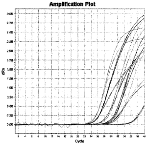
[00018] Figure 1 shows amplification curves for miRNAs obtained from a normal
sputum
sample.
[000191 Figure 2 shows amplification curves for a mixture of normal sputum and
A549 cells.
[000201 Figure 3A shows the amount of RNA amount ( g) in sputum during storage
at -20 C
over fourteen days.
[000211 Figure 3B shows the relative expression of miRNAs (miR-21, miR-92 and
U6) in
sputum samples during storage at -20 C over fourteen days.
[00022] Figure 3C shows the relative expression of miRNAs (miR-21, miR-92 and
U6) in
sputum samples spiked with A549 cells (105 cells in 200 l sputum) during
storage at -20 C over
fourteen days.
[00023] Figure 4A shows an amplification curve of miR-21 in A549 cells
extracted from
sputum samples. Figure 4B shows a standard curve of miR-21 for GM38 (normal
epithelium
fibroblast), A549 (non-small cell lung carcinoma), and MCF-7 (breast cancer)
cells.
[00024] Figures 5A-F show miRNA profiles for different types of cancers: (A)
plot showing
miRNA expression for different cell lines and miRNAs; (B) A549 cells (non-
small cell lung
carcinoma); (C) mes-1 cells (non-small cell lung carcinoma); (D) MCF-7 cells
(breast cancer);
(E) Du145 cells (prostate cancer); and (F) U118 cells (glioblastoma).
CA 02715518 2010-09-23
[00025] Figure 6 is a dendrogram showing hierarchical clustering based on the
fold of miRNA
expression of the cell lines.
[00026] Figure 7 shows miRNA amplification curves for normal and cancer cell
lines.
[00027] Figure 8 shows miRNA expression of normal (GM38) and cancer (A549,
H460,
H1792, mes-1, U118) cell lines.
[00028] Figure 9 is a dendrogram showing hierarchical clustering based on the
fold of miRNA
expression of normal (GM38) and cancer (A549, H460, H1792, mes-1, U118) cell
lines.
[00029] Figure 10 shows the amount of RNA ( g) in sputum samples from subjects
designated as D 1 and D2 (both cancer); D3 (successfully treated for cancer);
D4 (cancer-free);
and J16 (smoker control).
[00030] Figure 11 shows the relative quantity of selected miRNAs in sputum
samples from the
subjects of Figure 10.
[00031] Figure 12 shows the miRNA expression profiles of sputum samples from
the subjects
of Figure 10.
[00032] Figure 13 is a dendrogram showing hierarchical clustering based on
relatedness of
selected miRNAs.
[00033] Figure 14 is a dendrogram showing hierarchical clustering based on
miRNA
expression profiles (i.e., miR-21, miR-155, miR-210, miR-143, and hsa-miR-372)
of the sputum
samples of the subjects of Figure 10.
6
CA 02715518 2010-09-23
[00034] Figure 15 shows the miRNA expression profiles in sputum samples from
subjects
designated as Dl, D2, D6, D7 (cancer); D4, D5 (normal); J16 (normal smoker);
and SA-27
(normal smoker saliva).
[00035] Figure 16 is a dendrogram showing hierarchical clustering based on the
relatedness of
the sputum samples of the subjects of Figure 15.
Detailed Description of Preferred Embodiments
[00036] When describing the present invention, all terms not defined herein
have their
common art-recognized meanings. To the extent that the following description
is of a specific
embodiment or a particular use of the invention, it is intended to be
illustrative only, and not
limiting of the claimed invention. The following description is intended to
cover all alternatives,
modifications and equivalents that are included in the spirit and scope of the
invention, as
defined in the appended claims.
[00037] To facilitate understanding of the invention, the following
definitions are provided.
[00038] The term "microRNA" abbreviated as "miRNA" means a class of non-coding
RNA
molecules of about 19-25 nucleotides derived from endogenous genes which act
as post-
transcriptional regulators of gene expression. They are processed from longer
(ca 70-80 nt)
hairpin-like precursors termed pre-miRNAs by the RNAse III enzyme Dicer.
miRNAs assemble
in ribonucleoprotein complexes termed "miRNPs" and recognize their target
sites by antisense
complementarity, thereby mediating down-regulation of their target genes. Near-
perfect or
perfect complementarity between the miRNA and its target site results in
target mRNA cleavage,
whereas limited complementarity between the miRNA and the target site results
in translational
inhibition of the target gene.
[00039] The term "non-small cell lung carcinoma" abbreviated as "NSCLC" means
a group of
lung cancers comprising all the carcinomas except small cell carcinoma, and
including
7
CA 02715518 2010-09-23
adenocarcinoma of the lung, large cell carcinoma, and squamous cell carcinoma.
As used herein,
the terms "cancer" and "carcinoma" are synonymous and may be used
interchangeably.
[00040] The term "small cell lung carcinoma" abbreviated as "SCLC" means a
common,
highly malignant type of lung cancer, a form of bronchogenic carcinoma seen in
the wall of a
major bronchus, usually in a middle-aged person with a history of tobacco
smoking.
[00041] The term "sputum" means material (for example, mucus or phlegm) which
is
expectorated or sampled from the respiratory tract.
[00042] The term "threshold cycle" or "CT" means the fractional cycle number
at which
fluorescence has passed the fixed threshold.
[00043] In one embodiment, the present invention comprises sputum microRNA
expression
profiles which are associated with lung cancer. The expression profiles may be
used to screen a
subject for the disease. The microRNA expression profiles may be detected in
sputum from a
subject in order to discriminate lung cancer cells from epithelial or lung
fibroblast cells; lung
cancer from other cancer types; and between sub-types of lung cancer (i.e.,
either resistant or
sensitive to radiation and drugs). The miRNA expression profiles disclosed
herein are thus
diagnostic and prognostic markers of lung cancer.
[00044] In one embodiment, the invention comprises a method of screening a
subject for lung
cancer comprising the steps of.
a) obtaining a sputum sample from the subject; and
b) determining a subject microRNA expression profile from the sputum sample;
c) determining whether or not the subject has lung cancer by determining a
measure
of similarity or dissimilarity of the subject expression profile to at least
one
known lung cancer microRNA expression profile and a known control microRNA
expression profile;
wherein each of the subject and known expression profiles comprise the
expression levels of at
least two microRNAs.
8
CA 02715518 2010-09-23
[00045] In one embodiment, the method may distinguish between types of lung
cancer, by
comparing the miRNA expression profiles to known expression profiles from,
for.example, a
non-small cell lung carcinoma which is resistant to radiation and drugs,
and/or a non-small cell
lung carcinoma which is sensitive to radiation and drugs, and/or a small cell
lung carcinoma,
and/or a lung metastases originating from primary carcinomas of the breast,
prostate, brain, or
other tissue.
[00046] In one embodiment, the miRNA expression profile comprises the
expression levels of
at least two of miR-21, miR-92, miR-143, miR-145, miR-155, miR-210, miR-17-5p,
hsa-let-7a,
hsa-miR-182, hsa-miR-205, or hsa-miR-372. In one embodiment, the miRNA
expression profile
comprises the expression levels of miR-21, miR-155, miR-210, miR-143, and hsa-
miR-372. In
another embodiment, the miRNA expression profile comprises the expression
levels of either
miR-145 or hsa-miR-205, or both.
[00047] In one embodiment, the step of determining the miRNA expression
profile comprises
the use of a real-time quantitative PCR assay or micro-array analysis.
[00048] In one embodiment, the step of comparing the subject expression
profile comprises
grouping known microRNA expression profiles according to similarity of the
expressed
microRNAs and determining whether the subject expression profile is more
similar to one group
than the others. Similarity may be determined by statistical analysis, using
methods known to
one skilled in the art. In one embodiment, this grouping step comprises the
step of creating a
cluster diagram produced by hierarchical clustering. In one embodiment, the
cluster diagram
comprises a dendrogram.
[00049] miRNA profiling in sputum may be useful as a tool for cancer
detection,
classification, diagnosis and prognosis, since certain miRNA expression
profiles can be
correlated with certain cancers, or the absence of cancer. Thus, in the
development of one
embodiment of the present invention, it was determined whether a particular
miRNA expression
profile formed a signature or "barcode", which may be indicative of cancer
types and sub-types.
9
CA 02715518 2010-09-23
Twelve miRNA candidates were selected including eleven miRNAs related to
various cancer
types and one endogenous control miRNA (U6):
Table 1. miRNA candidates for miRNA profiling
miRNA Mature miRNA sequence SEQ Cancer type
ID:NO
miR-21 UAGCUUAUCAGACUGAUGUUGA 1 solid cancer cells
miR-92 UAUUGCACUUGUCCCGGCCUG 2 solid cancer cells
miR-143 UGAGAUGAAGCACUGUAGCUCA 3 all cancers except
miR-145 GUCCAGUUUUCCCAGGAAUCCCUU 4 stomach
miR-155 UUAAUGCUAAUCGUGAUAGGGG 5 lung, breast, colon
miR-210 CUGUGCGUGUGACAGCGGCUGA 6 lung, breast
miR-17-5p CAAAGUGCUUACAGUGCAGGUAGU 7 lung, breast, colon,
pancreas, prostate
hsa-let-7a UGAGGUAGUAGGUUGUAUAGUU 8 lung (NSCLC),
breast
hsa-miR-182 UUUGGCAAUGGUAGAACUCACA 9 lung (NSCLC)
hsa-miR-205 UCCUUCAUUCCACCGGAGUCUG 10 squamous cell lung
carcinoma
hsa-miR-372 AAAGUGCUGCGACAUUUGAGCGU 11 lung (NSCLC)
U6 GCAGGGGCCATGCTAATCTTCTCTGTATCG 12 Control
[00050) All twelve miRNAs in Table 1 exist in sputum from a subject without
lung cancer.
Table 2 shows the variation (OMCT SE) of duplicate samples which met the
required amount for
quantitative RT-PCR. Figure 1 shows the amplification curve of the miRNAs in
normal sputum.
CA 02715518 2010-09-23
Table 2. Accuracy and variation results of miRNAs in sputum
CT CT Mean CT SD ACT/miR-92 ACT /AACT
SE
miR-21 25.138 25.113 0.034 0.917 0.103
25.089
miR-145 29.494 29.648 0.219 5.452 0.239
29.803
miR-155 30.156 30.163 0.009 5.967 0.097
30.169
hsa-miR-205 27.966 27.748 0.308 3.552 0.323
27.530
miR-210 30.420 30.129 0.411 5.933 0.422
29.839
U6 24.797 24.812 0.022 0.616 0.100
24.828
miR-92 24.264 24.196 0.097 0.000 0.137
24.127
miR-17-5p 31.254 31.254 7.058
miR-143 36.604 36.515 0.125 12.319 0.158
36.427
hsa-miR-182 30.638 30.638 6.442
miR-392 36.532 36.744 0.299 12.548 0.315
36.956
hsa-let-7a 27.425 27.425 3.229
[00051] The stability of miRNAs in sputum was determined since sputum may
contain high
levels of RNase activity. Endogenous miRNA, rather than naked exogenous miRNA,
was used
as a marker due to its resistance to RNase activity. A549 cells (lung cancer -
NSCLC) were
spiked into sputum samples collected from a healthy subject to mimic sputum of
a subject with
lung cancer. Mixtures of 50 .d of sputum with different numbers of A549 cells
(0, 102, 104,108)
were used to determine the lowest detection limit (LOD), reproducibility and
variation. As
determined by UV, the LOD of A549 cells in sputum was 102 (Table 3). There is
no linear
relation between cell number and RNA concentration. From the difference
(0.0723-0.0426) of
104 and 102 cells, a very low LOD may be possible such as, for example, ten
cells.
11
CA 02715518 2010-09-23
Table 3. UV results of miRNA extracted from A549-sputum mixture
Cell + 50 l OD at 260 nm Mean OD OD SD Ratio 260/280 RNA ( g)
sputum
0.2384
106 0.2344 0.2364 0.0028 1.86 10.40
0.1006
104 0.0982 0.0982 0.0017 1.20 4.32
0.0734
102 0.0712 0.0723 0.0015 1.21 3.18
0.0440
0 0.0426 0.0426 0.0001 1.63 1.87
[00052] As determined by quantitative RT-PCR, the LOD of A549 cells in sputum
was 102 for
miR-21 (Table 4; Figure 2). The LOD of A549 cells in sputum was 104 for miR-
92. The
difference of LOD for different miRNA resulted from the different ambulance of
miRNA (i.e.,
related cell type and miRNA) in the same cells. Accuracy and variation of
quantitative RT-PCR
experiments were optimal (minimum CT SD 0.004). CT was not linear with cell
number in the
mixtures. As with the UV detection, a similar result was obtained in regard to
RNA amount and
cell number. With an increase of cell number added in sputum, the CT response
decreases
(equates to an increase in miRNA amount). Quantification of miR-21 expression
by real-time
RT-PCR showed that endogenous miRNA (miR-21) was clearly detected in the
samples.
12
CA 02715518 2010-09-23
Table 4. miRNA detection limit from quantitative RT-PCR
Cell + 50 l CT CT Mean CT SD Average SD
of sputum
20.14675
106 20.07125 20.109 0.053384
26.13311
104 25.9446 26.03885 0.133299 0.053
28.24446
miR-21 102 28.27815 28.26131 0.023825
28.87718
0 28.88331 28.88025 0.004336
LOD: Baseline-3 x SD = 28.883 (CT for sputum) - 0.159 = 28.724 > 28.2611
(CT for 102 cells)
LOD for miR-21 is 102 ins utum.
19.83921
106 19.03922 19.43922 0.565678*
23.8376
104 24.1454 23.9915 0.21765 0.133611
25.99367
miR-92 102 25.98723 25.99045 0.004549
26.18302
0 25.93039 26.05671 0.178633
LOD: Baseline-3 x SD = 26.057 (CT for sputum) - 0.399 = 25.718 > 25.990 (CT
for 102 cells) > 23.991
LOD for miR-92 is 104 in sputum.
* Noise spike: 1. bubbles in the reaction; 2. Evaporation during the
denaturation
step due to improper sealing or seal leaks
[00053] Sputum samples were stored at -20 C for 1-14 days. As measured by UV-
spectrometry, the RNA amount (jig) in sputum decreased over the fourteen day
period,
confirming that sputum contains high levels of RNase activity (Figure 3A).
However, there was
no effect on expression levels of miR-21, miR-92 or U6 in sputum samples
(Figure 3B), or
sputum samples spiked with A549 cells (105 cells in 200 l sputum) over the
same period (Figure
3C). The results indicate that endogenous miRNAs may be present in stable
forms in sputum
and reliably detected at various time points despite the presence of RNase
activity.
[000541 miRNA expression profiles were determined in vitro using normal and
cancer cell
lines (American Type Culture Collection, Manassas, VA, USA) (Table 5),
including four lung
13
CA 02715518 2010-09-23
cancer cell lines. MES-1 and H1792 lung cancer cell lines are resistant to
radiation and drugs in
contrast to A549 and H460 lung cancer cell lines which are sensitive to such
treatments.
Table 5. Cell lines and cancer type
Cell Line Cancer Type__
A549 Lung cancer (NSCLC)
MES-1 Lung cancer (NSCLC)
H460 Lung cancer (NSCLC)
H1792 Lung cancer (adenocarcinoma)
MCF-7 Breast cancer
DU145 Prostate cancer
U118 Glioblastoma
GM38 Normal epithelium fibroblast
MRC-5 Normal lunfibroblast
[00055] Initially, sputum samples collected from a healthy subject were spiked
in vitro with
cancer cells (A549 and MCF-7) to mimic the sputum of patients with cancer, and
to validate the
RT-PCR-based method described herein for determining miRNA expression profiles
in sputum.
GM38 cells (normal epithelium fibroblast) were used as the control. Briefly,
900 .tl of sputum
sample was added to duplicate 100 gl aliquots of each cell culture containing
either 3, 16, 80,
400, 2000 or 10000 cells. The total RNA was extracted according to the
procedure described in
Example 1. The expression of miRNAs was determined by quantitative RT-PCR as
described in
Examples 4 and 5. The results confirmed linearity between the RNA input and
the cycle
threshold (CL) values (Figures 4A and 4B). The miR-21 content among the three
cell lines was
greatest in MCF-7, followed by A549 and GM38 in that order. The assay had a
dynamic range
of at least six orders of magnitude (R.2 = 0.9986), and was capable of
detecting as few as three
cells in the sputum samples (Table 6).
Table 6. Lowest detection limit for miRNAs in A549 cancer cells
miRNA miR-21 miR-145 miR-205 miR-210 U6
LOD (cells) <10 400 80 <10 <10
[00056] The combinations of miRNAs expressed in the cancer cell lines, or the
controls, form
a profile indicative of a specific cancer type, or the absence of cancer.
miRNA expression
profiles were obtained for A549 (lung carcinoma sensitive to radiation and
drugs), mes-1 (lung
14
CA 02715518 2010-09-23
carcinoma resistant to radiation and drugs), MCF-7 (breast cancer), Du145
(prostate cancer), and
U118 (glioblastoma) cell lines (Figures 5A-5F). miR-145 was upregulated in
A549 cells and
downregulated in mes-1 cells. miR-145 thus shows potential as a marker in
sputum to
distinguish non-small cell lung carcinomas which are either sensitive (A549)
or resistant (mes-1)
to radiation and drugs. Further, hsa-miR-205, a highly specific marker for
squamous cell lung
carcinoma (NSCLC), was upregulated in A549 cells (Figure 5B) and absent in
Du145 cells
(prostate cancer) (Figure 5E).
[00057] In one embodiment, assessment of miRNA expression profiles was
performed using
hierarchical clustering which identifies relatively homogenous groups of cases
or variables based
on selected characteristics. In one embodiment, agglomerative hierarchical
clustering was used
which starts with each case as a cluster and combines new clusters until all
individuals are
grouped into one large cluster. Methods for combining clusters include, for
example, between-
group linkage, within-groups linkage, nearest neighbour, furthest neighbour,
centroid clustering,
median clustering, and Ward's method. In one embodiment, the method for
combining clusters
is between-group linkage. A convergence measure is used for measuring the
similarity and
divergence between cases (i.e., distance measuring). In one embodiment, the
convergence
measure is the Pearson correlation coefficient (denoted by r) which measures
the correlation or
linear dependence between two variables, giving a value between +1 and -1
inclusive. In one
embodiment, a correlation of less than 0.90 may be considered as indicative of
a significant
difference.
[00058] The distance at which the clusters are combined may be presented
graphically as a
dendrogram which connects the cases based upon their similarity scores. The
vertical lines show
joined clusters. The position of the line on the scale indicates the distance
at which clusters are
joined. The observed distances are resealed to fall into the range of 1 to 25;
however, the ratio of
the resealed distances within the dendrogram is the same as the ratio of the
original distances.
Cases grouped on a lower distance are considered more similar than cases
grouped at a higher
distance.
CA 02715518 2010-09-23
[00059] Cluster analysis was performed, designating the cell line as "case"
and the fold of
miRNA expression profiles as "variable" (Example 6). Table 7 sets out a
proximity matrix
which presents the information for the distances between the cases (cell
lines) and the clusters.
Table 7. Proximity matrix for cell lines
Case Correlation between vectors of values
(cell line) A549 mes-1 Du145 MCF-7 U118 GM38 MRC-5
A549 1.000 Ø993 0.703 0.234 -0.332 -0.470 0.000
mes-1 0.993 1.000 0.752 0.329 -0.259 -0.551 0.000
Du145 0.703 0.752 1.000 0.825 -0.359 -0.940 0.000
MCF-7 0.234 0.329 0.825 1.000 0.022 -0.967 0.000
U118 -0.332 -0.259 -0.359 0.022 1.000 0.113 0.000
GM38 -0.470 -0.551 -0.940 -0.967 0.113 1.000 0.000
MRC-5 0.000 0.000 0.000 0.000 0.000 0.000 1.000
Figure 6 is a dendrogram showing hierarchical clustering based on the fold of
miRNA expression
profiles of the cell lines. The seven cell lines are roughly separated into
four main clusters. The
first cluster contains the non-small cell lung carcinomas A549 (lung cancer
sensitive to radiation
and drugs) and mes-1 (lung cancer resistant to radiation and drugs). The
second cluster contains
Du145 (prostate cancer) and MCF-7 (breast cancer). MRC-5 (normal lung
fibroblast) is its own
cluster, clearly separated from all other clusters. A fourth cluster contains
U118 (glioblastoma)
and GM38 (normal epithelium fibroblast); however, as U118 and GM38 are grouped
together at
a higher distance, they are considered as less similar to each other. These
results indicate that
miRNA expression profiles can be used to discriminate between different types
of cancer (for
example, lung versus prostate and breast cancer).
[00060] It was determined whether miRNA expression profiles may distinguish
different sub-
types of a cancer. The miRNA expression profiles of four different lung cell
lines, namely A549
and H460 (both NSCLC sensitive to radiation and drugs), mes-1 (NSCLC resistant
to radiation
and drugs), and H 1792 (adenocarcinoma resistant to radiation and drugs), were
compared
(Figures 7-9). The GM3 8 (normal epithelium fibroblast) cell line was included
as a control,
while the U118 (glioblastoma) cell line was included as representing a
different cancer type (i.e.,
brain cancer). Cluster analysis was performed, designating the cell line as
"case" and the fold of
16
CA 02715518 2010-09-23
miRNA expression profile as "variable." Table 8 sets out a proximity matrix
which presents the
information for the distances between the cases (cell lines) and the clusters.
Table 8. Proximity matrix for lung cell lines
Case Correlation between vectors of values
(cell line) A549 H460 H1792 mes-1 U118 GM38
A549 1.000 0.925 0.429 0.440 0.577 0.000
H460 0.925 1.000 0.485 0.472 0.721 0.000
H1792 0.429 0.485 1.000 0.709 0.607 0.000
mes-1 0.440 0.472 0.709 1.000 0.141 0.000
U118 0.577 0.721 0.607 0.141 1.000 0.000
GM38 0.000 0.000 0.000 0.000 0.000 1.000
Figure 9 is a dendrogram showing hierarchical clustering based on the fold of
miRNA expression
profiles of the cell lines. The six cell lines are roughly separated into four
main clusters. The
first cluster contains the lung carcinomas A549 and H460 (both NSCLC sensitive
to radiation
and drugs). U118 (glioblastoma) is its own cluster, clearly separated from all
other clusters
(similarity <0.9). The third cluster contains H1792 (adenocarcinoma resistant
to radiation and
drugs) and mes-1 (NSCLC resistant to radiation and drugs). GM38 (normal
epithelium
fibroblast) is its own cluster, clearly separated from all other clusters
(similarity <0.9). These
results indicate that miRNA expression profiles can be used to discriminate
between different
sub-types of cancer (for example, NSCLC which are either resistant or
sensitive to radiation and
drugs).
[00061] miRNA expression profiles obtained from sputum samples may be
diagnostic and
prognostic markers of lung cancer, by comparison with known expression
profiles. miRNA
expression profiles were determined using sputum samples collected from five
subjects
designated as D 1 and D2 (both cancer); D3 (successfully treated for cancer);
D4 (cancer-free);
and J16 (smoker control) (Table 9).
17
CA 02715518 2010-09-23
Table 9. Data for sputum samples from subjects
Weight of Weight of Weight of Mean STD of Density of
1St 200 1 2d 200 1 3rd 200 1 weight weights sputum
Dl 0.2158 0.1948 0.2130 0.2079 0.0114 1.0393
D2 0.2050 0.2054 0.1914 0.2006 0.0080 1.0030
D3 0.2199 0.1983 0.2030 0.2071 0.0114 1.0353
D4 0.2377 0.1673 0.2028 0.2026 0.0352 1.0130
J16 0.2600 0.2516 0.2075 0.2397 0.0282 1.1985
[00062] The sputum samples were then homogenized (Example 2) for RNA
extraction as
described in Example 3. The amount of RNA ( g) and RNA concentration in 200 l
of sputum
sample from each subject was calculated (Example 3; Figure 10; Table 10).
Figure 10 and Table
reflect the quantity of all RNA including miRNA which comprises only 0.01 % of
all RNA
when extracted with the method described in Example 3.
Table 10. RNA concentration and quantity as determined using UV
spectrophotometer
OD1 OD2 mean STD Concentration RNA ( g) RNA STD
ng/ )
D1 0.2161 0.2252 0.2207. 0.006 353.04 32.4796 0.4632
D2 0.1393 0.141 0.1402. 0.001 224.24 20.6300 0.0865
D3 0.0501 0.0505 0.0500 0.0002 80.48 7.40416 0.0204
D4 0.0499 0.0503 0.0500 0.0002 80.16 7.37472 0.0204
J16 0.0453 0.0454 0.0907 7.07E-05 72.56 6.6755 0.0051
[00063] The relative quantity of selected miRNAs in 200 l of sputum sample
from each
subject was determined using quantitative RT-PCR (Figure 11; Examples 4 and
5). Figure 12
and Table 11 show that the miRNA profiles of sputum samples from each subject
appear to
differ.
18
CA 02715518 2010-09-23
Table 11. miRNA expression in sputum samples
D1 D2 D3 D4 J16
miR-21 1.7933 3.4762 0.6975 0.0000 1.0000
miR-145 0.2438 6.1787 0.5133 0.0678 1.0000
miR-155 1.0984 0.0090 0.3347 0.1046 1.0000
hsa-miR-205 0.0481 0.1547 0.0141 0.4936 1.0000
miR-210 0.0596 0.1528 0.1905 0.0702 1.0000
miR-17-5p 1.3634 3.2170 0.2932 0.3550 1.0000
miR-143 0.7846 0.7470 0.0563 0.0484 1.0000
hsa-miR-182 0.1985 0.4678 0.0251 0.0745 1.0000
hsa-miR-372 0.0206 0.2305 0.4129 0.7435 1.0000
hsa-let-7a 0.0048 0.0101 0.0381 0.1170 1.0000
[00064] Table 12 sets out a proximity matrix which presents the information
for the distances
between the cases (miRNAs) and the clusters.
Table 12. Proximity matrix for miRNAs
Matrix File hi ut
miR- miR- miR- hsa-miR- miR- miR-17- miR- hsa-miR- hsa-miR- hsa-let-
21 145 155 205 210 5 143 182 372 7a
miR-21 1.000 -0.116 -0.133 0.579 -0.587 0.882 -0.340 0.972 0.253 -0.244
miR-145 -0.116 1.000 0.479 0.609 0.018 -0.490 0.285 -0.211 0.417 0.497
miR-155 -0.133 0.479 1.000 0.563 0.732 -0.072 0.850 -0.096 0.899 0.981
hsa-miR-205 0.579 0.609 0.563 1.000 -0.031 0.353 0.376 0.578 0.814 0.526
miR-210 -0.587 0.018 0.732 -0.031 1.000 -0.286 0.901 -0.452 0.520 0.801
miR-17-5p 0.882 -0.490 -0.072 0.353 -0.286 1.000 -0.171 0.929 0.273 -0.176
miR-143 -0.340 0.285 0.850 0.376 0.901 -0.171 1.000 -0.197 0.777 0.926
hsa-miR-182 0.972 -0.211 -0.096 0.578 -0.452 0.929 -0.197 1.000 0.320 -0.179
hsa-miR-372 0.253 0.417 0.899 0.814 0.520 0.273 0.777 0.320 1.000 0.874
hsa-let-7a -0.244 0.497 0.981 0.526 0.801 -0.176 0.926 -0.179 0.874 1.000
[00065] Figure 13 is a dendrogram showing hierarchical clustering based on the
relatedness of
the miRNAs. The miRNAs are roughly separated into five main clusters. The
first cluster
contains miR-210 and hsa-let-7a. hsa-miR-182 is clearly separated from all
other clusters. A
third cluster contains hsa-miR-372 and hsa-miR-205. A fourth cluster contains
miR-155 and
miR-143. A fifth cluster contains miR-21, miR-17-5p and miR-145.
[00066] In one embodiment, miRNA expression profiles may comprise the
expression level of
at least two miRNAs, which are grouped in different clusters from each other.
19
CA 02715518 2010-09-23
[00067] From among the miRNAs analyzed above, five miRNAs (i.e., miR-21, miR-
155,
miR-210, miR-143, and hsa-miR-372) were selected for clustering analysis based
on miRNA
expression profiles of the sputum samples of the subjects. These miRNAs are
from different
clusters, except for miR-155 and miR-143, which are in the same cluster.
Although hsa-miR-
182 is in a separate cluster, it was not chosen as it is not associated with a
lung cancer. Table 13
sets out a proximity matrix which presents the information for the distances
between the cases
(sputum samples of the subjects) and the clusters.
Table 13. Proximity matrix for sputum samples
Correlation between vectors of values
D1 D2 D3 D4 J16
D1 1.000 0.779' 0.568 -0.600 0.000
D2 0.779 1.000 0.731 -0.373 0.000
D3 0.568 0.731 1.000 0.103 0.000
D4 -0.600 -0.373 0.103 1.000 0.000
J16 0.000 0.000 0.000 0.000 1.000
[00068] Figure 14 is a dendrogram showing hierarchical clustering based on the
relatedness of
the sputum samples. The sputum samples are roughly separated into three main
clusters. The
first cluster contains D 1 and D2 (both cancer) with the distance less than 0,
indicating their high
level of relatedness. D3 (treated for cancer) is separated from all other
clusters. A third cluster
contains D4 (cancer-free) and J16 (normal control), although the similarity
between D4 and J16
is zero (Table 13). Overall, D1 and D2 show significant difference with D3, D4
and J16,
indicating the miRNA expression profiles in sputum samples have potential in
distinguishing
between subjects with lung cancer and those without the disease.
[00069] In a further study, miRNA expression profiles were determined using
sputum samples
collected from subjects designated as Dl, D2, D6, D7 (cancer); D4, D5
(normal); J16 (normal
smoker); and SA-27 (normal smoker saliva). MRC-5 (normal lung fibroblast cell
line) was used
as the reference sample, and U6 as the endogenous control.
CA 02715518 2010-09-23
[00070] Figure 15 and Table 14 show the miRNA expression profiles in the
sputum samples
from each subject. From among the miRNAs analyzed in Table 14, five miRNAs
(i.e., miR-21,
miR-155, miR-210, miR-143, and hsa-miR-372) were selected for clustering
analysis based on
miRNA expression profiles of the sputum samples of the subjects. Table 15 sets
out a proximity
matrix which presents the information for the distances between the cases
(sputum samples of
the subjects) and the clusters.
[00071] Figure 16 is a dendrogram showing hierarchical clustering based on the
relatedness of
the expression profiles from the sputum samples. The expression profiles are
roughly separated
into four main clusters. The first cluster contains Dl, D2, D6 and D7 (all
cancer subjects), with
the distance less than 0, indicating their high level of relatedness. The
similarity of each pair is
greater than 0.90 (i.e., minimum 0.901 (Dl-D6) to maximum 0.993 (Dl-D2)). A
second cluster
contains D4, D5 and J16 (non-cancer subjects). MRC-5 (normal lung fibroblast
cell line) is
separated from all other clusters. SA-27 (normal smoker saliva) is also
separated from all other
clusters. ' Table 16 summarizes the diagnostic results. Overall, the results
indicate that the
miRNA expression profiles in sputum samples have potential in distinguishing
between normal
and cancer subjects.
21
CA 02715518 2010-09-23
O
r O T ~O O1
000 cl~ NO0 %0 N\ON 00
CTO~O 'r ON 0
0000 o oo 00000000 NO - N O O O00
O
co, M O O
O 0 N N < O O O O O Q O CO .-~
N O O O M
t, o O N N
O O W) "O .4 ~,O N 0
O O W) m CA 1O O
M N 00 ri ~ v1 OO 10 0 ~O N O O 00 L-- O
Or r0 r N MNM.0ON
0
O v1-NMI-O=-+ A010~0 U1 5 ON 0
OOCD OCOO
o oot- N
O O r c r 0
c) Ln O 0 0 M 00 00
M 0 00 O
At on r+ t` 0 o/ i
oNO~NO r14,-4 tiOO~00 t-0
.-~ M r-+ N 0. -+ ~ ~t =-i ri A O 00 0. et O on
0
0 0 9 C 0 0 .-+
M 00
OOOOrd0r1.4 P
v10 ev
^i OOOVO d 0\---1000 ,?
. %'0 O M-* t- N "60vt co 4-1 a 0
`4-.4 - 00
0N~OO\iNN-f "' 0
V) 0 o~ r
A~r1~~o.0,00
q o00 d
d o000'-4 It co
a ooo~nr--~rnNO
"O N 000 OMO 01 en M ' O C."
40 +r+Ov1'.OOM N NCO-
f~ O ~ O p
0 0 0 0 0 c, 0
~OOO~NetO~O 400 Vc!
0- O O O O O C r+
No
c~ ,6 In A
N CT .-+ O '~
O M M .M-~ M m r+ r+ N N
<C O O a O
M O O \O 00 o1 N r 0 ~ M d 000 O 8
G~~-4NNMO C700r- O
0 0
0 0 '1 1-4 vi
s~ o
^+ v1 N
(..~i N O Or1 000 00 M 'd "t N
d d ~O ~D p
c:) V) ,Ikn AN^'o
~~NN~on0M~ [~ 66
t0T N 00 N 00 M O
v~ r+ N N '-- c en e N en
v1
N N O O N 0 N O
~00N cr00~MOO NAa0
.-ion r- enONOO
00000000C)-4 O`''
V 000v1N~O000
o000d' M00000
OONNv'1rNro0
O- 000 N M.-+O
0 0 0 r+ 0 0 0 '--4 --i 00
AO
OOOd'~OMMN0
N 00O N 0 0 Q' M 000 O
N O
~i '-+ d O N V1 Ito)
N' AAAti AAA
.-.N~`atn~ord
AAA~AAAv~
CA 02715518 2010-09-23
Table 16. Diagnostic results from cluster analysis of miRNA profiles
Subject Dl D2 D4 J16 MRC-5 D5 D6 D7 SA-27
Sample cancer cancer normal Normal Normal normal cancer cancer Normal
smoker lung smoker
cells saliva
Diagnosis positive positive negative positive positive negative
[00072] In a further study, further diagnostic results demonstrate the
prognostic utility of
embodiments of the present invention by confirmation with actual diagnoses.
Table 17 shows
updated results from those patients and controls listed above:
Table 17. Summary of actual diagnoses corresponding (100%) to the blinded
Cluster
Analysis of miRNA profiling
Serial 1
sample o 2 3 5 6 7 8
Blinded D1 D2 D3 D4 D5 D6 D7
Lung Normal Normal
Actual Lung Lung Cancer/ Lung
Diagnosis Cancer Cancer Treatment- Non- Non- Cancer Lung Cancer
Controlled smoker smoker
Micro RNA Positive Positive Negative Negative Negative Positive Positive
Status
Sex M M M M M M M
Although not shown in Table 17, serial sample nos. 4 and 9 were control
subjects (blinded codes
C 1 and C2), both normal male smokers, with negative status from miRNA
expression profile
clustering.
[00073] In particular, the results for D3 as seen in Figure 14 and Table 17
indicate that the
treatment administered to D3 was effective. Upon treatment, the miRNA
expression profile
became less similar to the D 1 and D2 cluster, as seen in Figure 14.
Therefore, in one
embodiment, miRNA expression profile clustering may be used to monitor the
effectiveness of
treatment of subjects with lung cancer. Before treatment, the miRNA expression
profile of a
subject with lung cancer will be more similar, and will be grouped with,
expression profiles from
known lung cancers. After successful treatment, the miRNA expression profile
will change to be
less similar to the lung cancer profile, and more similar to normal profiles.
23
CA 02715518 2010-09-23
[00074] Exemplary embodiments of the present invention are described in the
following
Examples, which are set forth to aid in the understanding of the invention,
and should not be
construed to limit in any way the scope of the invention as defined in the
claims which follow
thereafter.
[00075] Example 1- RNA Extraction from Cell Lines
A TagMan MicroRNA Cell-to-CTTM Kit (Applied Biosystems Inc., Foster City, CA,
USA) was
used to extract RNA from the cell lines in accordance with the manufacturer's
instructions.
Briefly, 5x10⁵ cultured cells were plated into 96-well plates for six hours to
allow attachment,
and then washed with 4 C phosphate buffered saline. 49 gl of Lysis Solution
and 1 l DNase I
(provided by the supplier) was added into each well, and cells were incubated
for eight minutes
at room temperature. 5 l of Stop Solution (provided by the supplier) was
added to the lysate
and incubated for two minutes at room temperature to inactivate the lysis
reagents so that they
would not inhibit reverse transcription (RT) or polymerase chain reaction
(PCR). All cell lysates
were stored on ice for less than two hours or at -70 C for subsequent RT.
[00076] Example 2 - Collection and Homogenization of Sputum
Sputum samples were collected, stored at 4 C, and processed within one week of
collection. The
sputum was separated from saliva. A 200 l sample of sputum was transferred
into a 1.5 ml
nuclease-free tube. 400 1 of SputolysinTM solution (0.1 mg/mL, Sigma, Canada)
was added to
the tube, vortexed until the viscous sputum was lysed, and incubated at 37 C
for 30 minutes.
The homogenized sputum was stored at -20 C until processed for RNA extraction.
[00077] Example 3 - RNA Extraction from sputum
1000 L of TrizolTM (Invitrogen, USA) was added to the sputum sample tube
(containing 200 1
of homogenized sputum), mixed with a pipet to re-suspend the sample, and
vortexed for 20
seconds. 200 pl of chloroform was added, and the mixture was vortexed for 20
seconds and left
at room temperature for five minutes. The tube was centrifuged for 15 minutes
at 4 C to separate
the sample into an aqueous phase and a red organic phase. The aqueous phase
(approximately
500 l) was carefully transferred into a fresh 1.5 ml tube. 4 gl of glycogen
co-precipitant
24
CA 02715518 2010-09-23
(Ambion Inc., Austin, TX, USA) and 500 Al of isopropyl were added, gently
mixed, and left at
room temperature for twenty minutes. The tube was then centrifuged at 12000
rpm for 10
minutes, and for 15 minutes at 4 C to co-precipitate RNA with glycogen. The
RNA was
carefully removed, washed with 75% ethanol at 4 C, and centrifuged at 8000 rpm
at 4 C for two
minutes. RNA washing was repeated twice. The RNA was dissolved in 105 l of
nuclease-free
water and stored at -20 C until processed for reverse transcription and RT-
PCR. To determine
the RNA concentration and amount, 5 l of RNA was diluted with 95 l of
nuclease-free water,
and measured using a Beckman DUTM 7000 spectrophotometer (Beckman Coulter,
Fullerton,
CA, USA). The amount of RNA ( g) in a 200 0 sample may range from about 0.5 g
to 3.0 g.
The ratio of adsorption at 260 nm over 280 nm should be greater than 1Ø
The concentration of RNA is calculated as:
RNA ( g/ l) = OD260 x 20 x 40 [1]
The amount of RNA is calculated as:
Amount ( g) = OD260 x 20 x 40 x 0.1 [2]
[00078] Example 4 - Reverse Transcription
A TagMan MicroRNA Reverse Transcription Kit, primers, and StepOnePlusTM Real
Time
PCR system (Applied Biosystems Inc., Foster City, CA, USA) were used for miRNA
reverse
transcription in accordance with the manufacturer's instructions. Briefly, the
number of RT
reactions was calculated and a RT Master Mix was assembled for all the
reactions plus about
10% overage in a nuclease-free microcentrifuge tube on ice:
Table 17. RT Master Mix for single primer reactions
Component Each reaction
l OX RT Buffer 1.5 1
dNTP Mix 0.15 l
RNase Inhibitor 0.19 1
MultiScribe RT 1.0 1
Nuclease-free Water 4.16 1
Final Volume RT Master Mix 7.0 l
CA 02715518 2010-09-23
The components were mixed gently and placed on ice. The RT Master Mix was
distributed to
nuclease-free PCR tubes. 3.0 l of miRNA-specific primer was added to each
aliquot of RT
Master Mix followed by 5 l of sample lysate for a final 15 l reaction volume,
followed by
mixing and centrifugation at 1500 rpm for two minutes. The RT thermal cycler
program was run
according to the following settings:
Table 18. Thermal cycler settings for RT
Stage Reps Temperature Time
Primer annealing 1 1 16 C 30 min
Reverse transcription 2 1 42 C 30 min
RT inactivation 3 1 85 C 5 min
Hold 4 1 4 C indefinite (50 min
The completed RT reactions were stored at -70 C for subsequent quantitative RT-
PCR.
[00079] Example 5 - Quantitative RT-PCR
The comparative threshold cycle method (MOCT) was applied for miRNA profiling,
using normal
cell lines (GM38 or MRC-5) as reference samples and miR-92 as an endogenous
control. A
standard curve was generated to determine the limit of detection,
reproducibility and variation.
A TagMan Universal PCR Master Mix, Tagman MicroRNA Assay, primers, probes,
and
StepOnePlusTM Real Time PCR system (Applied Biosystems Inc., Foster City, CA,
USA) were
used for qRT-PCR in accordance with the manufacturer's instructions (Table
19).
26
CA 02715518 2010-09-23
C/] H O~
03
31 00
Q v. Ifl
tD ti h F :~ a
04.
r '=F' .yam .y~ ~,!
r r
~ ~" OC d' ~. d' K ~ ff K ' CC C' 'Q' a d' dC ' ~ .~ ~ r ~.a , Cd- =--3 th Q
~Ai m ti
-f r:
'ZI
r a!V cn c to m m T r r r .~ e- r r r r ~..~ VI Z
27
CA 02715518 2010-09-23
Briefly, the number of PCR assays was calculated and a PCR Cocktail was
assembled for all the
reactions plus about 10% overage in a nuclease-free microcentrifuge tube on
ice:
Table 20. RT-PCR Cocktail for Single Primer RT Reactions
Component Each reaction
Ta man Master Mix (2X) 10.0 1
Ta man MicroRNA Assay 1.0 gl
Nuclease-free water 7.67 1
Final volume RT-PCR Master Mix 18.67 l
The components were mixed gently and placed on ice. The PCR Cocktail was
distributed into
PCR tubes. 1.33 l of the RT product from Example 4 was added to each aliquot
of PCR
Cocktail for a final 20 gl reaction volume, followed by mixing and
centrifugation at 1500 rpm
for two minutes. The PCR instrument was run according to the following
settings:
Table 21. PCR Cycling Conditions
Stage Reps Temperature Time
Enzyme activation 1 1 95 C 10 min
PCR cycle 2 40 95 C 15 sec
60 C 1 min
[00080] Example 6 - Cluster Analysis
SPSS 13.0 software (SPSS Inc., Chicago, IL, USA) was used for hierarchical
clustering
according to the following steps:
a) Import data from an ExcelTM spreadsheet to SPSS 13.0 software;
b) Select Analysis for Cliffify and open Hierarchical menu;
c) Select Case for cluster analysis and statistics, and plots for Display;
d) Open Statistic and select Agglomeration Schedule and Proximity Matrix;
e) Open Plot and select All Cluster;
t) Open Method and select "between-group linkage" as the cluster method,
select
"Pearson-correlation" as the convergence measure; and
g) Start cluster analysis and retrieve output results.
28
CA 02715518 2010-09-23
[00081] As will be apparent to those skilled in the art, various
modifications, adaptations and
variations of the foregoing specific disclosure can be made without departing
from the scope of
the invention claimed herein.
References
[00082] The following references are incorporated herein by reference (where
permitted) as if
reproduced in their entirety. All references are indicative of the level of
skill of those skilled in
the art to which this invention pertains.
Barbarotto, E.; Schmittgen, T.D. and Calin, G.A. (2008) MicroRNAs and cancer:
profile,
profile, profile. Int. J. Cancer. 122(5):969-77.
DesJardins, L.E. (2000) Isolation of M. tuberculosis RNA from sputum. In
Methods in
Molecular Medicine, vol. 48: Antibiotic Resistance Methods and Protocols, ed.
S.H.
Gillespie, pp. 133-136.
Iorio, M.V.; Ferracin, M.; Liu, C.G.; Veronese, A.; Spizzo, R.; Sabbioni, S.;
Magri, E.; Pedriali,
M.; Fabbri, M.; Campiglio, M.; M6nard, S.; Palazzo, J.P.; Rosenberg, A.;
Musiani, P.;
Volinia, S.; Nenci, I.; Calin, G.A.; Querzoli, P.; Negrin, M. and Croce, C.M.
(2005)
MicroRNA gene expression deregulation in human breast cancer. Cancer Res.
65(16):7065-70.
Jay, C.; Nemunaitis, J.; Chen, P.; Fulgham, P. and Tong, A.W. (2007) miRNA
profiling for
diagnosis and prognosis of human cancer. DNA Cell Biol. 26(5):293-300. Review.
Johnson, S.M.; Grosshans, H.; Shingara, J.; Byrom, M.; Jarvis, R.; Cheng, A.;
Labourier, E.;
Reinert, K.L.; Brown, D. and Slack, F.J. (2005) RAS is regulated by the let-7
microRNA family. Cell 120(5):635-47.
Lacroix, J.; Becker, H.D.; Woerner, S.M.; Rittgen, W., Drings, P. and von
Knebel Doebertiz, M.
(2001) Sensitive detection of rare cancer cells in sputum and peripheral blood
samples of
patients with lung cancer by preprogrp-specific RT-PCR. Int. J Cancer 92:1-8.
Li, R.; Todd, N.W.; Qiu, Q.; Fan, T.; Zhao, R.Y.; Rodgers, W.H., et al. (2007)
Genetic deletions
in sputum as diagnostic markers for early detection of stage I non-small cell
lung cancer.
Clin. Cancer Res. 13:482-487.
Markou, A.; Tsaroucha, E.G.; Kaklamanis, L.; Fotinou, M.; Georgoulias, V. and
Lianidou, E.S.
(2008) Prognostic value of mature microRNA-21 and microRNA-205 overexpression
in
29
CA 02715518 2010-09-23
non-small cell lung cancer by quantitative real-time RT-PCR. Clin. Chem.
54(10):1696-
704. Epub 2008 Aug 21.
Qiu, Q.; Todd, N.W.; Li, P.; Peng, H.; Liu, Z.; Yfantis, H.G.; Katz, R.L.;
Stass, S.A. and Jiang,
F. (2008) Magnetic enrichment of bronchial epithelial cells from sputum for
lung cancer
diagnosis. Cancer 114:275-283.
Rabinowits, G.; Gercel-Taylor, C.; Day, J.M.; Taylor, D.D. and Kloecker, G.H.
(2009) Exosomal
microRNA: a diagnostic marker for lung cancer. Clin. Lung Cancer 10(l):42-6.
Rosenfeld, N.; Aharonov, R.; Meiri, E.; Rosenwald, S.; Spector, Y.; Zepeniuk,
M.; Benjamin,
H.; Shabes, N.; Tabak, S.; Levy, A.; Lebanony, D.; Goren, Y.; Silberschein,
E.; Targan,
N.; Ben-Ari, A.; Gilad, S.; Sion-Vardy, N.; Tobar, A.; Feinmesser, M.;
Kharenko, 0.;
Nativ, 0.; Nass, D.; Perelman, M.; Yosepovich, A.; Shalmon, B.; Polak-Charcon,
S.;
Fridman, E.; Avniel, A.; Bentwich, I.; Bentwich, Z.; Cohen, D.; Chajut, A.;
and
Barshack, I. (2008) MicroRNAs accurately identify cancer tissue origin. Nat.
Biotechnol. 26(4):462-9. Epub 2008 Mar 23.
Thunnissen, F.B. (2003) Sputum examination for early detection of lung cancer.
J Clin. Pathol.
11:805-810.
Volinia, S.; Calin, G.A.; Liu, C.G.; Ambs, S.; Cimmino, A.; Petrocca, F.;
Visone, R.; Iorio, M.;
Roldo, C.; Ferracin, M.; Prueitt, R.L.; Yanaihara, N.; Lanza, G.; Scarpa, A.;
Vecchione,
A.; Negrini, M.; Harris, C.C. and Croce, C.M. (2006) A microRNA expression
signature
of human solid tumors defines cancer gene targets. Proc Natl Acad Sci USA
103(7):2257-61. Epub 2006 Feb 3.
Yanaihara, N.; Caplen, N.; Bowman, E.; Seike, M.; Kumamoto, K.; Yi, M.;
Stephens, R.M.;
Okamoto, A.; Yokota, J.; Tanaka, T.; Calin, G.A.; Liu, C.G.; Croce, C.M.; and
Harris,
C.C. (2006) Unique microRNA molecular profiles in lung cancer diagnosis and
prognosis. Cancer Cell 9(3):189-98.
Xie, Y.; Todd, N.W.; Liu, Z.; Zhan, M.; Fang, H.; Peng, H.; Alattar, M.;
Deepak, J.; Stass, S.A.
and Jiang, F. (2009) Altered miRNA expression in sputum for diagnosis of non-
small
cell lung cancer. Lung Cancer 2009 May 13. [Epub ahead of print]