Note: Descriptions are shown in the official language in which they were submitted.
CA 02715543 2010-08-13
WO 2009/100531 PCT/CA2009/000171
ADJUSTABLE TISSUE OR NERVE CUFF AND METHOD OF USE
BACKGROUND OF THE INVENTION
The invention relates to the field of surgically implantable devices and
methods in the biomedical field. Implantable cuffs have been used for the
stimulation and recording of biological tissues, particularly nerves.
Stimulation of the
nervous system with nerve cuffs can result in recovery of lost sensory or
motor
function in individuals with neurological deficits. An example of such an
application
is the FreehandTM stimulator (Neurocontrol Corporation, Ohio, USA) that can
restore
a degree of hand function in an individual with a spinal cord injury.
Recording has
also been performed with implantable cuffs. Recording nerve function can relay
vital
information back to a processor that assists in decision-making based on the
activity
of the nerve. For example, in sleep apnea, patients implanted with nerve cuffs
rely
on the nerve cuff to be used for recording as well as stimulation when
necessary.
By targeting a nerve with an implanted nerve cuff, much less electrical
current is
required than for intra-muscular stimulation or surface stimulation.
Intramuscular
stimulation involves using an electrode directly in the muscle, whereas
surface
stimulation utilizes electrodes at the skin surface to activate nerves in the
general
area of interest. Surface stimulation is much less selective of the muscles it
can
stimulate as compared to nerve cuffs.
Most implantable electro-neuroprosthetics that target peripheral nerves use
some type of nerve cuff. Currently there are three primary types of nerve
cuffs used
to stimulate nerves with an electro-neuroprosthesis, namely C-shaped cuffs,
helical
cuffs, and nerve reshaping cuffs.
C-shaped nerve cuff electrodes are named for their c-shaped cross section.
They range from split cylinder, spiral and multi-compartmental designs. An
example
is seen in US Patent 6,600,956 to Maschino et al. Generally the cuff is made
of an
electrically insulative substrate with one or more imbedded electrically
conductive
elements designed to interact electrically with the nerve. The preferred
substrate is
biocompatible, the most common material being silicone rubber. The main draw
back to c-shaped nerve cuffs is that the internal diameter of the nerve cuff
needs to
be estimated prior to the surgery, and hence it can result in loose fitting
cuffs if made
1
CA 02715543 2010-08-13
WO 2009/100531 PCT/CA2009/000171
too large, or too constricting cuffs resulting in nerve damage if made too
small. This
can greatly increase costs as multiple sizes need to be made available to the
surgeon to minimize problems. Spiral electrodes that are self curling
alleviate the
size problem and can be removed with minimal force.
Helical cuffs such as shown in US Patent 5,964,702 to Grill et al. are built
much like spiral cuffs from a self curling substrate, but they are cut to look
like a
spring. One main draw back is that they need to be wrapped around the nerve,
which can be a time consuming process. Furthermore, helical cuffs rely
entirely on
the substrate properties to close properly as there is no closing mechanism.
This
can result in inappropriate contacts being made to the nerve. Helical cuffs
are also
susceptible to size constraints.
Nerve reshaping cuffs reshape the nerve to fit the cuff's internal space. An
example of a nerve reshaping cuff is illustrated in US Patent 5,634,462 to
Tyler et al.
This type of cuff relies on a force being applied to the nerve itself to
squeeze it into a
desired shape, either by using rigid structures or corrugations in the nerve
cuff. If
appropriate pressure is used, and enough space provided for the nerve, there
is a
possibility of using multiple electrically conductive units to isolate and
stimulate only
certain parts of the nerve. However, one risk is that damage to the nerve can
occur
during the installation. As well, a possible tensile strength decrease can
weaken the
nerve. In the case of large rigid structures near the nerve there is a further
risk for
increasing the incidence of inflammation in response to the mechanical
aggravation
of the tissues. The rigidity needed to shape the nerve in a corrugated nerve
cuff
such as in US Patent 5,634,462 also limits the ability of the cuff to
accommodate
different nerve sizes, so as above, different size cuffs must be provided for
different
nerve sizes. Adjusting the cuff intra-operatively to re-position conductive
elements,
or to adjust for size, is resisted by the design and rigidity of the
structure. Finally, the
corrugations of this type of device are designed to minimize contact points
with the
nerve, which for some applications limits the nerve surface which can be
directly
contacted with electrical contacts of nerve interacting devices.
In spite of the large number of available nerve cuff designs, there remains a
need for an adjustable size tissue cuff, that can be quickly installed, intra-
operatively
adjusted, and which places just the right amount of pressure on the nerve or
tissue
2
CA 02715543 2010-08-13
WO 2009/100531 PCT/CA2009/000171
to allow for ideal contact with conductive units without damaging the nerve.
As well,
given the many different functional electrical devices currently available
that rely on
peripheral nerve stimulation, there is a further need for a nerve cuff that is
not limited
to a single type of electrode lead design.
SUMMARY OF INVENTION
In one broad aspect, the invention provides an implantable, circumferentially
adjustable tissue cuff for circumferential attachment to an internal body
tissue. The
cuff includes a flat, thin, elastomeric strap formed of a biocompatible non-
conductive
material, the strap being elongated along a longitudinal axis, the strap
having a body
portion connected between a tail end portion and a head end portion and a
length in
excess of a circumference of the body tissue. The tail end portion and the
head end
portion are configured for adjustable length fastening one to the other when
wrapped
around the body tissue. Either of the following configurations may be
included:
a) The tail end portion may be formed with a plurality of longitudinally
spaced,
laterally paired locking projections while the head end portion is formed with
one or
more locking apertures; or
b) The tail end portion may be formed with a plurality of longitudinally
spaced
locking apertures while the head end portion is formed with one or more
laterally
paired locking projections.
In either configuration, each of the laterally paired locking projections is
shaped to
allow for passage through the locking apertures by flexing of the locking
projections
in an insertion direction through the locking aperture, and to restrict
movement in a
reversing direction through the locking aperture.
In another broad aspect, the invention provides a tissue cuff apparatus to
enable circumferential attachment of a tissue interacting device to an
internal body
tissue. The apparatus includes the above tissue cuff and one or more
implantable
tissue interacting devices attached to, imbedded in, or printed on the body
portion of
the strap. The tissue interacting device includes one or more conductive
elements
adapted to be in conducting proximity to the body tissue when the strap is
wrapped
around the body tissue. The tissue interacting device may be one adapted to
stimulate or record the body tissue, in which case, the conductive element is
3
CA 02715543 2010-08-13
WO 2009/100531 PCT/CA2009/000171
adapted to respond to one or more of electrical, thermal, auditory,
vibrational, light or
fluid stimulation. A type of conductive element is one or more electrical
contacts on
an inner face of the body portion of the strap. The apparatus may include
insulated
leads connecting the electrical contacts to a remote stimulating or recording
device.
An exemplary tissue interacting device is an implanted conductor or electrode
lead
adapted to be held in contact with the body tissue by the strap. Another
exemplary
tissue interacting device is a wireless stimulator attached to the strap, or
held within
the strap.
In yet another broad aspect, the invention provides a method for
circumferential attachment of a tissue cuff to an internal body tissue, the
method
comprising the steps of:
i. providing a tissue cuff as described above;
ii. wrapping the strap around the body tissue; and
iii. fastening the tail end portion and the head end portion together with
an
appropriate one of the laterally paired locking projection and locking
apertures, whereby the plurality of locking apertures or the plurality of
laterally
paired projections allows for a circumference of the tissue cuff to be
adjusted
intra-operatively for a particular circumference of the body tissue.
The method thus provides an intra-operative technique to adjust cuff size
around biological tissues, with the option to secure and lock the cuff to a
desired
size. Multiple sized nerve cuffs are no longer needed since during the
implantation
the cuff can be tightened or loosened for the best fit. This tissue cuff
allows for intra-
operative fine tuning. Test stimulations can be carried out and if an
inappropriate
result is seen the cuff can be moved or readjusted with ease to yield a better
result,
without tissue damage. Furthermore the cuff can be locked, stitched shut
and/or
anchored to nearby tissues to minimize migration of the cuff and potential
failure.
Unlike other nerve cuff designs, the cuff of this invention enables simple
manufacture with a simple planar 2D process from a flat sheet of substrate
material.
It can be stamped or laser cut from a flat biocompatible sheet of non-
conductive
material. The body portion of the cuff may then be attached to conductive
elements
such as electrode leads. The planar nature of the cuff apparatus also allows
for
photolithography and electroplating to be used in generating custom conductive
4
CA 02715543 2010-08-13
WO 2009/100531
PCT/CA2009/000171
elements and electronic circuits onto the body portion of the cuff. The cuff
simplicity
and size are conducive to endoscopic placement of this invention.
BRIEF DESCRIPTION OF THE DRAWINGS
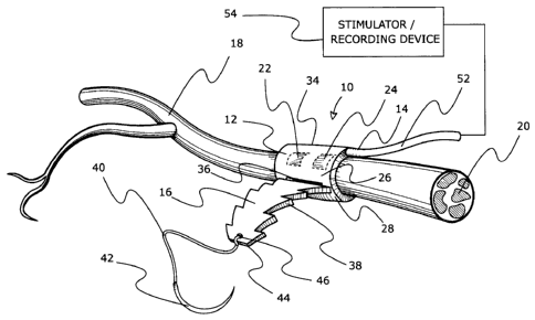
FIG. 1 is a schematic perspective view of one embodiment of the invention,
showing the nerve cuff wrapped around a nerve and fastened with the adjustable
closing mechanism. The figure shows a nerve interacting device in the form of
conductive elements on the inner face of the nerve cuff and insulated leads to
a
stimulator or recording device.
FIG. 2 is a schematic plan view of the inner face of the nerve cuff of FIG. 1,
showing the cuff and conductive elements connected to the stimulator or
recording
device.
FIG. 3 is a schematic perspective view of the nerve cuff fitted around a nerve
as in FIG. 1, illustrating the removal of the excess tail end with surgical
scissors
once the cuff is properly positioned.
FIG. 4 is a schematic perspective view of a nerve cuff similar to that of FIG.
1
fitted around a large nerve.
FIG. 5 is a schematic perspective view of a nerve cuff similar to that of FIG.
1
fitted around a small nerve.
FIG. 6 is a schematic plan view of a nerve cuff similar to that of FIG. 1, but
with a nerve interacting device in the form of a multiple contact electrode
lead.
FIG. 7 is a schematic plan view of the inner face of a nerve cuff illustrating
a
belt embodiment of the adjustable closing mechanism with multiple locking
apertures on the tail end of the cuff and showing two multiple contact
electrode
leads as nerve interacting devices.
FIG. 8 is a schematic plan view of the inner face of a nerve cuff illustrating
a
further embodiment of a closing mechanism which resists movement equally in
both
directions once closed, and showing connection to a single multiple contact
electrode lead as a nerve interacting device.
FIG. 9 is a side sectional and schematic view illustrating a method of locking
the adjustable nerve cuff of FIG. 1 by wrapping it around the nerve and using
a
needle and suture to lock the cuff in place with a suture to the cuff itself.
CA 02715543 2010-08-13
WO 2009/100531 PCT/CA2009/000171
FIG. 10 is a side sectional and schematic view illustrating the nerve cuff of
FIG. 6 prior to cutting and discarding the excess tail end.
FIG. 11 is a side sectional and schematic view illustrating a method of
anchoring the nerve cuff of FIG. 6 to nearby body tissue.
FIG. 12 is a schematic plan view of the inner face a nerve cuff with a closing
mechanism similar to that of FIGS. 1 and 6, but formed with custom printed
connections as the nerve interacting device.
FIG. 13 is a schematic plan view of the outer face of a nerve cuff with
printed
connections and imbedded electronics and having a loop closing mechanism.
FIG. 14 is a side sectional and schematic view of the installed nerve cuff of
FIG. 13 with printed connections and fully imbedded electronics, showing the
loop
closing mechanism in its locked position around the nerve.
FIG. 15 is a schematic plan view of the inner face of a nerve cuff with the
closing mechanism similar to that of FIG. 13, but showing a perpendicular
electrode
lead, and having printed conductive elements on the inner face of the nerve
cuff.
FIG. 16 is a side sectional and schematic view of the nerve cuff of FIG. 15 in
its locked position around the nerve.
FIG. 17 is a schematic perspective view of a nerve cuff fitted around a nerve
and holding a BIONTM wireless stimulator device in proximity to the nerve. The
BION
has an antenna for wireless transmission to a stimulator control or recording
device
control unit.
FIG. 18 is a schematic plan view of the inner face of a nerve cuff showing
conductive elements on the inner face of the nerve cuff and a wireless antenna
device on the outer face of the nerve cuff for wireless transmission to a
stimulator or
recording device control unit.
FIG. 19 is a schematic plan view of the inner face of the nerve cuff apparatus
used in the example of this application with an implanted conductor for nerve
stimulation. Exemplary but non-limiting dimensions are provided on the figure.
BRIEF DESCRIPTIONS OF THE PREFERRED EMBODIMENTS
The embodiments of the present invention are described by way of example
only and with reference to the figures in which similar reference numerals are
used
6
CA 02715543 2010-08-13
WO 2009/100531
PCT/CA2009/000171
in different figures to denote similar components. The tissue cuff of the
figures is
shown in the form of a nerve cuff, but the invention has broad application to
other
internal body tissues such as veins and arteries or other body tissues which
can be
encircled with a tissue cuff apparatus for purposes such as healing, attaching
other
devices or tissues, or immobilizing. While some dimensions are provided
herein, the
dimensions are non-limiting, and are provided as exemplary guidelines for
preferred
embodiments involving nerves, where typical nerve circumferences may be about
3
to 5 mm in diameter.
The nerve cuff apparatus of this invention as illustrated in FIGS 1 - 6 is
shown
generally at 10, and includes a nerve cuff 12 and a nerve interacting device
14. The
nerve cuff 12 consists of a a strap 16 formed of a thin, flat sheet of a non-
conductive, biocompatible, elastomeric material that can be wrapped around a
peripheral nerve 18. The nerve 18 is usually composed of multiple fascicles
20, so
adjustment of the nerve interacting device 14, relative to the fascicles 20
may be
desired during implantation (i.e., intra-operatively). The nerve interacting
device 14
includes conductive elements (in this case electrode units) 22, 24 imbedded in
the
strap 16 (or printed or attached) direct contact to the nerve 18. The non-
conductive
properties of the elastomeric material ensures that surrounding body tissue is
insulated from the electrode units 22, 24. The strap 16 is elongated with a
longitudinal axis along its length dimension, and a transverse width
dimension. The
length dimension is longer than that needed to wrap around the body tissue of
interest. The width dimension is sufficient to provide structural support for
the nerve
interacting device of interest and sufficient to be manipulated during
implantation.
The width dimension (which may be constant over the length, or varied) of the
strap
16 will depend on the thickness of the strap 16, and the particular
application for the
nerve cuff apparatus 10. The strap 16 is thin. For nerve applications, the
strap
thickness is preferably less than about 1 mm, more preferably less than about
0.5
mm, and still more preferably between about 0.15 - 0.35 mm. The strap 16 is
sufficiently thin that it remains elastic, pliable and flexible for
implanting, fastening,
and adjusting. One set of exemplary, non-limiting dimensions for nerves of
about 3
to 5 mm diameter is shown in FIG. 19.
The strap 16 includes a body portion 26 connected between (preferably
7
CA 02715543 2010-08-13
WO 2009/100531
PCT/CA2009/000171
integral with) a head end portion 28 and a tail end portion 30 (best seen in
FIG. 2).
The body portion 26 has an inner face 32 which faces the nerve to be
encircled, and
an outer face 34 which faces surrounding body tissues after implantation. The
head
and tail end portions 28, 30 are configured for adjustable length fastening
one to the
other around the nerve 18, and thus provide the adjustable locking or closing
mechanism of this invention. This leaves the body portion 26 isolated and
remote
from the adjustable length ends 28, 30, for stable and secure attachment to
the
nerve 18, and for separate and secure attachment to one or more nerve
interacting
devices 14. The adjustable length fastening is generally achieved by providing
the
extra length (i.e., a total length of the strap 16 which is in excess of an
expected
circumference of a body tissue to be encircled) in one or both of the head and
tail
end portions 28, 30. In general, the length of the body portion 26 will not be
greater
than the expected circumference of the body tissue to be encircled, so the
extra
length is provided in one or both of the head and tail end portions 28, 30 to
ensure a
secure attachment to the body tissue. In applications where the body tissue is
very
small, such as nerves, providing extra length in both the head and tail end
portions
28, 30 may be advantageous to assist in placement and manipulation during
implantation.
The head end portion 28 is shown in the embodiment of FIGS. 1 - 6 to be
formed with a transverse slot 36 as a locking aperture. The tail end portion
30 is
formed with a plurality of longitudinally spaced laterally paired locking
projections 38.
The paired locking projections 38 are spaced by narrower neck portions 39. The
locking projections 38 are shaped to allow for passage through the slot 36 by
flexing
in an insertion direction (i.e., in the direction of threading through the
slot 36 to
fasten around the nerve 18), and to restrict movement in the reversing
direction
through the slot 36 (i.e., in the direction to loosen the strap 16). The
flexibility of the
projections 38 permits them to be re-adjusted by the surgeon during
implantation in
the reverse direction if needed, but once the appropriate position is
achieved, the
projections 38 resist reverse movement through the slot 36. To achieve this
adjustable length fastening, the pairs of locking projections 38 have a
transverse
width at their widest points which exceeds the transverse width dimension of
the slot
36. Preferably, the narrow neck portions 39 have a maximum transverse width
8
CA 02715543 2010-08-13
WO 2009/100531
PCT/CA2009/000171
dimension no greater than the transverse width dimension of the slot 36. This
enables the strap 16 to lay flat against the nerve 18 when fastened. Further,
the
locking projections 38 are preferably shaped to assist in threading through
the slot
36. For instance, with the arrow shape projections 38 of FIGS. 1 - 6, the
double
toothed lateral edges are tapered to narrow inwardly toward the leading edge
44
(free end) of the tail end portion 30. Each pair of projections 38 at its
widest point
has a transverse width that extends transversely beyond the slot width in an
overlapping and locking mode. The extent of overlap of each projection 38
(i.e., on
each side of the slot 36) compared to the transverse slot width is preferably
at least
about 10% of the slot width dimension, more preferably about 15-30%. This
overlap
of the projections resists reverse movement of the projections 38 through the
slot
36. The length of the individual projections 38 and the number of
longitudinally
spaced paired projections 38 will vary to provide sufficient incremental
adjustments
around the nerve. The projection length and degree of overlap vary with such
factors as the type and thickness of the elastomeric material, the nature (ex.
size
and weight) of the nerve interacting device 14, and the nature and size of the
body
tissue being encircled, so the above dimensions are provided only as
guidelines.
The taper of the arrow shaped projections 38 (narrowing toward the leading
edge 44
of the tail end portion 30) provides a preferential sliding direction (in the
insertion
direction) when engaged in the slot 36. The tail end portion 30 may include a
suture 40 and needle 42 at its leading edge 44 to assist in threading through
the slot
36, and for locking and/or anchoring to the strap 16 once implanted (see FIGS.
9 -
11). The leading edge 44 might be formed with a suture connecting aperture 46,
or
the needle 42 can be used to attach to the leading edge 44 before or during
implantation.
The strap 16 is wrapped circumferentially around the nerve 18 in order to
create a good contact between the nerve 18 and the conductive elements 22, 24.
Insulated leads 48, 50 and 52 are shown leading to a remote stimulator or
recording
device 54, which might be implanted or external to the patient. The conductive
elements 22, 24 might be printed on, imbedded in or attached to (for example
with
adhesive) the inner face 32 of the body portion 26, by techniques known in the
art.
The conductive elements 22, 24 might be conductive metal or conductive rubber.
9
CA 02715543 2010-08-13
WO 2009/100531 PCT/CA2009/000171
Alternatively, the conductive elements might be designed to receive other than
electrical impulses, for example one or more of thermal, auditory,
vibrational, light or
fluid stimulation.
As shown in FIGS. 3-5, once properly positioned the excess at the tail end
portion 30 can be trimmed using medical scissors 55 to remove excess material
and
reduce mechanical irritation. The excess trimmed tail end material 56 (see
FIGS. 4,
5) containing the suture 40 and needle 42 can then be discarded. FIGS. 4 and 5
show how the same sized nerve cuff 12 can wrap around two different sized
nerves,
a large nerve 18 in FIG. 4 and a smaller nerve 18 in FIG. 5. The small nerve
18
generates longer excess material 56 when compared to the excess material 56
from
the large nerve 18, if cut the same distance (marked with a dotted line) from
the
projection 38 engaged in the head end slot 36.
FIGS. 6 - 8 show alternate embodiments of a nerve cuff apparatus of this
invention with nerve interacting devices in the form of one or more multiple
contact
electrode leads 60. Multiple contact electrode leads 60 include a plurality of
conductive elements 62 in order to achieve a specific stimulation or recording
result.
These leads 60 might be simply held in place by simple wrapping with the nerve
cuff
12, as in FIG. 6, or they might be held with a non-conductive biocompatible
adhesive
64 as shown in FIGS. 7 and 8.
FIG 7 illustrates an alternate closing/locking mechanism, namely a belt style
closure. The strap 66 is formed with a plurality of longitudinally spaced
slots 68
formed in the tail end portion 70. The head end portion 72 is formed with
laterally
paired locking projections 74. The leading edge 76 of the head end portion 72
forms
an elongated lead tab 78 to assist in threading into one of the slots 68. The
lead tab
78 may also be attached to a suture 80 and needle 82 as above described. The
preferred width dimensions of the projections 74, narrower neck portion 84 and
slots
68 are generally as set forth above. However, with the single pair of
projections 74
of this embodiment, it is preferable that the space 86 adjacent the narrower
neck
portion 84 between the head and body portions 72, 88 has a length component no
less than the thickness dimension of the strap 66. This assists in preventing
the
closure from re-opening.
In FIG. 8, the nerve cuff strap 90 is similar to that of FIG. 6, but the tail
end
CA 02715543 2010-08-13
WO 2009/100531 PCT/CA2009/000171
portion 92 is formed with laterally paired projections 94 which are rounded,
rather
than tapered. These rounded projections 94 resist movement in both directions
equally once fitted through the locking slot 96 formed in the head end portion
98.
The strap 90 is formed with an elongated lead tab 100 at the leading edge 102
of
the tail end portion 92 to facilitate threading the tail end portion 92 into
the locking
slot 96.
FIGS. 9 - 11 illustrate cross sectional views of different possible anchoring
and locking methods for a nerve cuff 12 similar to that of FIGS. 1 or 6. To
lock the
nerve cuff 12 in place it is possible to wrap the excess at the tail end
portion 30
around the cuff 12, as seen in FIG. 9. To prevent the cuff 12 from unraveling,
the
needle 42 and suture 40 can be used to tie the head end portion 28 with the
tail end
portion 30 via a stitch 104. This reduces the chance of the nerve cuff 12
unraveling,
and can be used to optimize contact between the conductive elements 22, 24 and
the nerve 18. The cuff 12 can also be left as is once the tail end portion 30
has
been inserted into the head end portion 28 as shown in FIG. 10 (or this tail
end
portion 30 may be cut as described above). The suture 40 and needle 42 can
also
be used to anchor the entire cuff 12 to nearby tissue 106 by stitching the
tail end
portion 30 via a stitch 108 to the nearby tissue 106, as seen in FIG. 11.
In FIG. 12 the nerve interacting device (or other tissue interacting device)
may
take the form of a circuit printed the body portion 110 of a nerve cuff strap
112,
between the head end and tail end portions 114, 116. In FIG. 12, the inner
face 117
of the body portion 110 is shown, but the circuit components might be printed
on
either or both sides, or the components may be imbedded in the strap 112. The
closing mechanism is similar to that shown in FIG. 6. The processes of
photolithography and electroplating can be used to generate custom conductive
element contact points 118, 119, 120, 121 that are unique in size and location
to suit
the nerve or tissue interacting device application. Some of these contact
points 118
- 121 can be linked to each other with conductive but insulated tracks 122.
In FIGS 13, 14, an electronic circuit is shown on the outer face 123 of a
nerve
cuff strap 124. Printing techniques as above-mentioned can be used to create
electric circuits such as pre-amplifiers, or entire stimulator/recording
devices that can
be placed directly on the of the strap 124. Exemplary electronic components
are
11
CA 02715543 2010-08-13
WO 2009/100531 PCT/CA2009/000171
shown as a micro processor 125, resistor 126, and capacitor 127, connected
with
can be connected with conductive and insulated tracks 128. These are shown on
the outer face 123 in FIGS. 13, 14, with the electrical contacts 131 being
shown on
the inner face 129 for contact with the nerve 18. The entire electronic
assembly on
the outer face 123 can be covered in an insulating biocompatible material 130
such
as silicone rubber to prevent direct tissue interaction with the electronics.
FIGS. 13, 14 also illustrate another embodiment for length adjustable closing
mechanism. The strap 124 with head and tail end portions 132, 134, and body
portion 133, has a loop 136 (ex. ring) formed at the head end portion 132. The
loop
136 sits above the plane of the strap 124, and may be connected to the strap
124,
for example by a biocompatible adhesive. The opening 137 formed between the
strap 124 and the loop 136 functions as a locking aperture to secure the
laterally
paired locking projections 138 formed on the tail end portion 134. As above,
the
width of the pairs of projections 138 at their widest points is greater than
the
transverse width of the loop opening 137. When the tail end portion 134 is
threaded
through the loop 136, the loop 136 rests on top of the head end portion 132
(best
seen in FIG. 14). The loop 136 might be provided as a separate ring which is
attached by adhesive, similar to the figures. Alternatively, the loop 136 and
strap
124 can be made from a single thin insulating, flexible sheet of biocompatible
material by folding side wings (not shown) inwardly to form the loop 136, and
fixing
with adhesive. The shape of the pairs of projections 138 shown in FIG. 13 is
generally tear drop shaped with extra downward taper (toward the body portion
133),
for ease of insertion in the loop 136, and to increase the resistance to
reverse
movement through the loop 136 once fastened in the loop 136. These tear drop
shaped projections 138 may also be used in slot embodiments (see FIG. 19). The
leading edge 140 of the tail end portion 134 is formed with an elongated lead
tab
142 having a transverse width at its leading edge 140 which is substantially
smaller
than the transverse width of the loop opening 137. This facilitates insertion
of the
tail end portion 134 through the opening 137.
In some applications it may be important to orient an electrode lead facing
perpendicularly to a nerve. A nerve cuff apparatus to accommodate this
orientation
is shown in FIGS. 15, 16. This orientation may be advantageous in endoscopic
12
CA 02715543 2010-08-13
WO 2009/100531 PCT/CA2009/000171
procedures. The nerve cuff strap 124 is similar to that of FIGS. 13, 14, so
FIGS. 15,
16 show like components with the same reference numerals. However, FIG. 15
shows the inner face 129 printed with conductive elements 144, 145 for
electrical
contact with the nerve 18. The insulated leads 146 from the elements 144, 145
are
oriented to be perpendicular to the nerve 18 on implantation (rather than
parallel as
in previous embodiments). This is the ideal application for the loop closure
mechanism. In FIG. 16, the loop 136 is shown in the closed position to orient
the
opening 137 above the inner face 129 (the strap is shown with the outer face
123 in
FIG. 14, so the loop opening 137 there is above the outer face 123).
FIG 17 and 18 illustrate a complete wireless stimulator anchored in immediate
proximity to the nerve 18. In FIG. 17, a wireless stimulator 150 such as a
BIONTM
from Advanced Bionics, LLC of California (see for example US Patent 5,193,539
to
Schulman et al.) is attached with adhesive (not shown) to the inner face 165
of the
body portion 154 of the nerve cuff strap. The BION 150 is a self sufficient
unit with
an outer shell that is conductive for electrical contact with the nerve 18.
The BION
receives data and/or power from an external control or recording unit 158 via
an
antenna 160 on the BION unit 150. Radio waves 162 (or other frequency waves)
may be used to control the unit 150 or transmit to the controller/recorder
158. The
nerve apparatus of the embodiment in FIG. 18 has conductive elements (example
metal contacts) 161, 163 printed, attached or imbedded at the inner face 164
of the
body portion 154 of strap 156 for direct contact with the nerve 18 once
installed.
The wireless control or recording unit 158 can be located externally to the
patient, or
may be implanted. The laterally paired projections 166 on the tail end portion
168
are shown as arrow shaped in FIG. 17 (as in FIG. 6) with needle 42, suture 40,
and
tear drop shaped in FIG. 18 (similar to FIG. 13). The leading edge 170 in FIG.
18 is
shown as forming an elongated tab 172, connected to needle 42 and suture 40,
similar to that in earlier figures.
It will be evident that alternate interlocking shapes of laterally paired
projections and/or locking apertures may be used in this invention. For
example, the
slots might be more oval shaped or circular shaped, with the projections being
similarly altered so as to still project in a transverse width direction
beyond the
transverse width dimension at the widest point of the slot. Alternatively, the
13
CA 02715543 2010-08-13
WO 2009/100531 PCT/CA2009/000171
projections might be shaped in 3D (and not just in 2D) to lock in the locking
aperture
to resist movement in the reversing direction. However, the above-described 2D
embodiments are preferred for their manufacturing simplicity and low cost, as
well as
for their ease of manipulation during implantation.
The tissue interacting devices useful in the tissue cuff apparatus of this
invention are wide ranging, with the above and following descriptions serving
only as
exemplary embodiments. Nerve stimulating devices are well known in the prior
art.
Nerve recording devices are also known. For example, nerve recordings from
sacral
root recordings intra-operatively as electroneurographic (ENGs) signals may be
obtained from either free electrodes or nerve cuffs. These are common in
procedures for spinal cord injured patients that focus on the sacral roots of
the spinal
cord. Devices that have both stimulation and recording capabilities might also
be
used, such as shown in US Patent 5,913,882 to King, designed for augmenting
electrical stimulation usefulness in pain control. Similarly, devices for
sleep apnea
via vagal nerve stimulation, or devices for Parkinson's disease in the form of
deep
brain stimulation, might be used with the nerve cuff of this invention.
The substrate materials for the strap extend to elastomeric materials which
provide sufficient elasticity, resiliency and strength in a thin flat format,
without the
corrugations, undulations or piercing projections of the prior art. The
materials are
biocompatible for implantation, and are preferably non-conductive to
protect/insulate
surrounding body tissue from any conductive elements (typically electrical
contacts).
Exemplary materials include flat sheets of silicone rubber elastomers, for
example
PDMS (polydimethylsiloxane), SilasticTM (a silicone rubber), and biocompatible
polyurethane polymers, and biocompatible polyimides. Generally, the sheets
have a
uniform thickness so that the strap is formed with a uniform thickness.
However, the
strap might alternatively be formed with increased thickness in the certain
body,
head or tail portions to increase the strength of one or more of these
sections for
particular applications. Other elastomeric biocompatible materials will be
known to
those skilled in the biomedical area. The substrate material may be coated or
impregnated with one or more active tissue agents, such as antibiotics,
proteins,
growth factors and the like, for applications such as healing.
For applications involving adhesives, the adhesives are biocompatible, with
14
CA 02715543 2010-08-13
WO 2009/100531 PCT/CA2009/000171
exemplary materials including silicone rubber, cyanoacrylates, and
polyethylene
glycol polymers. The latter group are advantageous in applications where a
biodegradable adhesive is desired.
Manufacturing involves the shaping, cutting or stamping of a sheet of non-
conductive biocompatible elastomeric material. Laser cutting is preferred,
particularly for the fine details and dimensions of the projections and slots.
The
conductive elements (for example conductive metals or conductive rubber) may
be
imbedded, attached or printed into or on the sheet. The entire cuff apparatus
can
then be sterilized prior to implantation.
Advantages and other features of the invention include:
1. One size fits various nerve sizes or configurations. The exact nerve sizes
are
typically not known in advance of implantation, so the length adjustability
for intra-
operative manipulation provides a more secure and stable attachment to the
nerve,
limiting additional surgical procedures needed in the event of device
migration.
2. Once fastened, the excess material in the tail end portion of the nerve
cuff can be
trimmed or sutured shut. The excess tail end material might alternatively
serve as
anchoring material by suturing to surrounding body tissue.
3. The adjustable fastening mechanism allows for intra-operative adjustment
for
different nerve sizes and re-positioning around the nerve until the desired
result is
obtained, minimizing post operative failures or migration of the apparatus..
4. The initial flat configuration makes the cuff easy to sterilize,
manufacture and
insert around the nerve.
5. The body portion being clear of the fastening head and tail end portions,
allows
for use with a wide range of conductive elements and nerve interacting
devices. For
instance, metal conductive elements and circuits can be printed on the inner
face of
the flat body portion in unique arrangements. In addition, or alternatively,
other
circuit components may be imbedded into the body portion or otherwise attached
(similar to electronic boards). The outer face of body portion may also carry
circuit
components, or serve to attach nerve interacting devices. Alternatively,
Silastic
materials can accommodate conductive and non-conductive rubber instead of
printed metal. Alternatively, the body portion can accommodate multiple
conductive
contacts, and can be used to secure a traditional barb/tube electrode close to
the
CA 02715543 2010-08-13
WO 2009/100531 PCT/CA2009/000171
nerve. Still alternatively, a BION may be secured close to the nerve with the
nerve
cuff to prevent shifting.
6. The needle and suture at the tail end allows for intuitive and minimally
destructive
approach to installing the cuff (as a guide). The needle may be metal, and the
suture a traditional suture. Alternatively, the needle might be plastic, and
the suture
a thin sheet of rubber.
Example
The nerve cuff apparatus of this invention in multiple of the preferred
embodiments has been tested in numerous animal trials where the application
was
an electrical nerve cuff. Following successful animal implanting, a plurality
of nerve
cuff apparatus 180 having the configuration and dimensions shown in FIG. 19
(not
drawn to scale) were implanted in a 51 year old spinal cord injured man. The
implantation was directed to restore upper extremity hand function in
conjunction
with a nerve stimulator device as described in U.S. Patent Application No.
2006/0184211 A published August 17, 2006, to Gaunt et al. The nerve cuff
straps
182 were each laser cut out of a biocompatible silicone rubber sheet 0.254 mm
thick. The implanted nerve cuff apparatus 180 included a monopolar conductor
184
attached to the body portion 186 of the nerve cuff strap 182 with a silicone
rubber
adhesive 188, cured prior to sterilization and implantation. The tail end
portion 190
was formed with tear drop shaped projections 194 as shown, and an elongated
lead
tab 196 to aid in manipulating into the slot 198 formed in the head end
portion 200.
The head end portion 200 of the nerve cuff strap 182 was lengthened with
excess
length material in order to aid in manipulation of the cuff apparatus 180
during
implantation. The nerve cuff apparatus 180 once circumferentially attached to
the
target nerves was tested with stimulation to verify proper positioning.
Position was
adjusted on each of the three implanted cuffs during the implantation
procedure (i.e.,
intra-operatively), until the most favorable results were observed. Each cuff
apparatus 180 was then trimmed (both the head and tail end portions 200, 190)
with
surgical scissors (as shown in FIG. 3). Five months later, all three
implantation sites
continued to stimulate the desired nerves, with no sign of apparatus migration
or
failure.
16
CA 02715543 2016-01-27
As used herein and in the claims, the word "comprising" is used in its non-
limiting
sense to mean that items following the word in the sentence are included and
that items
not specifically mentioned are not excluded. The use of the indefinite article
"a" in the
claims before an element means that one of the elements is specified, but does
not
specifically exclude others of the elements being present, unless the context
clearly
requires that there be one and only one of the elements. For example, "a slot"
as used
herein and in the claims may include multiple slots.
All references mentioned in this specification are indicative of the level of
skill in
the art of this invention. If any inconsistency arises between a cited
reference and the
present disclosure, the present disclosure takes precedence. Some references
provided herein provide details concerning the state of the art prior to the
filing of this
application, other references may be cited to provide additional or
alternative device
elements, additional or alternative materials, additional or alternative
methods of
analysis or application of the invention.
The terms and expressions used are, unless otherwise defined herein, used as
terms of description and not limitation. There is no intention, in using such
terms and
expressions, of excluding equivalents of the features illustrated and
described, it being
recognized that the scope of the invention is defined and limited only by the
claims
which follow. Although the description herein contains many specifics, these
should not
be construed as limiting the scope of the invention, but as merely providing
illustrations
of some of the embodiments of the invention.
One of ordinary skill in the art will appreciate that elements and materials
other
than those specifically exemplified can be employed in the practice of the
invention
without resort to undue experimentation. All art-known functional equivalents,
of any
such elements and materials are intended to be included in this invention
within the
scope of the claims, including without limitation the options and alternatives
mentioned
herein. The invention illustratively described herein suitably may be
practiced in the
absence of any element or elements, limitation or limitations which is not
specifically
disclosed herein.
17