Note: Descriptions are shown in the official language in which they were submitted.
CA 02715740 2010-08-17
WO 2009/104182 PCT/IL2009/000188
A DEVICE AND METHOD FOR DEPLOYING AND ATTACHING A PATCH TO A
BIOLOGICAL TISSUE
FIELD OF THE INVENTION
This invention generally relates to a device and method for repairing
biological tissue
aperture. More specifically, the present invention relates to a device and
method for
deploying and attaching a patch to a biological tissue.
BACKGROUND
An object of the present invention is to provide apparatus and a method for
performing
corrective surgery on internal wounds such as hernia where invasion of the
patient's body
tissues is minimized and resultant trauma is reduced.
A hernia is a protrusion of a tissue, structure, or part of an organ through
the muscular tissue
or the membrane by which it is normally contained. In other words a hernia is
a defect in the
abdominal wall through which a portion of the intra-abdominal contents can
protrude. This
often causes discomfort and an unsightly, visible bulge in the abdomen. When
such a hernia
defect occurs in the abdominal region, conventional corrective surgery has
required opening
the abdominal cavity by surgical incision through the major abdominal muscles.
While this
technique provides for effective corrective surgery of the hernia defect, it
has the
disadvantage of requiring a hospital stay of as much as a week, during which
pain is
frequently intense, and it requires an extended period of recuperation. After
the conventional
surgery patients frequently cannot return to a full range of activity and work
schedule for a
month or more. Accordingly, medical science has sought alternative-techniques
that are less
traumatic to the patient and provide for more rapid recovery.
Laparoscopy is the science of introducing a viewing instrument through a port
into a patient's
body, typically the abdominal cavity, to view its contents. This technique has
been used for
diagnostic purposes for more than 75 years. Operative laparoscopy is performed
through tiny
openings in the abdominal wall called ports. In most surgical techniques
several ports,
frequently three to six, are used. Through one port is inserted the viewing
device, which
conventionally comprises a fiber optic rod or bundle having a video camera
affixed to the
I
CA 02715740 2010-08-17
WO 2009/104182 PCT/IL2009/000188
outer end to receive and display images from inside the body. The various
surgical
instruments are inserted through other ports to do the surgery that normally
would be
performed through an open incision through the abdominal wall. Because the
laparoscopic
surgical techniques require only very small holes through the abdominal wall
or other
portions of the body, a patient undergoing such surgery may frequently leave
the hospital
within one day after the surgery and resume a full range of normal activities
within a few
days thereafter.
In repairing hernia the physician needs. to first deploy the patch and then to
attach the patch
to the tissue.
There are many patents and patent applications relating to attaching a
prosthesis implant to a
tissue via tacks. Each patent and patent application describes a different
attachment
mechanism via different anchoring means (see for example US patent 6,447,524).
Traditional
anchors used in surgery include clips, staples, or sutures, and may also be
referred to as tissue
anchors. These devices are usually made of a biocompatible material (or are
coated with a
biocompatible material), so that they can be safely implanted into the body.
Most tissue
anchors secure the tissue by impaling it with one or more posts or legs that
are bent or
crimped to lock the tissue into position. Thus, most traditional anchors are
rigid or are
inflexibly attached to the tissue. For example PCT No. W007/021834 describes
an anchor
having two curved legs that cross in a single turning direction to form a
loop. Those two
curved legs are adapted to penetrate tissue in a curved pathway. US patent
4,485,816 (refers
hereinafter as 816') describes surgical staple made of shape memory alloy. The
staple is
placed in contact of the tissue and then heated. The heating causes the staple
to change its
shape thus, penetrating the tissue.
US patent 6,893,452 (refers hereinafter as `452) describes a tissue attachment
device that
facilitates wound healing by holding soft tissue together under improved
distribution of
tension and with minimal disruption of the wound. interface and its nutrient
supplies. The
device has multiple sites for grasping the tissue using tines or prongs or
other generally
sharp, projecting points, protruding from a single, supportive backing. One of
the
embodiments described in `452 is the use of sharp projecting points protruding
from the
supportive backing in two different angles.
2
CA 02715740 2010-08-17
WO 2009/104182 PCT/IL2009/000188
US patent 6,517,584 (refers hereinafter as `584) describes a hernia patch
which includes at
least one anchoring device made of shape memory material. The anchoring
devices are
initially secured to the prosthesis by being interlaced through a web mesh
constituting the
prosthesis. The attachment is obtained by altering the attachment element's
shape from
rectilinear to a loop. shape due to heat induced shape memory effect.
Yet other patent literature relates to devices for endoscopic application of
surgical staples
adapted to attach surgical mesh to a body tissue.
An example of such a teaching is to be found in US patent 5,364,004, US patent
5,662,662,
US patent 5,634,584, US patent 5,560,224, US patent 5,588,581 and in US patent
5,626,587.
There are a few patent and patent applications teaching the deployment of
patches. For
example US patent 5,836,961 (refers hereinafter as `961) which relates to an
apparatus used
for developing an anatomic space for laparoscopic hernia repair and a patch
for use
therewith. The apparatus of patent `961 comprises a tubular introducer member
having a bore
extending therethrough. A tunneling shaft is slidably mounted in the bore and
has proximal
and distal extremities including a bullet-shaped tip. A rounded tunneling
member is mounted
on the distal extremity of the tunneling shaft. The apparatus comprises an
inflatable balloon.
Means is provided on the balloon for removably securing the balloon to the
tunneling shaft.
Means is also provided for forming a balloon inflation lumen for inflating the
balloon. The
balloon is wrapped on the tunneling shaft. A sleeve substantially encloses the
balloon and is
carried by the tunneling shaft. The sleeve is provided with a weakened region
extending
longitudinally thereof, permitting the sleeve to be removed whereby the
balloon can be
unwrapped and inflated so that it lies generally in a plane. The balloon as it
is being inflated
creates forces generally perpendicular to the plane of the balloon to cause
pulling apart of the
tissue along a natural plane to provide the anatomic space.
Although patent '961 relates to deploying means, patent '961 teaches a device
in which the
patch is attached to a balloon which is introduced into the abdominal cavity.
The deployment
is performed by inflating the balloon. In other words, a totally different
deploying means are
disclosed.
Furthermore, due to the relatively large volumes of balloons several
disadvantages are likely
to occur: (a) The visibility within the abdominal cavity might be damaged; (b)
The
3
CA 02715740 2010-08-17
WO 2009/104182 PCT/IL2009/000188
accessibility of the attachment means to the patch might be impaired; and, (c)
The
maneuverability of the patch within the abdominal cavity is limited.
Yet more, another major drawback to patent 1961, the inflated balloon lacks
any mechanical
stiffness which is needed for navigation of the patch to its position.
Another example for deploying the patch can be found in US patent no.
5,370,650 (refers
hereinafter as `650) which relates to an apparatus for positioning surgical
implants adjacent
to body tissue to facilitate the fastening of the implant to the body tissue.
Patent `650
provides an apparatus for positioning surgical implants adjacent to body
tissue, comprising
an outer tube having a proximal end, a distal end and a longitudinal axis; an
inner rod at least
partially disposed within the outer tube and slidable along said longitudinal
axis. The inner
rod has a proximal and a distal end portions. The inner rod distal end portion
further
comprises articulating means for pivoting at an angle with respect to the
longitudinal axis. A
looped support member having first and second end portions fixedly secured to
said distal
end portion of the inner rod; and a surgical implant releasably secured to the
looped support
member (a preferred embodiment illustrating the teaching of patent `650 is
illustrated in
figure 17).
The major difference between patent '650 and the present invention is the
actual patch
deployment mechanism.
While in patent '650, the looped support member 14 is transferred from a
deployed
configuration to a retracted- configuration by pushing and pulling tube 12, in
the proposed
technology the flexible arms are reconfigured from their initial stage (IS) to
their final stage
(FS) by the reciprocal movement the central shaft. In other words, while in
patent '650, the
patch is deployed due to the elasticity of the loop member (no force is
applied), in the present
application, the patch is deployed by actively and directly applying force on
the Flexible
arms by the surgeon.
Furthermore, the deployment of the patch in patent `650 is passive and
unidirectional; i.e.,
once the patch is deployed by pulling tube 12, the patch can not be un-
deployed and
reinserted into tube 12. In order to reinsert the patch into tube 12, the
patch must be refolded
and such an action can not be performed while the patch is within the patient.
Therefore, the
surgeon has only one chance to unfold the patch. This is in sharp contrary to
the present
invention in which the deployment of the patch is bidirectional and actively
controlled such
4
CA 02715740 2010-08-17
WO 2009/104182 PCT/IL2009/000188
that the patch can be deployed and un-deployed simply by the reconfiguration
of the flexible
arms (which a full description will be provided in the detail description).
Yet another major distinction between patent `650 and the proposed invention
is the fact that
in patent 1650 the looped support member 14 is preferably in a deployed (i.e.,
open)
configuration thereby insertion of the looped support member 14 into tube 12
will require the
physician to apply a significant amount of force in order to maintain the
looped support
member 14 in a closed configuration. On the contrary, in the present
invention, the flexible
arms can be actively configured to be constantly closed without any additional
force applied
by the physician. Therefore, the insertion of the device through a trocar is
facilitated.
Yet more, the present invention comprises a central shaft for providing the
device
mechanical stiffness for the backbone of the system which is needed for better
positioning of
the patch within the body. Further, by providing mechanical stiffness to the
backbone of the
system, it will enable the detachment of the patch from the deployment system.
Such a
mechanism is not disclosed nor claimed in patent '650.
Lastly, patent '650 describes no attachment mechanism for attaching the patch
to the tissue.
Further, some major, non obvious modification will have to be made in order to
enable
attachment between the patch and the tissue whilst using the device of patent
'650.
More patent literature can be found in PCT no. W008065653 (refers hereinafter
as `653)
relates to a device especially adapted to deploy a patch within a body cavity.
The device is an
elongate open-bored applicator (EOBP) and comprises (a) at least one
inflatable contour-
balloon, (b) at least one inflatable dissection balloon. The inflatable
contour-balloon and the
inflatable dissection balloon are adjustable and located at the distal
portion. The EOBP
additionally comprises (c) at least one actuating means located at the
proximal portion. The
actuating means is in communication with the inflatable contour-balloon and
the inflatable
dissection balloon. The actuating means is adapted to provide the inflatable
contour-balloon
and the inflatable dissection balloon with independent activation and/or de-
activation.
It should be pointed out that PCT `653 does not disclose nor claim means
adapted to anchor
the patch to the biological tissue.
Like patent '961, the deployment system describes in PCT '653 is an inflated
one, thus it is
fundamentally different from the proposed invention.
CA 02715740 2010-08-17
WO 2009/104182 PCT/IL2009/000188
All those patent and patent application demonstrate attachment means for
attaching the patch
to the tissue or means for deploying the patch within the body. However none
of the
literature found relates to a device especially adapted to deploy and attached
a patch to a
biological tissue.
Thus, there is still a long felt need for a device that can be used for both
deploying and
attaching a patch to a biological tissue.
Furthermore, there is still a long felt need for a deployment system that will
overcome the
above mentioned drawbacks and will provide a deployment system that will
enable the
following (i) a reversible deployment of the patch (i.e., enable the folding
and the unfolding
of said patch); (ii) a controlled deployment of the patch (i.e., the surgeon
applies force in
order to deploy the patch and therefore the deployment is actively
controlled); and, (iii) will
provide mechanical stiffness for the backbone of the system.
SUMMARY OF THE INVENTION
It is one object of the present invention to provide an integrated deployment
and attachment
device (DAD) comprising means adapted to deploy a patch and means adapted to
attach said
patch to a biological tissue within the body; wherein said DAD is adapted to
sequentially
deploy said patch within said body and attach said patch to said biological
tissue within said
body; further wherein said deployment of said patch is (i) controlled such
that a continuous
deployment is obtained; and, (ii) bidirectional such that said deployment is
fully reversible.
It is another. object of the present invention to provide the DAD as defined
above, wherein
said DAD is characterized by having a distal portion, adapted to be inserted
into a body and a
proximal portion, located adjacent to a user; said distal portion and said
proximal portion are
interconnected. along a main longitudinal axis via a tube (103); said tube
having a proximal
end (TP) connected to said proximal portion, and .a distal end (TD); said tube
accommodates
at least a portion of a central shaft (105); said central shaft (105) has a
proximal end (CSP)
accommodated within said tube (103) and a distal end (CSD) protruding from
said TD end;
said central shaft (105) is adapted to reciprocally move parallel to said main
longitudinal
axis within said tube (103);
said distal portion comprises: (i) at least two flexible. arm (FA) (104)
adapted to be reversibly
coupled to said patch; said FA having a proximal end (FAP) jointly connected
to said TD,
6
CA 02715740 2010-08-17
WO 2009/104182 PCT/IL2009/000188
and a distal end (FAD) jointly connected to said CSD; said FA (104) are
characterized by
having an initial stage (IS) at which said FA (104) are straight and parallel
to the longitudinal
axis of said central shaft (105); and, a final stage (FS) at which said FA
(104) are laterally
curved with respect to said longitudinal axis of said central shaft (105) such
that said patch is
deployed; said FA are adapted to reversibly transform from said IS to said FS
by said
reciprocate movement of said central shaft (105) towards and away from said
proximal
portion such that said deployment of said patch is bidirectional;
said FA (104) comprises (a) at least one attachment clip (108) adapted to
attach said patch
(106) to said biological tissue (501); and, (b) at least one connecting means
adapted to at
least partially reversibly connect said patch (106) to said FA (104);
said proximal portion comprising at least one handle (102) located outside
said body; said
handle is adapted to (i) reversibly transform said FA from said IS to said FS;
(ii) activate said
clip (108) such that said patch (106) is at least partially attached to said
tissue; and, (iii)
release said patch from said FA.
It is another object of the present invention to provide the DAD as defined
above, wherein
said connecting means are selected from at least one dedicated loop and\or
stretching means
(107) or patch-FA clip (1201) adapted to reversibly connect said patch to said
FA.
It is another object of the present invention to provide the DAD as defined
above, wherein
said clip is adapted to attach said patch to said biological tissue whilst
simultaneously
detaching from said FA.
It is another object of the present invention to provide the DAD as defined
above, wherein
said clip is adapted to first attach said patch to said biological tissue and
then to detach from
said FA.
It is another object of the present invention to provide the DAD as defined
above, wherein
said clip is characterized by having: (i) main portion (403) adapted to at
least partially
reversibly connected to said FA; (ii) at least one hooks (402) connected to
said main portion,
adapted to at least partially penetrate through said patch (106) to said
tissue (501) such that
an attachment between said patch and said tissue is obtained; (iii) a portion
(404) adapted to
reversibly connect to activation means; said activation means are adapted to
actuate said
hooks (402) such that said attachment is obtained.
7
CA 02715740 2010-08-17
WO 2009/104182 PCT/IL2009/000188
It is another object of the present invention to provide the DAD as defined
above, wherein
said clip additionally comprises securing means (701, 702) adapted to secure
and fix said clip
within said tissue and said patch.
It is another object of the present invention to provide the DAD as defined
above, wherein
said stretching means (107) and said activation means are selected from a
group consisting of
a wire.
It is another object of the present invention to provide the DAD as defined
above, wherein
said attachment between said patch and said tissue is obtained by a radial
motion of said clip
followed by a linear motion of said wire.
It is another object of the present invention to provide the DAD as defined
above, wherein
said attachment between said patch and said tissue is obtained by a linear
motion of said clip
followed by a linear motion of said wire.
It is another object of the present invention to provide the DAD as defined
above, wherein
said activation means is activation wire (112).
It is another object of the present invention to provide the DAD as defined
above, wherein
said activation wire (112) and/or said stretching wire 107 is made from a
group consisting of
biocompatible metal, shape memory materials, super elastic metals, non-
degradable polymer
and degradable polymers.
It is another object of the present invention to provide the DAD as defined
above, wherein
said clip is made from a group consisting of biocompatible metal, shape memory
materials,
super elastic metals, non-degradable polymer and degradable polymers.
It is another object of the present invention to provide the DAD as defined
above, especially
adapted to be used in procedures selected from a group consisting of hernia
surgeries,
minimal invasive heart surgeries, endoscopic colon surgeries.
It is another object of the present invention to provide the DAD as defined
above,
additionally comprising a cutting mechanism adapted to cut said stretching
means (107) in at
least one location such that said patch and said FA's are detached.
It is another object of the present invention to provide the DAD as defined
above, wherein
the detachment between said patch and said FA's is obtained by means
selected.from a group
consisting of transforming said FA's from said FS to said IS; mechanically
moving said
DAD away from said patch.
8
CA 02715740 2010-08-17
WO 2009/104182 PCT/IL2009/000188
It is another object of the present invention to provide the DAD as defined
above, wherein
said patch-FA clips (1201) comprises a body 1202 and at least one branch 1203
at least
partially protruding out of said body; said patch-FA clip 1201 is
characterized by (i) a main
longitudinal axis along which a reciprocal motion of said body 1203 is
enabled; (ii) at least
two positions enabled by said reciprocal motion; a first position in which
said branch 1203 is
perpendicular to the patch and a second, position in which said branch 1203 is
parallel to said
patch. '
It is another object of the present invention to provide the DAD as defined
above, wherein
said patch-FA clips (1201) comprises (i) a body 1202; (ii) at least one branch
1203 coupled
to said body and at least partially protruding out of said body; and, (iii) at
least one-envelope
covering (1204) at least partially covering said branch (1203); said patch-FA
clip 1201 is
characterized by at least two positions; a first position in which said branch
1203 is housed
within said envelope covering (1204) and perpendicular to the patch and a
second position
in which said envelope covering (1204) is removed and said branch 1203 is
parallel to said
patch.
It is another object of the present invention to provide the DAD as defined
above,
additionally comprising means (1501 and 1502) adapted to laterally rotate said
patch with
respect to said tissue, such that the right orientation of said patch is
obtained.
It is another object of the present invention to provide the DAD as defined
above,
additionally comprising at least one sleeve adapted to at least partially
reversibly cover said
patch such that insertion of said distal end into said patient through a
trocar is facilitated.
It is another object of the present invention to provide the DAD as defined
above, wherein
said sleeve additionally comprising at least one stopper positioned at the
distal end of said
stopper, said stopper is adapted to prevent said sleeve from insertion into
said patient.
It is another object of the present invention to provide a method for
deploying and attaching a
patch to a biological tissue. The method comprises steps selected inter alia
from:
a. obtaining a integrated deployment and attachment device (DAD); said DAD is
characterized by having a distal portion, adapted to be inserted into a.body
and
a proximal portion, located adjacent to a user; said distal portion and said
proximal portion are interconnected along a main longitudinal axis via a tube
(103); said tube having a proximal end (TP) connected to said proximal
9
CA 02715740 2010-08-17
WO 2009/104182 PCT/IL2009/000188
portion, and a distal end (TD); said tube accommodates at least a portion of a
central shaft (105); said central shaft (105) has a proximal end (CSP)
accommodated within said tube (103) and a distal end (CSD) protruding from
said TD end; said central shaft (105) is adapted to reciprocally move parallel
to said main longitudinal axis within said tube (103);
said distal portion comprises: (i) at least two flexible arm (FA) (104)
adapted
to be reversibly coupled to said patch; said FA having a proximal end (FAP)
jointly connected to said TD, and a distal end (FAD) jointly connected to said
CSD; said FA (104) are characterized by having an initial stage (IS) at which
said FA (104) are straight and parallel to the longitudinal axis of said
central
shaft (105); and, a final stage (FS). at which said FA (104) are laterally
curved
with respect to said longitudinal axis of said central shaft (105) such that
said
patch is deployed; said FA are adapted to reversibly transform from said IS to
said FS by said reciprocate movement. of said central shaft (105) towards and
away from said proximal portion;
said FA (104) comprises (a) at least one attachment clip (108) adapted to
attach said patch (106) to said biological tissue (501); and, (b) at least one
connecting means adapted to at least partially reversibly connect said patch
(106) to said FA (104);
said proximal portion comprising at least one handle (102) located outside
said body; said handle is adapted to (i) -reversibly transform said FA from
said
IS to said FS; (ii) activate said clip (108) such that said patch (106) is at
least
partially attached to said tissue; and, (iii) release said patch from said FA.
b. reversibly coupling said patch to said FA;
c. introducing said distal portion into said body cavity;
d. reversibly transforming said. FA from said IS to said FS; thereby deploying
said patch;
e. adjacently bringing said patch into contact with said biological tissue;
f. activating said at least one clip, thereby attaching said.patch to said
tissue;
g. detaching said at least one clip from said FA;.
h. detaching said patch from said FA;
CA 02715740 2010-08-17
WO 2009/104182 PCT/IL2009/000188
i. transforming said FA from said FS to said IS; and,
j. extracting said DAD from said body cavity.
It is another object of the present invention to provide the method as defined
above, wherein
said step of reversibly transforming said FA from said IS to said FS provides
a controlled
continuous deployment of said patch.
It is another object of the present invention to provide the method as defined
above, wherein
said step of reversibly transforming said FA from said IS to said FS provides
a bidirectional
fully reversible deployment.
It is another object of the present invention to provide the method as defined
above, wherein
said steps of detaching said patch from said FA and transforming said FA from
said FS to
said IS are performed simultaneously.
It is another object of the.present invention to provide the method as defined
above, wherein
said step of detaching said patch from said FA is performed by said step of
transforming said
FA from said FS to said IS.
It is another object of the present invention to provide the method as defined
above, wherein
said step of detaching said patch from said FA is performed by mechanically
moving said
DAD from said patch.
It is another object of the present invention to provide the method as defined
above, wherein
said steps of activating said clip and detaching said clip from said FA are
performed
simultaneously.
It is another object of the present invention to provide the method as defined
above, wherein
said step of activating said clip additionally comprising step of either
linearly moving and/or
radialy rotating said clip.
It is another object of the present invention to provide the method as defined
above, wherein
said step of detaching said patch from said FA comprising steps of cutting
said connecting
means at least one end; and, withdrawing said connecting means from the second
end.
It is another object. of the present invention to provide the method as
defined above,
additionally comprising the step of selecting said connecting means from a
group consisting
of biocompatible metal, shape memory materials, super elastic metals, non-
degradable
polymer and degradable polymers.
11
CA 02715740 2010-08-17
WO 2009/104182 PCT/IL2009/000188
It is another object of the present invention to provide the method as defined
above,
additionally comprising the step of selecting said clip from a group
consisting of
biocompatible metal, shape memory materials, super elastic metals, non-
degradable polymer
and degradable polymers.
It is another object of the present invention, to provide the method as
defined above,
additionally comprising a second step of attaching said patch to said
biological tissue using
conventional attaching means.
It is another object' of the present invention to provide the method as
defined above,
additionally comprising step of reversibly attaching said patch to said FA.
It is another object of the present invention to provide, the method as
defined above,
additionally comprising step of laterally rotating said patch with respect' to
said tissue, such
that the right orientation of said patch is obtained.
It is another object of the present invention to provide the method as defined
above,
additionally comprising step of at least partially covering said patch such
that insertion of
said distal end into said patient through a trocar is facilitated.
It is another object of the present invention to provide the method as defined
above,
additionally comprising step of preventing said sleeve additionally form
inserting into said
patient by means of at least one stopper.
It is another object of the present invention to provide a clip especially
adapted to attach a
patch to a biological tissue; said clip comprises (i) at least one hook
adapted to at least
partially penetrate through said patch to said biological tissue such that an
attachment
between said patch and said tissue is obtained; (ii) a portion adapted to at
least partially
reversibly connect to activation means; said activation means are adapted to
actuate said
hooks such that said attachment is obtained;
wherein said clip is actuated and said attachment is obtained by a linear
motion of said
activation means.
It is another object of the present invention to provide the clip as defined
above, wherein said
linear motion of said activation means is adapted to be converted into a
motion selected from
a group consisting of rotational motion, radial motion or linear motion of
said clip; said
motion of said clip is adapted to provide said attachment between said patch
and said tissue
via penetration of said at least one hook into said tissue.
12
CA 02715740 2010-08-17
WO 2009/104182 PCT/IL2009/000188
It is another object of the present invention to provide the clip as defined
above, additionally
comprises securing means adapted to secure and fix said clip within said
tissue and said
patch.
It is another object of the present invention to provide the clip as defined
above, wherein said
clip is made from a group consisting of biocompatible metal, shape memory
materials, super
elastic metals, non-degradable polymer and degradable polymers.
It is another object of the present invention to provide the DAD as defined
above, wherein
said DAD is characterized by having a distal portion, adapted to be inserted
into a body and a
proximal portion, located adjacent to a useq said distal portion and said
proximal portion are
interconnected along a main longitudinal axis via a tube (103); said tube
having a proximal
end (TP) connected to said proximal portion, and a distal end (TD); said tube
accommodates
at least a portion of a central shaft (105); said central shaft (105) has a
proximal end. (CSP)
accommodated within said tube (103) and a distal end (CSD) protruding from
said TD end;
said central shaft (105) is adapted to reciprocally move parallel to said main
longitudinal
axis within said tube (103);
said distal portion comprises: (i) at least two flexible arm (FA) (104) having
a proximal end
(FAP) jointly connected to said TD, and a distal end (FAD) jointly connected
to said CSD;
said FA (104) are characterized by having an initial stage (IS) at which said
FA (104) are
straight and parallel to the longitudinal axis of said central shaft (105);
and, a final stage (FS)
at which said FA (104) are laterally curved with respect to said longitudinal
axis of said
central shaft (105) such that said patch is deployed; said. FA are adapted to
reversibly
transform from said IS to said FS by said reciprocate movement of said central
shaft (105)
towards and away from said.proximal portion, such that (i) a controlled and
continuous
deployment is obtained; and, (ii) bidirectional, fully reversible deployment
is obtained;
said FA comprises (a) at least one connecting means adapted to at least
partially reversibly
connect said patch (106) to said FA (104);
said patch is coupled to at least one clip; said clip is adapted to attach
said patch (106) to said
biological tissue (501);
said proximal. portion comprising at least one handle (102) located outside
said body; said
handle is adapted to (i) reversibly transform said FA from said IS to said FS;
(ii) activate said
13
CA 02715740 2010-08-17
WO 2009/104182 PCT/IL2009/000188
clip (108) such that said patch .(106) is at least partially attached to said
tissue; and, (iii)
release said patch from said FA.
It is another object of the present invention to provide the DAD as defined
above, wherein
said connecting means are selected from at least one dedicated loop' and
stretching means
(107) or patch-FA clip 1201 adapted to reversibly connect said patch to said
FA.
It is another object of the present invention to provide the DAD as defined
above, wherein
said patch-FA clips 1201 comprises a body 1202 and at least one branch 1203 at
least
partially protruding out of said body; said patch-FA clip 1201 is
characterized by (i) a main
longitudinal axis along which a reciprocal motion of said body 1203 is
enabled; (ii) at least
two positions enabled by said reciprocal motion; a first position in which
said branch 1203 is
perpendicular to the patch and a second position in which said branch- 1203 is
parallel to said
patch.
It is another object of the present invention to provide the DAD as defined
above, wherein
said patch-FA clips (1201) comprises (i) a body 1202; (ii) at least one branch
1203 coupled
to said body and at least partially protruding out of said body; and, (iii) at
least one envelope
covering (1204) at least partially covering said branch (1203); said patch-FA
clip 1201 is
characterized by at least two positions; a first position in which said branch
1203 is housed
within said envelope covering (1204) and perpendicular to the patch and a
second position
in which said envelope covering (1204) is removed and said branch 1203 is
parallel to said
patch.
It is another object of the present invention to provide a patch especially
adapted to be
connected to a biological tissue; wherein said patch is connected to at least
one clip adapted
to attache said patch to said biological tissue.
It is another object of the present invention to provide a deployment device
(DD) adapted to
deploy a patch.within a body cavity; wherein said DD is characterize by having
a distal
portion, adapted to be inserted into a body and a proximal portion, located
adjacent to a user;
said distal portion and said proximal portion are interconnected along a main
longitudinal
axis via.a tube. (103); said tube having a proximal end (TP) connected to said
proximal
portion, and a distal end (TD); said tube accommodates at least a portion of a
central shaft
(105); said central shaft (105), has a proximal end (CSP) accommodated within
said tube
14
CA 02715740 2010-08-17
WO 2009/104182 PCT/IL2009/000188
(103) and a distal end (CSD) protruding from said TD end; said central shaft
(105) is
adapted to reciprocally move parallel to said main longitudinal axis within
said tube (103);
said distal portion comprises: (i) at least two flexible arm (FA) (104)
adapted to be reversibly
coupled to said patch; said FA having a proximal end (FAP) jointly connected
to said TD,
and a distal end (FAD) jointly connected to said CSD; said FA (104) are
characterized by
having an initial stage (IS) at which said FA (104) are straight and parallel
to the longitudinal
axis of said central shaft (105); and, a final stage (FS) at which said FA
(104) are laterally
curved with respect to said longitudinal axis of said central shaft (105) such
that said patch is
deployed; said FA are adapted to reversibly transform from said IS to said FS
by said
reciprocate movement of said central shaft (105) towards and away from said
proximal
portion;
said FA comprises at least one connecting means adapted to at least partially
reversibly
connect said patch (106) to said FA (104);
said proximal portion comprising at least one handle (102) located outside
said body; said
handle is adapted to (i) reversibly transform said FA from said IS to said FS;
and, (ii) release
said patch from said FA;
wherein said deployment of said patch is (i) controlled such that a continuous
deployment is
obtained; and, (ii) bidirectional such that said deployment is fully
reversible.
It is another object of the present invention to provide the DD as defined
above, wherein said
connecting means are selected from at least one dedicated loop and stretching
means (107) or
patch-FA clip 1201 adapted to reversibly connect said patch to said FA.
It is another object of the present invention to provide the DD as defined
above, wherein said
patch-FA clips 1201 comprises a body 1202 and at least one branch 1203 at
least partially
protruding out of said body; said patch-FA clip 1201 is characterized by (i) a
main
longitudinal axis along which a reciprocal motion of said body 1203 is
enabled; (ii) at least
two positions enabled by said reciprocal motion; a first position in which
said branch 1203 is
perpendicular to the patch and a second position in which said branch 1203 is
parallel to said
patch.
It is another object of the present invention to provide the DD as defined
above, wherein said
patch-FA clips (1201) comprises (i) a body 1202; (ii) at least one branch 1203
coupled to
said body and at least partially protruding out of said body; and, (iii) at
least one envelope
CA 02715740 2010-08-17
WO 2009/104182 PCT/IL2009/000188
covering (1204) at least partially covering said branch (1203); said patch-FA
clip 1201 is'
characterized by at least two positions; a first position in which said branch
1203 is housed
within said envelope covering (1204) and perpendicular to the patch and a
second position
in which said envelope covering (1204) is removed and said branch 1203 is
parallel to said
patch.
It is another object of the present invention to provide the DD as defined
above, wherein said
stretching means are selected from a group consisting of a wire.
It is another object of the present invention to provide the DD as defined
above, wherein said
stretching wire 107 is made from a group consisting of biocompatible metal,
shape memory
materials, super elastic metals, non-degradable polymer and degradable
polymers.
It is another object of the present invention to provide the DD as defined
above, additionally
comprising a cutting mechanism adapted to cut said stretching means (107) such
that said
patch and said FA's are detached.
It is another object of the present invention to provide the DD as defined
above, wherein the.
detachment between said patch and said FA's is obtained by means selected from
a group
consisting of transforming. said FA's from said FS to said IS; mechanically
moving said
DAD away from said patch.
It is another object of the present invention to provide the DD as defined
above, especially
adapted to hernia surgeries.
It is another object of the present invention to provide the DD as defined
above, additionally
comprising means (1501 and 1502) adapted to laterally rotate said patch with
respect to said
tissue, such that the right orientation of said patch is obtained.
It is another object of the present invention to provide the DD as defined
above, additionally
comprising at least one sleeve at least partially covering said patch such
that insertion of said
distal end into said patient through a trocar is facilitated.
It is another object of the present invention to provide the DD as defined
above, wherein said
sleeve additionally comprising a stopper positioned at the distal end of said
stopper, said
stopper is adapted to prevent said sleeve from insertion into said patient.
It is another object of the present invention to provide a method for
deploying within a body
cavity. The method comprises steps selected inter alia from:
16
CA 02715740 2010-08-17
WO 2009/104182 PCT/IL2009/000188
a. obtaining a deployment device characterize by having a distal portion,
adapted
to be inserted into a body and a proximal portion, located adjacent to a user;
said distal portion and said proximal portion are interconnected along a main
longitudinal axis via a tube (103); said tube having a proximal end (TP)
connected to said proximal portion, and a distal end (TD); said tube
accommodates at least a portion of a central shaft (105); said central shaft
(105) has a proximal end (CSP) accommodated within said tube (103) and a
distal end. (CSD) protruding from said TD end; said central shaft (105) is
adapted to reciprocally move parallel to said main longitudinal axis within
said tube (103);
said distal portion comprises: (i) at least two flexible arm (FA) (104)
adapted
to be reversibly coupled to said patch; said FA having a proximal end (FAP)
jointly connected to said TD, and a distal end (FAD) jointly connected to said
CSD; said FA (104) are characterized by having an initial. stage (IS) at which
said FA (104) are straight and parallel to the longitudinal axis of said
central
shaft (105); and, a final stage (FS) at which said FA (104) are laterally
curved
with respect to said longitudinal axis of said central shaft (105) such that
said
patch is deployed; said FA are adapted to reversibly transform from said IS to
said FS by said reciprocate movement of said central shaft (105) towards and
away from said proximal portion;
said FA comprises at least one connecting means adapted to at least partially
reversibly connect said patch (106) to said FA (104);
said proximal portion comprising at least one handle (102) located outside
said body; said handle is adapted to (i) reversibly transform said FA from
said
IS to said FS; and, (ii) release said patch from said FA.;
b. reversibly coupling said patch to said FA;
c. inserting said distal portion- into said body cavity;
d. reversibly transforming said FA from said IS to said FS; thereby deploying
said patch;
e. detaching said patch from said FA;
f. transforming said FA from said FS to said IS; and,
17
CA 02715740 2010-08-17
WO 2009/104182 PCT/IL2009/000188
g. extracting said DAD from said body cavity.
It is another object of the present invention to provide the method as.defined
above, wherein
said step of reversibly transforming said FA from said IS to said FS provides
a controlled
continuous deployment of said patch.
It is another object of the present invention to provide the method as defined
above, wherein
said step of reversibly transforming said FA from said IS to said FS provides
a bidirectional
fully reversible deployment.
It is another object of the present invention to provide the method as defined
above, wherein
said step of detaching said patch from said FA comprising steps of cutting
said connecting
means in at least one end; and, withdrawing said connecting means from
the'second end.
It is another object of the present invention to provide the method as defined
above,
additionally comprising the step of selecting said connecting means from a
group consisting
of biocompatible metal, shape memory materials, super elastic metals, non-
degradable
polymer and degradable polymers.
It is another object of the present invention to provide the method as defined
above,
additionally comprising a second step of attaching said patch to said
biological tissue using
conventional attaching means.
It is another object of the present invention to provide the method as defined
above,
additionally comprising step of reversibly attaching said patch to said FA.
It is another object of the present invention to provide the method as defined
above, wherein
said steps of detaching said patch from said FA and transforming said FA from
said FS to
said IS are performed simultaneously.
It is another object of the present invention to provide the method as defined
above, wherein
said step of detaching said patch from said FA is performed by said step of
transforming said
FA from said FS to said IS.
It is another object of the present invention to provide the method as defined
above, wherein
said step of detaching said patch from said FA is performed by mechanically
moving said
DAD from said patch.
It is another object of the present invention to provide the method as defined
above,
additionally comprising step of laterally rotating said patch with respect to
said tissue, such
that the right orientation of said patch is obtained.
18
CA 02715740 2010-08-17
WO 2009/104182 PCT/IL2009/000188
It is another object of the present invention to provide the method as defined
above,
additionally comprising step of at least partially covering said patch such
that insertion of
said distal end into said patient through a trocar is facilitated.
It is another object of the present invention to provide the method as defined
above,
additionally comprising step of preventing said sleeve from inserting into
said patient by
means of at least one stopper.
It is another object of.the present invention to provide a deployment device
(DD) adapted to
deploy. a patch within a body cavity; wherein said DD is characterize by
having a distal
portion, adapted to be inserted into a body and a proximal portion, located
adjacent to a user;
said distal portion and said proximal portion are interconnected along a main
longitudinal
axis via a tube (103); said tube having a proximal end (TP) connected to said
proximal
portion, and a distal end (TD); said tube accommodates at least a portion of a
central shaft
(105); said central shaft (105) has a proximal end (CSP) accommodated within
said tube
(103) and a distal end (CSD) protruding from said TD end; said central shaft
(105) is
adapted to reciprocally move parallel to said main longitudinal axis within
said tube (103);
said distal portion comprises: (i) at least two flexible arm (FA) (104) having
a proximal end
(FAP) jointly connected to said TD, and a distal end (FAD) jointly connected
to said CSD;
each of said FA (104) comprises at least two portions jointly, coupled
together; said FA (104)
are characterized by having an initial stage (IS) at which said FA (104) are
straight and
parallel to the longitudinal axis of said central shaft (105); and, a final
stage (FS) at which
said FA (104) are perpendicular with respect to said longitudinal axis of.
said central shaft
(105); said FA are adapted to reversibly transform from said IS to said FS by
said reciprocate
movement of said central shaft (105) and via said joint towards and away from
said proximal
portion;
said FA comprises at least one extension (1801) comprises at least one
connecting means
adapted to at least partially reversibly connect said patch (106) to said
extension (1801);
said proximal portion comprising at least one handle (102) located outside
said body; said
handle is adapted to (i) reversibly transform said FA from said IS to said FS;
and, (ii) release
said patch from said FA;,
wherein said deployment of said patch is (i) controlled such that a continuous
deployment is
obtained; and, (ii) bidirectional such that said deployment is fully
reversible.
19
CA 02715740 2010-08-17
WO 2009/104182 PCT/IL2009/000188
It is another object of the present invention to provide the DD as defined
above, wherein said
connecting means are selected from at least one dedicated loop and stretching
means (107) or
patch-FA clip 1201 adapted to reversibly connect said patch to said FA.
It is another object of the present invention to provide the DD as defined
above, wherein said
patch-FA clips 1201 comprises a body 1202 and at least one branch 1203 at
least partially
protruding out of said body; said patch-FA clip 1201 is characterized by (i) a
main
longitudinal axis along which a reciprocal motion of said body 1203 is
enabled; (ii) at least
two positions enabled by said reciprocal motion; a first position in which
said branch 1203 is
perpendicular to the patch and a second position in which said branch 1203 is
parallel to said
patch.
It is another object of the present invention to provide the DD as defined
above, wherein said
patch-FA clips (1201) comprises (i) a body 1202; (ii) at least one branch 1203
coupled to
said body and at least partially protruding out of said body; and, (iii) at
least one envelope
covering (1204) at least partially covering said branch (1203); said patch-FA
clip 1201 is
characterized by at least two positions; a first position in which said branch
1203 is housed
within said envelope covering (1204) and perpendicular to the patch and a
second position
in which said envelope covering (1204) is removed and said branch 1203 is
parallel to said
patch.
It is another object of the present invention to provide the DD as defined
above, wherein said
stretching means are selected from a group consisting of a wire.
It is another object of the present invention to provide the DD as defined
above, wherein said
stretching wire 107 is made from a group consisting of biocompatible metal,
shape memory
materials, super elastic metals, non-degradable polymer and degradable
polymers.
It is another object of the present invention to provide the DD as defined
above, wherein the
detachment between said patch and said FA's. is obtained by means selected
from a group
consisting, of transforming said FA's from said FS to said IS; mechanically
moving said
DAD away from said patch.,
It is another object of the present invention to provide the DD as defined
above, especially
adapted to hernia surgeries.
CA 02715740 2010-08-17
WO 2009/104182 PCT/IL2009/000188
It is another object of the present invention to provide the DD as defined
above, additionally
comprising means (1501 and 1502) adapted to laterally rotate said patch with
respect to said
tissue, such that the right orientation of said patch is obtained.
It'is still an object of the present invention to provide the DD as defined
above, additionally
comprising at least one sleeve at least partially covering said patch such
that insertion of said
distal end into said patient through a trocar is facilitated.
It is lastly an object of the present invention to provide the DD as defined
above, wherein
said sleeve additionally comprising a stopper positioned at the distal end of
said stopper, said
stopper is adapted to prevent said sleeve from insertion into said patient.
BRIEF DESCRIPTION OF THE DRAWINGS
The invention is herein described, by way of example only, with reference to
the
accompanying drawings, wherein:
FIGS. 1A and 113 are a schematic diagram showing a device which is a preferred
embodiment of the present invention.
FIGS. 2A-2D illustrate the patch deployment process.
FIGS. 2E-2F represent a side view of the distal portion of device 100 once the
patch is
deployed.
FIGS. 2G-2I illustrate the distal portion 101 of device 100 in.a 3D
configuration.
FIGS. 3A-3C illustrate a number of options for the folding of patch 106 prior
to inserting the
distal end 101 to the body.
FIGS. 4A-4B illustrate one possible option for the attachment clips.
FIGS. 5A-5D illustrate the attachment between a patch 106 and a tissue 501.
FIGS. 6A-6F illustrate means adapted to reversibly, connect clips 108 to the
FAs 104.
FIGS. 7A-7B illustrate the clip 108 according to another preferred embodiment
of the present
invention.
FIGS. 7C-7N illustrate another embodiments of clips 108. Said clip 108 is
activated by
pulling.
FIGS. 8A-8F illustrate several embodiments for the connection between the
activation wire
112 and the clip 108.
21
CA 02715740 2010-08-17
WO 2009/104182 PCT/IL2009/000188
FIGS. 9A-9D represent a cross sectional view of the mechanism 901 for cutting
the
activation wire 112 and the stretching wire. 107.
FIGS. 1OA-10E represent the proximal portion 102 in different stages of the
deployment and
the attachment.
FIGS. 1 l and 12A-12G illustrate different coupling (connecting)- means
between the patch
106 and the FAs 104 (i.e., the patch-FA clips 1201).
FIGS. 12H-12J illustrate an approach of mounting the patch 106 on the
deployment system
(i.e., another embodiment to the patch-FA clips 1201).
FIGS. 12K-12Q illustrate another approach of mounting the patch 106 on the
deployment
system (i.e., another embodiment to the patch-FA clips 1201).
FIGS. 13A-13F illustrate an alternative embodiment for attaching patch 106 to
the tissue 501
by several clips 108.
FIGS. 14A-14D illustrate an alternative detachment mechanism between the patch
106 and
the FAs 104.
FIGS. 15A-I5D, illustrate the controllable/flexible joint 103.
FIGS 16A-16C illustrate the patch insertion sleeve.
FIG. 17 illustrates a deployment system according to prior art.
FIGS. 18A-18D illustrates another preferred embodiment of the deployment
system.
DETAIL DESCRIPTION OF THE SPECIFIC EMBODIMENTS
The following description is provided, alongside all chapters of the present
invention, so as to
enable any person skilled in the art to make use of the invention and sets
forth the best modes
contemplated by the inventor of carrying out this invention. Various
modifications, however,
is adapted to remain apparent to those skilled in the art, since the generic
principles of the
present invention have been defined specifically to provides a device and
method for
deploying and attaching a patch to a biological tissue.
The present provides a deployment and attachment device (DAD) wherein the DAD
is
adapted to both deploy a patch within the body and to attach the patch to a
biological tissue
within the body.
It should be emphasized that the DAD is adapted to sequentially deploy said
patch within
said body and attach said patch to said biological tissue within said body,
such that the
22
CA 02715740 2010-08-17
WO 2009/104182 PCT/IL2009/000188
deployment of said patch is (i) controlled so as a continuous deployment is
obtained; and, (ii)
bidirectional so as said deployment is fully reversible.
The present invention also provides a method for deploying and attaching a
patch to a
biological tissue. The method comprises steps_ selected inter alia from:
a. obtaining a DAD ;
b. inserting the distal portion into the body cavity;
c. reversibly transforming the FA from the IS to the FS; thereby deploying the
patch;
d. adjacently bringing the patch into contact with the biological tissue;
e. activating the clip, thereby attaching the patch to the tissue;
f. detaching the clip from the FA;
g. detaching the patch from the FA;
h. transforming the FA from the FS to the IS;
i. extracting the DAD from the body.cavity.
The present invention additionally provides a clip especially adapted to
attach a patch to a
biological tissue; the clip comprises (i) at least one hook adapted to at
least partially penetrate
through the patch to the biological tissue such that an attachment between the
patch and the
tissue is obtained; (ii) a portion adapted to reversibly connect activation
means; the activation
means are adapted to actuate the hooks such that the attachment is obtained.
The clip is
actuated and the attachment is obtained by a linear motion of the activation
means.
Still the present invention provides a deployment device (DD) adapted to
deploy a patch
within a body cavity; wherein the DD is characterize by having a distal
portion, adapted to be
inserted into a body and a proximal portion, located adjacent to a user. The
distal portion and
the proximal portion are interconnected along a main longitudinal axis via a
tube; the tube
having a proximal end (TP) connected to the proximal portion, and a distal end
(TD); the
tube accommodates at least a portion of a central shaft;-the central shaft has
a proximal end
(CSP) accommodated within the tube and a distal end (CSD) protruding from the
TD end;
the central shaft is adapted to reciprocally move parallel to the main
longitudinal axis within
the tube. The distal portion comprises: (i) at least two flexible arm (FA)
having a proximal
end (FAP) connected via a joint to the TD, and a distal end. (FAD) connected
via a joint to
the CSD; the FA are characterized by having an initial stage (IS) at which the
FA are straight
23
CA 02715740 2010-08-17
WO 2009/104182 PCT/IL2009/000188
and parallel to the longitudinal axis of the central shaft; and, a final stage
(FS) at which the
FA are laterally curved with respect to the longitudinal axis of the central
shaft such that the
patch is deployed; the FA are adapted to reversibly transform from the IS to
the FS by the
reciprocate movement of the central shaft towards and away from the proximal
portion. The
FA comprises (a) at least one dedicated loop and stretching means adapted to
reversibly
connect the patch to the FA. The proximal portion comprising at least one
handle located
outside the body; the handles adapted to (i) reversibly transform the FA from
the IS to the
FS; and, (ii) release the patch from the FA.
Yet it is an object of the present invention to.provide a method for deploying
within a body
cavity. The method comprises steps selected inter alia from:
a. obtaining a DD as defined above;
b. inserting the distal portion into the body cavity;
c. reversibly transforming the FA from the IS to the FS; thereby deploying the
patch;
d. detaching the patch from the FA;
e. transforming the FA from the FS to the IS; and,
f. extracting the DAD from the body cavity.
It should be emphasized that some of the major advantages of the present
invention, with
respect to the prior art, is to provide a deployment system or a deployment
and attachment
system that enables (a) an actively deployment - the deployment is actively
controlled by the
surgeon (as opposed, to passive deployment); (b) the deployment is continuous
(analogous
and not binary. such that several deployment levels can be obtained); and, (c)
the deployment
is bidirectional such that it can be fully reversible.
The term `close form' or `initial stage' refers hereinafter to the state of
the flexible side arms
FA in their initial stage as can be seen from figure 2A.
The term `open form' or `final.stage' refers hereinafter to the state of the
flexible side arms
in their final stage as can be seen from figure 2C or 2D.
The term `bidirectional' or `fully reversible deployment' refers hereinafter
to the
deployment of the patch, which according to the present invention, is fully
reversible. In
other words, the patch deployment is bidirectional, i.e., the patch can be
fully folded (i.e.,
deployed within the body) and then, if the surgeon desires, the patch can be
fully unfolded
24
CA 02715740 2010-08-17
WO 2009/104182 PCT/IL2009/000188
simply by the reconfiguration of the flexible arms from the initial stage to
the final stage and
vice versa.
The term `controlled deployment' refers hereinafter to a patch deployment
which is
continuous; i.e., the deployment is not binary but analogous - there are
several deployment
levels. This is in contrast so conventional deployment system is now days (see
for example
patent 5,370,650, figure 17), in which the deployment of the patch. relies
upon the elasticity
of a loop member surrounding the patch such that the patch can be either fully
folded or fully
unfolded. No intermediate are enabled. In the present invention there can be
several
deployment stages.
The term `aneurysm' refers hereinafter to an aneurysm (or aneurism) is a
localized, blood-
filled dilation (balloon-like bulge) of a blood vessel caused by disease or
weakening of the
vessel wall.
The term `Photolithography' or `photochemical lithography' refers hereinafter
to a
process used in microfabrication to selectively remove parts of a thin film
(or the bulk of a
substrate). It uses light to transfer a geometric pattern from a photomask to
a light-sensitive
chemical (photoresist, or simply "resist") on the substrate. A series of
chemical treatments
then engraves the exposure pattern into the material underneath the
photoresist.
The term `laser cutting' refers hereinafter to a technology that uses a laser
to cut materials.
The term "Biocompatible materials" refers hereinafter to materials that have
the ability to
perform with an appropriate host response in a specific application.
Biocompatible materials
have the quality of not having toxic or injurious effects on biological
systems.
The term "self-dissolving materials" or "biodegradable materials" refers
hereinafter to
materials that are degraded by the body's enzymatic pathways through a
reaction against
"foreign" material. Some urologists may prefer self-dissolving materials in
catheter simply
because then they don't have to go necessarily through the procedure of
removing them
afterwards. Examples of self-dissolving polymers are Polydioxanone (PDO),
Polycaprolactone (PCL), Polylactic acid (PLA), Polyglycolic acid (PGA), Adipic
acid, PEG
and glutamic acid
The term "shape memory materials". refers hereinafter to materials which can
"remember"
there original geometry. After a sample of shape memory materials has been
deformed from
its original geometry, it regains its original geometry by itself during
heating (one-way
CA 02715740 2010-08-17
WO 2009/104182 PCT/IL2009/000188
effect) or, at higher ambient temperatures, simply during unloading (pseudo-
elasticity or
superelasticity). The thermally induced shape-memory effect has been described
for different
material classes: polymers, such as polyurethanes, poly(styrene-block-
butadiene),
Polydioxanone and polynorbornene, metallic alloys, such as copper-zinc-
aluminium-nickel,
copper-aluminium-nickel, and nickel-titanium (NiTi) alloys.
The term "Hernia" refers hereinafter for umbilical hernia, hiatal hernia,
ventral hernia,
postoperative hernia, epigastric hernia, spiegelian hernia, inguinal hernia
and femoral hernia,
generally any abdominal wall related hernia.
The term "orientation of the patch" refers hereinafter to the ability to
laterally rotate the
patch within the abdominal cavity. Since the shape of the patch is not
symmetrical (i.e.,
rectangular or i.e., ellipse) -it has different directions. Therefore it is
highly important to
orient the patch (i.e., laterally rotate it) so as the right
direction/orientation will face the
tissue/hernia.
The term "minimally invasive surgery" refers hereinafter to procedures that
avoid open
invasive surgery in favor of closed or local surgery with fewer traumas.
Furthermore, the
term refers to a procedure that is carried out by entering the body through
the skin or through
a body cavity or anatomical opening, but with the smallest damage possible.,
Before explaining the figures, it should be understood that the invention is
not limited in its
application to the details of construction and the arrangement of the
components set forth in
the following description or illustrated in the drawings. The invention can be
carried out in
various ways.
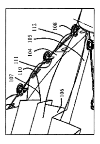
Reference is now made to figures la and lb which describes a preferred
embodiment. of the
present invention.
Figure la is a general view of the deployment and attachment device and figure
Ib is a closer
view of a portion of said deployment and attachment device. According to that
embodiment a
device 100 which is adapted for deployment and attachment of prosthetic mesh
during a
minimal invasive (Laparoscopic) hernia repair surgery is provided: The
deployment- and
attachment device (DAD) 100 comprises 2 main portions: distal portion 101, and
a proximal
portion 102. The distal portion is adapted to be inserted into a body during
the surgery via a
trocar. The distal portion is also adapted to deploy and attach a hernia patch
to the patient's
tissue surface. The proximal portion 102 comprises a handle 113 which provides
the surgeon
26
CA 02715740 2010-08-17
WO 2009/104182 PCT/IL2009/000188
with the ability to control the deployment and attachment of the patch. The
two portions are
connected via a tube 103.
The distal portion comprises of at least 2 flexible side arms (FA) 104 adapted
to be bended
laterally. The FA are connected at their distal end to the distal end of a
central flexible shaft
105, and at their proximal end to the distal end of the tube 103, the
connection is made using
a flexible joint. The central flexible shaft 105 is adapted to reciprocally
move within the tube
103 thereby spread and deploy the patch 106.
A prosthetic hernia repair patch 106 is folded in between the flexible arms
(FA) 104 and
connected to them via stretching means or especially a wire 107 which passes
through the
patch and a plurality of, dedicated loops 110 located on the FAs 104. The two
ends of the
wire are connected to the proximal portion 102. A plurality of dedicated
hernia clips 108 are
connected to the FA 104 at special connection points 111. Sais clips 108 are
adapted to attach
the patch 106 to the tissue. All the clips 108 are connected together by at
least one wire
(activation wire) 112 which will serve as their activation means. One end of
the activation
wire 112 is connected to the proximal portion 102.
The patch 106 is initially coupled to the FAs by a stretching wire 107 and is
folded in
between the FAs 104 such that it can be inserted into the body within a trocar
114. Once the
patch is inserted it is deployed by the central shaft 105 and the FAs 104.
Next, the physician
brings the patch into adjacent contact with the tissue. Then, the patch is
attached to the tissue
by clips 108 which are activated by the activation wire 112. Once the patch is
attached to the
tissue the activation wire 112 and the stretching wire 107 are cut via a
dedicated cutting
mechanism positioned in the distal end of tube 103. Next, the stretching wire
is pulled
towards the proximal portion and extracted. By doing so, the patch is no
longer coupled to
the FAs 104. Next, the FAs brought back into their initial stage, which
enables their
extraction from the body through the trocar 114.
Reference is now made to figures 2A-2D which illustrate the patch deployment
process. The
initial stage is described in figure 2A at which the two FAs are parallel and
straight ('close
form'). Furthermore, the patch 106 is folded in between the two FAs (FIG 2A).
Once the
distal portion has been inserted into the abdominal cavity, the physician
deploys the patch by
pressing the handle 113 (see figure 1, 10A-10E) at the proximal portion 102.
Pressing handle
113 results in a movement of the central shaft 105 toward the proximal portion
102. As a
27,
CA 02715740 2010-08-17
WO 2009/104182 PCT/IL2009/000188
result, the distance between the distal end of the central shaft 105 and the
distal end of the
tube 103 become shorter. Since the FAs 104 are connected to the distal end of
the central
shaft and the distal end of the tube 10; and since the distance -becomes
shortened the FAs
buckle and bend laterally, thereby forming an eye shape loop as described at
FIG 2B. At this
point, the two FAs are in their final stage ('open form'). It should be
pointed out that, whilst
the FAs 104 are bended, a continues tension at the stretching wire 107 is
maintained. The
continues tension results in the deployment of the patch 106 together with the
bending of the
FAs 104. Once the FAs 104 reach their final stage, the patch 106 is completely
unfolded and
deployed (FIG 2C). At this point the physician brings the patch to be in
contact with the
tissue and attaches the patch in a way which will be discus further on. Once
the patch have
been attached to the tissue, the physician detaches it from the FAs 104 by
releasing one end
of the stretching wire 107 and pulling it toward the proximal portion 102 (FIG
2D).
It should be pointed out that the FAs 104 are flexible in .the lateral
direction and very stiff on
the perpendicular direction such that on applied pressure (by the central
shaft) they buckle
only laterally. Furthermore, due to the fact that the FAs are very stiff on
the perpendicular
direction, the. applied pressure by the central shaft will be equally
distributed along the FAs.
If the pressure was not equally distributed and was concentrated only at the
edges (close to
the central shaft), the central portion of the FAs would not be able to apply
sufficient
pressure on the tissue to enable an attachment.
Reference is now made to figures 2E-2F which illustrate a side view of the
distal portion of
device 100 once the patch is deployed. As can be seen from the figures the
device 100 will be
able to adjust itself under pressure applied by the physician so as to bring
the patch 106 into
full contact with the tissue 501. This is due to the fact that the central
shaft 105 is flexible.
Another option for this capability of device 100 (to be adjustable) is to
locate a joint 220
between the distal end of tube 103 and the proximal end of the FA's 104.
Reference is now made to figs. 2G-21 which illustrate the distal portion 101
of device 100
adapted to deploy and attach a patch onto a curved surface, i.e. 3D
configuration. The 3D
device additionally comprises at least one flexible, arm 221 in a 3D
configuration. The figure.
2G represent the 3D device in which the FAs 221 and 104 are in a close
configuration (initial
stage) and figure 2H represent the FAs 221 and 104 in the open configuration
(final stage).
28
CA 02715740 2010-08-17
WO 2009/104182 PCT/IL2009/000188
Figure 21 represents the 3D device with the patch 106. Deploying and attaching
the patch will
be done essentially the same as for the 2D device.
FIG 3 describes a number of options for the folding of patch 106 prior to
inserting the distal
end 101 to the body. In all of the drawings a front cross section is seen
showing the patch
106, the FA 104, the clip 108 and the trocar 114. FIG 3A describes the most
simple form of
folding the patch 106. As can be seen from the figure, the patch 106 is folded
between the
two FA 104 in a zigzag form. The main advantage of this form is the fact that
this fold is
reversible. I.e. it is most likely that the patch will return to this form of
folding from an
unfolded state when FAs return to their close form. This enables a fast and
easy extraction
from the body in case the patch was not attached to the tissue.
FIG 3B describes the most efficient fold. This folding enable to use largest
patch since it
exploits and utilizes almost the entire available space in the trocar 114.
Another advantage of
this folding it the fact the patch is located above the clips 108 when it is
in the unfolded
stage, reducing the risk of entanglement between the patch 106 and the clips
108.
FIG 3C describes a variation of the previous patch folding. This folding is
simpler to
implement and it also have the advantage of reducing the entanglement risk as
mentioned
before.
Reference, is now made to. Figures 4A-4B which illustrate the attachment clips
and their use
during the attachment process.
FIGS. 4A-4B describe a preferred embodiment of the clip 108. The clip
comprises of a main
portion 401 which is connected to at least 2 lateral curved hooks 402 adapted
to penetrate the
tissue. The main portion is reversibly connected to the FAs 104 by a central
connection area
403. The clip 108 additionally comprises a connection point 404 to the
activation wire 112.
In a preferred embodiment of the present invention the connection point 404 is
positioned
laterally to the main portion 401. It should be pointed out that the
activation wire 112
connects all the clips 108 together. Furthermore, as can be seen from figure
4A the two hooks
402 are titled with regards to the main portion 401. This incline is of much
importance. This
incline ,is responsible for the fact that the hooks' edges 410 are constantly
presses against the
tissue prior to the clip's activation; such that once the clips are activated,
the edges 410, will
penetrate the tissue and not only slide along the patch 106 surface.
29
CA 02715740 2010-08-17
WO 2009/104182 PCT/IL2009/000188
The clip 108 can be made of any biocompatible metal (such as stainless steel,
titanium),
shape memory materials, super elastic metals (such as Nitinol i.e. NiTi), non-
degradable
polymer (such as polyurethane, PVC, PTFE (i.e. Teflon), PC (polycarbonate),
degradable
polymers (such as PLA; PGA, PLLA, PCL, PDS).
It should be pointed out that the clips 108 can be produced by photochemical
lithography
methods, laser cutting method.
The FA can be made of any biocompatible metal (such as stainless steel,
titanium), shape
memory materials, super elastic metals (such as Nitinol i.e. NiTi), non-
degradable polymer
(such as polyurethane, PVC, PTFE (i.e. Teflon), PC (polycarbonate).
The activation wire 112 and the stretching wire 107 can be made of any
biocompatible metal
(such as stainless steel, titanium), shape memory materials, super elastic
metals (such as
Nitinol i.e. NiTi), non-degradable polymer (such as polyurethane, PVC, PTFE
(i.e. Teflon),
PC (polycarbonate), degradable polymers (such as PLA, PGA, PLLA, PCL, PDS).
Reference is now made to figures 5A-5D which describes the attachment between
a patch
106 and a tissue 501. Figure 5A illustrates the tissue 501, the patch 106, the
clips 108, the
FAs 104 and the activation wire 112. Once the physician brings the patch
adjacent and in
contact with the tissue (figure 5B), the activation wire 112 is then pulled,
generating a
rotational moment which. rotates the clip with regards to the FAs 104. The
rotational
movement inserts the hooks 402 into the tissue 501 through the patch 106,
thereby providing
a strong attachment between the patch 106 and the tissue 501(Figure 5C). The
connection
between the clip 108 and the FAs 104 is made in such a way that the clips 108
are secured to
the FAs 104 prior to the attachment; and, the clips 108 detach from the FAs
104 once they
are attached to the tissue 501 (figure 5D).
Reference is now made to figures 6A-6C which illustrate means adapted to
reversibly
connect clips 108 to the FAs 104. According to this embodiment, clips 108 are
connected to
the FA's by hooks. As can be seen from the figure 6A the hooks 601 protrude
from'the FA's
into portion 403 of the clip 108. The clips are secured to the FA's due to L
shape of hook
601.
Two niches 602 are located on two opposite side along portion 403 perimeter.
Prior to
activating the clips (i.e. pulling activation. wire 112), the niches 602 are
not aligned together
with the hooks 601. Once the clips are activated, or in other words they
rotate, the hooks 601
CA 02715740 2010-08-17
WO 2009/104182 PCT/IL2009/000188
are aligned with the niches 602 (see figure 6B) such that the attachment
between the clips
and the FA is cancelled and the clips 108 are released from the FA 104 (see
figure 6C).
Reference is now made to figures 6D-6F which illustrate means according to
another
embodiment adapted to reversibly connect clips 108 to the FAs 104. According
to this
embodiment, clips 108 are connected to the FA's by a dedicated screw. As can
be seen from
figure 6D, screw 603 protrude from the FA's into portion 403 of the clip 108:
The clips are
screwed into screw 603 thereby secured to the FA'.s.
The clips can be detached from the FAs by screwing out the clips from screw
603 (see figure
6E). Once the clips 108 are screwed out from the screw 603 they are released
from the FAs
(see figure 6F).
It should be pointed that it is in the scope of the present invention wherein
the clips 108 can
be attached to the tissue and detach from the FA simultaneously or it can be
done in two
different steps.
Reference is now made to figures 7A-7B which illustrate the clip 108 according
to another
preferred embodiment of the present invention. According to this embodiment,
the clip 108
includes at least one additional hook 701 located on the lateral hooks 402.
This hook 701 is
adapted to prevent the reverse rotation (hence the release) of the clip 108
from the tissue 501.
It is acknowledged that the attachment between the patch 106 and the tissue
501 can be
annulled if the clip 106 rotates in the reverse rotational motion when
subjected to external
loads. Therefore, the additional hook 701 prevents this reverse rotational
motion with
minimal. interference to the forward rotation. Clip 108 may additionally
comprise at least one
hook 702 which is adapted to prevent any unwanted movement between the patch
106 and
the clip 108.
It is in the scope of the present invention wherein attachment clips,
activated by pulling, are
provided.. Another example for such clips is demonstrated in figures 7C-7H.
Figure 7C
illustrates an arrow-like clip. 108. The Clip 108 is characterized by having a
plate 711 and an
arrow-like shaped hook 712. Hook 712 is adapted to penetrate the patch 106 and
the tissue
501. Plate 711 also comprises groove cut 713 and a dedicated aperture 714. As
described
before, clip 10.8 has a connection point 403 to the activation wire 112.
Figures 7D-7G illustrate the steps needed for attaching the clip to the tissue
501.
31
CA 02715740 2010-08-17
WO 2009/104182 PCT/IL2009/000188
Figure 7D illustrates the clip 108 coupled to the FA 104 and being brought
into adjacent
contact with the tissue 501. Figure 7E illustrates the clip 108 being presets
against the tissue
501. The next step is attaching the patch to the tissue via clips 108 (figure
7F). The
attachment is obtained by pulling the activation wire 112. Once the clip 108
is attached to the
tissue 501, it detaches from the FA 104 (figure 7G).
Figures 7H-7J illustrate a closer view of the arrow-like clip 108 and how it
detaches from the
FAs 104.
Figure 7H illustrate the clip 108 attached to the FAs 104. The attachment.
between the clip
108 and the FA is provided by a dedicated hook 715 which is inserted into the
groove cut 713
in the plate 711.
When the activation wire 112 is pulled (by the handle 1002) the clips 108 are
pulled towards
the proximal portion 102. By pulling the clips 108, the arrow like hook 712
penetrates the
tissue 501. The result of this pulling is the movement of hook 715 within the
groove cut 713
until said hook 715 reaches the dedicated' aperture 714. Aperture 714 is
adapted to fit the
dimensions, shape and size of the hook 715. Once hook 715 reaches the aperture
714 (figure
71) the clip 108 can be detach from the FA 104 (figure 7J).
Figures 7K-7N illustrate a clip 108 according to another embodiment of the
present
invention. This clip 108 is characterized by a plate 711 and a sharp curved
edge 720. Plate
711 is attached to the FAs 104 by a screw or by a pin 721 (see figure 7N). Pin
721 is adapted
to be reversibly connected to a dedicated conection area 730 within clip 108.
As described
before, clip 108 has a connection point 403 which is adapted to be connected
to the activation
wire 112.
Figures 7L-7-- illustrate the steps needed for attaching the clip to the
tissue 501.
Figure 7L illustrate the clip 108 attached to the FAs 104 and being brought
into adjacent
contact with the tissue 501. When the activation wire 112 is pulled (by the
handle 1002),
clips 108 are rotated and thus, their sharp edge 720 penetrates the tissue 501
(figure 7M).
Once the sharp edge 720 penetrates the tissue 501, the clip 108 can be
detached from the FA
104 (figure 7N). The detachment can be obtained by extracting the pin 721 from
the clip 108.
Another option for the detachment is by the rotational motion of the clip 108
itself. In this
case the clip 108 is attached to the FA 104 by a screw. The rotational motion
needed for the
32
CA 02715740 2010-08-17
WO 2009/104182 PCT/IL2009/000188
attachment of the clip to the tissue will also be used for detaching (by
unscrewing) the clip
from the FA 104.
Reference is now made to figures 8A-8F which illustrate several alternative
embodiments for
the connection between the activation wire 112 and the clip 108. One option to
connect the
activation wire 112 and the clip 108 is as.described in figure 8A. The
activation wire can
enter the connection point 404 (which can be an aperture) and glued or tied to
it. Another
option is demonstrated in figure 8B in which the activation wire is glued
parallel to the
connection point which has a rectangular profile providing sufficient
attachment surface.
Figure 8C represents another alternative in which a number of apertures are
provided. The
activation wire 112 can enter the apertures in a zig-zag form or back and
forth look thereby
providing a glue-less attachment. Figures 8D - 8F represent another possible
embodiment
for the connection between the activation wire 112 and ,the connection point
404. According
to this embodiment, a fork like portion 404 encapsulates the wire 112.
Once the patch 106 is attached to the tissue 501 the stretching wire 107 and
the activation
wire 112 are cut.
Reference is now, made to figures 9A-9D which represent one possible
embodiment
implementing a mechanism 901 for cutting the activation wire 112 and the
stretching wire
107. Those figures illustrate a cross section view of the cutting mechanism
901. As can be
seen from the figures, the cutting mechanism 901 is located at the distal end
of tube 103. This
location was chosen for two reasons: (i) the fact that the edge of the
activation wire 112
should be as shorter as possible once the wire has been cut (this leftover
from the wire.
remain in the body, therefore it is preferable that the leftover would be as
short as possible);
and, (ii) the fact that the stretching wire 107 is pulled out of the body. In
order to extract the
wire 107, it has to pass towards the patch and through the perimeter of the
patch. To enable
an easy extraction of wire 107 it is preferred to cut the wire as close as
possible to the patch -
i.e. in the distal end of tube 103.
Figures 9A-9B illustrate a 3D cross section of the cutting mechanism 901 and
figures 9C-9D
illustrate a 2D cross section of the cutting mechanism 901.
The cutting mechanism 901 comprises a dedicated cutting pin 902 is placed
inside tube 103
near the distal end. The cutting pin 902 is connected to the distal -end of a
cutting activation
wire 903. The proximal end of the cutting activation wire 903 is connected to
the proximal
33
CA 02715740 2010-08-17
WO 2009/104182 PCT/IL2009/000188
portion 102 of device 100. Pulling the cutting activation wire 903 will result
in a reciprocate
movement of the cutting pin 902 (which is parallel to tube 103 longitudinal
axis).
Both the cutting pin 902 and the tube 103 have lateral holes (904 and 905
respectfully)
through which the activation wire 112 and\or the stretching wire 107 are
passing through.
Furthermore the activation wire 112 and the stretching wire 107 can move
freely inside the
holes (904 and 905). Once the patch 106 is attached to the tissue 501 the
physician now
needs to cut both the activation wire and the stretching wire. In order to cut
those wires the
physician will press the cutting handle 115 (the handle will be discussed in
further details in
figure 10) at the proximal portion 102. As a result the cutting activation
wire 903 and the
cutting pin 902 will be pulled toward the proximal portion 102 and a shear
force will be
implemented on the activation wire 112 and/or the stretching wire 107, hence
cutting them to
two sections as illustrated in figure 9B.
It is further in the scope of the present invention wherein a single mechanism
is adapted to
cut both the activation wire 112 and the stretching wire 107. It is further in
the scope of the
present invention wherein two separate mechanisms are adapted to cut the
activation wire
112 and the stretching wire 107.
In a preferred embodiment of the present invention, at least a portion of the
activation wire
remains within the body, thereby providing additional fixation to the clips.
This additional
fixation is needed in case one of the clips detaches from the patch and may
wander inside the
body, causing complications.
It is in the scope of the present invention wherein the entire activation wire
112 detaches
from the clips 108, and extracted from the body.
Reference is now made to figures 10A-10E which represent the proximal. portion
102 in
different stages of the deployment and the attachment. As can be seen from the
figure, the
proximal end 102 can comprises numerous handles. A dedicated handle 113 is
adapted to
reversibly transform the FAs 104 from their close stage (initial stage) to
their open stage
(final stage). A second handle 1001 is adapted to activate clips 108 such that
the patch 106 is
at least partially attached to the tissue 501 by pulling the activation wire
112. Handle 1002 is
adapted to release the patch 106 from the FAs, by cutting the stretching. wire
107. Handle
1002 is also adapted to cut the activation wire 112. Button 1003 is adapted to
release handle
113 such that the FAs 104 return to their close stage.
34
CA 02715740 2010-08-17
WO 2009/104182 PCT/IL2009/000188
Figure 10A illustrates the initial stage at which none of the handles are
pressed. In figure
1013, handle 113 is presses thereby transforming the FAs 104 from the close
stage to the open
stage thereby deploying the patch 106. After the patch 106 is deployed, the
physician can
move and rotate the deployed patch 106. This is preformed in order to bring
said patch
adjacent to the tissue. The physician can apply pressure needed for the
attachment process.
When the patch is located adjacent to the tissue, handle 1001 is presses
(figure IOC) thereby
activating the clips (by pulling the activation wire 112) and the patch is now
attached to the
tissue. After the patch is securably attached to the tissue, handle 115 is
presses, thereby the
cutting the stretching wire 107 and the activation wire ,l 12 (figure IOD).
Now the patch is
released from the FAs 104 and the FAs can return to the close stage and be
extracted from
the body (by pressing on button 1002, figure. IOE).
It is in the scope of the present invention wherein the device 100 which is
adapted to deploy
and attach a patch is useful in minimal invasive heart surgeries for attaching
a patch to the
heart, for preventing heart failure due to aneurysm.
It is in the scope of the present invention wherein the device 100 which is
adapted to deploy
and attach a patch is useful in endoscopic colon surgeries.
It is another object of the present invention to provide a deployment and
attachment device in
which clips 108 are at least partially connected to the patch (instead of the
FA) prior to the
attachment. In this embodiment the clips 108 are initially coupled to the
patch and not to the
FAs. Furthermore, in this embodiment the role of the FAs is to deploy the
patch and to press
it against the tissue.
Reference is now being made to FIGs 11 and 12A-12G which describe different
coupling
means between the patch 106 and. the FAs 104.
One of the advantages of the coupling means is the fact that no wires are
used. Thus,
enabling fast and simple mounting of the patch 106 on top of the FAs 104
during surgery.
The coupling is based on dedicated patch-FA clips 1201 which are connected to
the FAs 104.
Reference is now made to figure 12A which presents a closer view of the clip
1201. The clip
1201 has a main portion 1202 and at least one flexible branches 1203 extruding
(or
protruding) out from the main portion 1202. When the branches 1203 are not
subjected to
external load, they buckle laterally, therefore, provide attachment between
the FA 104 and
the patch 106.
CA 02715740 2010-08-17
WO 2009/104182 PCT/IL2009/000188
Reference is now made to figures 12A-12C which describe the method of de-
activating the
clip 1201 (i.e., disconnecting the clip 1201 from the patch 106).
Once the patch 106 is attached to the tissue 501, the user could disconnect
the patch by
pulling the FA 104 away from the tissue. As a result the branches 1203 are
deformed (from a
position being parallel to the tissue to a potion being perpendicular to the
same). Thus, the
branches 1203 pass through their entrance hole at the patch 106 (Fig 12A) and
are
disconnected from the patch.
Once the clips 1201 are disconnected from the patch it resumes its original
shape (in which
the branches 1203 are in a parallel position) - see figure 12C.
According to one embodiment of the present invention, in order to allow
correct folding and
unfolding of the patch 106, without the creation of tension on the patch 106
and on the FA
104, some of the clips 1201 could move freely along the FA 104, while others
will be fixed
to their place.
Figs 12D-12G describe the process of mounting the patch 106 on the deployment
system. In
order to enable simple insertion of the clip 1201 through the patch 106, the
clips 1201 are
delivered to the user together with a sharp cap (i.e., envelope covering) 1204
as can be seen
from Fig 12D.
The cap will accommodate the branches 1203 in a vertically alignment (in
relation to the FA
104). In a preferred embodiment, the user (a surgeon or a nurse) will insert
the cap 1204
whilst accommodating the clip 1201 through the patch during the surgery (see
Fig 12E).
Once all the clips 1201 are inserted. through the patch the caps 1204 are
removed from each
individual clip 1201 (see Fig 12F). As a result the. branches 1203 buckle
laterally (i.e., into a
parallel position in relation to the tissue). Thus, providing attachment
between FA 104 and
the patch 106 (see Fig 12G).
Figs 12H-12J illustrate an alternative approach of mounting the patch 106 on
the deployment
system. According to this approach, the clip 1201 comprises two separate
portions: (a) a
main portion 1205 which is connected the FA 104; and, (b) a second portion
1206. Portion
1206 have at least one branch 1208 connected to a pressing area 1207.
Initially, the branches
1208 are partially inserted into a channel within the main. portion 1205, such
that they are
vertically aligned, and their distal end protrudes out from the top end of the
main portion
36
CA 02715740 2010-08-17
WO 2009/104182 PCT/IL2009/000188
1205 (see Fig 121). The main role of portion 1205 is to retain the branches
1208 from
buckling laterally.
Said attachment. between patch 106 and FAs 104 is obtained by inserting the
patch 106
through the branches 1208 (see Fig 121) and then portion 1206 is pressed
upward, toward the
patch. The branches, which is no longer confined by the main portion 1205,
buckle laterally,
thus provide the said attachment between patch 106 and FA 104 (see Fig 12J).
Figs 12K-12Q describe another alternative approach of mounting the patch 106
on the
deployment system. In this approach, the branches 1208 of each clip are bended
radially,
toward or away the center of the patch (see Fig 12K).
Once the patch 106 is attached to the tissue 501, the FAs 104 are closed. As a
result the
branches 1208 move radially, therefore, disconnecting form the patch 106, as
can be seen at
Figs 12M-12Q. In a preferred embodiment, the mounting process of patch 106 on
top FAs
104 is similar to the previously described approach as can be seen in Figs 12N-
12P
Reference is now being made to Figs 13A-13F which describes an alternative
embodiment
for attaching patch 106 to the tissue 501 by several clips 108. In this
embodiment, as can be
seen in Fig 13A and Fig 13D, the clip 108 is connected to a wire 1301 which is
incorporated
within the patch 106. The connection between clip 108 and wire 1301 is at a
wire connection
area 1306 and will enable free rotation of the clip 108 around the wire 1301.
The clip 108 will have a central plate 1305 and at least two sharp arms 1304
connected.to
each side of the plate. In a preferred embodiment, the arms 1304 are curved.
An activation
wire 1302 will be coupled to the plate 1305. Once the wire 1302 is pulled,
during surgery, the
entire clip 108 is rotated around wire 1301.
The activation wires 1302 from each clip 108 will be connected to a central
activation area
1303 (see figure 13B) which will pull all the activation wires 1302 toward the
center of the
patch 106 once the attachment between patch 106 and tissue 501 is needed. The
central
activation area 1303 will be activated by pulling the. clip activation wire
112, or by rotating
the central shaft 105.
As can be seen in Figs 13B and 13E, once the central activation area 1303 is
activated, each
activation wire 1302 is pulled toward the center, therefore inducing
rotational movement of
each of the clips 108 around wire 1301. As a result, the arms 1304 (of each
clip. 108), are
37
CA 02715740 2010-08-17
WO 2009/104182 PCT/IL2009/000188
inserted through the patch 106 and the tissue 501. Thus, providing attachment
between the
tissue 501 and the patch 106.
Once the attachment (between the patch and the tissue) is achieved, the wire
1302 is
disconnected from the central activation area 1303 in order to enable proper
detachment
between the connected patch 106 and the rest of the deployment system.
In a preferred embodiment of the current invention the connection between clip
108 and the
activation wire 1302 is considerably weaker than the rest of the wire 1302 but
strong enough
to rotate the clip 108. Once the clip is fully inserted into the tissue, the
activation wire will be
pulled using sufficient'force for disconnecting it for the clip 108 (Fig 13C,
13F). In other
words, pulling the activation wire 1302 in F amount of force enables the
insertion of the clip
108 into the tissue; and, pull the activation wire 1302 in F1 amount of force
(F1 is
sufficiently greater than F) enables the disconnection of the activation wire
1302 from clip
108.
Reference is now being made for Figs 14A-14D which describe an alternative
detachment
mechanism between the patch 106 and the FAs 104. According to this embodiment,
the
central shaft 105 is extended from the proximal side of handle 102 (Fig 14A),
therefore the
unfolding process can be achieved be pulling the central shaft proximally.
The distal end of shaft 105 is inserted to a sleeve 1401 located at the distal
end on the FAs
104. The sleeve 1401 have two lateral holes 1402 initially concentric to a
hole 1403 at the
distal end of the central shaft. When hole 1402 and hole 1403 are aligned, the
stretching wire
107 can pass through them (see Fig 14C).
The stretching wire is kept constantly in tension, therefore keeps a
sufficient tension applied
on the patch 106 during the unfolding process, and prevents wrinkles. Once the
patch 106 is
attached to the tissue 501, the user will rotate handle 1404 which is located
at the proximal
end of the central shaft 105 (Fig 14B). This rotational movement cuts the
stretching wire 107
at the distal end of the central shaft 105 (Fig 14D). This cutting cuts the
stretching wire 107
into two halfs (sections). Since there .the stretching wire 107 are tensed,
the two ends of the
already cut-wire 107 will be instantly pulled toward the proximal end of the
system, therefore
canceling the attachment between patch 106 and the FAs 104.
In the. foregoing description, embodiments of the invention, including
preferred
embodiments, have been presented for the purpose of illustration and
description. They are
38
CA 02715740 2010-08-17
WO 2009/104182 PCT/IL2009/000188
not intended to be exhaustive or to limit the invention to the precise form
disclosed. Obvious
modifications or variations are possible in light of the above teachings. The
embodiments
were chosen and described to provide the best illustration of the principals
of the invention
and its practical application, and to enable one of ordinary skill in the art
to utilize the
invention in various embodiments and with various modifications as are suited
to the
particular use contemplated. All such modifications and variations are within
the. scope of the
invention as determined by the appended claims when interpreted in accordance
with the
breadth they are fairly, legally, and equitably entitled.
Reference is now made to figs. 15A-15D which illustrate an embodiment in which
the patch
can be laterally rotate with respect to the tissue such that the right
orientation of the patch is
facing the tissue or the hernia.
Reference is now being made for Figs 15A-15B, which describes an embodiment in
which at
least a part of tube 103 is a controllable and flexible joint 1502. This joint
is especially
adapted to allow fine adjustment of the lateral angle between distal portion
101 and the
proximal portion 102 during the procedure.
In other words, the controllable and flexible joint is provided in order to
adjust the right
orientation of the patch with regards to the tissue or the hernia.
Such adjustment is needed in order to aline the patch 106 with a desirable
lateral position
with regards to tissue 501. According to this embodiment, the controllable and
flexible joint
1502 is made of flexible material (e.g. polymer) and can be curved according
to
predetermined angle in its distal end. The controllable and flexible joint
1502 is housed by a
rigid alignment tube 1501. Said rigid alignment tube 1501 can be reciprocally
moved along it
longitudinal axis.
It should be emphasized that the controllable and flexible joint 1502 has an
intrinsic spring-
like properties; i.e., the controllable and flexible joint 1502, when is
unloaded, returns to its
original curved/bent shape.
At the initial state (Fig . 15A) the controllable and flexible joint 1502 is
completely
encapsulated within the rigid alignment tube 1501 such that controllable and
flexible joint
1502 it forced to be straight and linear, once the distal portion 101 is
inserted into the patient
body and lateral angle adjustment is required, the rigid alignment tube 1501
is pulled toward
the proximal portion 102; as a result, the controllable and flexible joint
1502, which is no
39
CA 02715740 2010-08-17
WO 2009/104182 PCT/IL2009/000188
longer supported by the rigid alignment tube 1501 , is bent/curved into its
original form, thus
providing the desire angle between the proximal portion 102 and the distal
portion 101 (Fig
15B).
By controlling the location of the rigid alignment tube 1501 with respect to
the
controllable/flexible joint 103, a fine adjustment of the angle is obtained.
As mentioned
above, the control over the rigid alignment tube's 1501 location is provided
by the amount of
pulling or pushing of said rigid alignment tube 1501 towards and away from the
proximal
portion 102.
Since the surgeon controls said rigid alignment tube's 1501 location, he
therefore controls the
angle between the distal portion 101 and proximal portion 102.
The movement (and thus the angle between the distal portion 101 and proximal
portion 102)
is adjusted by the angle control means 1502 which is located at the proximal
portion 102.
Reference is now made to figures 15C-15D which illustrate a top view of the
system. In
Figure 15C the rigid alignment tube 1501 is fully housing/encapsulating the
controllable and
flexible joint 1502 and thus the angle between the distal portion 101 and
proximal portion
102 is 0 degrees.
Figure 15D also illustrates a top view of the system. However, in figure 15D
the rigid
alignment tube 1501 is not fully housing/encapsulating the controllable and
flexible joint
1502, thus the controllable and flexible joint 1502 is curved/bent according
to the location of
the rigid alignment tube's 1501 with respect to the controllable and flexible
joint 1502.
Therefore, an angle A is obtained between the distal portion 101 and proximal
portion 102.
Reference is now being made to Figs 16A-16C which describe an embodiment of
the patch
insertion sleeve. Such a sleeve/cover is needed in order to facilitate, to
ease and to catalyze
the insertion of the distal end 101 and the patch 106 in to the patient's
body.
According to this embodiment the insertion sleeve 1601 is an elongated rigid
tube with a
cone shaped expansion 1602 at its proximal end and a stopper 1603 near its
distal end. Once
the patch 106 is mounted and folded on the distal portion 101 (during the
surgery or during
the assembly process prior to the surgery), it is inserted, together with the
distal portion 101,
into the insertion sleeve 1601 trough the cone shape expansion 1602, such that
the distal end
of the insertion sleeve 1601 reaches the distal end of the distal portion 101
(Fig 16B).
CA 02715740 2010-08-17
WO 2009/104182 PCT/IL2009/000188
The overall complex is then inserted to the patient's body through a trocar
114. The outside
diameter of the insertion sleeve 1601 at the portion between its distal end
and the stopper
1603 is smaller or equal to the inside diameter of the trocar 114, such that
this portion can be
inserted into the trocar 114. Once the stopper 1603 reaches the trocar 114
proximal end, the
distal portion 101 and the patch 106 slide out of the insertion sleeve and
into the trocar 114
and the patent body, while the insertion sleeve is slide backward along the
rigid alignment
tube 1501 or the tube 103. At the final stage (Fig 16C), the distal portion
101 and the patch
106 is completely inserted into the patient's body.
Reference is now being made to figures 18A-18D which illustrate an additional
embodiment
of the deployment mechanism. This embodiment provides larger patches
deployment using
the same initial length of the distal portion 101; in addition, it will allow
a simpler reversible
attachment between patch 106 and the distal FA 104.
According to this embodiment each FA 104 additionally comprises a long rod
1801 which is
aligned parallel to the central shaft.. The rods 1801 are connected to the FA
104 via at least
one joint or flexible portion.
According to this embodiment, the patch 106 is reversibly connected to the
rods1801 rather
than the FAs 104. The patch 106 is deployed by a reciprocal movement of the
central shaft
105 toward the proximal portion. As a result, the rods 1801 are laterally
moved away from
each other, providing patch deployment.
Figure 18D illustrate the above embodiment incorporated with the patch 106.
41