Note: Descriptions are shown in the official language in which they were submitted.
CA 02716031 2012-03-14
Specification
1. Title of the Invention
METHOD FOR PRODUCING ARTIFICIAL BONE AND ARTIFICIAL BONE
PRODUCED BY THE METHOD
2. Field of the Invention
The present invention relates to a method for producing
an artificial bone used in surgery of human bodies and others
by utilizing a three-dimensional shaping method and an
artificial bone based on the method.
3. Description of the Related Art
There is a trend that demand for transplantation of an
artificial bone for a bone part of a human body where a defect
or damage has occurred has increased in line with the development
of medical technology.
As shown in Patent Document 1, there has been extensively
used a method for producing an artificial bone in which a layer
of one or more types of powder selected from metals, resins
and ceramics is subjected to laser sintering based on artificial
bone image data and the sintered layer is laminated.
Incidentally, it is an inevability in molding artificial
bones that an artificial bone is molded accurately at both ends
and their vicinities constituting a joined part to a human bone
part.
1
CA 02716031 2012-03-14
However, in a conventional method for producing an artificial
bone, no particular attention has been paid or no device has
been made in this respect. And Patent Document 1 is no exception.
Further, the joined part of an artificial bone is required
to be made stronger than other regions in order to prevent fatigue
or friction resulting from joining.
However, despite the fact that the above-described laser
sintering has been adopted, conventional techniques have failed
to provide a configuration in which particular attention is
paid to this respect.
[Prior Art Documents]
[Patent Document]
[Patent Document I] W02007/122783
4 .Summary of the Invention
Problems to be Solved by the Invention
An object of the present invention is to provide a method
for producing an artificial bone capable of realizing accurate
molding at a joined part with appropriate strength and an
artificial bone based on the method.
[Means for Solving the Problems]
In order to attain the above object, a basic construction
of the present invention is made up of the following:
(1) A method for producing an artificial bone in which
2
CA 02716031 2012-03-14
electromagnetic waves or electron beams are irradiated to a
layer of one or more types of powder selected from metal
biomaterials, ceramics for anartificial bone andplastic resins
for an artificial bone based on image data corresponding to
a shape of the artificial bone, thereby effecting sintering
or melting, and the thus at least two sintered layers or the
thus melted and at least two solidified layers is laminated,
the method for producing the artificial bone wherein a surface
finish step is adopted that inner faces and/or outer faces of
the both ends and their vicinities configuring a joined part
to a human bone part are polished by a rotating tool based on
the image data, and irradiation of electromagnetic waves or
electron beams at both ends and their vicinities configuring
the joined part is made greater than that at other regions,
(2) an artificial bone which is produced by any one of
the above-described methods of (1).
5. Brief Description of the Drawings
Figs. 1 show an artificial bone which is hollow inside.
Fig. 1 (a) is a sketch showing a pipe-shaped artificial bone,
Fig. 1 (b) is a sketchshowingapartiallypipe-shapedartificial
bone, and Fig. (c) is a sketch showing a combination of the
pipe shaped artificial bone with the partially pipe-shaped
artificial bone.
3
CA 02716031 2012-03-14
Figs. 2 show an artificial bone in which the interior
of a peripheral wall along the longitudinal direction is in
a three-dimensional meshed state. Fig. 2 (a) is a
cross-sectional view taken in the longitudinal direction, Fig.
2 (b) is a cross-sectional view taken in a direction orthogonal
to the longitudinal direction (the cross section shown in. 2
(b) shows a portion taken along the line A to A in Fig. 2 (a) ) .
Figs. 3 show an artificial bone which forms a hollow
peripheral wall along the longitudinal direction. Fig. 3 (a)
is a side elevational view where the peripheral wall is in a
meshed state, and Fig. 3 (b) is a side elevational view where
the peripheral wall is in a pore aggregate state.
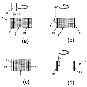
Figs. 4 explain that powder is subjected to irradiation
by electromagnetic waves or electron beams and polished by a
rotating tool, thereby molding an artificial bone. Fig. 4 (a)
is a cross-sectional view showing a sintering step in which
electromagnetic waves or electron beams are irradiated, Fig.
4 (b) is a cross-sectional view showing a polishing step in
which an outer wall of the sintered region is molded, Fig. 4
(c) is a cross-sectional view showing a laminating step in which
powder is additionally laminated after completion of the
polishing step to mold the outer wall, and Fig. 4 (d) is a
cross-sectional view showing a step in which the inner wall
4
CA 02716031 2012-03-14
,
. .
,
..
is molded after completion of steps (a) , (b) and (c) (the white
arrows indicate a state that a rotating tool rotates around
and the solid arrows indicate a state that the rotating tool
rotates on its own axis) .
Fig. 5 is a block diagram showing a case where a CAD/CAM
system is applied to the present invention.
[Description of the Symbols]
1: artificial bone
11: end
2: powder
21: sintered region
3: CAD/CAM system
31: CAD system
32: CAM system
4: NC controller
5: apparatus for irradiating electromagnetic waves or electron
beams
6: rotating tool
7: electromagnetic waves or electron beams
6. Detailed Description
In general, an artificial bone 1 adopts any one of a
configuration in which a peripheral wall is made hollow inside
as shown in Figs. 1 and a configuration in which the peripheral
CA 02716031 2012-03-14
,
. .
wall is in a meshed state of a three-dimensional structure inside
as shown in Figs. 2. (In Figs. 2, the meshed state of the
three-dimensional structure is provided all over a region along
the longitudinal direction, but a configuration may also be
adopted in which a meshed state is provided at a partial region
such as both ends and the inside of the peripheral wall besides
both ends forming a joined part and positions of their
neighborhoods . )
However, the above-described hollow artificial bone
includes any one of the pipe shape, the partial pipe shape and
the combination of them as shown in Figs. 1 (a) , (b) and (c) .
Further, for the purpose of infiltration of body fluid or
allowing body fluid to enter into human tissue, as shown in
Figs. 3 (a) and (b) , there may be adopted an artificial bone
in which a meshed state or a pore aggregate state is provided
at all or some regions of a peripheral wall along the longitudinal
direction. (In Figs. 3 (a) and (b) , there is adopted an
artificial bone in which the meshed state or the pore aggregate
state is provided at regions other than both ends 11 and their
vicinities. However, as a matter of course, it is possible
to adopt such a configuration in which any one of these states
also covers the both ends 11 and their vicinities.)
In any mode, the artificial bone 1 is joined to a human
6
CA 02716031 2012-03-14
, .
. ,
bone at both ends and their vicinities.
In most cases, the artificial bone 1 is firmly joined
to a human bone with a screw in such a manner that the artificial
bone 1 is placed outside and the human bone is placed inside.
However, as an exception, they can be joined in such a manner
that the human bone is placed outside and the artificial bone
1 is placed inside.
Nevertheless, at both ends 11 and their vicinities
configuring a joined part, an artificial bone is required to
be molded accurately according to the shape of a human bone.
Further, for the purpose of avoiding friction and fatigue at
the joined part, the joined part is required to be made greater
in strength than other regions.
In the previously described basic construction (1) , as
shown in Figs . 4 (a) and (c) , based on the conventional techniques
in which electromagnetic waves or electron beams 7 are irradiated
to a layer of one or more types of powder 2 selected from metal
biomaterials, ceramics for the artificial bone 1 and plastic
resins for the artificial bone 1 to effect sintering and these
sintered layers are laminated sequentially, inner faces and/or
outer faces of the ends 11 and their vicinities where joining
is performed are polished by a rotating tool 6, thereby
conducting final shaping as shown in Figs. 4(b) and (d) . And,
7
CA 02716031 2012-03-14
. ,
. .
an accurately joined face is provided.
Then, where a maximum diameter of surface roughness based
on the polishing by the rotating tool 6 is to be 10 pm, it is
possible to provide an extremely accurate shaping and match
the needs of medical practices.
There is found no particular trouble resulting from
polishing by the rotating tool 6 on inner faces of the ends
11 and their vicinities configuring a joined part. Therefore,
in this respect, the basic construction (1) has technical value .
In an artificial bone 1 where an inner face other than
the ends 11 and their vicinities are bent or in an artificial
bone 1 where apart further inside the ends 11 and theirvicinities
is increased in diameter, an ordinary rotating tool 6 smaller
in rotating diameter may cause trouble in polishing and molding
an inner face.
However, even in these cases, for example, a specially
shaped rotating tool having an enlarged rotating diameter at
the leading end can be used to overcome the above trouble.
The basic construction (1) also includes a method for
polishing and polishing both inner faces and outer faces of
the ends 11 and their vicinities. In this configuration, it
is possible not only to provide accurate molding on an inner
face to be joined but also to mold a smooth outer face at the
8
CA 02716031 2012-03-14
. .
..
end 11 by polishing and polishing, thereby avoiding unnecessary
muscle adhesion.
With attention given to the above situation, the basic
construction (1) has adopted a surface finish step in which
a region other than a joined part to a human bone part on an
outer face of the artificial bone 1 maybe polished by the rotating
tool 6.
There is such a case that a complicated shape is formed
at a leading end of the joined end 11 to a human bone part.
In this case, an embodiment having a polishing step in
which leading end faces at both ends are polished by the rotating
tool 6 enables accurately shaping the leading end which is
complicated in shape, therefore it is favorably applicable.
In normal molding, an outer face is polished and molded
by the rotating tool 6 after being sintered by means of
electromagnetic waves or electron beams 7 and molded, then
laminated further, while in most cases an inner face is polished
and molded after completion of polishing and molding of the
outer face.
Where the leading end faces of the both ends 11 configuring
the joined part are polished by the rotating tool 6, these faces
may be polished before or after polishing of the inner face.
In most cases, these faces are polished before that.
9
CA 02716031 2012-03-14
. .
..
In the basic construction (1) , irradiation at the ends
11 and their vicinities configuring a j oined part is made greater
than that at other regions, thereby increasing the strength
of the joined part and decreasing the friction and fatigue of
the artificial bone 1 at the joined part as much as possible.
To set an irradiation dose at the ends 11 and their
vicinities, either one of which the irradiation dose per unit
area is increased or the irradiation time is prolonged can be
selected.
Where a three-dimensional meshed state or a pore aggregate
state is formed at all or some of a peripheral wall along the
longitudinal direction as shown in Figs. 3 (a) and (b) , in order
to maintain necessary strength at a region covering an
intermediate portion of the peripheral wall, irradiation dose
of electromagnetic waves or electron beams 7 can be set greater
than other regions free of the above state.
However, it is also possible that, depending on an area
of the meshed region, the number and dimension of a pore aggregate
state or an area formed by the aggregate state, such selection
can be made that the region concerned is made lower in strength
than other regions free of the above state and equal in strength
to a human bone.
Where irradiation dose per unit area or irradiation time
CA 02716031 2012-03-14
is changed in the basic construction (1) and the embodiments
shown in Figs. 3 (a) and (b) , in most cases, it is changed by
such an embodiment that a CAD/CAM system 3 shown in Fig. 5 is
adopted, a CAD system 31 is used to set image data corresponding
to a shape of the artificial bone 1, and the CAD system 31 or
a CAM system 32 is used to set irradiation dose per unit area
or irradiation time of electromagnetic waves or electron beams
7 at individual regions of the artificial bone 1.
In the embodiment adopting the CAD/CAM system 3, where
electromagnetic waves or electron beams 7 are changed at each
of predetermined regions based on the set irradiation dose per
unit area or the set irradiation time of the electromagnetic
waves or electron beams 7 corresponding to individual regions
of the artificial bone 1, the artificial bone 1 at the
predetermined region changes in strength. Therefore,
appropriate moving velocity and/or rotating velocity where
polishing is performed by the rotating tool 6 also change.
In coping with the above-described situation, an
embodiment is preferably adopted that in accordance with
irradiation dose per unit area or irradiation time of
electromagnetic waves or electron beams 7, the CAD system 31
or the CAN system 32 is used to set the moving velocity and/or
rotating velocity of the rotating tool 6 as well.
11
CA 02716031 2012-03-14
. =
In general, where a spot diameter to be irradiated with
electromagnetic waves or electron beams 7 is set less than 100
pm, not only the ends 11 and their vicinities but also other
regions can be molded accurately and finely.
Metal biomaterials include Ti-6A1-7Nb, pure Ti, Ti-6A1-4V,
Ti-29Nb-13Ta-16Zr, Ti-15Mo-5Zr-3A1, Ti-
5A1-5V-5Cr,
Ti-15Zr-4Nb-4Ta, Co-Cr alloy, SUS3162, and SUS630. Ceramics
for the artificial bone 1 include calcium phosphates (such as
hydroxyapatite, a-calcium phosphate and-calcium phosphate) .
Plastic resins for the artificial bone 1 preferably include
polycarbonate and polyester in terms of strength.
7. Example
Hereinafter, an explanation will be made by referring
to an example.
[Example]
In the example, powder 2 which is metal biomaterial powder
or substantially composed of the metal biomaterial powder is
adopted as laminated powder 2 at both ends and their vicinities
configuring a joined part.
In this example, only metal biomaterial powder or powder
substantially composed of the metal biomaterial powder is used
at both ends and their vicinities, thus making it possible to
maintain necessary strength and also cope with friction and
12
CA 02716031 2013-01-09
fatigue at the j oined part, in addition to the basic construction
(1).
Where powder 2 other than the powder described above in
the example is adopted at regions other than the both ends 11
and their vicinities, the powder 2 is switched to the
above-described powder to effect laminating at a stage where
the both ends 11 and their vicinities are subjected to
irradiation. Therefore, in the example, two or more nozzles
are preferably used for spraying the powder 2.
8. Effects of the Invention
Based on the previously described basic constructions
(1) and (2), in the case of the artificial bone of the present
invention, it is possible to accurately mold the artificial
bone at the ends and their vicinities configuring a joined part
to a human bone with necessary strength and to exert functions
fundamentally required for an artificial bone.
The present invention is widely applicable in producing
and using artificial bones.
13