Note: Descriptions are shown in the official language in which they were submitted.
CA 02716390 2015-09-23
f
ENHANCED DELIVERY OF A THERAPEUTIC TO OCULAR
TISSUES THROUGH IONTOPHORESIS
BACKGROUND
Corticosteroids are widely prescribed therapeutics. Systemic, topical and
injected formulations are routinely employed for a variety of ophthalmic
conditions.
In particular, topical applications account for the widest use of non-
invasively
delivered corticosteroids for ocular disorders. This approach, however,
suffers from
low bioavailability and, thus, limited efficacy.
Dexarnethasone, member of the glucocorticoid class of steroid hormones,
acts as an anti-inflammatory and immunosuppressant. Ocular formulations are
used
that allow for diffusion of dexamethasone across an ocular membrane, however,
such topical formulations suffer from slow, inadequate and uneven uptake.
Because
current ocular delivery methods achieve low ocular exposures, frequent
applications
are required and compliance issues are significant.
Topical dexamethasone applications involving ocular iontophoresis have not
been described. Based on commercially-available, columbic-controlled
iontophoresis for topical applications to the skin of a variety of
therapeutics, it is
clear that even well understood pharmaceuticals require customized
formulations for
iontophoresis. These alterations maximize dosing effectiveness, improve the
safety
and manage commercial challenges. The known technical formulation challenges
presented by dermatological applications may translate in to ocular delivery.
However, ocular iontophoresis presents additional formulation needs. Thus,
developing novel formulations that are ideally suited for ocular iontophoretic
delivery of corticosteroids is required. Such formulations include many
variables,
including: API concentration, solute, excipients, stabilizers, buffering
agents,
CA 02716390 2010-08-24
WO 2009/108626 PCT/US2009/034977
delivery applicator, iontophoretic dose, etc. Developing corticosteroids
suitable for
non-invasive local ocular delivery will significantly expand treatment options
for
ophthalmologists.
SUMMARY
Described herein are devices and methods for enhancing the delivery of
negatively charged compounds into and through tissues, e.g., the eye. More
specifically, the methods and devices described herein utilize iontophoresis
to
actively deliver a compound, e.g., dexamethasone phosphate, into a mammalian
eye.
The methods and devices focus on developing corticosteroid formulations and
use of
these formulations to maximize drug delivery, e.g., through iontophoresis, and
patient safety. These novel formulations are suitable for treating a variety
of
inflammatory-mediated ocular disorders. The formulations, which include
different
strengths of the active phaiiiiaceutical ingredient (API), are capable of
being used
with different iontophoretic doses (e.g., current levels and application
times). These
solutions can, for example: (1) be appropriately buffered to manage initial
and
terminal pHs, (2) be stabilized to manage shelf-life (chemical stability),
and/or (3)
include other excipients that modulate osmolarity. Furthermore, the drug
product
solutions are crafted to minimize the presence of competing ions. These unique
dosage forms can address a variety of therapeutic needs. Ocular iontophoresis
is a
novel, non-invasive, out-patient approach for delivering substantial amounts
of APIs
into many ocular tissues. This non-invasive approach can lead to results
comparable
to or better than those achieve with ocular injections, without the
significant risk of
infection associated with the latter.
One embodiment is directed to a method for iontophoretically delivering a
corticosteroid, corticosteroid derivative, prodrug or salt thereof into the
eye of a
subject, comprising: a) administering the compound to the eye of the subject;
and b)
performing ocular iontophoresis under conditions such that the pH is between
about
2.5 and about 6.5, thereby delivering the compound into the eye. In a
particular
embodiment, the corticosteroid is a dexamethasone compound, derivative
thereof.
In a particular embodiment, the starting pH is about 5.7. In a particular
embodiment, the corticosteroid is in the faun of a prodrug. In a particular
2
BOST_964298.1
CA 02716390 2010-08-24
WO 2009/108626 PCT/US2009/034977
embodiment, the corticosteroid is delivered by injection prior to
iontophoresis. In a
particular embodiment, the method of injection is selected from the group
consisting
of: an intracameral injection, an intracorneal injection, a subconjonctival
injection, a
subtenon injection, a subretinal injection, an intravitreal injection and an
injection
into the anterior chamber. In a particular embodiment, the corticosteroid is
administered topically prior to iontophoresis. In a particular embodiment, the
topical administration comprises providing the corticosteroid in a form
selected from
the group consisting of: a liquid solution, a paste and a hydrogel. In a
particular
embodiment, the corticosteroid is embedded in a foam matrix. In a particular
embodiment, the corticosteroid is supported in a reservoir. In a particular
embodiment, the step of ocular iontophoresis is carried out prior to, during
or after
the step of administering the corticosteroid. In a particular embodiment, the
compound is delivered by an iontophoretic dose of about 1.7 x 10⁴ mA=min to
about
120 mA=min, e.g., between about 10 mA=min and about 30 mA=min. In a particular
embodiment, the iontophoretic dose is about 20 mA=min. In a particular
embodiment, the compound is delivered at a current of about 4.0 mA for a
period of
about 5 minutes. In a particular embodiment, the compound is delivered at a
variable or fixed current of less than about 10 mA. In a particular
embodiment, the
compound is delivered for a time of less than about 10 minutes.
One embodiment is directed to a kit for iontophoretically delivering
dexamethasone into the eye of a subject, wherein the kit is to be used for
iontophoresis between a pH range of about 2.5 to about 6.5, and an apparatus
for
iontophoretically delivering the compound into the eye of a subject.
One embodiment is directed to a dexamethasone formulation suitable for
ocular iontophoretic delivery into the eye of a subject. In a particular
embodiment,
the dexamethasone is in the form of a prodrug. In a particular embodiment,
iontophoretic delivery is to be performed in a pH range of between about 2.5
and
about 6.5. In a particular embodiment, the pH is about 5.7.
One embodiment is directed to a device for delivering dexamethasone,
comprising: a) a reservoir comprising at least at least one medium comprising
a
dexamethasone formulation, the reservoir extending along a surface intended to
cover a portion of an eyeball; and b) an electrode associated with the
reservoir so as
3
BOST_964298.1
CA 02716390 2010-08-24
WO 2009/108626
PCT/US2009/034977
to, when polarized, supply an electric field directed through the medium and
toward
a surface of the eye, wherein at least a portion of the dexamethasone
formulation is
delivered transdermally through the surface of the eye through iontophoresis.
In a
particular embodiment, the reservoir comprises: a) a first container for
receiving the
at least one medium comprising the dexamethasone formulation; b) a second
container for receiving an electrical conductive medium comprising electrical
conductive elements; and c) a semi-permeable membrane positioned between the
first and second containers, the semi-permeable membrane being permeable to
electrical conductive elements and non-permeable to the active substances.
One embodiment is directed to a method for treating a corticosteroid
sensitive ophthalmic disease in a mammal, comprising administering an
effective
amount of a corticosteroid by ocular iontophoresis. In a particular
embodiment, the
ophthalmic disease is selected from the group consisting of: uveitis, dry eye,
post
operative inflammation and corneal graft rejection. In a particular
embodiment, the
corticosteroid is dexamethasone phosphate. In a particular embodiment,
administration of dexamethasone phosphate occurs in a single dose.
BRIEF DESCRIPTION OF THE DRAWINGS
FIG. 1 is a schematic overview of the iontophoresis apparatus and procedure.
FIG. 2 is a graph showing in vitro delivery of DEX phosphate.
FIG. 3 is a graph showing in vitro delivery of DEX phosphate using varying
sodium citrate concentrations.
FIG. 4 is a graph showing linear dependence of DEX phosphate flux on
applied current (mean SD, n = 4).
FIG. 5 is an image showing the setup of iontophoretic dosing in New
Zealand rabbit eyes with the EyeGate II device and generator.
FIG. 6 is a graph showing tear flow measurement in rabbits injected in the
lacrimal gland with either Concanavalin A or phosphate-buffered saline (n = 8
for
each group). Rabbits were given a single iontophoretic dose of either
dexamethasone phosphate or phosphate-buffered saline on Day 2. Tear flow was
measured with Schirmer strips and was recorded as the distance in mm of flow
in 5
minutes. (* = P < 0.01).
4
BOST_964298.1
CA 02716390 2010-08-24
WO 2009/108626 PCT/US2009/034977
FIG. 7 are representative slit-lamp microscope images of fluorescein staining
on the ocular surface of rabbits on Day 8 of the study. Left panel: Group 1 ¨
Rabbit
had Con A-induced dry eye and was iontophoretically treated with saline on Day
2.
Middle Panel: Group 2¨ Rabbit had Con A-induced dry eye and was
iontophoretically treated with Dex-P on Day 2. Right panel: Group 3 ¨ Rabbit
was
injected with saline and was iontophoretically treated with saline on Day 2.
FIG. 8 is a graph showing fluorescein staining score in rabbits after a single
iontophoretic dose of either dexamethasone phosphate solution or phosphate-
buffered saline in the lacrimal gland (n = 8 for each group). * = P < 0.01).
FIG. 9 is a graph showing the expression of interleukin-lbeta (IL-113) in the
lacrimal glands and corneas of rabbits on Day 4 or Day 8 after lacrimal gland
injection of concanavalin A or saline and iontophoretic treatment with
dexamethasone phosphate or saline on Day 2. n = 4, * = P < 0.01. No
significant
difference was noted in the cornea, indicating a specific lacrimal gland
inflammatory
response.
FIG. 10 is a graph showing expression of transforming growth factor beta-1
(TGF-131) in the lacrimal glands and corneas of rabbits on Day 4 or Day 8
after
lacrimal gland injection of concanavalin A or saline and iontophoretic
treatment
with dexamethasone phosphate or saline on Day 2. n = 4, * = P <0.01. No
significant difference was noted in the cornea, indicating a specific lacrimal
gland
inflammatory response.
DETAILED DESCRIPTION
The process of iontophoresis involves applying a current to an ionizable
substance, for example a drug product, to increase its mobility across a
surface.
Three principle forces govern the flux caused by the current. The primary
force is
electrochemical repulsion, which propels like charged species through surfaces
(tissues). The earliest investigations of iontophoresis involve transdermal
applications.
When an electric current passes through an aqueous solution containing
electrolytes and a charged material (for example, the active pharmaceutical
ingredient or API), several events occur: (1) the electrode generates ions,
(2) the
5
BOST_964298.1
CA 02716390 2010-08-24
WO 2009/108626 PCT/US2009/034977
newly generated ions approach/collide with like charged particles (typically
the drug
being delivered), and (3) the electrorepulsion between the newly generated
ions
force the dissolved/suspended charged particles (the API) into and/or through
the
surface adjacent (tissue) to the electrode. Continuous application of
electrical
current drives the API significantly further into the tissues than is achieved
with
simple topical administration. The degree of iontophoresis is proportional to
the
applied current and the treatment time. Corticosteroids can be delivered at
fixed or
variable current settings ranging from, for example, about 1 mA to about 10
mA.
The overall iontophoretic dose is a function of current and time. The
iontophoretic
dose, for example, can be applied over a period of less than about 10 minutes,
less
than about 15 minutes, less than about 20 minutes, or about 5 minutes.
Iontophoresis occurs in water-based preparations, where ions can be readily
generated by electrodes. Two types of electrodes can be used to produce ions:
(1) inert electrodes and (2) active electrodes. Each type of electrode
requires
aqueous media containing electrolytes. Iontophoresis with an inert electrode
is
governed by the extent of water hydrolysis that an applied current can
produce. The
electrolysis reaction yields either hydroxide (cathodic) or hydronium (anodic)
ions.
Some formulations contain buffers, which can mitigate pH shifts caused by
these
ions. The presence of certain buffers introduces like charged ions that can
compete
with the drug product for ions generated electrolytically, which can decrease
delivery of the drug product. The electrical polarity of the drug delivery
electrode is
dependent on the chemical nature of the drug product, specifically its
pKa(s)/isoelectric point and the initial dosing solution pH. It is primarily
the
electrochemical repulsion between the ions generated via electrolysis and the
drug
product's charge that drives the drug product into tissues. Thus,
iontophoresis offers
a significant advantage over topical drug application, in that it increases
drug
absorption. The rate of drug delivery may be adjusted by varying the applied
current, as determined by one of skill in the art.
Ocular iontophoresis has been reported in the literature, but the fundamental
understanding of this approach for drug delivery, especially at the typically
much
higher currents used, is not at the same level as that for transdermal
electrotransport.
The present invention, therefore, is directed to unexpected discoveries about
the
6
BOST_964298.1
CA 02716390 2010-08-24
WO 2009/108626
PCT/US2009/034977
formulations and conditions for using particular DEX phosphate formulations
for
ocular iontophoresis. In particular, electrical properties of the sclera
(charge,
permselectivity, pI) and the basics of iontophoretic transport of model
anionic
species (e.g., buffered DEX phosphate) are described.
Definitions
As used herein, the tem'. "subject" refers to an animal, in particular, a
mammal, e.g., a human.
As used herein, the term "efficacy" refers to the degree to which a desired
effect is obtained. Specifically, the term refers to the degree to which
dexamethasone or a prodrug thereof is effective in treating inflammation. The
term
"efficacy" as used in the context of the present invention, also refers to
relief or
reduction of one or more symptoms or clinical events associated with
inflammation.
As used herein, "anterior uveitis" refers to an intraocular inflammation of
the
anterior portion of the uvea (i.e., the iris and ciliary body). "Iritis"
refers to an
inflammation of the iris only, while "iridocyclitis" involves both the iris
and the
ciliary body. The terms "anterior uveitis", "iritis", and "iridocyclitis" are
often used
synonymously. Anterior uveitis is termed "acute" when the inflammation lasts
less
than 12 weeks or "chronic" when it lasts longer. Chronic anterior uveitis is
characterized by a duration of greater than three months and the recurrence of
the
disease with multiple episodes. Recurrence indicates the return of intraocular
inflammation after a period of quiescence.
As used herein, "DEX" generally refers to dexamethasone compounds,
derivatives and salts thereof, e.g., dexamethasone phosphate, dexamethasone
sodium
phosphate. As used herein, the term "derivative" can refer to a chemical
modification, for example, of a corticosteroid.
As used herein, "glucocorticoids" refers to corticosteroids, often useful in
treating various inflammation disorders. Glucocorticoids or corticosteroids,
like
dexamethasone, suppress inflammation by inhibiting, for example, edema, fibrin
deposition, capillary deposition, and phagocytic migration of the inflammatory
response. As in other tissues, corticosteroids do not appear to have specific
effects
in ocular tissues but exert a broad spectrum of anti-inflammatory activity.
The
7
BOST_964298.1
CA 02716390 2010-08-24
WO 2009/108626 PCT/US2009/034977
effects of corticosteroids in ocular tissues include: 1) reduction of the
cellular
immune response, 2) reduction of inflammatory vascular permeability, 3)
stabilization of the blood-aqueous barrier, 4) limitation of fibrinoid
exudation, 4)
inhibition of fibroblast transdifferentiation, 5) inhibition of epithelial
proliferation,
6) inhibition of inflammatory corneal neovascularization, 7) retardation of
wound
healing, 8) elevation of intraocular pressure, and 9) induction of cataract.
Corticosteroids also inhibit leukocyte movement to the inflamed site and may
reduce
the ability of leukocytes to remain in the inflamed areas.
Active Pharmaceutical Ingredients (APIs)
The present invention is directed to methods and formulations comprising
one or more of DEX, DEX phosphate and DEX sodium phosphate. Active
substances, e.g., dexamethasone and formulations thereof, are preferably
present in a
concentration between approximately 0.1 mg and approximately 100 mg per ml of
medium.
The active substances are ionizable by themselves or are in a form that
facilitates their ionization. Thus, it is possible to bond active substances
to additives
presenting terminating ions, such as, for example, a polymer, a dendrimer, a
polymer
nanoparticle or a microsphere, or a liposome (the active substance is then
contained
in the aqueous core and not in the wall of the liposome). Various other
examples of
techniques for improving active substances ionization are known in the art
(Bourlais,
C. et al., Frog. Retin Eye Res., 17:33-58, 1998; Ding, S., Pharm. Sci. Tech.
Today,
1:328-335 1998; Lallemand, F. et al., Eur. I Pharm. Biopharm., 56:307-318,
2003).
Methods for Treating Ocular Inflammation
Corticosteroids have unparalleled anti-inflammatory effects and rapid onset
of action. Corticosteroid ophthalmic solutions have been used to treat acute
inflammatory conditions in the anterior eye tissues (McGhee, C. et al., Drug
Saf,
25:33-55, 2002). Two clinical studies, for example, demonstrate that topical
application of a potent corticosteroid using a short-term, intensive-dosing
regimen
alleviates acute dry eye signs and symptoms in patients with moderate to
severe
keratoconjunctivitis sicca (KCS) who were unresponsive to artificial tear
supplementation (Marsh, P and Pflugfelder, S., Ophthalmology, 106:811-816,
1999;
8
BOST_964298.1
CA 02716390 2010-08-24
WO 2009/108626 PCT/US2009/034977
Hong, S. et al., J. Ocul. Pharmacol. Ther., 23:78-82, 2007). Patients
experienced
dry eye signs and symptoms relief for time periods that extended significantly
beyond the active dosing period, suggesting that the treatment modified the
underlying causative inflammatory pathology. Topical corticosteroids remain
the
mainstay treatment for corneal graft rejection episodes. The pharmacological
effects
of steroids include blockage of the prostaglandin synthesis by inhibiting
phospholipase A2 and the lipo-oxygenase pathways, decrease of both cellular
and
fibrinous exudation, inhibition of chemotaxis and phagocytosis, restoration of
capillary permeability, stabilisation of the lysosomal membranes of
polymorphonuclear cells (PMN), and inhibition of graft vascularization.
Anterior uveitis encompasses a wide range of etiologies; the most common
form of anterior uveitis is of unknown etiology. The signs and symptoms of
uveitis
vary with etiology and location of inflammation. Anterior uveitis is
differentiated
from more common types of ocular inflammation by its presentation of pain or
photophobia, circumlimbal redness and anterior chamber cells and flare.
Patients
with anterior uveitis may exhibit symptoms of pain in one eye unless the
anterior
uveitis is secondary to a systemic disease, in which case pain or redness is
not
necessarily a symptom. Common vision-threatening complications of anterior
uveitis (e.g., posterior subcapsular cataract (PSC), glaucoma and macular
edema)
generally occur due to its recurrent nature.
Medical management of anterior uveitis depends on severity and consists of
topical or systemic corticosteroid treatment and often with cycloplegics. When
topical steroid drops are used, the frequency of treatment can range from
every 15 to
minutes, to every hour, or to every other day depending on the severity of the
25 inflammation being treated. The role of corticosteroids in treating
anterior uveitis is
to decrease inflammation by reducing, for example, the production of exudates,
stabilizing cell membranes, inhibiting the release of lysozyme by
granulocytes, and
suppressing the circulation of lymphocytes. Cycloplegics serve three purposes
in
the treatment of anterior uveitis: 1) to relieve pain by immobilizing the
iris; 2) to
30 prevent adhesion of the iris to the anterior lens capsule (posterior
synechia), which
can lead to iris bombe and elevated intraocular pressure (I0P); and 3) to
stabilize the
blood-aqueous barrier and help prevent further protein leakage (flare).
9
BOST_964298.1
CA 02716390 2010-08-24
WO 2009/108626
PCT/US2009/034977
The steroid hormone dexamethasone [9-fluoro-1113,17,21-trihydroxy-16a-
methylpregna-1,4-diene-3,20-dione] belongs to the class of glucocorticoid
steroid
hormones that can suppress the inflammatory response to a variety of agents of
mechanical/surgical, chemical, and/or immunological nature. The anti-
inflammatory activity of dexamethasone administered systemically is about six
to
ten times greater than that of prednisone or prednisolone and about 30 to 40
times
more potent than cortisone. Dexamethasone (DEX) has been shown to be effective
in suppressing and/or blocking inflammation in the eye in human clinical
studies and
in rabbit models.
DEX is currently available in multiple commercial forms, which include
some prodrugs: dexamethasone base (alcohol), acetate or disodium phosphate.
DEX
and its prodrugs can be administered orally, topically, by intravenous or
intramuscular injection or inhaled. In ophthalmology, DEX disodium phosphate
(Decadron , Merck & Co.) 0.1% solution has been used. Although 0.1% solutions
are widely used for ocular treatments, the doses and durations of treatment
vary
considerably across individual patients. DEX phosphate 0.1% solutions do not
readily penetrate the intact cornea. Selection of the DEX dose for treatment
of
ocular inflammation is based mostly on clinical effectiveness data, with
supportive
information from pharmacology and phannacokinetic studies.
Patients with anterior uveitis are typically treated aggressively with a
potent
topical steroid agent during the initial stage of inflammation, and re-
evaluated at
frequent intervals, with a schedule of steroid tapering dictated by clinical
response,
as determined by one of skill in the art. Thus, in practice, the principal
means of
regulating the dosage of a topically applied corticosteroid is to vary the
frequency
with which the medication is instilled. When a maximal effect is desired,
topical
steroids are administered hourly, or even more frequently. In very severe
cases of
anterior uveitis, prednisolone acetate 1% or dexamethasone alcohol 0.1% may be
required hourly around the clock, together with periocular and/or oral
corticosteroids
as adjunctive therapy. Compliance with these regimens is often a consideration
when treatment effectiveness is being evaluated. Most treatment failures with
topical steroids are believed to be due to poor patient compliance, inadequate
dosing, or abrupt or rapid tapering schedules.
BOST_964298.1
CA 02716390 2015-09-23
In addition to uveitis, other conditions suitable for treatment by
iontophoresing dexamethasone into the eye include, for example, dry eye,
diabetic
macular edema, age-related macular degeneration, and other inflammatory eye
conditions.
Ocular Iontophoresis Apparatus
Devices for delivering, for example, dexamethasone and suitable
formulations thereof, have been described (U.S. Pat. No. 6,154,671; U.S. Pub.
App.
No. 2006/0142706; U.S. Pub. App. No. 2005/0245856; WO 2006/072887; and U.S.
Pub. App. No. 2007/0123814).
In a preferred embodiment, an iontophoretic device, with a topical applicator,
is used to perform ocular iontophoresis. An example of such a device is
described
below, however, one of skill in the art would appreciate that other devices
suitable
for ocular iontophoresis are useful for using the formulations and methods of
the
present invention.
The iontophoresis applicator is annular in shape, and designed to fit over the
sclera of the eye, to allow direct delivery of drug to the eye. The inner
diameter of
the applicator is the same diameter as the average cornea to help facilitate
the
centering of the device on the eye. The active contact surface between the eye
and
the applicator consists of soft polyurethane hydrophilic foam; this foam
serves as the
reservoir for the dexamethasone phosphate solution to be delivered during
treatment.
The electrode is inert and annular in shape to match the shape and size of the
foam.
The foam reservoir can be made of hydrophilic foam that facilitates the
reservoir filling process and helps eliminate air bubbles in the system. The
distal
part of the applicator and the foam reservoir of the applicator function as
the
interface between the eye and the device. The dimensions of these components
are
specifically designed to fit over the sclera, 1 mm from the limbus. The inside
diameter of the applicator serves as a viewing port to aid in placement and
centration
of the applicator.
11
CA 02716390 2010-08-24
WO 2009/108626 PCT/US2009/034977
The dimensioning and shape of the reservoir is such that the molecules to be
delivered are distributed in a homogeneous manner and on the large ocular area
so
as to minimize their action per area unit, and thus to preserve the
superficial ocular
tissue from too much stress, and also to deliver the produce precisely in
targeted
intraocular tissues with avoiding systemic absorption. A larger surface area
allows a
lower electric field resident time on the eyeball and limits the current
density on it.
The application surface of the reservoir can be chosen for covering a target
area. It is thus not only the surface area, but also the shape of the
reservoir that can
be adapted for reaching the purpose of maximizing a homogeneous distribution
of
active substances. The reservoir of the device, for example, can be adapted to
administer the active substances via at least a part of the cornea alone, or
at least a
part of the sclera and at least a part of the cornea, or at least a part of
the sclera
alone. In some embodiments, the application surface of the reservoir is
annular and
capable of extending around the optical axis of the eyeball.
The medium housed in the reservoir extends from a surface of the eyeball.
The medium can include, for example, a natural or synthetic gel member, a
natural
or synthetic foam that is geometrically and compositionally compatible for
ocular
applications for receiving the active substances in solution, or a single
solution.
Electrically-conductive media, such as, for example, water or hydrogel, can
also be
placed in the reservoir to guide and conduct the electric field through the
reservoir to
the surface of the eyeball. The medium can also contain supplemental agents,
such
as, for example, electrolytes, stability additives, medicament preserving
additives,
pH regulating buffers, PEGylating agents and any other agent that, when
associated,
increase the half-life and/or bioavailability.
The applicator electrode can be made of, for example, a flat film with a
silver
coating on one surface and a conductive carbon coating on the other surface.
The
silver coated surface of the electrode is in contact with the source connector
pin and
helps disperse the current evenly around the electrode. The conductive carbon
is in
contact with the drug product in the foam reservoir and serves to transfer the
current
to the drug product; the carbon surface is inert and does not react with the
drug
product. The electrode is, for example, about 6 rum away from the surface of
the
eye to minimize any potential thermal effects from the applicator electrode.
12
BOST_964298.1
CA 02716390 2015-09-23
A passive or return electrode can be placed on a portion of the body (to
"loop" current through the body), for example on an ear, the forehead or a
cheek.
As with the active electrode, the passive electrode can include an anode or a
cathode
depending upon whether the active substances are cationic or anionic. The
return
electrode can be very similar to, for example, a standard TENS type electrode.
It
consists of multiple layers of conductive materials that are designed to allow
the
current to pass out from the patient and back to the constant current
generator. The
electrode is flat, rectangular in shape and sized to fit on the forehead. A
commercially-available conductive gel adhesive secures the electrode to the
patient.
io The active, or applicator electrode, can be advantageously arranged, in
operation, to present current density of about 10 rnA/cm2 or less, and to be
polarized
for about ten minutes or less. In some embodiments, the device includes a
protective layer optionally formed on a surface of the active electrode so as
to
protect it or to protect the inactive substances from metallic contaminants,
as
described in FR 04/04673.
The device can be advantageously arranged in such a
manner that the distance between the active electrode and the ocular surface
is
chosen to prevent any damage of the ocular tissue due to the electric field. A
distance from the ocular surface to the active electrode can be chosen, for
example,
zo to be at least about 4 mm.
The transfer system can be comprised of a syringe and spike, serving to
transfer the drug product from a standard vial to the foam reservoir of the
applicator.
The spike, which can be fabricated from plastic, has a sharp end that is used
to
perforate the top seal of the glass vial containing the DEX phosphate
ophthalmic
solution. The distal end of the transfer system mates with the applicator to
facilitate
the transfer of drug product from the syringe to the applicator reservoir.
Alternatively, the transfer system can be provided as a sterile, single-use,
disposable
product.
The iontophoresis generator can be a hand held battery operated device
designed to deliver a constant current to the applicator in the predetermined
range
used for iontophoretic delivery of the drug product. The generator
automatically
ramps up the current at a predetermined rate to the desired current, as
determined by
13
CA 02716390 2010-08-24
WO 2009/108626 PCT/US2009/034977
one of skill in the art.
Iontophoresis Parameters
Several interdependent factors influence the overall efficacy and safety of a
particular topical steroid preparation in the treatment of ocular inflammatory
disease.
These include the ability of a topical steroid to penetrate through the
cornea, sclera
or blood-ocular barrier, relative anti-inflammatory potency and duration of
action,
the dose and frequency of administration and the adverse event profile. Given
the
medical imperative to intervene early and aggressively in eyes with, for
example,
anterior uveitis, and the high frequency of administration required to achieve
adequate therapeutic levels of steroid in the anterior chamber, alternative
methods of
steroid delivery into the eye are of clinical interest.
Described herein are pertinent solution parameters that produce a DEX
sodium phosphate formulation effective for delivery by ocular iontophoresis.
Both
the upper and lower effectiveness limits of each parameter are described, and
one of
skill in the art would know how to adjust these parameters to produce, for
example,
a controlled rate of drug delivery. The parameters considered are as follows:
1. pH This is measured by a calibrated pH meter. Various pH
ranges are obtained by pH adjustment with acid or base using
various buffering systems including, for example, phosphate
buffers..
2. Conductivity This is measured by a calibrated pH/conductivity meter.
Various conductivity ranges are obtained by altering the salt
(e.g., NaC1, KC1, etc.) concentration.
3. Osmolarity. This is measured by a calibrated osmometer. Various
osmolarity ranges are obtained by addition of, for example,
mannitol.
4. Ionic Strength Various ionic strengths are obtained by the addition of
various
ionic compounds (e.g., NaC1, KC1, CaCl2, MgC12, etc.). Ionic
strength is determined by using the following calculation:
= 2_, C,z;-
14
BOST_964298.1
CA 02716390 2010-08-24
WO 2009/108626 PCT/US2009/034977
where I is ionic strength, C, is the concentration of the ith
molecule, and z, is the charge of the ith molecule.
5. Viscosity This is measured by a calibrated viscometer. Various
viscosities are obtained by the addition of, for example,
various polyethylene glycol species (PEG's).
Other parameters that are considered in optimizing delivery of DEX include,
for example, use of inert versus active electrodes, choice of buffer system,
choice of
excipient (possibly required for adjusting osmolarity), compound charge (e.g.,
PKa
and pI), compound solubility, API concentration, compound stability, choice of
drug
stabilizer, co-solvents and emulsions.
The applicator used to deliver the drug formulation utilizes an electrode
(inert or active) that stimulates the electrolysis of water to produce ions
(hydroxide
or hydronium), which are required to deliver charged molecules. An anion at
physiological pH, cathodic delivery (generating hydroxide ions), therefore, is
required to deliver DEX phosphate. This process generates hydroxide ions that
promote movement of the anionic DEX phosphate into the ocular tissues, and
concurrently raises the pH of the drug solution. The drug product solution
offers
sufficient buffering capacity to accommodate all hydroxide ions generated with
dosing. The unique physicochemical properties of DEX phosphate, specifically
the
two pKa's of DEX phosphate, allow the production of a highly water soluble
formulation with significant buffering capacity.
An aqueous formulation of DEX would not be suitable for ocular
iontophoresis because DEX lacks a charged group and has very limited aqueous
solubility (0.1 mg/mL). These two shortcomings are overcome by utilizing the
prodrug of dexamethasone, e.g., dexamethasone phosphate, which offers an
additional advantage, internal buffering capacity. The finished drug product
intended for iontophoretic delivery in patients with anterior uveitis is an
aqueous
solution of DEX phosphate (at a concentration of about, for example, 40 mg/mL,
between about 25 and 50 mg/mL; and between about 10 and 100 mg/mL) produced
by methods known in the art (e.g., by suspending the API in water for
injection and
BOST_964298.1
CA 02716390 2010-08-24
WO 2009/108626 PCT/US2009/034977
then adjusting the pH of the solution to 5.7 with sodium hydroxide). As the
solution
becomes less acidic, DEX phosphate dissolves, resulting in a clear solution.
In one
embodiment, the finished drug product can be filter sterilized and aseptically
filled
into USP Type 1 glass vials. The vials can be closed with, for example,
bromobutyl
rubber stoppers and an aluminum overseal. The vials of finished drug product
can
be stored at about 2-8 C, protected from light. The product can be warmed to
room
temperature prior to administration.
EXEMPLIFICATION
Example 1. Conditions for Ocular Iontophoresis of DEX
In vitro testing was performed at 3 mA using a 10 mg/mL solution in
100 mM sodium citrate at pH ¨ 5.66. Approximately 1% transferred to receptor
using cathodic delivery.
In vitro testing was perfoinied using four different concentrations of sodium
citrate buffer to examine the effect of reducing the number of competing ions
on
transport efficiency of DEX. Decreasing the amount of sodium citrate increased
DEX flux (see FIGS. 1 and 2).
Other conditions are varied including, for example, eliminating the pH change
from
the lower concentrations of sodium citrate solutions and using various non-
charged
excipients to modulate the donor solution osmolarity.
Example 2. DEX Electrotransport Across Rabbit Sclera with an Inert Electrode
Described herein is a study of ocular iontophoresis; specifically, a
characterization of the barrier's peilliselectivity and to establish structure-
transport
relationships. The electrotransport of model anionic compounds (DEX phosphate)
has been examined across rabbit sclera. DEX phosphate, a widely used
ophthalmic
drug, was chosen as model negatively-charged agent. It is a further goal to
examine
whether drug flux across the sclera can be optimized using the same strategies
that
have proven successful for skin and, in particular, to confirm that linear
"flux-
current" relationships also apply at the higher current densities used in
ocular
delivery.
16
BOST_964298.1
CA 02716390 2010-08-24
WO 2009/108626 PCT/US2009/034977
Methods
All transscleral iontophoresis studies were performed in side-by-side
diffusion cells (transport area = 0.2 cm2, volume = 4 mL) with excised rabbit
sclera.
The tissue was freed from the conjuctiva, extraocular muscles and retina. The
sclera
was clamped between the two half-cells, with the conjunctival side facing the
drug
solution. Pt or Ag/AgC1 electrodes were used to deliver the constant current,
which
was provided by a power supply. Each experiment was performed in at least
quadruplicate. Appropriate passive, no-current controls were performed.
Cathodal trans-scleral iontophoresis of DEX phosphate was conducted at 0.5,
1, and 2 mA for 2 hours. The donor solution was 0.4% w/v DEX phosphate in
water. The receptor solution was again phosphate-buffered saline at pH 7.4. A
limited number of experiments were also carried out, in this instance, using
sheep
sclera. The data from these studies were indistinguishable from those obtained
using
the corresponding rabbit membranes. Samples of the receptor phase were assayed
for dexamethasone by HPLC.
Results
Iontophoretic delivery of dexamethasone phosphate across the sclera was
facile, and the fluxes achieved after one hour were directly proportional to
the
applied current (FIG. 4).
Example 3.
Testing was perfatined using dexamethasone and the two prodrugs, DEX
sodium phosphate and DEX phosphate. Based on comparative pharmacokinetic
data, DEX phosphate was selected as a suitable prodrug for iontophoretic
delivery.
Since dexamethasone is considered to be the active moiety of the prodrugs,
this
section describes the pharmacology of dexamethasone.
Published literature supports the phaiinacologic effect of dexamethasone,
particularly in models of ocular inflammation. A number of experiments have
been
reported that characterize the pharmacologic effects of dexamethasone, both in
vitro
and in vivo. Often prodrugs of dexamethasone are used in these pharmacology
studies, and it is assumed that the conversion of these prodrugs to
dexamethasone
occurs relatively rapidly and completely. These combined data support that
17
BOST_964298.1
CA 02716390 2010-08-24
WO 2009/108626 PCT/US2009/034977
dexamethasone efficiently and effectively inhibits inflammation. The in vitro
and in
vivo studies leading to these findings are described herein.
Described herein are formulations and methods for delivering DEX to a
subject. The iontophoretic delivery of therapeutic agents into the eye is of
interest as
a means of non-invasively achieving higher drug levels inside the eye by
promoting
the movement of charged substances (drug products) across biological membranes
by applying a low electrical current forming an electrical field. The electric
field
causes electrorepulsions between the newly formed ions and the drug product,
which
propels the drug product into ocular tissue. The iontophoretic delivery of an
aqueous dosing solution of dexamethasone phosphate, an anion at physiological
pH,
requires cathodic electrolysis with, for example, an inert electrode. This
process
generates hydroxide ions that promote movement of the anionic dexamethasone
phosphate into the ocular tissues, while concurrently raising the pH of the
drug
product solution. The unique physicochemical properties of DEX phosphate,
specifically the two pKa's (1.9 and 6.4) of dexamethasone phosphate, however,
allow the production of a highly water soluble formulation (40 mg/mL) with
significant buffering capacity (initial pH 5.7-5.8) to accommodate hydroxide
ions
generated.
The biophysical and biological mechanisms responsible for the tissue
penetration of active products are not well understood. Most transdermal
models are
based on the modified Nernst-Planck equation. According to this equation,
total
flux is the sum of active and passive transport mechanisms: passive diffusion,
electrorepulsion, and electroosmosis flux, which are summarized in the Nernst-
Planck equation below:
FlUXtotal = FlUXpassive + FlUXelectric + FlUXosmotic
FLUXToTAL = -D/(DC/DX) + (D.Z.V.F.C;)/(K.T) C.0
where:
D = Diffusion coefficient (characteristic of the biological membrane)
dc/dx = Concentration gradient
z = valence
18
BOST_964298.1
CA 02716390 2010-08-24
WO 2009/108626
PCT/US2009/034977
V = Electrical field
F = Faraday's constant
K = Boltzmann's constant
T = Temperature
Ci = Ionized drug concentration
C = Drug concentration
u = convective flow of water
In Vitro Testing
In vitro experiments were conducted to evaluate drug product stability under
iontophoresis. These experiments employed Ussing chambers, using a wide range
of iontophoretic doses (e.g., up to 120 mA=min). Compound concentrations were
measured using HPLC analysis coupled to a UV detector, and standard curves
were
generated by testing solutions at various concentrations.
The donor and receiving chambers are connected by a ball and socket joint
with freshly harvested rabbit scleral tissue compressed into the joint (using
the cell
clamp and tension knob). A 40 mg/mL aqueous DEX phosphate solution (pH
adjusted to 5.7 with 1.0 N aqueous sodium hydroxide) was placed in the donor
chamber. The receiving chamber was filled with 0.9% saline. After standing at
room temperature for up to 120 minutes, samples were removed from the donor
and
accepting chambers to appraise DEX phosphate and dexamethasone concentrations.
Next, inert electrodes were placed into the donor and acceptor chambers. The
connecting wires were configured at the generator in order to produce cathodic
iontophoresis. At a variety of time points, aliquots were removed from the
donor
and receiving chambers in order to quantify dexamethasone, dexamethasone
phosphate, and any impurities. On average, little or no
dexamethasone/dexamethasone phosphate was transferred passively (without
current) and up to 5% of the material was fluxed across the membrane (with
current). For up to 120 minutes, no significant impurities were detectable in
the
donor or receiving chambers. A linear proportional drug product concentration
relationship was obtained.
19
BOST_964298.1
CA 02716390 2010-08-24
WO 2009/108626
PCT/US2009/034977
Approximately 95% of the original DEX phosphate concentration was
present in the donor chamber. The residual solution contained one quantifiable
material (concentration >0.5%). The quantifiable material represented <5% of
the
total area under the curve based on HPLC (UV detection), which was
dexamethasone (based on comparison to a reference standard). No other
quantifiable materials were detected.
The receptor chamber contained <5% of the total DEX phosphate that was
present at the beginning of the study in the donor chamber. Within the
receptor
chamber solution, 95% of the material was dexamethasone phosphate. The balance
of the material, which represented <5% of the total area under the curve based
on
HPLC (UV detection), was dexamethasone (based on comparison to a reference
standard). No other quantifiable materials were detected.
Absorption and Ocular Tissue Concentrations
The ocular tissue concentrations of DEX phosphate (the prodrug) and
dexamethasone (active moiety) two hours after topical administration,
subconjunctival injection and constant coulomb iontophoresis delivery of DEX
disodium phosphate were evaluated in 42 male and female Fauve de Bourgogne
pigmented rabbits (6/group). The seven treatments were single doses
administered
to the right eye as follows:
Group 1: Iontophoretic device placed on right eye loaded with Sterile Water
for Injection; no current was applied;
Group 2: Iontophoretic delivery of DEX disodium phosphate with
iontophoretic device at 2.5 mA for 5 minutes (device loaded with
0.5 mL of DEX disodium phosphate 10 mg/mL solution, Sigma)
Group 3: Iontophoretic delivery of DEX disodium phosphate with
iontophoretic device 2.5 mA for 5 minutes (device loaded with
0.5 mL of DEX disodium phosphate 40 mg/mL solution, Sigma)
Group 4: Iontophoretic delivery of DEX disodium phosphate with
iontophoretic device 2.5 mA for 5 minutes (device loaded with
0.5 mL of DEX disodium phosphate 10 mg/mL solution, Abraxis)
BOST_964298.1
CA 02716390 2010-08-24
WO 2009/108626 PCT/US2009/034977
Group 5: Subconjunctival injection of DEX disodium phosphate (0.75 mL
of
DEX disodium phosphate 40 mg/mL solution, Sigma)
Group 6: Subconjunctival injection of DEX disodium phosphate (0.75 mL
of
DEX disodium phosphate 10 mg/mL solution, Abraxis)
Group 7: Topical instillation of DEX disodium phosphate (0.05 mL of DEX
disodium phosphate 10 mg/mL solution, Abraxis)
Ocular tissues and plasma collected 2 hours post dosing were analyzed for
DEX phosphate and dexamethasone concentration. Samples were analyzed by an
ELISA or HPLC-MS/MS method. Iontophoresis or subconjunctival administration
provided higher ocular tissue concentrations of DEX phosphate and
dexamethasone
compared to topical instillation. Subconjunctival administration resulted in
very
high concentrations of DEX phosphate and dexamethasone in conjunctiva and
choroid tissue. Other ocular tissues had high levels of dexamethasone and DEX
phosphate. Aqueous humor concentrations correlated with iris-ciliary body
tissue
concentrations two hours post dose for all dosing modalities investigated.
Vitreous
humor concentrations correlated with retina concentrations two hours post dose
of
all dosing modalities. Systemic exposure at two hours post dosing was very low
(<100 ng/mL) for iontophoresis and topical administration of DEX disodium
phosphate. Subconjunctival administration resulted in low but measurable
plasma
zo levels (<4000 ng/mL) at two hours post dose.
The pharmacokinetics of dexamethasone and DEX phosphate after
iontophoretic administration by the iontophoretic device were characterized in
24
female New Zealand White rabbits. Dexamethasone phosphate (60 mg/mL) was
administered iontophoretically at 3 mA for 5 minutes as a single dose to both
eyes or
DEX phosphate 40 mg/mL was iontophoretically delivered once daily for 3
consecutive days to both eyes. Ocular tissues and plasma were analyzed for DEX
phosphate and dexamethasone concentrations by an HPLC-MS/MS method in serial
samples collected post dosing. Dose proportional increases in plasma and
ocular
tissue concentrations and exposure measures of dexamethasone were observed
after
iontophoretic administration of the 40 mg/mL versus 60 mg/mL DEX phosphate
solution (Table 1).
21
BOST_964298.1
CA 02716390 2010-08-24
WO 2009/108626
PCT/US2009/034977
Table 1.
Single Dose-40 mg/mL Dex P Single
Dose-60 mg/mL Dex P
Ocular Tissue
Dex AUC0-241,
or Plasma Dex AUCo-th Dex AU CO-24h Dex AUC0-61,
(pg.h/g or
(pg.h/g or pg.h/mL) (pg.h/g or lig.h/mL) (pg.h/g or pg.h/mL)
pg.h/mL)
Aqueous
56.5 73.8 123 132
Humor
Vitreous 1.5 2.2 2.3 3.0
Choroid 24.9 35.9 49.5 66.7
Plasma 1.6 3.3 3.7 6.6
Single Dose-40 mg/mL Dex P Single
Dose-60 mg/mL Dex P
Ocular Tissue
or Plasma Dex C. Dex T Dex C. ' Dex T.
(pg/g or pg/mL) (hours) (pg/g or pg/mL) (hours)
Aqueous
16.6 2 40.5 2
Humor
Vitreous 0.360 2 0.657 2
Choroid 7.43 0.25 12.5 0.25
Plasma 0.342 0.25 0.997 2
Dex P = Dexannethasone Phosphate; Dex = Dexamethasone; AUC = area under the
concentration-time curve over a specified time period; Tmax = time to maximum
concentration; Cmax = maximum concentration
Peak dexamethasone concentrations in ocular tissues or plasma occurred
relatively rapidly, within two hours post iontophoretic dosing. Significant
ocular
tissue concentrations of dexamethasone occurred up to six hours post
iontophoretic
dosing. In general, dexamethasone and DEX phosphate were nearly completely
cleared from plasma and ocular tissues within 48 hours after iontophoretic
administration. The choroid tissue concentration did not decline as rapidly as
that of
the other ocular tissues. While choroid tissue concentrations of dexamethasone
and
DEX phosphate were measurable at 48 hours post iontophoretic delivery of DEX
phosphate, they were generally less than 10% of peak choroid concentrations.
Compared to peak concentrations of dexamethasone and DEX phosphate, plasma
and ocular tissue concentrations were relatively low at 24 hours post
iontophoretic
administration. At 24 hours post dosing, ocular tissues and plasma
concentrations
were less than 10% of peak dexamethasone or DEX phosphate concentrations in
all
tissues except for the choroid. Dexamethasone and DEX phosphate concentrations
in aqueous humor correlated with concentrations in the iris-ciliary body.
22
BOST_964298.1
CA 02716390 2010-08-24
WO 2009/108626 PCT/US2009/034977
The effect of pH and chemical form of DEX phosphate on dexamethasone
and DEX phosphate plasma and ocular tissue concentrations after delivery by
constant coulomb iontophoresis was evaluated in 6 female New Zealand White
rabbits. The treatments included DEX phosphate 40 mg/mL pH 5.8 made from
DEX phosphate free acid, DEX phosphate 40 mg/mL pH 5.8 made from DEX
phosphate disodium salt, and DEX phosphate 40 mg/mL pH 7.0 made from DEX
phosphate disodium salt. A single iontophoretic dose of 2.5 mA for 5 minutes
was
administered. Dexamethasone concentrations in plasma and ocular tissues were
higher after iontophoretic delivery of DEX phosphate formulations prepared
from
DEX phosphate free acid when compared to foimulations prepared from DEX
phosphate disodium salt.
Table 2.
EVALUATION OF RABBITS /FAUVE DE STERILE WATER IN DEVICE; DEX-P AND
DEX
TOPICAL, B0URG0GNE/42 M&F NO CURRENT; CONCENTRATIONS AT T=2
SUBCONJUNCTIVAL 6/GROUP HOURS DETERMINED IN
INJECTION AND SIGMA DEX DISODIUM P OCULAR TISSUES
AND PLASMA
CONSTANT COULOMB 10 MG/ML AND 40 MG/ML
IONTOPHORESIS IONTOPHORETIC DOSE OF IONTOPHORESIS
OR
DELIVERY OF 2.5 MA FOR 5 MIN; SUBCONJ. DOSING
PROVIDE
DEXAMETHASONE HIGHER TISSUE
DISODIUM PHOSPHATE ABRAXIS, DEX DISODIUM P CONCENTRATIONS
OF DEX-P
IN FAUVE DE 10MG/ML, IONTOPHORESIS + DEX IN ALL
TISSUE
BOURGOGNE RABBITS 2.5mA FOR 5MIN; COMPARED TO TOPICAL
INSTILLATION.
ABRAXIS DEX DISODIUM P
10MG/ML, TOPICAL; SUBCONJUNC. DOSING
RESULTED IN VERY HIGH
ABRAXIS DEX DISODIUM P CONCENTRATIONS OF DEX-P
10 MG/ML AND AND DEX IN
CONJUNCTIVA
SIGMA DEX DISODIUM P AND CHOROID TISSUE.
40MG/ML
SUBCONJUNCTIVAL AQUEOUS HUMOR
INJECTION; CONCENTRATIONS
CORRELATE WITH IRIS-CILIARY
SINGLE DOSE TO RIGHT BODY TISSUE
EYE. CONCENTRATIONS 2 H
POST
DOSE FOR ALL TESTED
DOSING MODALITIES.
VITREOUS HUMOR
CONCENTRATIONS
CORRELATE WITH RETINA
CONCENTRATIONS 2 H POST
DOSE OF ALL TESTED DOSING
MODALITIES.
SYSTEMIC EXPOSURE IS VERY
LOW FOR IONTOPHORESIS
AND TOPICAL DOSES.
SUBCONJUNC. DOSING
RESULTED IN LOW BUT
23
BOST_964298.1
CA 02716390 2010-08-24
WO 2009/108626 PCT/US2009/034977
MEASURABLE PLASMA LEVELS
AT 2 H.
EVALUATION OF THE RABBITS / NEW DEX-P 40MG/ML AND DOSE PROPORTIONAL
PK CURVE OF ZEALAND WHITE/ 60MG/ML INCREASES IN PLASMA
AND
DEXAMETHASONE 24 F OCULAR TISSUE
PHOSPHATE DOSE: 3mA FOR 5MIN CONCENTRATIONS
AND
ADMINISTERED BY EXPOSURES WERE
OBSERVED
CONSTANT COULOMB SINGLE DOSE OR ONCE A AFTER
IONTOPHORETIC
IONTOPHORESIS USING DAY FOR 3 CONSECUTIVE ADMINISTRATION
OF THE 40
THE EYEGATE II DAYS TO BOTH EYES. MG/ML VERSUS 60
MG/ML
DEVICE IN NEW DEXAMETHASONE
ZEALAND RABBITS PHOSPHATE SOLUTION.
DEX AND DEX-P WERE
NEARLY COMPLETELY
CLEARED FROM PLASMA AND
OCULAR TISSUES 48H AFTER
IONTOPHORETIC
ADMINISTRATION. DEX AND
DEX-P CONCENTRATIONS
WERE VERY LOW AFTER 24H
IN PLASMA AND OCULAR
TISSUES.
DEX CONCENTRATIONS IN
AQUEOUS HUMOR CORRELATE
WITH CONCENTRATIONS IN
IRIS-CILIARY BODY TISSUE.
EFFECT OF PH ON RABBITS! NEW DEX-P 40MG/ML PH 5.8 DEX
CONCENTRATIONS IN
DELIVERY OF ZEALAND WHITE/6 F FROM DEX-P FREE ACID, PLASMA AND
OCULAR TISSUES
DEXAMETHASONE WERE HIGHER AFTER
PHOSPHATE BY DEX-P 40MG/ML PH 5.8 IONTOPHORETIC
DELIVERY OF
CONSTANT COULOMB FROM DEX-P DISODIUM DEX-P
FORMULATIONS
IONTOPHORESIS USING SALT PREPARED FROM DEX-P
FREE
EYEGATE II DEVICE IN ACID WHEN COMPARED TO
NEW ZEALAND RABBITS DEX-P 40mG/mL pH 7.0 FORMULATIONS
PREPARED
FROM DEX-P DISODIUM FROM DEX-P DISODIUM
SALT.
SALT
DOSE: 2.5mA FOR 5MIN
_____________________________________ SINGLE DOSE.
Example 4.
Additional parameters for iontophoretic delivery are varied. Conditions
include, for example, the following:
Use of active or inert electrodes;
Varying osmolarity (typically from about 200-240 mOsm/L);
Varying the starting pH from about 2.5 to about 6.5 (typically from about
5.7-5.8);
Buffer: none or use of buffering systems known in the art;
Choice of excipient;
Drug product concentration (typically about 40 mg/mL);
Choice of drug product stabilizer: none (in cases where a stabilizer can be an
irritant), or other stabilizer known in the art (see below);
24
BOST_964298.1
CA 02716390 2010-08-24
WO 2009/108626
PCT/US2009/034977
Varying co-solvents; and/or
Varying emulsions
Other conditions are also varied to optimize iontophoretic delivery, for
example, osmolarity can range from, for example, about 200-600 mOsm/L, from
about 250-500 mOsm/L, from about 300-400 mOsm/L, or from about 200-550
mOsm/L. One of skill in the art would know how to vary osmolarity to achieve
optimized results.
The starting pH, typically about 2.5-7.5 can also be varied within this range
to achieve optimized results, for example, a range of about 3.0-6.5, about 3.5-
6.0,
about 4.0-6.0, or about 5.0-6.0 can be used.
One of skill in the art would know how to vary the buffer system used to
achieve a particular pH range. Exemplary buffer systems include, for example,
lithium, sodium, potassium acetate, citrate, tartrate, etc.
One of skill in the art would know how to vary the choice of excipient,
which could be used to adjust osmolarity, for example, by using non-charged
sugars.
One of skill in the art will recognize that conditions will vary based on
parameters such as, for example, the pKa of the compound to be delivered, the
compound solubility, the concentration of the compound to be delivered (for
example, for dexamethasone, from about 1-100 mg/mL, about 5-80 mg/mL, about
10-50 mg/mL, or from about 20-50 mg/mL).
Examples of conditions include, for example, the following:
A.
Electrode: Inert
Device: EyeGate II applicator
Current pole: cathodic
Current range: 0.01-10 mA
Dose time: 1 second ¨ 10 minutes
Total iontophoretic dose (current x time in minutes): 0.01-100 mAmin
B.
Electrode: Inert
Device: EyeGate II applicator
Current pole: cathodic
Current range: 0.1-10 mA
Dose time: 30 seconds-10 minutes
Total iontophoretic dose (current x time in minutes): 0.1-100 mAmin
BOST_964298.1
CA 02716390 2010-08-24
WO 2009/108626 PCT/US2009/034977
C.
Electrode: Inert
Device: EyeGate II applicator
Current pole: cathodic
Current range: 0.5-10 mA
Dose time: 30 seconds ¨ 5 minutes
Total iontophoretic dose (current x time in minutes): 0.5-50 mAmin
Preferred DEX formulations include, for example:
A.
Electrode: Active and inert
Osmolarity: 200-600 mOsm/L
Starting pH: 3.5-8.5
Vehicle: water for injection
Stabilizers: benzyl alcohol, benzalkonium chloride, EDTA, Citrate, Bisulfite,
Metabisulfite
Concentration: 1-100 mg/mL
Storage: aerobic and anerobic
B.
Electrode: Inert
Osmolarity: 200-400 mOsm/L
Starting pH: 5.4-6.4
Vehicle: water for injection
Stabilizers: 0.1% benzyl alcohol, 0.01% benzalkonium chloride, 0.1%
EDTA, 0.65% Citrate, 0.1% Bisulfite, 0.1% Metabisulfite
Buffer: lithium, sodium, potassium acetate, citrate, tartrate, etc
Choice of excipient: non-charged sugars
Concentration: 1-60 mg/mL
Storage: aerobic and anerobic
C.
Electrode: Inert
Osmolarity: 200-300 mOsm/L
Starting pH: 5.7-6.1
Vehicle: water for injection
Concentration: 40 mg/mL
Example 5. Single-Dose Treatment With Dexamethasone Phosphate Resolves
Concanavalin A-Induced Dry Eye in Rabbits
Current treatment options for dry eye include long-term treatment with
artificial tears, topical corticosteroids such as prednisolone, and punctal
plugs, which
may result in immediate effects. These treatments can be combined with topical
cyclosporine A (Restasis0), which can take up to six months to improve
symptoms.
26
BOST_964298.1
CA 02716390 2010-08-24
WO 2009/108626 PCT/US2009/034977
Daily, multiple doses of topical corticosteroids are required for
effectiveness. Long-
term dexamethasone treatment, however, can have negative effects such as
elevated
intraocular pressure. The efficacy of a single iontophoretically-delivered
dexamethasone phosphate (Dex-P) in rabbits with concanavalin A-induced dry eye
was assessed.
Induction of Dry Eye in Rabbits
300 ug of Concanavalin A (Sigma) in 30 mL of phosphate-buffered saline
(PBS) or PBS alone were injected into the lacrimal glands of white New Zealand
rabbits to induce inflammation leading to dry eye symptoms, which is a well-
established model of dry eye syndrome.
Iontophoretic Drug Delivery
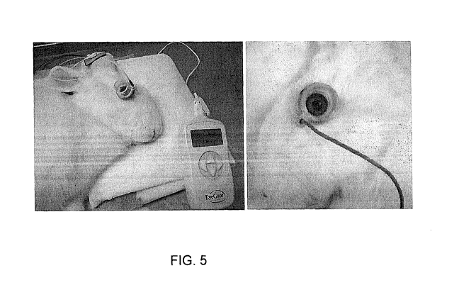
48 hours after lacrimal gland injection, rabbits were given a single 15
mA=min (-3.0 mA for 5 min) iontophoretic dose of dexamethasone phosphate
(40 mg/mL) or phosphate-buffered saline using the EyeGate II device (EyeGate
Pharmaceuticals, Inc) (FIG. 5). The animals were assigned to the following
treatment groups:
Group 1: Con A injection on Day 0, Treatment with Dex-P on Day 2
Group 2: Con A injection on Day 0, Treatment with PBS on Day 2
Group 3: PBS injection on Day 0, Treatment with PBS on Day 2
Group 4: PBS injection on Day 0 with no subsequent treatment
Clinical Observations
Animals were observed daily following Con A injection for signs of ocular
inflammation. Tear flow was measured using Schirmer strips in all groups on
Days
0, 1, 2, 4, 7, and 8 after Con A injection (FIG. 6). Signs of ocular surface
damage
were assessed on Days 0, 2, 4, and 8 using fluorescein staining and slit-lamp
microscopy (FIGS. 7 and 8). Staining was scored from 0 to 2 for superior,
central,
and inferior cornea for a total possible score of 6.
27
BOST_964298.1
CA 02716390 2015-09-23
Cytokine Assays
Animals were euthanized on Day 4 or Day 8 following Con A injection.
Upon sacrifice, the cornea and lacrimal gland were removed and snap frozen in
liquid nitrogen followed by storage at -80 C. All samples were homogenized by
hand in a ground-glass homogenizer in 0.5 mL of PBS + 10 mM EDTA.
Interleukin-l-beta (IL-1P), FIG. 9, and transforming growth factor beta-1 (TGF-
I31),
FIG. 10, were measured in lacrimal gland and corneal extracts using human IL-
10 or
TGF431 ELISA kit (R&D Systems DuoSet ELISA development system) according
to manufacturer's instructions. Results were normalized for total protein
concentration measured in the protein assay. Due to the high homology between
rabbit and human IL-1f3 and TGF-pl, human kits are appropriate for detecting
the
rabbit cytokine.
Conclusions
A single iontophoretic dose of dexamethasone phosphate increases tear flow
in rabbits and decreases the amount of ocular surface damage compared to
control
groups. Reduced IL-l1 and TGF-p1 expression is observed in the lacrimal glands
of eyes treated with a single iontophoretic dose of dexamethasone phosphate
compared to saline treatment and control groups. No significant elevation of
inflammatory eytokines in the cornea is observed on Day 4 and Day 8,
indicating a
specific inflammatory response of the lacrimal gland. A single iontophoretic
dose of
corticosteroid is a safer and more effective alternative than multiple, daily
topical
doses.
OTHER EMBODIMENTS
Other embodiments will be evident to those of skill in the art. It should be
understood that the foregoing detailed description is provided for clarity
only and is
merely exemplary. The scope of the claims should not be limited by the
preferred
embodiments set forth in the examples, but should be given the broadest
interpretation consistent with the description as a whole.
28