Note: Descriptions are shown in the official language in which they were submitted.
CA 02716411 2015-04-30
25771-1811
Apparatus for the separation of plasma
The present invention relates to an apparatus for absorbing blood and
separating blood
components, e.g. blood plasma, as a sample liquid.
The present invention relates to microfiuidic systems or apparatus. The
following remarks
apply to apparatus in which capillary forces act and are particularly crucial
to the operation.
Apparatuses for separating blood plasma from blood are known from US 4,906,439
A and
WO 01/24931 Al in each case, in which a plurality of groove-like or capillary-
like individual
channels are provided for receiving the blood plasma and carrying it away. The
disadvantage
here is that the channels fill up with the sample liquid in the form of blood
plasma at different
rates or not at all. Therefore a uniform liquid front cannot be achieved. This
is problematic in
terms of diagnostics as there is not a defined amount available at the same
time or, for
example, dry chemicals or the like cannot simultaneously be dissolved by the
sample liquid
in the desired or necessary amounts.
The present invention relates to an improved apparatus and an improved
process for absorbing blood and separating blood components, such as blood
plasma, as a
sample liquid, while permitting optimised filling of the channel with sample
liquid and
providing capillary contact between the separation element and the channel and
preferably
improving the diagnostic or investigative possibilities.
The present invention relates to a device for creating fluidic capillary
contact between
separation elements, particularly membranes and a conveying channel. This
allows
optimal, rapid and uniform filling of the channel and prevents unwanted
trapped air.
Preferably the sample liquid is absorbed by capillary forces in a channel
which is open in
construction at least on one narrow or longitudinal side, so as to form a
lateral liquid stop for
the sample liquid in the channel and to enable the sample liquid to be guided
in the channel
without any side walls. In particular a recess laterally adjoins the open side
of the channel.
This ensures, by a simple method, that the sample liquid is prevented from
being pushed
forwards - i.e. the channel being filled more rapidly - in the region where a
side wall would
otherwise be provided. This allows the filling speed to be evened out over the
entire cross-
1
CA 02716411 2015-04-30
25771-1811
section of the channel, so that an at least substantially uniform or straight
liquid front can be
achieved during the filling of the channel.
The laterally open construction of the channel ensures an improved,
particularly optimum
venting when the channel is fillled with sample liquid.
Moreover the surface of the sample liquid which is held or guided without any
side walls
enables the sample liquid to be examined directly, particularly by the
focussing of light
thereon, without a side wall or the like that would otherwise be provided.
Preferably the recess is of a trough-like construction and totally surrounds
the channel which
is open on all sides, in particular. Thus, especially with very fine
structures, the inner edges
that would normally occur at the transition from the flat sides to the narrow
sides and that
have particularly high capillary forces can be done away with completely.
However, the same
is also true when the channel is open at the sides only in parts.
Alternatively the recess or side wall laterally adjoining the channel may also
be capable of
being filled with sample liquid or some other liquid. The recess or side wall
is then
constructed - particularly with regard to its size, curvatures or wetting
characteristics - or
configured by means of deflector elements such that the fill speed of the
channel with the
sample liquid is greater than or equal to the fill speed of the recess or
along the side wall in
the direction of filling - particularly the longitudinal direction - of the
channel. In this way,
also, it is possible to prevent the liquid front from advancing laterally as
it is filled with sample
liquid.
Another proposed process for determining a parameter in the blood plasma or a
blood
component is characterised in that in a microfluidic system, immediately after
blood cells
have been held back or separated off, a component or parameter of the blood
plasma is
determined directly by means of one or more chemicals. This allows rapid and
inexpensive
analysis or determination of the parameter using an apparatus of simple and
compact
construction.
2
CA 02716411 2015-04-30
25771-1811
In one apparatus aspect, the present invention relates to an apparatus for
absorbing
blood and separating blood components as a sample liquid, comprising: a feed
device for receiving the blood; a separating device for separating blood
components
as the sample liquid; a channel which takes up the sample liquid by capillary
forces;
and a fill device for filling the channel with the sample liquid in an inlet
or feed region
of the channel, wherein the separating device includes a membrane and at least
one
of the membrane and a base of the feed region is domed, convexly curved, and
projects into the filling device from a periphery of the membrane or base to a
single
apex in a direction of filling and a spacing of the membrane from a base of
the
channel is from Ito 100 microns.
Further advantages, features, properties and aspects of the present invention
will
become apparent from the following description of preferred embodiments by
reference to the drawings. These show:
Fig. 1 a schematic section through a proposed apparatus according to a first
embodiment;
Fig. 2 a schematic plan view of a carrier of the filled apparatus according to
Fig. 1;
Fig. 3 a schematic section through the apparatus on the line 111-11I according
to Fig. 2;
Fig. 4 a schematic longitudinal section through the apparatus on the line IV-
IV
according to Fig. 2;
2a
CA 02716411 2010-08-25
P01-2358/W0/1 ¨ PCT filing text
Fig. 5 a schematic plan view of a carrier of a proposed apparatus according to
a second
embodiment;
Fig. 6 a schematic plan view of a carrier of a proposed apparatus according to
a third
embodiment;
Fig. 7 a schematic plan view of a detail of a carrier of a proposed apparatus
according to a
fourth embodiment;
Fig. 8 a schematic section through a detail of the apparatus on the line VIII-
VIII according to
Fig. 7;
Fig. 9 a schematic longitudinal section through a proposed apparatus according
to a fifth
embodiment; and
Fig. 10 a schematic section through a proposed apparatus according to a sixth
embodiment;
Fig. 11A, B, C and Fig. 12 an embodiment with a convex membrane arrangement;
Fig. 13, Fig. 14, Fig. 15 and Fig. 16 embodiments with a membrane made convex
by a
punch;
Fig. 17 and Fig. 18 embodiments with convex carrier bases;
Fig. 19 and Fig. 19A, B, C embodiments with an inlaid insert;
Fig. 20 an embodiment with a vent;
Fig. 21 an embodiment with a welded membrane and
Fig. 22, Fig. 22 A, Fig. 23 and Fig. 23A a widening of the inlet cross-section
of the channel.
In the figures the same reference numerals are used for identical or similar
parts, while
corresponding or comparable properties and advantages are achieved even if the
relevant
description is not repeated.
Fig. 1 shows in schematic section a first embodiment of a proposed apparatus
(1) for
absorbing and/or diagnosing a sample liquid (2), particularly blood plasma or
the like. The
apparatus (1) has a channel (3) which takes up the sample liquid (2) by means
of capillary
forces. The channel (3) is of open construction, at least on one narrow side
or longitudinal
side (4), on both narrow or longitudinal sides (4) in the embodiment shown, as
indicated in
Fig. 1.
Finally, adjoining the open sides (4) is a recess (5) which is preferably in
the form of a groove
or trough in the embodiment shown.
Thus, a lateral liquid stop for the sample liquid (2) - therefore an obstacle
to flow that cannot
be overcome by capillary forces - is formed in the channel (3) and the sample
liquid (2) can
be guided along the open sides (4) in the channel (3) without any side walls.
In the embodiment shown the apparatus (1) has a carrier (6) and an associated
cover (7),
between which are formed the channel (3) and the recess (5). If necessary,
only the carrier
3
CA 02716411 2010-08-25
P01-2358/W0/1 ¨ PCT filing text
(6) is cut away to form the necessary structures and the cover (7) is of even
construction,
preferably at least substantially free from recesses. However the situation
may also be
reversed. If necessary, however, both the carrier (6) and the cover (7) may be
recessed
and/or constructed with projections to form the desired structures and
optionally designed to
receive chemicals, reagents, investigation means or the like (not shown).
The recess (5) preferably adjoins the channel (3) with a sharp edge, as shown
in Fig. 1. In
the embodiment shown, the recess (5) is formed only in the carrier (6) and
thus, in the view
shown in Fig. 1 it extends substantially only downwards in relation to a
lateral projection of
the channel (3). The recess (5) may however extend upwards or on both sides of
the lateral
projection of the channel (3) - i.e. upwards and downwards, in particular, as
desired.
The recess (5) which is preferably rectangular in cross-section leads to an
increase in the
cross-section of a kind, particularly stepwise or sudden, that causes the
capillary forces to be
reduced so that the above-mentioned liquid stop for the sample liquid (2) is
formed in the
transition from the channel (3) to the recess (5), as indicated in Fig. 1.
The channel (3) is preferably defined or formed by only two opposing,
particularly
substantially flat surfaces or flat sides (8) and (9), which are formed by the
carrier (6) or the
cover (7) in the embodiment shown and run parallel. If required, therefore,
the recess (5)
may be omitted altogether and the channel (3) be formed for example by two
suitable strips
or the like, at a suitable spacing for the production of the desired capillary
forces.
Fig. 2 shows in schematic plan view the carrier (6) of the apparatus (1)
without a cover (7),
but partially filled with the sample liquid (2) up to the liquid front V. In
the embodiment shown,
the recess (5) extends along the open side(s) (4) of the channel (3),
preferably at least along
opposing open longitudinal sides (4). Moreover, in the embodiment shown the
channel (3) is
constructed to be laterally open on all sides and the recess (5) is
accordingly of encircling
design.
The channel (3) is thus surrounded by the recess (5) on all sides.
Preferably the recess (5) adjoins those narrow sides or longitudinal sides (4)
of the channel
(3) which extend at least substantially parallel to the main direction of
filling F of the channel
(3) with sample liquid (2), as indicated in Fig. 2. Consequently, the recess
(5) preferably
extends at least partially parallel to the main direction of filling F.
According to another alternative embodiment which will be described
hereinafter with
reference to Fig. 10 it is also possible for the recess (5) to fill up with
the sample liquid (2) or
with another liquid that is immiscible with the sample liquid (2), in
particular, such as oil or the
like. In this case, however, the recess (5) is constructed so that its fill
speed is at most as
great as the fill speed of the channel (3), to allow it to be filled as
uniformly as possible with
sample liquid (2). The fill speeds each relate to the filling or advancing of
the liquid front V in
4
CA 02716411 2010-08-25
P01-2358/W0/1 ¨ PCT filing text
the main direction of filling F.
Alternatively the recess (5) may also be flushed only with the other liquid
before the sample
liquid (2) is introduced.
The channel (3) preferably comprises a substantially rectangular and/or flat
cross-section,
particularly at right-angles to the main direction of filling F.
The height H of the channel (3) indicated in Fig. 1 - i.e. the spacing of the
preferably parallel
surfaces (8) and (9) that delimit the channel (3) - is at most 2000 microns,
preferably at most
500 microns, particularly about 50 to 200 microns. The recess (5) preferably
leads to a
stepwise or sudden increase in the height H and hence to the formation of the
desired liquid
stop. In particular, the height H of the recess (5) is at least twice as great
as the height H of
the channel (3).
The width B of the channel (3) is preferably about 100 to 5000 microns,
particularly about
200 to 4000 microns.
The height H of the channel (3) is substantially less, particularly by at
least a factor of 5 or
10, than the width B of the channel (3).
The capacity of the channel (3) is preferably less than 1 ml, particularly
less than 100
particularly preferably not more than 10 IA
The apparatus (1) thus forms a microfluidic system. In particular, the
apparatus (1) serves
for microfluidic diagnosis for medical or non-medical or other investigations.
The channel (3) and hence its main direction of filling F and main extension
plane E
preferably extend at least substantially horizontally in the position of use.
Depending on the
intended use or design solution, however, a different alignment is also
possible, particularly
as the uptake or filling of the channel (3) with sample liquid (2) is
preferably determined or
carried out primarily by capillary forces alone.
Thus, the main direction of filling F may extend horizontally or at an angle,
while the main
extension plane E extends vertically, for example, so that the channel (3) is
therefore aligned
on edge.
The channel (3) preferably forms at least one reservoir for the sample liquid
(2), particularly
for diagnostic purposes. The channel (3) may optionally contain a chemical
(not shown),
particularly a dry chemical or the like. However, investigations on the sample
liquid (2) may
also be carried out in some other way.
In the embodiment shown, the channel (3) comprises at least one deflector
element for
influencing and particularly evening out the filling thereof with the sample
liquid (2).
According to an alternative embodiment, the channel (3) preferably comprises
regularly
distributed elevations (10) as deflector elements. These are arranged
particularly in rows, at
right angles, preferably perpendicularly, or longitudinally with respect to
the main direction of
filling F, particularly alternately offset at right angles. The elevations
(10) are the rows offset
CA 02716411 2010-08-25
P01-2358/W0/1 ¨ PCT filing text
in the main direction of filling F. In this way the sample liquid (2) can be
made to fill the
channel (3) row by row, and as a result advance with a substantially straight
liquid front V in
the main direction of filling F.
If necessary, the surface density, the spacing and/or the size of the
elevations (10) may vary,
particularly as a function of their respective distances from an inlet for the
sample liquid (2)
into the channel (3), not shown in Figs. 1 and 2.
The elevations (10) are preferably in the form of webs, humps or columns,
particularly with a
round or polygonal base surface. However, it would also be possible to provide
depressions
instead.
Alternatively or additionally the channel (3) may comprise at least one trough
(11) or a web
as the deflector element extending transversely or longitudinally with respect
to the main
direction of filling F of the channel (3). The groove-like trough (11) which
is preferably
provided and which is in particular rectangular or semi-circular in cross-
section has a
substantially lesser depth than the recess (5) and therefore forms a purely
temporary liquid
stop for evening out the liquid front V. In this way it can be ensured that
the sample liquid (2)
does not fill the trough (11) and then the subsequent channel region until
after it has filled the
channel (3) over its entire cross-section.
It should be emphasised that by combining the guidance of the sample liquid
(2) and
deflector elements without the use of side walls it is possible to achieve a
highly uniform
filling of the channel (3) by capillary forces with a liquid front V which
extends at least
substantially in a straight line or perpendicular to the main directional
filling F.
Alternatively, the channel (3) and/or a reservoir, collecting chamber,
collecting region or the
like formed thereby may also be at least substantially smooth or flat in
construction, i.e.
without deflector elements, in particular.
Fig. 3 shows another schematic section through the apparatus (1) with the
cover (7) along
the line in Fig. 2.
The apparatus (1) has at least one vent (12) associated with the channel (3),
which is
connected not directly to the channel (3) but to the recess (5). Thus there is
no need for an
additional liquid stop for the vent (12) to prevent the sample liquid (2) from
escaping through
the vent (12). The construction of the channel (3) which is preferably
laterally open on all
sides allows optimum venting while the channel (3) is filling with the sample
liquid (2) so that
unwanted air inclusions can be reliably prevented.
The sample liquid (2) can be conveyed to the channel (3) preferably
perpendicularly to the
channel extent E, particularly vertically, in the position of use.
The apparatus (1) has a feed device (13) for taking up and supplying sample
liquid (2) to the
channel (3). In the embodiment shown the feed device (13) comprises an
opening,
particularly a perforation (14) in the cover (7), preferably for receiving
blood or the like, and a
6
CA 02716411 2010-08-25
P01-2358/W0/1 ¨ PCT filing text
separating device (15) such as a filter, membrane or the like for separating
off blood plasma
as the sample liquid (2). The separating device (15) in the embodiment shown
is inserted in
a gap (16) in the cover (7) opening towards the carrier (6) and covers the
perforation (14).
Preferably the separating device (15) is fixedly connected to the cover (7),
for example by
welding or gluing or is held thereby by frictional or interlocking engagement.
The separating
device (15) is directly in contact with the channel (3), by means of a flat
side in the
embodiment shown, and in particular the separating device (15) lies on
preferably column-
shaped structures (17) or the like in the channel (3) in a feed region (18) of
the channel (3).
The structures (17) are preferably provided with wedge-shaped recesses or the
like to deflect
the blood plasma or sample liquid (2) by capillary forces to the channel
surface opposite the
separating device (15), in this instance to the base surface (8) of the
channel (3) formed by
the carrier (6), and thus ensure total filling between the base surface (8)
and the cover (7) or
of the feed region (18) with sample liquid (2).
The structures (17) form a filling device for (totally) filling the channel
(3) between the cover
(7) and base surface (8) with sample liquid (2). However, this filling device
may also be
constructed differently, as will be explained later on by means of the fifth
embodiment.
Next the sample liquid (2) - after overcoming the first trough (11) in the
embodiment shown -
is sucked further into the channel (3) by capillary forces, as indicated by
the main direction of
filling F in Fig. 2.
Fig. 4 shows in schematic longitudinal section the preferred structure of the
proposed
apparatus (1) according to the first embodiment, in which a drop of blood (19)
which has
been supplied is shown for illustration purposes.
The separating device (15) may if necessary contain a chemical, particularly a
dry chemical,
particularly to allow the separation of blood plasma as the sample liquid (2)
from the blood
(19) as desired in the embodiment shown, or to assist this separation and/or
if necessary to
allow lysing of cells. The separation or further conveying take place
particularly solely by
capillary forces. Preferably, only a single channel (3) for receiving or
conveying the sample
liquid (2) adjoins the feed device (13). The channel (3) should be understood
as being a
single capillary. If necessary, however, the channel (3) may lead in different
directions or to
different areas or may branch, as will be explained hereinafter with reference
to the second
embodiment according to Fig. 5 and the third embodiment according to Fig. 6.
Figs. 5 and 6 each show a plan view of the carrier (6) of the apparatus (1)
according to the
second or third embodiment, without a cover (7) in each case.
In the second embodiment shown in Fig. 5 the channel (3) extends, starting
from the feed
device (13) or the feed region (18), onto opposite sides or in opposite
directions, for example
7
CA 02716411 2010-08-25
P01-2358/W0/1 ¨ PCT filing text
in order to carry out different investigations, tests or the like
simultaneously. This produces a
substantially elongate arrangement.
In the third embodiment shown in Fig. 6 a cross-shaped configuration is
provided. The
channel (3) extends in four different directions. Thus, for example, four
different
investigations, tests, reactions or the like can be carried out
simultaneously.
Both in the second embodiment and in the third embodiment the recess (5) is
preferably
again provided for at least partial guidance of the sample liquid (2) in the
channel (3) without
the use of side walls. In particular, the recess (5) surrounds the entire
channel configuration
completely, while the channel (3) may preferably be constructed so as to be
laterally open on
all sides.
Figs. 7 and 8 show a fourth embodiment of the proposed apparatus (1) and
specifically Fig. 7
shows a plan view of the carrier (6) without a cover (7) and Fig. 8 shows a
sectional view
along the line VIII-VIII in Fig. 7 with the cover (7) present. The channel (3)
here forms a
collecting chamber (20) for the sample liquid (2). The collecting chamber (20)
is in turn
substantially flat in construction and comprises, as necessary, the elevations
(10) shown
and/or other deflector elements or the like.
The apparatus (1) according to the fourth embodiment comprises a device (21),
particularly
an optical fibre or the like, for conducting light into the sample liquid (2),
particularly for
measuring fluorescence. The light strikes the free surface of the sample
liquid (2) in the
region of an open side (4) of the channel (3) and as a result of a
correspondingly steep
direction of impact which is preferably substantially perpendicular to the
surface of the liquid,
it enters the sample liquid (2) as indicated by the arrow (22). The gas
(air)/sample liquid (2)
interface is used for the entry of light. This avoids the need for the light
having to be guided
by a side wall, as would normally be present, and thus undesirably scattered
or causing
fluorescence.
As indicated in Fig. 7, the incident light beam (22) is preferably reflected
many times by total
reflection at the sample liquid (2)/gas (air) interface. This is achieved by
making the angle
between the surface perpendicular and the incident light beam greater than the
critical angle
of the total reflection. The ground or base surface of the collecting chamber
(20) which is
delimited and defined by the encircling recess (5) is designed accordingly in
order to achieve
the desired beam guidance and total reflection, by a suitable polygonal
configuration in the
embodiment shown. The light (22) radiated in is used for fluorescence
determination or
fluorescence spectroscopy. The sample liquid (2) particularly marker molecules
or the like
contained therein, which are present as chemicals in the channel (3) and
dissolved by
sample liquid, are excited by a particular wavelength. This leads to electron
transitions in the
molecules which revert to their original state after a certain length of time,
emitting a photon.
8
CA 02716411 2010-08-25
P01-2358/W0/1 ¨ PCT filing text
The radiation emitted is indicated by arrows (23) in Fig. 8 and can be
detected by a detector
(24). In order to exclude the influence of the elevations (10) or other
deflector elements on
the incident light beam (22), the plane of the light beam is arranged above
these structures
or at a spacing from them. Moreover, the light beam plane runs at least
substantially parallel
to the main extension plane or in the main extension plane E of the channel
(3) or of the
collecting chamber (20).
The light radiation and light guidance provided ensures substantially total
excitation of the
sample liquid (2) or of marker molecules or the like contained therein and at
the same time
allow the use of micro-structures such as the elevations (10) or similar
deflector elements.
Capturing the emitted light beams (23) at right angles, particularly
perpendicularly, to the
direction of incidence (22) is optimum with regard to disengagement from the
incident light.
Fig. 9 shows a schematic longitudinal section through a fifth embodiment of
the proposed
apparatus (1).
Compared with the first embodiment, the filling device for filling the channel
(3) comprises,
between the two flat sides (8) and (9), particularly for deflecting the blood
plasma or the
sample liquid (2) from the separating device (15) or from the cover surface
(9) to the
opposing base surface (8) so as to form a spatial meniscus between the two
surfaces or flat
sides (8) and (9), a slope or ramp (25), alternatively or in addition to the
structures (17), said
slope or ramp (25) correspondingly reducing the channel height H or even
allowing it to
become zero. In particular, the separating device (15) may be in direct
contact with the ramp
(25) or may rest directly thereon. The filling device mentioned may also be
referred to or
understood as a device for wetting the cover and base.
The schematic sectional view shown in Fig. 10 shows a sixth embodiment of the
proposed
apparatus (1). Here, the recess (5) laterally adjoining the channel (3) can be
filled by the
sample liquid (2) and constructed, particularly on the basis of a
corresponding rounding-off of
its side wall (26) and/or by the formation of corresponding guide elements
such as elevations
(10) or the like, such that the fill speed of the recess (5) in the main
direction of filling F - i.e.
perpendicular to the plane of the drawing in the representation shown in Fig.
10 - does not
exceed the fill speed of the channel (3), so as to prevent unwanted lateral
advancing of the
liquid front. It should be noted that in the embodiment shown the height H of
the recess (5)
corresponds to only about the height H of the channel (3). However, it is
preferably greater.
The proposed apparatus (1) is suitable for all kinds of tests, investigations
or the like. In
particular, it allows immunological or biochemical testing, for example, of
blood (19), blood
plasma or the like.
According to one alternative embodiment the channel (3) may have a number of
investigation
regions or collecting regions (20) which can be filled with the test liquid
(2) one after another.
9
CA 02716411 2010-08-25
P01-2358/W0/1 ¨ PCT filing text
Thus, it is possible for example to carry out various investigations one after
the other and/or
to expose the sample liquid (2) successively to different reagents,
particularly dry chemicals,
which are dissolved one after another.
According to another alternative embodiment, a second investigation or
collecting region (20)
may adjoin a first investigation or collecting region (20), the second region
preferably having
a substantially greater capillarity, for example by the use of an inserted
nonwoven fabric or
the like. The sample liquid (2), after the first region has been filled and in
particular after a
dry chemical provided therein, as necessary, has been dissolved, can then be
sucked or
conveyed into the second region, the dry chemical being washed out of the
first region and in
this way, for example, a further investigation is made possible in the first
and/or second
region.
According to another alternative embodiment, a first chemical, particularly a
dry chemical, is
provided preferably in the feed device (13) or separating device (15), and at
least one
second chemical, particularly a dry chemical, is provided preferably in the
channel (3) or
collecting region (20). This allows effective handling or influencing of the
sample liquid (2),
blood (19) or the like. Preferably, in order to investigate blood plasma, the
first chemical is
designed to prevent or delay clotting of the blood (19). For this purposes,
EDTA (Ethylene
Dyamine Tetra-acytic Acid) can be used as the first chemical, for example, in
order to
produce EDTA blood. The EDTA binds the calcium in the blood which is necessary
as
Factor IV for blood clotting.
Then the second chemical, preferably a mixture of chemicals, is used for an
investigation or
to determine one or more parameters in the blood plasma, such as glucose,
ketones or
lactate.
Preferably, in order to investigate at least one intra-cellular parameter such
as the
haemoglobin value or the calcium value in the blood (19), the first chemical
is designed to
lyse cells such as blood cells and release calcium or the like. Lysine buffer
is used for this
purpose, for example.
Then the second chemical, preferably a mixture of chemicals, is used to
investigate or
determine the parameter, particularly the calcium content. One ingredient of
the mixture,
preferably the chelating agent 8-hydroxyquinoline, is used to eliminate
magnesium ions from
the reaction as these would interfere with the reaction. Another cornplexing
agent, preferably
0-cresolphthalein, forms a coloured complex with calcium under alkaline
conditions.
The extinction of the colour complex is proportional to the calcium
concentration at a
wavelength of 570nm. It is determined directly in the channel (3) or
collecting region (20) or
optionally after removal. However, other measurements or procedures are also
possible. In
particular, the extinction may also be used at different wavelength and/or for
determining
CA 02716411 2010-08-25
P01-2358/W0/1 ¨ PCT filing text
different complexes, parameters or the like. The same applies to other,
preferably optical
methods of measurement, such as fluorescence measurements or the like.
According to yet another alternative embodiment, a removal opening (not shown)
is provided
in the cover (7) and/or in the carrier (6), to allow the removal of sample
liquid (2), particularly
blood plasma or the like which has been separated off. The removal opening is
preferably
connected to an at least relatively large-capacity storage region (20) or the
like of the
channel (3), to provide a desired or sufficient removal volume.
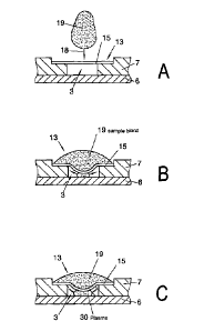
As a rule, the spacing of the membrane surface from the channel base is equal
to the height
of the channel, as shown in Figure 11A. The problem then arises that the
separating
element for the blood separation may constitute a fluidic barrier to the
unimpeded flow of the
plasma into the channel. This is caused by the fact that a membrane or a
filter element is
used as the separating element, the membrane or the filter element consisting
of a woven
fibre network or a porous material. The materials used may be artificial
fibres combined or
compressed into a fleece or porous ceramics as well as metal meshes. The
filter material
has as a result of the mesh structure film branched channels with a high
capillary force,
which cause fluidic components to be retained in the filter or membrane. The
membrane has
a pore size of 0.01 microns to 1.2 microns, particularly 0.2 to 0.6 microns.
The membrane
density is from 50 microns to 500 microns, preferably 120 gm to 180 gm. The
porosity, i.e.
the proportion of the volume of the membrane which is not constructed with
material, is 40-
90%, preferably 70 to 80%. The pore material used may be various materials
such as nylon,
particularly isotropically foamed nylon 60, with a pore volume of more than
70% and a pore
size of 0.5 microns or also preferably hydrophilic polyvinyldifluoride with a
pore size of 0.6
microns.
If a drop of blood is then placed in the entry region in the feed device (18),
a hemispherical
drop of blood forms on the surface of the membrane (15) as can be seen in Fig.
11b. As a
result of gravity and fluid pressure, the blood plasma flows through the
channels of the
membrane (15), holding back the larger blood particles, and forms, as a result
of the
hydraulic pressure, a plasma film or plasma drop on the underside which
adheres to the
membrane. Because of the small amounts of blood or plasma or, in particular,
when there is
a large dead space to be filled between the membrane and the base of the
channel, there
may be no fluidic adhesion between the plasma and the channel. Admittedly, a
plasma
stream often flows along the channel walls to the channel base, in particular,
and slowly fills
the channel (3) or the collecting region (20). However, the start of the
filling process is
delayed by this, resulting in undesirably long flow times which have a
negative effect on the
function of a diagnostic or analytical apparatus connected to the fluidic
channel structure.
11
CA 02716411 2010-08-25
1301-2358/W0/1 ¨PCT filing text
The dead space in the filling region between the separating element and the
channel thus
acts as an impedance or a resistance for the flow rate of the plasma.
A further aim of the present invention is to set this impedance to a
controlled level,
particularly to reduce it to a minimum.
Advantageously, the flow resistance can be minimised by constructing the
separating
element (15) as shown in Fig. 11B. For this, the separating element (15) is
made convex in
the direction of the channel base, so that it preferably rests on the channel
base in a central
region or alternatively the apex of the convex shape extends to close to the
channel base.
The spacing of the separating element from the bottom of the filling device,
particularly from
the channel base, is preferably from 1 micron to 100 microns, particularly
from 10 microns to
25 microns.
This ensures that the plasma liquid emerging from the underside of the
membrane as a
result of gravity or hydraulic pressure wets the channel directly and starting
from this wetting
point flows into the channel, as schematically shown in Figs. 11B and 11C.
In a preferred embodiment the diameter of the membrane (15) used is from 2 to
10 mm,
particularly 250 to 350 microns.
Advantageously, the height value W of the convexity or apex, as schematically
shown in Fig.
12a, is in the region of the membrane density. In the Example shown
previously, with a
membrane thickness of 250 microns, the value W is preferably from 100 microns
to 300
microns. The height value of the convexity should advantageously correspond
roughly to the
height of a channel in the inlet region or of a chamber located underneath the
conveying
membrane (15). As the depth of the channel constructed according to Figure 1
is preferably
50-200 microns, the height W of the convexity may also vary in the range from
50 to 200
microns, according to the depth of the trough.
Advantageously, the channel walls and particularly the channel base comprise
elements (10)
that increase the volume flow in capillary manner as shown in Fig. 2.
Particularly
advantageously, an element (17) with vertical nodges is inserted on the
channel base, as
disclosed in AP 101 3341 Bl. The geometry of the notch initiates and assists a
vertical flow
from the filling region through the separating element to the channel base.
Fig. 12 shows elements of this kind on the channel base, while a plurality of
elements are
arranged relative to one another on the base of the channel such that, as a
result of the
capillary winding of the interstices, there is a horizontal volume flow of the
fluid or plasma in a
collecting chamber (20) in the direction of the channel.
Advantageously, the convex curvature of the membrane is obtained by upsetting
the
membrane as it is secured in the direction of its centre, so that it is flexed
through to the
centre. This can also be achieved by making the diameter of the membrane
greater than the
12
CA 02716411 2010-08-25
P01-2358/W0/1 ¨ PCT filing text
diameter of the space in which the membrane is secured, particularly glued.
With a
corresponding retaining tool (not shown) that has a convex surface shape, the
membrane is
placed and glued in the fixing region. The convex shape of the tool causes the
deflection of
the membrane to be formed.
As an alternative to gluing, thermal processes such a welding, particularly
ultra-sound
welding, can be used for fixing the membrane, while the membrane is
advantageously
pressed in between two plastic elements of the apparatus with a pre-shaped
retaining tool in
this case as well.
As an alternative to shaping the separating element of the membrane during the
fixing
process, the convex deflection of the separating element can also take place
beforehand by
stamping the shape into the separating element.
With metal filter elements it is possible, for example, to press or bend them
into a domed,
particularly convex shape.
In the case of non-woven materials, a pressing operation in a correspondingly
shaped tool
with the application of pressure and/or temperature and/or additional chemical
fixing agents
or adhesives would also be possible. Alternatively, even during the
manufacture of the
nonwoven material from synthetic fibres, a convex shape might advantageously
be
impressed during the needling and consolidation of the nonwoven fabric.
In another advantageous embodiment the separating element is in two or more
parts,
particularly in two layers, while a flexible membrane is provided on a fixed
holding element,
particularly a membrane holder (31), as shown in Fig. 12A. Advantageously, the
membrane
is glued in the outer region of the funnel-shaped retaining element, but may
also be secured
by clamping elements. The funnel-shaped membrane holder or shaping insert (29)
has a
through-opening or bore (32) in a central region, so that when the funnel is
filled with blood
the latter can pass through the opening (32) into the membrane.
In another advantageous embodiment of the invention according to Fig. 13, a
membrane (15)
is provided as a separating device (15). When a drop of blood (19) is added to
the feed
region (18) and the opening (14) of the cover (6), the drop comes to rest on
the membrane
(15) which is flat in Fig. 13A.
In the following step, as can be seen from Fig. 13B, a ram (28) is inserted in
the opening
(14), this ram deforming the membrane surface towards the interior of the
channel, so as to
produce a convex membrane shape.
13
CA 02716411 2010-08-25
P01-2358/W0/1 ¨ PCT filing text
The punch (28) is preferably also domed at its end which comes into contact
with the
membrane.
The insertion of the ram (28) may be done both manually by an operator or
using an
automatic operating device with actuating drive means. In the latter case the
ram (28) is
mounted on a positioning drive, the positioning drive moving the ram such that
it presses the
membrane downwards towards the base of the channel. The positioning drive can
be
operated by piezo-electrical positioning members or a stepping motor or other
suitable
mechanical or electrical actuating means. Preferably, the ram (28) is moved
downwards as
a function of the analysis step that is to be carried out.
On the apparatus, sensors may be provided for automatic movement of the ram
(28). These
sensors detect the feeding in of a drop of blood (19) and actuate the ram or
punch (28) by
means of a control device, especially a microprocessor which records and
processes the
sensor signals.
In another embodiment of the invention according to Fig. 14, the channel (3)
is formed by a
recess (5) in the carrier (6) and a congruent cut-out in the cover (7). The
cover (7) has an
opening in the end region of the channel (3). This opening (14) is closed off
towards the top
of the cover (7) by a pressure element (33).
The cover element (33) comprises a punch (28) and an opening (14) through
which a drop of
blood (19) can be fed into the feed region (18). In the cut-out (16) of the
cover is provided a
separating element (15), particularly a filter membrane, which is secured
therein.
This securing may be done for example by means of gluing or welding to the
cover (7). In
the manufacture of the apparatus (1) according to Fig. 14, in a first step the
membrane is
attached to the cover (7). In a subsequent manufacturing step the cover (7)
provided with
the membrane and the carrier (6) are joined together.
In another manufacturing step following the first step, the covering element
(33) is attached
to the cover (7) as a result of which the ram (28) convexly deforms the
membrane so that the
dead space in the feed device (13) is reduced and the apex of the convex
membrane (27)
extends close to the base of the channel. This results in an apparatus (1) in
which there is a
significantly reduced fluid resistance between the membrane (27) and the
channel (3).
In one embodiment of the invention according to Figure 15, the punch (28) is
inserted in a
bore in the carrier (6) in the feed region. For this, the shaft of the punch
has a first section
14
CA 02716411 2010-08-25
P01-2358/W0/1 ¨PCT filing text
adjoining the head of the punch, which corresponds in length to the thickness
of the carrier
(6) in the region of the bore and seals off the bore when the punch is
inserted.
A second portion of the shaft of the punch is provided with notches or
profiling or has
perforations running through the longitudinal direction of the shaft. This
second section of
the shaft of the punch extends from the base of the feed region (18),
particularly the base of
the channel (3) or the base of a collecting chamber (20), up to the separating
element (15)
and is in contact with the latter, so that the profiled shaft of the punch
establishes a vertical
fluid connection between the base and a membrane (15).
Particularly advantageously, the shaft of the punch may be provided with
elevations or
deflector elements (10) on its preferably cylindrical outer surface, which
assists capillary flow
of the plasma that is separated off.
Fig. 16 shows an apparatus in which the cover element (33) is provided with a
central bore
(32) through which a drop of blood (19) is introduced into the feed region
(18).
The cover element (33) is attached to the cover (7), for example ultra-
sonically welded
thereto.
The pressure element (33), cover (7) and carrier (6) are preferably made of
plastics.
The membrane (27) is clamped between the cover (7) and the carrier (6),
particularly welded
in position.
In the embodiment according to Figure 16, the pressure element (33) is in the
form of a
shaping insert (29), the shaping insert (29) being of 3-dimensional
construction in the
direction of the channel (3) such that the membrane is convexly curved and
comes into
fluidic contact with the base of the channel (3) particularly with the
deflector element (10) on
the base of the channel.
In an embodiment according to Fig. 17, during the manufacture of the apparatus
(1), the
separating element (15) is first connected to the cover (7) and closes off the
feed region (18)
of the feed device (13) downwards towards the entry region of the channel.
The carrier element (6) which has a cut-out in the form of a channel (3), is
designed, in the
region of the opening of the cover (7) i.e. in the region of the membrane (27)
mounted flatly
on the cover (7), such that in a region of the carrier (6) arranged opposite
the central region
of the opening the surface of the carrier extends beyond the junction plane
between the
CA 02716411 2010-08-25
P01-2358/W0/1 ¨ PCT filing text
carrier (6) and cover (7). This can be achieved, for example, by having the
surface of the
carrier (6) bulging convexly inwards into this region, as shown in Fig. 7,
pressing against the
membrane (27) and deflecting it accordingly to match the shape of the carrier
(6).
Advantageously the carrier surface is also provided in this region with
deflector elements (10)
which ensure that the plasma has a horizontal fluid flow.
In the embodiment according to Fig. 18 the apparatus (1) is constructed in
three layers. The
carrier (6) is connected to the cover via an intermediate element (34), in
this case the
channel element (34) that forms the channel. For this purpose the intermediate
element (34)
is glued to the carrier (6) and to the cover (7), for example. The
intermediate element (34) is
preferably a double-sided adhesive film. The channel structures, particularly
the channel (3),
are formed as cut-outs (16) in the intermediate element, for example by
standing out of the
finished shape or as cut-outs during the moulding or casting process.
Advantageously, in this embodiment in which all the guide channels and fluid
chambers are
arranged in the intermediate element, the cover (7) and the carrier (6) may be
constructed as
planar elements without recesses (5) for the fluid-conveying structures, thus
considerably
reducing the use of precise, high-cost micro-forming tools and making
manufacture easier.
In the embodiment according to Fig. 18, in order to establish fluidic contact
and reduce the
flow resistance in the inlet region of the channel, the carrier element (6) is
to be shaped in
the direction of the membrane (27), particularly at least one peg (37) is to
be inserted in the
carrier element (6), this peg projecting from the junction plane between the
channel element
(34) and the carrier element (6) towards the membrane and providing fluidic
contact with the
membrane.
The peg (37) may for example be introduced directly into the carrier element
during the
manufacture of the carrier (6) by moulding or subsequently by mechanical
and/or thermal
embossing.
In an embodiment shown in Fig. 19 the channel (3) is formed as a recess (5) in
the carriers
(6).
The separating device (15), in this case a filter element (15), is arranged in
a recess (16) and
forms, together with the inlayed element or insert (35), the tensile dyed
device (13). If a drop
(19) of blood is fed into the feed region (18), the blood is absorbed and
filtered by the filter
(15), and the plasma emerging in the direction of the channel is taken up by
the insert (35)
arranged on the channel side of the filter (15).
16
CA 02716411 2010-08-25
P01-2358/W0/1 ¨PCT filing text
The insert (35) is geometrically designed so that its height corresponds
substantially to the
height of the gap between the channel base and the underside of the filter
(15) and the insert
(35) makes contact both with the channel base or chamber base in the inlet
region and also
with the underside of the filter. Preferably, the insert (35) is an inlayed
element (35) which
means that when the carrier (6) is connected to the cover (7) the insert (35)
is secured by
being held by the contact pressure between the filter and the carrier (6).
The insert may be produced for example as an 0-ring from an elastic plastics
material or
from rubber.
In a preferred embodiment of the insert (35) according to Fig. 19B the latter
consists of
another filter material which may be a porous ceramic material, a sponge made
of fibre
material, a metal grid or mesh element or some other suitable element made of
structures
which have channels.
Other materials that may be used are gel-like sponges or polymers such as
polysaccherides
or silicones. Examples of such polymers are sacarodes, polyaryamide or
agarose.
Advantageously, reagents such as anticoagulants (K2EDTA) may be introduced
into the
spongy or gel-like material.
In another preferred embodiment according to Fig. 19A the insert (35) is in
the form of a
horseshoe-shaped inlaid element (35). The inlaid element may consist of pore-
free plastics
material but it is also possible to produce the inlaid element from one of the
above-mentioned
channel-carrying materials.
Particularly preferably, the inlaid element has at least one notch (36) in its
edge region,
particularly a plurality of notches (36) which assist with vertical
discharging of the plasma into
the plasma chamber (20) of channel (3). Advantageously the cross-section of
the inlaid parts
may also be wedge-shaped, as shown by the section A-A in Fig. 19A, the apex of
the wedge
being in contact with the membrane surface and thereby establishing fluid
contact.
The air present in the volume of the feed region may be included in the
filling region when a
drop of blood is added.
On the one hand this may have the effect of forcing air out of the region
underneath the filter
(15), which rests on the peg (37) according to Figure 20 into the channel.
These air bubbles
present major flow resistance and are therefore undesirable. In addition there
may be an
accumulation of air which will build up a counter pressure to the hydraulic
pressure of the
17
CA 02716411 2010-08-25
P01-2358/W0/1 ¨ PCT filing text
plasma and constitute a serious flow impedance. Advantageously, therefore, a
lateral vent
(12) is provided in the collecting chamber (20) at right angles to the channel
(3). A vent of
this kind on the feed device (13) may be provided in all the embodiments
according to
Figures 1 to 23.
To ensure that the filter (15) is tightly sealed in a structure having a
carrier (6), a cover (7)
and an intermediate element (34), the filter is provided, in its fixing
region, with a
compression member (38) which compresses the filter material in the region of
the
compressing member (38). In an apparatus of this kind according to Fig. 21, a
recess (5) is
formed in a carrier element (6), to form the channel (3). The intermediate
element is a plastic
part in the form of a film provided with adhesive on both sides, the adhesive
establishing
contact by sticking both to the cover (7) and to the carrier (6) and attaching
them to one
another. Arranged in contact with the filter is an insert (35) in the form of
an inlaid part (35) in
the region of the feed device.
In an embodiment according to Fig. 18, the separating device or the filter
(15) is glued or
welded into the cover (7), welding being carried out for example by ultra-
sound or by thermal
welding.
In one embodiment of this arrangement according to Fig. 22 the feed region is
shown from
above. The plan view shows the intermediate element (34) which is, for
example, a channel
element with cut-outs which form a sample collecting chamber (20) in the feed
region and a
channel (3). Of the underlying carrier (6), the deflector elements (10) can be
seen in the
region of the sample chamber (20).
Above the plane of the intermediate element is schematically shown the weld
line (fixing line)
(39) which, lying in the upper cover (7), obstructs the filter or the filter
membrane (15) with
the cover. The intermediate element is, in particular, a film which is
provided with an
adhesive on both sides.
When a drop of blood is added, the plasma separated off flows into the
collecting chamber
(20) and is carried away into the channel (3) with the assistance of the
deflector elements.
The inlet region of the channel (3) constitutes a clear and abrupt reduction
in the flow cross-
section.
As shown in Fig. 22A, air may flow into the channel (3) as a result of which a
stream of air
bubbles may enter the channel, significantly increasing the flow resistance or
bringing the
18
CA 02716411 2010-08-25
P01-2358/W0/1 ¨PCT filing text
flow to a complete standstill. In the region of the fixing line (39) in
particular, there may be a
transverse influx of air (40) as the filter material, as a result of the
compression at this point
during welding, leaves behind a cavity out of which air can flow inwards.
In an advantageous embodiment of the transition from the sample chamber (20)
to the
channel (3), it is therefore provided according to Fig. 23 that the cross-
section of flow be
reduced continuously from the collecting chamber to the channel along a
transitional region.
This may take place stepwise, for example, by reducing the cross-sectional
area, as shown
in Figure 22, until the reduced cross-section (41) is about 2 to 5 times the
cross-section of
the channel (3).
As shown in Figure 23A the cross-sectional step (41) is arranged so that the
encircling fixing
line (39) slows down the outlet region from the collecting chamber (20) in the
region of the
cross-sectional step and does not form a crossover to the channel (3), thereby
preventing a
direct influx of air (40) into the channel (3).
Admittedly, air flows into the region of the cross-sectional step (41) in this
embodiment of the
flow, but as it is has a larger cross-section it takes correspondingly longer
for an influx of air
bubbles to block this cross-section and possibly lead to a break-off of the
fluid current.
In a number of the embodiments shown, the base of the channel (3) or the base
of the feed
device (18) comprises deflector elements (10). These deflector elements help
to assist the
wetting by a vertical flow of fluid. When suitably positioned relative to one
another the
interspaces with a capillary action between the deflector elements (10) also
assist a
horizontal flow of fluid.
One feature that is common to all these embodiments is that the deflector
elements are not
an essential operational component. The capillary gap present between a filter
or a
membrane (15) and the base of a channel (3) or a feed device (18) also acts in
the same
way as a deflector element (10), as the curvature of the base towards the
membrane at the
contact surfaces or feed surfaces result in wedge-shaped capillary gaps of low
height and
high capillarity.
19